Article

Prmt7 Downregulation in Mouse Spermatogonia Functions through miR-877-3p/Col6a3

1
Institute of Animal Sciences, Chinese Academy of Agricultural Sciences, Beijing 100193, China
2
College of Animal Science and Technology, China Agricultural University, Beijing 100193, China
*
Author to whom correspondence should be addressed.
These authors contributed equally to this work.
Life 2022, 12(8), 1194; https://doi.org/10.3390/life12081194
Submission received: 24 June 2022 / Revised: 14 July 2022 / Accepted: 30 July 2022 / Published: 5 August 2022
(This article belongs to the Section Physiology and Pathology)

Abstract

:
Protein arginine methyltransferases 7 (Prmt7) is expressed in male germ cells, including primordial germ cells, gonocytes, and spermatogonia. Our previous study demonstrated that Prmt7 downregulation reduced the proliferation of GC-1 cells (a cell line of mouse immortalized spermatogonia). However, how Prmt7 regulates spermatogonial proliferation through miRNA and the target gene remains elusive. Here, we experimentally reduced the Prmt7 expression in the GC-1 cells and subjected them to miRNA sequencing to explore the miRNA profile and its Prmt7-responsive members. In total, 48 differentially expressed miRNAs (DEmiRNAs), including 36 upregulated and 12 downregulated miRNAs, were identified. After verifying the validity of sequencing results through qRT-PCR assays in randomly selected DEmiRNAs, we predicted the target genes of these DEmiRNAs. Next, we combined DEmiRNA target genes and previously identified differentially expressed genes between Prmt7 knockdown and control groups of GC-1 cells, which resulted in seven miRNA/target gene pairs. Among these miRNA/target gene pairs, we further detected the expression of Col6a3 (collagen type VI alpha 3) as the target gene of mmu-miR-877-3p. The results suggested that Prmt7 downregulation in mouse spermatogonia might function through miR-877-3p/Col6a3. Overall, these findings provide new insights into the role of Prmt7 in male germ cell development through miRNA and target genes.

1. Introduction

Spermatogenesis, as a highly specialized process in male reproduction, is based on the self-renewal and differentiation of spermatogonia. This process encompasses mitosis from spermatogonia to spermatocytes, followed by meiosis from spermatocytes to spermatids, resulting in haploid spermatids, which undergo a final differentiation into spermatozoa [1]. To date, numerous studies have identified several key regulators of spermatogonial proliferation, such as extrinsic growth factors (including GDNF, EGF, IGF1, and FGF2) and intrinsic genes (including Plzf, Gfra1, and Etv5) [2,3]. However, the underlying molecular regulation of spermatogonial proliferation remains still unclear.
In our previous study, we found that protein arginine methyltransferase 7 (Prmt7), the only type III PRMT (a type of PRMT that only catalyzes the formation of monomethylarginine of its protein substrates), can replace Sox2 in combination with Oct4, Nanog, and Klf4 to successfully produce induced pluripotent stem cells [4]. Our further study revealed that Prmt7 was highly expressed in mouse testes at both mRNA and protein levels and Prmt7 knockdown reduced spermatogonial proliferation [5]. In addition, Prmt7 is reportedly expressed in male gonocytes and spermatogonia [6]. Prmt7 depletion causes defects in germ cell proliferation as well as a reduced number of primordial germ cells in mice [7]. These findings confirmed the crucial role of Prmt7 in male germ cell development. Moreover, Prmt7 is also positively involved in the pluripotency of embryonic stem cells [8]. It mediates the repression of miR-221-3p and miR-221-5p, which can further target and downregulate the expression of Oct4, Nanog, and Sox2, thus, playing crucial roles in maintaining the pluripotency of mouse embryonic stem cells [9]. Therefore, we propose that Prmt7 can regulate gene expression through miRNAs, playing roles in spermatogonial proliferation.
It is well known that miRNAs are a class of single-stranded small noncoding RNAs that are approximately 22 nt in length and they post-transcriptionally regulate protein synthesis by degrading mRNA via base pairing to partially complementary sequences in the 3′ UTR of their target mRNAs [10]. Thus, miRNAs play crucial roles in many biological processes, including the cell cycle [11], cell differentiation [12], metabolism [13], cell apoptosis [14], and embryo development [15]. In the gonads, particularly in the testes, miRNAs are ubiquitously present in spermatogonia, Sertoli cells, Leydig cells, and mature spermatozoa. There is growing evidence demonstrating that miRNAs are essential for male germ cell development and differentiation [10,16,17]. For example, let-7 family miRNAs are highly expressed in type B spermatogonia and primary spermatocytes, highlighting their potential role in spermatogenesis [18]. MiR-146 levels are decreased with spermatogonial differentiation, and miR-146 modulates the effects of retinoic acid on spermatogonial differentiation [19]. MiR-15 downregulation is associated with the abnormal morphology and motility of porcine sperm samples [20]. In addition, miR-20, miR-106a, miR-34c, miR-221, miR-222, miR-146a, and miR-21 are present and involved in the regulation of mouse spermatogonial stem cells [19,21,22,23,24]. These findings demonstrate the potential roles of miRNAs in male reproduction, especially in germ cell development.
Thus far, the interplay between Prmt7 and miRNAs is still poorly understood. It has only been reported that Prmt7 can downregulate miR-24-2, miR-221-3p, and miR-221-5p to maintain the pluripotency of mouse embryonic stem cells [9,25]. However, whether Prmt7 regulates miRNAs in spermatogonia remains unknown. Thus, in the current study, we aimed to determine if and how Prmt7 regulates miRNAs in the mouse spermatogonial cell line, GC-1 cells. To this end, we performed Prmt7 siRNA and negative control siRNA transfection into GC-1 cells, followed by small RNA sequencing. The sequencing results on a subset of DEmiRNAs were verified using qRT-PCR. By comparing the predicted target genes of these DEmiRNAs with our previous transcriptome results, we finally screened seven miRNA/target gene pairs, among which we verified the expression of Col6a3, the target gene of mmu-miR-877-3p. These results suggested that the proliferation of GC-1 spermatogonia might be mediated through the Prmt7/mmu-miR-877-3p/Col6a3 pathway, which helps better understand the role of Prmt7 in male germ cell development through its miRNA/target gene pathway.

2. Materials and Methods

2.1. Cell Culture and Treatment

The mouse GC-1 spermatogonial cell line was purchased from iCell (iCell-m022, Shanghai, China). Cells were cultured in a high-glucose Dulbecco’s modified Eagle’s medium (Lonza), supplemented with a 10% fetal bovine serum (Gibco), 100 U/mL penicillin, and 100 μg/mL streptomycin (Invitrogen, Carlsbad, CA, USA) at 37 °C in the humidified incubator with 5% CO2. SiRNA transfection was performed using a Lipofectamine RNAiMAX reagent (Invitrogen), according to the manufacturer’s instructions. Briefly, GC-1 cells were seeded into 6-well plates at a density of 1 × 10⁵ cells/well. Then, the cells were incubated in an incubator at 37 °C with 5% CO2 overnight. Prmt7 siRNA or negative control (NC) siRNA at a concentration of 50 nM was transfected into GC-1 cells using Lipofectamine RNAiMAX reagent (Invitrogen). The siRNAs of Prmt7 (target sequence: TCATGGATGATATGATTAA) and negative control (unpublic) were designed and synthesized by RiboBio (Guangzhou, China). After 48 h of transfection, the cells were collected for further assays.

2.2. RNA Extraction and qRT-PCR

Total RNA was extracted from the GC-1 cells 48 h after the siRNA transfection using a TRIzol reagent (Invitrogen). For the mRNA relative expression assay, 1 μg RNA was reverse transcribed using the PrimeScriptTM RT reagent kit (TaKaRa, Dalian, China), according to the manufacturer’s instructions. Then, qRT-PCR was performed with a final volume of the 20 μL reaction system, which contained 10 μL SYBR® Premix Ex TaqTM (TaKaRa), 1 μL cDNA, 0.4 μL forward primer (work concentration 0.2 μM), 0.4 μL reverse primer (work concentration 0.2 μM), 0.4 μL ROX reference dye II, and 7.8 μL sterile distilled H2O. β-actin was used as an internal control. For the miRNA relative expression assay, qRT-PCR was performed utilizing the miRNA qRT-PCR SYBR® Kit (Sangon, Shanghai, China) in a 20 μL reaction system, which contained 10 μL master mix, 2 μL cDNA, 0.5 μL forward primer, 0.5 μL reverse primer, 1 μL ROX reference dye (L), and 6 μL RNase-free water. U6 was used as the control. qRT-PCR was carried out on an ABI 7500 Fast Real-Time PCR system (Applied Biosystems, Waltham, MA, USA). The relative expressions of Prmt7 mRNA and miRNAs were normalized to β-actin and U6 expressions, respectively, and calculated using the 2ΔΔct method [26]. Primer sequences used for qRT-PCR of miRNAs and RNAs are listed in Table 1. Results are presented as fold changes relative to the control.

2.3. Protein Extraction and Western Blotting

Proteins were extracted from the GC-1 cells and transfected with siRNA for 48 h. After denaturing, the proteins were electrophoresed on 10% SDS-PAGE gel and then transferred to nitrocellulose membranes. After being blocked with 5% nonfat milk at room temperature for 1 h, the membranes were incubated with primary antibodies at 4 °C overnight. Later, the membranes were washed and further incubated with an HRP-conjugated secondary antibody at room temperature for 1 h. Then, the membranes were developed and visualized on a Tanon-5200 Chemiluminescent Imaging System. The primary antibodies used were PRMT7 (1:1000, #14762) and β-actin (1:1000, #4970) from Cell Signaling Technologies (Danvers, MA, USA). The intensity of each protein band was analyzed using ImageJ software and the protein expression levels were normalized to β-ACTIN.

2.4. miRNA Sequencing

Total RNA was extracted from GC-1 cells and transfected with Prmt7 siRNA or NC siRNA using a TRIzol reagent (Invitrogen). The RNA purity was examined via NanoPhotometer® spectrophotometer (IMPLEN, Westlake Village, CA, USA). The RNA concentration was measured with a Qubit® RNA Assay Kit in a Qubit® 2.0 Flurometer (Life Techonologies, Carlsbad, CA, USA). RNA integrity was tested with a RNA Nano 6000 Assay Kit of Agilent Bioanalyzer 2100 system (Agilent Technologies, Santa Clara, CA, USA).
Small RNA libraries were derived from 3 μg RNA per sample of spermatogonia for each group (Prmt7 siRNA-transfected group and NC siRNA-transfected group) in triplicate. Sequencing libraries were generated using an NEB-Next® Multiplex Small RNA Library Prep Set for Illumina® (NEB, Ipswich, MA, USA), following the manufacturer’s protocols. The index codes were added to attribute sequences to each sample. Then, the clustering of index-coded samples was performed on a cBot Cluster Generation System using a TruSeq Cluster Kit v3-cBot-HS (Illumina), based on the manufacturer’s instructions. Thereafter, the libraries were sequenced on an Illumina Hiseq 2500 platform. Fifty bp single-end reads were generated (Novogene, Beijing, China). The miRNA sequencing raw data were deposited in NCBI with BioProject accession number PRJNA858498.

2.5. miRNA Sequencing Data Analysis

Raw data were firstly processed and further filtered to obtain the clean reads. The small RNA tags were mapped to a reference sequence using Bowtie (bowtie-0.12.9) [27]. Then, their expression and distribution were analyzed. Mapped small RNA tags were used to identify known miRNAs with MiRBase 20.0 as reference. The modified software mirdeep2 (mirdeep2_0_0_5) and srna-tools-cli were used to obtain the potential miRNAs, as well as to draw the secondary structures [28]. In addition, small RNA tags were mapped to the Repeat Masker and Rfam database to remove tags derived from protein-coding genes, repeat sequences, rRNA, tRNA, snRNA, and snoRNA. The novel miRNAs were predicted from the hairpin structure characteristics of a miRNA precursor. The software miREvo (miREvo_v1.1) and mirdeep2 were used integratedly to predict novel miRNAs by exploring the secondary structures, Dicer cleavage sites, and the minimum free energy of small RNA tags unannotated in the former steps [28,29]. The DESeq R package (1.8.3) was used to analyze the differential expression of miRNAs, and adjusted p < 0.05 was defined as a significantly differential expression of miRNAs between Prmt7 siRNA-transfected and NC siRNA-transfected GC-1 cells [30].

2.6. Prediction and Functional Enrichment Analyses of miRNA Target Genes

A miRNA target gene prediction was performed using two online tools: miRanda (miRanda-3.3a) and RNAhybrid [31]. The target genes of DEmiRNAs were determined using the intersection predicted with these two databases, followed by being subjected to GO term and KEGG pathway analyses with GOseq and KOBAS, respectively [32,33,34]. GO terms and KEGG pathways were considered significantly enriched when adjusted p < 0.05.

2.7. Statistical Analysis

The qRT-PCR data were analyzed using GraphPad Prism 5 and the results were presented as mean ± SEM. The statistical significance was calculated through Student’s t-test from three independent experiments. p < 0.05 was considered statistically significant.

3. Results

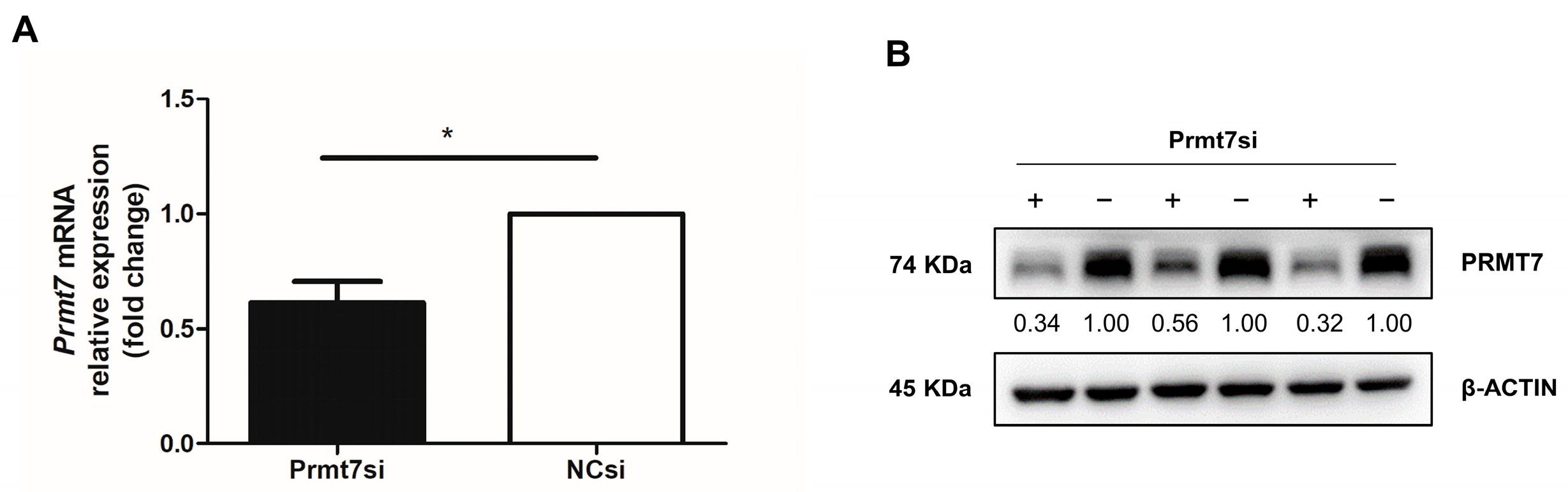
3.1. Validation of Prmt7 Knockdown in GC-1 Cells before miRNA Sequencing

Our previous work identified that Prmt7 regulated the proliferation of GC-1 cells (a type of mouse spermatogonial cell line) as the Prmt7 knockdown reduced their proliferation [5]. However, the underlying molecular mechanisms are still unclear. It is known that miRNAs play essential roles in male germ cell development. Thus, to identify the miRNAs regulated by Prmt7 and shed light on the mechanisms involved in the proliferation of mouse spermatogonia, we performed small RNA sequencing in GC-1 cells, transfected with Prmt7 siRNA or negative control (NC) siRNA. Before sequencing, we examined the inhibitory effect of Prmt7 siRNA in GC-1 cells. The results showed that Prmt7 expression was significantly reduced after the Prmt7 siRNA transfection for 48 h at both the mRNA and protein levels (Figure 1A,B).

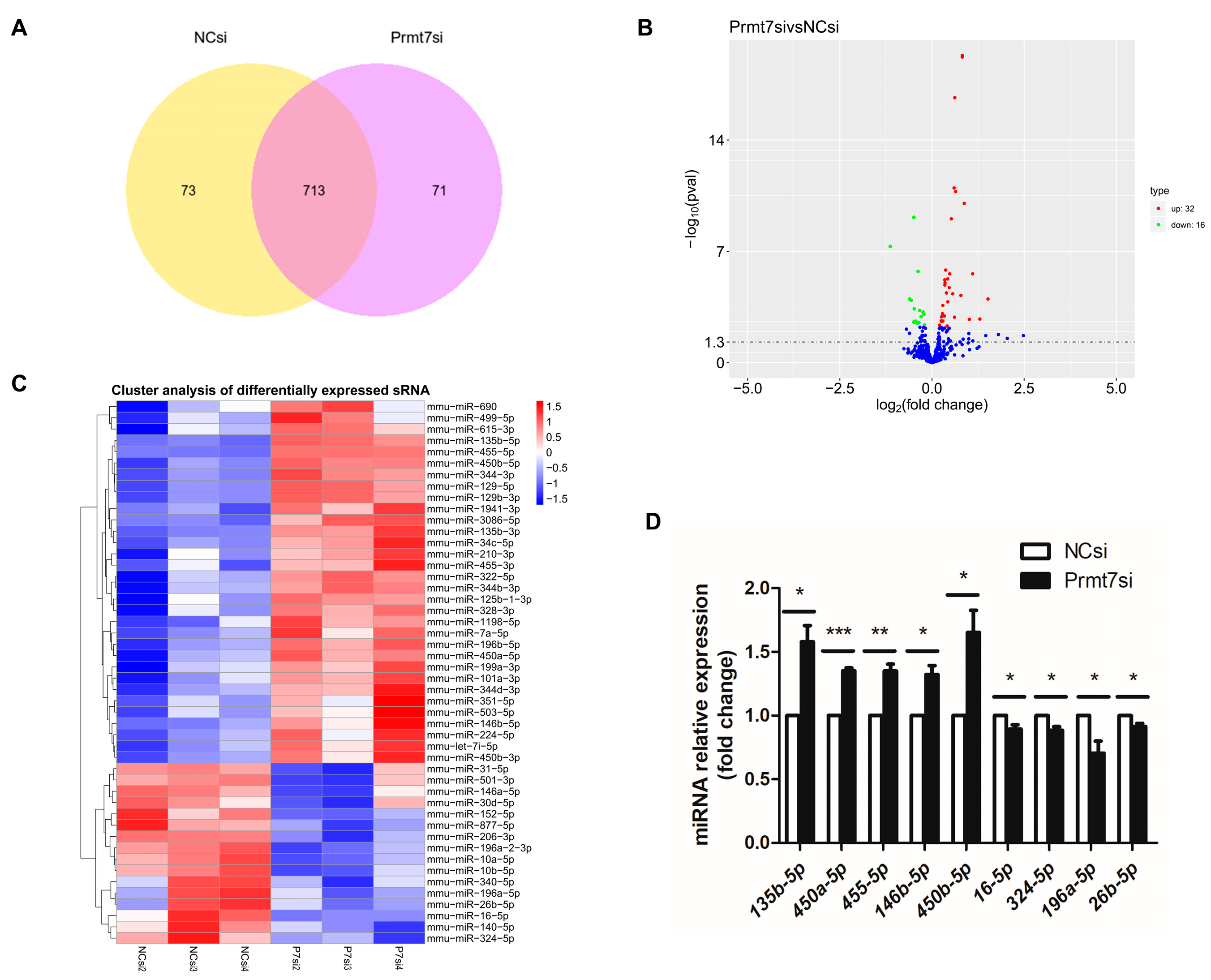
3.2. Analysis of Differentially Expressed miRNAs upon Prmt7 Downregulation

We investigated the miRNAs which were differentially expressed in the Prmt7 siRNA-transfected GC-1 cells relative to the NC siRNA-transfected GC-1 cells, making a further distinction between upregulation and downregulation. In total, 713 miRNAs were detected in both groups with three replicates (the NC siRNA group and Prmt7 siRNA group) (Figure 2A). Of the 713 miRNAs, 48 miRNAs were differentially expressed based on an adjusted p < 0.05. Of these, 32 and 16 miRNAs were upregulated and downregulated, respectively (Figure 2B). A cluster analysis was also performed to investigate the expression patterns of these differentially expressed miRNAs (DEmiRNAs), as shown in Figure 2C. Prior to further analysis, we verified the DEmiRNAs using an independent method. Specifically, we conducted qRT-PCR on a subset of randomly selected upregulated and downregulated miRNAs. Relative expression levels were normalized to the expression level of U6 snRNA. The qRT-PCR results confirmed the sequencing data and validated that mmu-miR-135b-5p, mmu-miR-450a-5p, mmu-miR-455-5p, mmu-miR-146b-5p, and mmu-miR-450b-5p were significantly upregulated (p < 0.05, p < 0.01, and p < 0.001), whereas, mmu-miR-16-5p, mmu-miR-324-5p, mmu-miR-196a-5p, and mmu-miR-26b-5p were significantly downregulated (p < 0.05, Figure 2D) in the Prmt7 siRNA group, compared to the NC siRNA group. Thus, the miRNA sequencing results were credible.

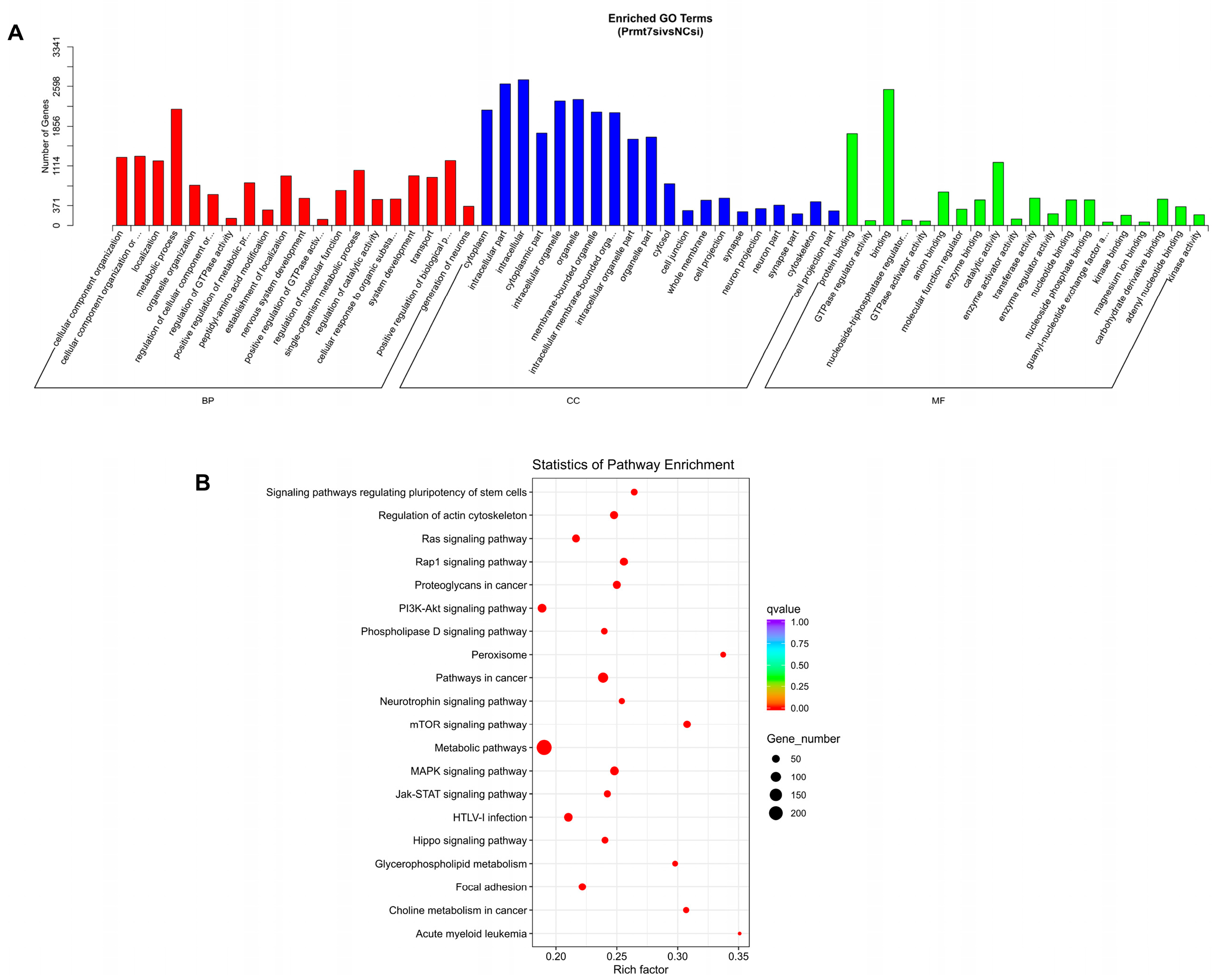
3.3. Functional Enrichment Analysis of Target Genes of DEmiRNAs

miRNA target prediction was performed using two online tools: miRanda (miRanda-3.3a) and RNAhybrid. The target genes of DEmiRNAs were determined using the intersection predicted with these two databases. Then, we performed functional enrichment analysis of these intersection target genes. The GO enrichment analysis revealed that these target genes were categorized according to their inclusion in the ontologies biological process (BP), cellular component (CC), and molecular function (MF), resulting in 227 BP terms, 59 CC terms, and 43 MF terms (adjusted p < 0.05; Supplementary File S1). The top 20 GO terms for the three ontologies are shown in Figure 3A. The target genes of the DEmiRNAs were mainly enriched in BP terms of the cellular component organization or biogenesis, localization, and metabolic process; in CC terms of cytoplasm and intracellular part; and in MF terms of protein binding, GTPase regulator activity, and binding. Furthermore, the KEGG pathway analysis revealed 165 significantly enriched pathways (adjusted p < 0.05; Supplementary File S2) and the top 20 pathways are shown in Figure 3B. The most significantly enriched pathways (top three) associated with Prmt7 expression were metabolic pathways, pathways in cancer, and the mTOR signaling pathway.

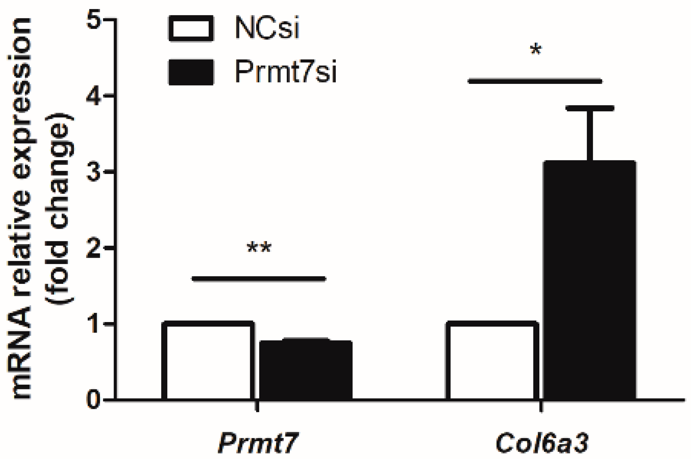
3.4. The Possible Molecular Mechanisms Underlying the GC-1 Proliferation Regulated by Prmt7/miRNA/Target Gene

To identify the possible molecular mechanisms underlying the GC-1 proliferation regulated by the Prmt7/miRNA/target gene, we screened the intersection genes between the DEmiRNA target genes and our previously obtained differentially expressed genes (DEGs) from the RNA sequencing of GC-1 cells (NC siRNA group vs. Prmt7 siRNA group, related raw data were deposited in NCBI with accession PRJNA779471). In total, we obtained 37 target genes as DEGs (Supplementary File S3). Through further screening, seven genes were uniquely regulated by a single miRNA (Table 2). Among these miRNA/target gene pairs, we verified the expression of Col6a3 (collagen type VI alpha 3), as a target gene of mmu-miR-877-3p. The results showed that with the downregulated Prmt7 expression, the mRNA level of Col6a3 was upregulated (Figure 4). These data suggest that the proliferation of GC-1 spermatogonial cells might be associated with the Prmt7/mmu-miR-877-3p/Col6a3 regulatory pathway.

4. Discussion

Prmt7, the only type III PRMT, plays a crucial role in the development of mice and humans [35]. Recent studies, including our own, have demonstrated the positively regulatory roles of Prmt7 in the proliferation of male germ cells [7], which implies its possible function in male reproduction. Growing evidence indicates that male germ cell development relies on miRNAs [16,17]. However, the mechanism by which Prmt7 regulates the miRNA and target genes in male germ cells remains unclear. In the current study, we examined the miRNA profile in GC-1 cells, a cell line of immortalized mouse spermatogonia, featuring a reduced expression of Prmt7, which was achieved by transfection with Prmt7 siRNA, compared to NC siRNA as a control. As a result, we gained new information regarding the molecular mechanisms of Prmt7 in spermatogonial proliferation, mediated through miRNA and the target gene.
After the miRNA sequencing of six libraries (two groups, three replicates in each group), we identified 713 miRNAs, including 48 differentially expressed miRNAs (DEmiRNAs), of which 32 were upregulated and 16 were downregulated (adjusted p < 0.05). On a subset of these hits, we confirmed the differential miRNA expression using qRT-PCR, in particular, the upregulation of miR-135b-5p, miR-450a-5p, miR-455-5p, miR-146b-5p, and miR-450b-5p, as well as the downregulation of miR-16-5p, miR-324-5p, miR-196a-5p, and miR-26b-5p in Prmt7 siRNA-transfected GC-1 cells, compared with NC siRNA-transfected GC-1 cells. Among upregulated miRNAs, miR-146b-5p inhibits cancers, including gallbladder cancer [36], papillary thyroid cancer [37], nonsmall cell lung cancer [38], colorectal cancer [39], and pancreatic cancer [40]. In addition, we noted the levels of miR-450a-5p, which is a proadipogenic miRNA that is able to promote adipogenesis [41]. The roles of these DEmiRNAs are involved in cell proliferation, cancer growth and metastasis, and adipogenesis, which are similar to the functions of Prmt7. For example, Prmt7 deficiency can lead to the defective proliferation of mouse primordial germ cells during the embryonic stage [7]. Prmt7 downregulation reduced GC-1 mouse spermatogonial proliferation [5]. Prmt7 overexpression promotes the malignant progression of nonsmall cell lung cancer cells [42] and the growth of renal cell carcinoma [43]. Prmt7 expression is also associated with the metastasis of breast cancer [44]. Both Prmt7-deficient mice and patients with Prmt7 mutations reportedly exhibit obesity [45,46]. Studies further revealed a repressive role of Prmt7 in adipogenesis in that Prmt7 depletion in preadipocytes resulted in enhanced adipogenesis and vice versa [47]. Overall, these DEmiRNAs could be the downstream effectors of Prmt7.
Next, we predicted the target genes of these DEmiRNAs and performed a functional enrichment analysis. The GO analysis identified significantly enriched BP terms, including metabolic processes and nervous system development, which were associated with Prmt7, since Prmt7 depletion contributed to reduced oxidative metabolism in skeletal muscle [45]. The loss-of-function of Prmt7 in mice and humans causes an increased fat mass, suggesting the regulatory role of Prmt7 in metabolism [35]. Moreover, patients with Prmt7 mutations exhibit neuron-deficient phenotypes, and, similarly, Prmt7-knockout mice exhibit deficient social behaviors [46,48]. In addition, a KEGG pathway analysis for the target genes predicted from these DEmiRNAs was performed. Among the top 20 significantly enriched pathways, we identified pathways in cancer, signaling pathways regulating the pluripotency of stem cells, and the PI3K-Akt signaling pathway, all of which are reportedly associated with Prmt7 expression. For example, high expression levels of Prmt7 are correlated with the proliferation and metastasis of breast cancer cells, lung cancer tissues, and clear cell renal cell carcinoma tissues [42,43,44,49]. In clear-cell renal cell carcinoma, Prmt7 can maintain β-catenin expression to regulate cell proliferation, which demonstrates the possible role of Prmt7 in regulating the Wnt/β-catenin signaling pathway in cancer. Regarding the involvement of Prmt7 in the regulation of stem cell pluripotency, Prmt7 plays a crucial role in maintaining the pluripotency of embryonic stem cells [9,25]. Based on the targeted genes in this pathway, we proposed that Prmt7 could function in stem cells through the Jak-STAT, MAPK, Wnt, and PI3K-Akt signaling pathways. Moreover, the PI3K-Akt signaling pathway, as a well-known regulator, is involved in many biological processes, such as cell proliferation, apoptosis, metabolism, growth, and survival. This pathway is activated in multiple types of human cancers, including ovarian cancer, brain cancer, breast cancer, and metastatic renal cell carcinoma [50], and maintains the pluripotency of stem cells through the PI3K/Akt/Sox2 axis [51]. Notably, with increased Prmt7 expression, the phosphorylation of Akt and substrates of mTORC1 were enhanced, suggesting the regulatory roles of Prmt7 in the Akt/mTORC pathway [52]. Overall, we propose that the miRNAs differentially expressed in response to the expression of Prmt7 and their target genes in these pathways form a functional signaling pathway in cell proliferation.
To better determine the possible molecular mechanisms underlying the GC-1 proliferation regulated by Prmt7/miRNA/target gene, we determined the intersection genes between the DEmiRNA target genes and our previously obtained DEGs from the RNA sequencing of GC-1 cells from the NC siRNA group and Prmt7 siRNA group. In total, 37 genes in common were obtained, which meant they were considered as both target genes of DEmiRNAs and DEGs. By deleting the nonunique miRNA/target gene pairs, we finally obtained seven genes which were uniquely regulated by single miRNAs. Then, we detected the expression of Col6a3 (collagen type VI alpha 3, as the target gene of mmu-miR-877-3p) in Prmt7-downregulated and control GC-1 cells. The results showed Col6a3 expression was increased with Prmt7 downregulation, suggesting that Prmt7 downregulation in mouse spermatogonia might function through miR-877-3p/Col6a3. It has only been reported that Col6a3 is associated with cancer [53,54], muscular dystrophy [55,56], and obesity [57,58], which exhibits the possible similar roles of Prmt7. However, thus far, there is no evidence showing the role of mmu-miR-877-3p or Col6a3 in male reproduction. Therefore, further study should be performed in the future to help better understand the regulatory roles of Prmt7 in male reproduction.

5. Conclusions

In summary, to identify the miRNAs regulated by Prmt7 in spermatogonia and further explore the possible pathway involved in Prmt7 regulation in mouse spermatogonial cell line GC-1 cells, we performed small RNA sequencing, as well as integrated analyses with previous RNA sequencing data for Prmt7 siRNA and NC siRNA-transfected GC-1 cells. As a result, we identified 48 differentially expressed miRNAs, among which 32 and 16 miRNAs were upregulated and downregulated, respectively. The intersection between the target genes of these DEmiRNAs and previously obtained DEGs using RNA sequencing was further screened and seven miRNA/target gene pairs were obtained. Further detection suggested that Prmt7 downregulation in mouse spermatogonia might function through miR-877-3p/Col6a3. These data will provide insight into the further exploration of the roles of Prmt7 in male germ cell development and in male reproduction.

Supplementary Materials

The following supporting information can be downloaded at: https://www.mdpi.com/article/10.3390/life12081194/s1, Supplementary File S1: GO terms of target genes; Supplementary File S2: KEGG pathways of target genes; Supplementary File S3: intersection gene screening.

Author Contributions

Conceptualization, B.W., H.G. and Y.M.; methodology, H.G., B.W., M.Z., J.G., Z.L. and F.G.; validation, H.G., M.Z. and J.G.; formal analysis, H.G., M.Z., Z.L. and F.G.; data curation, B.W., H.G. and Z.L.; writing—original draft preparation, B.W., H.G., M.Z. and Z.L.; writing—review and editing, B.W. and Y.M.; supervision, B.W. and Y.M.; project administration, B.W. and Y.M.; funding acquisition, B.W. and Y.M. All authors have read and agreed to the published version of the manuscript.

Funding

This research was funded by the National Natural Science Foundation of China, grant number 32172738, the Chinese Academy of Agricultural Sciences: Major Scientific Research Tasks for Scientific and Technological Innovation Project, grant number CAAS-ZDRW202006, and the Agricultural Science and Technology Innovation Program, grant number ASTIP-IAS05.

Data Availability Statement

The data presented in this study are available on request from the corresponding author.

Acknowledgments

We thank all the staff in the group and institute who helped with this work.

Conflicts of Interest

The authors declare no conflict of interest.

References

  1. Griswold, M.D. Spermatogenesis: The Commitment to Meiosis. Physiol. Rev. 2016, 96, 1–17. [Google Scholar] [CrossRef] [PubMed] [Green Version]
  2. Kubota, H.; Brinster, R.L. Spermatogonial stem cells. Biol. Reprod. 2018, 99, 52–74. [Google Scholar] [CrossRef] [PubMed] [Green Version]
  3. Mei, X.X.; Wang, J.; Wu, J. Extrinsic and intrinsic factors controlling spermatogonial stem cell self-renewal and differentiation. Asian J. Androl. 2015, 17, 347–354. [Google Scholar] [CrossRef] [PubMed]
  4. Wang, B.; Pfeiffer, M.J.; Drexler, H.C.; Fuellen, G.; Boiani, M. Proteomic Analysis of Mouse Oocytes Identifies PRMT7 as a Reprogramming Factor that Replaces SOX2 in the Induction of Pluripotent Stem Cells. J. Proteome Res. 2016, 15, 2407–2421. [Google Scholar] [CrossRef] [Green Version]
  5. Wang, B.; Zhang, M.; Guo, J.; Liu, Z.; Zhou, R.; Guo, F.; Mu, Y. The Effects of Flavonoid Apigenin on Male Reproductive Health: Inhibition of Spermatogonial Proliferation through Downregulation of Prmt7/Akt3 Pathway. Int. J. Mol. Sci. 2021, 22, 12209. [Google Scholar] [CrossRef]
  6. Jelinic, P.; Stehle, J.C.; Shaw, P. The testis-specific factor CTCFL cooperates with the protein methyltransferase PRMT7 in H19 imprinting control region methylation. PLoS Biol. 2006, 4, e355. [Google Scholar] [CrossRef]
  7. Chen, M.; Wang, Y.; Lin, L.; Dong, F.; Wu, H.; Bao, S.; Gao, F. PRMT7 is involved in regulation of germ cell proliferation during embryonic stage. BioChem. Biophys. Res. Commun. 2020, 533, 938–944. [Google Scholar] [CrossRef]
  8. Buhr, N.; Carapito, C.; Schaeffer, C.; Kieffer, E.; Van Dorsselaer, A.; Viville, S. Nuclear proteome analysis of undifferentiated mouse embryonic stem and germ cells. ElectrophoResis 2008, 29, 2381–2390. [Google Scholar] [CrossRef]
  9. Chen, T.Y.; Lee, S.H.; Dhar, S.S.; Lee, M.G. Protein arginine methyltransferase 7-mediated microRNA-221 repression maintains Oct4, Nanog, and Sox2 levels in mouse embryonic stem cells. J. Biol. Chem. 2018, 293, 3925–3936. [Google Scholar] [CrossRef] [Green Version]
  10. Chen, X.; Li, X.; Guo, J.; Zhang, P.; Zeng, W. The roles of microRNAs in regulation of mammalian spermatogenesis. J. Anim. Sci. Biotechnol. 2017, 8, 35. [Google Scholar] [CrossRef]
  11. Bueno, M.J.; Malumbres, M. MicroRNAs and the cell cycle. Biochim. Biophys. Acta 2011, 1812, 592–601. [Google Scholar] [CrossRef] [Green Version]
  12. Lakshmipathy, U.; Love, B.; Goff, L.A.; Jörnsten, R.; Graichen, R.; Hart, R.P.; Chesnut, J.D. MicroRNA expression pattern of undifferentiated and differentiated human embryonic stem cells. Stem. Cells Dev. 2007, 16, 1003–1016. [Google Scholar] [CrossRef] [Green Version]
  13. Chen, B.; Li, H.; Zeng, X.; Yang, P.; Liu, X.; Zhao, X.; Liang, S. Roles of microRNA on cancer cell metabolism. J. Transl. Med. 2012, 10, 228. [Google Scholar] [CrossRef] [Green Version]
  14. Hwang, H.W.; Mendell, J.T. MicroRNAs in cell proliferation, cell death, and tumorigenesis. Br. J. Cancer 2007, 96, R40–R44. [Google Scholar] [CrossRef]
  15. Liu, W.; Niu, Z.; Li, Q.; Pang, R.T.; Chiu, P.C.; Yeung, W.S. MicroRNA and Embryo Implantation. Am. J. Reprod. Immunol. 2016, 75, 263–271. [Google Scholar] [CrossRef]
  16. Kotaja, N. MicroRNAs and spermatogenesis. Fertil. Steril. 2014, 101, 1552–1562. [Google Scholar] [CrossRef]
  17. Wang, L.; Xu, C. Role of microRNAs in mammalian spermatogenesis and testicular germ cell tumors. Reproduction 2015, 149, R127–R137. [Google Scholar] [CrossRef] [Green Version]
  18. Luo, M.; Hao, L.; Hu, F.; Dong, Y.; Gou, L.; Zhang, W.; Wang, X.; Zhao, Y.; Jia, M.; Hu, S.; et al. MicroRNA profiles and potential regulatory pattern during the early stage of spermatogenesis in mice. Sci. China Life Sci. 2015, 58, 442–450. [Google Scholar] [CrossRef] [Green Version]
  19. Huszar, J.M.; Payne, C.J. MicroRNA 146 (Mir146) modulates spermatogonial differentiation by retinoic acid in mice. Biol. Reprod. 2013, 88, 15. [Google Scholar] [CrossRef]
  20. Curry, E.; Safranski, T.J.; Pratt, S.L. Differential expression of porcine sperm microRNAs and their association with sperm morphology and motility. Theriogenology 2011, 76, 1532–1539. [Google Scholar] [CrossRef]
  21. He, Z.; Jiang, J.; Kokkinaki, M.; Tang, L.; Zeng, W.; Gallicano, I.; Dobrinski, I.; Dym, M. MiRNA-20 and mirna-106a regulate spermatogonial stem cell renewal at the post-transcriptional level via targeting STAT3 and Ccnd1. Stem. Cells 2013, 31, 2205–2217. [Google Scholar] [CrossRef] [Green Version]
  22. Huang, Z.; Tang, D.; Gao, J.; Dou, X.; Cheng, P.; Peng, D.; Zhang, Y.; Mao, J.; Zhang, L.; Zhang, X. miR-34c disrupts spermatogonial stem cell homeostasis in cryptorchid testes by targeting Nanos2. Reprod. Biol. Endocrinol. 2018, 16, 97. [Google Scholar] [CrossRef]
  23. Niu, Z.; Goodyear, S.M.; Rao, S.; Wu, X.; Tobias, J.W.; Avarbock, M.R.; Brinster, R.L. MicroRNA-21 regulates the self-renewal of mouse spermatogonial stem cells. Proc. Natl. Acad. Sci. USA 2011, 108, 12740–12745. [Google Scholar] [CrossRef] [Green Version]
  24. Yang, Q.E.; Racicot, K.E.; Kaucher, A.V.; Oatley, M.J.; Oatley, J.M. MicroRNAs 221 and 222 regulate the undifferentiated state in mammalian male germ cells. Development 2013, 140, 280–290. [Google Scholar] [CrossRef] [Green Version]
  25. Lee, S.H.; Chen, T.Y.; Dhar, S.S.; Gu, B.; Chen, K.; Kim, Y.Z.; Li, W.; Lee, M.G. A feedback loop comprising PRMT7 and miR-24-2 interplays with Oct4, Nanog, Klf4 and c-Myc to regulate stemness. Nucleic. Acids Res. 2016, 44, 10603–10618. [Google Scholar] [CrossRef]
  26. Livak, K.J.; Schmittgen, T.D. Analysis of relative gene expression data using real-time quantitative PCR and the 2(-Delta Delta C(T)) Method. Methods 2001, 25, 402–408. [Google Scholar] [CrossRef]
  27. Langmead, B.; Trapnell, C.; Pop, M.; Salzberg, S.L. Ultrafast and memory-efficient alignment of short DNA sequences to the human genome. Genome Biol. 2009, 10, R25. [Google Scholar] [CrossRef] [Green Version]
  28. Friedländer, M.R.; Mackowiak, S.D.; Li, N.; Chen, W.; Rajewsky, N. miRDeep2 accurately identifies known and hundreds of novel microRNA genes in seven animal clades. Nucleic Acids Res. 2012, 40, 37–52. [Google Scholar] [CrossRef]
  29. Wen, M.; Shen, Y.; Shi, S.; Tang, T. miREvo: An integrative microRNA evolutionary analysis platform for next-generation sequencing experiments. BMC Bioinform. 2012, 13, 140. [Google Scholar] [CrossRef] [Green Version]
  30. Sun, J.; Nishiyama, T.; Shimizu, K.; Kadota, K. TCC: An R package for comparing tag count data with robust normalization strategies. BMC Bioinform. 2013, 14, 219. [Google Scholar] [CrossRef] [Green Version]
  31. Enright, A.J.; John, B.; Gaul, U.; Tuschl, T.; Sander, C.; Marks, D.S. MicroRNA targets in Drosophila. Genome Biol. 2003, 5, R1. [Google Scholar] [CrossRef] [PubMed] [Green Version]
  32. Kanehisa, M.; Araki, M.; Goto, S.; Hattori, M.; Hirakawa, M.; Itoh, M.; Katayama, T.; Kawashima, S.; Okuda, S.; Tokimatsu, T.; et al. KEGG for linking genomes to life and the environment. Nucleic Acids Res. 2008, 36, D480–D484. [Google Scholar] [CrossRef] [PubMed]
  33. Young, M.D.; Wakefield, M.J.; Smyth, G.K.; Oshlack, A. Gene ontology analysis for RNA-seq: Accounting for selection bias. Genome Biol. 2010, 11, R14. [Google Scholar] [CrossRef] [PubMed] [Green Version]
  34. Mao, X.; Cai, T.; Olyarchuk, J.G.; Wei, L. Automated genome annotation and pathway identification using the KEGG Orthology (KO) as a controlled vocabulary. Bioinformatics 2005, 21, 3787–3793. [Google Scholar] [CrossRef]
  35. Akawi, N.; McRae, J.; Ansari, M.; Balasubramanian, M.; Blyth, M.; Brady, A.F.; Clayton, S.; Cole, T.; Deshpande, C.; Fitzgerald, T.W.; et al. Discovery of four recessive developmental disorders using probabilistic genotype and phenotype matching among 4,125 families. Nat. Genet. 2015, 47, 1363–1369. [Google Scholar] [CrossRef] [PubMed]
  36. Ouyang, B.; Pan, N.; Zhang, H.; Xing, C.; Ji, W. miR-146b-5p inhibits tumorigenesis and metastasis of gallbladder cancer by targeting Toll-like receptor 4 via the nuclear factor-κB pathway. Oncol. Rep. 2021, 45, 15. [Google Scholar] [CrossRef]
  37. Peng, Y.; Fang, X.; Yao, H.; Zhang, Y.; Shi, J. MiR-146b-5p Regulates the Expression of Long Noncoding RNA MALAT1 and Its Effect on the Invasion and Proliferation of Papillary Thyroid Cancer. Cancer Biother. Radiopharm. 2021, 36, 433–440. [Google Scholar] [CrossRef]
  38. Li, Y.; Zhang, H.; Dong, Y.; Fan, Y.; Li, Y.; Zhao, C.; Wang, C.; Liu, J.; Li, X.; Dong, M.; et al. MiR-146b-5p functions as a suppressor miRNA and prognosis predictor in non-small cell lung cancer. J. Cancer 2017, 8, 1704–1716. [Google Scholar] [CrossRef] [Green Version]
  39. Zhu, Y.; Wu, G.; Yan, W.; Zhan, H.; Sun, P. miR-146b-5p regulates cell growth, invasion, and metabolism by targeting PDHB in colorectal cancer. Am. J. Cancer Res. 2017, 7, 1136–1150. [Google Scholar]
  40. Lin, F.; Wang, X.; Jie, Z.; Hong, X.; Li, X.; Wang, M.; Yu, Y. Inhibitory effects of miR-146b-5p on cell migration and invasion of pancreatic cancer by targeting MMP16. J. Huazhong Univ. Sci. Technol. Med. Sci. 2011, 31, 509. [Google Scholar] [CrossRef]
  41. Zhang, Y.; Yu, M.; Dai, M.; Chen, C.; Tang, Q.; Jing, W.; Wang, H.; Tian, W. miR-450a-5p within rat adipose tissue exosome-like vesicles promotes adipogenic differentiation by targeting WISP2. J. Cell Sci. 2017, 130, 1158–1168. [Google Scholar] [CrossRef] [Green Version]
  42. Cheng, D.; He, Z.; Zheng, L.; Xie, D.; Dong, S.; Zhang, P. PRMT7 contributes to the metastasis phenotype in human non-small-cell lung cancer cells possibly through the interaction with HSPA5 and EEF2. Onco. Targets 2018, 11, 4869–4876. [Google Scholar] [CrossRef] [Green Version]
  43. Liu, F.; Wan, L.; Zou, H.; Pan, Z.; Zhou, W.; Lu, X. PRMT7 promotes the growth of renal cell carcinoma through modulating the β-catenin/C-MYC axis. Int. J. Biochem. Cell Biol. 2020, 120, 105686. [Google Scholar] [CrossRef]
  44. Liu, Y.; Li, L.; Liu, X.; Wang, Y.; Liu, L.; Peng, L.; Zhang, Y. Arginine methylation of SHANK2 by PRMT7 promotes human breast cancer metastasis through activating endosomal FAK signalling. eLife 2020, 9, e57617. [Google Scholar] [CrossRef]
  45. Jeong, H.J.; Lee, H.J.; Vuong, T.A.; Choi, K.S.; Choi, D.; Koo, S.H.; Cho, S.C.; Cho, H.; Kang, J.S. Prmt7 Deficiency Causes Reduced Skeletal Muscle Oxidative Metabolism and Age-Related Obesity. Diabetes 2016, 65, 1868–1882. [Google Scholar] [CrossRef] [Green Version]
  46. Agolini, E.; Dentici, M.L.; Bellacchio, E.; Alesi, V.; Radio, F.C.; Torella, A.; Musacchia, F.; Tartaglia, M.; Dallapiccola, B.; Nigro, V.; et al. Expanding the clinical and molecular spectrum of PRMT7 mutations: 3 additional patients and review. Clin. Genet. 2018, 93, 675–681. [Google Scholar] [CrossRef] [Green Version]
  47. Leem, Y.E.; Bae, J.H.; Jeong, H.J.; Kang, J.S. PRMT7 deficiency enhances adipogenesis through modulation of C/EBP-β. BioChem. Biophys. Res. Commun. 2019, 517, 484–490. [Google Scholar] [CrossRef]
  48. Fiorica, P.N.; Wheeler, H.E. Transcriptome association studies of neuropsychiatric traits in African Americans implicate PRMT7 in schizophrenia. PeerJ 2019, 7, e7778. [Google Scholar] [CrossRef] [Green Version]
  49. Yao, R.; Jiang, H.; Ma, Y.; Wang, L.; Wang, L.; Du, J.; Hou, P.; Gao, Y.; Zhao, L.; Wang, G.; et al. PRMT7 induces epithelial-to-mesenchymal transition and promotes metastasis in breast cancer. Cancer Res. 2014, 74, 5656–5667. [Google Scholar] [CrossRef] [Green Version]
  50. Alzahrani, A.S. PI3K/Akt/mTOR inhibitors in cancer: At the bench and bedside. Semin. Cancer Biol. 2019, 59, 125–132. [Google Scholar] [CrossRef]
  51. Schaefer, T.; Steiner, R.; Lengerke, C. SOX2 and p53 Expression Control Converges in PI3K/AKT Signaling with Versatile Implications for Stemness and Cancer. Int. J. Mol. Sci. 2020, 21, 4902. [Google Scholar] [CrossRef]
  52. Gutiérrez-Venegas, G.; González-Rosas, Z. Apigenin reduce lipoteichoic acid-induced inflammatory response in rat cardiomyoblast cells. Arch. Pharm. Res. 2017, 40, 240–249. [Google Scholar] [CrossRef]
  53. Duan, Y.; Liu, G.; Sun, Y.; Wu, J.; Xiong, Z.; Jin, T.; Chen, M. COL6A3 polymorphisms were associated with lung cancer risk in a Chinese population. Respir. Res. 2019, 20, 143. [Google Scholar] [CrossRef] [Green Version]
  54. Hu, Y.; Li, J.; Luo, H.; Song, W.; Yang, J. Differential Expression of COL1A1, COL1A2, COL6A3, and SULF1 as Prognostic Biomarkers in Gastric Cancer. Int. J. Gen. Med. 2021, 14, 5835–5843. [Google Scholar] [CrossRef]
  55. Das Bhowmik, A.; Karthik, T.; Uppin, M.; Sundaram, C.; Dalal, A. Targeted next generation sequencing reveals novel splice site mutations in COL6A3 gene in a patient with congenital muscular dystrophy. Neurol. India 2018, 66, 1812–1814. [Google Scholar]
  56. Pan, T.C.; Zhang, R.Z.; Markova, D.; Arita, M.; Zhang, Y.; Bogdanovich, S.; Khurana, T.S.; Bönnemann, C.G.; Birk, D.E.; Chu, M.L. COL6A3 protein deficiency in mice leads to muscle and tendon defects similar to human collagen VI congenital muscular dystrophy. J. Biol. Chem. 2013, 288, 14320–14331. [Google Scholar] [CrossRef] [Green Version]
  57. Dankel, S.N.; Svärd, J.; Matthä, S.; Claussnitzer, M.; Klöting, N.; Glunk, V.; Fandalyuk, Z.; Grytten, E.; Solsvik, M.H.; Nielsen, H.J.; et al. COL6A3 expression in adipocytes associates with insulin resistance and depends on PPARγ and adipocyte size. Obesity 2014, 22, 1807–1813. [Google Scholar] [CrossRef]
  58. McCulloch, L.J.; Rawling, T.J.; Sjöholm, K.; Franck, N.; Dankel, S.N.; Price, E.J.; Knight, B.; Liversedge, N.H.; Mellgren, G.; Nystrom, F.; et al. COL6A3 is regulated by leptin in human adipose tissue and reduced in obesity. Endocrinology 2015, 156, 134–146. [Google Scholar] [CrossRef] [Green Version]
Figure 1. The expression of Prmt7 in GC-1 spermatogonial cells after Prmt7 siRNA (Prmt7si) or negative control siRNA (NCsi) transfection for 48 h. (A) Relative Prmt7 mRNA expression detected with qRT-PCR in Prmt7si- and NCsi-transfected GC-1 cells for 48 h. Data from three independent experiments are represented as mean ± SEM. * indicates statistical significance. * p < 0.05. (B) The protein level of PRMT7 detected with Western blotting in Prmt7si-(shown by “+”) and NCsi-transfected (shown by “−”) GC-1 cells for 48 h. The intensity of each protein band was analyzed using ImageJ software and the pro-tein expression levels were normalized to β-ACTIN.
Life 12 01194 g001
Figure 2. Differentially expressed miRNAs (DEmiRNAs) between Prmt7 siRNA-transfected and NC siRNA-transfected GC-1 cells. (A) Total miRNAs identified in NC siRNA-transfected (left) and Prmt7 siRNA-transfected GC-1 cells (right). (B) Volcano plot of DEmiRNAs in Prmt7 siRNA-transfected and NC siRNA-transfected GC-1 cells (adjusted p < 0.05). Red and green dots represent miRNAs that were upregulated and downregulated, respectively, in the Prmt7 siRNA group, compared to NC siRNA group. (C) Clustered heatmap of DEmiRNAs between the Prmt7 siRNA-transfected and the NC siRNA-transfected GC-1 cells (adjusted p < 0.05). (D) Verification of the randomly selected DEmiRNAs using qRT-PCR in the Prmt7 siRNA group and the NC siRNA group. U6 was used as an internal control. Data from the three independent experiments are represented as mean ± SEM. * indicates statistical significance. * p < 0.05, ** p < 0.01, and *** p < 0.001.
Life 12 01194 g002
Figure 3. Functional enrichment analysis of target genes. (A) GO analysis showed that target genes predicted from DEmiRNAs were involved in 3 classes of GO terms: biological process (BP) shown in red, cellular component (CC) shown in blue, and molecular function (MF) shown in green. The top 20 significantly enriched GO terms are shown. (B) KEGG pathway analysis of the target genes predicted from DEmiRNAs. The top 20 enriched KEGG pathways are shown in the bubble diagram. The larger the bubble size, the higher the number of target genes that were enriched. The bubble color changing from red to purple indicates the increasing p value.
Life 12 01194 g003
Figure 4. Relative Prmt7 and Col6a3 mRNA expression detected using qRT-PCR in Prmt7 siRNA (Prmt7si)- and negative control siRNA (NCsi)-transfected GC-1 cells for 48 h. Data from three independent experiments are represented as mean ± SEM. * indicates statistical significance. * p < 0.05, ** p < 0.01.
Life 12 01194 g004
Table 1. Primers used for qRT-PCR assessing the relative expression of RNAs and miRNAs in mouse GC-1 spermatogonia.
miRNA Primer Sequences
Prmt7Forward primerGGAGATTGCCAGGTCATCCT
Reverse primerGAAGTCAGCCCCTGCAGTAA
Col6a3Forward primerGAGATGGGGTTGGCAGTGAA
Reverse primerGCATTGATCAACGGAGTCGC
β-actinForward primerTGTGCTGTCCCTGTATGCCTCT
Reverse primerTAGATGGGCACAGTGTGGGTGA
mmu-miR-135b-5pForward primerCGCGTATGGCTTTTCATTCCTATGTGA
mmu-miR-450a-5pForward primerCGCGCTTTTGCGATGTGTTCCTAATAT
mmu-miR-455-5pForward primerCGCTATGTGCCTTTGGACTACATCG
mmu-miR-146b-5pForward primerCCGCTGAGAACTGAATTCCATAGGCT
mmu-miR-450b-5pForward primerGCGCGTTTTGCAGTATGTTCCTGAATA
mmu-miR-16-5pForward primerCCGTAGCAGCACGTAAATATTGGCG
mmu-miR-324-5pForward primerTATATACGCATCCCCTAGGGCATTGG
mmu-miR-196a-5pForward primerCGCGTAGGTAGTTTCATGTTGTTGGG
mmu-miR-26b-5pForward primerGCGCGTTCAAGTAATTCAGGATAGGT
β-actin and U6 were served as controls of RNAs and miRNAs, respectively.
Table 2. Intersection genes by combining target genes of differentially expressed miRNAs and differentially expressed genes between NC siRNA group and Prmt7 siRNA group GC-1 cells.
Intersection GeneLog2 FCmiRNALog2 FC
Fut7−1.62mmu-miR-135b-3p1.29
Atp2c21.25mmu-miR-140-5p−0.22
Gm144061.38mmu-miR-30d-5p−0.40
Cracdl1.03mmu-miR-31-5p−0.57
Hmcn21.12mmu-miR-324-5p−0.50
Lefty21.69mmu-miR-501-3p−0.49
Col6a31.08mmu-miR-877-5p−0.49
Publisher’s Note: MDPI stays neutral with regard to jurisdictional claims in published maps and institutional affiliations.

