Article

The Entomopathogenic Nematodes H. bacteriophora and S. carpocapsae Inhibit the Activation of proPO System of the Nipa Palm Hispid Octodonta nipae (Coleoptera: Chrysomelidae)

1
State Key Laboratory of Ecological Pest Control for Fujian and Taiwan Crops, Department of Plant Protection, Fujian Agriculture and Forestry University, 15, Shangxiadian Road, Cangshan District, Fuzhou 350002, China
2
Department of Crop Protection, Faculty of Agriculture, Bayero University Kano, Gwarzo Road, Kano P.M.B. 3011, Nigeria
3
Department of Entomology, College of Plant Protection, Nanjing Agricultural University, Nanjing 210095, China
*
Author to whom correspondence should be addressed.
These authors contributed equally to this work.
Life 2022, 12(7), 1019; https://doi.org/10.3390/life12071019
Submission received: 27 May 2022 / Revised: 25 June 2022 / Accepted: 27 June 2022 / Published: 9 July 2022
(This article belongs to the Special Issue Advances in Plant Defensive Biochemical Activity)

Abstract

:
Entomopathogenic nematodes are biocontrol agents of invasive insect pests in soil and cryptic habitats. Nipa palm hispid, Octodonta nipae, is a pest of palm trees in Sothern China. To address its increasing damage, environmentally friendly control methods are required. This study aimed to test efficacy of Heterorhabditis bacteriophora and Steinernema carpocapsae on O. nipae and investigated the influence of secondary metabolites, nematodes, and their isolated cuticles on the activation of O. nipae’s prophenoloxidase system using qPCR analysis. Our data revealed that O. nipae were less susceptible to H. bacteriophora than S. carpocapsae and penetrations of infective juveniles were higher with S. carpocapsae treatment than H. bacteriophora. Moreover, expression levels of the serine protease P56, prophenoloxidase activation factor 1, PPO and serine protease inhibitor 28 upon S. carpocapsae and H. bacteriophora infections were generally downregulated at all times. However, upon heating, the cuticles lost their inhibitory effects and resulted in upregulation of the PPO gene. Similarly, the addition of arachidonic acid reversed the process and resulted in the upregulation of the PPO gene compared to the control. Further work is needed to identify toxic substances secreted by these EPNs to evade O. nipae’s immune system.

1. Introduction

The movement of destructive pest species from one country or region to another is on the rise. Nipa palm hispid Octodonta nipae (Maulik) is not an exception [1,2,3,4,5,6,7,8,9]. This pest is one of the most destructive invasive insects, reportedly causing economic damage to palm trees in China, mainly in Hainan, Guangdong, Fujian, etc. [9,10,11,12,13,14,15,16]. However, due to hazard caused by insecticides to the environment, the use of chemical control of this pest remains elusive. Several attempts were made by different studies to control this beetle using non-chemical methods [17,18,19]. One of these was the use of Tetrastichus brontispae Ferrière (Hymenoptera: Eulophidae) [20,21,22,23,24,25,26,27,28] entomopathogenic nematode Steinernema carpocapsae [17,18,19] and Metarrhizium anisopliae [29,30,31,32]. More eco-friendly and cost-effective methods of control of O. nipae are needed.
Heterorhabditidae and Steinernematidae are families of entomopathogenic nematodes (EPNs) mainly used for biological control of different economically invasive pests at field and laboratory levels [33,34,35,36,37]. They have advantages over the conventional chemical control because they have the ability to actively search for their hosts in cryptic habitats like leaf surfaces [38,39,40,41]. In addition, EPNs are known to be more effective against the larvae and other pre-adult stages of insects because they enter the host body easily [42]. EPNs are reported to be used for the control of leaf-eating caterpillars and other insect pests of many crops [43]. Many researchers have studied the efficacy of EPNs in controlling the adult western corn rootworm, Diabrotica virgifera LeConte [37,44], flea beetles, Phyllotreta spp. [45], and Colorado potato beetle, Leptinotarsa decemlineata (Say) found on leaf surfaces [40]. Similarly, the use of EPN for the control of O. nipae larvae was conducted in our laboratory using S. carpocapsae and found to be virulent at different concentrations and time points, as published in Sanda et al. [19]. However, experiments to test the efficacy of Heterorhabditis bacteriophora on O. nipae larvae have yet to be conducted.
Insects employ both humeral and cellular immune reactions in response to attacks by pathogens such as bacteria, fungi, and nematodes. These innate immune mechanisms include nodulation, phagocytosis, encapsulations, production of antimicrobial peptides, and activation of PPO system, which leads to melanization and subsequent pathogen death by septicemia [46,47]. Insect PPO activation system is one of the immune reactions against nematodes and bacteria that involves several proteinases, which are regulated by specific inhibitors. When pathogens like nematodes or bacteria attack insects, the hemolymph pattern-recognition proteins such as Peptidoglycan recognition proteins (PGRP) and C-type lectin bind the pathogens’ surface polysaccharides, thereby inducing the activation of initiator protease(s). In addition, enzymes released by the pathogens, as well as tissue damage due to the infection, may activate the proPO system [48,49]. These proteases trigger a protease cascade, activating a series of terminal serine proteases such as proPO-activating enzymes (PPAE), proPO-activating proteinase (PAP), or proPO-activating factors (PPAF) to cleave PPO and form active phenoloxidase (PO). However, the excessive production of these proteases is regulated by serine proteinase inhibitors (serpins) that limit the reaction speed and avoid excessive melanization [49,50]. In O. nipae, three full-length cDNAs of PPAFs (OnPPAFs) were cloned, namely OnPPAF3, OnPPAF1, and OnPPAF2. They were highly expressed in hemolymph except OnPPAF2, which shows low transcript abundance. However, knockdown of OnPPAF1 and OnPPAF3 showed a reduction in hemolymph phenoloxidase activity and inhibition of hemolymph melanization [51,52,53,54].
Moreover, different insect hosts responded differently to EPNs infections depending on the species and strain. Each nematode species uses different strategies to evade and suppress melanization and escape cellular encapsulation. Our previous study showed that EPNs of the family Steinernematids, S. carpocapsae use their body surface cuticle to escape host hemocyte encapsulation and at the same time inhibit its proPO system activity [19]. These are believed to be triggered by the body surface of the Steinernematids due to the presence of some lipid compounds on its body cuticle. Additionally, pathogenicity of nematodes is believed to be essentially modulated by the nematode’s excreted and secreted products (ESPs), which are proteases and protease inhibitors that act against effectors of the host’s immune system [55]. These ESPs have been detected in S. carpocapsae and are involved in host immune modulation [23,56,57].
Eicosanoids are other insect cellular and humoral immune mediators against several microbial infections [58,59]. They are mainly synthesized from arachidonic acid (AA) (5,8,11,14-eicosatetraenoic acid). AA is linked to phospholipids at the sn2 position and subsequently released upon microbial challenge by the catalytic activity of phospholipase A2 (PLA2) [60]. Biosynthesis of eicosanoids could also be inhibited by a nematode–bacteria complex [61]. Inhibition of PLA2 due to suppression of eicosanoids results in subsequent inhibition of phenoloxidase activation [62]. The symbiotic bacteria produce secondary metabolites such as Oxindole, Dexamethasone (DEX), and Benzylideneacetone (BZA) that suppress the activity of PLA2, thereby inhibiting the host immune responses [63,64]. These metabolites also restrict the spread of hemocytes and inhibit nodulation, antimicrobial peptides (AMPs) expression, and phenoloxidase activity [65,66,67]. However, the molecular nature of how H. bacteriophora and S. carpocapsae interact with O. nipae humoral immune system remains unknown. The present study, therefore, was conducted to determine the survival rates of O. nipae larvae at different concentrations of H. bacteriophora. Similarly, we evaluated the penetration abilities of both H. bacteriophora and S. carpocapsae against the third instar larvae of O. nipae. Further, we tested the differences in expression levels of four selected genes involved in proPO activation in O. nipae larvae. Finally, the effects of some selected secondary metabolites and isolated cuticles from nematodes on O. nipae proPO gene expression were investigated.

2. Materials and Methods

2.1. Experimental Samples

H. bacteriophora H06 (Poinar) and S. carpocapsae All (Weiser) were obtained from the Guangdong Institute of Applied Biological Resources, China [68]. Nematodes were cultured using larvae of greater wax moth, Galleria mellonella [69]. Infective Juveniles (IJs) were stored in distilled water at 15 °C and were used in all experiments within 15 days of emergence from the host. Before the experiments, nematodes were kept at 25 °C for 60 min [70]. O. nipae adults were collected from Hainan Island, China, and reared with small pieces of fortunes windmill palm, Trachycarpus fortunei (Hook) in the laboratory for many generations, after which healthy third instar larvae were selected and used for this study. The insects were maintained at 25 ± 1 °C, relative humidity of 80 ± 5%, and a photoperiod of 12 light: 12 dark hours, as previously described by Sanda et al. [18].

2.2. Reagents

The phospholipase A2 inhibitor, dexamethasone (DEX: (11β,16α)-9-fluoro-11,17,21-trihydroxy-16-methylpregna-1,4-diene-3), the eicosanoid precursor, arachidonic acid (AA: 5,8,11,14-eicosatetraenoic acid), serine proteases inhibitor, phenylmethanesulfonyl (PMSF) fluoride, and dimethyl sulfoxide (DMSO) were purchased from Sigma Aldrich (Shanghai, China) Trading Co., Ltd. (Sigma Aldrich Research Biochemicals, Pudong District, Shanghai, China).

2.3. Virulence of Entomopathogenic Nematodes on O. nipae Larvae

Here, the effects of EPN concentrations on mortality of O. nipae larvae were explored. In addition, the ability of the IJs to penetrate the O. nipae larvae at 0, 25, 50 and 100 IJs/larva concentrations was also experimented, and complete randomized design (CRD) was used as experimental design. Bioassay was conducted to determine the survival of O. nipae larvae at different concentrations of H. bacteriophora as fully described in our previous experiment using S. carpocapsae [18]. In the same vein, bioassay was conducted to determine the IJs penetration of both H. bacteriophora and S. carpocapsae. After 72 h of inoculation, ten dead larvae (cadavers) were picked from the well plates, rinsed with distilled water, and dissected under dissecting stereo-microscope (Nikon SMZ745T Stereo, Camera; Nikon DS-fi2)-Nikon company, Japan. The number of penetrated IJs were counted and recorded. Distilled water was used as control and each treatment contained 30 larvae, replicated three times to confirm the results.

2.4. Expression of Four Selected Prophenoloxidase Activation Genes in O. nipae Post-Nematodes Infections

We intended to determine the effects of EPN infections on the mRNA expression level of four selected genes involved in the activation of the proPO system of O. nipae larvae infected with the two nematode species. These include Serine Protease P56 (SPP56), prophenoloxidase activation factor 1 (PPAF1), PPO, and serine protease inhibitor 28 (SPI28) genes (Table 1). Third instar larvae of O. nipae were infected with H. bacteriophora and S. carpocapsae at 100 IJS per larva, as described above. Larvae samples were collected from each treatment at 8, 16, and 24 h after inoculation. Total RNA isolation, cDNA Synthesis, and qRT-PCR analysis and calculations were performed as described in Sanda et al. [19]. Ribosomal protein S3 (rpS3) was use as a reference gene, and qRT-PCR analysis was performed in triplicate for each biological replicate.

2.5. Influence of Nematodes Isolated Cuticle on the Expression Analysis of PPO Gene

To determine whether nematode body cuticle have an influence on immune expression of O. nipae PPO gene, H. bacteriophora and S. carpocapsae body cuticles were isolated and purified as described in our previous study [19]. The O. nipae larvae were injected with 112 nL of purified isolated cuticles from H. bacteriophora and S. carpocapsae and the same amount of phosphate buffered saline (PBS) as control. Another set of the cuticles were subjected to heat treatment at 100 °C for 20 min. The heat-treated cuticles were injected in the same way as mentioned above. Total RNA isolation and qRT-PCR analysis were performed in triplicate for each biological replicate according to Sanda et al. [19] to check the PPO gene expression levels due to the untreated cuticle, heat-treated cuticle, and control treatments.

2.6. Influence of Dexamethasone (DEX) and Arachidonic Acid (AA) on PO Activity in O. nipae

We determined the inhibition effects of Dexamethasone’s presence on the phenoloxidase activity in an in vitro assay with hemolymph of O. nipae larvae, as described in our previous study [19]. Secondly, AA was added on inhibitors’ treatments to reverse the inhibition effects caused by dexamethasone. All the kinetics were performed as described in Sanda et al. [19]. For control treatment, only 20 µL of distilled water was added in place of various treatments. The relative activity of phenoloxidase was measured with a spectrophotometer (SpectraMax, Molecular Devices Corporation, San Jose, CA, USA) at 490 nm 5 min−1, and at 20 °C. The experiments were run in triplicate to confirm the results.

2.7. Combine Effects of Dexamethasone (DEX), Arachidonic Acid (AA), and Nematodes Treatments on the Expression Level of PPO Gene

We injected the O. nipae larvae with eicosanoid biosynthesis inhibitor, dexamethasone (DEX) and precursor, arachidonic acid (AA) individually and in combination with H. bacteriophora and S. carpocapsae to ascertain their roles on the expression of PPO gene. The inhibitor, DEX, was dissolved in 100% dimethyl sulfoxide (DMSO) at an initial concentration of 1 M and subsequently diluted to 5 mM concentration. Three treatment groups were prepared for injection into the O. nipae larvae at 112 nL using a Nanoliter 2010 injection system (World Precision Instruments, Sarasota, FL, USA). The first group was injected with DEX only to test its inhibitory effects on O. nipae proPO activation system, and PBS was used as control. The second group of the larvae was initially infected with nematodes and then injected with DEX after 8 h post-infection. The last group was infected with nematodes + AA to recover the effects of nematode inhibition. Samples were taken at 24 h after treatment for RNA isolation. Total RNA isolation and qRT-PCR analysis were performed in triplicate for each biological replicate according to Sanda et al. [19] to investigate the PPO gene expression levels of each group.

2.8. Statistical Analyses

All statistical analyses were performed using IBM SPSS Statistics version 22 (IBM Corporation, New York, USA) (SPSS, RRID: SCR_002865). Data were corrected for control mortality using Abbott formula [34] and percentage data were square-root transformed to meet the assumptions of normality and homogeneity of variances. The percentage-corrected cumulative mortality data, penetration data, were analyzed by one way analysis of variance (ANOVA). When ANOVA showed a significance effect (p ˂ 0.05), means were compared or separated using least significance differences (LSD). The level of mRNA expression of some selected genes in proPO activation system of O. nipae was transformed by Logarithmic function and analyzed using the Student’s t-test. Differences between mean values were analyzed and considered significant when p < 0.05 or considered extremely significant when p < 0.0001 concerned the control values.

3. Results

3.1. Survival of O. nipae Larvae Infected with H. bacteriophora and S. carpocapsae

We previously evaluated the pathogenicity of S. carpocapsae at different concentrations by plotting the survival curve at different time points [19]. Significant differences in survival of O. nipae larvae at different concentrations were reported and at 24 and 48 h post-treatments [19]. In this study, the pathogenicity of H. bacteriophora on the survival of O. nipae larvae was evaluated. The results showed that the survival ability of O. nipae larvae was significant at different time points and H. bacteriophora IJs concentrations (χ2 = 42.170, df = 4, p = 0.001, Log-Rank Test). The O. nipae larvae survived for 140 h longer at lower concentrations of 25 and 50 IJs/larva than at 100 IJs/larvae, as shown in Figure 1.
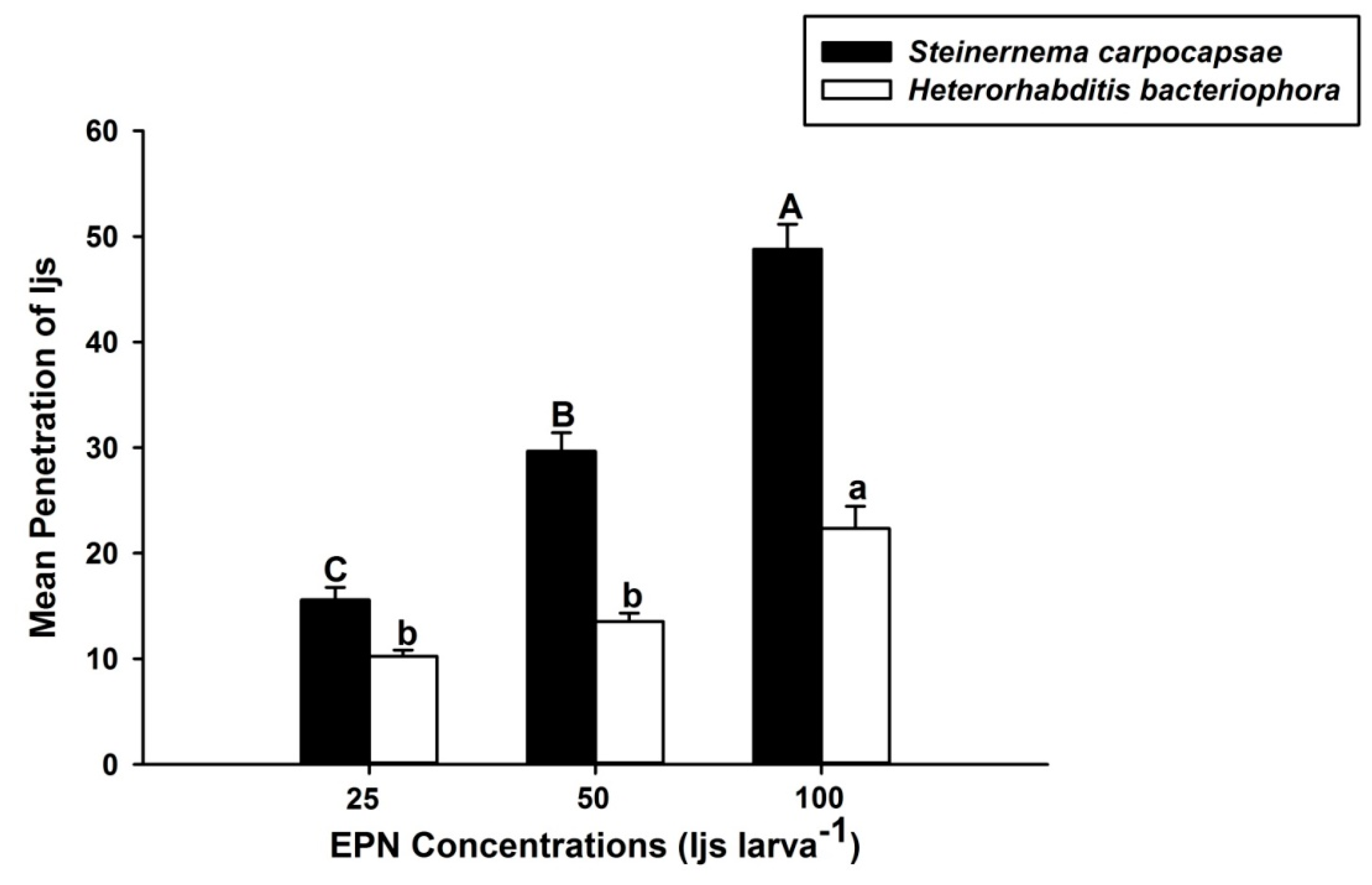
Similarly, the results for the penetration assay revealed that the IJs of S. carpocapsae and H. bacteriophora penetrated the hemocoel of the larvae of O. nipae at the concentration of 25, 50, and 100 IJs/larva (Figure 2). However, the mean penetration potential of S. carpocapsae was significantly higher (F2,6 = 60.84, p < 0.01) than that of H. bacteriophora (F2,6 = 23.99, p = 0.01) in O. nipae larvae, as shown in Figure 2. The mean penetration of IJs increased with increasing IJs concentration, in both S. carpocapsae and H. bacteriophora.

3.2. S. carpocapsae and H. bacteriophora Infection Suppress the Activation of O. nipae PPO System

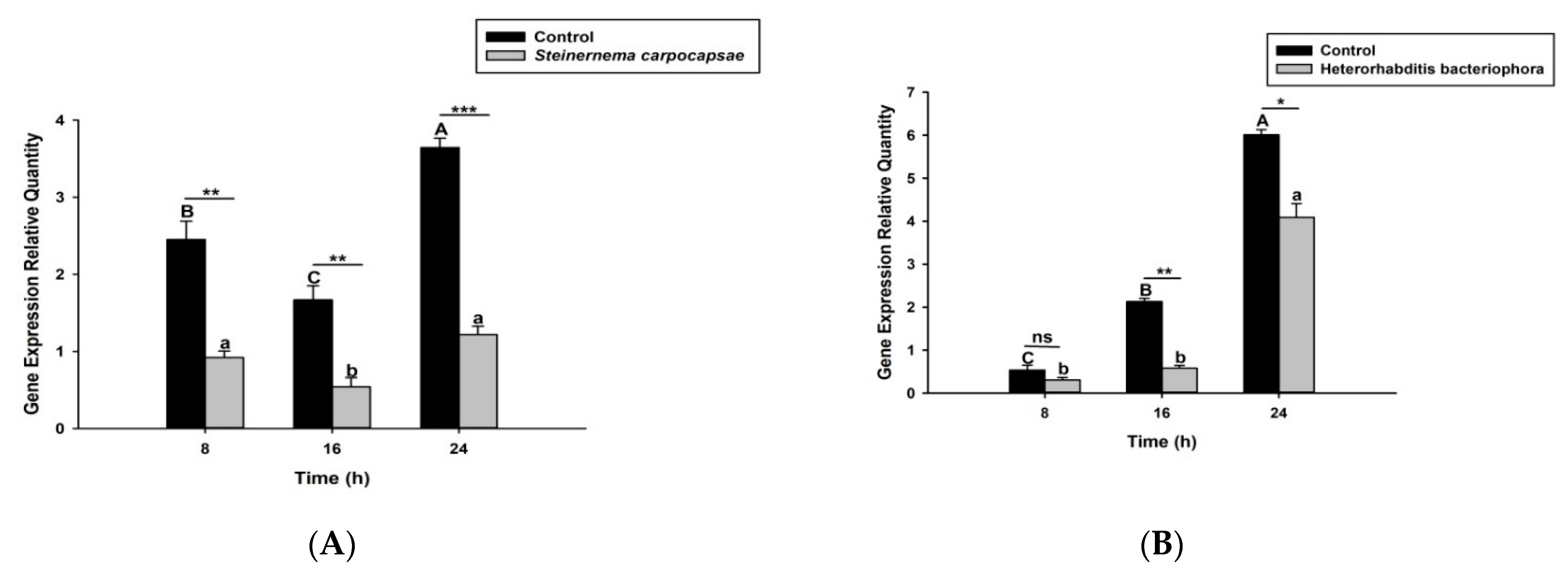
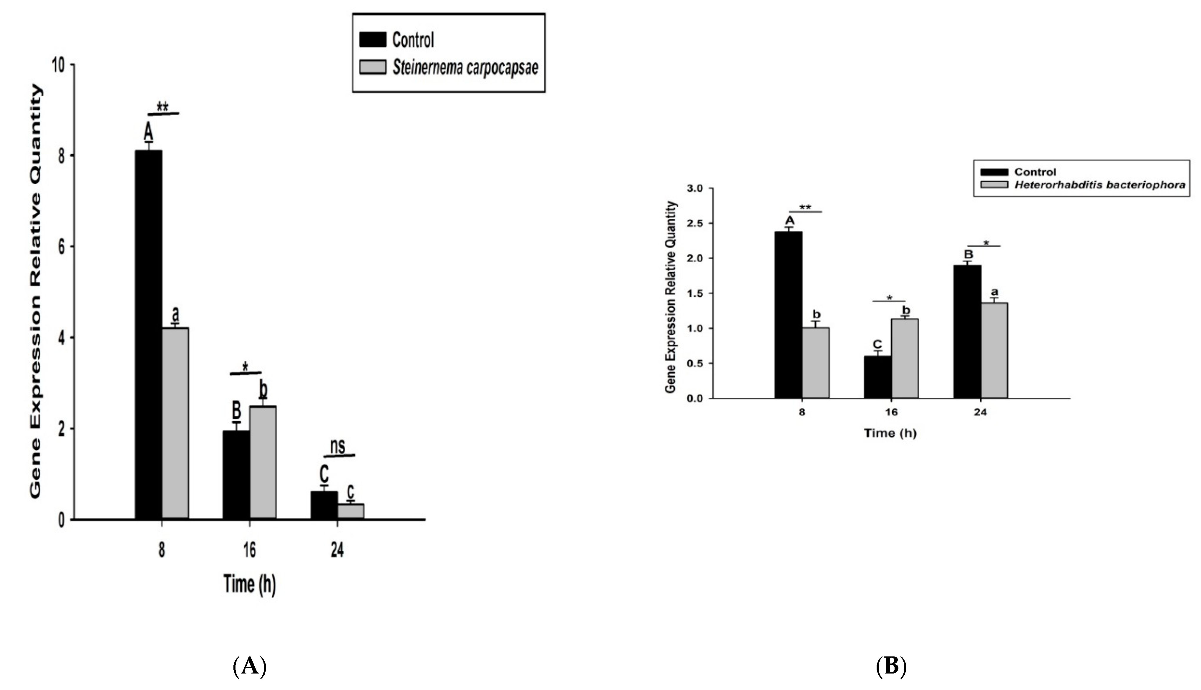
Here we investigated and compared the expression levels of four O. nipae prophenoloxidase enzymes (including the Serine Protease P56, SPP56; prophenoloxidase activation factor 1, PPAF1; PPO; and serine protease inhibitor 28, SPI28) between H. bacteriophora- and S. carpocapsae-treated larvae at three distinct time intervals. Our qRT-PCR results reveals that SPP56 was significantly upregulated (t4 = 1.346, p = 0.022) after H. bacteriophora challenge and insignificantly upregulated (t4 = 2.93, p = 0.383) upon S. carpocapsae treatment at 8 h post-infection (Figure 3A,B). At 16 and 24 h post-treatment, however, SPP56 was downregulated upon a challenge with both nematodes (Figure 3A,B). These translate to differences in the invasion strategies between the two nematodes. PPAF1 was downregulated at all time points following treatment with both nematodes, except at 16 h where H. bacteriophora infection results in significant upregulation (t4 = 1.88, p = 0.047) of the gene (Figure 4A,B). Similarly, the mRNA expression levels of PPO gene were completely downregulated at 8, 16, and 24 h post-S. carpocapsae and H. bacteriophora treatments (Figure 5A,B). As expected, the serine protease inhibitor SIP28 was downregulated in all nematodes treatments at 8 and 24 h time points, except at 16 h where it appeared upregulated for S. carpocapsae (t4 = 1.31, p = 0.032) and H. bacteriophora (t4 = 1.58, p = 0.040) treatments (Figure 6A,B). To sum it up, the S. carpocapsae and H. bacteriophora treatments successfully inhibit the PPO activation system of O. nipae, except at a few time-points where it shows upregulation.

3.3. The Heat-Treated Cuticles Positively Expresses the PPO Gene in O. nipae

The expression level of O. nipae PPO gene challenged with nematodes isolated cuticles was generally low compared to the control treatment. The mRNA level of the PPO gene was insignificantly regulated at 16 h (t2 = 1.28, p = 0.272 ns) but significantly downregulated at 24 h (t2 = 1.817, p = 0.001) after injection of the isolated cuticles from the two nematodes (Figure 7). To further confirm this, we subjected the isolated cuticles to heat treatment at 100 °C for 20 min and injected them into the larvae as described above. The results establish that the expression levels of PPO gene are higher than the controls, especially at late hours after injection in both treatments. A significant increase in mRNA level of PPO gene was obtained at 24 h post-H. bacteriophora (t2 = 2.93, p = 0.030) and S. carpocapsae (t2 = 1.31, p = 0.001) heat-treated cuticles injections. At 16 h after injection of S. carpocapsae cuticle, insignificance (t2 = 1.27, p = 0.152) upregulation of PPO gene was recorded (Figure 7A,B).

3.4. Addition of Arachidonic Acid Reverses the Inhibitions of Phenoloxidase Activity Caused by Dexamethasone

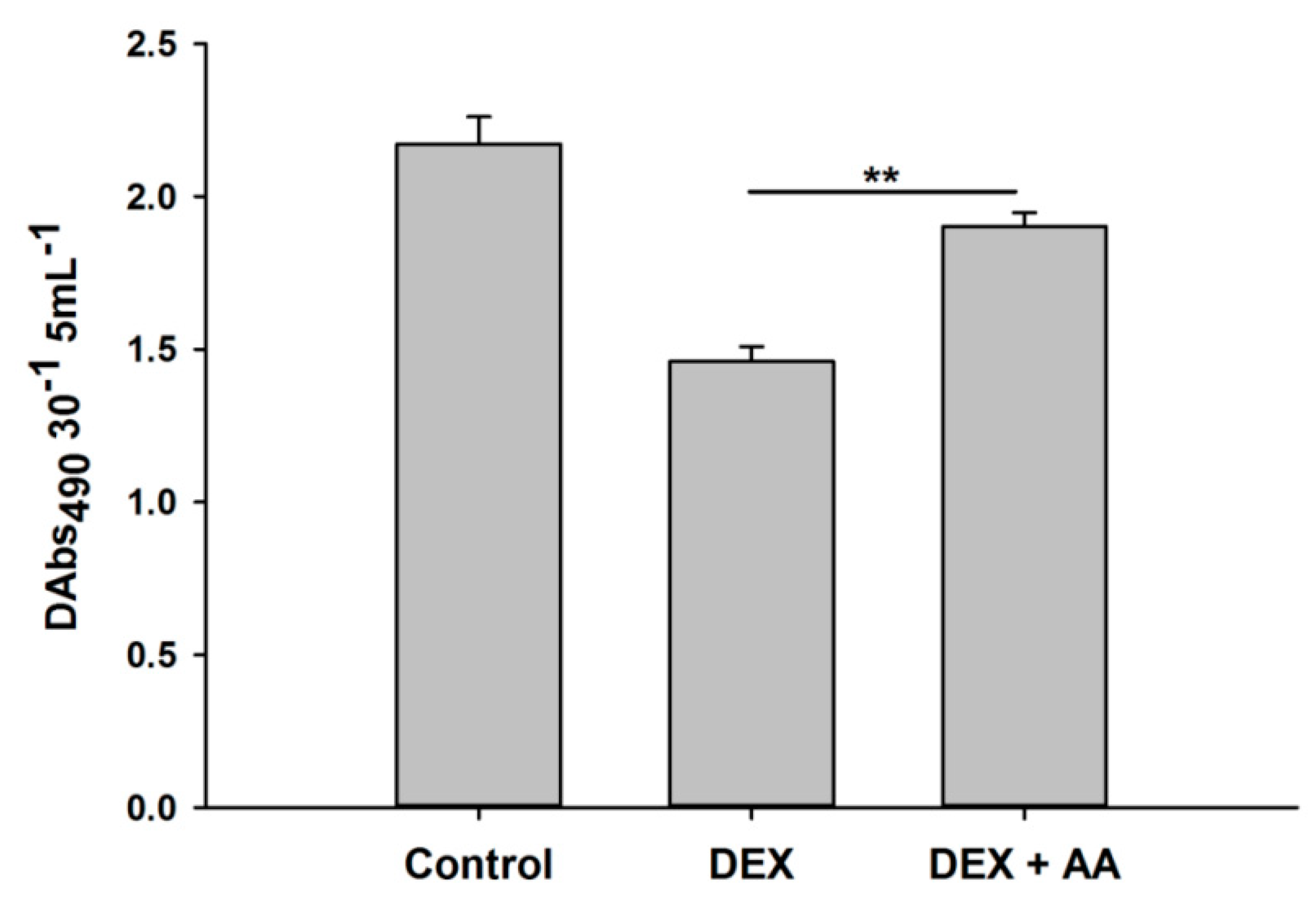
In this study, phenoloxidase-inhibitory activities of eicosanoid biosynthesis inhibitors and DEX treatments compared to controls were observed. There were significant decreases in phenoloxidase enzyme activities in hemolymph of O. nipae larvae treated with DEX (F2,6 = 42.13, p = 0.002). However, the addition of AA rescued the phenoloxidase-inhibitory activities caused by DEX treatments (Figure 8). There were significant increases in phenoloxidase activities in hemolymph of O. nipae larvae treated with DEX +AA (F2,6 = 43.13, p = 0.001).

3.5. Treatment with DEX Suppresses the Expression of the PPO Gene in O. nipae

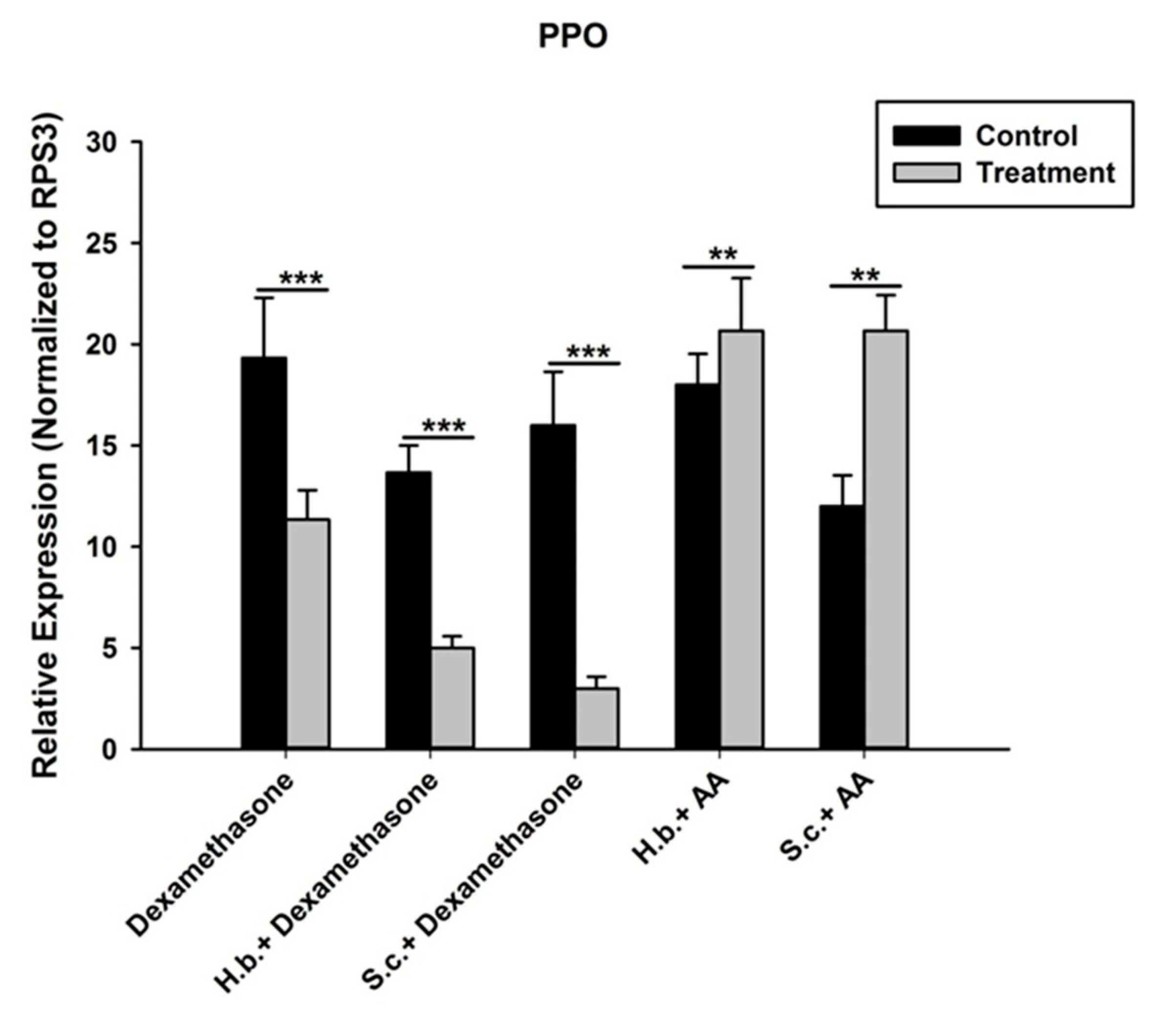
To further ascertain the role of this eicosanoid biosynthesis inhibitor DEX, we determined the expression level of PPO gene in treated larvae samples. Our results revealed that the expression level of PPO gene was downregulated significantly upon treatment of DEX (t4 = 4.20, p = 0.001) compared with control (Figure 9).
Secondly, injections of DEX to nematodes-treated larvae further suppressed the expression level of PPO gene. There were significant further downregulations of PPO gene in H. bacteriophora plus DEX (t4 = 2.928, p = 0.001) and S. carpocapsae plus DEX (t4 = 1.91, p = 0.002) treatments at 24 h. Interestingly, injection of AA (precursor of eicosanoid biosynthesis) to nematode-treated larvae reverses the inhibition effects of both DEX and nematode plus DEX-treated larvae on the PPO gene expression. The mRNA level of PPO gene in H. bacteriophora plus AA (t4 = 1.346, p = 0.024) and S. carpocapsae plus AA (t4 = 2.93, p = 0.030) treatment were highly significant at 24 h after treatments.

4. Discussion

Entomopathogenic nematodes modulate their hosts differently, depending on the strategies used to escape the host defense mechanisms. For example, Zang and Maizels [55] described the pathogenicity of insect pathogenic nematodes against their target hosts as resulting from an “arms race” between the insect and the nematodes. The findings from this study suggested that O. nipae larvae were more susceptible to H. bacteriophora. Compared to our previous results, the virulence of S. carpocapsae [19] over H. bacteriophora can be attributed to their differences in host-searching strategies, as reported by Grewal et al. [38] and Koppenhöfer et al. [71]. Additionally, insect cellular immune responses differ among species and depend on the invading species. For instance, G. mellonella hemocytes can recognize H. bacteriophora IJs, but not S. carpocapsae and S. glaseri nematodes [72]. This is similar to our previous experiment where S. carpocapsae efficiently resists being encapsulated and melanized within the O. nipae’s hemolymph and most of the nematodes were observed moving freely in the hemolymph even at 24 h post-incubation, whereas H. bacteriophora was identified, encapsulated and subsequently melanized by O. nipae’s hemolymph [19]. Moreover, Memari et al. [73] reported that H. bacteriophora was less virulent to carob moth Ectomyelois ceratoniae Zeller (LC50 = 426.9 IJs larva−1) when compared to both S. carpocapsae and S. feltiae (LC50 = 2 IJs/larva) at concentrations similar to this study. Additionally, Sharifi et al. [74] have reported that S. carpocapsae caused higher mortality of Osphranteria coerulescens than H. bacteriophora, as evident by their LC50 of 2.7 and 9.0 IJs larva−1, respectively. This is consistent with quite a number of reports for different insect pest control [75,76,77,78,79,80].
In any successful biological control program using EPNs as control agents, the nematodes should have the ability to not only penetrate their hosts but also reproduce inside them. The routes of entry of IJs into the host body cavity differ among nematode species and insect hosts. These require some mechanical processes by the nematodes to bypass the insects’ natural barriers, such as hairs [81]. According to Sharifi et al. [74], different hosts and EPN species have different penetration rates. In this study, we reported that differences exist between concentrations of S. carpocapsae and H. bacteriophora with regards to the mean penetration potential within the host larvae. This is also similar to a report by Susurluk et al. [82], which established higher penetration rates of S. carpocapsae into turf pest, Dorcadion pseudopreissi, and lower in H. bacteriophora treatments. Similarly, higher penetration rates were also reported for Steinernematids in previous works by Phan et al. [83], Salari et al. [34], Salem et al. [84], Koppenhöfer et al. [71], and Caroli et al. [85].
During host infection, nematodes contend the humoral and cellular host immune responses through passive and active strategies, in which nematodes avoid triggering an immune response by being recognised as ‘self’ as in S. carpocapsae. They also suppress the hosts’ immune responses after being recognised as ‘non-self’ [86,87]. In addition, nematodes modulate their hosts’ immune responses by releasing toxic molecules (such as proteases and inhibitors) during infection [88,89,90]. Activation of proPO results in the synthesis of phenoloxidase (PO), which serves as a reactive intermediate for melanin biosynthesis, an important component of insect’s innate immunity which can be activated by serine proteinases [55]. However, some EPNs have developed some means of preventing proPO activation by producing serpin-type inhibitors or other factors specifically interfering with the proteolytic activation of proPO or its upstream components. Here, we dissected the expression levels of four proPO activation enzymes of O. nipae infected with S. carpocapsae and H. bacteriophora. From our results, we found that nearly all the genes involved were downregulated in both nematodes and at all time points, except in a few cases. Initially, when the serine protease SPP56 was majorly downregulated, the whole enzymes involved in the activation of proPO cascades are shut down. This is consistent with our previous study where O. nipae’s phenoloxidase activity was found to be slightly suppressed due to infection of both H. bacteriophora and S. carpocapsae at early stages of infection [19].
Additionally, the S. carpocapsae isolated cuticles escaped being melanized by O. nipae’s hemolymph in vitro and were found to be completely melanized, however, when isolated cuticles were heat-killed before treatment. This is in line with some previous reports that show success in parasitization of Steinernematids is aided by their body surface cuticles [89,90,91,92,93]. Similarly, S. feltiae was shown to prevent the antibacterial immune response in G. mellonella by their body surface [94]. Akhurst and Boemare [40] suggest that cuticles of parasitic nematodes in addition to secreted molecules are involved in immune-evasion and suppression of host’s defences during the invasion. It was also reported to depress the host immune system, such as the proPO system, when injected into the hemocoel. Therefore, it was hypothesized that the nematode disguises as ‘self’ with the hemolymph proteins of the invading host by coating its body surface [94]. Yi et al. [59] showed that the involvement of S. carpocapsae or H. bacteriophora cuticles caused suppressions of humoral and cellular immune processes, such as phenoloxidase activity, haemocytes vitality, phagocytosis and antimicrobial activity. In this study, we confirm that the purified, isolated cuticles suppress the expression level of PPO gene in both nematodes’ treatments. However, upon heating, the cuticles lost their inhibitory effects on O. nipae’s immune responses and resulted in the upregulation of the PPO gene at both 16 and 24 h after treatment.
Furthermore, eicosanoids mediate basic humoral and cellular immune response mechanisms in insects [89]. These include nodulation, phagocytosis, encapsulation, and phenoloxidase activation systems. The results of the present experiment revealed that the eicosanoid biosynthesis inhibitor DEX suppresses the expression of the PPO gene in O. nipae, similar to that caused by treatment with H. bacteriophora and S. carpocapsae. In contrast, the addition of AA reversed the process and resulted in the upregulation of the PPO gene compared to the control treatment. In a study by Yi et al. [59], injection of the eicosanoid biosynthesis inhibitors dexamethasone and indomethacin induced similar depression in the key innate immune parameters, such as PO activity, compared to the nematode cuticles treatment in G. mellonella. However, injection of larvae with AA reverses the inhibitory effects of these inhibitors. Similarly, benzaldehyde and its derivative treatments resulted in the inhibition of PO activity in Pieris rapae larvae [95]. This is supported by the study of Ullah et al. [96] in which, in addition to the inhibition of nodule formation, the PO activity was also inhibited at different concentrations of benzaldehyde in G. mellonella larvae. Similarly, treatments with BZA (benzylideneacetone) in another study resulted in the immunosuppression of PO activities in Plutella xylostella and S. exigua [97,98,99]. We conclude that various eicosanoid inhibitors play a crucial role in the death of target insects.

5. Conclusions

Overall, this study provides the first data on the use of H. bacteriophora and S. carpocapsae for biological control of O. nipae larvae at different concentrations in the laboratory. We further dissected the expression levels of four selected proPO activation enzymes infected with H. bacteriophora and S. carpocapsae and found out that all of the genes were downregulated from both nematodes’ treatments. These were further confirmed by the counter experiments of heat-treated cuticles and AA treatment, which reversed the inhibitory effects caused by the nematodes and the eicosanoid inhibitor. Therefore, we speculated that these nematodes devised some means to evade and suppress the immune system of O. nipae, either by secretion of toxins or by manipulation of the host system. Future work will focus on the identification and functional analysis of the secretions used by S. carpocapsae and H. bacteriophora against O. nipae immune system.

Author Contributions

Data curation, N.B.S.; Formal analysis, N.B.S.; Funding acquisition, Y.H.; Investigation, N.B.S.; Methodology, N.B.S.; Project administration, Y.H.; Resources, Y.H.; Software, B.H.; Supervision, Y.H.; Validation, B.H.; Visualization, B.H.; Writing—original draft, N.B.S.; Writing—review & editing, B.H. All authors have read and agreed to the published version of the manuscript.

Funding

We are very grateful to the grant from the National Key R & D Program of China (2017YFC1200605) and Fujian Science and Technology Special Project (2017NZ0003-1-6).

Institutional Review Board Statement

Not applicable.

Informed Consent Statement

Not applicable.

Acknowledgments

We thank Richou Han and his associate Xun Yan for providing us with Entomopathogenic nematodes for the experiment.

Conflicts of Interest

The authors declare no conflict of interest.

References

  1. Hou, Y.; Weng, Z. Temperature-dependent development and life table parameters of Octodonta nipae (Coleoptera: Chrysomelidae). Environ. Entomol. 2010, 39, 1676–1684. [Google Scholar] [CrossRef] [PubMed]
  2. Hou, Y.; Wu, Z.; Wang, C.; Xie, L. The Status and Harm of Invasive Insects in Fujian, China; Biological invasions: Problems and countermeasures; Science Press: Beijing, China, 2011; pp. 111–114. [Google Scholar]
  3. Vassiliou, V.A.; Kazantzis, E.; Melifronidou-Pantelidou, A. First report of the nipa palm hispid Octodonta nipae on queen palms in Cyprus. Phytoparasitica 2011, 39, 51–54. [Google Scholar] [CrossRef]
  4. Zhang, X.; Tang, B.; Hou, Y. A rapid diagnostic technique to discriminate between two pests of palms, Brontispa longissima and Octodonta nipae (Coleoptera: Chrysomelidae), for quarantine applications. J. Econ. Entomol. 2015, 108, 95–99. [Google Scholar] [CrossRef] [Green Version]
  5. Li, J.; Zhang, X.; Hou, Y.; Tang, B. Effects of multiple mating on the fecundity of an invasive pest (Octodonta nipae): The existence of an intermediate optimal female mating rate. Physiol. Entomol. 2014, 39, 348–354. [Google Scholar] [CrossRef]
  6. Li, J.-L.; Tang, B.-Z.; Hou, Y.-M.; Xie, Y.-X. Molecular cloning and expression of the vitellogenin gene and its correlation with ovarian development in an invasive pest Octodonta nipae on two host plants. Bull. Entomol. Res. 2016, 106, 642–650. [Google Scholar] [CrossRef] [PubMed]
  7. Chen, Z.; Chen, J.; Zhang, X.; Hou, Y.; Wang, G. Development of microsatellite markers for the nipa palm hispid beetle, Octodonta nipae (Maulik). Can. J. Infect. Dis. Med. Microbiol. 2018, 2018, 9139306. [Google Scholar] [CrossRef] [Green Version]
  8. Peng, L.; Chen, L.; Li, J.; Hou, Y.; Chen, Y. Mate recognition and antennal morphology of Octodonta nipae (Coleoptera: Chrysomelidae) adults. J. Asia-Pac. Entomol. 2018, 21, 268–278. [Google Scholar] [CrossRef]
  9. Peng, L.-f.; Li, J.-l.; Hou, Y.-m.; Zhang, X. Descriptions of immature stages of Octodonta nipae (Maulik) (Coleoptera, Chrysomelidae, Cassidinae, Cryptonychini). ZooKeys 2018, 764, 91–109. [Google Scholar] [CrossRef]
  10. Hou, Y.; Miao, Y.; Zhang, Z. Leaf consumption capacity and damage projection of Octodonta nipae (Coleoptera: Chrysomelidae) on three palm species. Ann. Entomol. Soc. Am. 2014, 107, 1010–1017. [Google Scholar] [CrossRef]
  11. Hou, Y.; Miao, Y.; Zhang, Z. Study on life parameters of the invasive species Octodonta nipae (Coleoptera: Chrysomelidae) on different palm species, under laboratory conditions. J. Econ. Entomol. 2014, 107, 1486–1495. [Google Scholar] [CrossRef] [Green Version]
  12. Hua, R.; Hou, Y.; Shi, Z. Changes in the contents of physiologically active substances in Octodonta nipae (Coleoptera: Chrysomelidae) after low temperature acclimation. Acta Entomol. Sin. 2014, 57, 265–273. [Google Scholar]
  13. Feng, S.; Hou, Y. Lipopoly saccharide-induced immune response of Octodonta nipae (Coleoptera: Chrysomelidae) adults in relation to their genders. Acta Entomol. Sin. 2015, 58, 28–37. [Google Scholar]
  14. Ali, H.; Muhammad, A.; Sanda Bala, N.; Hou, Y. The endosymbiotic Wolbachia and host COI gene enables to distinguish between two invasive palm pests; coconut leaf beetle, Brontispa longissima and hispid leaf beetle, Octodonta nipae. J. Econ. Entomol. 2018, 111, 2894–2902. [Google Scholar] [CrossRef] [Green Version]
  15. Ali, H.; Muhammad, A.; Islam, S.U.; Islam, W.; Hou, Y. A novel bacterial symbiont association in the hispid beetle, Octodonta nipae (Coleoptera: Chrysomelidae), their dynamics and phylogeny. Microb. Pathog. 2018, 118, 378–386. [Google Scholar] [CrossRef] [PubMed]
  16. Ali, H.; Muhammad, A.; Sanda, N.B.; Huang, Y.; Hou, Y. Pyrosequencing uncovers a shift in bacterial communities across life stages of Octodonta nipae (Coleoptera: Chrysomelidae). Front. Microbiol. 2019, 10, 466. [Google Scholar] [CrossRef] [PubMed]
  17. Sanda, N.; Wudil, B.; Hou, Y. Effect of Temperatures on the Pathogenicity of Entomopathogenic Nematodes (Steinernema carpocapsae and Heterorhabditis bacteriophora) for Biocontrol of Octodonta nipae (Maulik). Niger. J. Entomol. 2020, 36, 88–95. [Google Scholar] [CrossRef]
  18. Sanda, N.B.; Hou, B.; Muhammad, A.; Ali, H.; Hou, Y. Exploring the role of Relish on antimicrobial peptide expressions (AMPs) upon nematode-bacteria complex challenge in the Nipa Palm Hispid Beetle, Octodonta nipae Maulik (Coleoptera: Chrysomelidae). Front. Microbiol. 2019, 10, 2466. [Google Scholar] [CrossRef]
  19. Sanda, N.B.; Muhammad, A.; Ali, H.; Hou, Y. Entomopathogenic nematode Steinernema carpocapsae surpasses the cellular immune responses of the hispid beetle, Octodonta nipae (Coleoptera: Chrysomelidae). Microb. Pathog. 2018, 124, 337–345. [Google Scholar] [CrossRef]
  20. Tang, B.; Chen, J.; Hou, Y.; Meng, E. Transcriptome immune analysis of the invasive beetle Octodonta nipae (Maulik)(Coleoptera: Chrysomelidae) parasitized by Tetrastichus brontispae Ferrière (Hymenoptera: Eulophidae). PLoS ONE 2014, 9, e91482. [Google Scholar] [CrossRef]
  21. Tang, B.-Z.; Meng, E.; Zhang, H.-J.; Zhang, X.-M.; Asgari, S.; Lin, Y.-P.; Lin, Y.-Y.; Peng, Z.-Q.; Qiao, T.; Zhang, X.-F. Combination of label-free quantitative proteomics and transcriptomics reveals intraspecific venom variation between the two strains of Tetrastichus brontispae, a parasitoid of two invasive beetles. J. Proteom. 2019, 192, 37–53. [Google Scholar] [CrossRef] [Green Version]
  22. Tang, B.; Xu, L.; Hou, Y. Effects of rearing conditions on the parasitism of Tetrastichus brontispae on its pupal host Octodonta nipae. BioControl 2014, 59, 647–657. [Google Scholar] [CrossRef]
  23. Toubarro, D.; Avila, M.M.; Montiel, R.; Simões, N. A pathogenic nematode targets recognition proteins to avoid insect defenses. PLoS ONE 2013, 8, e75691. [Google Scholar] [CrossRef] [PubMed] [Green Version]
  24. Meng, E.; Li, J.; Tang, B.; Hu, Y.; Qiao, T.; Hou, Y.; Lin, Y.; Chen, Z. Alteration of the phagocytosis and antimicrobial defense of Octodonta nipae (Coleoptera: Chrysomelidae) pupae to Escherichia coli following parasitism by Tetrastichus brontispae (Hymenoptera: Eulophidae). Bull. Entomol. Res. 2019, 109, 248–256. [Google Scholar] [CrossRef] [PubMed]
  25. Meng, E.; Qiao, T.; Tang, B.; Hou, Y.; Yu, W.; Chen, Z. Effects of ovarian fluid, venom and egg surface characteristics of Tetrastichus brontispae (Hymenoptera: Eulophidae) on the immune response of Octodonta nipae (Coleoptera: Chrysomelidae). J. Insect Physiol. 2018, 109, 125–137. [Google Scholar] [CrossRef] [PubMed]
  26. Meng, E.; Tang, B.; Hou, Y.; Chen, X.; Chen, J.; Yu, X.-Q. Altered immune function of Octodonta nipae (Maulik) to its pupal endoparasitoid, Tetrastichus brontispae Ferrière. Comp. Biochem. Physiol. Part B Biochem. Mol. Biol. 2016, 198, 100–109. [Google Scholar] [CrossRef]
  27. Meng, E.; Tang, B.; Li, J.; Fu, L.; Hou, Y. The first complete genome sequence of an iflavirus from the endoparasitoid wasp Tetrastichus brontispae. Arch. Virol. 2021, 166, 2333–2335. [Google Scholar] [CrossRef]
  28. Meng, E.; Tang, B.; Sanchez-Garcia, F.J.; Qiao, T.; Fu, L.; Wang, Y.; Hou, Y.-M.; Wu, J.-L.; Chen, Z.-M. The first complete genome sequence of a novel Tetrastichus brontispae RNA virus-1 (TbRV-1). Viruses 2019, 11, 257. [Google Scholar] [CrossRef] [Green Version]
  29. Howard, F.; Moore, D.; Giblin-Davis, R.; Abad, R. Insects on Palms; CABI International: Wallingford, UK, 2001. [Google Scholar]
  30. Chen, Q.; Peng, Z.; Xu, C.; Tang, C.; Lu, B.; Jin, Q.; Wen, H.; Wan, F. Biological assessment of Tetrastichus brontispae, a pupal parasitoid of coconut leaf beetle Brontispa longissima. Biocontrol Sci. Technol. 2010, 20, 283–295. [Google Scholar] [CrossRef]
  31. Nguyen, H.; Oo, T.; Ichiki, R.; Takano, S.; Murata, M.; Takasu, K.; Konishi, K.; Tunkumthong, S.; Chomphookhiaw, N.; Nakamura, S. Parasitisation of Tetrastichus brontispae (Hymenoptera: Eulophidae), a biological control agent of the coconut hispine beetle Brontispa longissima (Coleoptera: Chrysomelidae). Biocontrol Sci. Technol. 2012, 22, 955–968. [Google Scholar] [CrossRef]
  32. Xu, L.; Lan, J.; Hou, Y.; Chen, Y.; Chen, Z.; Weng, Z. Molecular identification and pathogenicity assay on Metarhizium against Octodonta nipae (Coleoptera: Chrysomelidae). Chin. J. Appl. Entomol. 2011, 48, 922–927. [Google Scholar]
  33. Kaya, H.K.; Gaugler, R. Entomopathogenic nematodes. Annu. Rev. Entomol. 1993, 38, 181–206. [Google Scholar] [CrossRef]
  34. Abbott, W.S. A method of computing the effectiveness of an insecticide. J. Econ. Entomol. 1925, 18, 265–267. [Google Scholar] [CrossRef]
  35. Taşkesen, Y.E.; Yüksel, E.; Canhilal, R. Field Performance of Entomopathogenic Nematodes against the Larvae of Zabrus spp. Clairville, 1806 (Coleoptera: Carabidae). Uluslararası Tarım Ve Yaban Hayatı Bilimleri Derg. 2021, 7, 429–437. [Google Scholar]
  36. Öğretmen, A.; Yüksel, E.; Canhilal, R. Susceptibility of larvae of wireworms (Agriotes spp.) (Coleoptera: Elateridae) to some Turkish isolates of entomopathogenic nematodes under laboratory and field conditions. Biol. Control. 2020, 149, 104320. [Google Scholar] [CrossRef]
  37. Özdemir, E.; Inak, E.; Evlice, E.; Yüksel, E.; Delialioğlu, R.A.; Susurluk, I.A. Effects of insecticides and synergistic chemicals on the efficacy of the entomopathogenic nematode Steinernema feltiae (Rhabditida: Steinernematidae) against Leptinotarsa decemlineata (Coleoptera: Chrysomelidae). Crop Prot. 2021, 144, 105605. [Google Scholar] [CrossRef]
  38. Grewal, P.S.; Gaugler, R.; Kaya, H.K.; Wusaty, M. Infectivity of the entomopathogenic nematode Steinernema scapterisci (Nematoda: Steinernematidae). J. Invertebr. Pathol. 1993, 62, 22–28. [Google Scholar] [CrossRef]
  39. Arthurs, S.; Heinz, K.; Prasifka, J. An analysis of using entomopathogenic nematodes against above-ground pests. Bull. Entomol. Res. 2004, 94, 297–306. [Google Scholar] [CrossRef]
  40. Trdan, S.; Vidrih, M.; Andjus, L.; Laznik, Ž. Activity of four entomopathogenic nematode species against different developmental stages of Colorado potato beetle, Leptinotarsa decemlineata (Coleoptera, Chrysomelidae). Helminthologia 2009, 46, 14–20. [Google Scholar] [CrossRef] [Green Version]
  41. Trdan, S.; Vidrih, M.; Valic, N. Activity of four entomopathogenic nematode species against young adults of Sitophilus granarius (Coleoptera: Curculionidae) and Oryzaephilus surinamensis (Coleoptera: Silvanidae) under laboratory conditions. J. Plant Dis. Prot. 2006, 113, 168–173. [Google Scholar] [CrossRef]
  42. Hara, A.; Kaya, H.; Gaugler, R.; Lebeck, L.; Mello, C. Entomopathogenic nematodes for biological control of the leafminer, Liriomyza trifolii (Dipt.: Agromyzidae). Entomophaga 1993, 38, 359–369. [Google Scholar] [CrossRef]
  43. Binda-Rossetti, S.; Mastore, M.; Protasoni, M.; Brivio, M.F. Effects of an entomopathogen nematode on the immune response of the insect pest red palm weevil: Focus on the host antimicrobial response. J. Invertebr. Pathol. 2016, 133, 110–119. [Google Scholar] [CrossRef] [PubMed]
  44. Levine, E.; Oloumi-Sadeghi, H. Management of diabroticite rootworms in corn. Annu. Rev. Entomol. 1991, 36, 229–255. [Google Scholar] [CrossRef]
  45. Yan, X.; Han, R.; Moens, M.; Chen, S.; De Clercq, P. Field evaluation of entomopathogenic nematodes for biological control of striped flea beetle, Phyllotreta striolata (Coleoptera: Chrysomelidae). BioControl 2013, 58, 247–256. [Google Scholar] [CrossRef]
  46. Li, X.-Y.; Cowles, R.; Cowles, E.; Gaugler, R.; Cox-Foster, D. Relationship between the successful infection by entomopathogenic nematodes and the host immune response. Int. J. Parasitol. 2007, 37, 365–374. [Google Scholar] [CrossRef]
  47. Shi, M.; Chen, X.-Y.; Zhu, N.; Chen, X.-X. Molecular identification of two prophenoloxidase-activating proteases from the hemocytes of Plutella xylostella (Lepidoptera: Plutellidae) and their transcript abundance changes in response to microbial challenges. J. Insect Sci. 2014, 14, 179. [Google Scholar] [CrossRef] [Green Version]
  48. Galko, M.J.; Krasnow, M.A.; Arias, A.M. Cellular and genetic analysis of wound healing in Drosophila larvae. PLoS Biol. 2004, 2, e239. [Google Scholar] [CrossRef] [Green Version]
  49. Cerenius, L.; Lee, B.L.; Söderhäll, K. The proPO-system: Pros and cons for its role in invertebrate immunity. Trends Immunol. 2008, 29, 263–271. [Google Scholar] [CrossRef]
  50. Kanost, M.; Gorman, M. Phenoloxidases in insect immunity. In Insect Immunology; Academic Press: Cambridge, MA, USA, 2008; Volume 10, p. B978-012373976. [Google Scholar]
  51. Zhang, H.; Tang, B.; Lin, Y.; Chen, Z.; Zhang, X.; Ji, T.; Zhang, X.; Hou, Y. Identification of three prophenoloxidase--activating factors (PPAFs) from an invasive beetle Octodonta nipae Maulik (Coleoptera: Chrysomelidae) and their roles in the prophenoloxidase activation. Arch. Insect Biochem. Physiol. 2017, 96, e21425. [Google Scholar] [CrossRef]
  52. Zhang, H.-J.; Lin, Y.-P.; Liu, M.; Liang, X.-Y.; Ji, Y.-N.; Tang, B.-Z.; Hou, Y.-M. Functional conservation and division of two single-carbohydrate-recognition domain C-type lectins from the nipa palm hispid beetle Octodonta nipae (Maulik). Dev. Comp. Immunol. 2019, 100, 103416. [Google Scholar] [CrossRef]
  53. Zhang, X.-M.; Zhang, H.-J.; Liu, M.; Liu, B.; Zhang, X.-F.; Ma, C.-J.; Fu, T.-T.; Hou, Y.-M.; Tang, B.-Z. Cloning and immunosuppressive properties of an acyl-activating enzyme from the venom apparatus of Tetrastichus brontispae (Hymenoptera: Eulophidae). Toxins 2019, 11, 672. [Google Scholar] [CrossRef] [Green Version]
  54. Zhang, H.-J.; Lin, Y.-P.; Li, H.-Y.; Wang, R.; Fu, L.; Jia, Q.-C.; Hou, Y.-M.; Tang, B.-Z. Variation in Parasitoid Virulence of Tetrastichus brontispae during the Targeting of Two Host Beetles. Int. J. Mol. Sci. 2021, 22, 3581. [Google Scholar] [CrossRef] [PubMed]
  55. Zang, X.; Maizels, R.M. Serine proteinase inhibitors from nematodes and the arms race between host and pathogen. Trends Biochem. Sci. 2001, 26, 191–197. [Google Scholar] [CrossRef] [Green Version]
  56. Yadav, S.; Daugherty, S.; Shetty, A.C.; Eleftherianos, I. RNAseq analysis of the Drosophila response to the entomopathogenic nematode Steinernema. G3 Genes Genomes Genet 2017, 7, 1955–1967. [Google Scholar] [CrossRef] [PubMed] [Green Version]
  57. Cooper, D.; Eleftherianos, I. Parasitic nematode immunomodulatory strategies: Recent advances and perspectives. Pathogens 2016, 5, 58. [Google Scholar] [CrossRef] [PubMed] [Green Version]
  58. Srikanth, K.; Park, J.; Stanley, D.W.; Kim, Y. Plasmatocyte-spreading peptide influences hemocyte behavior via eicosanoids. Arch. Insect Biochem. Physiol. 2011, 78, 145–160. [Google Scholar] [CrossRef] [PubMed]
  59. Yi, Y.; Wu, G.; Lv, J.; Li, M. Eicosanoids mediate Galleria mellonella immune response to hemocoel injection of entomopathogenic nematode cuticles. Parasitol. Res. 2016, 115, 597–608. [Google Scholar] [CrossRef]
  60. Balsinde, J.; Winstead, M.V.; Dennis, E.A. Phospholipase A2 regulation of arachidonic acid mobilization. FEBS Lett. 2002, 531, 2–6. [Google Scholar] [CrossRef]
  61. Sadekuzzaman, M.; Park, Y.; Lee, S.; Kim, K.; Jung, J.K.; Kim, Y. An entomopathogenic bacterium, Xenorhabdus hominickii ANU101, produces oxindole and suppresses host insect immune response by inhibiting eicosanoid biosynthesis. J. Invertebr. Pathol. 2017, 145, 13–22. [Google Scholar] [CrossRef]
  62. Kim, H.; Keum, S.; Hasan, A.; Kim, H.; Jung, Y.; Lee, D.; Kim, Y. Identification of an entomopathogenic bacterium, Xenorhabdus ehlersii KSY, from Steinernema longicaudum GNUS101 and its immunosuppressive activity against insect host by inhibiting eicosanoid biosynthesis. J. Invertebr. Pathol. 2018, 159, 6–17. [Google Scholar] [CrossRef]
  63. Eom, S.; Park, Y.; Kim, Y. Sequential immunosuppressive activities of bacterial secondary metabolites from the entomopahogenic bacterium Xenorhabdus nematophila. J. Microbiol. 2014, 52, 161–168. [Google Scholar] [CrossRef]
  64. Sadekuzzaman, M.; Gautam, N.; Kim, Y. A novel calcium-independent phospholipase A2 and its physiological roles in development and immunity of a lepidopteran insect, Spodoptera exigua. Dev. Comp. Immunol. 2017, 77, 210–220. [Google Scholar] [CrossRef] [PubMed]
  65. Park, J.-A.; Kim, Y. Phospholipase A2 inhibitors in bacterial culture broth enhance pathogenicity of a fungus Nomuraea rileyi. J. Microbiol. 2012, 50, 644–651. [Google Scholar] [CrossRef] [PubMed]
  66. Duvic, B.; Jouan, V.; Essa, N.; Girard, P.-A.; Pages, S.; Abi Khattar, Z.; Volkoff, N.-A.; Givaudan, A.; Destoumieux-Garzon, D.; Escoubas, J.-M. Cecropins as a marker of Spodoptera frugiperda immunosuppression during entomopathogenic bacterial challenge. J. Insect Physiol. 2012, 58, 881–888. [Google Scholar] [CrossRef] [Green Version]
  67. Ahmed, S.; Kim, Y. Differential immunosuppression by inhibiting PLA2 affects virulence of Xenorhabdus hominickii and Photorhabdus temperata temperata. J. Invertebr. Pathol. 2018, 157, 136–146. [Google Scholar] [CrossRef]
  68. Yan, X.; Liu, X.; Han, R.; Chen, S.; De Clercq, P.; Moens, M. Osmotic induction of anhydrobiosis in entomopathogenic nematodes of the genera Heterorhabditis and Steinernema. Biol. Control. 2010, 53, 325–330. [Google Scholar] [CrossRef]
  69. Kaya, H.K.; Aguillera, M.; Alumai, A.; Choo, H.Y.; De la Torre, M.; Fodor, A.; Ganguly, S.; Hazır, S.; Lakatos, T.; Pye, A. Status of entomopathogenic nematodes and their symbiotic bacteria from selected countries or regions of the world. Biol. Control. 2006, 38, 134–155. [Google Scholar] [CrossRef]
  70. White, G. A method for obtaining infective nematode larvae from cultures. Science 1927, 66, 302–303. [Google Scholar] [CrossRef]
  71. Koppenhöfer, A.M.; Brown, I.M.; Gaugler, R.; Grewal, P.S.; Kaya, H.K.; Klein, M.G. Synergism of entomopathogenic nematodes and imidacloprid against white grubs: Greenhouse and field evaluation. Biol. Control. 2000, 19, 245–251. [Google Scholar] [CrossRef]
  72. Ebrahimi, L.; Niknam, G.; Dunphy, G. Hemocyte responses of the Colorado potato beetle, Leptinotarsa decemlineata, and the greater wax moth, Galleria mellonella, to the entomopathogenic nematodes, Steinernema feltiae and Heterorhabditis bacteriophora. J. Insect Sci. 2011, 11, 75. [Google Scholar] [CrossRef] [Green Version]
  73. Memari, Z.; Karimi, J.; Kamali, S.; Goldansaz, S.H.; Hosseini, M. Are entomopathogenic nematodes effective biological control agents against the carob moth, Ectomyelois ceratoniae? J. Nematol. 2016, 48, 261. [Google Scholar] [CrossRef] [Green Version]
  74. Sharifi, S.; Karimi, J.; Hosseini, M.; Rezapanah, M. Efficacy of two entomopathogenic nematode species as potential biocontrol agents against the rosaceae longhorned beetle, Osphranteria coerulescens, under laboratory conditions. Nematology 2014, 16, 729–737. [Google Scholar] [CrossRef] [Green Version]
  75. Shapiro-Ilan, D.I.; Cottrell, T.E. Susceptibility of the lesser peachtree borer (Lepidoptera: Sesiidae) to entomopathogenic nematodes under laboratory conditions. Environ. Entomol. 2006, 35, 358–365. [Google Scholar] [CrossRef]
  76. Susurluk, I.A. Influence of temperature on the vertical movement of the entomopathogenic nematodes Steinernema feltiae (TUR-S3) and Heterorhabditis bacteriophora (TUR-H2), and infectivity of the moving nematodes. Nematology 2008, 10, 137–141. [Google Scholar] [CrossRef]
  77. Batalla-Carrera, L.; Morton, A.; García-del-Pino, F. Efficacy of entomopathogenic nematodes against the tomato leafminer Tuta absoluta in laboratory and greenhouse conditions. BioControl 2010, 55, 523–530. [Google Scholar] [CrossRef]
  78. Ashtari, M.; Karimi, J.; Rezapanah, M.R.; Hassani-Kakhki, M. Biocontrol of leopard moth, Zeuzera pyrina L. (Lep.: Cossidae) using entomopathogenic nematodes in Iran. IOBC/Wprs Bull. 2011, 66, 333–335. [Google Scholar]
  79. Yadav, A.K. Efficacy of indigenous entomopathogenic nematodes from Meghalaya, India against the larvae of taro leaf beetle, Aplosonyx chalybaeus (Hope). J. Parasit. Dis. 2012, 36, 149–154. [Google Scholar] [CrossRef] [Green Version]
  80. Nagesh, M.; Kumar, N.; Shylesha, A.; Srinivasa, N.; Javeed, S.; Thippeswamy, R. Comparative virulence of strains of entomopathogenic nematodes for management of eggplant grey weevil, Myllocerus subfasciatus Guerin (Coleoptera: Curculionidae). Indian J. Exp. Biol. 2016, 54, 835–842. [Google Scholar]
  81. Jung, S.; Kim, Y. Synergistic effect of Xenorhabdus nematophila K1 and Bacillus thuringiensis subsp. aizawai against Spodoptera exigua (Lepidoptera: Noctuidae). Biol. Control. 2006, 39, 201–209. [Google Scholar] [CrossRef]
  82. Susurluk, I.A.; Kumral, N.A.; Peters, A.; Bilgili, U.; Açıkgöz, E. Pathogenicity, reproduction and foraging behaviours of some entomopathogenic nematodes on a new turf pest, Dorcadion pseudopreissi (Coleoptera: Cerambycidae). Biocontrol Sci. Technol. 2009, 19, 585–594. [Google Scholar] [CrossRef]
  83. Phan, K.; Tirry, L.; Moens, M. Pathogenic potential of six isolates of entomopathogenic nematodes (Rhabditida: Steinernematidae) from Vietnam. BioControl 2005, 50, 477–491. [Google Scholar] [CrossRef]
  84. Hedayat-allah, M.S.; Hussein, M.A.; Hafez, S.E.; Hussein, M.A.; Sayed, R.M. Ultrastructure changes in the haemocytes of Galleria mellonella larvae treated with gamma irradiated Steinernema carpocapsae BA2. J. Radiat. Res. Appl. Sci. 2014, 7, 74–79. [Google Scholar]
  85. Caroli, L.; Glazer, I.; Gaugler, R. Entomopathogenic nematode infectivity assay: Comparison of penetration rate into different hosts. Biocontrol Sci. Technol. 1996, 6, 227–234. [Google Scholar] [CrossRef]
  86. Brehélin, M.; Drif, L.; Boemare, N. Depression of defence reactions in insects by Steinernematidae and their associated bacteria. In Proceedings of the Proceedings and Abstracts, Vth International Colloquium on Invertebrate Pathology and Microbial Control, Adelaide, Australia, 20–24 August 1990; pp. 213–217. [Google Scholar]
  87. DUNPHY, G.B.; HURLBERT, R.E. Interaction of avirulent transpositional mutants of Xenorhabdus nematophilus ATCC 19061 (Enterobacteriaceae) with the antibacterial systems of non-immune Galleria mellonella (Insecta) larvae. J. Gen. Appl. Microbiol. 1995, 41, 409–427. [Google Scholar] [CrossRef] [Green Version]
  88. Brooks, C.L.; Dunphy, G.B. Protein kinase A affects Galleria mellonella (Insecta: Lepidoptera) larval haemocyte non-self responses. Immunol. Cell Biol. 2005, 83, 150–159. [Google Scholar] [CrossRef]
  89. Stanley, D.; Miller, J.; Tunaz, H. Eicosanoid actions in insect immunity. J. Innate Immun. 2009, 1, 282–290. [Google Scholar] [CrossRef]
  90. Balasubramanian, N.; Toubarro, D.; Simoes, N. Biochemical study and in vitro insect immune suppression by a trypsin-like secreted protease from the nematode Steinernema carpocapsae. Parasite Immunol. 2010, 32, 165–175. [Google Scholar] [CrossRef]
  91. Brivio, M.F.; Pagani, M.; Restelli, S. Immune suppression of Galleria mellonella (Insecta, Lepidoptera) humoral defenses induced by Steinernema feltiae (Nematoda, Rhabditida): Involvement of the parasite cuticle. Exp. Parasitol. 2002, 101, 149–156. [Google Scholar] [CrossRef]
  92. Brivio, M.F.; Mastore, M.; Moro, M. The role of Steinernema feltiae body-surface lipids in host–parasite immunological interactions. Mol. Biochem. Parasitol. 2004, 135, 111–121. [Google Scholar] [CrossRef]
  93. Sheykhnejad, H.; Ghadamyari, M.; Ghasemi, V.; Jamali, S.; Karimi, J. Haemocytes immunity of rose sawfly, Arge ochropus (Hym.: Argidae) against entomopathogenic nematodes, Steinernema carpocapsae and Heterorhabditis bacteriophora. J. Asia-Pac. Entomol. 2014, 17, 879–883. [Google Scholar] [CrossRef]
  94. Brivio, M.F.; Moro, M.; Mastore, M. Down-regulation of antibacterial peptide synthesis in an insect model induced by the body-surface of an entomoparasite (Steinernema feltiae). Dev. Comp. Immunol. 2006, 30, 627–638. [Google Scholar] [CrossRef]
  95. Wang, Q.; Ke, L.; Xue, C.; Luo, W.; Chen, Q. Inhibitory Kinetics of p-Substituted Benzaldehydes on Polyphenol Oxidase from the Fifth Instar of Pieris Rapae L. Tsinghua Sci. Technol. 2007, 12, 400–404. [Google Scholar] [CrossRef]
  96. Ullah, I.; Khan, A.L.; Ali, L.; Khan, A.R.; Waqas, M.; Hussain, J.; Lee, I.-J.; Shin, J.-H. Benzaldehyde as an insecticidal, antimicrobial, and antioxidant compound produced by Photorhabdus temperata M1021. J. Microbiol. 2015, 53, 127–133. [Google Scholar] [CrossRef] [PubMed]
  97. Kwon, B.; Kim, Y. Benzylideneacetone, an immunosuppressant, enhances virulence of Bacillus thuringiensis against beet armyworm (Lepidoptera: Noctuidae). J. Econ. Entomol. 2008, 101, 36–41. [Google Scholar] [CrossRef]
  98. Kim, G.S.; Kim, Y. Up-regulation of circulating hemocyte population in response to bacterial challenge is mediated by octopamine and 5-hydroxytryptamine via Rac1 signal in Spodoptera exigua. J. Insect Physiol. 2010, 56, 559–566. [Google Scholar] [CrossRef] [PubMed]
  99. Seo, S.; Lee, S.; Hong, Y.; Kim, Y. Phospholipase A2 inhibitors synthesized by two entomopathogenic bacteria, Xenorhabdus nematophila and Photorhabdus temperata subsp. temperata. Appl. Environ. Microbiol. 2012, 78, 3816–3823. [Google Scholar] [CrossRef] [PubMed] [Green Version]
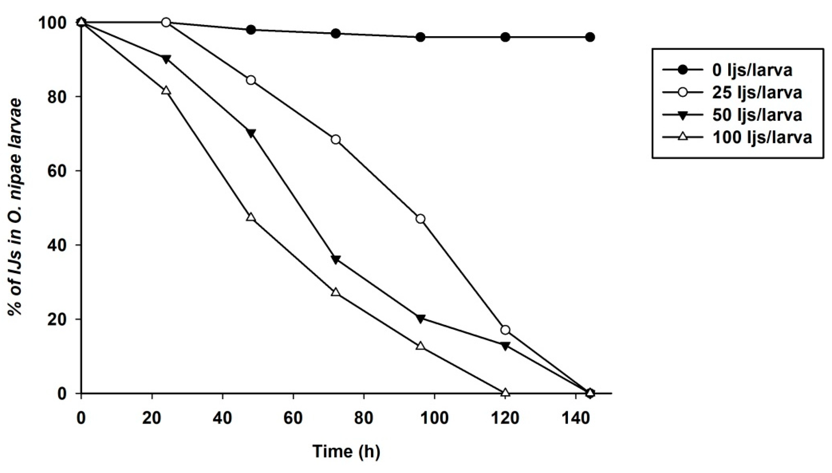
Figure 1. Survival curve of O. nipae larvae infected with H. bacteriophora at different concentrations are significantly different, (p < 0.0001, log-rank test).
Life 12 01019 g001
Figure 2. Mean penetration of infective juveniles of Heterorhabditis bacteriophora and Steinernema carpocapsae at different concentrations into third instars of Octodonta nipae. Means with the same letter are not significantly different, capital letters represent means of S. carpocapsae penetration, lowercase letters represent means of H. bacteriophora penetration.
Life 12 01019 g002
Figure 3. Transcription of Serine Protease P56 (SPP56) gene in O. nipae larvae infected with (A) S. carpocapsae and (B) H. bacteriophora at 8, 16, and 24 h after treatments. Error bars labeled with different letters are significantly different (one-way ANOVA followed by LSD test, p < 0.05). The asterisks *** (p < 0.0001); ** (p < 0.001); * (p < 0.01) indicates different significant levels between the control and nematode treatments at the indicated time period, while “ns” indicates no significant difference.
Life 12 01019 g003
Figure 4. Transcription of prophenoloxidase Activation Factor 1 (PPAF1) gene in O. nipae larvae infected with (A) S. carpocapsae and (B) H. bacteriophora at 8, 16, and 24 h after treatments. Error bars labeled with different letters are significantly different (one-way ANOVA followed by LSD test, p < 0.05). The asterisks *** (p < 0.0001); ** (p < 0.001); * (p < 0.01) indicate different significant levels between the control and nematode treatments at the indicated time period, while “ns” indicates no significant difference.
Life 12 01019 g004
Figure 5. Transcription of prophenoloxidase (PPO) gene in O. nipae larvae infected with (A) S. carpocapsae and (B) H. bacteriophora at 8, 16, and 24 h after treatments. Error bars labeled with different letters are significantly different (one-way ANOVA followed by LSD test, p < 0.05). The asterisks *** (p < 0.0001); ** (p < 0.001); * (p < 0.01) indicate different significant levels between the control and nematode treatments at the indicated time period, while “ns” indicates no significant difference.
Life 12 01019 g005
Figure 6. Transcription of serine protease inhibitor 28 (SPI28) gene in O. nipae larvae infected with (A) S. carpocapsae and (B) H. bacteriophora at 8, 16, and 24 h after treatments. Error bars labeled with different letters are significantly different (one-way ANOVA followed by LSD test, p < 0.05). The asterisks; ** (p < 0.001); * (p < 0.01) indicate different significant levels between the control and nematode treatments at the indicated time period, while “ns” indicates no significant difference.
Life 12 01019 g006
Figure 7. Transcription of prophenoloxidase (PPO) gene in O. nipae larvae injected with (A) H. bacteriophora purified cuticle (B) S. carpocapsae purified cuticle. Treatments with heat-killed cuticles reversed the influence of untreated cuticles on the PPO expressions. H.b.—H. bacteriophora and S.c.—S. carpocapsae. The asterisks *** (p < 0.0001); ** (p < 0.001); * (p < 0.01) indicates different significant levels between the control and nematode treatments at the indicated time period, while “ns” indicates no significant difference.
Life 12 01019 g007
Figure 8. Effects of Dexamethasone (DEX) and Oxindole on inhibition of phenoloxidase (PO) activity in third instar O. nipae larvae. The addition of Arachidonic acid (AA) reverses the inhibition of PO activity caused by metabolite treatments. The asterisks ** (p < 0.001) indicate highly significant levels between the control and treatments.
Life 12 01019 g008
Figure 9. Influence of DEX and nematodes plus DEX, on expression of prophenoloxidase (PPO) gene in O. nipae larvae. AA rescues DEX and nematodes plus DEX-suppressed expression of PPO gene in O. nipae larvae. The asterisks *** (p < 0.0001); ** (p < 0.001); indicate different significant levels between the control and nematode treatments at the indicated time period.
Life 12 01019 g009
Table 1. Prophenoloxidase activation genes Primers used for the qRT-PCR analysis.
Gene NameForward Primer (5′→3′)Reverse Primer (5′→3′)
qSPP56CGGTTGGTGGAAAGTGTCAGCCCTCGTTGTCCAGCTTCTA
qSPI28TCGCCTTAGTGATAGCGTGTACAGGGCTAGGGAAAACTCC
qPPAF1GATCACCGGCGACAAAGAAACAGCTTGTTGGGATTGCCTT
qPPOGTATCTTGTCACCCAATAGAGCAAACGATTCAAGATGCCTGT
q-RPS3-FGACGGTGTCTTCAAAGCTGAATTTCTGTACGTGTCGGGGT
Publisher’s Note: MDPI stays neutral with regard to jurisdictional claims in published maps and institutional affiliations.