Article

Association of the Genetic Variation in the Long Non-Coding RNA FENDRR with the Risk of Developing Hypertrophic Cardiomyopathy

by
Elías Cuesta-Llavona
1,2,3,
Rebeca Lorca
1,2,3,4,
Valeria Rolle
2,
Belén Alonso
1,
Sara Iglesias
1,
Julian Rodríguez-Reguero
1,4,
Israel David Duarte-Herrera
1,
Sergio Pérez-Oliveira
1,
Alejandro Junco-Vicente
1,
Claudia García Lago
2,
Eliecer Coto
1,2,3,4,5,* and
Juan Gómez
1,2,3,4,6,*
1
Hospital Universitario Central de Asturias (HUCA), 33011 Oviedo, Spain
2
Instituto de Investigación Sanitaria del Principado de Asturias (ISPA), 33011 Oviedo, Spain
3
Redes de Investigación Cooperativa Orientadas a Resultadosen Salud (RICORs), 28029 Madrid, Spain
4
Unidad de Cardiopatías Familiares del HUCA, 33011 Oviedo, Spain
5
Medicicine Department, Universidad de Oviedo, 33003 Oviedo, Spain
6
CIBER-Enfermedades Respiratorias, 28029 Madrid, Spain
*
Authors to whom correspondence should be addressed.
Life 2022, 12(6), 818; https://doi.org/10.3390/life12060818
Submission received: 4 April 2022 / Revised: 20 May 2022 / Accepted: 28 May 2022 / Published: 30 May 2022
(This article belongs to the Special Issue Advances in Heart Disease)

Abstract

:
Background: In around 40–60% of Hypertrophic Cardiomyopathy (HCM) cases pathogenic variants are not identified. Our aim was to evaluate the possible association of lncRNAs with the risk of developing HCM. Methods: We sequenced 10 lncRNAs coding genes that have been associated with cardiovascular disease in a discovery cohort (238 HCM patients and 212 controls) by NGS, and genotyped rs74035787 G>A and rs1424019 A>G polymorphism in a validation cohort (962 HCM patients and 923 controls). Finally, we sequenced the FENDRR promoter by Sanger sequencing. Results: We observed by NGS that FENDRR rs39527, rs39529 and rs40384 polymorphisms were significantly associated with HCM in our cohort (p = 0.0284; OR: 0.24, 95%CI: 0.07–0.86). NGS results were confirmed by genotyping rs74035787 polymorphism (p = 0.001; OR:0.38, 95%CI: 0.21–0.66). Moreover, it is also associated when stratification by sex (p = 0.003; OR:0.20, 95%CI: 0.06–0.53), and age (≥50 years old p = 0.001, OR:0.33, 95%CI: 0.16–0.63) Moreover, the risk of HCM in the carriers of the GG genotype of the rs1424019 polymorphism was significantly higher than that of the AA/AG genotypes carriers in the elderly subjects (p = 0.045, OR:1.24, 95%CI: 1.01–1.53). On the other hand, we observed significant differences in the rs74035787 A/rs1424019 G haplotype frequency (p = 0.0035; OR: 0.20, 95%CI: 0.07–0.59). Conclusions: Our study suggested a significant association between FENDRR gene variants and HCM.

1. Introduction

Hypertrophic Cardiomyopathy (HCM) is the most common hereditary heart disease, with a prevalence of 1/500 (0.2%) [1] and it is caused by pathogenic variants, mainly in genes encoding sarcomere proteins [2,3]. Meanwhile, next generation sequencing (NGS) has revolutionized the study of the genetic basis of heritable diseases andexome sequencing has proven to be an extremely useful tool for identifying new genes associated with various pathologies; however, in around 40–60% of HCM cases, the causal pathogenic variant is not identified, including cases with a family history of the disease. Most pathogenic variants are associated with clinical heterogeneity, incomplete penetrance, and variable phenotypic expressiveness, including carriers in the same family [4]. Studies suggest that there are other genetic factors that could partially explain the risk of developing HCM or modify the phenotype among carriers anda group of candidate genes to be evaluated are those that encode long-non-coding RNAs (lncRNAs), whose variation escapes the informatics prediction tools applicable to the exome. These RNAs are molecules with a length of more than 200 nucleotides which have a role in the modulation of gene expression and signaling pathways. They perform important roles in various physiological processes through epigenetic modulations (gene silencing), regulating alternative splicing or as molecular sponges. In addition, they can interfere with the translation of proteins by blocking, stabilizing or destabilizing the mRNA. In addition, lncRNAs have low levels of expression, conservation, and high tissue specificity. These RNAs are classified by their gene location in sense, intronic sense, antisense, bidirectional, or intergenic lncRNA [5,6]. Furthermore, the expression of a large number of lncRNAs has been characterized in the cardiovascular system both in physiological and pathological conditions (Table 1) [7,8,9,10,11,12,13,14,15,16,17,18].
Regarding HCM, there are studies that have pointed to an association between lncRNAs and this pathology. It was reported that H19 regulates cardiac hypertrophy through miR-675 and genetic variation at this lncRNA is associated with the risk of HCM [12,19], that MIAT perform like a miR-150 molecular sponge [13], MHRT antagonizes the role of Brg1 in cardiac hypertrophy [9], PVT1 modulates the pathological cardiac hypertrophy via miR-196b [18] and TINCR regulates cardiac hypertrophy through the epigenetic silencing of CaMKII [16];however, only a limited number of lncRNAs have been identified as regulators of cardiac hypertrophy. The aim of this study is to characterize the genetic variations of several lncRNA and their possible role in the risk of developing hypertrophic cardiomyopathy.

2. Materials and Methods

2.1. Patients and Controls

The study included a discovery cohort of 238 index cases of HCM and 212 healthy controls. All the study participants were Caucasian from the region of Asturias (Northern Spain). The patients were recruited through the Familial Cardiomyopathies Unit of Hospital Universitario Central Asturias, the national reference center for these pathologies. Healthy controls were recruited through the primary care centers of the region. They were an elderly cohort with neither a HCM diagnosis nor alterations in their electrocardiograms. Since HCM is an age-dependent disease, the controls were thus selected to have a mean age higher than the patients to reduce the possibility of not identifying late-onset HCM-candidate variants. An additional validation cohort was recruited to confirm the NGS results. This cohort included 962 HCM patients (199 harboring pathogenic/likely pathogenic variants according to the American College of Medical Genetics and Genomics criteria [20], 20.7%) recruited as the discovery cohort, and 923 healthy controls of the same region. The only inclusion criterion was the informed consent for the genetic study. The main demographic values were obtained from the clinical history. The study was approved by the Ethics Committee of Principado de Asturias (Oviedo, Spain) and informed consent was obtained from all patients or their representatives. Table 2 summarizes the main characteristic of these cohorts.

2.2. Next Generation Sequencing

We obtained DNA from blood leukocytes of both cohorts and performed a NGS of a total of 10 lncRNAs genes that have been associated with the cardiovascular system after an exhaustive bibliography review (H19, KCNQ1OT1, MHRT, CARMEN, FENDRR, TINCR, ANRIL, MIAT, PVT1, MALAT1) (Supplementary Table S1). NGS were performed in the discovery cohort by semiconductor chips technology (Ion Torrent) in an Ion GeneStudio S5 Plus Sequencer (Thermo Fisher Scientific, Waltham, MA, USA). The detailed procedure was previously reported [2,21]. The raw data was processed with the Torrent Suite v5 software. Reads assembling and variant identification were performed with the Variant Caller (VC). The Ion Reporter (Thermo Fisher Scientific), and HD Genome One (DREAMgenics S.L., Oviedo, Asturias, Spain) software were used for variant annotation, including population, functional, disease-related and in silico predictive algorithms. The Integrative Genome Viewer (IGV, Broad Institute, Cambridge, MA, USA) was used for the analysis of depth coverage, sequence quality, and variant identification.

2.3. Genotyping Studies

PCR-RFLP with restriction enzyme NcoI was performed in the validation cohort to genotype FENDRR rs74035787 G>A polymorphism. The results were visualized in a 4% agarose gel electrophoresis (Supplementary Figure S1). Furthermore, the rs1424019 A>G polymorphism was genotyped by real-time PCR with Taqman probes (Thermo Fisher Scientific, assay id. C_12107263_10: GGGTGAGTGACTGAGCACGCCGCTT[A/G]TTCAGGGGCCTGTGCGGTCTGGGTC).

2.4. FENDRR Promoter Sanger Sequencing

Additionally, we sequenced the FENDRR promoter in the HCM patients. The main objective was to search for polymorphisms that could be associated with HCM, to define the LD between rs1424019 and other promoter SNPs and identify how these polymorphisms affect the FENDRR expression. A fragment that covered the FENDRR promoter was amplified with the primers 5′GACCGCCTACCCACTCTC and 5′GCACGCGGAATTCTCTATTATTAT (706 bp). This amplified fragment was Sanger sequenced with BigDye chemistry in an ABI3130xl (Thermo Fisher Scientific, Waltham, MA, USA).

2.5. Statistic Analysis

Statistical analyses were performed using R statistical software (version 4.2.0). Continuous variables were expressed as the median [Q1, Q3] and compared with a Mann–Whitney U test. Categorical variables were expressed as a number and percentage n (%) and compared with Fisher’s exact test. Univariate and multivariate logistic regression was used to compare the genotype and allelic frequencies in the polymorphisms identified by NGS and genotyping studies. Regressions were adjusted by sex but not age, as the controls were purposefully older than the cases. In some cases where regular logistic regression could not be performed due to a zero cell, Firth’s bias-reduced penalized-likelihood logistic regression was used. Odds Ratio (OR), 95% confidence intervals (95%CI) and p-values are reported. The correlation between the FENDRR polymorphisms and the risk of HCM was determined based on the distribution of allele frequency. A value of p < 0.05 was considered to be significant. In addition, we performed an online bioinformatic prediction of the effect of the nucleotide changes on RNA folding with the RNA Web Server tool (https://rth.dk/resources/rnasnp/, accesed on 20 March 2022) [22].

3. Results

3.1. Next Generation Sequencing in Discovery Cohort

We observed by NGS that FENDRR rs39527 A>G, rs39529 G>C and rs40384 T>C polymorphisms (Supplementary Figure S2) were associated with the risk of developing HCM in our cohort by significant differences between the patients and healthy controls (0.006 vs. 0.03; p = 0.0284, OR: 0.25, 95%CI: 0.07–0.86). Therefore, we performed an online bioinformatic prediction of the effect of these nucleotide changes in FENDRR folding with the RNA Web Server tool. We observed that the rs40384 T>C polymorphism caused important FENDRR secondary structure differences (Figure 1).

3.2. Genotyping Studies in Validation Cohort

NGS results were confirmed by PCR-RFLP (Table 3 and Table 4) with restriction enzyme NcoI, in the validation cohort through genotyping for the rs74035787 G>A polymorphism (0.009 vs. 0.029; p = 0.001, OR: 0.38, 95%CI: 0.21–0.66), which is in linkage disequilibrium (LD) with the three FENDRR polymorphisms identified by NGS. These data imply a statistics power of 91.4% for this variant. We also genotyped by Taqman assay the rs1424019 A>G polymorphism, a FENDRR promoter SNP which could have a role in the regulation of the expression (Table 3 and Table 4). No significant differences were observed between the patients and controls (0.75 vs. 0.74; p = 0.333). The allele frequencies for these polymorphisms in our population were close to the reported among individuals from European ancestry (Eurx 1000 genomes; Supplementary Figure S3).
In addition, we stratified the cohort by age (≥50 years old, and <50) and gender (Table 5). The results showed that the risk of developing HCM in the carriers of the A allele (GA/AA) of the FENDRR rs74035787 polymorphism was significantly lower than that of the G allele (GG genotype) carriers in the elderly subjects and females (p = 0.001, OR: 0.33, 95%CI: 0.16–0.63; p = 0.003, OR: 0.20, 95%CI: 0.06–0.53, respectively; Table 5).
The risk of HCM in the carriers of the GG genotype of the rs1424019 polymorphism was significantly higher than that of the AA/AG genotypes carriers in the elderly subjects (p = 0.045, OR:1.24, 95%CI:1.01–1.53), meanwhile a trend was shown in males (p = 0.063, OR:1.25, 95%CI: 0.99–1.58), but not in the young, and female subjects (p> 0.05; Table 5)
Additionally, we calculated the haplotype frequency between the two FENDRR SNPs, rs74035787 and rs1424019, in the patients and healthy controls. Significant differences were seen between the patients and healthy controls in the rs74035787 A/rs1424019 G haplotype frequency (0.002 vs. 0.010; p = 0.0035, OR: 0.20, 95%CI: 0.07–0.59; (Table 6)).

3.3. FENDRR Promoter Sanger Sequencing

Finally, in order to investigate variants in the FENDRR which could modify its expression, we sequenced the FENDRR promoter in the HCM patients. We identified several variants, all previously reported, including rs1364225 C>G, rs3812976 A>G and rs3841240 TGTTTG/TG polymorphisms. These SNPs were in high LD with rs1424019 A>G; however, we have not identified any rare or not yet described variant in our cohort.

4. Discussion

The FOXF1Adjacent Noncoding Developmental Regulatory RNA (FENDRR) is a lncRNA that is transcribed bidirectionally with FOXF1 on the opposite strand. Both genes share a region containing their respective promoters [23]. Supplementary Figure S4 shows thecellular localization and expression for the FENDRR gene across all cell lines. This lncRNA performs its role through epigenetic modulations, or as molecular sponges of microRNAs and transcription factors. FENDRR is involved in the development and progression of various types of cancer by regulating cell proliferation, migration, invasion, and apoptosis [24,25,26] and plays important roles in cardiac lineage involvement through interaction with Polycomb repressive complex 2 (PRC2) and the TrxG/MLL complex to regulate the activation of a group of cardiac differentiation genes in cardiac progenitor cells [7,8]. Although FENDRR is found to be essential for proper heart and body wall development in mice, there is no clear evidence about the role of FENDRR in human cardiovascular diseases. A recent study found that FENDRR inhibits hypoxia/reoxygenation-induced cardiomyocyte apoptosis by p53 degradation and this would protect myocardial cells from ischemia/reperfusion injury [27].
According to our results, it was observed that the FENDRR rs39527 A>G, rs39529 G>C and rs40384 T>C polymorphisms were significantly associated with the risk of developing HCM in our cohort, having a protective role to develop HCM. Furthermore, these results were confirmed by PCR-RFLP in the validation cohort for the rs74035787 G>A polymorphism, which is in LD with the three polymorphisms identified by NGS. Therefore, this study identified the significant protective effect of the rare allele in the FENDRR rs39527 G, rs39529 C, rs40384 C and rs74035787 A polymorphisms in this disease in a Spanish cohort. In addition, we observed that the two RNA transcripts for the rs40384 T>C showed important secondary structure differences. This suggests that these polymorphisms would induce structural rearrangements in the FENDRR lncRNA that sequester or expose the binding sites of various molecules or transcription factors involved in the cardiac hypertrophy regulation. Moreover, the secondary structure would play a fundamental role in the functionality of lncRNAs.
Additionally, the rs1424019 A>GGG genotype was more frequent in elderly and male patients in the validation cohort. Furthermore, we identified three polymorphisms (rs1364225, rs3812976 and rs3841240) through FENDRR promoter Sanger sequencing that were in high LD with rs1424019. These SNPs could modify FENDRR expression in different tissues. Several studies showed by RNAseq that FENDRR expression was higher in lung, urinary bladder, gallbladder, esophagus, prostate, and the colon, and was almost undetectable in heart, kidney and liver [28,29]. On the other hand, dysregulation FENDRR expression has been linked to tumorigenesis, fibrosis, and inflammatory diseases [23]. In a recent study, it was observed a significant negative correlation of FENDRR expression levels with left ventricular mass index in peripheral blood mononuclear cells of patients with essential hypertension in relation to left ventricular hypertrophy, suggesting that FENDRR could possibly have a cardioprotective role [30]. However, another study observed that FENDRR is required for the pathogenesis of cardiac fibrosis in a mouse heart by sponging the miR-106b, which is a negative regulator of pro-fibrotic Smad3 [31]. In addition, the NIH Genotype-Tissue Expression (GTEx) project showed that the GG genotype in rs1424019 polymorphism had lower FENDRR expression than the AA and AG genotype in esophagus muscularis and colon [28]. According to Kontaraki JE. et al. and GTEx Consortium results, our study suggests that the GG genotype (58.4% in patients vs. 55.3% in controls) would decrease the FENDRR expression in the heart and increase the risk of developing HCM. Meanwhile the AA + AG genotype was more frequent in controls (41.61% vs. 44.7%), which could increase the FENDRR expression and decrease the risk of developing this pathology. Therefore, these promoter polymorphisms would affect the FENDRR expression in the heart and the risk of developing HCM. This could be due to the fact that FENDRR overexpression inhibits cell proliferation, since this lncRNA performs through an interaction with PRC2 to promote the target gene promoters’ methylation, thus reducing the cardiac genes expression; however, the functional relevance of these changes requires validation by functional studies.
To further analyze the genetic association between the risk of HCM and FENDRR polymorphisms in different subpopulations, we performed stratified analyses to investigate the effects of age and gender. We found that elderly people and female carriers of the rs74035787 A allele (GA/AA) were significantly lower risk than GG carriers. These results are in concordance with the proved fact that females have a lower prevalence of HCM, and a latter onset age [32,33,34,35,36,37,38]. In addition, the risk of HCM in the rs1424019 polymorphism G allele (GG genotype) carriers in the elderly patients was higher than that of the A allele (AA/AG) carriers.
Finally, we found significant differences in the rs74035787 A/rs1424019 G haplotype frequency, with it being lower in patients than in the controls. This suggests that this haplotype reduces the risk of developing HCM and performs a significant protective effect in this pathology. In this way, the A allele in the rs74035787 polymorphism could neutralize the pro-hypertrophic effect of the G allele in the rs1424019 polymorphism.

5. Limitations of the Study

Baseline characteristics as body mass index, comorbidities…, etc., were not available either for the patients or the healthy controls. Moreover, functional studies to measure FENDRR expression levels had not been performed. These results should be validated by additional studies.

6. Conclusions

In conclusion, we identified a significant association between rs39527 G, rs39529 C, rs40384 C and rs74035787 A FENDRR alleles in HCM, which suggests a protective role of these alleles, mainly in females. These variants could involve a change in the structure of FENDRR that would modify the regulation of gene expression controlled by this lncRNA. To our knowledge, this is the first study which associates FENDRR lncRNA with Hypertrophic Cardiomyopathy, increasing the scope of the genetic mechanism involved in the disease; however, the functional relevance of these changes requires additional experimental validation.

Supplementary Materials

The following supporting information can be downloaded at: https://www.mdpi.com/article/10.3390/life12060818/s1, Supplementary file with Methods: 1. DNA Extraction; 2. Next Generation Sequencing; 3. PCR-RFLP. Figure S1: NcoI digestion and electrophoresis size-fractioning (RFLP) on 4% agarose gel of the rs74035787 G>A in the validation cohort. In addition to the polymorphic site, the rs74035787 PCR-fragment contained two NcoI restriction sites that were visualized as constant bands in agarose gels. PCR size: 584 bp; alleles size: G (274 + 222 + 88 bp) and A (496 + 88 bp). M: DNA marker (1 kb DNA ladder), Figure S2: FENDRR polymorphism identified by next generation sequencing, Figure S3: Reported frequencies of the rs74035787 and rs1424019 polymorphisms in populations worldwide. Data accessed at the ensembl web (www.ensembl.org). Afr: Africans, Amr: Americans, EAS: East-Asians, EUR: Europeans, SAS: South-Asians; CEU: Utah residents of European ancestry, FIN: Finns, GBR: Great Britain Caucasians, IBS Spanish, TSI: Toscany. Figure S4: FENDRR cellular localization and expression. Bars representing CN-RCI values for the FENDRR gene across all cell lines. Expression values (FPKMs) for the FENDRR gene are shown for cytoplasm (on top of the bar) and nucleus (on the bottom) compartments. Bars are colored by their absolute nuclear expression. Data accessed at the lncATLAS (crg.eu), Table S1: Genes included in the NGS. Refs. [2,21,39] are cited in the Supplementary File.

Author Contributions

Conceptualization, J.G. and E.C.; methodology, J.G. and E.C.; formal analysis, E.C.-L. and V.R.; investigation, E.C.-L., R.L., B.A., S.I., J.R.-R., I.D.D.-H., S.P.-O., A.J.-V. and C.G.L.; resources, J.G.; data curation, E.C.-L.; writing—original draft preparation, E.C.-L.; writing—review and editing, E.C. and J.G.; supervision, E.C. and J.G.; project administration, E.C. and J.G.; funding acquisition, J.G. All authors have read and agreed to the published version of the manuscript.

Funding

This research was funded by Instituto de Salud Carlos III (ISCIII), grant number PI17/00648.

Institutional Review Board Statement

The study was conducted in accordance with the Declaration of Helsinki.

Informed Consent Statement

Informed consent was obtained from all subjects involved in the study.

Data Availability Statement

Further data is available by mailing to the corresponding author ([email protected]).

Acknowledgments

We would like to thank all participants of the study for their collaboration.

Conflicts of Interest

The authors declare no conflict of interest.

References

  1. Maron, B.J.; Maron, M.S. Hypertrophic cardiomyopathy. Lancet 2013, 381, 242–255. [Google Scholar] [CrossRef]
  2. Gómez, J.; Lorca, R.; Reguero, J.R.; Morís, C.; Martín, M.; Tranche, S.; Alonso, B.; Iglesias, S.; Alvarez, V.; Díaz-Molina, B.; et al. Screening of the Filamin C Gene in a Large Cohort of Hypertrophic Cardiomyopathy Patients. Circ. Cardiovasc. Genet. 2017, 10, e001584. [Google Scholar] [CrossRef] [PubMed] [Green Version]
  3. Marian, A.J.; Braunwald, E. Hypertrophic Cardiomyopathy: Genetics, Pathogenesis, Clinical Manifestations, Diagnosis, and Therapy. Circ. Res. 2017, 121, 749–770. [Google Scholar] [CrossRef] [PubMed]
  4. Lorca, R.; Gómez, J.; Martín, M.; Cabanillas, R.; Calvo, J.; León, V.; Pascual, I.; Morís, C.; Coto, E.; Reguero, J.J.R. Insights Into Hypertrophic Cardiomyopathy Evaluation Through Follow-up of a Founder Pathogenic Variant. Rev. EspCardiol 2019, 72, 138–144. [Google Scholar] [CrossRef] [PubMed]
  5. Ma, L.; Bajic, V.B.; Zhang, Z. On the classification of long non-coding RNAs. RNA Biol. 2013, 10, 925–933. [Google Scholar] [CrossRef]
  6. Kopp, F.; Mendell, J.T. Functional Classification and Experimental Dissection of Long Noncoding RNAs. Cell 2018, 172, 393–407. [Google Scholar] [CrossRef] [Green Version]
  7. Grote, P.; Wittler, L.; Hendrix, D.; Koch, F.; Währisch, S.; Beisaw, A.; Macura, K.; Bläss, G.; Kellis, M.; Werber, M.; et al. The tissue-specific lncRNA Fendrr is an essential regulator of heart and body wall development in the mouse. Dev. Cell 2013, 24, 206–214. [Google Scholar] [CrossRef] [Green Version]
  8. Grote, P.; Herrmann, B.G. The long non-coding RNA Fendrr links epigenetic control mechanisms to gene regulatory networks in mammalian embryogenesis. RNA Biol. 2013, 10, 1579–1585. [Google Scholar] [CrossRef] [Green Version]
  9. Han, P.; Li, W.; Lin, C.-H.; Yang, J.; Shang, C.; Nurnberg, S.T.; Jin, K.K.; Xu, W.; Lin, C.-Y.; Lin, C.-J.; et al. A long noncoding RNA protects the heart from pathological hypertrophy. Nature 2014, 514, 102–106. [Google Scholar] [CrossRef] [Green Version]
  10. Ounzain, S.; Micheletti, R.; Arnan, C.; Plaisance, I.; Cecchi, D.; Schroen, B.; Reverter, F.; Alexanian, M.; Gonzales, C.; Ng, S.-Y.; et al. CARMEN, a human super enhancer-associated long noncoding RNA controlling cardiac specification, differentiation and homeostasis. J. Mol. Cell. Cardiol. 2015, 89, 98–112. [Google Scholar] [CrossRef] [Green Version]
  11. Zhao, J.; Li, L.; Peng, L. MAPK1 up-regulates the expression of MALAT1 to promote the proliferation of cardiomyocytes through PI3K/AKT signaling pathway. Int. J. Clin. Exp. Pathol. 2015, 8, 15947–15953. [Google Scholar] [PubMed]
  12. Liu, L.; An, X.; Li, Z.; Song, Y.; Li, L.; Zuo, S.; Liu, N.; Yang, G.; Wang, H.; Cheng, X.; et al. The H19 long noncoding RNA is a novel negative regulator of cardiomyocyte hypertrophy. Cardiovasc. Res. 2016, 111, 56–65. [Google Scholar] [CrossRef] [PubMed]
  13. Zhu, X.H.; Yuan, Y.X.; Rao, S.L.; Wang, P. LncRNA MIAT enhances cardiac hypertrophy partly through sponging miR-150. Eur. Rev. Med. Pharmacol. Sci. 2016, 20, 3653–3660. [Google Scholar] [PubMed]
  14. Haemmig, S.; Simion, V.; Yang, D.; Deng, Y.; Feinberg, M.W. Long noncoding RNAs in cardiovascular disease, diagnosis, and therapy. Curr. Opin. Cardiol. 2017, 32, 776–783. [Google Scholar] [CrossRef] [PubMed]
  15. Coto, E.; Calvo, D.; Reguero, J.R.; Morís, C.; Rubín, J.M.; Díaz-Corte, C.; Gil-Peña, H.; Alosno, B.; Iglesias, S.; Gómez, J. Differential methylation of lncRNA KCNQ1OT1 promoter polymorphism was associated with symptomatic cardiac long QT. Epigenomics 2017, 9, 1049–1057. [Google Scholar] [CrossRef]
  16. Shao, M.; Chen, G.; Lv, F.; Liu, Y.; Tian, H.; Tao, R.; Jiang, R.; Zhang, W.; Zhuo, C. LncRNA TINCR attenuates cardiac hypertrophy by epigenetically silencing CaMKII. Oncotarget 2017, 8, 47565–47573. [Google Scholar] [CrossRef] [Green Version]
  17. Xu, B.; Fang, Z.; He, S.; Wang, J.; Yang, X. ANRIL polymorphism rs4977574 is associated with increased risk of coronary artery disease in Asian populations: A meta-analysis of 12,005 subjects. Medicine 2018, 97, e12641. [Google Scholar] [CrossRef]
  18. Wu, Q.; Chen, Q.; Wang, J.; Fan, D.; Zhou, H.; Yuan, Y.; Shen, D. Long non-coding RNA Pvt1 modulates the pathological cardiac hypertrophy via miR-196b-mediated OSMR regulation. Cell. Signal. 2021, 86, 110077. [Google Scholar] [CrossRef]
  19. Gómez, J.; Lorca, R.; Reguero, J.R.; Martín, M.; Morís, C.; Alonso, B.; Iglesias, S.; Díaz-Molina, B.; Avanzas, P.; Coto, E. Genetic variation at the long noncoding RNA H19 gene is associated with the risk of hypertrophic cardiomyopathy. Epigenomics 2018, 10, 865–873. [Google Scholar] [CrossRef]
  20. Richards, S.; Aziz, N.; Bale, S.; Bick, D.; Das, S.; Gastier-Foster, J.; Grody, W.W.; Hegde, M.; Lyon, E.; Spector, E.; et al. Standards and guidelines for the interpretation of sequence variants: A joint consensus recommendation of the American College of Medical Genetics and Genomics and the Association for Molecular Pathology. Genet. Med. 2015, 17, 405–424. [Google Scholar] [CrossRef] [Green Version]
  21. Gómez, J.; Reguero, J.R.; Morís, C.; Martín, M.; Alvarez, V.; Alonso, B.; Iglesias, S.; Coto, E. Mutation analysis of the main hypertrophic cardiomyopathy genes using multiplex amplification and semiconductor next-generation sequencing. Circ. J. 2014, 78, 2963–2971. [Google Scholar] [CrossRef] [PubMed] [Green Version]
  22. Sabarinathan, R.; Tafer, H.; Seemann, S.E.; Hofacker, I.L.; Stadler, P.F.; Gorodkin, J. The RNAsnp web server: Predicting SNP effects on local RNA secondary structure. Nucleic Acids Res. 2013, 41, W475–W479. [Google Scholar] [CrossRef] [Green Version]
  23. Szafranski, P.; Stankiewicz, P. Long Non-Coding RNA FENDRR: Gene Structure, Expression, and Biological Relevance. Genes 2021, 12, 177. [Google Scholar] [CrossRef] [PubMed]
  24. Cheng, C.; Li, H.; Zheng, J.; Xu, J.; Gao, P.; Wang, J. FENDRR Sponges miR-424-5p to Inhibit Cell Proliferation, Migration and Invasion in Colorectal Cancer. Technol. Cancer Res. Treat. 2020, 19, 1–9. [Google Scholar] [CrossRef]
  25. Zheng, Q.; Zhang, Q.; Yu, X.; He, Y.; Guo, W. FENDRR: A pivotal, cancer-related, long non-coding RNA. Biomed. Pharmacother. 2021, 137, 111390. [Google Scholar] [CrossRef] [PubMed]
  26. Yuxuan, C.; Wang, B.; Cai, Y.; Chengfu, Y.; Meng, E.; Guo, C.; Zhou, G.; Yuan, C. The Therapeutic Value and Molecular Mechanisms of lncRNA FENDRR in Human Cancer. Curr. Pharm. Des. 2021, 27, 4100–4106. [Google Scholar] [CrossRef]
  27. Li, X.; Ni, L.; Wang, W.; Zong, L.; Yao, B. LncRNA Fendrr inhibits hypoxia/reoxygenation-induced cardiomyocyte apoptosis by downregulating p53 expression. J. Pharm. Pharmacol. 2020, 72, 1211–1220. [Google Scholar] [CrossRef]
  28. GTEx Consortium. The Genotype-Tissue Expression (GTEx) project. Nat. Genet. 2013, 45, 580–585. [Google Scholar] [CrossRef]
  29. Fagerberg, L.; Hallström, B.M.; Oksvold, P.; Kampf, C.; Djureinovic, D.; Odeberg, J.; Habuka, M.; Tahmasebpoor, S.; Danielsson, A.; Edlund, K.; et al. Analysis of the human tissue-specific expression by genome-wide integration of transcriptomics and antibody-based proteomics. Mol. Cell. Proteom. 2014, 13, 397–406. [Google Scholar] [CrossRef] [Green Version]
  30. Kontaraki, J.E.; Marketou, M.E.; Kochiadakis, G.E.; Maragkoudakis, S.; Konstantinou, J.; Vardas, P.E.; Parthenakis, F.I. The long non-coding RNAs MHRT, FENDRR and CARMEN, their expression levels in peripheral blood mononuclear cells in patients with essential hypertension and their relation to heart hypertrophy. Clin. Exp. Pharmacol. Physiol. 2018, 45, 1213–1217. [Google Scholar] [CrossRef]
  31. Gong, L.; Zhu, L.; Yang, T. Fendrr involves in the pathogenesis of cardiac fibrosis via regulating miR-106b/SMAD3 axis. Biochem. Biophys. Res. Commun. 2020, 524, 169–177. [Google Scholar] [CrossRef] [PubMed]
  32. Maron, B.J.; Gardin, J.M.; Flack, J.M.; Gidding, S.S.; Kurosaki, T.T.; Bild, D.E. Prevalence of hypertrophic cardiomyopathy in a general population of young adults. Echocardiographic analysis of 4111 subjects in the CARDIA Study. Coronary Artery Risk Development in (Young) Adults. Circulation 1995, 92, 785–789. [Google Scholar] [CrossRef] [PubMed]
  33. Olivotto, I.; Maron, M.S.; Adabag, S.; Casey, S.A.; Vargiu, D.; Link, M.S.; Udelson, J.E.; Cecchi, F.; Maron, B.J. Gender-related differences in the clinical presentation and outcome of hypertrophic cardiomyopathy. J. Am. Coll. Cardiol. 2005, 46, 480–487. [Google Scholar] [CrossRef] [PubMed] [Green Version]
  34. Towbin, J.A. Hypertrophic cardiomyopathy. Pacing Clin. Electrophysiol. 2009, 32 (Suppl. S2), S23–S31. [Google Scholar] [CrossRef]
  35. Geske, J.B.; Ong, K.C.; Siontis, K.C.; Hebl, V.B.; Ackerman, M.J.; Hodge, D.O.; Miller, V.M.; Nishimura, R.A.; Oh, J.K.; Schaff, H.; et al. Women with hypertrophic cardiomyopathy have worse survival. Eur. Heart J. 2017, 38, 3434–3440. [Google Scholar] [CrossRef] [Green Version]
  36. Huang, F.Y.; Shah, J.P.; Pu, X.B.; Hagar, A.; Chen, S.J. Influence of Gender on Clinical Characteristics and Outcomes in Chinese Patients with Hypertrophic Cardiomyopathy. Am. J. Med. Sci. 2020, 360, 517–524. [Google Scholar] [CrossRef]
  37. Seo, J.; Hong, Y.J.; Kim, Y.J.; Lkhagvasuren, P.; Cho, I.; Shim, C.Y.; Ha, J.-W.; Hong, G.-R. Prevalence, functional characteristics, and clinical significance of right ventricular involvement in patients with hypertrophic cardiomyopathy. Sci. Rep. 2020, 10, 21908. [Google Scholar] [CrossRef]
  38. Butters, A.; Lakdawala, N.K.; Ingles, J. Sex Differences in Hypertrophic Cardiomyopathy: Interaction with Genetics and Environment. Curr. Heart Fail. Rep. 2021, 18, 264–273. [Google Scholar] [CrossRef]
  39. Miller, S.A.; Dykes, D.D.; Polesky, H.F. A simple salting out procedure for extracting DNA from human nucleated cells. Nucleic Acids Res. 1988, 16, 1215. [Google Scholar] [CrossRef]
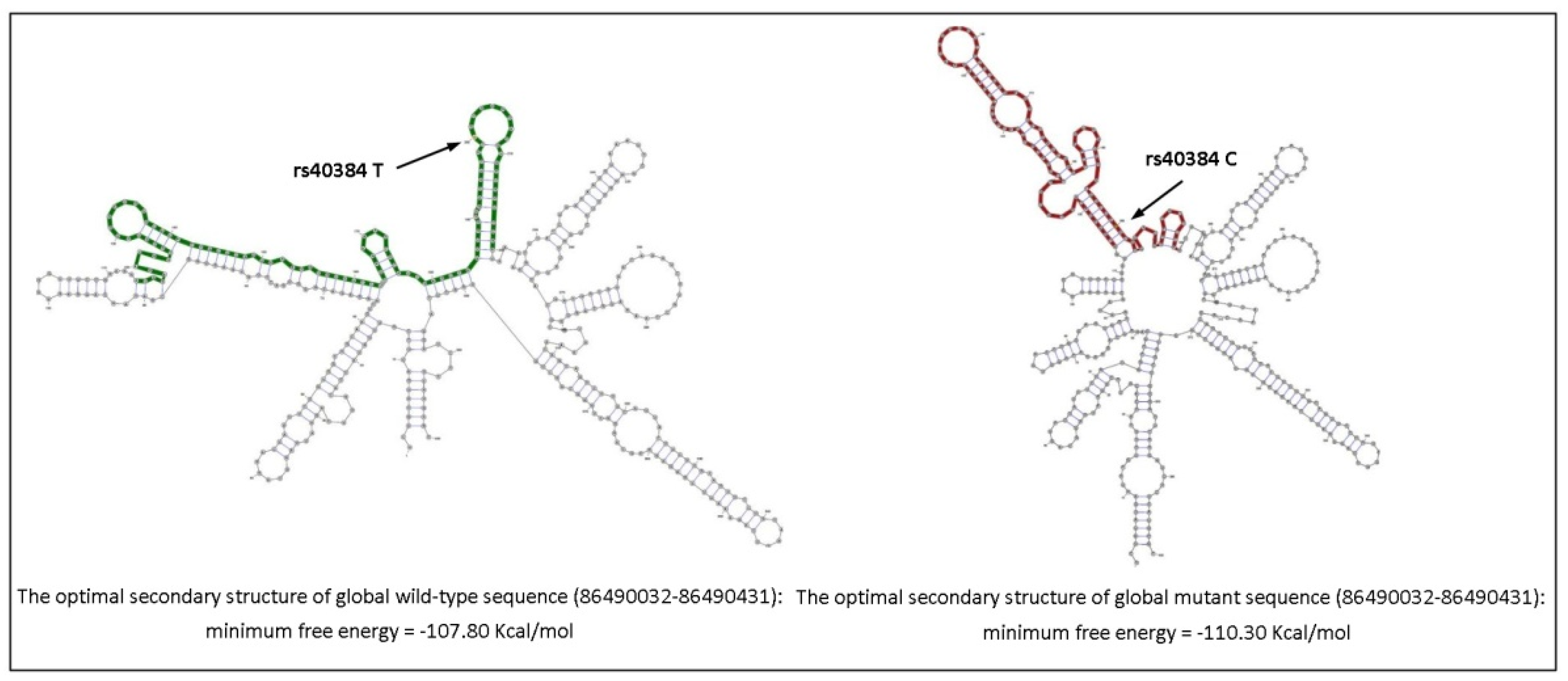
Figure 1. Structural differences in lncRNA FENDRR related to the rs40384 T>C polymorphism. The structural folding of FENDRR was predicted with http://rth.dk/resources/rnasnp/. Arrows indicate the T/C nucleotide change.
Life 12 00818 g001
Table 1. Examples of lncRNAs and their mechanism of action in the cardiovascular system in normal physiological conditions or disease states.
lncRNAsMechanism of ActionReferences
FENDRRCardiovascular embryogenesis, interaction with PRC2[7,8]
MHRTAntagonizes the role of Brg1 in cardiac hypertrophy[9]
CARMENRegulation of cardiac differentiation[10]
MALAT1Regulates the proliferation of endothelial cells and cardiomyocytes, epigenetic and alternative splicing regulation[11]
H19Regulates cardiac hypertrophy through miR-675[12,19]
MIATRegulates cardiac hypertrophy, miR-150 molecular sponge[13]
KCNQ1OT1Long QT syndrome, regulator of gene expression[15]
TINCRRegulates cardiac hypertrophy, epigenetic silencing of CaMKII[16]
ANRILStructure and function of vascular smooth muscle, regulator of gene expression[17]
PVT1Modulates the pathological cardiac hypertrophy via miR-196b[18]
Table 2. Main characteristics of the discovery and validation cohort.
DiscoveryValidation
ControlsPatientsp-ValueControlsPatientsp-Value
(N = 212)(N = 238) (N = 923)(N = 962)
Age72.0 [64.0, 76.0]61.0 [51.0, 69.0]<0.00166.0 [55.8, 78.0]60.0 [48.0, 69.0]<0.001
Sex
 Men91 (45.0%)148 (63.0%)<0.001518 (56.1%)620 (64.4%)<0.001
 Women111 (55.0%)87 (37.0%) 405 (43.9%)342 (35.6%)
Sarcomere
 No-186 (78.2%)--763 (79.3%)-
 Yes-52 (21.8%) -199 (20.7%)
Table 3. Descriptive results of genotype and allele frequencies of the rs74035787 and rs1424019 polymorphisms in the validation cohort. Results are shown as overall cases, stratified by gender, and by age (≥50 years old).
PatientsControlsTotal PatientsTotal Controls
MenWomen≥50< 50MenWomen≥50<50
(N = 620)(N = 342)(N = 691)(N = 271)(N = 518)(N = 405)(N = 763)(N = 160)(N = 962)(N = 923)
rs74035787
GG (n (%))607 (97.9%)338 (98.8%)680 (98.4%)265 (97.8%)499 (96.3%)382 (94.3%)727 (95.3%)154 (96.3%)945 (98.2%)881 (95.4%)
GA (n (%))13 (2.10%)4 (1.17%)11 (1.59%)6 (2.21%)19 (3.67%)23 (5.68%)36 (4.72%)6 (3.75%)17 (1.77%)42 (4.55%)
G (Freq.)0.9900.9940.9920.9890.9820.9720.9760.9810.9910.977
A (Freq.)0.0110.0060.0080.0110.0180.0280.0240.0190.0090.023
rs1424019
AA (n (%))53 (8.55%)26 (7.60%)55 (7.96%)24 (8.86%)42 (8.11%)30 (7.41%)58 (7.60%)14 (8.75%)79 (8.21%)72 (7.80%)
AG (n (%))212 (34.2%)109 (31.9%)222 (32.1%)99 (36.5%)208 (40.2%)133 (32.8%)286 (37.5%)55 (34.4%)321 (33.4%)341 (36.9%)
GG (n (%))355 (57.3%)207 (60.5%)414 (59.9%)148 (54.6%)268 (51.7%)242 (59.8%)419 (54.9%)91 (56.9%)562 (58.4%)510 (55.3%)
A (Freq.)0.2560.2350.2400.2710.2820.2380.2630.2590.2490.263
G (Freq.)0.7440.7650.7600.7290.7180.7620.7370.7410.7510.737
Table 4. Univariate and multivariate analysis of rs74035787 and rs1424019 variants. Since healthy controls were selected older on purpose, only sex was added to the multivariate analysis.
UnivariateMultivariate
PredictorsOdds RatioCIp-ValueOdds RatioCIp-Value
rs74035787GGReferenceReference
GA0.380.21–0.660.0010.380.21–0.670.001
Women 0.710.59–0.85<0.001
GReferenceReference
A0.380.21–0.660.0010.390.21–0.670.001
Women 0.710.62–0.81<0.001
rs1424019GGReferenceReference
AA o AG0.880.73–1.050.1650.860.72–1.040.112
Women 0.700.58–0.84<0.001
GReferenceReference
A0.930.80–1.080.3330.920.79–1.060.248
Women 0.700.62–0.80<0.001
Table 5. Stratified analysis of rs74035787 and rs1424019 polymorphisms and the risk of HCM in the validation cohort. Stratification criteria were gender and age (≥50 years old).
PredictorsWomenMen
Odds RatioCIp-ValueOdds RatioCIp-Value
rs74035787GA0.200.06–0.520.0030.560.27–1.140.115
A0.200.06–0.530.0030.570.27–1.140.118
rs1424019GG1.030.77–1.390.8301.250.99–1.580.063
G1.020.80–1.290.8961.140.94–1.370.173
Predictors≥50 yo<50 yo
Odds RatioCIp-ValueOdds RatioCIp-Value
rs74035787GA0.330.16–0.630.0010.700.21–2.360.559
Women0.870.71–1.080.02080.390.25–0.60<0.001
A0.330.16–0.630.0010.710.21–2.350.563
Women0.870.75–1.010.0730.390.29–0.53<0.001
rs1424019GG1.241.01–1.530.0450.920.61–1.380.686
Women0.860.70–1.060.1620.390.25–0.60<0.001
G1.140.96–1.350.1290.930.68–1.280.674
Women0.870.75–1.010.0620.390.29–0.53<0.001
Table 6. Haplotype frequencies between rs74035787 and rs1424019 polymorphisms in the validation cohort.
PolymorphismHaplotype Frequency
rs74035787 G>Ars1424019 A>GPatients
N = 962
Controls
N = 923
p-ValueOR95%CI
GG1441 (0.749)1341 (0.726)0.11601.130.97–1.30
GA466 (0.242)464 (0.251)0.51470.950.82–1.10
AG4 (0.002)19 (0.010)0.00350.200.07–0.59
AA13 (0.007)22 (0.012)0.10310.560.28–1.12
Publisher’s Note: MDPI stays neutral with regard to jurisdictional claims in published maps and institutional affiliations.