Article

Anaplasma phagocytophilum Transmission Activates Immune Pathways While Repressing Wound Healing in the Skin

by
Jacob Underwood
1,2,
Cristina Harvey
1,
Elizabeth Lohstroh
1,
Branden Pierce
1,
Cross Chambers
1,
Stephanie Guzman Valencia
1 and
Adela S. Oliva Chávez
1,*
1
Department of Entomology, Texas A&M University, College Station, TX 77845, USA
2
Navy Entomology Center of Excellence, United States Navy, Jacksonville, FL 32212, USA
*
Author to whom correspondence should be addressed.
Life 2022, 12(12), 1965; https://doi.org/10.3390/life12121965
Submission received: 15 October 2022 / Revised: 18 November 2022 / Accepted: 21 November 2022 / Published: 24 November 2022
(This article belongs to the Special Issue Tick-Transmitted Diseases)

Abstract

:
Anaplasma phagocytophilum, the causative agent of human granulocytic anaplasmosis (HGA), is an obligate intracellular bacterium transmitted by the bite of black-legged ticks, Ixodes scapularis. The main host cells in vertebrates are neutrophils. However, the first site of entry is in the skin during tick feeding. Given that the initial responses within skin are a crucial determinant of disease outcome in vector-borne diseases, we used a non-biased approach to characterize the transcriptional changes that take place at the bite during I. scapularis feeding and A. phagocytophilum transmission. Experimentally infected ticks were allowed to feed for 3 days on C57BL/6J mice to allow bacterial transmission and establishment. Skin biopsies were taken from the attachment site of uninfected ticks and A. phagocytophilum-infected ticks. Skin without ticks (intact skin) was used as baseline. RNA was isolated and sequenced using next-generation sequencing (NGS). The differentially expressed genes were used to identify over-represented pathways by gene ontology (GO) and pathway enrichment (PE). Anaplasma phagocytophilum transmission resulted in the activation of interferon signaling and neutrophil chemotaxis pathways in the skin. Interestingly, it also led to the downregulation of genes encoding extracellular matrix (ECM) components, and upregulation of metalloproteinases, suggesting that A. phagocytophilum delays wound healing responses and may increase vascular permeability at the bite site.

1. Introduction

The black-legged tick, Ixodes scapularis, has been spreading in recent years to more locations across the northeastern and midwestern United States. It is a competent vector of seven different pathogens, known to cause illness in humans, including Anaplasma phagocytophilum [1]. Anaplasma phagocytophilum is the causative agent of human granulocytic anaplasmosis (HGA), formerly known as human granulocytic ehrlichiosis (HGE), which was discovered infecting a cluster of men in the upper Midwest in 1994 [2]. Later, similar cases were reported in other parts of the U.S. where Lyme disease is prevalent [3]. Most recently, 5655 cases were reported to the CDC in 2019, making HGA the second most common tick-borne disease in the U.S. [4]. In most cases, the illness is self-limiting, and patients will recover with or without antibiotic treatment. Symptoms will typically develop 1–2 weeks following a bite by an infected tick, can lead to hospitalization in around 36% of confirmed cases, and presents mortality rates of around 0.2–1% [5].
Anaplasma phagocytophilum is a Gram-negative bacterium of small size (0.4 to 1.5 μm) that replicates inside neutrophils within small vacuoles termed “morulae” [6]. This bacterium lacks several of the enzymes involved in peptidoglycan and lipopolysaccharide biosynthesis [6,7], which are two pathogen-associated molecular patterns (PAMPs) commonly recognized by vertebrate immune responses. Nevertheless, stimulation of peripheral blood leukocytes from healthy donors with cell free bacteria or recombinant A. phagocytophilum outer protein P44 (rP44) induces the expression of proinflammatory cytokines [8]. Similarly, high levels of proinflammatory cytokines, such as interferon (Ifn)-γ, interleukin (IL)-12p70, and IL-10, are detected in HGA patients [9]. Further, the elevated levels of these cytokines appear to be associated with the severity and pathology of the disease in humans and murine models [9,10,11]. Ifn-γ is involved in the control of A. phagocytophilum infection in mice [10,11,12] and during the in vitro infection of Hoxb8 neutrophils [13]. In vivo experiments using murine models indicate that secretion of Inf-γ correlates with Stat1 phosphorylation [14]. Stat1 knock-out in mice leads to increased bacterial loads, severe disease pathology, increased spleen size, higher cytokines/chemokines levels in plasma, and reduced iNOS induction [15]. These studies were focused on systemic immune responses, and little is known about the immune events that take place during A. phagocytophilum transmission at the bite site.
Despite the importance of the skin as the site of entry and establishment of A. phagocytophilum, only a few experiments have been conducted to understand the events that occur at the bite site. In 2010, Granquist et al. [16] described the site of A. phagocytophilum transmission in lambs naturally infested with Ixodes ricinus ticks. Histological inspection of the bite sites showed evidence of inflammation, accumulation of immune cells such as neutrophils, macrophages, and other mononuclear cells, and the deterioration of the collagen matrix. Bacteria were associated with neutrophils and macrophages. However, the time of feeding for each tick could not be determined since animals were naturally infested. A later study similarly reported the presence of A. phagocytophilum within neutrophils in the skin of experimentally infected sheep later infested with ticks [17]. This study also reported significantly higher numbers of neutrophils in the skin of the experimentally infected sheep independent of the presence of feeding ticks, suggesting that A. phagocytophilum infection increases migration of neutrophils to the skin. Although this study considered the changes in expression of eight immune-related genes, the responses by each animal were variable and no conclusions on the activation of immune signaling pathways could be drawn. Thus, an important knowledge gap exists on the signaling and immunological events that take place during transmission of A. phagocytophilum by ticks.
The present study explores the gene expression changes that occur at the bite site during the transmission of A. phagocytophilum. We describe an upregulation in genes involved in Ifn-γ signaling, defense responses to viruses, neutrophil chemotaxis, and interleukin-1 responses. Interestingly, A. phagocytophilum transmission appears to decrease the expression of genes implicated in extracellular matrix (ECM) organization and wound healing responses. Given that the skin is the initial site of A. phagocytophilum establishment, an understanding of the immunological events that take place during initial infection will help us uncover potential signaling pathways and immune responses that can be exploited to stop infection.

2. Materials and Methods

2.1. Anaplasma Phagocytophilum Culture

HL60 cells (CCL-240™) were obtained from ATCC (Manassas, VA, USA). HL60 cell cultures were maintained in RPMI media (Corning, Manassas, VA, USA) supplemented with 10% fetal bovine serum (Gibco, Whaltham, MA, USA), 1% Glutamax (Gibco, Whaltham, MA, USA), and 1% amphotericin B (Corning, Manassas, VA, USA) and incubated at 37 °C with 5% CO2, as previously described [18]. Cells were maintained until the cell density became optimal for passage or for infection with A. phagocytophilum (~1 to 5 × 10⁵ cells/mL). Cells were passaged as follows: 2 mL of cell culture was transferred into a 25 cm2 tissue culture flask (Fisher Scientific, Pittsburgh, PA, USA) and supplemented with 18 mL of freshly prepared culture media. This was repeated every 3 to 5 days, until cells reached passage 10 when they were discarded, and a new culture was recovered from liquid nitrogen (LN2).
For infection with A. phagocytophilum, 2 mL of uninfected HL60 cell cultures (at approximately 5 × 10⁵ cells/mL) was inoculated with 500 μL of A. phagocytophilum-infected HL60 cells (at approximately 2 × 10⁵ cells/mL, with ~90% infection). Anaplasma phagocytophilum was cultured in HL60 cells for up to 5 days. Infections were monitored by placing 1 mL of the suspended cell culture onto a microscope slide, and spinning with a CytoSpin 4 (Thermo Scientific, Whaltham, MA, USA) at 800× g for 5 min. The infected cells were stained using the Richard-Allan Scientific™ Three-Step Stain Kit (Thermo Scientific, Whaltham, MA, USA), according to manufacturer’s specifications. The morulae within cells were observed by light microscopy with an Olympus model BX43F (Shinjuku City, Tokyo, Japan). Infections were passaged when the percentage of infection was greater than 90%, determined by counting 100 HL60 cells with observable morulae. Bacterial cultures were maintained for up to 5 passages before freezing or infecting mice, using the procedures described below.

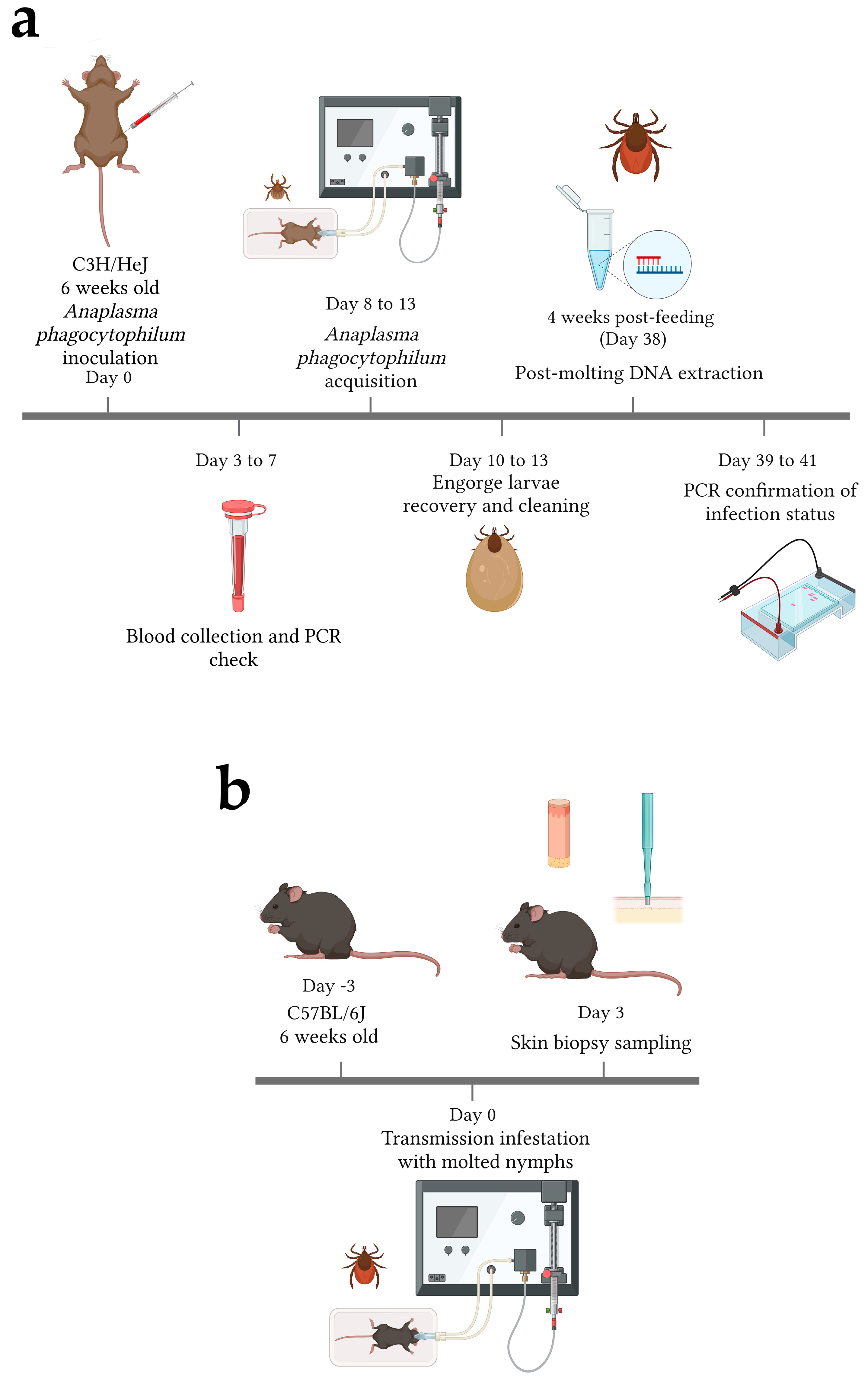
2.2. Mice Infections

C3H/HeJ male mice of 6 weeks of age (The Jackson Laboratory, Bar Harbor, ME, USA) were used for pathogen acquisition due to their high susceptibility to infection from Gram-negative bacteria, including to A. phagocytophilum infection [19]. The susceptibility of this mouse strain to Gram-negative bacteria is associated with a mutation in the cytoplasmic domain of Toll-like receptor 4 (TLR4) [20] and has shown impaired inflammatory and innate immune responses under several conditions [21,22]. Mice were injected intraperitoneally (i.p.) with 100 μL A. phagocytophilum-infected HL60 containing 1 × 10⁷ bacteria, using 27-gauge needles (Figure 1a). Cells were spun down at 300× g for 10 min, culture media was removed, and the cells were suspended in 1× PBS. The number of bacteria was estimated using the previously described formula [23]. Control mice received an injection of 100 μL 1× PBS.
To confirm infections, cheek bleeds were performed on the mice on days 3, 5, and 7 post-infection (p.i.), collecting 20 to 100 μL of blood in microvettes 500 K3E (Sarstedt, Nümbercht, Germany) after anesthesia with 1.25% to 2% isoflurane (Figure 1a). Blood was used for DNA extraction with the DNeasy Blood & Tissue Kit (QIAGEN, Hilden, Germany), following the manufacturer’s instructions. DNA quantity and quality was assessed using a NanoQuant Infinite M200 Pro (Tecan, Switzerland). PCR amplification of mouse actin was performed to confirm the absence of contaminants and PCR inhibitors. PCR analyses of the A. phagocytophilum rpoB and the 16s rRNA genes were performed on the blood to confirm infection with A. phagocytophilum. PCRs were prepared using GoTaq Flexi DNA polymerase (Promega, Madison, WI, USA). Amplification was completed using the following PCR cycling conditions: 1 denaturing cycle for 3 min at 95 °C, followed by 34 cycles of 1-min denaturation at 95 °C, 1 min at the annealing temperature (Table 1) and an extension of 72 °C for 30 s. A final extension step of 5 min at 72 °C was performed. Predicted product sizes are displayed in Table 1. PCR product sizes were confirmed by gel electrophoresis and compared with a 100 bp ladder (NEB, Ipswich, MA, USA). Gels were visualized with an iBright FL1500 Imaging System (Thermo Scientific, Whaltham, MA, USA) to confirm the infection of mice (Figure S1a).

2.3. Tick Infestations

For A. phagocytophilum tick infections, 200 larval I. scapularis ticks were placed on mice after confirming infection in the blood (Figure 1a). Mice were separated into individual mesh bottom cages placed above a water trap to collect the fed ticks. Mice were anesthetized for 30 min with 1.25% to 2% isoflurane to allow the larvae to attach. Engorged ticks were collected from the water baths after 3, 4 and 5 days of feeding (Figure 1a). The mice were euthanized with CO2, followed by cervical fracture and heart puncture exsanguination. The collected ticks were washed in 2% bleach and autoclaved water, placed into groups of 25, and allowed to molt into nymphs.
DNA was purified from 5 pooled nymphs from each group of infected and control ticks to confirm infection or lack of. Ticks were placed at −80 °C for 1 h and DNA was isolated using the Quick-DNA/RNA Miniprep kit (Zymo, Irvine, CA, USA), according to the manufacturer’s instructions. DNA quantity and quality were tested as described above, and a PCR on the I. scapularis actin gene was performed to determine the presence of PCR inhibitors. Anaplasma phagocytophilum infection (or lack of) was confirmed by PCR amplification of the rpoB and p44 genes (Table 1; Figure 1a). Amplification of the PCR products was carried out using similar cycling conditions as described above. Predicted product sizes are displayed in Table 1. Positive and negative PCRs were confirmed by gel electrophoresis as described above. Additionally, the relative levels of bacterial infection were assessed by qPCR, using the ΔCt value of A. phagocytophilum p44 normalized by tick actin with the following formula:
Δ C t = 2 ( c t   A n a p l a s m a   p 44 c t   t i c k   a c t i n )
qPCRs were performed using PowerUp™ SYBR™ Green Master Mix (Applied Biosystems, Whaltham, MA, USA), using same primers as for PCR analysis (Table 1). Amplification, melt curves, and data were analyzed with CFX Maestro Software (Bio-Rad, Hercules, CA, USA).
Due to the potential effect of the mutation of C3H/HeJ mice tlr4 in the local immune responses to A. phagocytophilum transmission, we decided to use a different mouse strain. The C57BL/6J mouse strain has previously been used for the study of murine systemic immune responses during A. phagocytophilum infection and the role of IFN-γ/STAT1 [11], therefore, we used this same strain to define local immune responses to bacterial transmission. A. phagocytophilum-infected and -uninfected nymphs were used to infest 6 week old C57BL/6J male mice (The Jackson Laboratory Bar Harbor, ME, USA). Twenty-five I. scapularis nymphs were placed on each mouse. Mice were anesthetized as previously described for the larvae (Figure 1b). The ticks were allowed to feed for 3 days to allow 24 h post-transmission of A. phagocytophilum [28]. After this time, the mice were euthanized with CO₂, followed by cervical fracture. Three (3) mm skin biopsies were taken of the bite sites, utilizing Integra disposable biopsy punches (Militex, Saint-Pries, France) for RNAseq, and 5 mm skin biopsies were taken for qRT-PCR (Figure 1b). Skin samples far from where the ticks were located were taken to determine the gene expression in intact skin (baseline). The partially engorged nymphs were collected for excision of their midguts and salivary glands. The skin was placed in 500 μL RNALater (Invitrogen, Carlsbad, CA, USA).

2.4. RNA-Seq and Pathway Analysis of Skin Biopsies

RNA extraction, library preparations, sequencing reactions and bioinformatic analysis were conducted at GENEWIZ, LLC. (South Plainfield, NJ, USA) as follows: total RNA was extracted using Qiagen RNeasy Plus Universal mini kit following the manufacturer’s instructions (Qiagen, Hilden, Germany). The quantity and quality of the RNA samples were assessed using a Qubit 2.0 Fluorometer (Life Technologies, Carlsbad, CA, USA) and an Agilent TapeStation 4200 (Agilent Technologies, Palo Alto, CA, USA), respectively.
The RNA libraries were prepared using the NEBNext Ultra II RNA Library Prep Kit (NEB, Ipswich, MA, USA), following the manufacturer’s instructions for Illumina. Briefly, mRNAs were first enriched with Oligo(dT) beads, followed by fragmentation for 15 min at 94 °C and cDNA synthesis. cDNA was adenylated at 3′ends and end repaired. Universal adapters were ligated to cDNA fragments, followed by index addition and library enrichment by limited-cycle PCR. The quality of the libraries was validated on the Agilent TapeStation (Agilent Technologies, Palo Alto, CA, USA). Libraries were quantified with a Qubit 2.0 Fluorometer (Invitrogen, Carlsbad, CA, USA) and quantitative PCR (KAPA Biosystems, Wilmington, MA, USA).
The libraries were sequenced on an Illumina HiSeq instrument (4000) according to the manufacturer’s instructions, using a 2 × 150 bp paired end (PE) configuration. HiSeq Control Software (HCS) v2.0.12 was used for image analysis and base calling. After investigating the quality of the raw data, possible adapter sequences and nucleotides with poor quality were trimmed. The trimmed reads were mapped to the reference genome, using STAR aligner v.2.5.2b. Unique gene hit counts were calculated using featureCounts from Subread package v.1.5.2. Only unique reads that fell within exon regions were counted. The complete sequencing data were deposited on NCBI, and accession numbers can be found in the data availability section.
Upregulated and downregulated genes with adjusted p-value (padj) < 0.05 and log2 fold changes of 1 or more (−1 or less for downregulated genes) were used to identify pathway over-representation using Reactome (https://reactome.org/, accessed on 19 February 2022). Only pathways with p < 0.05 and false discovery rate (FDR) < 0.05 were considered as over-represented.

2.5. qRT-PCR of Skin Biopsies

RNA was extracted from the skin using TRIZOL (Invitrogen, Whaltham, MA, USA) according to the manufacturer’s specifications with small modifications. Briefly, the RNALater was washed off the tissues with 1× phosphate buffered saline (PBS), and skin samples were quickly flash froze with LN2. The frozen tissue was homogenized with a mortar and pestle. One (1) mL of TRIZOL was added to the tissue. The aqueous phase was taken and mixed 1:1 with 70% ethanol. RNA was then isolated using PureLink™ RNA Mini Kit (Ambion, Carlsbad, CA, USA), according to the manufacturer’s indications. RNA was quantified and quality was assessed using a NanoQuant Infinite M200 Pro (Tecan, Switzerland). RNA (100 ng) was used to synthesize cDNA with the Verso cDNA Synthesis Kit (Thermo Scientific, Whaltham, MA, USA). qPCRs were performed using PowerUp™ SYBR™ Green Master Mix (Applied Biosystems, Whaltham, MA, USA). Interferon gamma (Ifn-g), interleukin 1β (Il1b), interferon regulatory factor 1 (Irf1), S100 calcium binding protein A8 (S100a8), Aggrecan (Acan) and Matrilin 3 (matn3) were amplified using the primers described in Table 1. The amplifications were performed in a CFX Opus 96 Real-Time PCR instrument (Bio-Rad, Hercules, CA, USA). Amplification, melt curves, and data were analyzed with CFX Maestro Software (Bio-Rad, Hercules, CA, USA). The relative differences were calculated using the ΔΔCt method [29] as follows:
Δ Δ C T = ( 2 ( C t   g e n e   o f   i n t e r e s t   t r e a t m e n t C t   a c t i n   t r e a t m e n t ) ) ( 2 ( C t   g e n e   o f   i n t e r e s t   i n t a c t   s k i n C t   a c t i n   i n t a c t   s k i n ) )
The expression of the genes was normalized to mouse actin (Table 1). The expression of immune genes in skin samples at the bite site of uninfected and infected ticks was normalized to that of the intact skin (baselines). Statistical differences in gene expression between conditions was evaluated using an unpaired two-tailed t-test with GraphPad Prism 9.3.1 (GraphPad Software, San Diego, CA, USA). Outliers were determined using GraphPad 9.3.1.
The specificity of the primers was confirmed by RT-PCR of RNA isolated from mouse hearts followed by Sanger sequencing (Etonbio, Union, NJ, USA). The forward and reverse sequences were assembled with Geneious Prime (Biomatters, Inc., San Diego, CA, USA) and identity was confirmed through BLAST (NCBI). Accession numbers for the sequences amplified by the primers used here in can be found in the data availability statement.

3. Results

3.1. Tick Feeding Induces the Expression of Neutrophil Chemotaxis and Inflammatory Responses in the Skin

Pathogen-free certified larvae fed upon naïve mice until repletion. Following molting, the absence of A. phagocytophilum infection in control nymphs was confirmed by DNA extraction, followed by negative amplification of p44 and RpoB genes from A. phagocytophilum (Figure S1b). Uninfected (control) nymphs fed upon C57BL/6J for 3 days, and 2 mm skin biopsy punches were taken from the bite site. RNAseq and differential gene expression (DEG) analyses were performed on RNA isolated from the skin samples. A total of 1213 genes were differentially regulated upon tick feeding, when compared with the intact skin (Table 2). A total of 797 genes were upregulated and 416 were downregulated during tick feeding. Principal component analysis (PCA) demonstrated a clear separation of the transcriptomic profiles between the intact skin and uninfected tick bite sites (Figure 2a). Volcano plot analysis of intact skin samples versus uninfected tick bite sites showed the upregulation of several immune-related genes (including genes encoding chemokines and chemoattractant Saa3), and the downregulation of iron homeostasis genes (such as Hamp2) and transcription factors (such as RorC) (Figure 2b).
Heatmap analysis of the 30 most differentially regulated genes resulted in five gene clusters of co-regulated gene (Figure 2c). Cluster 1 included keratine-encoding genes important in epithelial cell integrity in the skin (krt16 and krt6b); enzymes involved in phospholipid and heme metabolism, and glycolysis (Plbd1, Hmox1, and Eno1); and to signaling proteins, including Arrdc3 that allows G protein signaling, and Saa3. Cluster 2 comprised several lectins, such as Chil3, Sell, and Clec4d; the glycoprotein Cd300lf; and 2610528a11Rik, a gene encoding G protein-coupled receptor 15 ligand induced during several inflammatory conditions in the skin [30]. Cluster 3 contained a gene without known function (Gm45819) and a circadian transcriptional repressor (Cry1). Cluster 4 was formed by two chemokines, one involved in macrophage attraction (Ccl7), and Ccl2 that is a chemoattractant for monocytes and basophils. It also contained a lipocalin (Lcn2), a metalloproteases inhibitor (Timp1), and a proteoglycan (Prg4). Cluster 5 contained two genes involved in chromosome segregation (Kif22 and Nuf2), immune response associated genes (Slfn4, S100a9, S100a8, and Il1b), epidermal maintenance and development (Stfa3 and Stfa1), and other genes (Slc15a3 and Adamts4). All clusters showed upregulation of the co-regulated genes, with the exception of Cluster 3 that showed higher levels of expression in the intact skin (baseline; Figure 2c). Enrichment analysis based on gene ontology (GO) (Figure S2) showed an over-representation of genes associated with immune responses, neutrophil chemotaxis, cell division, and chromosome segregation. Pathway enrichment (PE) analysis (Table S1) of upregulated genes demonstrated enrichment of interleukin-10 signaling associated genes, neutrophil degranulation, Th2 signaling, and mitotic spindle, potentially indicating the division of keratynocytes in response to the inflammation and damage associated with the tick bite as previously reported in mouse biopsies [25]. Downregulated genes did not show any significant enrichment in pathways (Table S2).

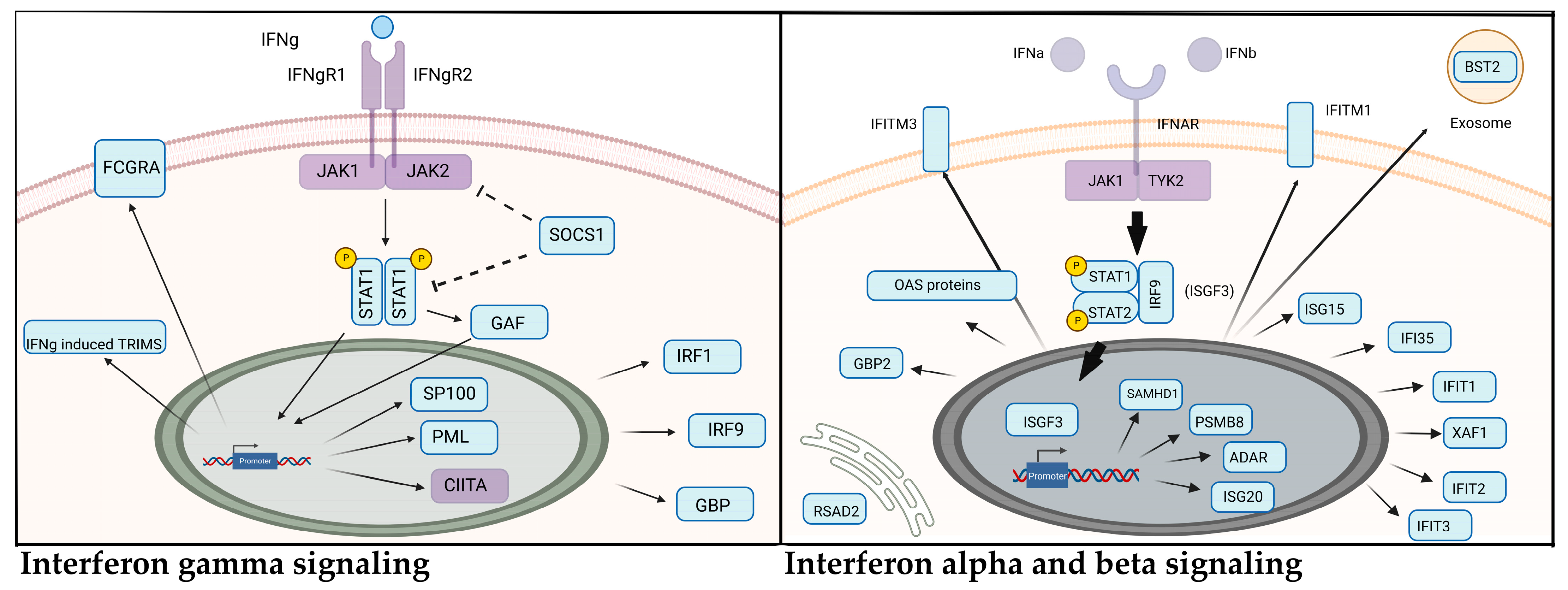
3.2. Anaplasma Phagocytophilum Transmission Induces the Upregulation of Interferon Signaling Genes

Ixodes scapularis larvae fed upon A. phagocytophilum PCR-positive mice (Figure S1a). Ticks were allowed to molt and DNA was extracted from five nymphs from each group to confirm infection status. Anaplasma phagocytophilum-positive nymphs (Figure S1b) with varying relative levels of bacterial infection (Figure S1c) were infested onto naïve C57BL/6J mice for 3 days. Skin samples were taken from the bite site and RNA was isolated for RNAseq analysis. DEG analysis identified a total of 2559 DEGs between the intact skin and skin samples from the bite site of Anaplasma phagocytophilum-infected ticks, including 1417 upregulated and 1142 downregulated genes. Comparatively, 622 DEGs were identified when uninfected tick and infected tick bite sites were compared. This encompassed 476 upregulated and 146 downregulated genes, indicating a synergistic effect between A. phagocytophilum and the tick (Table 2). PCA showed the separation of control samples and samples from the bite site of A. phagocytophilum-infected ticks and of uninfected ticks. Similar separation was observed between intact skin and infected ticks’ bite sites. In both cases, the presence of one outlier from the A. phagocytophilum ticks was detected (Figure 3a and Figure S3a). Nevertheless, this sample showed similar co-regulatory gene expression as the other samples (Figure 3c and Figure S3a). Anaplasma4, on the other hand, showed a slightly lower upregulation of immune genes when compared with skin samples from bite sites of uninfected ticks and intact skin (Figure 3c and Figure S3c; Supplementary Files S1 and S2). This might be due to lower levels of infection in the tick or due to a delayed attachment of the tick leading to less time for A. phagocytophilum transmission, although this is speculative since we did not test bacterial numbers in the skin.
Analysis of the gene expression using volcano plots showed several immune-related genes that are significantly upregulated during transmission of A. phagocytophilum when compared with uninfected ticks’ bite sites (Figure 3b), including the gene encoding tumor necrosis factor (tnf), and chemokines ccl4 and cxcl10, which are attractants of macrophages, natural killer cells, and T cells. Interestingly, genes involved in extracellular matrix formation were downregulated; for example, the gene encoding matn3. Heatmap analysis identified six clusters of co-regulated genes (Figure 3c). The first cluster upregulated in Anaplasma transmission skin samples, included the genes encoding for the interferon gamma inducible GTPase Ifgga3 protein (Gm4841), lymphocyte antigen 6c2 (Ly6c2; which acts as an acetylcholine receptor), the endoplasmic reticulum/Golgi membrane-spanning 4-domains subfamily A, member 4C (Ms4a4c), and the ubiquitin-like modifier protein ISG15 (Isg15). The second cluster contained two interferon-induced genes (Ifi213 and Oas3), regulators of Th1 cytokine secretion (Phf11b and Phf11d), and an enzyme involved in arginine biosynthesis (Ass1), which showed upregulation in the skin samples from infected ticks bite sites.
A third cluster of upregulated genes involved Slfn4, the gene encoding an interferon regulatory factor 7 (Irf7), and the interferon-induced gene Oasl2. Two genes encoding G receptor interacting proteins were also found in this group (Rgs5 and Slc9a3r1). The fourth cluster only contained two genes slightly upregulated during A. phagocytophilum transmission: an interferon activated gene (Ifi214) and a pseudogene (Gm654). The fifth cluster contained the highest number of genes and included several immune-related genes (Gzmb, Gm12185, Trim30b, Ccl4, and Tnf) and two lipid transport proteins (Apol9b and Apol9a). The last cluster included genes encoding two glycoproteins (Cd300lf and Clec4e) and an interferon-induced protein (Ifi44), which are involved in innate immune responses (Figure 3c). Several of the immune-related clusters upregulated in A. phagocytophilum transmission sites versus controls were also observed when compared with intact skin (baseline; Figure S3c).
Gene ontology (GO) enrichment analysis of the differentially regulated genes during A. phagocytophilum transmission identified the enrichment of genes involved in the cellular response to interferon β (Ifn-β) and Ifn-γ (Figure 4). Enrichment of interferon signaling was also detected using PE analysis of the significantly upregulated genes (Table 3 and Table S3). Interestingly, antiviral mechanisms also showed enrichment in GO and pathway analysis, likely due to the intracellular nature of A. phagocytophilum infection. Other interleukin signaling pathways (IL-1, IL-10, and IL-12) were also observed in the enrichment analysis. Th2 responses were enriched to a lesser degree and likely represented the responses to the tick feeding, as these pathways were significantly over-represented in uninfected tick bite sites, unlike interferon signaling that was not found to be upregulated during uninfected tick feeding (Table S1). This suggests that A. phagocytophilum transmission leads to the activation of Th1 signaling pathways in the skin.
Pathway enrichment (PE) analysis of downregulated genes during A. phagocytophilum showed an over-representation of pathways involved in ECM integrity, including ECM organization, fibrils formation, and several collagen organization enzymes (Table 4). Some genes involved in these pathways were downregulated in uninfected tick bite sites (Figure S2). Nevertheless, pathways involved in ECM integrity were not over-represented in uninfected tick bite sites (Table S2). Curiously, although the upregulation of interferon and interleukin signaling pathways were detected during A. phagocytophilum transmission when compared with intact skin (Table S3), the ECM organization pathways were not over-represented in downregulated genes (Table S4).

3.3. Differentially Expressed Genes (DEGs) Stimulated during Tick Feeding and A. Phagocytophilum Transmission

To determine genes and pathways that are affected by both the tick feeding and A. phagocytophilum transmission, the DEGs that are upregulated and downregulated during the bite of uninfected and infected ticks, when compared with intact skin expression, were identified (Supplemental File S7). Pathway enrichment (PE) of genes upregulated in both conditions showed an over-representation of interleukin-10 signaling, neutrophil degranulation, chemokine signaling, Th2 cytokine signaling, and other pathways enriched during tick feeding compared with intact skin expression (Table 5 and Table S1), corroborating that the enrichment of these pathways during A. phagocytophilum transmission is the result of the synergetic effect of the tick and bacterial transmission. By comparison, PE analysis of genes upregulated during transmission of A. phagocytophilum solely identified an over-representation of interferon signaling genes, interleukin-10 signaling, and other immune response pathways (Table S5), confirming the effect of the bacterial transmission on these signaling pathways. In the case of the DEGs identified as upregulated during the tick bite only, no significant enrichment of pathways was detected (Table S6).
In the case of shared downregulated genes, no significant PE was observed (Table 6), suggesting that the transmission of A. phagocytophilum leads to the downregulation of distinct pathways when compared with tick feeding. The analysis of genes downregulated only during A. phagocytophilum transmission, identified pathways involved in muscle contraction, collagen related pathways, and extracellular matrix organization (Table S7). As in the case of shared downregulated genes, the PE analysis of genes downregulated during uninfected tick feeding did not identify any significantly over-represented pathways (Table S8). These results were similar to the analysis of all DEGs downregulated during tick feeding versus expression in intact skin (Table S2). This confirmed that A. phagocytophilum transmission, and not the tick feeding, is responsible for the detected effects on ECM organization. Whether this effect is due to bacterial manipulation of cells in the skin or due to changes in tick saliva during infection with A. phagocytophilum remains to be determined.

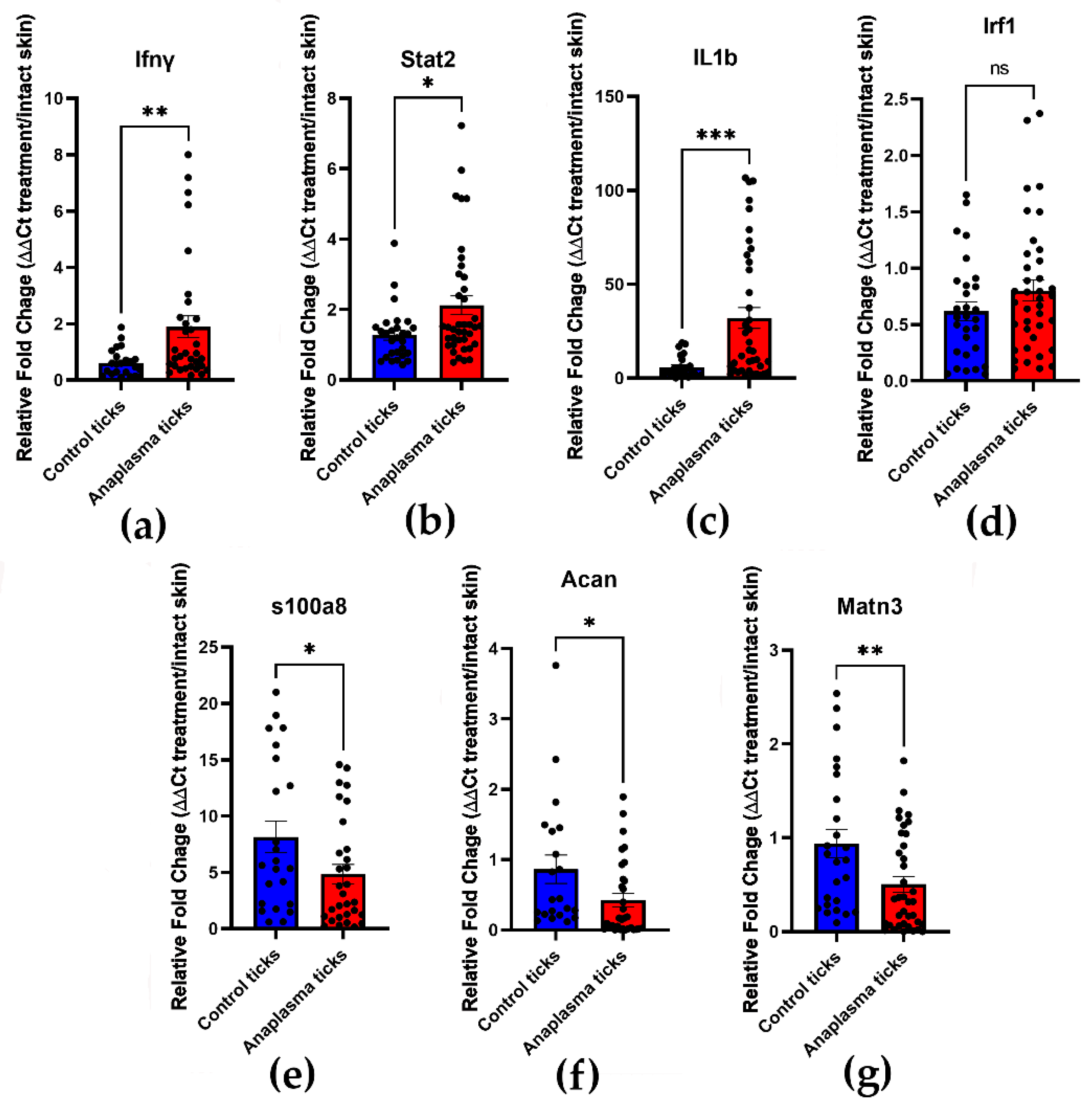
3.4. Confirmation of Th1 Cytokines Upregulation and Downregulation of ECM Genes by qRT-PCR

The expression patterns of selected genes identified by RNAseq were confirmed by qRT-PCR. RNA was isolated from skin biopsies of the bite site of uninfected and A. phagocytophilum ticks and were normalized to the gene expression of intact skin from the same mice. Similar to the tick batches used for the infestation of mice during the RNAseq experiments, relative bacterial levels were highly variable (Figure S1d). The upregulation of Ifn-γ, stat2, and il1β during A. phagocytophilum transmission were validated (Figure 5a–c). Although Irf1 was slightly upregulated in the bite site of A. phagocytophilum ticks, this difference was not statistically significant (Figure 5d). S100a8, Acan, and matn3 were upregulated in samples taken from uninfected tick bite sites (Figure 5e–g). Acan and matn3 encode for proteins with roles in ECM integrity; thus, validating the upregulation of interferon and interleukin-1 mediated signaling related genes and the downregulation of genes involved in ECM integrity. Interestingly, although the relative expression of immune genes (Infg, stat2, and il1β) was significantly lower in intact skin when compared with the bite site of A. phagocytophilum-infected ticks, the expression of Acan and matn3 was not significantly different (Figure S4), thus, confirming that expression of these ECM integrity genes is similar in intact skin and A. phagocytophilum transmission sites.

4. Discussion

Initial responses within the skin can ultimately define the outcome of infection by a vector-borne pathogen. Skin cells, including immune cells, keratinocytes, and endothelial cells, serve as the site of initial replication for vector-borne viruses, bacteria, and parasites [31,32,33,34,35]. Arthropod inoculation of some viruses can lead to increased severity of pathologies associated with infection [31]. Further, arthropod saliva influences responses beyond the skin. For example, inoculation of mosquito salivary gland extracts (SGE) enhanced the migration of neutrophils and dendritic cells into draining lymph nodes and boosted the pathogenesis of the virus during antibody-dependent enhancement of dengue [34]. The role of single salivary proteins in the stimulation of immune cells and the enhanced pathogenesis of disease has also been investigated in mosquito models. In Zika virus (ZIKV), neutrophil-stimulating factor 1 (NeSt1) activates neutrophils and leads to augmented early virus replication [36]. Similarly, proteins within Lutzomia longipalpis saliva affect neutrophil function, inducing macrophage migration into the bite site and increasing Leishmania chagasi and Leishmania infantum replication [32,33,35]. Thus, arthropod saliva and other vector-derived factors are important determinants of pathogenesis of vector-borne pathogens.
In the case of tick-borne pathogens, little is known of the cellular and immune factors that influence their establishment and how tick saliva and tick modulatory molecules affect pathogen replication and pathogenesis [37]. In the case of Rickettsia parkeri, although tick saliva and tick feeding increase skin pathology during bacterial infection, bacterial replication at the inoculation site was reduced in the presence of tick salivary components [38,39]. As in the case of mosquitoes, Amblyomma maculatum feeding resulted in variable infiltration of macrophages and neutrophils in rhesus monkeys’ skin, whereas bacterial inoculation at the tick feeding site led to marked neutrophil and macrophage translocation at the dermis [39]. Ixodes scapularis feeding also led to a significant increase in the number of neutrophils and macrophages in murine skin [25], which was consistent with previous transcriptional analysis of tick bite sites in murine models [40] and our results (Figure S2). According to our results, genes involved in neutrophil chemotaxis and degranulation were induced during I. scapularis feeding and A. phagocytophilum transmission (Figure 3 and Figure S5). This includes the upregulation of CXCL1 and CXCL2 and the receptors CCR5, CCR7, and CCRL2 (Supplementary File S8), which are involved in the activation, migration, effector, and antigen presentation functions of neutrophils [41,42]. Pathways enrichment (PE) analysis of genes upregulated during tick feeding and A. phagocytophilum transmission when compared with intact skin showed that neutrophil degranulation pathways are enriched in both (Table 5), suggesting that this effect may be mainly in response to tick feeding. This is corroborated by the absence of enrichment during A. phagocytophilum transmission versus feeding by uninfected ticks (Table 3). Interestingly, early experiments on sheep demonstrated that A. phagocytophilum infection led to higher number of neutrophils in the skin in the presence or absence of tick feeding [17]; whether A. phagocytophilum alone triggers neutrophil chemotaxis to the skin remains to be determined. Studies in the 1970s suggested that neutrophils may be involved in the pathology of tick bites of ixodid ticks [43]; nevertheless, what function neutrophils play during A. phagocytophilum transmission remains unexplored.
Among other immune pathways affected by the transmission of A. phagocytophilum in the skin was an increased activation of genes involved in interferon signaling (Table 3 and Table 5 and Figure 6). Type I (17 members, including Ifn-α and Ifn-β) and type II (Ifn-γ) interferons are important against viral and bacterial infections [44,45]. Their interaction with receptors within the membrane of cells activate signaling cascades that lead to the expression of immune-related genes [44]. The importance of Ifn-γ signaling in the immune response against systemic A. phagocytophilum infection has been demonstrated in several studies [10,11,12,13,14]. However, the significance of these cytokines and signaling pathways during early infection is unknown. Type I and type II interferon signaling pathways confer resistance to R. parkeri infection in murine models [46]. Double knockout mice with mutations in the type I IFN receptor (Ifnar1 −/−) and Ifn-γ receptor (Ifngr1 −/−) resulted in eschar development in the skin and lethality after intradermal (i.d.) infection of as low as 102 bacteria when compared with 107 when injected intravenously (i.v.), suggesting that IFN signaling in the skin may be important for protective immunity [46,47]. Furthermore, infection and vaccination with mutant R. parkeri strains showed that Ifnar1 −/−; Ifngr1 −/− double knockout mice may be a good tool for the study of rickettsial pathogenesis, and as models to evaluate vaccine candidates [46]. As with SFG Rickettsia, A. phagocytophilum does not produce disease in mice. Given our results, and that bacterial numbers increase during systemic infection of A. phagocytophilum in Ifn-γ / mice [11], it is possible that these receptors are also important for initial immune responses in the skin. However, more studies are needed to understand the role of IFN signaling during bacterial establishment.
Unexpectedly, A. phagocytophilum transmission also resulted in the downregulation of extracellular matrix (ECM) organization related genes (Table 4). Several genes encoding ECM structural components, such as collagen, thrombospondin-5, integrin ITGA10, Matrilin 3, aggrecan, elastin, fibronectin, and fibrillin, were downregulated during A. phagocytophilum transmission by unknown mechanisms (Figure 5; Supplementary File S4). Enrichment of genes involved in collagen-related pathways was also observed when DEGs unique to Anaplasma phagocytophilum transmission, when compared with intact skin, were analyzed (Table S7). A liver-derived protease—plasmin—degrades collagens, fibronectin, and other components of the ECM. Several pathogens, including Borrelia spp., exploit plasmin to facilitate their invasion into tissues [48]. Interestingly, α2-macroglobulin (A2m), a protein involved in the regulation of plasmin to avoid excessive proteolysis, is downregulated in the skin during A. phagocytophilum transmission (Supplementary File S4). Whether this results in more damage in the ECM due to plasmin activity during A. phagocytophilum early infection remains to be determined. A second mechanism of ECM degradation that is manipulated by pathogens is the induction of host metalloproteinases [48]. Anaplasma phagocytophilum is known to stimulate the release of metalloproteinases during infection of neutrophils and during coinfection with Borrelia burgdorferi [49,50]. We detected the upregulation of several metalloproteinases in the site of A. phagocytophilum transmission, including MMP1b, MMP8, MMP13, and MMP25 (Supplementary File S1). Both Granquist et al. [16] and Reppert et al. [17] reported the presence of infected neutrophils within the bite site of infected ticks and uninfected ticks on experimentally infected hosts, respectively. Although it is possible that infected neutrophils may be the source of this upregulation, the synergistic effect of tick saliva was also detected as these metalloproteinases were also upregulated in the tick-only control, except for MMP1b (Supplementary Files S4 and S5). Increased permeability of the ECM may facilitate the dissemination of the infection, warranting the further exploration of the molecular mechanisms and effects of the downregulation of ECM integrity genes.

5. Conclusions

Overall, our results indicate that A. phagocytophilum transmission leads to the activation of interferon signaling pathways and the upregulation of Th1 cytokines such as Tnfα, Ifn-γ, and other cytokines and chemokines associated with inflammation in the skin. The inflammatory environment developed in the skin, results in the activation of genes involved in neutrophil chemotaxis, confirming previously reported findings. Further, transmission of this tick-borne pathogen also causes a downregulation of genes involved in ECM integrity and an upregulation of metalloproteinases that may increase vascular leakage, indicating that A. phagocytophilum transmission and early infection actively delays wound healing responses and may affect vascular permeability at the bite site. Understanding the initial responses at the site of transmission of tick-borne pathogens can assist us to discover protective signaling pathways and identify models that can be used to distinguish factors that define disease pathogenesis and protective immunity, as previously shown with R. parkeri [46].

Supplementary Materials

The following supporting information can be downloaded at: https://www.mdpi.com/article/10.3390/life12121965/s1, Figure S1: Confirmation of Anaplasma phagocytophilum infection in mice and ticks; Figure S2: Gene ontology (GO) analysis of upregulated genes in uninfected tick bite sites; Figure S3: Transcriptional profile of immune genes during Anaplasma phagocytophilum transmission compared with intact skin (baseline) samples; Figure S4: Relative expression of select differentially expressed genes in intact skin (baseline), uninfected tick bite sites (control ticks), and Anaplasma phagocytophilum transmission (Ap ticks); Figure S5: Gene ontology (GO) analysis of upregulated genes during Anaplasma phagocytophilum transmission when compared with intact skin samples; Table S1: Over-represented pathways upregulated genes intact skin (baseline) v control; Table S2: Over-represented pathways downregulated genes intact skin (baseline) v uninfected tick bite sites (control); Table S3: Over-represented pathways upregulated genes intact skin (baseline) v Anaplasma; Table S4: Over-represented pathways downregulated genes intact skin (baseline) v Anaplasma; Supplemental File S1: Differential_expression_analysis_table control v Anaplasma; Table S5: Pathway enrichment (PE) analysis of upregulated genes unique to infected tick bite sites when compared with intact skin; Table S6: Pathway enrichment (PE) analysis of upregulated genes unique to uninfected tick bite sites when compared with intact skin; Table S7: Pathway enrichment (PE) analysis of downregulated genes unique to infected tick bite sites when compared with intact skin; Table S8: Pathway enrichment (PE) analysis of downregulated genes unique to uninfected tick bite sites when compared with intact skin; Supplementary File S1: Differential_expression_analysis_table control anaplasma; Supplemental File S2: Differential_expression_analysis_table intact skin (baseline) Anaplasma; Supplemental File S3: Differential_expression_analysis_table intact skin (baseline) v uninfected tick bite sites (control); Supplemental File S4: Differential_expression_analysis_table significant genes uninfected tick bite sites (control) v anaplasma (only significant p-value); Supplemental File S5: Differential_expression_analysis_table significant genes intact skin (baseline) v uninfected tick bite sites (control) (only significant p-value); Supplemental File S6: Differential_expression_analysis_table intact skin (baseline) v anaplasma (significant p-value); Supplemental File S7: Differentially regulated genes (DEGs) shared during response to tick feeding and Anaplasma phagocytophilum transmission compared with intact skin; Supplemental File S8: Differentially expressed chemokines, chemokine receptors, cytokine, and cytokine receptors.

Author Contributions

J.U. and A.S.O.C. were involved in the experimental design; J.U., S.G.V. and A.S.O.C. performed cell and bacterial cultures; J.U., C.C., S.G.V., E.L., B.P. and A.S.O.C. performed pathogen acquisition and transmission experiments; J.U., C.C. and A.S.O.C. verified the infection of the animals and ticks; J.U. and A.S.O.C. collected skin samples for RNAseq experiments; A.S.O.C. generated pathway analysis based on RNA-seq data; S.G.V. and E.L. designed primers; A.S.O.C. and E.L. verified the specificity of the qRT-PCR primers; E.L., C.H., B.P. and A.S.O.C. purified RNA from skin samples for qRT-PCR; E.L., C.H., B.P. and A.S.O.C. performed cDNA synthesis; A.S.O.C. performed qPCR; J.U. and A.S.O.C. wrote this manuscript; A.S.O.C. and C.C. edited the final version. All authors have read and agreed to the published version of the manuscript.

Funding

Funding was provided the Texas A&M University start-up funds, USDA National Institute of Food and Agriculture Hatch-MultiState Project #TEX0-1-7714, and the T3 triads for transformation grant from Texas A&M University to AOC.

Institutional Review Board Statement

Animal studies were conducted according to the animal use protocol #2019-0194 approved by the institutional animal care and use committee (IACUC) of Texas A&M University.

Data Availability Statement

RNAseq raw sequences were deposited into NCBI with accession # as follows: Intact skin (Baseline) 1 = SRR18095180; Intact skin (Baseline) 2 = SRR18097073; Intact skin (Baseline) 3 = SRR18105828; Intact skin (Baseline) 4 = SRR18106047; Intact skin (Baseline) 5 = SRR18106143; Uninfected tick bite site (Control) 1 = SRR18106222; Uninfected tick bite site (Control) 2 = SRR18147549; Uninfected tick bite site (Control) 3 = SRR18147908; Uninfected tick bite site (Control) 4 = SRR18148061; Uninfected tick bite site (Control) 5 = SRR18148857; Anaplasma 1 = SRR18150648; Anaplasma 2 = SRR18150707; Anaplasma 3 = SRR18150718; Anaplasma 4 = SRR18151819; and Anaplasma 5 = SRR18153448. The sanger sequences from the qRT-PCR primers were deposited into NCBI under the following accession numbers: Ifng = OM427501; Il1b = OL792765; Irf1 = OL792763; Stat2 = OL792766; S100a8 = OL792762; Acan = OM427502; Matn3 = OM628810.

Acknowledgments

We thank Brenda Galvan-Leal and Sydney Orsborn for assistance with mice checks. We also acknowledge the help by personnel at the Comparative Medicine Program (CMP). RNA sequencing and analysis was performed by GENEWIZ as fee for service. Ixodes scapularis larvae were provided by the CDC and Oklahoma State University Tick Lab. We thank Albert Mulenga at the College of Veterinary Medicine at Texas A&M University for providing the cages for the tick experiments and Munderloh at the University of Minnesota for providing Anaplasma phagocytophilum original culture.

Conflicts of Interest

The authors declare no conflict of interest.

References

  1. Eisen, R.J.; Eisen, L. The Blacklegged Tick, Ixodes scapularis: An increasing public health concern. Trends Parasitol. 2018, 34, 295–309. [Google Scholar] [CrossRef] [PubMed]
  2. Bakken, J.S.; Dumler, J.S.; Chen, S.M.; Eckman, M.R.; Van Etta, L.L.; Walker, D.H. Human granulocytic ehrlichiosis in the upper Midwest United States. A new species emerging? JAMA 1994, 272, 212–218. [Google Scholar] [CrossRef] [PubMed]
  3. Wallace, B.J.; Brady, G.; Ackman, D.M.; Wong, S.J.; Jacquette, G.; Lloyd, E.E.; Birkhead, G.S. Human granulocytic ehrlichiosis in New York. Arch. Intern. Med. 1998, 158, 769–773. [Google Scholar] [CrossRef]
  4. CDC. Epidemiology and Statistics. Available online: https://www.cdc.gov/anaplasmosis/stats/index.html (accessed on 28 February 2022).
  5. Bakken, J.S.; Dumler, J.S. Human granulocytic anaplasmosis. Infect. Dis. Clin. N. Am. 2015, 29, 341–355. [Google Scholar] [CrossRef] [PubMed] [Green Version]
  6. Ismail, N.; McBride, J.W. Tick-borne emerging infections: Ehrlichiosis and Anaplasmosis. Clin. Lab. Med. 2017, 37, 317–340. [Google Scholar] [CrossRef]
  7. Otten, C.; Brilli, M.; Vollmer, W.; Viollier, P.H.; Salje, J. Peptidoglycan in obligate intracellular bacteria. Mol. Microbiol. 2018, 107, 142–163. [Google Scholar] [CrossRef] [Green Version]
  8. Kim, H.Y.; Rikihisa, Y. Expression of interleukin-1beta, tumor necrosis factor alpha, and interleukin-6 in human peripheral blood leukocytes exposed to human granulocytic ehrlichiosis agent or recombinant major surface protein P44. Infect. Immun. 2000, 68, 3394–3402. [Google Scholar] [CrossRef] [Green Version]
  9. Dumler, J.S.; Barat, N.C.; Barat, C.E.; Bakken, J.S. Human granulocytic anaplasmosis and macrophage activation. Clin. Infect. Dis. 2007, 45, 199–204. [Google Scholar] [CrossRef]
  10. Akkoyunlu, M.; Fikrig, E. Gamma interferon dominates the murine cytokine response to the agent of human granulocytic ehrlichiosis and helps to control the degree of early rickettsemia. Infect. Immun. 2000, 68, 1827–1833. [Google Scholar] [CrossRef] [Green Version]
  11. Martin, M.E.; Caspersen, K.; Dumler, J.S. Immunopathology and ehrlichial propagation are regulated by interferon-gamma and interleukin-10 in a murine model of human granulocytic ehrlichiosis. Am. J. Pathol. 2001, 158, 1881–1888. [Google Scholar] [CrossRef]
  12. Wang, T.; Akkoyunlu, M.; Banerjee, R.; Fikrig, E. Interferon-gamma deficiency reveals that 129Sv mice are inherently more susceptible to Anaplasma phagocytophilum than C57BL/6 mice. FEMS Immunol. Med. Microbiol. 2004, 42, 299–305. [Google Scholar] [CrossRef] [PubMed] [Green Version]
  13. Gussmann, K.; Kirschnek, S.; von Loewenich, F.D. Interferon-γ-dependent control of Anaplasma phagocytophilum by murine neutrophil granulocytes. Parasit. Vectors 2017, 10, 329. [Google Scholar] [CrossRef] [PubMed]
  14. Choi, K.S.; Dumler, J.S. Anaplasma phagocytophilum, interferon gamma production and Stat1 signaling. Microbiol. Immunol. 2013, 57, 207–212. [Google Scholar] [CrossRef] [PubMed] [Green Version]
  15. Choi, K.S.; Scorpio, D.G.; Dumler, J.S. Stat1 negatively regulates immune-mediated injury with Anaplasma phagocytophilum infection. J. Immunol. 2014, 193, 5088–5098. [Google Scholar] [CrossRef] [PubMed] [Green Version]
  16. Granquist, E.G.; Aleksandersen, M.; Bergström, K.; Dumler, S.J.; Torsteinbø, W.O.; Stuen, S. A morphological and molecular study of Anaplasma phagocytophilum transmission events at the time of Ixodes ricinus tick bite. Acta Vet. Scand. 2010, 52, 43. [Google Scholar] [CrossRef] [Green Version]
  17. Reppert, E.; Galindo, R.C.; Ayllón, N.; Breshears, M.A.; Kocan, K.M.; Blouin, E.F.; de la Fuente, J. Studies of Anaplasma phagocytophilum in sheep experimentally infected with the human NY-18 isolate: Characterization of tick feeding sites. Ticks Tick Borne Dis. 2014, 5, 744–752. [Google Scholar] [CrossRef] [PubMed]
  18. Oliva Chávez, A.S.; Fairman, J.W.; Felsheim, R.F.; Nelson, C.M.; Herron, M.J.; Higgins, L.; Burkhardt, N.Y.; Oliver, J.D.; Markowski, T.W.; Kurtti, T.J.; et al. An O-Methyltransferase is required for infection of tick cells by Anaplasma phagocytophilum. PLoS Pathog. 2015, 11, e1005248. [Google Scholar] [CrossRef] [Green Version]
  19. Blas-Machado, U.; de la Fuente, J.; Blouin, E.F.; Almazán, C.; Kocan, K.M.; Mysore, J.V. Experimental infection of C3H/HeJ mice with the NY18 isolate of Anaplasma phagocytophilum. Vet. Pathol. 2007, 44, 64–73. [Google Scholar] [CrossRef]
  20. Poltorak, A.; He, X.; Smirnova, I.; Liu, M.Y.; Van Huffel, C.; Du, X.; Birdwell, D.; Alejos, E.; Silva, M.; Galanos, C.; et al. Defective LPS signaling in C3H/HeJ and C57BL/10ScCr mice: Mutations in Tlr4 gene. Science 1998, 282, 2085–2088. [Google Scholar] [CrossRef] [PubMed] [Green Version]
  21. Suganami, T.; Mieda, T.; Itoh, M.; Shimoda, Y.; Kamei, Y.; Ogawa, Y. Attenuation of obesity-induced adipose tissue inflammation in C3H/HeJ mice carrying a Toll-like receptor 4 mutation. Biochem. Biophys. Res. Commun. 2007, 354, 45–49. [Google Scholar] [CrossRef]
  22. Gosemann, J.H.; van Griensven, M.; Barkhausen, T.; Kobbe, P.; Thobe, B.M.; Haasper, C.; Pape, H.C.; Krettek, C.; Hildebrand, F.; Frink, M. TLR4 influences the humoral and cellular immune response during polymicrobial sepsis. Injury 2010, 41, 1060–1067. [Google Scholar] [CrossRef]
  23. McClure Carroll, E.E.; Wang, X.; Shaw, D.K.; O’Neal, A.J.; Oliva Chavez, A.S.; Brown, L.J.; Boradia, V.M.; Hammond, H.L.; Pedra, J.H.F. p47 licenses activation of the immune deficiency pathway in the tick Ixodes scapularis. Proc. Natl. Acad. Sci. USA 2019, 116, 205–210. [Google Scholar] [CrossRef] [PubMed] [Green Version]
  24. Wang, X.; Shaw, D.K.; Hammond, H.L.; Sutterwala, F.S.; Rayamajhi, M.; Shirey, K.A.; Perkins, D.J.; Bonventre, J.V.; Velayutham, T.S.; Evans, S.M.; et al. The Prostaglandin E2-EP3 receptor axis regulates Anaplasma phagocytophilum-mediated NLRC4 inflammasome activation. PLoS Pathog. 2016, 12, e1005803. [Google Scholar] [CrossRef] [Green Version]
  25. Oliva Chávez, A.S.; Wang, X.; Marnin, L.; Archer, N.K.; Hammond, H.L.; Carroll, E.E.M.; Shaw, D.K.; Tully, B.G.; Buskirk, A.D.; Ford, S.L.; et al. Tick extracellular vesicles enable arthropod feeding and promote distinct outcomes of bacterial infection. Nat. Commun. 2021, 12, 3696. [Google Scholar] [CrossRef] [PubMed]
  26. Johnson, T.L.; Graham, C.B.; Maes, S.E.; Hojgaard, A.; Fleshman, A.; Boegler, K.A.; Delory, M.J.; Slater, K.S.; Karpathy, S.E.; Bjork, J.K.; et al. Prevalence and distribution of seven human pathogens in host-seeking Ixodes scapularis (Acari: Ixodidae) nymphs in Minnesota, USA. Ticks Tick Borne Dis. 2018, 9, 1499–1507. [Google Scholar] [CrossRef] [PubMed]
  27. Mastronunzio, J.E.; Kurscheid, S.; Fikrig, E. Postgenomic analyses reveal development of infectious Anaplasma phagocytophilum during transmission from ticks to mice. J. Bacteriol. 2012, 194, 2238–2247. [Google Scholar] [CrossRef] [PubMed] [Green Version]
  28. Hodzic, E.; Fish, D.; Maretzki, C.M.; De Silva, A.M.; Feng, S.; Barthold, S.W. Acquisition and transmission of the agent of human granulocytic ehrlichiosis by Ixodes scapularis ticks. J. Clin. Microbiol. 1998, 36, 3574–3578. [Google Scholar] [CrossRef] [Green Version]
  29. Livak, K.J.; Schmittgen, T.D. Analysis of relative gene expression data using real-time quantitative PCR and the 2(-Delta Delta C(T)) Method. Methods 2001, 25, 402–408. [Google Scholar] [CrossRef] [PubMed]
  30. Dainichi, T.; Nakano, Y.; Doi, H.; Nakamizo, S.; Nakajima, S.; Matsumoto, R.; Farkas, T.; Wong, P.M.; Narang, V.; Moreno Traspas, R.; et al. C10orf99/GPR15L regulates proinflammatory response of keratinocytes and barrier formation of the skin. Front. Immunol. 2022, 13, 825032. [Google Scholar] [CrossRef] [PubMed]
  31. Briant, L.; Desprès, P.; Choumet, V.; Missé, D. Role of skin immune cells on the host susceptibility to mosquito-borne viruses. Virology 2014, 464–465, 26–32. [Google Scholar] [CrossRef] [PubMed]
  32. Passelli, K.; Billion, O.; Tacchini-Cottier, F. The impact of neutrophil recruitment to the skin on the pathology induced by Leishmania infection. Front. Immunol. 2021, 12, 649348. [Google Scholar] [CrossRef] [PubMed]
  33. Prates, D.B.; Araújo-Santos, T.; Luz, N.F.; Andrade, B.B.; França-Costa, J.; Afonso, L.; Clarêncio, J.; Miranda, J.C.; Bozza, P.T.; Dosreis, G.A.; et al. Lutzomyia longipalpis saliva drives apoptosis and enhances parasite burden in neutrophils. J. Leukoc. Biol. 2011, 90, 575–582. [Google Scholar] [CrossRef]
  34. Schmid, M.A.; Glasner, D.R.; Shah, S.; Michlmayr, D.; Kramer, L.D.; Harris, E. Mosquito saliva increases endothelial permeability in the skin, immune cell migration, and Dengue pathogenesis during Antibody-Dependent Enhancement. PLoS Pathog. 2016, 12, e1005676. [Google Scholar] [CrossRef] [PubMed] [Green Version]
  35. Teixeira, C.R.; Santos, C.D.S.; Prates, D.B.; Dos Santos, R.T.; Araújo-Santos, T.; de Souza-Neto, S.M.; Borges, V.M.; Barral-Netto, M.; Brodskyn, C.I. Lutzomyia longipalpis Saliva Drives Interleukin-17-Induced Neutrophil Recruitment Favoring Leishmania infantum Infection. Front. Microbiol. 2018, 9, 881. [Google Scholar] [CrossRef]
  36. Hastings, A.K.; Uraki, R.; Gaitsch, H.; Dhaliwal, K.; Stanley, S.; Sproch, H.; Williamson, E.; MacNeil, T.; Marin-Lopez, A.; Hwang, J.; et al. Aedes aegypti NeSt1 protein enhances Zika virus pathogenesis by activating neutrophils. J. Virol. 2019, 93, e00395-19. [Google Scholar] [CrossRef] [PubMed] [Green Version]
  37. Wikel, S. Ticks and tick-borne pathogens at the cutaneous interface: Host defenses, tick countermeasures, and a suitable environment for pathogen establishment. Front. Microbiol. 2013, 4, 337. [Google Scholar] [CrossRef] [PubMed] [Green Version]
  38. Banajee, K.H.; Verhoeve, V.I.; Harris, E.K.; Macaluso, K.R. Effect of Amblyomma maculatum (Acari: Ixodidae) saliva on the acute cutaneous immune response to Rickettsia parkeri infection in a murine model. J. Med. Entomol. 2016, 53, 1252–1260. [Google Scholar] [CrossRef] [Green Version]
  39. Banajee, K.H.; Embers, M.E.; Langohr, I.M.; Doyle, L.A.; Hasenkampf, N.R.; Macaluso, K.R. Amblyomma maculatum feeding augments Rickettsia parkeri infection in a Rhesus Macaque model: A pilot study. PLoS ONE 2015, 10, e0135175. [Google Scholar] [CrossRef]
  40. Heinze, D.M.; Wikel, S.K.; Thangamani, S.; Alarcon-Chaidez, F.J. Transcriptional profiling of the murine cutaneous response during initial and subsequent infestations with Ixodes scapularis nymphs. Parasit. Vectors 2012, 5, 26. [Google Scholar] [CrossRef] [PubMed] [Green Version]
  41. Del Prete, A.; Martínez-Muñoz, L.; Mazzon, C.; Toffali, L.; Sozio, F.; Za, L.; Bosisio, D.; Gazzurelli, L.; Salvi, V.; Tiberio, L.; et al. The atypical receptor CCRL2 is required for CXCR2-dependent neutrophil recruitment and tissue damage. Blood 2017, 130, 1223–1234. [Google Scholar] [CrossRef] [PubMed]
  42. Capucetti, A.; Albano, F.; Bonecchi, R. Multiple roles for chemokines in neutrophil biology. Front. Immunol. 2020, 11, 1259. [Google Scholar] [CrossRef] [PubMed]
  43. Tatchell, R.J.; Moorhouse, D.E. Neutrophils: Their role in the formation of a tick feeding lesion. Science 1970, 167, 1002–1003. [Google Scholar] [CrossRef]
  44. Platanias, L.C. Mechanisms of type-I- and type-II-interferon-mediated signalling. Nat. Rev. Immunol. 2005, 5, 375–386. [Google Scholar] [CrossRef]
  45. Hile, G.A.; Gudjonsson, J.E.; Kahlenberg, J.M. The influence of interferon on healthy and diseased skin. Cytokine 2020, 132, 154605. [Google Scholar] [CrossRef] [PubMed]
  46. Burke, T.P.; Engström, P.; Tran, C.J.; Langohr, I.M.; Glasner, D.R.; Espinosa, D.A.; Harris, E.; Welch, M.D. Interferon receptor-deficient mice are susceptible to eschar-associated rickettsiosis. eLife 2021, 10, e67029. [Google Scholar] [CrossRef] [PubMed]
  47. Burke, T.P.; Engström, P.; Chavez, R.A.; Fonbuena, J.A.; Vance, R.E.; Welch, M.D. Inflammasome-mediated antagonism of type I interferon enhances Rickettsia pathogenesis. Nat. Microbiol. 2020, 5, 688–696. [Google Scholar] [CrossRef]
  48. Singh, B.; Fleury, C.; Jalalvand, F.; Riesbeck, K. Human pathogens utilize host extracellular matrix proteins laminin and collagen for adhesion and invasion of the host. FEMS Microbiol. Rev. 2012, 36, 1122–1180. [Google Scholar] [CrossRef] [Green Version]
  49. Choi, K.S.; Grab, D.J.; Dumler, J.S. Anaplasma phagocytophilum infection induces protracted neutrophil degranulation. Infect. Immun. 2004, 72, 3680–3683. [Google Scholar] [CrossRef] [Green Version]
  50. Grab, D.J.; Nyarko, E.; Barat, N.C.; Nikolskaia, O.V.; Dumler, J.S. Anaplasma phagocytophilum-Borrelia burgdorferi coinfection enhances chemokine, cytokine, and matrix metalloprotease expression by human brain microvascular endothelial cells. Clin. Vaccine Immunol. 2007, 14, 1420–1424. [Google Scholar] [CrossRef]
Figure 1. Schematic representation of acquisition and transmission feedings. (a) C3H/HeJ mice were inoculated with Anaplasma phagocytophilum-infected HL60 cells or PBS (control mice). Blood samples were taken on days 3, 5, and 7, and infection (or lack of) was confirmed by PCR. Animals were infested with pathogen-free certified larvae on day 8 once infectious status had been confirmed. Engorged larvae were recovered starting at day 10 (3 days after infestation in mice) until day 13 (5 days post infestation in mice). Larvae were allowed to molt for 1 month and nymphs were tested for A. phagocytophilum infection by PCR and gel electrophoresis. (b) After confirmation of infection (or lack of), nymphs were infested into C57BL/6J and were allowed to feed for 3 days. Skin biopsies were collected from the bite site of Anaplasma phagocytophilum-infected ticks, uninfected ticks, and from intact skin. Illustration was created using BioRender.
Life 12 01965 g001
Figure 2. Transcriptional profile of immune genes during tick feeding. (a) Principal component analysis (PCA) of the transcriptional profiles of intact skin samples (baseline = pink dots) versus uninfected control ticks (blue dots). (b) The global transcriptional change across intact skin (baseline) and skin samples collected from uninfected tick bite sites (control) was visualized by a volcano plot. Each data point in the scatter plot represents a gene. The log2 fold change of each gene is represented on the x-axis and the log10 of its adjusted p-value is on the y-axis. Genes with an adjusted p-value less than 0.05 and a log2 fold change greater than 1 represent upregulated genes (red dots). Genes with an adjusted p-value less than 0.05 and a log2 fold change less than −1 are indicated by blue dots (downregulated genes). (c) A bi-clustering heatmap was used to visualize the expression profile of the top 30 differentially expressed genes sorted by their adjusted p-value by plotting their log2 transformed expression values in samples. The pink squares at the top represent “control” uninfected ticks, whereas the intact skin (baseline) is shown in light blue.
Life 12 01965 g002
Figure 3. Transmission of Anaplasma phagocytophilum leads to Th1 and interferon signaling induction. (a) Principal component analysis (PCA) of the transcriptional profiles of skin samples from uninfected tick bite sites (control; pink dots) versus skin samples from infected tick bite sites (Anaplasma; blue dots). (b) The global transcriptional change across the skin samples collected from uninfected (control) and infected tick bite sites (Anaplasma) was visualized by a volcano plot. Each data point in the scatter plot represents a gene. The log2 fold change of each gene is represented on the x-axis and the log10 of its adjusted p-value is on the y-axis. Genes with an adjusted p-value less than 0.05 and a log2 fold change greater than 1 (upregulated) are indicated by red dots. Genes with an adjusted p-value less than 0.05 and a log2 fold change less than −1 (downregulated) are indicated by blue dots. (c) A bi-clustering heatmap was used to visualize the expression profile of the top 30 differentially expressed genes sorted by their adjusted p-value by plotting their log2 transformed expression values in samples. The pink squares at the top represent infected tick bite sites (Anaplasma). The light blue squares are the skin samples from uninfected tick’s bite sites (control).
Life 12 01965 g003
Figure 4. Gene ontology (GO) analysis of genes differentially expressed during transmission of Anaplasma phagocytophilum.
Life 12 01965 g004
Figure 5. qRT-PCR confirmation of DEGs in skin samples taken from the bite site of uninfected and Anaplasma phagocytophilum-infected ticks. The ΔΔCt method was used to calculate the fold change gene expression on skin samples from uninfected tick bite sites (control; blue) versus A. phagocytophilum-infected (Anaplasma; red). Gene expression levels were normalized to actin and divided by normalized gene expression in intact skin. The expression of: (a) interferon γ (Ifn-γ; ** p = 0.0084); (b) signal transducer and activator of transcription 2 (stat2; * p = 0.0132); (c) interleukin β (Il1β; *** p = 0.0005); (d) interferon regulatory factor 1 (Irf1; ns: not significant); (e) S100 calcium binding protein A8 (S100a8; * p = 0.0412); (f) Aggrecan (Acan; * p = 0.0352); and (g) matn3 (matn3; ** p = 0.0091). Each dot in the graph represents a single skin sample. The height of the bar and the lines symbolize the average and the standard mean of the error (SEM), respectively. The graphs are representative from three independent experiments.
Life 12 01965 g005
Figure 6. Interferon gamma and alpha/beta signaling related genes upregulated during Anaplasma phagocytophilum transmission. Components in light blue represent genes that are upregulated during transmission of A. phagocytophilum when compared with the bite site from uninfected ticks. Purple proteins are encoded by genes that were not differentially expressed. Figure created with BioRender based on pathway enrichment analysis from Reactome.
Life 12 01965 g006
Table 1. Primers used for PCR and qRT-PCR amplifications.
Primer NamePrimer SequenceTa *Product Size (bp)Reference
Mactin F5′-ACGCAGAGGGAAATCGTCCGTGAC-3′60 °C101[24]
Mactin R5′-ACGCGGGAGGAAGAGGATGCGGCAGTG-3′60 °C
Actin Is F5′-GGTCATCACAATCGGCAA-3′54 °C108[25]
Actin Is R5′-ATGGAGTTGTACGTGGTCTC-3′54 °C
P44 F5′-ATGGAAGGTAGTGTTGGTTATGGTATT-3′56 °C77[26]
P44 R5′-TTGGTCTTGAAGCGCTCGTA-3′56 °C
16s rRNA F5′-GGTGAGTAATGCATAGGAATC-3′53 °C108[27]
16s rRNA R5′-GCTCATCTAATAGCGATAAATC-3′53 °C
rpoB F5′-CTTTATCCTGCTTTAGAACAACATC-3′52 °C286[18]
rpoB R5′-GGTCCGTATGGTCTGGTTACT-3′52 °C
Ifng F5′-AGCGTCATTGAATCACACCT-3′54 °C196This study
Ifng R5′-ATCAGCAGCGACTCCTTTTC-3′54 °C
IL1βF5′-CCTGTGTAATGAAAGACGGC-3′54 °C216This study
IL1βR5′-TGTCCTGACCACTGTTGTTT-3′54 °C
Irf1 F5′-ATAACTCCAGCACTGTCACC-3′54 °C177This study
IrF1 R5′-AAGGTCTTCGGCTATCTTCC-3′54 °C
Stat2 F5′- TGGGACTTCGGCTTCTTGAC-3′57 °C247This study
Stat2 R5′- TCTTGGGATTTGGGCTGAGC-3′57 °C
S100a8 F5′-CACCATGCCCTCTACAAGAA-3′54 °C161This study
S100a8 R5′-CCCACTTTTATCACCATCGC-3′54 °C
Acan F5′-CAGATGGCACCCTCCGATAC-3′57 °C151This study
Acan R5′-GACACACCTCGGAAGCAGAA-3′57 °C
Matn3 F5′-GAGGGTGGCTGTGGTGAACT-3′59 °C160This study
Matn3 R5′-GGCTTCCTCCATCGCTGTCT-3′59 °C
* Annealing temperatures.
Table 2. Significantly differentially regulated genes affected by tick feeding and Anaplasma phagocytophilum transmission.
ComparisonUpregulated GenesDownregulated GenesTotal Significantly DEGs
Intact skin v uninfected tick bite sites 7974161213
Intact skin v Anaplasma-infected tick bite141711422559
Uninfected tick bite sites v Anaplasma-infected tick bite476146622
Table 3. Pathway enrichment (PE) analysis of upregulated genes during transmission of Anaplasma phagocytophilum.
Pathway IdentifierPathway NameNumber
Entities Found
Number
Entities
Total
Entities
p-Value
Entities FDR
R-HSA-909733Interferon alpha/beta signaling501881.11 × 10−161.52 × 10⁻¹⁴
R-HSA-6783783Interleukin-10 signaling38861.11 × 10−161.52 × 10⁻¹⁴
R-HSA-913531Interferon signaling813941.11 × 10−161.52 × 10⁻¹⁴
R-HSA-1280215Cytokine signaling in immune system14910921.11 × 10−161.52 × 10⁻¹⁴
R-HSA-168256Immune system21326841.11 × 10−161.52 × 10⁻¹⁴
R-HSA-449147Signaling by interleukins706434.66 × 10−155.32 × 10⁻¹³
R-HSA-877300Interferon gamma signaling412501.18 × 10−141.15 × 10⁻¹²
R-HSA-380108Chemokine receptors bind chemokines17571.40 × 10−101.21 × 10⁻⁸
R-HSA-6785807Interleukin-4 and interleukin-13 signaling262112.48 × 10−71.88 × 10⁻⁵
R-HSA-1169410Antiviral mechanism by IFN-stimulated genes14941.97 × 10−50.001342146
R-HSA-375276Peptide ligand-binding receptors202031.29 × 10−40.007973888
R-HSA-1169408ISG15 antiviral mechanism11833.99 × 10−40.022716167
R-HSA-9705462Inactivation of CSF3 (G-CSF) signaling6276.44 × 10−40.03349729
Table 4. Pathway enrichment (PE) analysis of downregulated genes during transmission of Anaplasma phagocytophilum.
Pathway IdentifierPathway NameNumber
Entities Found
Number
Entities
Total
Entities
p-Value
Entities FDR
R-HSA-1474244Extracellular matrix organization293291.11 × 10−163.26 × 10⁻¹⁴
R-HSA-2022090Assembly of collagen fibrils and other multimeric structures13677.42 × 10−135.98 × 10⁻¹¹
R-HSA-1474290Collagen formation151048.05 × 10−135.98 × 10⁻¹¹
R-HSA-1474228Degradation of the extracellular matrix171488.20 × 10−135.98 × 10⁻¹¹
R-HSA-8948216Collagen chain trimerization11443.13 × 10−121.72 × 10⁻¹⁰
R-HSA-1650814Collagen biosynthesis and modifying enzymes13763.52 × 10−121.72 × 10⁻¹⁰
R-HSA-3000178 13795.66 × 10−122.38 × 10⁻¹⁰
R-HSA-216083Integrin cell surface interactions11863.39 × 10−91.22 × 10⁻⁷
R-HSA-1442490Collagen degradation10695.56 × 10−91.78 × 10⁻⁷
R-HSA-419037NCAM1 interactions6449.59 × 10−62.78 × 10⁻⁴
R-HSA-1566948Elastic fiber formation6461.23 × 10−53.20 × 10⁻⁴
R-HSA-8874081MET activates PTK2 signaling5322.86 × 10−56.86 × 10⁻⁴
R-HSA-3000171Non-integrin membrane-ECM interactions6615.86 × 10−50.001289879
R-HSA-375165NCAM signaling for neurite outgrowth6701.24 × 10−40.002108039
R-HSA-186797Signaling by PDGF6701.24 × 10−40.002108039
R-HSA-3656244Defective B4GALT1 causes B4GALT1-CDG (CDG-2d)391.38 × 10−40.002108039
R-HSA-3656225Defective CHST6 causes MCDC1391.38 × 10−40.002108039
R-HSA-3656243Defective ST3GAL3 causes MCT12 and EIEE15391.38 × 10−40.002108039
R-HSA-8875878MET promotes cell motility5451.41 × 10−40.002108039
R-HSA-2022854Keratan sulfate biosynthesis4377.38 × 10−40.010327613
R-HSA-2129379Molecules associated with elastic fibers4388.14 × 10−40.011395335
R-HSA-2022857Keratan sulfate degradation3220.0018195010.023653508
R-HSA-2243919Crosslinking of collagen fibrils3240.0023255310.027906369
R-HSA-1638074Keratan sulfate/keratin metabolism4520.0025421290.029455221
R-HSA-399710Activation of AMPA receptors270.0026777470.029455221
R-HSA-6806834Signaling by MET5880.0027814690.03059616
R-HSA-1369062ABC transporters in lipid homeostasis3290.003945690.039456898
R-HSA-8951671RUNX3 regulates YAP1-mediated transcription290.0043646790.043646794
Table 5. Pathway enrichment (PE) analysis of upregulated genes shared between skin samples from uninfected tick bite sites and infected tick bite sites when compared with intact skin samples.
Pathway IdentifierPathway NameNumber
Entities Found
Number
Entities Total
Entities
p-Value
Entities FDR
R-HSA-6783783Interleukin-10 signaling36867.12 × 10−141.17 × 10⁻¹⁰
R-HSA-6798695Neutrophil degranulation614804.41 × 10−133.62 × 10⁻¹⁰
R-HSA-380108Chemokine receptors bind chemokines24577.27 × 10−113.97 × 10⁻⁸
R-HSA-6785807Interleukin-4 and interleukin-13 signaling422112.18 × 10−88.94 × 10⁻⁶
R-HSA-2500257Resolution of sister chromatid cohesion281344.57 × 10−71.50 × 10⁻⁴
R-HSA-2467813Separation of sister chromatids271951.10 × 10−63.00 × 10⁻⁴
R-HSA-141424Amplification of signal from the kinetochores22947.82 × 10−60.001602622
R-HSA-141444Amplification of signal from unattached kinetochores via a MAD2 inhibitory signal22947.82 × 10−60.001602622
R-HSA-5663220RHO GTPases activate formins241491.20 × 10−50.002177163
R-HSA-68877Mitotic prometaphase312111.79 × 10−50.002933421
R-HSA-9648025EML4 and NUDC in mitotic spindle formation241213.02 × 10−50.004498095
R-HSA-69618Mitotic spindle checkpoint221111.34 × 10−40.018203955
Table 6. Pathway enrichment (PE) analysis of downregulated genes shared between skin samples from uninfected tick bite sites and infected tick bite sites when compared with intact skin samples.
Pathway IdentifierPathway NameNumber Entities FoundNumber Entities TotalEntities
p-Value
Entities FDR
R-HSA-400253Circadian clock91050.0017230.60096047
R-HSA-5682910LGI-ADAM interactions3140.0057750.60096047
R-HSA-3000480Scavenging by Class A receptors5490.0089650.60096047
R-HSA-8874081MET activates PTK2 signaling4320.0096010.60096047
R-HSA-3000178ECM proteoglycans6790.0167750.60096047
R-HSA-2022870Chondroitin sulfate biosynthesis3250.0268320.60096047
R-HSA-1482922Acyl chain remodeling of PI3250.0268320.60096047
R-HSA-391903Eicosanoid ligand-binding receptors3250.0268320.60096047
R-HSA-8949275RUNX3 regulates immune response and cell migration2100.027410.60096047
R-HSA-8875878MET promotes cell motility4450.0291350.60096047
R-HSA-1482925Acyl chain remodeling of PG3260.0296330.60096047
R-HSA-1442490Collagen degradation5690.0331480.60096047
R-HSA-430116GP1b-IX-V activation signaling2120.0381880.60096047
R-HSA-3000170Syndecan interactions3290.0389240.60096047
R-HSA-1482801Acyl chain remodeling of PS3310.0458430.60096047
R-HSA-6785807Interleukin-4 and interleukin-13 signaling102110.0464930.60096047
R-HSA-1650814Collagen biosynthesis and modifying enzymes5760.0467240.60096047
R-HSA-391908Prostanoid ligand receptors2140.0502960.60096047
R-HSA-2214320Anchoring fibril formation2150.05680.60096047
Publisher’s Note: MDPI stays neutral with regard to jurisdictional claims in published maps and institutional affiliations.

