Article

Helicobacter pylori-Positive Gastric Biopsies—Association with Clinical Predictors

by
Anca Negovan
1,*,†,
Andreea-Raluca Szőke
2,3,*,†,
Simona Mocan
4 and
Claudia Bănescu
5
1
Department of Clinical Science-Internal Medicine, “George Emil Palade” University of Medicine, Pharmacy, Science, and Technology of Târgu Mureș, 540139 Târgu Mureș, Romania
2
Department of Pathophysiology, “George Emil Palade” University of Medicine, Pharmacy, Science, and Technology of Târgu Mureș, 540139 Târgu Mureș, Romania
3
Pathology Department, Mureș County Clinical Hospital, 540011 Târgu Mureș, Romania
4
Pathology Department, Emergency County Hospital of Targu Mures, 540136 Târgu Mureș, Romania
5
Genetics Laboratory, Center for Advanced Medical and Pharmaceutical Research, “George Emil Palade” University of Medicine, Pharmacy, Science and Technology of Târgu Mureș, 540139 Târgu Mureș, Romania
*
Authors to whom correspondence should be addressed.
These authors contributed equally to this paper.
Life 2022, 12(11), 1789; https://doi.org/10.3390/life12111789
Submission received: 4 October 2022 / Revised: 26 October 2022 / Accepted: 1 November 2022 / Published: 4 November 2022
(This article belongs to the Special Issue Helicobacter pylori)

Abstract

:
Introduction: Although Helicobacter pylori’s role in gastric oncogenesis is well-known, only a fraction of infected patients develop cancer. Hence, more factors are supposed to be involved. The objectives of the present study were to investigate the impact of clinicopathological parameters on Helicobacter pylori status. Methods: The study included 1522 patients referred for endoscopy: study group consisted of 557 patients with Helicobacter pylori-positive biopsies confirmed using histochemical stains or immunohistochemistry methods; and the control group consisted of 965 patients with Helicobacter pylori-negative status on histology. Results: Severe endoscopic lesions were more frequent in the Helicobacter pylori group (p < 0.001), with no difference noticed in the distribution of premalignant gastric lesions (p = 0.82). Anemia and dyslipidemia were independent factors associated with Helicobacter pylori-positive biopsies (p < 0.05). Non-steroidal anti-inflammatory therapy was more frequently administered in the study group, while proton-pump inhibitors had an anti-Helicobacter pylori activity on histology (p < 0.0001). Conclusion: In the studied population, patients with Helicobacter pylori-positive biopsies had a more frequent history of gastrotoxic medication, severe endoscopic lesions, and anemia. Helicobacter pylori was unpredictable by gastrointestinal symptoms. The frequency of premalignant gastric lesions was similar irrespective of the actual status of infection, underlining the importance of unintentional clearance of bacteria in old infection and the remaining risk for cancer in this population.

1. Introduction

Gastric cancer (GC), the fourth leading cause of cancer-related death worldwide, is associated with Helicobacter (H.) pylori infection, considered the most consistent etiological factor in both the precursor lesions (atrophic gastritis (AG), intestinal metaplasia (IM)) and subsequent GC development [1,2].
Because of the high prevalence of H. pylori and the low survival rate for GC estimated at around 15.5% in eastern Europe, prevention and management of infected patients are still challenges for clinicians [3]. Although H. pylori is a recognized cause of GC, only a fraction of infected individuals develop cancer. Hence, in positive H. pylori patients, other potential risk factors for GC are supposed to be involved, with the cellular and molecular events and the tumor microenvironment being the mechanisms widely studied in the last decade [4,5,6].
Diagnostic testing for H. pylori infection can be divided into non-invasive (serology, stool antigen, and urea breath tests) and invasive methods (culture, histology, and urease tests) when there is an indication for endoscopy [7].
Histology can document H. pylori infection and appreciate the degree of inflammation or associated pathology such as AG, IM, dysplasia, or cancer. Special stains like Giemsa and specific immunohistochemical (IHC) methods are used for histopathologic detection of H. pylori infection in gastric biopsies [8,9].
The objectives of the present study were to investigate the impact of clinicopathological factors, including gastrointestinal symptoms, chronic gastroprotective and gastrotoxic drug consumption, social habits, comorbidities, mucosal lesions, and premalignant gastric lesions (AG and IM), in patients with positive H. pylori status on biopsies (histological/IHC detected) in comparison with patients with negative biopsies.

2. Materials and Methods

2.1. Study Population

We performed a case-control, retrospective, observational study that enrolled all sequential patients who underwent an esophagogastroduodenoscopy (EGD) between 2018–2021 at Medical Clinic No. 2 in Târgu Mureș Emergency County Hospital, Romania.
All the patients included in the study presented indications for endoscopy because of dyspeptic symptoms (particularly unresponsive to therapy or associated with alarm signs), duodenogastric reflux symptoms, anemia, or endoscopic surveillance for malignity. A subset of patients with cardiovascular diseases underwent an endoscopy to assess the gastrointestinal bleeding risk before initiating the antithrombotic therapy.
Of the 1726 consecutive patients, 204 (11.8%) patients met one of the following exclusion criteria: previous H. pylori eradication therapy, severe medical conditions (malignity, active digestive hemorrhages requiring endoscopic hemostasis, severe heart diseases, end-stage renal disease, dementia), and missing data and were excluded from further analysis. The cases with predominant corpus atrophy and positive serology for autoimmune gastritis were also excluded from the present study.
The remaining 1522 patients were divided into two groups: study group—557 patients confirmed with positive H. pylori status on biopsy using histochemical stains or immunohistochemistry methods; and control group—965 patients with negative H. pylori status on all biopsy sites (Scheme 1).

2.2. Data Collection

Medical records and endoscopic and histologic results were reviewed and noted in a designed database.
The current symptoms: epigastric pain, heartburn, regurgitation, flatulence, and nausea/vomiting were registered. Proton-pump inhibitors (PPI) therapy was considered if administered for at least one month before investigation. Non-steroidal anti-inflammatory drugs (NSAIDs—ibuprofen, ketoprofen, dexketoprofen, etc.), antiplatelet drugs (clopidogrel, low-dose aspirin (LDA)), and anticoagulants (antivitamin K, low-weight molecular heparin, and new oral anticoagulants—apixaban, rivaroxaban, dabigatran) were noted if consumed at least one month in regular doses before the upper digestive endoscopy. Data about drug consumption was obtained using both direct interviews and medical records.
The medical history of the patients was checked for comorbidities: respiratory disease (asthma/chronic obstructive pulmonary disease), cardiovascular diseases (ischemic heart disease, congestive heart failure, valvular heart disease), chronic osteoarticular diseases, diabetes mellitus, and cerebrovascular disease (ischemic and hemorrhagic stroke). They were noted as present/absent. Cut-off values of hemoglobin levels according to WHO statements for anemia were considered <12 g/dL in women and <13 g/dL in men, without distinction between the macrocytic and microcytic forms [10]. Dyslipidemia was defined as elevated total (≥240 mg/dL) or low-density lipoprotein (≥190 g/dL) cholesterol levels associated or not with hypertriglyceridemia (>150 mg/dL) [11].
Social behavior (alcohol, tobacco smoking) was considered if patients declared consumption of at least ten units (10 mL) of pure alcohol weekly and >5 cigarettes/day, including smoking cessation in the past five years.

2.3. Endoscopic and Histologic Findings

During the endoscopic examination, the presence of reflux esophagitis, hiatal hernia, and biliary reflux were noted. The Los Angeles Classification System was used [12] in the endoscopic assessment of reflux esophagitis. Patients with endoscopic appearance of esophageal columnar metaplasia lined at least 1 cm in a circular or tongue shape (suspected as Barrett’s esophagus) were also included. Reflux esophagitis was recorded in the study as present or absent.
We used a modified Lanza Score for the evaluation of endoscopic findings [13]. Grade 0: no visible gastro-duodenal lesions; Grade 1: one erosion or petechiae (hemorrhagic area without mucosal defect); Grade 2: 2 to 10 erosions/petechiae; Grade 3: more than 10 erosions/petechiae; and Grade 4: more than 25 erosions/petechiae or stomach ulcer. Patients with Lanza scores of 3 and 4 were registered with severe endoscopic lesions.
Five gastric tissue biopsies were obtained according to the protocol of Sydney for H. pylori detection: two from the antrum, two from the corpus (the greater and the lesser curvature), and one from the angular notch and sent in 5 different containers. Fixed formalin biopsies were embedded in paraffin, and 3-5 microns sections were processed and stained with Hematoxylin and Eosin (H&E), PAS-alcian blue, and modified Giemsa. Chronic inflammation of the gastric mucosa, activity, AG, and IM were assessed according to the Updated Sydney System. AG and complete and incomplete IM were considered premalignant gastric lesions.
The diagnosis of reactive gastropathy relied on histologic features such as foveolar hyperplasia, mucin depletion in surface epithelial cells, fibromuscular replacement of the lamina propria, and capillary congestion.
Patients were considered to have H. pylori-negative status when all the biopsy specimens were negative. H. pylori-positive status was defined if the bacteria were identified in at least one gastric tissue. IHC was performed when H. pylori was not seen with H&E or Giemsa stains, but there were histological features that raised the suspicion of H. pylori infection: significant active gastritis and patients with PPI therapy associated with persistent inflammation. The immunohistochemistry technique for H. pylori detection used FLEX polyclonal rabbit anti-H. pylori antibody, Ready to Use (DakoAutostainer), and EnVision FLEX, High pH as a visualization system.

2.4. Statistical Analysis

Continuous variables are presented as the median and interquartile range (IQR). Categorical data are summarized using frequencies and percentages. Fisher’s exact test was utilized for the comparison of categorical data; the Mann–Whitney U-test was applied for the comparison of continuous variables. All significant factors and factors whose unadjusted estimated significance level was p ≤ 0.25 in univariate analysis were selected as candidates for multivariate logistic regression. The partial likelihood ratio test was applied to decide the analyzed variables in the final model. All tests were two-sided. A p-value < 0.05 was considered statistically significant. All data were computed using the JASP (JASP graphical program for statistical analysis supported by the University of Amsterdam, Amsterdam, The Netherlands) program v.0.12.1.

3. Results

The prevalence of H. pylori-positive biopsies among the study participants was 57.7%, the majority of them (95%) having pangastritis on histology. From 243 (15.9%) cases for which IHC was performed, 49 biopsies (20.1%) were positive for H. pylori. Patients diagnosed with positive H. pylori status were statistically significantly younger: median (Q1–Q3) 61 years (51–70) for cases vs. 64 years (53–72) for controls, U = 293387, p = 0.0029. Homogenous sex distribution in both the study and the control group was observed (Table 1).
All the investigated symptoms (epigastric pain, heartburn, nausea/vomiting, bloating) had a similar frequency between the two groups (all p > 0.05). No association was noticed between H. pylori and endoscopic findings such as reflux esophagitis, biliary reflux, and hiatal hernia (p > 0.05).
Severe mucosal lesions (Lanza scores 3 and 4) were more frequent in patients with positive H. pylori biopsies; the relationship was maintained in univariate and multivariate logistic regression analysis (p < 0.001) (Table 2).
The histologic assessment of the control group demonstrated 380 (39.4%) patients with reactive gastropathy, 388 (40.2%) patients with chronic inactive gastritis, 66 (6.8%) with chronic active gastritis, and 131 (13.6%) without histologic lesions. There was no difference noticed in terms of premalignant gastric lesions (AG and intestinal IM) between the two groups (p > 0.05).
The distribution of other coexisting conditions (cardiovascular, respiratory, chronic kidney diseases, or diabetes mellitus) was similar (all p > 0.05). Cardiovascular diseases had the highest prevalence of all studied chronic conditions, with 626 (41.3%) patients from all the study participants.
Patients with histologically positive H. pylori status presented more frequent dyslipidemia, which remained an independent factor in multivariate logistic regression analysis (p = 0.03) (Table 3). Anemia was associated with H. pylori in the multivariate regression model, irrespective of concomitant NSAIDs and severe endoscopic lesions (p < 0.01). When included in a logistic regression model alongside other chronic diseases associated with systemic inflammation (diabetes mellitus, chronic kidney disease, cardiovascular diseases, cerebrovascular disease, and chronic respiratory diseases), the relationship between anemia and H. pylori remained significant (p = 0.03).
NSAID treatment was statistically significantly more common in the H. pylori group, while PPI therapy was negatively associated with the bacterial load on general histochemical stains (p < 0.001). Compared with their counterparts, patients with H. pylori in biopsy samples were recorded more often as smokers and with a positive history of alcohol consumption in univariate but not multivariate regression analysis (p > 0.05).
The histologic aspects of H. pylori gastritis are seen in Figure 1.

4. Discussion

It is known that geographical differences in the prevalence of H. pylori infection and GC exist in all parts of the world [14]. The prevalence of premalignant gastric lesions (AG and IM) is largely unknown and the discrepancy in study results comes from the selection of cases, lack of data in the general population, evaluation based on different biopsy protocols, or using serologic evaluation for AG.
Although the prevalence of H. pylori infection decreased over the last decades due to improvements in sanitary and eradication methods, recent data from a systematic review demonstrated high H. pylori rates in Eastern Europe [3]. These data confirm the validity of H. pylori-positive biopsies proportion (57.7%) prevalence in the present study. In Table 4, we compared our results with those reported in other biopsy studies worldwide, supporting the mentioned differences.
The present study enrolled Romanian, consecutive patients investigated by endoscopy and systematically evaluated with biopsy from the antrum and corpus. Of these patients, almost 40% had premalignant gastric lesions (AG/IM). The proportion is high compared with similar Western studies (11%) but comparable with that published in the endoscopic Asian population (57%), suggesting that the risk of GC in unintentionally eradicated subjects is theoretically equal to that of successfully eradicated cases [15,16,17,18,19].
Histochemical stains are used to evaluate H. pylori gastritis, while IHC is recommended in cases with persistent inflammation associated with premalignant gastric lesions (AG, M), PPI therapy, and follow-up biopsies after eradication treatment for H. pylori if no bacteria were found using general histochemical stains [20].
It is already known that PPIs can lead to false-negative results on biopsy urease, urea breath, and stool antigen tests because the therapy reduces the number of microorganisms that may only be present in the oxyntic mucosa [20,21,22]. Additionally, because PPI therapy may favor the colonization of the gastric mucosa with numerous other microorganisms due to the low-acid environment, H. pylori antibodies are recommended for determining the presence of H. pylori among different types of bacteria. However, cases with few bacteria can be missed inclusively in immunohistochemical specimens. Furthermore, other Helicobacter species can stain positive with antibodies (especially polyclonal) for H. pylori, and morphologic differences are difficult to be made on immunohistochemical stains [21].
Studies highlighted that under unfavorable conditions (alkaline pH, aerobiosis) or exposure to PPIs and various antimicrobial agents, H. pylori changes from the bacillary to the coccoid morphology. Furthermore, the latest research suggests that coccoid forms contribute to recurrences after H. pylori eradication treatment [23,24]. On the other hand, the coccoid forms should be differentiated from other bacterial cocci and fungi from the stomach. For these purposes, the IHC method can be used because it also stains the coccoid forms of the bacteria [21].
Well-developed countries registered a global decrease in H. pylori prevalence, including the younger generation [25], while the study results demonstrated that patients with positive biopsies were younger compared with patients from the control group. Because of aging of the mucosa, PPIs, or antibiotics with anti-H. pylori activity [20,21,22], elderly patients with these therapies prescribed throughout life may have an unintentional clearance of the bacteria and may be underdiagnosed using the general histochemical stains.
On the other hand, antibiotic resistance to H. pylori infection is of great concern, particularly clarithromycin-resistant strains of H. pylori, which are identified in more than 20% of infected patients from central and eastern Europe. In this order, biopsies obtained during esophagogastroduodenoscopy can be used in culture or bimolecular tests evaluating the clarithromycin-resistant H. pylori strains [26,27].
In terms of gender, the study shows a similar distribution of H. pylori between men and women, in line with other studies from western Europe [3].
The results of our study do not demonstrate any symptoms associated with H. pylori. Many positive H. pylori patients do not develop dyspeptic symptoms, or the symptoms are transitory. However, H. pylori can cause symptoms in some patients. Therefore, H. pylori gastritis must be excluded before a diagnosis of functional dyspepsia [28,29,30].
Both NSAIDs and H. pylori are independent risk factors for ulcer disease [31]. NSAID treatment and severe endoscopic lesions were statistically significantly more frequent in H. pylori-positive biopsies, supporting the importance of H. pylori eradication in reducing peptic ulcer bleeding and the development of NSAID-induced mucosal lesions.
H. pylori can lead to GC through sequential steps of the Correa precancerous cascade (AG, IM, dysplasia) [2,32]. In the present study, positive status was not associated with the concomitant presence of premalignant gastric lesions (AG and IM). Inflammation caused by H. pylori infection damages the structure of gastric mucosa, with subsequent development of AG and IM. As a result, H. pylori will disappear because the bacteria cannot survive in the intestinal-type mucosa, native or metaplastic. Corpus atrophy with pseudopyloric metaplasia can resemble antral mucosa; thus, it is essential to distinguish antral mucosa from oxyntic mucosa with extensively glandular atrophy and pseudopyloric metaplasia on the microscopic examination [20,21].
Although in several studies H. pylori infection is linked to extra-digestive disorders (coronary heart disease, cerebrovascular and respiratory diseases, diabetes mellitus, end-stage kidney disease) [25,33], there were no statistically significant differences between the groups in the distribution of these comorbidities. In contrast, we found positive H. pylori specimens significantly and independently associated with dyslipidemia. Recent epidemiological research reports H. pylori as a risk factor for dyslipidemia, arteriosclerosis, and cardiovascular diseases. Additionally, the bacteria promotes the activation of platelets and maintains chronic inflammation, leading to arterial hypertension and endothelial dysfunction [34,35,36].
Anemia remained an independent factor associated with positive biopsies in the multivariate regression model when adjusted for other covariates (severe endoscopic lesions and NSAIDs consumption), supporting the evidence indicating an increased likelihood of iron-deficiency anemia in H. pylori gastritis because of reduced acid secretion and iron malabsorption [37]. Additionally, anemia of inflammation develops in patients with chronic conditions that cause persistent immune-cells activation, including chronic pulmonary diseases, congestive heart failure, chronic kidney disease, etc. [38]. Anemia associated with systemic inflammation in chronic diseases was excluded as an error source when analyzed in a multivariate logistic regression alongside diabetes mellitus, chronic kidney disease, cardiovascular diseases, cerebrovascular disease, and chronic respiratory disease.
Both smoking and alcohol, studied in relationship with GC, were found as risk factors for GC development. The existing data about H. pylori and smoking suggest an increased persistence of the infection and a higher prevalence in smokers [39,40,41]. In this study, the smoking role was suppressed by the other variables when included in the logistic regression model, suggesting that additional factors are necessary to facilitate the H. pylori infection. Chronic alcohol abuse is reported to reduce bacteria acquisition [42]. Statistics revealed that alcohol consumption was positively associated with H. pylori in the univariate, but not in multivariate regression analysis.
The present study included a subset of patients with an increased frequency of cardiovascular diseases and gastrotoxic medication belonging to a population from eastern Europe with a still high prevalence of H. pylori. The results have shown that positive gastric biopsies were not associated with specific digestive symptoms, suggesting the low predictability of symptoms and the utility of the test-and-treat strategy in young patients with uninvestigated dyspepsia and no alarming symptoms in high prevalence H. pylori regions [20,28,30].
Research performed in a European population with increased prevalence of H. pylori infection and GC, similar to Asian populations, may contribute to understanding and improving strategies for GC prevention, especially in the selection of cases requiring surveillance. Our results highlight the importance of histological and IHC studies in patients with PPI consumption in whom non-invasive tests for H. pylori may lead to false-negative results and delay of treatment, further increasing the risk for cancer.
The study has all the limitations of a retrospective, observational, and single-center study but represents a real-life “perspective” of systematic concomitant assessment of clinical and endoscopic parameters associated with the histopathological findings.

5. Conclusions

In the studied population, patients diagnosed with H. pylori infection from biopsies reported a more frequent history of gastrotoxic medication and developed severe endoscopic lesions and anemia more often. The infection was unpredictable by dyspepsia or other symptoms. In patients without previous eradication therapies for H. pylori infection, the frequency of premalignant gastric lesions was similar irrespective of the actual status of infection, underlining the importance of unintentional clearance of bacteria in old infection and the remaining risk for cancer in this population.

Author Contributions

Conceptualization, A.N. and A.-R.S.; Data curation, A.N. and A.-R.S.; Formal analysis, C.B.; Funding acquisition, A.N. and C.B.; Investigation, A.N., A.-R.S. and S.M.; Methodology, A.N., A.-R.S. and C.B.; Project administration, A.N. and C.B.; Resources, A.N. and C.B.; Software, A.-R.S.; Supervision, S.M. and C.B.; Validation, S.M. and C.B.; Visualization, S.M. and C.B.; Writing—original draft, A.-R.S.; Writing—review & editing, A.N., S.M. and C.B. All authors have read and agreed to the published version of the manuscript.

Funding

This research was funded by an Internal Research Grant from the George Emil Palade University of Medicine, Pharmacy, Sciences and Technology of Targu Mures (Nr. 615/12/17.01.2019).

Institutional Review Board Statement

The study was conducted following the Declaration of Helsinki and approved by The Ethical Committee of Targu Mures County Emergency Clinical Hospital (10846/15.04.2019) and of George Emil Palade University of Medicine, Pharmacy, Science and Technology of Targu Mures, Romania (282/19.07.2019).

Informed Consent Statement

Informed consent was obtained from all subjects involved in the study.

Data Availability Statement

Data can be found with A.-R.S.

Acknowledgments

The authors thank the patients for agreeing to be included in the study and the nurse Simina Todoran for collaborative effort during investigations and data collection.

Conflicts of Interest

The authors declare no conflict of interest.

References

  1. Sung, H.; Ferlay, J.; Siegel, R.L.; Laversanne, M.; Soerjomataram, I.; Jemal, A.; Bray, F. Global Cancer Statistics 2020: GLOBOCAN Estimates of Incidence and Mortality Worldwide for 36 Cancers in 185 Countries. CA Cancer J. Clin. 2021, 71, 209–249. [Google Scholar] [CrossRef] [PubMed]
  2. Schulz, C.; Schütte, K.; Mayerle, J.; Malfertheiner, P. The role of the gastric bacterial microbiome in gastric cancer: Helicobacter pylori and beyond. Ther. Adv. Gastroenterol. 2019, 12, 1756284819894062. [Google Scholar] [CrossRef] [PubMed] [Green Version]
  3. Venneman, K.; Huybrechts, I.; Gunter, M.J.; Vandendaele, L.; Herrero, R.; Van Herck, K. The epidemiology of Helicobacter pylori infection in Europe and the impact of lifestyle on its natural evolution toward stomach cancer after infection: A systematic review. Helicobacter 2018, 23, e12483. [Google Scholar] [CrossRef] [PubMed]
  4. Baj, J.; Korona-Głowniak, I.; Forma, A.; Maani, A.; Sitarz, E.; Rahnama-Hezavah, M.; Radzikowska, E.; Portincasa, P. Mechanisms of the Epithelial-Mesenchymal Transition and Tumor Microenvironment in Helicobacter pylori-Induced Gastric Cancer. Cells 2020, 9, 1055. [Google Scholar] [CrossRef] [PubMed] [Green Version]
  5. Fu, M.; Gu, J.; Jiang, P.; Qian, H.; Xu, W.; Zhang, X. Exosomes in gastric cancer: Roles, mechanisms, and applications. Mol. Cancer 2019, 18, 41. [Google Scholar] [CrossRef] [Green Version]
  6. Yeoh, K.G.; Tan, P. Mapping the genomic diaspora of gastric cancer. Nat. Rev. Cancer 2022, 22, 71–84. [Google Scholar] [CrossRef]
  7. Pohl, D.; Keller, P.M.; Bordier, V.; Wagner, K. Review of current diagnostic methods and advances in Helicobacter pylori diagnostics in the era of next generation sequencing. World J. Gastroenterol. 2019, 25, 4629–4660. [Google Scholar] [CrossRef]
  8. Best, L.M.; Takwoingi, Y.; Siddique, S.; Selladurai, A.; Gandhi, A.; Low, B.; Yaghoobi, M.; Gurusamy, K.S. Non-invasive diagnostic tests for Helicobacter pylori infection. Cochrane Database Syst. Rev. 2018, 15, CD012080. [Google Scholar] [CrossRef] [Green Version]
  9. Bordin, D.S.; Voynovan, I.N.; Andreev, D.N.; Maev, I.V. Current Helicobacter pylori Diagnostics. Diagnostics 2021, 11, 1458. [Google Scholar] [CrossRef]
  10. Ko, C.W.; Siddique, S.M.; Patel, A.; Harris, A.; Sultan, S.; Altayar, O.; Falck-Ytter, Y. AGA Clinical Practice Guidelines on the Gastrointestinal Evaluation of Iron Deficiency Anemia. Gastroenterology 2020, 159, 1085–1094. [Google Scholar] [CrossRef]
  11. Arnett, D.K.; Blumenthal, R.S.; Albert, M.A.; Buroker, A.B.; Goldberger, Z.D.; Hahn, E.J.; Himmelfarb, C.D.; Khera, A.; Lloyd-Jones, D.; McEvoy, J.W.; et al. ACC/AHA Guideline on the Primary Prevention of Cardiovascular Disease: A Report of the American College of Cardiology/American Heart Association Task Force on Clinical Practice Guidelines. J. Am. Coll. Cardiol. 2019, 74, 177–232. [Google Scholar] [CrossRef]
  12. Galmiche, J.P.; Johnson, F.; Hongo, M.; Richter, J.E.; Spechler, S.J.; Tytgat, G.N.; Wallin, L. Endoscopic assessment of oesophagitis: Clinical and functional correlates and further validation of the Los Angeles classification. Gut 1999, 45, 172–180. [Google Scholar] [CrossRef] [Green Version]
  13. Yanagawa, A.; Fukumura, T.; Matsui, H.; Uemura, H.; Endo, T.; Nakagawa, T.; Mizushima, Y. Possible mechanisms of gastroduodenal mucosal damage in volunteers treated with nonsteroidal antiinflammatory drugs—The usefulness of prodrugs. J. Rheumatol. 1992, 19, 1075–1082. [Google Scholar]
  14. Kadar, Z.; Jung, I.; Orlowska, J.; Szentirmay, Z.; Sugimura, H.; Turdean, S.; Simona, G. Geographic particularities in incidence and etiopathogenesis of sporadic gastric cancer. Pol. J. Pathol. Off. J. Pol. Soc. Pathol. 2015, 66, 254–259. [Google Scholar] [CrossRef]
  15. Huang, R.J.; Ende, A.R.; Singla, A.; Higa, J.T.; Choi, A.Y.; Lee, A.B.; Whang, S.G.; Gravelle, K.; D’Andrea, S.; Bang, S.J.; et al. Prevalence, Risk Factors, and Surveillance Patterns for Gastric Intestinal Metaplasia Among Patients Undergoing Upper Endoscopy with Biopsy. Gastrointest. Endosc. 2020, 1, 70–77. [Google Scholar] [CrossRef]
  16. Zhang, M.; Liu, S.; Hu, Y.; Bao, H.B.; Meng, L.N.; Wang, X.T.; Lu, B. Biopsy strategies for endoscopic screening of pre-malignant gastric lesions. Sci. Rep. 2019, 9, 14909. [Google Scholar] [CrossRef] [Green Version]
  17. Olmez, S.; Aslan, M.; Erten, R.; Sayar, S.; Bayram, I. The Prevalence of Gastric Intestinal Metaplasia and Distribution of Helicobacter pylori Infection, Atrophy, Dysplasia, and Cancer in Its Subtypes. Gastroenterol. Res. Pract. 2015, 2015, 434039. [Google Scholar] [CrossRef] [Green Version]
  18. Esmaeilzadeh, A.; Goshayeshi, L.; Bergquist, R.; Jarahi, L.; Khooei, A.; Fazeli, A.; Hoseini, B. Characteristics of gastric precancerous conditions and Helicobacter pylori infection among dyspeptic patients in north-eastern Iran: Is endoscopic biopsy and histopathological assessment necessary? BMC Cancer 2021, 21, 1143. [Google Scholar] [CrossRef]
  19. Kishikawa, H.; Ojiro, K.; Nakamura, K.; Katayama, T.; Arahata, K.; Takarabe, S.; Nishida, J. Previous Helicobacter pylori infection-induced atrophic gastritis: A distinct disease entity in an understudied population without a history of eradication. Helicobacter 2020, 25, e12669. [Google Scholar] [CrossRef] [Green Version]
  20. Malfertheiner, P.; Megraud, F.; O’Morain, C.A.; Gisbert, J.P.; Kuipers, E.J.; Axon, A.T.; Bazzoli, F.; Gasbarrini, A.; Atherton, J.; Graham, D.Y.; et al. Management of Helicobacter pylori infection-the Maastricht V/Florence Consensus Report. Gut 2017, 66, 6–30. [Google Scholar] [CrossRef] [Green Version]
  21. Lewin, K.J.; Jain, D. Lewin, Weinstein and Riddell’s Gastrointestinal Pathology and its Clinical Implications, 2nd ed.; Wolters Kluwer Health: Philadelphia, PA, USA, 2014; pp. 599–614. [Google Scholar]
  22. Malfertheiner, P.; Kandulski, A.; Venerito, M. Proton-pump inhibitors: Understanding the complications and risks. Nat. Rev. Gastroenterol. Hepatol. 2017, 14, 697–710. [Google Scholar] [CrossRef] [PubMed]
  23. Patel, S.K.; Pratap, C.B.; Jain, A.K.; Gulati, A.K.; Nath, G. Diagnosis of Helicobacter pylori: What should be the gold standard? World J. Gastroenterol. 2014, 20, 12847–12859. [Google Scholar] [CrossRef] [PubMed]
  24. Sarem, M.; Corti, R. Rol de las formas cocoides de Helicobacter pylori en la infección y la recrudescencia (Role of Helicobacter pylori coccoid forms in infection and recrudescence). Gastroenterol. Hepatol. 2016, 39, 28–35. [Google Scholar] [CrossRef] [PubMed]
  25. Roberts, S.E.; Morrison-Rees, S.; Samuel, D.G.; Thorne, K.; Akbari, A.; Williams, J.G. Review article: The prevalence of Helicobacter pylori and the incidence of gastric cancer across Europe. Aliment. Pharmacol. Ther. 2016, 43, 334–345. [Google Scholar] [CrossRef] [PubMed]
  26. Charitos, I.A.; D’Agostino, D.; Topi, S.; Bottalico, L. 40 Years of Helicobacter pylori: A Revolution in Biomedical Thought. Gastroenterol. Insights 2021, 12, 111–135. [Google Scholar] [CrossRef]
  27. Megraud, F.; Bruyndonckx, R.; Coenen, S.; Wittkop, L.; Huang, T.D.; Hoebeke, M.; Bénéjat, L.; Lehours, P.; Goossens, H.; Glupczynski, Y. Helicobacter pyloric resistance to antibiotics in Europe in 2018 and its relationship to antibiotic consumption in the community. Gut 2021, 70, 1815–1822. [Google Scholar] [CrossRef]
  28. Black, C.J.; Paine, P.A.; Agrawal, A.; Aziz, I.; Eugenicos, M.P.; Houghton, L.A.; Hungin, P.; Overshott, R.; Vasant, D.H.; Rudd, S.; et al. British Society of Gastroenterology guidelines on the management of functional dyspepsia. Gut 2022, 71, 1697–1723. [Google Scholar] [CrossRef]
  29. Crowe, S.E. Helicobacter pylori Infection. N. Engl. J. Med. 2019, 380, 1158–1165. [Google Scholar] [CrossRef]
  30. Malfertheiner, P.; Megraud, F.; Rokkas, T.; European Helicobacter and Microbiota Study Group. Management of Helicobacter pylori infection: The Maastricht VI/Florence consensus report. Gut 2022, 71, 1724–1762. [Google Scholar] [CrossRef]
  31. Sarri, G.L.; Grigg, S.E.; Yeomans, N.D. Helicobacter pylori and low-dose aspirin ulcer risk: A meta-analysis. J. Gastroenterol. Hepatol. 2019, 34, 517–525. [Google Scholar] [CrossRef]
  32. Negovan, A.; Iancu, M.; Fülöp, E.; Bănescu, C. Helicobacter pylori and cytokine gene variants as predictors of premalignant gastric lesions. World J. Gastroenterol. 2019, 25, 4105–4124. [Google Scholar] [CrossRef]
  33. Bu, X.L.; Yao, X.Q.; Jiao, S.S.; Zeng, F.; Liu, Y.H.; Xiang, Y.; Liang, C.R.; Wang, Q.H.; Wang, X.; Cao, H.Y.; et al. A study on the association between infectious burden and Alzheimer’s disease. Eur. J. Neurol. 2015, 22, 1519–1525. [Google Scholar] [CrossRef]
  34. Xia, X.; Zhang, L.; Wu, H.; Chen, F.; Liu, X.; Xu, H.; Cui, Y.; Zhu, Q.; Wang, M.; Hao, H.; et al. CagA+ Helicobacter pylori, Not CagA Helicobacter pylori, Infection Impairs Endothelial Function Through Exosomes-Mediated ROS Formation. Front. Cardiovasc. Med. 2022, 9, 881372. [Google Scholar] [CrossRef]
  35. Xu, Z.; Li, J.; Wang, H.; Xu, G. Helicobacter pylori infection and atherosclerosis: Is there a causal relationship? Eur. J. Clin. Microbiol. Infect. Dis. 2017, 36, 2293–2301. [Google Scholar] [CrossRef]
  36. Watanabe, J.; Hamasaki, M.; Kotani, K. The Effect of Helicobacter pylori Eradication on Lipid Levels: A Meta-Analysis. J. Clin. Med. 2021, 10, 904. [Google Scholar] [CrossRef]
  37. Hudak, L.; Jaraisy, A.; Haj, S.; Muhsen, K. An updated systematic review and meta-analysis on the association between Helicobacter pylori infection and iron deficiency anemia. Helicobacter 2017, 22, e12330. [Google Scholar] [CrossRef]
  38. Weiss, G.; Ganz, T.; Goodnough, L.T. Anemia of inflammation. Blood 2019, 133, 40–50. [Google Scholar] [CrossRef] [Green Version]
  39. Poorolajal, J.; Moradi, L.; Mohammadi, Y.; Cheraghi, Z.; Gohari-Ensaf, F. Risk factors for stomach cancer: A systematic review and meta-analysis. Epidemiol. Health 2020, 42, e2020004. [Google Scholar] [CrossRef]
  40. Deng, W.; Jin, L.; Zhuo, H.; Vasiliou, V.; Zhang, Y. Alcohol consumption and risk of stomach cancer: A meta-analysis. Chem. Biol. Interact. 2021, 336, 109365. [Google Scholar] [CrossRef]
  41. Butt, J.; Varga, M.G.; Wang, T.; Tsugane, S.; Shimazu, T.; Zheng, W.; Abnet, C.C.; Yoo, K.Y.; Park, S.K.; Kim, J.; et al. Smoking, Helicobacter pylori Serology, and Gastric Cancer Risk in Prospective Studies from China, Japan, and Korea. Cancer Prev. Res. 2019, 12, 667–674. [Google Scholar] [CrossRef] [Green Version]
  42. Liu, S.Y.; Han, X.C.; Sun, J.; Chen, G.X.; Zhou, X.Y.; Zhang, G.X. Alcohol intake and Helicobacter pylori infection: A dose-response meta-analysis of observational studies. Infect. Dis. 2016, 48, 303–309. [Google Scholar] [CrossRef] [PubMed]
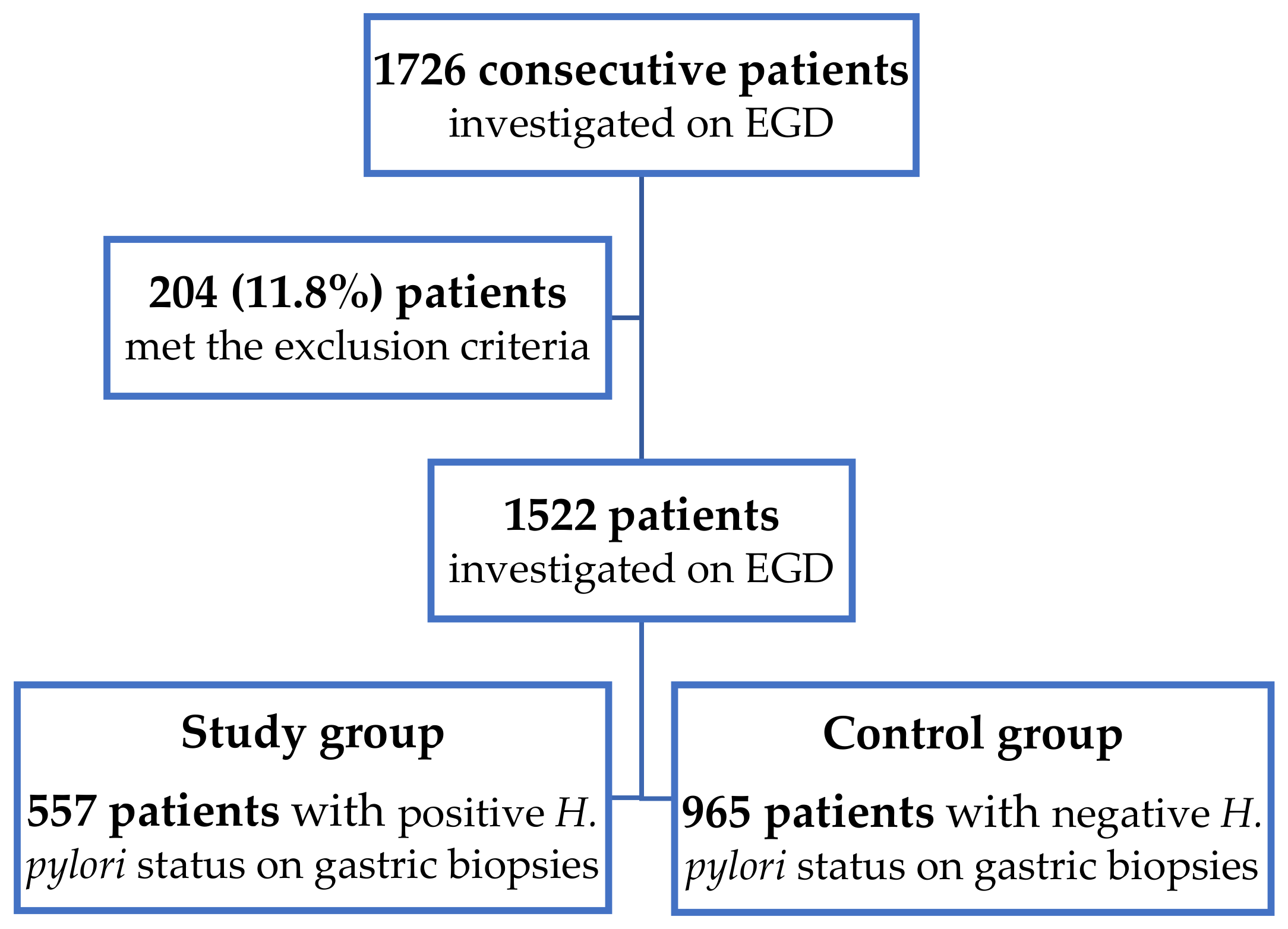
Scheme 1. Patients’ selection.
Life 12 01789 sch001
Figure 1. (a) Histologic aspects in Helicobacter pylori gastritis, Hematoxylin and Eosin staining; magnification, ×40. (b) Positive Giemsa stain for Helicobacter pylori microorganisms, magnification, ×40. (c) Immunohistochemical staining for Helicobacter pylori with numerous positive bacteria attached to the luminal surface of the foveolar cells; magnification, ×40.
Life 12 01789 g001aLife 12 01789 g001b
Table 1. Group differences between demographic and clinical characteristics.
ParameterStudy Group
(Positive H. pylori Status)
N = 557
Control Group
(Negative H. pylori Status)
N = 965
p-Value *OR95% CI
n%n%
Male gender29352.646347.90.06271.220.99 to 1.50
Age > 50 years42676.477179.80.11960.810.63 to 1.05
Endoscopic and histologic findings
Reflux esophagitis11921.320421.10.941.010.78 to 1.30
Severe endoscopic lesions13023.315616.1<0.0011.571.21 to 2.04
Biliary reflux19535.034235.40.910.980.78 to 1.22
Hiatal hernia16429.431232.30.250.870.69 to 1.09
Premalignant gastric lesions a21338.237839.10.820.970.78 to 1.20
Comorbidities
Cardiovascular diseases22540.340141.50.660.950.77 to 1.17
Cerebrovascular diseases386.8383.90.011.781.12 to 2.83
Chronic respiratory diseases8314.916416.90.310.850.64 to 1.14
Diabetes mellitus10518.815916.40.261.170.89 to 1.54
Anemia11019.722623.40.100.800.62 to 1.04
Dyslipidemia 25345.438740.10.041.241.00 to 1.53
Drug consumption
Antiplatelet drugs21338.232633.70.081.210.97 to 1.50
Anticoagulants6712.013614.00.270.830.60 to 1.14
NSAIDs b10418.612212.6<0.011.581.19 to 2.11
PPIs c20737.149050.7<0.00010.570.46 to 0.70
Symptoms
Epigastric pain29853.550352.10.631.050.85 to 1.30
Heartburn15026.924625.40.541.070.85 to 1.36
Regurgitation376.6545.50.431.200.77 to 1.84
Nausea/Vomiting10719.215616.10.031.341.02 to 1.75
Bloating13023.320621.30.361.120.87 to 1.44
Social behaviour
Tobacco smoking d10518.813013.4<0.011.491.12 to 1.97
Alcohol consumption e14225.420120.80.041.301.01 to 1.66
a Premalignant gastric lesions: atrophic gastritis and/or intestinal metaplasia; b NSAIDs: non-steroidal anti-inflammatory drugs; c PPIs: proton-pump inhibitors; d Over 5 cigarettes/day; e More than 2 units/day, 1 unit = 10 mL pure alcohol; * Obtained from Chi-square or Fisher’s exact tests; OR: odds ratio; CI: 95% confidence interval; PPI: proton-pump inhibitors.
Table 2. Results from univariate binary logistic regression.
ParameterStatistics Zp-Value *Crude OR95% CI
Male gender1.870.061.220.99 to 1.50
Age > 50 years−1.560.110.810.63 to 1.05
Antiplatelet drugs1.750.081.210.97 to 1.50
NSAIDs a3.16<0.011.581.19 to 2.11
PPIs b−5.11<0.0010.570.46 to 0.71
Severe endoscopic lesions3.43<0.0011.571.21 to 2.04
Cerebrovascular disease2.450.011.781.12 to 2.83
Dyslipidemia2.020.041.241.00 to 1.53
Smoking c2.78<0.011.491.12 to 1.97
Alcohol consumption d2.090.031.301.01 to 1.66
Anemia−1.660.090.800.62 to 1.04
Hiatal hernia−1.170.240.870.69 to 1.09
a NSAIDs: non-steroidal anti-inflammatory drugs; b PPIs: proton-pump inhibitors; c Over 5 cigarettes/day; d More than 2 units/day, 1 unit = 10 mL pure alcohol; * Crude p values obtained from Wald’s test; Response variable: presence of Helicobacter pylori infection; Crude OR: unadjusted odds ratio; CI: 95% confidence interval.
Table 3. The multivariable logistic regression model.
Parameterb eSEp-Value *Adjusted OR95% CI
NSAIDs a0.470.15<0.011.601.18 to 2.18
PPIs b−0.600.11<0.0010.540.43 to 0.68
Severe endoscopic lesions0.500.14<0.0011.651.24 to 2.19
Dyslipidemia0.250.110.031.281.02 to 1.62
Anemia−0.390.14<0.010.670.50 to 0.89
Male gender0.140.110.201.160.92 to 1.46
Smoking c0.230.190.211.260.87 to 1.84
Alcohol consumption d−0.060.170.710.940.67 to 1.31
Cerebrovascular disease0.490.250.051.641.00 to 2.70
a NSAIDs: non-steroidal anti-inflammatory drugs; b PPIs: proton-pump inhibitors; c Over 5 cigarettes/day; d More than 2 units/day, 1 unit = 10 mL pure alcohol; e Estimated unstandardized regression coefficients; SE: standard error; * Wald’s test adjusted p-value; CI: 95% confidence interval.
Table 4. Geographic prevalence of H. pylori infection and premalignant gastric lesions (AG and IM) in gastric biopsies.
N * H. pyloriAGIMCountryAuthor
152257.7% 35.9%Romania
36826.9%57.3% ChinaZhang et al. [15]
17710 11.7%USHuang et al. [16]
4050 13.8%TurkishOlmez et al. [17]
58580.2%12.6%15.2%IranEsmaeilzadeh et al. [18]
* N: number of patients included in the study.
Publisher’s Note: MDPI stays neutral with regard to jurisdictional claims in published maps and institutional affiliations.