Review

Heat Shock Protein 70 and 90 Family in Prostate Cancer

1
Department of Urology, Zhongnan Hospital of Wuhan University, Wuhan 430000, China
2
Department of Surgery and Biomedical Sciences, Cooper Medical School of Rowan University, Camden, NJ 08028, USA
*
Author to whom correspondence should be addressed.
These authors contributed equally to this work.
Life 2022, 12(10), 1489; https://doi.org/10.3390/life12101489
Submission received: 20 August 2022 / Revised: 19 September 2022 / Accepted: 21 September 2022 / Published: 26 September 2022
(This article belongs to the Special Issue Heat Shock Proteins in Cancer Treatment)

Abstract

:
Prostate cancer (PCa) is the second most frequent cancer that affects aging men worldwide. However, its exact pathogenesis has not been fully elucidated. The heat shock protein (HSP) family has cell-protective properties that may promote tumor growth and protect cancer cells from death. On a cellular level, HSP molecules have a strong relationship with multiple important biological processes, such as cell differentiation, epithelial–mesenchymal transition (EMT), and fibrosis. Because of the facilitation of HSP family molecules on tumorigenesis, a number of agents and inhibitors are being developed with potent antitumor effects whose target site is the critical structure of HSP molecules. Among all target molecules, HSP70 family and HSP90 are two groups that have been well studied, and therefore, the development of their inhibitors makes great progress. Only a small number of agents, however, have been clinically tested in recruited patients. As a result, more clinical studies are warranted for the establishment of the relationship between the HSP70 family, alongside the HSP90 molecule, and prostate cancer treatment.

1. Introduction

Prostate cancer (PCa) is the second most frequently diagnosed cancer in men worldwide, with an incidence of 14.1% and a mortality of 6.8% in 2020 [1]. The incidence rate of PCa has been increasing in the vast majority of countries over recent decades, while death rates have varied greatly worldwide (up to 10-fold) [2]. Despite its high incidence and mortality, the complexity of PCa pathogenesis makes it difficult to offer a radical treatment for PCa patients. Growing evidence has been published to elucidate its pathogenesis associating with androgen/androgen receptor (AR), estrogen, genetic changes, alterations of signal pathways, inflammation and infections, leptin and so on [2]. More mechanistic studies are still necessary.
Heat shock proteins (HSPs) are conserved molecular chaperones that have cytoprotective properties and are upregulated in response to multiple pathophysiological stresses induced by extensive stimulations, such as high temperature, hypoxia and infectious agents (bacterial and viral) [3]. HSPs are categorized into six subfamilies based on the molecular weight: HSP110, HSP90, HSP70, HSP40, small HSPs, as well as chaperonin families [4]. Among all these HSPs, HSP70s (the HSP70 family) and HSP90s (the HSP90 family) are the most two well-studied molecular families with multiple functions. HSP70s are composed of 13 members principally including inducible HSP70 (HSP72 or HSPA1), constitutive heat shock homologous protein 70 (HSC70), glucose-regulated protein 78 (GRP78) and mortalin (GRP75) [5]. HSP90s consist of four members: two in cytosol (HSP90AA1 and HSP90AB1), one in endoplasmic reticulum (ER) (GRP94, also called HSP90B1) and one in mitochondria (TRAP1) [5,6]. The HSP70 and HSP90 family members share the similar regulatory pattern that both require co-chaperones to regulate their functional cycles [7]. These two molecular families are thought to serve as housekeeping genes with basic expression levels under normal conditions; upon exposure to the external stress factors, they are overexpressed to fight against the stressed response. Accumulating evidence has paid attention to the involvement of HSP70 and HSP90 molecular family in multiple biological processes related to tumorigenesis, such as tumor growth, invasion, metastasis. On a cellular level, HSP70s and HSP90s are involved in chaperone activity, protein folding, cell survival and proliferation, cell apoptosis, autophagy, cell differentiation, epithelial–mesenchymal transition (EMT), fibrosis, DNA repair, etc. [8,9,10,11,12] (Figure 1), contributing to the initiation, development and progression of prostate cancer. Based on the functions of HSP70s and HSP90s in tumorigenesis, a number of agents targeting the critical structure of two molecular families have been successfully developed. These developed agents are divided into three categories: molecular inhibitors, aptamers and immune biologics: antibodies and vaccines. However, only a small number of agents have been subjected to the clinical trial with only three agents tested in recruited PCa patients. In this review, we focus on the important role of HSP70s and HSP90s in PCa pathogenesis. We also highlight the potential value of two molecular families as therapeutic targets in PCa treatment.

2. Prostate Cancer

The malignant transformation of the prostate follows a multi-step process, first prostatic intraepithelial neoplasia (PIN), then localized prostate cancer, followed by locally infiltrated advanced prostate adenocarcinoma, and finally leading to metastatic prostate cancer [13]. The etiology of prostate cancer is complicated and has not been fully elucidated. Known etiologies of prostate cancer include but are not limited to androgen/androgen receptor (AR), EMT, genetics, and pathway alterations.
Androgens regulate the development, maturation, and maintenance of the prostate and affect the proliferation and differentiation of the luminal epithelium. Androgen exposure plays an important role in the occurrence and maintenance of prostate cancer. AR is expressed in almost all primary and metastatic PCa, most of which initially respond to androgen deprivation. Androgen deprivation can improve the prognosis of prostate cancer patients, enabling reduced metastasis and prolonged survival [2]. Thompson and his colleagues treated subjects with finasteride (one of the 5α-reductase inhibitors (5ARI)) and placebo, observing that the incidence rate of prostate cancer was significantly lower in the experimental group than in the control group. 5ARI can reduce the incidence rate of prostate cancer by 24.8% by inhibiting the conversion of testosterone (T) into more effective dihydrotestosterone (DHT) [14].
Normally, the epithelium is a monolayer tightly held by adhesive proteins, and this immobilization prevents movement of cells from the monolayer [15]. In the course of PCa development, epithelial cells are thought to undergo EMT process. It is shown that epithelial cell markers (such as E-cadherin and ocludins) are downregulated, resulting in loss of cell adhesion, while mesenchymal markers (such as vimentin and N-cadherin) are upregulated, thereby allowing tumor cells to migrate or transfer to different organs [16]. Furthermore, growth factors and cytokines, such as transforming growth factors β (TGF- β) [17], epidermal growth factor (EGF) [18], and insulin-like growth factor (IGF) [19], as well as signaling pathways, such as mitogen activated protein kinase (MAPK) and phosphatidylinositol 3-kinase (PI3K) [20], are able to regulate the expression of E-cadherin, N-cadherin and ZEB1 and therefore influence EMT levels, which ultimately affects the progression of prostate cancer. This indicates that EMT is essential in the occurrence and metastasis of prostate cancer.
A large number of studies have shown that genetics plays a critical role in the initiation and development of prostate cancer. In the mid-20th century, Woolf et al. [21] published a report on family aggregation for the first time. This observation was supported by subsequent case–control and cohort studies [22]. Zeegers et al. [23] conducted a meta-analysis on 33 epidemiological studies of prostate cancer and found that relative risk was 2.53 (95% confidence interval, 2.24–2.85) for first-degree family members. The relative risk increases with the number of affected family members, degree of relationship, and age. In addition, the alterations of gene expression are also considered to be related to the development of prostate cancer, such as HOXB13, BRCA1, BRCA2, as well as our protagonists HSP90 and HSP70 [24,25,26,27]. In an in vitro experiment, HOXB13 and MEIS1 interact to regulate the proliferation and apoptosis of multiple prostate cancer cell lines (DU145, E006AAT, PC3, CWR22RV1, LNCaP, LNCaP C4-2, and VCap) [28], which is believed to regulate the development of prostate cancer. Early studies showed that BRCA1 and BRCA2 mutations have an increased risk of prostate cancer. It is estimated that BRCA1 mutations increase prostate cancer risk by 1.8 to 3.5 times and BRCA2 by 4.6 to 8.6 times. BRCA-related prostate cancer is more likely to metastasize and deteriorate in the late stage [29,30]. As for HSP70 and HSP90 molecules, we will focus on them in the following section.

3. HSP Family

3.1. HSP70 Family

As the most important member in the whole HSP family, HSP70s are upregulated in response to various cellular stresses and able to protect cells from fatal damage [31,32]. HSP70 family molecules have been shown to be involved in cell apoptosis, proliferation, and differentiation. Human HSP70s are widely expressed in a wide range of cell components (such as ER, proteasome, ribosome, mitochondria, and lysosomal membrane). It is believed that the expression of HSP70s is increased in many cancers, such as PCa, breast cancer, which may be associated with the tumorigenesis mechanism [33]. In patients with PCa, serum levels of HSP70s were significantly higher than in patients without PCa [34]. In addition, castration resistant prostate cancer (CRPC) is associated with increased reliance on HSP70s [35], and GRP78 expression is significantly elevated in metastatic CRPC compared with localized prostate cancer [36]. Moreover, GRP75 expression correlates with increased risk of high-grade prostate adenocarcinoma [37]. However, a report published in 2000 indicated that the expression of HSP70 molecules was unaltered in early prostate cancers but was reduced in morphologically advanced cancers compared with non-neoplastic prostate epithelium [38]. These findings suggest that HSP70s may serve as a diagnostic indicator and prognostic indicator for PCa.

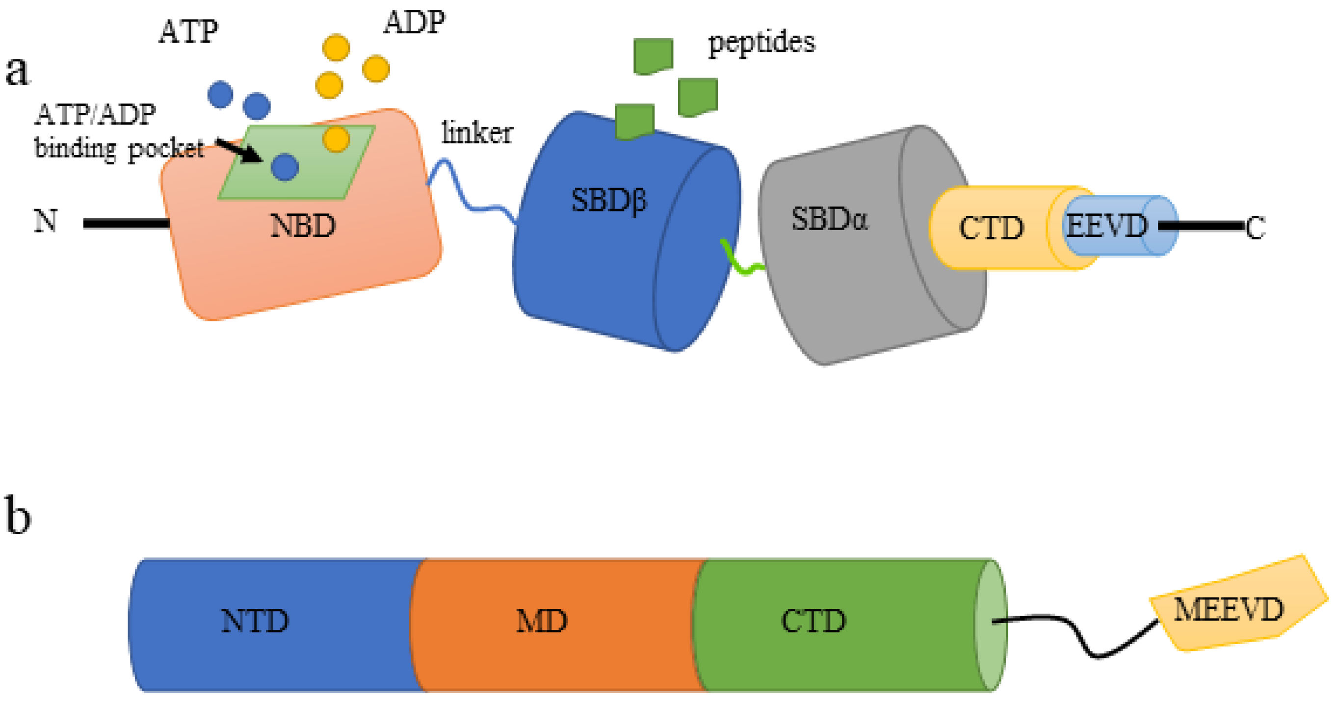
3.1.1. Structure of HSP70

The HSP70 family is highly conserved in evolution and consists of two major domains. One is the N-terminal nucleotide-binding domain (NBD) responsible for ATPase activity, and the other is the C-terminal substrate-binding domain (SBD) required for peptide binding. The two domains are connected by a linker (Figure 2a). The N-terminal NBD provides ATP/ADP pockets for ATP binding, which is critical for the ATPase reaction that is required for protein folding and release [8]. HSP70 binding protein 1(HSPBP1) and Bcl2-associated athanogene 1 (BAG-1), the functional orthologous nucleotide exchange factors of HSP70s, catalyze the release of ADP from HSP70s while inducing different conformational changes in the ATPase domain of HSP70s. An appropriate exchange rate of ADP/ATP is crucial for the chaperone dependent protein folding process [39]. SBD is further divided into two subdomains, namely SBDα and SBDβ, and followed by a C-terminal domain (CTD) that ends with an EEVD motif. The subdomain SBD β is a peptide binding capsule with which peptides can be bound as substrates. The binding and dissociation of substrates are necessary for HSP70 family members to fold substrates. Allosteric effects between functional domains modulate the ATPase cycle, in turn, polypeptide uptake and release by HSP70s [8,40].

3.1.2. HSP70s and AR

HSP70s can maintain the androgen receptor (AR) inactive phase [41]. These chaperones are released in the presence of androgen, prompting transactivation and receptor clustering. Previous studies showed that HSP70s, assisted by its co-chaperone HSP40, are able to recognize a region of the AR named N-terminal domain (NTD) including the FQNLF motif. This motif activates AR by interacting with the AR ligand binding domain (LBD). This suggests that HSP70s and the LBD compete for the FQNLF motif and in turn regulate AR activation [41]. In addition, HSP70s are molecular chaperones for AR variants lacking the LBD domain, ensuring their stability and function in cooperation with HSP40 molecule, which is disadvantageous in the treatment of CRPC patients [42]. These findings suggest that inhibition of HSP70s may suppress the promotive effects of androgens on PCa. Dong et al. demonstrated that the HSP70 molecule is a cofactor of the N-terminal domain (NTD) of AR in prostate cancer cells. HSP70 inhibition by siRNA significantly downregulated the expression of endogenous AR and concomitantly suppressed the transcriptional activity of AR. This suggests that HSP70s play an important role in AR activation [24]. Proteostasis is a potential mechanism that contributes to cancer cell survival and drug resistance. The proteostasis of AR/AR-V7 can be maintained by the interaction of STUB1, a ubiquitin ligase, with HSP70. STUB1 dissociates AR/AR-V7 from HSP70, leading to AR/AR-v7 ubiquitination and degradation. Inhibition of HSP70 significantly inhibits prostate tumor growth and improves anti androgen therapy in CRPC via AR/AR-V7 inhibition. Clinically, HSP70 expression is upregulated and correlates with AR/AR-V7 levels in high Gleason score PCa, suggesting that HSP70 may be a predictor of androgen dependent prostate cancer [43]. Moreover, the dynamic composition of the AR folding complex and AR function are influenced by BAG-1M and HSPBP1. As HSPBP1 and BAG-1 protein levels increase, AR function is inhibited [39].

3.1.3. HSP70s and Cell Apoptosis and Proliferation

In mammals, alterations in HSP70s levels significantly affect cell apoptosis and proliferation. Mammalian sterile 20-like kinase 1 (Mst1) is a ubiquitously expressed serine/threonine kinase whose activation leads to apoptosis. HSP70 overexpression mediated the degradation of Mst1in the proteasome-dependent pathway, inhibiting the proapoptotic effect of Mst1, which in turn suppresses the apoptosis in two prostate cancer cell lines: LNCaP and DU145 [44]. A study by Gibbons et al. [45] also showed that HSP72, a member of the HSP70 family, could inhibit apoptosis of prostate tumor cells through the Mcl-1, Bcl-2, Bcl-X (L), and glutathione-S-transferase (GST) pathways. Jones et al. found that HSP72 directly inhibited apoptosis in a dose-dependent manner. Depletion of HSP70 using antisense oligonucleotides against HSP70 mRNA or the bioflavonoid drug quercetin can lead to tumor cell PC-3 apoptosis in the absence of stress [46]. HSP70s bind and fold newly synthesized cyclinD1 and are involved in the assembly of cyclinD1/cyclin-dependent kinase 4 (CDK4). CyclinD1 is amplified and overexpressed in many cancers. Cyclin D1/CDK4/6 holoenzyme complex is stimulated by the mitotic signal cascade to accelerate the proliferation of cancer cells [47].

3.1.4. HSP70s and EMT, Migration, Invasion, and Metastasis

Previous studies have confirmed that HSP70s play a key role in tumor EMT, invasion, migration, and metastasis in vitro, including but not limited to lung cancer [48], colorectal cancer [49], glioblastoma [50], breast cancer [51], and Pca [52,53]. Cultrara et al. knocked down GRP78 in PC3 cells, which in turn reduced the expression of E-cadherin (E-cad) significantly, and then affected the EMT process of tumor cells [53]. Teng et al. showed that decreased expression of HSP70 and 90 family members in prostate cancer cells leads to loss of invasion. This effect is mediated in part by controlling the critical invasion-promoting capacity of the WASF3 protein [52].

3.2. HSP90 Family

HSP90s are highly conserved in biological evolution with an approximately 90 kDa molecular weight, accounting for approximately 1–2% of the total protein in mammalian cells. They are molecular chaperones that promote de novo synthesis and misfolded protein folding, thereby counteracting their aggregation [9,54]. HSP90s are involved in essential cellular processes and regulatory pathways such as apoptosis, cell cycle control, protein folding and degradation, and signaling events [9]. Similar to HSP70s, HSP90s are also highly expressed in PCa and promotes the malignant progression and metastasis of PCa, which could assist in diagnosing PCa and judging patient prognosis [55,56].

3.2.1. Structure of HSP90s

HSP90s function in vivo as a homodimer [57]. The HSP90 monomer consists of three highly conserved domains: (a) the amino terminal domain (NTD), which mediates binding to ATP; (b) the middle domain (MD), involved in ATP hydrolysis and HSP90s binding to client proteins; (c) the C-terminal domain (CTD), responsible for the formation of dimers (Figure 2b). The CTD also contains a C-terminal met-Glu-Glu-Val-Asp (MEEVD) motif, which is critical for interactions with co-chaperones containing tetratricopeptide repeat (TPR) domains [58]. Similar to the HSP70s, NTD and MD are connected by a long, flexible, and stretchable charged linker. This linker could regulate the binding of NTD to MD, which in turn affects the function of HSP90s [58].

3.2.2. HSP90s and AR

HSP90s form a complex with AR in the cytoplasm to maintain the natural state of AR in cells, thus stabilizing AR before ligand binding. Inhibition of HSP90s leads to AR degradation and its cytoplasmic accumulation [59]. Vanaja et al. [60] treated prostate cancer cells LNCaP for 24 h with the HSP90 inhibitor geldamycin (GA) and found that GA reduced the androgen-induced AR protein level to 15%. At the same time, the expression of androgen upregulated genes such as immunophilin protein FKBP51 and prostate specific antigen (PSA) also decreased significantly. Furthermore, the maintenance of AR high affinity hormone binding conformation by HSP90s was confirmed in vitro by Fang and his colleague [61]. HSP90s push the equilibrium toward the high affinity hormone binding conformation. In this state, the binding of androgens leads to further structural changes that promote AR activation.

3.2.3. HSP90s and Cell Apoptosis and Proliferation

HSP90s are widely involved in biological processes such as apoptosis, proliferation, and cycle regulation through protein folding. AC245100.4 is a LncRNA present in the cytoplasm and its association with prostate cancer was reported in previous studies [62]. Cui et al. [63] showed that AC245100.4 binds to HSP90s to alter its chaperone function by changing the levels of client proteins. When overexpressed AC245100.4, the levels of IKKα, p-IκB, p-P65, and CyclinD1 were increased, and the proliferation ability of prostate cancer cells was significantly increased. Additionally, GA could inhibit the pro-proliferative effect of AC245100.4. This suggests that AC245100.4 binding to HSP90s promotes prostate cancer progression by NF κB pathways. In addition, HSP90 can also regulate the proliferation and apoptosis of prostate cancer cells through AR, ERBB2, Akt, c-RAF, survivin, EGFR, IGFR-1, STAT3, ERK, CDK-4, and CDK-6 signaling pathways [64]. In an in vivo experiment, TRAP1 transgenic mice showed an accelerated incidence of invasive prostate adenocarcinoma characterized by increased cell proliferation and reduced cell apoptosis, and conversely, homozygous deletion of TRAP1 delayed prostate tumorigenesis in mice without affecting hyperplasia or prostate intraepithelial neoplasia [65]. This evidence suggests that HSP90s can participate in the development of prostate cancer by regulating cell proliferation and cell apoptosis.

3.2.4. HSP90s and EMT, Migration, Invasion, and Metastasis

In eukaryotes, HSP90s are associated with EMT, invasion, migration, and metastasis of many tumors, such as oral cancer [66], breast cancer [67], and prostate cancer [52,68,69]. Tumor secreted extracellular HSP90 (eHSP90) initiates EMT in prostate cancer cells, promoting prostate tumor growth and invasion in vivo. Mechanistically, eHSP90 initiates sustained activation of MEK/ERK, a key signal that promotes transcriptional upregulation of EZH2 (a methyltransferase closely related to EMT) and recruitment to the E-cadherin promoter. The eHSP90-EZH2 pathway orchestrates an extended series of EMT-related events, including tumor cell migration and metastasis [68]. Additionally, eHSP90 could enhance cell motility and shifts cell morphology toward a mesenchymal phenotype in an ERK and matrix metalloproteinase-2/9-dependent manner. Conversely, inhibition of eHSP90 arrested PCa migration and shifted cell morphology towards an epithelial phenotype [69].

4. HSP70 Family in Prostate Cancer Treatment

In addition to hormonal therapy that removes androgen (androgen deprivation treatment (ADT)) or blocks androgen action, another important medical therapy for prostate cancer is chemotherapy mainly used for castration-resistant prostate cancer (CRPC). Radical prostatectomy remains the gold standard for PCa treatment, as drug therapies are not a radical cure even if the tumor is localized within the prostate capsule [2]. Attempts have been made over the recent decades to explore novel biomarkers of prostate cancer and develop them into novel drugs in avoidance of surgical trauma. As an important biomarker for multiple cancers, the HSP70 family has been considered as one of the promising targets that can be developed into drugs for cancer treatment. In this section, we review the agents targeting HSP70 family molecules that may be used for PCa treatment in future.

4.1. Small Molecular Inhibitors

Small molecular inhibitors against different HSP70 family molecules have not been successfully developed for clinical use, with only one agent being subjected to clinical trial. The first well-studied inhibitor, 2-phenylethynesulfonamide (PES) or pifithrin-µ, has been documented to bind to C-terminal SBD of HSP70 and therefore disrupt the association of HSP70s with its co-chaperone HSP40 and other client proteins such as proapoptotic ones APAF-1 and p53 [70]. This interaction of PES with HSP70 molecule induces either autophagic cell death or apoptotic, caspase-dependent, cell death, which depends on different cell types [71,72]. PES is such a potent antitumor agent in vitro and in vivo that this agent shows potent cytotoxicity in multiple cancer cells including LNCaP95 prostate cancer cells, especially when in combination with other inhibitors or medications [73,74,75].
Inhibitors that target N-terminal ATP-binding domain of HSP70s include 15-deoxyspergualin (15-DSG), VER-155008, MKT-077 (the only one subjected to clinical trial) and so on. These agents inhibit HSP70s function by attacking ATP-binding domain of HSP70 and disrupting its ATPase activity, thus blocking cellular proliferation or stimulating cell apoptosis [76,77]. 15-DSG is a natural immunosuppressive agent with a mild effect on cancerous cells compared with other inhibitors [78]. More efficacious agents are MAL3-101, the second-generation inhibitor, and its derivatives [76]. These inhibitors are minimally effective when administered alone for clinical use, but when in combination with other agents, such as HSP90 inhibitor 17-AAG and PS-341(bortezomib), they are shown to have improved efficacy [79]. VER-155008 is an adenosine-derived compound that attacks N-terminal ATP-binding domain of HSP70 family molecules. In vitro studies have demonstrated that VER-155008 is able to induce either caspase-dependent cell death or non-caspase-dependent cell death [77]. The cytotoxic effect of VER-155008 can be improved when administered in combination with HSP90 inhibitors including NVP-AUY922 [80]. The rhodacyanine dye analog MKT-077 is the only agent that has been clinically tested. MKT-077 is thought to serve as a metabolic poison in the mitochondria of cancerous cells where it induces G1 arrest and cell apoptosis [81]. However, the clinical trial of MKT-077 was halted due to the observation of nephrotoxicity, principally renal magnesium wasting. Park and his team recently reported a HSP70 inhibitor apoptosol (AZ) that inhibits the ATPase activity of HSP70s on lysosomal membrane and triggers lysosomal membrane permeabilization (LMP), ultimately inducing lysosome-mediated apoptosis of cancer cells [82,83]. This inhibitor may have higher specificity than above-mentioned HSP70 inhibitors as lysosomal HSP70s are rarely found in normal cells. The derivative of AZ, Az-TPP-O3, was also observed to have potent proapoptotic effect on cancer cells. Az-TPP-O3 primarily attacks the mitochondrial HSP70 molecule mortalin and blocks the interaction of mortalin and p53, which drives a process called mitochondrial outer membrane permeabilization (MOMP) and leads to mitochondria-mediated apoptosis due to caspase activation [82]. Unfortunately, these two unique inhibitors have not been tested in cancer patients, much less in prostate cancer patients. As a result, though all these inhibitors theoretically have strong antitumor activity, temporarily, none of them are appropriate to be developed for clinical use.

4.2. Immunotherapeutic Approaches

A promising strategy to inhibit the expression of HSP70s is the application of monoclonal antibodies (mAb) [79]. The mAb targets HSP70s more accurately with less side-effects than small molecular inhibitors due to its high antigen specificity. A mAb called cmHsp70.1 has completed phase I clinical trial and is being subjected to phase II trial. This monoclonal antibody can recognize the extracellular motif—TKDNNLLGRFELSG (TDK) of membrane HSP70 molecule [84]. In colon cancer, this antibody has been shown to reduce the weight and volume of tumors and promote patients’ survival when administered alone [84].
The development of HSP70 vaccines provides another immunotherapeutic approach for cancers. Several vaccines composed of disease specific epitopes and HSP70 DNA have been successfully developed and subjected to clinical trials. For example, a vaccine called pNGVL4a-Sig/E7(detox)/HSP70 DNA was clinically tested in patients with cervical intraepithelial neoplasia [85]. Another clinical trial has tested the efficacy and toxicity of another vaccine made with HSP70s in chronic myelogenous leukemia [79]. Moreover, a novel immunotherapeutic approach focusing on natural killer (NK) cells based adoptive immunotherapy is being clinically evaluated in which Hsp70-peptide TKD/IL-2 activated, autologous NK cells are employed [79]. To date, no clinical studies of HSP70-related immunotherapy have been performed in patients with prostate cancer. This must be a promising area in the exploration of PCa treatment.

4.3. A Novel Tool with High Specificity: Aptamers

In addition to small molecular inhibitors and antibodies, recent findings have uncovered the inhibitory effect of aptamers, a new category of targeting agents, on functions of HSP70 family molecules. Aptamers (i.e., DNA, RNA aptamers), alongside peptide aptamers, are able to bind to molecules of interest with high specificity and high affinity, exhibiting a functional similarity to antibodies. The differences between two biologics are that aptamers have relatively smaller molecular weight with decreased immunogenicity. Similar to aforesaid small molecular inhibitors, the target site of aptamers is also the NBD or SBD of HSP70 family molecules and the binding of aptamers to the target site can impair the function of HSP70s, as a result of which cancerous cells are susceptible to cell death. Screened from aptamer libraries, A17 has been considered as the most potent aptamer until now. In HeLa cells, A17 is shown to promote cell death (mainly apoptosis) and exhibit its antitumor properties in vivo when combined with cisplatin, but have no such effects when administered alone [86]. A mechanistic study has revealed that this proapoptotic effect of A17 comes from its attack on the NBD of HSP70s that disrupts HSP70 function [86]. Furthermore, the A17 aptamer can inactivate the HSP70 chaperone, but not the one of HSP90 or HSC70 [87]. All above-mentioned agents were included in a table (Table 1).

5. HSP90 Family in Prostate Cancer Treatment

As the most well-studied family of HSPs, HSP90 molecules and their client proteins have been shown to play fundamental roles in tumorigenesis [11]. Targeting HSP90s can lead to the proteasomal degradation of their clients including AR and ultimately influence the signal transduction related to the initiation, development and progression of cancers. This simultaneous inhibitory effects of targeting HSP90 family molecules on AR and other client proteins render HSP90 blockade an attractive therapeutic strategy for prostate cancer [64].

5.1. Natural HSP90 Inhibitors and Their Derivatives

The N-terminal of HSP90 molecules responsible for ATP binding is one of the binding sites for most natural HSP90 inhibitors. These molecular inhibitors ubiquitously have higher affinity for the binding site than natural nucleotides, which are able to inhibit its cycling between ATP- and ADP- bound conformations and impair the functions of HSP90 family molecules [79]. The targeting therapy using HSP90 inhibitors for cancer treatment begins with geldanamycin (GM), an agent derived from Streptomyces hygroscopicus with potent antitumor activity [88]. However, its phase I clinical trial was terminated due to its structural instability and hepatotoxicity. Radicicol (RD), another natural inhibitor derived from Monosporium bonorden, exhibits powerful in vitro antitumor properties by disrupting the critical ATP-binding site of HSP90s, but its structural instability makes it ineffective in vivo. Different from GM and RD, the natural inhibitor Novobiocin destabilizes the client proteins of HSP90 by binding to C-terminal domain of HSP90 and therefore inhibits cancer cell growth [89,90,91]. Importantly, targeting HSP90 C-terminus is one of the approaches to avoid the compensatory heat shock response, induced by targeting the HSP90 ATP-binding site, that stimulates the expression of multiple HSPs, such as HSP90, HSP70, clusterin. Unfortunately, the efficacy and safety of Novobiocin have not been rigorously tested in vivo.
The first generation GM derivatives are 17-AAG (also called tanespimycin or 17-allylamino-17-demethoxygeldanamycin) and 17-DMAG (also called as alvespimycin or 17-dimethylaminoethylamino-17-demethoxygeldanamycin). 17-AAG has been evaluated in the phase II clinical trial, but its poor water solubility and oral bioavailability restricts its development [92]. 17-DMAG shows improved solubility compared with 17-AAG; however, this agent has dose-limiting toxicities: peripheral neuropathy and renal dysfunction [93]. The next generation GM derivative, IPI-504 (retaspimycin), has been shown to have minimal efficacy and unacceptable toxicity in a phase II clinical study in CRPC patients following IPI-504 treatment [94]. This agent was therefore eliminated from therapeutic choices of prostate cancers.
The natural derivatives of RD include NVP-AUY922 and AT13387 which also attack the ATP-binding pocket of HSP90 molecules. NVP-AUY922 is the first generation RD derivative and has strong proapoptotic activity in vitro and powerful antitumor properties in an ex vivo model of prostate cancer. Nevertheless, NVP-AUY922 has not been clinically tested in patients with metastatic CRPC (mCRPC). The second generation derivative, AT13387, shows in vitro and in vivo antitumor activities against prostate cancer, with long duration of action as one of its most important characteristics [64,95]. Theoretically, the use of longer-acting HSP90 inhibitors is more likely to maintain antitumor efficacy with less systemic exposure and side effects [95]. AT13387 has been involved in multiple phase I or II clinical trials, some of which have been completed (NCT01294202, NCT01685268, NCT00878423, NCT01246102) [79]. In addition, a phase I/II clinical trial is in progress to compare the efficacy when administered alone or combined with abiraterone acetate [64]. Recent studies reported a chalcone derivative, SU086, that has potent proapoptotic effect on multiple PCa cell lines including AR-positive cell (C4-2) and AR-negative cell lines (DU145 and PC-3) [96]. This supports a notion that SU086 is active in PCa cells independence of AR status. Mechanistically, SU086 influences the glycolysis process of prostate cancer cells by impairing the function of HSP90 [97]. The clinical value of this inhibitor still needs validation by a number of clinical studies.

5.2. Synthetic HSP90 Inhibitors

Similar to natural inhibitors, the HSP90 N-terminus is also the important target site of synthetic HSP90 inhibitors, among which purine based compounds are one of the choices for drug development [79]. PU-H71 and PU-DZ8 are structurally analogous to ADP but have higher affinity for HSP90. This competitive effect between these two compounds and ADP hinders the binding of ADP and then impairs HSP90 functions. CNF-2024 is also a purine-scaffold compound with high oral bioavailability and has been shown to induce lymphoma cell death [98]. To date, no clinical studies have been performed to evaluate the efficacy and safety of PU series in PCa patients, therefore more clinical studies are warranted.
The screening of ATP-binding proteins using the ATP-affinity column discovered the 2-aminobenzamide derivative SNX-5422 (PF-04929113) [99]. This compound is an orally bioavailable prodrug of PF 04928473 (SNX-2112), a selective HSP90 inhibitor, and has been subjected to phase I clinical trials in patients with solid tumors including PCa as well as hematological malignancies such as lymphomas and chronic lymphocytic leukemia (CLL) [100,101,102]. Due to its ocular toxicity observed in animal models and in a phase I study, the development of SNX-5422 was terminated and waited for further evaluation [102]. Ganetespib (STA-9090), one of the RD derivatives, is a chemically synthetic compound with potent activity against prostate cancer xenografts. A phase II clinical trial, however, shows a negligible efficacy of ganetespib in 17 patients with mCRPC (only 2 patients had progression-free survival (PFS) of more than 4 months) [103]. As a compound targeting HSP90 C-terminus analogous to novobiocin, its analogue KU174 also has powerful antitumor activity against prostate cancer in vitro and in vivo without induction of the heat shock response [104]. Unfortunately, this compound has not been clinically evaluated to date. All above-mentioned inhibitors we mentioned were included in Table 2.

6. Conclusions

In aggregate, blocking HSP70 and HSP90 family is an emerging therapeutic target in cancer. Inhibitors and protein aptamers may be developed into various chemotherapeutic drugs while both monoclonal antibodies and vaccines are the critical parts of immunotherapies. The cytotoxicity of these agents make them able to potently reduce the size of tumors, however, only a small part of agents have been clinically tested in PCa patients showing therapeutic effects on prostate cancer. In addition, the side effect caused by these inhibitors is always not a negligible problem. Therefore, a number of clinical studies are still necessary to further evaluate the efficacy and safety of present HSP inhibitors and simultaneously the development novel agents targeting HSP70s and HSP90s will become increasingly important in the next few years.

Author Contributions

Conceptualization, X.F. and J.L.; writing—original draft preparation, X.F., J.L., X.Y. and M.E.D.; writing—review and editing, X.F. and X.Z. All authors have read and agreed to the published version of the manuscript.

Funding

This research received no external funding.

Institutional Review Board Statement

Not applicable.

Informed Consent Statement

Not applicable.

Data Availability Statement

Not applicable.

Conflicts of Interest

The authors declare no conflict of interest.

References

  1. Sung, H.; Ferlay, J.; Siegel, R.L.; Laversanne, M.; Soerjomataram, I.; Jemal, A.; Bray, F. Global Cancer Statistics 2020: GLOBOCAN Estimates of Incidence and Mortality Worldwide for 36 Cancers in 185 Countries. CA A Cancer J. Clin. 2021, 71, 209–249. [Google Scholar] [CrossRef] [PubMed]
  2. Stein, R. Campbell Walsh Wein Urology, Chapter107: Epidemiology, Etiology, and Prevention of Prostate Cancer, PART XIV, VOLUME 3. Aktuelle Urol. 2021, 3, 25. [Google Scholar] [CrossRef]
  3. Gething, M.J.; Sambrook, J. Protein folding in the cell. Nature 1992, 355, 33–45. [Google Scholar] [CrossRef] [PubMed]
  4. Ratajczak, W.; Lubkowski, M.; Lubkowska, A. Heat Shock Proteins in Benign Prostatic Hyperplasia and Prostate Cancer. Int. J. Mol. Sci. 2022, 23, 897. [Google Scholar] [CrossRef]
  5. Kampinga, H.H.; Hageman, J.; Vos, M.J.; Kubota, H.; Tanguay, R.M.; Bruford, E.A.; Cheetham, M.E.; Chen, B.; Hightower, L.E. Guidelines for the nomenclature of the human heat shock proteins. Cell Stress Chaperones 2009, 14, 105–111. [Google Scholar] [CrossRef]
  6. Johnson, J.L. Evolution and function of diverse Hsp90 homologs and cochaperone proteins. Biochim. Biophys. Acta 2012, 1823, 607–613. [Google Scholar] [CrossRef]
  7. Albakova, Z.; Mangasarova, Y.; Albakov, A.; Gorenkova, L. HSP70 and HSP90 in Cancer: Cytosolic, Endoplasmic Reticulum and Mitochondrial Chaperones of Tumorigenesis. Front. Oncol. 2022, 12, 829520. [Google Scholar] [CrossRef]
  8. Fu, X.; Liu, H.; Liu, J.; DiSanto, M.E.; Zhang, X. The Role of Heat Shock Protein 70 Subfamily in the Hyperplastic Prostate: From Molecular Mechanisms to Therapeutic Opportunities. Cells 2022, 11, 2052. [Google Scholar] [CrossRef]
  9. Schopf, F.H.; Biebl, M.M.; Buchner, J. The HSP90 chaperone machinery. Nat. Rev. Mol. Cell Biol. 2017, 18, 345–360. [Google Scholar] [CrossRef]
  10. Milani, A.; Basirnejad, M.; Bolhassani, A. Heat-shock proteins in diagnosis and treatment: An overview of different biochemical and immunological functions. Immunotherapy 2019, 11, 215–239. [Google Scholar] [CrossRef]
  11. Calderwood, S.K. Heat shock proteins and cancer: Intracellular chaperones or extracellular signalling ligands? Philos. Trans. R. Soc. London. Ser. B Biol. Sci. 2018, 373, 20160524. [Google Scholar] [CrossRef] [PubMed]
  12. Dubrez, L.; Causse, S.; Borges Bonan, N.; Dumétier, B.; Garrido, C. Heat-shock proteins: Chaperoning DNA repair. Oncogene 2020, 39, 516–529. [Google Scholar] [CrossRef] [PubMed]
  13. Wang, G.; Zhao, D.; Spring, D.J.; DePinho, R.A. Genetics and biology of prostate cancer. Genes Dev. 2018, 32, 1105–1140. [Google Scholar] [CrossRef] [PubMed]
  14. Thompson, I.M.; Goodman, P.J.; Tangen, C.M.; Lucia, M.S.; Miller, G.J.; Ford, L.G.; Lieber, M.M.; Cespedes, R.D.; Atkins, J.N.; Lippman, S.M.; et al. The influence of finasteride on the development of prostate cancer. New Engl. J. Med. 2003, 349, 215–224. [Google Scholar] [CrossRef]
  15. Yang, J.; Weinberg, R.A. Epithelial-mesenchymal transition: At the crossroads of development and tumor metastasis. Dev. Cell 2008, 14, 818–829. [Google Scholar] [CrossRef]
  16. Nauseef, J.T.; Henry, M.D. Epithelial-to-mesenchymal transition in prostate cancer: Paradigm or puzzle? Nat. Rev. Urol. 2011, 8, 428–439. [Google Scholar] [CrossRef]
  17. Xu, J.; Lamouille, S.; Derynck, R. TGF-beta-induced epithelial to mesenchymal transition. Cell Res. 2009, 19, 156–172. [Google Scholar] [CrossRef]
  18. Cho, K.H.; Choi, M.J.; Jeong, K.J.; Kim, J.J.; Hwang, M.H.; Shin, S.C.; Park, C.G.; Lee, H.Y. A ROS/STAT3/HIF-1α signaling cascade mediates EGF-induced TWIST1 expression and prostate cancer cell invasion. Prostate 2014, 74, 528–536. [Google Scholar] [CrossRef]
  19. Graham, T.R.; Zhau, H.E.; Odero-Marah, V.A.; Osunkoya, A.O.; Kimbro, K.S.; Tighiouart, M.; Liu, T.; Simons, J.W.; O’Regan, R.M. Insulin-like growth factor-I-dependent up-regulation of ZEB1 drives epithelial-to-mesenchymal transition in human prostate cancer cells. Cancer Res. 2008, 68, 2479–2488. [Google Scholar] [CrossRef] [PubMed]
  20. Gennigens, C.; Menetrier-Caux, C.; Droz, J.P. Insulin-Like Growth Factor (IGF) family and prostate cancer. Crit. Rev. Oncol. Hematol. 2006, 58, 124–145. [Google Scholar] [CrossRef]
  21. Woolf, C.M. An investigation of the familial aspects of carcinoma of the prostate. Cancer 1960, 13, 739–744. [Google Scholar] [CrossRef]
  22. Eeles, R.A.; Dearnaley, D.P.; Ardern-Jones, A.; Shearer, R.J.; Easton, D.F.; Ford, D.; Edwards, S.; Dowe, A. Familial prostate cancer: The evidence and the Cancer Research Campaign/British Prostate Group (CRC/BPG) UK Familial Prostate Cancer Study. Br. J. Urol. 1997, 79 (Suppl. S1), 8–14. [Google Scholar] [CrossRef] [PubMed]
  23. Zeegers, M.P.; Jellema, A.; Ostrer, H. Empiric risk of prostate carcinoma for relatives of patients with prostate carcinoma: A meta-analysis. Cancer 2003, 97, 1894–1903. [Google Scholar] [CrossRef] [PubMed]
  24. Dong, J.; Wu, Z.; Wang, D.; Pascal, L.E.; Nelson, J.B.; Wipf, P.; Wang, Z. Hsp70 Binds to the Androgen Receptor N-terminal Domain and Modulates the Receptor Function in Prostate Cancer Cells. Mol. Cancer Ther. 2019, 18, 39–50. [Google Scholar] [CrossRef] [PubMed]
  25. Centenera, M.M.; Carter, S.L.; Gillis, J.L.; Marrocco-Tallarigo, D.L.; Grose, R.H.; Tilley, W.D.; Butler, L.M. Co-targeting AR and HSP90 suppresses prostate cancer cell growth and prevents resistance mechanisms. Endocr. Relat. Cancer 2015, 22, 805–818. [Google Scholar] [CrossRef] [PubMed]
  26. Castro, E.; Goh, C.; Olmos, D.; Saunders, E.; Leongamornlert, D.; Tymrakiewicz, M.; Mahmud, N.; Dadaev, T.; Govindasami, K.; Guy, M.; et al. Germline BRCA mutations are associated with higher risk of nodal involvement, distant metastasis, and poor survival outcomes in prostate cancer. J. Clin. Oncol. Off. J. Am. Soc. Clin. Oncol. 2013, 31, 1748–1757. [Google Scholar] [CrossRef]
  27. Attard, G.; Parker, C.; Eeles, R.A.; Schröder, F.; Tomlins, S.A.; Tannock, I.; Drake, C.G.; de Bono, J.S. Prostate cancer. Lancet 2016, 387, 70–82. [Google Scholar] [CrossRef]
  28. Johng, D.; Torga, G.; Ewing, C.M.; Jin, K.; Norris, J.D.; McDonnell, D.P.; Isaacs, W.B. HOXB13 interaction with MEIS1 modifies proliferation and gene expression in prostate cancer. Prostate 2019, 79, 414–424. [Google Scholar] [CrossRef]
  29. Castro, E.; Eeles, R. The role of BRCA1 and BRCA2 in prostate cancer. Asian J. Androl. 2012, 14, 409–414. [Google Scholar] [CrossRef]
  30. Eeles, R.; Goh, C.; Castro, E.; Bancroft, E.; Guy, M.; Al Olama, A.A.; Easton, D.; Kote-Jarai, Z. The genetic epidemiology of prostate cancer and its clinical implications. Nat. Rev. Urol. 2014, 11, 18–31. [Google Scholar] [CrossRef]
  31. Kumar, S.; Stokes, J., 3rd; Singh, U.P.; Scissum Gunn, K.; Acharya, A.; Manne, U.; Mishra, M. Targeting Hsp70: A possible therapy for cancer. Cancer Lett. 2016, 374, 156–166. [Google Scholar] [CrossRef] [PubMed]
  32. Ibrahim, I.M.; Abdelmalek, D.H.; Elfiky, A.A. GRP78: A cell’s response to stress. Life Sci. 2019, 226, 156–163. [Google Scholar] [CrossRef] [PubMed]
  33. Saini, J.; Sharma, P.K. Clinical, Prognostic and Therapeutic Significance of Heat Shock Proteins in Cancer. Curr. Drug Targets 2018, 19, 1478–1490. [Google Scholar] [CrossRef] [PubMed]
  34. Abe, M.; Manola, J.B.; Oh, W.K.; Parslow, D.L.; George, D.J.; Austin, C.L.; Kantoff, P.W. Plasma levels of heat shock protein 70 in patients with prostate cancer: A potential biomarker for prostate cancer. Clin. Prostate Cancer 2004, 3, 49–53. [Google Scholar] [CrossRef] [PubMed]
  35. Shkedi, A.; Taylor, I.R.; Echtenkamp, F.; Ramkumar, P.; Alshalalfa, M.; Rivera-Márquez, G.M.; Moses, M.A.; Shao, H.; Karnes, R.J.; Neckers, L.; et al. Selective vulnerabilities in the proteostasis network of castration-resistant prostate cancer. Cell Chem. Biol. 2022, 29, 490–501.e494. [Google Scholar] [CrossRef] [PubMed]
  36. Pootrakul, L.; Datar, R.H.; Shi, S.R.; Cai, J.; Hawes, D.; Groshen, S.G.; Lee, A.S.; Cote, R.J. Expression of stress response protein Grp78 is associated with the development of castration-resistant prostate cancer. Clin. Cancer Res. Off. J. Am. Assoc. Cancer Res. 2006, 12, 5987–5993. [Google Scholar] [CrossRef]
  37. Hirth, C.G.; Vasconcelos, G.R.; da Cunha, M.; Leite, C.H.B.; Dornelas, C.A. Immunoexpression of HSPA9 and CUL2 in prostatic tissue and adenocarcinoma. Ann. Diagn. Pathol. 2022, 56, 151843. [Google Scholar] [CrossRef]
  38. Cornford, P.A.; Dodson, A.R.; Parsons, K.F.; Desmond, A.D.; Woolfenden, A.; Fordham, M.; Neoptolemos, J.P.; Ke, Y.; Foster, C.S. Heat shock protein expression independently predicts clinical outcome in prostate cancer. Cancer Res. 2000, 60, 7099–7105. [Google Scholar]
  39. Knapp, R.T.; Wong, M.J.; Kollmannsberger, L.K.; Gassen, N.C.; Kretzschmar, A.; Zschocke, J.; Hafner, K.; Young, J.C.; Rein, T. Hsp70 cochaperones HspBP1 and BAG-1M differentially regulate steroid hormone receptor function. PLoS ONE 2014, 9, e85415. [Google Scholar] [CrossRef]
  40. Albakova, Z.; Armeev, G.A.; Kanevskiy, L.M.; Kovalenko, E.I.; Sapozhnikov, A.M. HSP70 Multi-Functionality in Cancer. Cells 2020, 9, 587. [Google Scholar] [CrossRef]
  41. Eftekharzadeh, B.; Banduseela, V.C.; Chiesa, G.; Martínez-Cristóbal, P.; Rauch, J.N.; Nath, S.R.; Schwarz, D.M.C.; Shao, H.; Marin-Argany, M.; Di Sanza, C.; et al. Hsp70 and Hsp40 inhibit an inter-domain interaction necessary for transcriptional activity in the androgen receptor. Nat. Commun. 2019, 10, 3562. [Google Scholar] [CrossRef] [Green Version]
  42. Moses, M.A.; Kim, Y.S.; Rivera-Marquez, G.M.; Oshima, N.; Watson, M.J.; Beebe, K.E.; Wells, C.; Lee, S.; Zuehlke, A.D.; Shao, H.; et al. Targeting the Hsp40/Hsp70 Chaperone Axis as a Novel Strategy to Treat Castration-Resistant Prostate Cancer. Cancer Res. 2018, 78, 4022–4035. [Google Scholar] [CrossRef] [PubMed]
  43. Liu, C.; Lou, W.; Yang, J.C.; Liu, L.; Armstrong, C.M.; Lombard, A.P.; Zhao, R.; Noel, O.D.V.; Tepper, C.G.; Chen, H.W.; et al. Proteostasis by STUB1/HSP70 complex controls sensitivity to androgen receptor targeted therapy in advanced prostate cancer. Nat. Commun. 2018, 9, 4700. [Google Scholar] [CrossRef] [PubMed]
  44. Ren, A.; Yan, G.; You, B.; Sun, J. Down-regulation of mammalian sterile 20-like kinase 1 by heat shock protein 70 mediates cisplatin resistance in prostate cancer cells. Cancer Res. 2008, 68, 2266–2274. [Google Scholar] [CrossRef] [PubMed]
  45. Gibbons, N.B.; Watson, R.W.; Coffey, R.N.; Brady, H.P.; Fitzpatrick, J.M. Heat-shock proteins inhibit induction of prostate cancer cell apoptosis. Prostate 2000, 45, 58–65. [Google Scholar] [CrossRef]
  46. Jones, E.L.; Zhao, M.J.; Stevenson, M.A.; Calderwood, S.K. The 70 kilodalton heat shock protein is an inhibitor of apoptosis in prostate cancer. Int. J. Hyperth. Off. J. Eur. Soc. Hyperthermic Oncol. N. Am. Hyperth. Group 2004, 20, 835–849. [Google Scholar] [CrossRef]
  47. Qie, S.; Diehl, J.A. Cyclin D degradation by E3 ligases in cancer progression and treatment. Semin. Cancer Biol. 2020, 67, 159–170. [Google Scholar] [CrossRef]
  48. Ling, X.; Wan, J.; Peng, B.; Chen, J. Hsp70 Promotes SUMO of HIF-1α and Promotes Lung Cancer Invasion and Metastasis. J. Oncol. 2021, 2021, 7873085. [Google Scholar] [CrossRef]
  49. Nikotina, A.D.; Vladimirova, S.A.; Komarova, E.Y.; Alexeev, D.; Efremov, S.; Leonova, E.; Pavlov, R.; Kartsev, V.G.; Polonik, S.G.; Margulis, B.A.; et al. Prevention of High Glucose-Mediated EMT by Inhibition of Hsp70 Chaperone. Int. J. Mol. Sci. 2021, 22, 6902. [Google Scholar] [CrossRef]
  50. Huang, P.; Guo, Y.; Zhao, Z.; Ning, W.; Wang, H.; Gu, C.; Zhang, M.; Qu, Y.; Zhang, H.; Song, Y. UBE2T promotes glioblastoma invasion and migration via stabilizing GRP78 and regulating EMT. Aging 2020, 12, 10275–10289. [Google Scholar] [CrossRef]
  51. Zhang, R.; Meng, Z.; Wu, X.; Zhang, M.; Zhang, S.; Jin, T. Mortalin promotes breast cancer malignancy. Exp. Mol. Pathol. 2021, 118, 104593. [Google Scholar] [CrossRef] [PubMed]
  52. Teng, Y.; Ngoka, L.; Mei, Y.; Lesoon, L.; Cowell, J.K. HSP90 and HSP70 proteins are essential for stabilization and activation of WASF3 metastasis-promoting protein. J. Biol. Chem. 2012, 287, 10051–10059. [Google Scholar] [CrossRef] [PubMed]
  53. Cultrara, C.N.; Kozuch, S.D.; Ramasundaram, P.; Heller, C.J.; Shah, S.; Beck, A.E.; Sabatino, D.; Zilberberg, J. GRP78 modulates cell adhesion markers in prostate Cancer and multiple myeloma cell lines. BMC Cancer 2018, 18, 1263. [Google Scholar] [CrossRef] [PubMed]
  54. Hoter, A.; El-Sabban, M.E.; Naim, H.Y. The HSP90 Family: Structure, Regulation, Function, and Implications in Health and Disease. Int. J. Mol. Sci. 2018, 19, 2560. [Google Scholar] [CrossRef] [PubMed]
  55. Leav, I.; Plescia, J.; Goel, H.L.; Li, J.; Jiang, Z.; Cohen, R.J.; Languino, L.R.; Altieri, D.C. Cytoprotective mitochondrial chaperone TRAP-1 as a novel molecular target in localized and metastatic prostate cancer. Am. J. Pathol. 2010, 176, 393–401. [Google Scholar] [CrossRef] [PubMed]
  56. Hoter, A.; Rizk, S.; Naim, H.Y. The Multiple Roles and Therapeutic Potential of Molecular Chaperones in Prostate Cancer. Cancers 2019, 11, 1194. [Google Scholar] [CrossRef]
  57. Wayne, N.; Bolon, D.N. Dimerization of Hsp90 is required for in vivo function. Design and analysis of monomers and dimers. J. Biol. Chem. 2007, 282, 35386–35395. [Google Scholar] [CrossRef]
  58. Buchner, J. Hsp90 & Co.—A holding for folding. Trends Biochem. Sci. 1999, 24, 136–141. [Google Scholar] [CrossRef]
  59. Chen, L.; Li, J.; Farah, E.; Sarkar, S.; Ahmad, N.; Gupta, S.; Larner, J.; Liu, X. Cotargeting HSP90 and Its Client Proteins for Treatment of Prostate Cancer. Mol. Cancer Ther. 2016, 15, 2107–2118. [Google Scholar] [CrossRef]
  60. Vanaja, D.K.; Mitchell, S.H.; Toft, D.O.; Young, C.Y. Effect of geldanamycin on androgen receptor function and stability. Cell Stress Chaperones 2002, 7, 55–64. [Google Scholar] [CrossRef]
  61. Fang, Y.; Fliss, A.E.; Robins, D.M.; Caplan, A.J. Hsp90 regulates androgen receptor hormone binding affinity in vivo. J. Biol. Chem. 1996, 271, 28697–28702. [Google Scholar] [CrossRef] [PubMed] [Green Version]
  62. Liu, Y.; Zhang, R.; Zhao, N.; Zhang, Q.; Yan, Z.; Chang, Z.; Wei, Y.; Wu, C.; Xu, J.; Xu, Y. A comparative analysis reveals the dosage sensitivity and regulatory patterns of lncRNA in prostate cancer. Mol. Biosyst. 2016, 12, 3176–3185. [Google Scholar] [CrossRef] [PubMed]
  63. Cui, R.; Liu, C.; Lin, P.; Xie, H.; Wang, W.; Zhao, J.; Jiang, S.; Shi, J.; Yu, X. LncRNA AC245100.4 binds HSP90 to promote the proliferation of prostate cancer. Epigenomics 2020, 12, 1257–1271. [Google Scholar] [CrossRef] [PubMed]
  64. Azad, A.A.; Zoubeidi, A.; Gleave, M.E.; Chi, K.N. Targeting heat shock proteins in metastatic castration-resistant prostate cancer. Nat. Rev. Urol. 2015, 12, 26–36. [Google Scholar] [CrossRef] [PubMed]
  65. Lisanti, S.; Garlick, D.S.; Bryant, K.G.; Tavecchio, M.; Mills, G.B.; Lu, Y.; Kossenkov, A.V.; Showe, L.C.; Languino, L.R.; Altieri, D.C. Transgenic Expression of the Mitochondrial Chaperone TNFR-associated Protein 1 (TRAP1) Accelerates Prostate Cancer Development. J. Biol. Chem. 2016, 291, 25247–25254. [Google Scholar] [CrossRef]
  66. Ono, K.; Sogawa, C.; Kawai, H.; Tran, M.T.; Taha, E.A.; Lu, Y.; Oo, M.W.; Okusha, Y.; Okamura, H.; Ibaragi, S.; et al. Triple knockdown of CDC37, HSP90-alpha and HSP90-beta diminishes extracellular vesicles-driven malignancy events and macrophage M2 polarization in oral cancer. J. Extracell. Vesicles 2020, 9, 1769373. [Google Scholar] [CrossRef]
  67. Zhang, A.; Qi, X.; Du, F.; Zhang, G.; Li, D.; Li, J. PNSA, a Novel C-Terminal Inhibitor of HSP90, Reverses Epithelial-Mesenchymal Transition and Suppresses Metastasis of Breast Cancer Cells In Vitro. Mar. Drugs 2021, 19, 117. [Google Scholar] [CrossRef]
  68. Nolan, K.D.; Franco, O.E.; Hance, M.W.; Hayward, S.W.; Isaacs, J.S. Tumor-secreted Hsp90 subverts polycomb function to drive prostate tumor growth and invasion. J. Biol. Chem. 2015, 290, 8271–8282. [Google Scholar] [CrossRef]
  69. Hance, M.W.; Dole, K.; Gopal, U.; Bohonowych, J.E.; Jezierska-Drutel, A.; Neumann, C.A.; Liu, H.; Garraway, I.P.; Isaacs, J.S. Secreted Hsp90 is a novel regulator of the epithelial to mesenchymal transition (EMT) in prostate cancer. J. Biol. Chem. 2012, 287, 37732–37744. [Google Scholar] [CrossRef]
  70. Gyrd-Hansen, M.; Nylandsted, J.; Jaattela, M. Heat shock protein 70 promotes cancer cell viability by safeguarding lysosomal integrity. Cell Cycle 2004, 3, 1484–1485. [Google Scholar] [CrossRef]
  71. Leu, J.I.J.; Pimkina, J.; Frank, A.; Murphy, M.E.; George, D.L. A Small Molecule Inhibitor of Inducible Heat Shock Protein 70. Mol. Cell 2009, 36, 15–27. [Google Scholar] [CrossRef] [PubMed] [Green Version]
  72. Steele, A.J.; Prentice, A.G.; Hoffbrand, A.V.; Yogashangary, B.C.; Hart, S.M.; Lowdell, M.W.; Samuel, E.R.; North, J.M.; Nacheva, E.P.; Chanalaris, A.; et al. 2-phenylacetylenesulfonamide (PAS) induces p53-independent apoptotic killing of B-chronic lymphocytic leukemia (CLL) cells. Blood 2009, 114, 1217–1225. [Google Scholar] [CrossRef] [PubMed]
  73. Kaiser, M.; Kühnl, A.; Reins, J.; Fischer, S.; Ortiz-Tanchez, J.; Schlee, C.; Mochmann, L.H.; Heesch, S.; Benlasfer, O.; Hofmann, W.K.; et al. Antileukemic activity of the HSP70 inhibitor pifithrin-μ in acute leukemia. Blood Cancer J. 2011, 1, e28. [Google Scholar] [CrossRef] [PubMed]
  74. Kita, K.; Shiota, M.; Tanaka, M.; Otsuka, A.; Matsumoto, M.; Kato, M.; Tamada, S.; Iwao, H.; Miura, K.; Nakatani, T.; et al. Heat shock protein 70 inhibitors suppress androgen receptor expression in LNCaP95 prostate cancer cells. Cancer Sci. 2017, 108, 1820–1827. [Google Scholar] [CrossRef]
  75. Kumar, S.; Gurshaney, S.; Adagunodo, Y.; Gage, E.; Qadri, S.; Sharma, M.; Malik, S.; Manne, U.; Singh, U.P.; Singh, R.; et al. Hsp70 and gama-Semino protein as possible prognostic marker of prostate cancer. Front. Biosci. 2018, 23, 1987–2000. [Google Scholar] [CrossRef]
  76. Rodina, A.; Vilenchik, M.; Moulick, K.; Aguirre, J.; Kim, J.; Chiang, A.; Litz, J.; Clement, C.C.; Kang, Y.; She, Y.; et al. Selective compounds define Hsp90 as a major inhibitor of apoptosis in small-cell lung cancer. Nat. Chem. Biol. 2007, 3, 498–507. [Google Scholar] [CrossRef]
  77. Massey, A.J.; Williamson, D.S.; Browne, H.; Murray, J.B.; Dokurno, P.; Shaw, T.; Macias, A.T.; Daniels, Z.; Geoffroy, S.; Dopson, M.; et al. A novel, small molecule inhibitor of Hsc70/Hsp70 potentiates Hsp90 inhibitor induced apoptosis in HCT116 colon carcinoma cells. Cancer Chemother. Pharmacol. 2010, 66, 535–545. [Google Scholar] [CrossRef]
  78. Nadeau, K.; Nadler, S.G.; Saulnier, M.; Tepper, M.A.; Walsh, C.T. Quantitation of the interaction of the immunosuppressant deoxyspergualin and analogs with Hsc70 and Hsp90. Biochemistry 1994, 33, 2561–2567. [Google Scholar] [CrossRef]
  79. Chatterjee, S.; Burns, T.F. Targeting Heat Shock Proteins in Cancer: A Promising Therapeutic Approach. Int. J. Mol. Sci. 2017, 18, 1978. [Google Scholar] [CrossRef]
  80. Chatterjee, M.; Andrulis, M.; Stühmer, T.; Müller, E.; Hofmann, C.; Steinbrunn, T.; Heimberger, T.; Schraud, H.; Kressmann, S.; Einsele, H.; et al. The PI3K/Akt signaling pathway regulates the expression of Hsp70, which critically contributes to Hsp90-chaperone function and tumor cell survival in multiple myeloma. Haematologica 2013, 98, 1132–1141. [Google Scholar] [CrossRef]
  81. Britten, C.D.; Rowinsky, E.K.; Baker, S.D.; Weiss, G.R.; Smith, L.; Stephenson, J.; Rothenberg, M.; Smetzer, L.; Cramer, J.; Collins, W.; et al. A phase I and pharmacokinetic study of the mitochondrial-specific rhodacyanine dye analog MKT 077. Clin. Cancer Res. Off. J. Am. Assoc. Cancer Res. 2000, 6, 42–49. [Google Scholar]
  82. Park, S.H.; Baek, K.H.; Shin, I.; Shin, I. Subcellular Hsp70 Inhibitors Promote Cancer Cell Death via Different Mechanisms. Cell Chem. Biol. 2018, 25, 1242–1254.e1248. [Google Scholar] [CrossRef] [PubMed] [Green Version]
  83. Kirkegaard, T.; Roth, A.G.; Petersen, N.H.; Mahalka, A.K.; Olsen, O.D.; Moilanen, I.; Zylicz, A.; Knudsen, J.; Sandhoff, K.; Arenz, C.; et al. Hsp70 stabilizes lysosomes and reverts Niemann-Pick disease-associated lysosomal pathology. Nature 2010, 463, 549–553. [Google Scholar] [CrossRef] [PubMed]
  84. Stangl, S.; Gehrmann, M.; Riegger, J.; Kuhs, K.; Riederer, I.; Sievert, W.; Hube, K.; Mocikat, R.; Dressel, R.; Kremmer, E.; et al. Targeting membrane heat-shock protein 70 (Hsp70) on tumors by cmHsp70.1 antibody. Proc. Natl. Acad. Sci. USA 2011, 108, 733–738. [Google Scholar] [CrossRef] [PubMed]
  85. Trimble, C.L.; Peng, S.; Kos, F.; Gravitt, P.; Viscidi, R.; Sugar, E.; Pardoll, D.; Wu, T.C. A phase I trial of a human papillomavirus DNA vaccine for HPV16+ cervical intraepithelial neoplasia 2/3. Clin. Cancer Res. Off. J. Am. Assoc. Cancer Res. 2009, 15, 361–367. [Google Scholar] [CrossRef]
  86. Rérole, A.L.; Gobbo, J.; De Thonel, A.; Schmitt, E.; Pais de Barros, J.P.; Hammann, A.; Lanneau, D.; Fourmaux, E.; Demidov, O.N.; Micheau, O.; et al. Peptides and aptamers targeting HSP70: A novel approach for anticancer chemotherapy. Cancer Res. 2011, 71, 484–495. [Google Scholar] [CrossRef]
  87. Jego, G.; Hazoume, A.; Seigneuric, R.; Garrido, C. Targeting heat shock proteins in cancer. Cancer Lett. 2013, 332, 275–285. [Google Scholar] [CrossRef]
  88. Patel, H.J.; Modi, S.; Chiosis, G.; Taldone, T. Advances in the discovery and development of heat-shock protein 90 inhibitors for cancer treatment. Expert Opin. Drug Discov. 2011, 6, 559–587. [Google Scholar] [CrossRef]
  89. Allan, R.K.; Mok, D.; Ward, B.K.; Ratajczak, T. Modulation of chaperone function and cochaperone interaction by novobiocin in the C-terminal domain of Hsp90: Evidence that coumarin antibiotics disrupt Hsp90 dimerization. J. Biol. Chem. 2006, 281, 7161–7171. [Google Scholar] [CrossRef]
  90. Heddle, J.G.; Barnard, F.M.; Wentzell, L.M.; Maxwell, A. The interaction of drugs with DNA gyrase: A model for the molecular basis of quinolone action. Nucleosides Nucleotides Nucleic Acids 2000, 19, 1249–1264. [Google Scholar] [CrossRef]
  91. Wu, L.X.; Xu, J.H.; Zhang, K.Z.; Lin, Q.; Huang, X.W.; Wen, C.X.; Chen, Y.Z. Disruption of the Bcr-Abl/Hsp90 protein complex: A possible mechanism to inhibit Bcr-Abl-positive human leukemic blasts by novobiocin. Leukemia 2008, 22, 1402–1409. [Google Scholar] [CrossRef] [PubMed]
  92. Pacey, S.; Banerji, U.; Judson, I.; Workman, P. Hsp90 inhibitors in the clinic. Handb. Exp. Pharmacol. 2006, 172, 331–358. [Google Scholar] [CrossRef]
  93. Kummar, S.; Gutierrez, M.E.; Gardner, E.R.; Chen, X.; Figg, W.D.; Zajac-Kaye, M.; Chen, M.; Steinberg, S.M.; Muir, C.A.; Yancey, M.A.; et al. Phase I trial of 17-dimethylaminoethylamino-17-demethoxygeldanamycin (17-DMAG), a heat shock protein inhibitor, administered twice weekly in patients with advanced malignancies. Eur. J. Cancer 2010, 46, 340–347. [Google Scholar] [CrossRef] [PubMed]
  94. Oh, W.K.; Galsky, M.D.; Stadler, W.M.; Srinivas, S.; Chu, F.; Bubley, G.; Goddard, J.; Dunbar, J.; Ross, R.W. Multicenter phase II trial of the heat shock protein 90 inhibitor, retaspimycin hydrochloride (IPI-504), in patients with castration-resistant prostate cancer. Urology 2011, 78, 626–630. [Google Scholar] [CrossRef] [PubMed]
  95. Graham, B.; Curry, J.; Smyth, T.; Fazal, L.; Feltell, R.; Harada, I.; Coyle, J.; Williams, B.; Reule, M.; Angove, H.; et al. The heat shock protein 90 inhibitor, AT13387, displays a long duration of action in vitro and in vivo in non-small cell lung cancer. Cancer Sci. 2012, 103, 522–527. [Google Scholar] [CrossRef] [PubMed]
  96. Rice, M.A.; Kumar, V.; Tailor, D.; Garcia-Marques, F.J.; Hsu, E.C.; Liu, S.; Bermudez, A.; Kanchustambham, V.; Shankar, V.; Inde, Z.; et al. SU086, an inhibitor of HSP90, impairs glycolysis and represents a treatment strategy for advanced prostate cancer. Cell Rep. Med. 2022, 3, 100502. [Google Scholar] [CrossRef]
  97. Plymate, S.R.; Sprenger, C.; Haffner, M.C. Starving lethal prostate cancer by targeting heat shock proteins and glycolytic enzymes. Cell Rep. Med. 2022, 3, 100493. [Google Scholar] [CrossRef]
  98. Böll, B.; Eltaib, F.; Reiners, K.S.; von Tresckow, B.; Tawadros, S.; Simhadri, V.R.; Burrows, F.J.; Lundgren, K.; Hansen, H.P.; Engert, A.; et al. Heat shock protein 90 inhibitor BIIB021 (CNF2024) depletes NF-kappaB and sensitizes Hodgkin’s lymphoma cells for natural killer cell-mediated cytotoxicity. Clin. Cancer Res. Off. J. Am. Assoc. Cancer Res. 2009, 15, 5108–5116. [Google Scholar] [CrossRef]
  99. Fadden, P.; Huang, K.H.; Veal, J.M.; Steed, P.M.; Barabasz, A.F.; Foley, B.; Hu, M.; Partridge, J.M.; Rice, J.; Scott, A.; et al. Application of chemoproteomics to drug discovery: Identification of a clinical candidate targeting hsp90. Chem. Biol. 2010, 17, 686–694. [Google Scholar] [CrossRef]
  100. Chandarlapaty, S.; Sawai, A.; Ye, Q.; Scott, A.; Silinski, M.; Huang, K.; Fadden, P.; Partdrige, J.; Hall, S.; Steed, P.; et al. SNX2112, a synthetic heat shock protein 90 inhibitor, has potent antitumor activity against HER kinase-dependent cancers. Clin. Cancer Res. Off. J. Am. Assoc. Cancer Res. 2008, 14, 240–248. [Google Scholar] [CrossRef]
  101. Okawa, Y.; Hideshima, T.; Steed, P.; Vallet, S.; Hall, S.; Huang, K.; Rice, J.; Barabasz, A.; Foley, B.; Ikeda, H.; et al. SNX-2112, a selective Hsp90 inhibitor, potently inhibits tumor cell growth, angiogenesis, and osteoclastogenesis in multiple myeloma and other hematologic tumors by abrogating signaling via Akt and ERK. Blood 2009, 113, 846–855. [Google Scholar] [CrossRef] [PubMed]
  102. Rajan, A.; Kelly, R.J.; Trepel, J.B.; Kim, Y.S.; Alarcon, S.V.; Kummar, S.; Gutierrez, M.; Crandon, S.; Zein, W.M.; Jain, L.; et al. A phase I study of PF-04929113 (SNX-5422), an orally bioavailable heat shock protein 90 inhibitor, in patients with refractory solid tumor malignancies and lymphomas. Clin. Cancer Res. Off. J. Am. Assoc. Cancer Res. 2011, 17, 6831–6839. [Google Scholar] [CrossRef] [PubMed] [Green Version]
  103. Thakur, M.K.; Heilbrun, L.K.; Sheng, S.; Stein, M.; Liu, G.; Antonarakis, E.S.; Vaishampayan, U.; Dzinic, S.H.; Li, X.; Freeman, S.; et al. A phase II trial of ganetespib, a heat shock protein 90 Hsp90) inhibitor, in patients with docetaxel-pretreated metastatic castrate-resistant prostate cancer (CRPC)-a prostate cancer clinical trials consortium (PCCTC) study. Investig. New Drugs 2016, 34, 112–118. [Google Scholar] [CrossRef] [PubMed]
  104. Eskew, J.D.; Sadikot, T.; Morales, P.; Duren, A.; Dunwiddie, I.; Swink, M.; Zhang, X.; Hembruff, S.; Donnelly, A.; Rajewski, R.A.; et al. Development and characterization of a novel C-terminal inhibitor of Hsp90 in androgen dependent and independent prostate cancer cells. BMC Cancer 2011, 11, 468. [Google Scholar] [CrossRef] [Green Version]
Figure 1. Schematic illustration of the influencing factors of HSPs alteration and its relationship with PCa pathogenesis: External stresses such as hyperthermia, hypoxia and infectious agents stimulate the expression of HSPs, which further affects protein folding, pyroptosis and apoptosis, autophagy, EMT, and DNA repair and ultimately triggers the pathophysiological process of prostate cancer.
Life 12 01489 g001
Figure 2. Structure of HSP70s and HSP90s (a) HSP70s consist of two main domains, namely, the N-terminal nucleotide-binding domain (NBD) responsible for ATPase activity and the C-terminal substrate-binding domain (SBD) required for peptide binding. The linker connects two domains. The N-terminal NBD provides ATP/ADP pockets for ATP binding. SBD is further divided into two subdomains (SBDα and SBDβ) and followed by a C-terminal ending with an EEVD motif. The main function of SBDβ is to bind substrate peptides, which is the critical step of substrate folding. (b) HSP90s monomers are composed of an amino terminal domain (NTD), an intermediate domain (MD), and a C-terminal domain (CTD). CTD contains a C-terminal met-Glu-Glu-Val-Asp (MEEVD) motif. The three domains function differently: the NTD mediates binding to ATP, the MD is involved in ATP hydrolysis and HSP90 binding to client proteins and the CTD is responsible for the formation of dimers.
Life 12 01489 g002
Table 1. Current agents targeting HSP70s.
AgentsTarget SiteClinical EvaluationReference
1. Small molecular inhibitors
(1) PESSBDYES [70,71,72,73,74,75]
(2) 15-DSGNBDNO [78]
(3) MAL3-101NBDNO [76,79]
(4) VER-155008NBDNO [77,80]
(5) MKT-077NBDHalted [81]
(6) AZNBDNO [82,83]
(7) Az-TPP-O3NBDNO [82]
2. Immunotherapeutic agents
(1) cmHsp70.1TDKPhase II (on going) [84]
(2) pNGVL4a-Sig/E7(detox) /HSP70 DNA-Phase I [85]
3. Aptamers
A17NBDNO [86,87]
NBD: nucleotide-binding domain; SBD: substrate-binding domain; TDK: extracellular motif—TKDNNLLGRFELSG; -: not reported.
Table 2. Current agents targeting HSP90s.
AgentsTargeting
Site
Clinical EvaluationReference
1. Natural inhibitors and derivatives
(1) GMNTDHalted [88]
(2) NovobiocinCTDNO [89,90,91]
(3) 17-AAGNTDPhase II [92]
(4) 17-DMAGNTDNO [93]
(5) IPI-504NTDHalted [94]
(6) AT13387NTDPhase I and II [64,79,95]
(7) SU086 NO [96,97]
2. Synthetic inhibitors
(1) CNF-2024NTDNO [98]
(2) SNX-5422NTDPhase I [99,100,101,102]
(3) STA-9090NTDPhase II [103]
(4) KU174CTDNO [104]
NTD: amino terminal domain; CTD: C-terminal domain; -: not reported.
Publisher’s Note: MDPI stays neutral with regard to jurisdictional claims in published maps and institutional affiliations.

