Article

Ultrasound-Verified Peripheral Arthritis in Patients with HLA-B*35 Positive Spondyloarthritis

by
Daniela Šošo
1,2,*,
Jure Aljinović
1,2,
Sanja Lovrić Kojundžić
3,
Ivanka Marinović
1,2,
Esma Čečuk Jeličić
4 and
Daniela Marasović Krstulović
5
1
Department of Rehabilitation Medicine and Rheumatology, University Hospital Centre Split, 21 000 Split, Croatia
2
Department of Health Studies, University of Split, 21 000 Split, Croatia
3
Department of Diagnostic and Interventional Radiology, University Hospital Centre Split, 21 000 Split, Croatia
4
Department of Transfusion Medicine, University Hospital Centre Split, 21 000, Split, Croatia
5
Department of Rheumatology and Clinical Immunology, University Hospital Centre Split, 21 000 Split, Croatia
*
Author to whom correspondence should be addressed.
Life 2021, 11(6), 524; https://doi.org/10.3390/life11060524
Submission received: 14 May 2021 / Revised: 29 May 2021 / Accepted: 2 June 2021 / Published: 4 June 2021
(This article belongs to the Special Issue Imaging in Inflammatory Bowel Disease)

Abstract

:
Background: We aimed to investigate possible association between the HLA-B*35 allele and peripheral arthritis, tenosynovitis and enthesitis. Methods: Ultrasound of peripheral joints and tendons was performed in 72 HLA-B*35 positive patients with preliminary diagnosis of undifferentiated axial form of spondyloarthitis and joint and tendon pain. Patients with other known types of axial and peripheral spondyloarthritis were excluded as well as patients with other known types of arthritis. Results: Pathological changes were found in the joints of 33 (46%) patients and on the tendons in 13 (18%) patients. The most common ultrasound findings were joint effusion and synovial proliferation with positive power Doppler signal grade 1. The most common ultrasound finding in patients with painful tendons was tenosynovitis. A higher disease activity and an increased incidence of elevated CRP (≥5 mg/L) were more often observed in the group with positive ultrasound findings. Conclusion: In this study, we showed that the HLA-B*35 allele could be a potential risk factor for developing peripheral arthritis, but not for tenosynovits and enthesitis in patients with the undifferentiated axial form of spondyloarthritis. This result may influence the follow up of these patients, especially since it gives us an opportunity to consider the use of different types of DMARDs in the treatment of these patients.

1. Introduction

Spondyloarthrtis (SpA) is a group of diseases that includes ankylosing spondylitis (AS), reactive arthritis (ReA), arthritis related to inflammatory bowel disease (IBD), psoriatic arthritis (PsA), juvenile Spa and the undifferentiated form of SpA (un-SpA) [1]. The patients with un-SpA may over time evolve to an overt form of SpA [2,3,4]. Depending on whether the spine or peripheral joints and entheses are predominantly affected by inflammation, we distinguish between axial (axSpA) [5] and peripheral SpA [6]. Inflammation can lead to ankylosis of the spine, resulting in spinal rigidity. The clinical picture of patients with peripheral SpA is characterized by peripheral joints arthritis, enthesitis and dactylitis [7,8]. SpA is also often accompanied by HLA-B*27 positivity [9], which, along with sacroiliitis, is one of two major features of axSpA [5]. Despite the unclear mechanism, HLA-B*27 alleles are crucial in the development of SpA [10,11]. Studies have shown that in the gut and synovial tissues, β2 microglobulin (β2m), a noncovalent part of the MHC-I complex, reduces HLA-B*27 proper folding, thereby activating the interleukin-23/interleukin-17 (IL-23/IL-17) pathway. Activation of the IL-23/IL-17 pathway leads to inflammation of spine and peripheral joints, implying that this process is associated with intestinal inflammation [12,13]. In addition to this genetic influence, many studies have shown the importance of the gut microenvironment, and particularly the gut microbiome in SpA, so it was reasonable to assume that there is a gut–joint axis in the pathogenesis of these diseases [14,15]. IL-23/IL-17 pathway dysfunction was not only detected in SpA but also in IBD, psoriasis and rheumatoid arthritis (RA) [16]. IL-23 and IL-17 are thought to be the major cytokines for axSpA and PsA [17] and the clinical picture of these patients can be greatly mitigated by secukinumab and ixekizumab, anti-IL-17 monoclonal antibodies [18]. On the other hand, these monoclonal antibodies did not manage to reduce the severity of Chron’s disease in clinical trials. Results from some studies have suggested that these drugs could even worsen the symptoms of IBD. In addition to IL-17, tumor necrosis factor-α (TNF-α) has also been shown to be an important cytokine in the pathogenesis of these diseases. TNF-α blockade has been shown to be effective not only in SpA but also in IBD, psoriasis and uveitis [19].
In addition to HLA-B*27, some studies showed an increased incidence of some other HLA alleles in HLA-B*27 negative SpA patients, especially HLA-B*35 allele [20,21,22,23]. Kamanli et al. and Said-Nahal et al. found higher frequency of HLA-B*35 in HLA- B*27 negative SpA patients [24,25] and genetic research on ancient human remains showed an association of HLA-B*40, HLA-B*27 and HLA-B*35 alleles in individuals with rheumatic diseases, particular in individuals with SpA [26].
This association is based on studies in which sacroiliitis was detected by conventional X-ray but the structural lesions visible on X-ray are a sign of advanced disease and therefore diagnosing with X-rays may delay diagnosis of SpA for seven years [27]. Apart from its inability to detect early sacroilliitis, there is also significant observer variation in reading radiographs of sacroiliac (SI) joints [28]. For these reasons, and especially for the development of biologic drugs that effectively treat these diseases, there was a need for early and accurate diagnosis of SpA. Since sacroiliitis, along with HLA-B*27 positivity, is one of the two main features of axSpA, the Assessment of SpodyloArthritis International Society (ASAS) classification criteria for axSpA [5] included acute inflammation of the sacroiliac joints seen by magnetic resonance imaging (MRI) as a feature of early sacroiliitis and only definite radiographic sacroiliitis as a feature of advanced disease [29].
Given that we also noticed an increased frequency of the HLA-B*35 allele in patients with symptoms of axSpA and without any other known cause of axSpA, we conducted a study in which we confirmed the connection between sacroiliitis detected by MRI and the HLA-B*35 allele in patients with un-axSpA [30].
The importance of this connection lies in the fact that patients with SpA but without HLA-B*27 positivity, recent infections, psoriasis, or IBD often go unrecognized and inadequately treated.
On the other hand, some authors have linked HLA-B*35 to peripheral arthritis. Dubost et al. suggested an association between the HLA-B*35 and peripheral arthritis, which they assumed was a new entity [31], and Moroldo et al. showed the association between HLA-B*35 and pauciarticular-onset juvenile RA [32]. Additionally, in 2000, Orchard et al. found a connection between the HLA-B*35 allele and peripheral arthropathy as a part of the clinical picture of SpA related to IBD [33]. Ultrasound (US) was not used to evaluate joint and tendon problems.
On the other hand, in our study [30] some HLA-B*35 patients complained of pain and swelling of the joints and tendons or these symptoms were detected by a physician during a clinical examination. We found that the HLA-B*35 allele was associated with a five-fold greater chance of finding peripheral joint synovitis in un-axSpa patients. We assumed that these findings were part of the clinical picture of un-axSpA and not a separate clinical entity and not exclusively a part of SpA related to IBD.
In order to investigate a possible association of HLA-B*35 positivity and these peripheral manifestations in patients with un-axSpA, we performed US examination of painful and swollen joints and tender tendons and entheses to see if these data match the clinical picture of other known forms of spondyloarthritis.

2. Materials and Methods

This study was performed at the Rheumatology Outpatient Clinic at University Hospital Centre in Split, Croatia, from April 2017 to January 2019. US was performed in 72 HLA-B*35 positive patients with preliminary diagnosis of un-axSpA and joint and tendon problems. These patients were part of our cross-sectional study that showed an association between sacroiliitis detected by MRI and HLA-B*35 allele after patients with HLA-B*27 allele, psoriasis, IBD, preceding infections and juvenile SpA were excluded [30]. Detailed history taking and additional laboratory analysis were performed to rule out other types of arthritis.
The level of disease activity was determined by the Disease Activity Score 28 ESR (DAS28ESR) and Disease Activity Score 28 CRP (DAS28CRP) [34]. The level of functional limitation was determined by the Health Assessment Questionnaire (HAQ) [35]. DAS 28ESR, DAS28CRP and HAQ are the instruments for evaluating and monitoring patients with peripheral arthritis. They were included in daily clinical practice and in clinical research after being extensively validated.
HLA genotyping was performed at the Department of Transfusion Medicine, University Hospital Centre in Split, Croatia. Genomic DNA was extracted from EDTA-blood samples using the High Pure PCR Template Preparation Kit (Roche Diagnostics GmbH, Germany). HLA alleles were detected by the PCR-sequence specific oligonucleotide probing method, using the commercially available Immucor Lifecodes HLA-SSO typing kit (Immucor Transplant Diagnostics, Inc, Stamford, CT, USA) and the standard polymerase chain reaction sequence-specific priming protocol for Olerup SSP® typing kits (Olerup GmbH, Vienna, Austria) [36]. HLA typing was performed using Luminex® 100/200ᵀᴹ System analyzer (Luminex Corporation, Austin, TX, USA).
Ultrasonography: All patients with joint and tendon problems underwent US examination (LOGIQ e, General Electric, Shanghai, China). US examinations were performed by a single reader (J.A.) according to European League Against Rheumatism (EULAR) guidelines [37]. The reader passed Level 2 of the Competency Assessment in Musculoskeletal Ultrasound. He is also a qualified teacher of the basic and intermediate EULAR course [38]. The intra-observer variability was checked; ten randomly selected US findings were re-analyzed by the same reader.
Data analysis: Statistical analysis was performed using SPSS 20 statistical package (IBM Corp, Armonk, NY, USA). We used the Kolmogorov–Smirnov test to determine whether quantitative data follow a normal distribution. Since the data did not follow the normal distribution, we used the non-parametric Mann–Whitney U test [39]. We also used χ2 and logistic regression, and the results with p < 0.05 were considered statistically significant.

3. Results

Demographic and baseline characteristics of the 72 enrolled patients are shown in Table 1.
In our group of 72 HLA-B*35 positive patients, 57 (79%) complained of pain and swelling of the joints and 14 (19%) of pain and swelling of tendons, or these symptoms were detected by a physician during a clinical examination. Positive US findings were found in the joints of 33 (46%) patients and on the tendons in 13 (18%) patients. Most patients had the asymmetric and oligoarticular form of the disease. One third of the 72 HLA-B*35 positive axSpA patients had grade 1 hypertrophic synovitis (32%) on the B mode scale (Figure 1) However, when those patients were scanned in PD (power Doppler) mode a substantial number of grade 2 and grade 3 PD signal was found in these patients (26% and 13%, respectively). If we take into account all 72 patients, then 8% of them had a grade 2 PD signal and 4% of them had a grade 3 PD signal. Six of the 13 patients with erosions had only inactive erosions without signs of arthritis. The wrists, metacarpophalangeal (MCP), proximal (PIP) and distal interphalangeal (DIP), and knees were most commonly affected, accounting for 12 (17%), 11 (15%), 8 (11%), 8 (11%) and 10 (14%) patients, respectively. The most common US finding in patients with painful tendons was tenosynovitis (Figure 1).
We have shown that patients with positive ultrasound findings have a higher level of CRP and a higher level of disease activity (p < 0.05), but we cannot say with certainty that this is indeed the case because the 95% confidence interval (CI) includes the null value (Table 2).
Although higher CRP levels were more often observed in the group with positive US findings, there was no statistically significant association between elevated acute phase reactant levels (ESR ≥ 29 mmHg and CRP ≥ 5 mg/L) and positive US findings (p > 0.05) (Table 3).
There was no statistically significant association between bone marrow oedema (BMO) at SI joints, the main criteria for diagnosis of sacroiliitis on MRI findings, and the presence of arthritis (χ2 = 1.51; p = 0.019), tenosynovitis (χ2 = 0.031; p = 0.186) or enthesitis.

4. Discussion

The association between HLA-B*35 allele and SpA, whether in the axial or the peripheral form of the disease, has long been known [20,21,22,23,24,25,26,31,32,33]. Indeed, in our study, a large proportion of the HLA-B*35 positive patients with preliminary diagnosis of un-axSpA had joint and tendon pain and swelling according to patients or their physicians [30], which we evaluated by US. In current practice in rheumatology, US is used to evaluate peripheral joint and periarticular abnormalities, whether to confirm or refute them [40] or to detect their subclinical presence, which would otherwise be missed on examination [41]. This is especially important when clinical signs are sparse and serological tests fail to conclusively differentiate between some types of arthritis. The data on the number of joints involved, the size of the joints involved (large or small) and the pattern of joint involvement (symmetrical or asymmetrical), although sometimes similar in different types of arthritis [42], can still help decide which type of arthritis a particular patient has. In our patients with undifferentiated form of axSpA, US detected mostly asymmetric and oligoarticular arthritis of the lower limbs, which would correspond to the clinical picture of other types of SpA, including AS, ReA, and type 1 arthritis related to IBD for which association with the HLA-B*35 allele has been described [33,43]. The high proportion of hand involvement could be explained by the fact that some forms of the SpA, such as type 2 arthritis related to IBD and the psoriatic form of SpA, can affect the hands [44,45]. Most of our patients had grade 1 hypertrophic synovitis with PD showing mild alteration grading 1. It can be argued that some patients with the synovial hypertrophy grade 1 and PD grade 1 can be found in a healthy population [46] but our study design tried to limit this accidental finding by examining only the patients with pain and swelling. This fact gives rise to the possibility that some subclinical synovitis were not detected. We consider this as a possible limitation of this study since some studies showed that SpA patients can have subclinical synovitis [47]. On the other hand, we had patients with PD signal grade 2 and 3, which is uncommon or absent in healthy subjects or in patients with osteoarthritis.
Although the correlation between mild peripheral arthritis and the HLA-B*35 allele has already been described by Dubost et al., they had not confirmed it by US [31], nor had Orchard et al. in patients with SpA related to IBD [33]. Additionally, Dubost et al. suggested that this arthritis is a separate clinical entity but we assume that it is more likely part of the clinical picture of SpA, whether as a part of peripheral SpA or as a part of axSpA. Anti-citrullinated protein antibody (ACPA) and rheumatoid factor (RF) were detected in a few patients, but none of them fulfilled the classification criteria for RA nor for other known types of arthritis [48]. Only slightly higher disease activity and slightly higher levels of CRP were found in patients with a positive US finding. The reason lies in the fact that patients suffering from a peripheral form of SpA, in contrast to patients suffering from RA, for example, are less likely to have destructive forms of arthritis accompanied by high inflammatory parameters [49].
Only a few patients complained of tendon problems and US mostly showed tenosynovitis. Enthesitis was detected in two patients only. Tenosynovitis and enthesitis were expected given that they are the key respective sonopathologies in SpA [50] but the small number with these findings was unexpected. In our opinion, the reason for this lies in the fact that we evaluated only tender sites. Additionally, some of our patients were obese and enthesitis in obese patients is often difficult to diagnose clinically [51]. It is possible that due to obesity we detected a smaller number of enthesitis cases and consequently, fewer enthetes were evaluated by US. In addition, it is known that these patients have a significant prevalence of asymptomatic musculoskeletal involvement [52] and although there are some disagreements as to which entheses should be examined, these sites must be evaluated whether they are painful or not. The fact that this was not done we consider also to be a limitation of this study. Additionally, all US examinations were performed by a single examiner blinded from the clinical data. He was a unique expert in the field in our institution according to the competency of Assessment in EULAR Musculoskeletal Ultrasound. For this reason, the inter-observer variability was not determined. Therefore, the fact that only one examiner performed US and that no intra-observer variability was determined is considered a limitation of the study. In our study the patients were mostly female, which is not common for spondyloarthritis, so this could be a subject of further research.
In rheumatology, the presence of IBP and pain in the joints and tendons always raises the suspicion of the presence of SpA. The presence of the HLA-B*27 allele, psoriasis, IBD, or recent infection in these patients facilitates the diagnosis of this disease. In the absence of these features, patients often go unrecognized and inadequately treated.
Given that we have shown that the HLA-B*35 allele could be a potential risk factor for developing sacroiliitis as well as for peripheral arthritis in patients with un-axSpA, we believe that the presence of the HLA-B*35 allele justifies a complete rheumatological assessment just like patients with differentiated forms of the disease. Yet, since the patients with un-SpA may over time evolve to an overt disease, more specifically into PsA and arthritis related to IBD, the presence of HLA-B*35 should, in rheumatologists, raise awareness of the possible development of psoriasis and IBD or even the existence of subclinical inflammatory lesions in the gut. This is of particular importance because it is known that up to 70% of AS patients have clinically silent gut inflammation, detected by ileocolonoscopy, and 7% of them will develop IBD [51]. It is possible that disruption of the epithelial intestinal barrier is likely to alter both the local and systemic inflammatory response representing the mechanism of SpA. On the other hand, some studies have suggested that anti-IL-17 blockers could worsen the symptoms of IBD. Therefore, the possibility that these patients may develop IBD or that they already have clinically silent gut inflammation should raise caution with rheumatologists when choosing a biologic drug given the tissue-specific roles of IL-17, which are probably detrimental in the joints and protective in the gut. Treatment with anti-TNF blockers might be a better choice for some of these patients.
In conclusion, although we have come a long way in understanding the pathogenesis of SpA, in everyday clinical practice we have, in addition to the clinical picture and imaging, only help from HLA typing in diagnosing these diseases. Analysis of non-HLA gene involvement and the complex process of antigen processing and presentation are only available in scientific studies. Therefore, early implementation of MRI in detection of sacroiliitis and US in detection of arthritis, enthesitis and dactylitis in HLA-B*35 positive patients with a preliminary un-axSpA diagnosis is justified to better estimate the prevalence of SpA. The presence of the HLA-B*35 allele in un-axSpA patients can also raise awareness of the possible development of psoriasis and IBD and help in the choice of therapeutic modalities. Given the very effective modalities of treatment of these diseases, early initiation of treatment with targeted therapies can prevent the development of joint damage and disability and increase patients’ quality of life. Additionally, a multidisciplinary approach, i.e., cooperation between rheumatologists, gastroenterologists, dermatologists and radiologists, is needed for monitoring and treatment of these patients as well as for further research in understanding the mechanism of these diseases and their more effective treatment.

Author Contributions

Conceptualization, D.Š.; Methodology, D.Š., J.A. and D.M.K.; Validation, S.L.K. and E.Č.J.; Formal analysis, D.Š.; Investigation, D.Š., J.A., S.L.K. and E.Č.J.; Resources, D.Š., I.M. and E.Č.J.; Data curation, D.Š.; Writing—original draft preparation, D.Š., J.A. and D.M.K.; Writing—review and editing, D.Š. and J.A.; Visualization, D.Š.; Supervision, D.M.K.; Project administration, D.Š. All authors have read and agreed to the published version of the manuscript.

Funding

This research received no external funding.

Institutional Review Board Statement

The study was conducted according to the guidelines of the Declaration of Helsinki, and approved by the Ethical Committee of the Clinical Hospital Centre Split (500-03/17-01/08, 2181-147-01/06/M.S.-17-2, 20 April 2017).

Informed Consent Statement

Informed consent was obtained from all subjects involved in the study. Written informed consent has been obtained from the patients to publish this paper.

Data Availability Statement

The data presented in this study are available on request from the corresponding author.

Acknowledgments

The authors thank all the study patients and their research enrollers. The authors thank Ana Poljičanin for technical support and critical reading of the manuscript and Vesna Čapkun for data analysis.

Conflicts of Interest

The authors declare no conflict of interests.

References

  1. Dougados, M.; Hochberg, M.C. Why is the concept of spondyloarthropathies important? Best Pract. Res. Clin. Rheumatol. 2002, 16, 495–505. [Google Scholar] [CrossRef] [PubMed]
  2. Raychaudhuri, S.P.; Deodhar, A. The classification and diagnostic criteria of ankylosing spondylitis. J. Autoimmun. 2014, 48, 128–133. [Google Scholar] [CrossRef] [PubMed]
  3. Spondyloarthritis in over 16 s: Diagnosis and Management; National Institute for Health and Care: London, UK, 2017.
  4. Braun, J.; Sieper, J. Building consensus on nomenclature and disease classification for ankylosing spondylitis: Results and discussion of a questionnaire prepared for the International Workshop on New Treatment Strategies in Ankylosing Spondylitis, Berlin, Germany, 18–19 January 2002. Ann. Rheum. Dis. 2002, 61 (Suppl. 3), iii61–iii67. [Google Scholar] [PubMed]
  5. Rudwaleit, M.; van der Heijde, D.; Landewe, R.; Listing, J.; Akkoc, N.; Brandt, J.; Braun, J.; Chou, C.T.; Estévez, E.C.; Dougados, M.; et al. The development of Assessment of SpondyloArthritis international Society classification criteria for axial spondyloarthritis (part II): Validation and final selection. Ann. Rheum. Dis. 2009, 68, 777–783. [Google Scholar] [CrossRef] [PubMed] [Green Version]
  6. Rudwaleit, M.; Landewe, R.; van der Heijde, D.; Listing, J.; Brandt, J.; Braun, J.; Burgos-Vargas, R.; Estévez, E.C.; Davis, J.; Dijkmans, B.; et al. The development of Assessment of SpondyloArthritis international Society classification criteria for axial spondyloarthritis (part I): Classification of paper patients by expert opinion including uncertainty appraisal. Ann. Rheum. Dis. 2009, 68, 770–776. [Google Scholar]
  7. Poddubnyy, D.; Rudwaleit, M. Early spondyloarthritis. Rheum. Dis. Clin. N. Am. 2012, 38, 387–403. [Google Scholar] [CrossRef]
  8. Schett, G.; Lories, R.; D’Agostino, M.-A.; Elewaut, D.; Kirkham, B.; Soriano, E.R.; McGonagle, D. Enthesitis: From pathophysiology to treatment. Nat. Rev. Rheumatol. 2017, 13, 731–741. [Google Scholar] [CrossRef] [PubMed]
  9. Chen, B.; Li, J.; He, C.; Li, D.; Tong, W.; Zou, Y.; Xu, W. Role of HLA-B27 in the pathogenesis of ankylosing spondylitis (Review). Mol. Med. Rep. 2017, 15, 1943–1951. [Google Scholar] [CrossRef] [Green Version]
  10. Dashti, N.; Mahmoudi, M.; Aslani, S.; Jamshidi, A. HLA-B*27 subtypes and their implications in the pathogenesis of ankylosing spondylitis. Gene 2018, 670, 15–21. [Google Scholar] [CrossRef]
  11. Brewerton, D.A.; Hart, F.D.; Nicholls, A.; Caffrey, M.; James, D.C.; Sturrock, R.D. Ankylosing spondylitis and HL-A 27. Lancet 1973, 1, 904–907. [Google Scholar] [CrossRef]
  12. Colbert, R.A.; DeLay, M.L.; Layh-Schmitt, G.; Sowders, D.P. HLA-B27 misfolding and spondyloarthropathies. Adv. Exp. Med. Biol. 2009, 649, 217–234. [Google Scholar] [PubMed] [Green Version]
  13. Tran, T.M.; Dorris, M.L.; Satumtira, N.; Richardson, J.A.; Hammer, R.E.; Shang, J.; Taurog, J.D. Additional human beta2-microglobulin curbs HLA-B27 misfolding and promotes arthritis and spondylitis without colitis in male HLA-B27-transgenic rats. Arthritis Rheum. 2006, 54, 1317–1327. [Google Scholar] [CrossRef] [PubMed]
  14. Rath, H.C.; Herfarth, H.H.; Ikeda, J.S.; Grenther, W.B.; Hamm, T.E., Jr.; Balish, E.; Taurog, J.D.; E Hammer, R.; Wilson, K.H.; Sartor, R.B. Normal luminal bacteria, especially Bacteroides species, mediate chronic colitis, gastritis, and arthritis in HLA-B27/human beta2 microglobulin transgenic rats. J. Clin. Investig. 1996, 98, 945–953. [Google Scholar] [CrossRef] [PubMed] [Green Version]
  15. Taurog, J.D.; A Richardson, J.; Croft, J.T.; A Simmons, W.; Zhou, M.; Fernández-Sueiro, J.L.; Balish, E.; E Hammer, R. The germfree state prevents development of gut and joint inflammatory disease in HLA-B27 transgenic rats. J. Exp. Med. 1994, 180, 2359–2364. [Google Scholar] [CrossRef]
  16. Mahmoudi, M.; Aslani, S.; Nicknam, M.H.; Karami, J.; Jamshidi, A.R. New insights toward the pathogenesis of ankylosing spondylitis; genetic variations and epigenetic modifications. Mod. Rheumatol. 2017, 27, 198–209. [Google Scholar] [CrossRef]
  17. Paine, A.; Ritchlin, C.T. Targeting the interleukin-23/17 axis in axial spondyloarthritis. Curr. Opin. Rheumatol. 2016, 28, 359–367. [Google Scholar] [CrossRef] [PubMed] [Green Version]
  18. McGonagle, D.G.; McInnes, I.B.; Kirkham, B.W.; Sherlock, J.; Moots, R. The role of IL-17A in axial spondyloarthritis and psoriatic arthritis: Recent advances and controversies. Ann. Rheum. Dis. 2019, 78, 1167–1178. [Google Scholar] [CrossRef] [Green Version]
  19. Gossec, L.; Baraliakos, X.; Kerschbaumer, A.; de Wit, M.; McInnes, I.; Dougados, M.; Primdahl, J.; McGonagle, D.G.; Aletaha, D.; Balanescu, A.; et al. EULAR recommendations for the management of psoriatic arthritis with pharmacological therapies: 2019 update. Ann. Rheum. Dis. 2020, 79, 700–712. [Google Scholar]
  20. Wagener, P.; Zeidler, H.; Eckert, G.; Deicher, H. Increased frequency of HLA-Bw62 and Bw35 CREG antigens in HLA-B27 negative ankylosing spondylitis. Z. Rheumatol. 1984, 43, 253–257. [Google Scholar]
  21. Yamaguchi, A.; Tsuchiya, N.; Mitsui, H.; Shiota, M.; Ogawa, A.; Tokunaga, K.; Yoshinoya, S.; Juji, T.; Ito, K. Association of HLA-B39 with HLA-B27-negative ankylosing spondylitis and pauciarticular juvenile rheumatoid arthritis in Japanese patients. Evidence for a role of the peptide-anchoring B pocket. Arthritis Rheum. 1995, 38, 1672–1677. [Google Scholar] [CrossRef]
  22. Khan, M.A.; Kushner, I.; Braun, W.E. A subgroup of ankylosing spondylitis associated with HLA-B7 in American blacks. Arthritis Rheum. 1978, 21, 528–530. [Google Scholar] [CrossRef]
  23. Khan, M.A.; Kushner, I.; Braun, W.E. B27-negative HLA-BW16 in ankylosing spondylitis. Lancet 1978, 1, 1370–1371. [Google Scholar] [CrossRef]
  24. Kamanli, A.; Ardicoglu, O.; Godekmerdan, A. HLA-b27 subtypes in patients with spondylarthropathies, IgE levels against some allergens and their relationship to the disease parameters. Bratisl. Lek. Listy 2009, 110, 480–485. [Google Scholar]
  25. Said-Nahal, R.; Miceli-Richard, C.; Berthelot, J.; Duché, A.; Dernis-Labous, E.; Le Blévec, G.; Saraux, A.; Perdriger, A.; Guis, S.; Claudepierre, P.; et al. The familial form of spondylarthropathy: A clinical study of 115 multiplex families. Groupe Francais d’Etude Genetique des Spondylarthropathies. Arthritis Rheum. 2000, 43, 1356–1365. [Google Scholar] [CrossRef] [Green Version]
  26. Laza, I.M.; Ventades, N.G.; Hervella, M.; de-la-Rua, C. Contribution of ancient human remains analysis to the understanding of the variability in HLA-B gene variants in relation to the diagnosis of spondyloarthropathies. J. Autoimmun. 2018, 94, 70–82. [Google Scholar] [CrossRef] [PubMed]
  27. Khmelinskii, N.; Regel, A.; Baraliakos, X. The Role of Imaging in Diagnosing Axial Spondyloarthritis. Front. Med. 2018, 5, 106. [Google Scholar] [CrossRef] [Green Version]
  28. Van Tubergen, A.; Heuft-Dorenbosch, L.; Schulpen, G.; Landewe, R.; Wijers, R.; van der Heijde, D.; van Engelshoven, J.; van der Linden, S. Radiographic assessment of sacroiliitis by radiologists and rheumatologists: Does training improve quality? Ann. Rheum. Dis. 2003, 62, 519–525. [Google Scholar] [CrossRef] [PubMed] [Green Version]
  29. Mandl, P.; Navarro-Compán, V.; Terslev, L.; Aegerter, P.; Van Der Heijde, D.; D’Agostino, M.A.; Baraliakos, X.; Pedersen, S.J.; Jurik, A.G.; Naredo, E.; et al. EULAR recommendations for the use of imaging in the diagnosis and management of spondyloarthritis in clinical practice. Ann. Rheum. Dis. 2015, 74, 1327–1339. [Google Scholar] [CrossRef] [PubMed]
  30. Soso, D.; Aljinovic, J.; Marinovic, I.; Kojundzic, S.L.; Jelicic, E.C.; Krstulovic, D.M. The occurrence of sacroiliitis in HLA-B*35-positive patients with undifferentiated spondyloarthritis. A cross sectional MRI study. Clin. Rheumatol. 2020, 39, 2299–2306. [Google Scholar] [CrossRef]
  31. Dubost, J.J.; Demarquilly, F.; Soubrier, M.; Coussediere, C.; Ristori, J.M.; Sauvezie, B.J. HLA and self-limiting, unclassified rheumatism. A role for HLA-B35? J. Rheumatol. 1999, 26, 2400–2403. [Google Scholar] [PubMed]
  32. Moroldo, M.B.; Donnelly, P.; Saunders, J.; Glass, D.N.; Giannini, E.H. Transmission disequilibrium as a test of linkage and association between HLA alleles and pauciarticular-onset juvenile rheumatoid arthritis. Arthritis Rheum. 1998, 41, 1620–1624. [Google Scholar] [CrossRef]
  33. Orchard, T.R.; Thiyagaraja, S.; Welsh, K.I.; Wordsworth, B.P.; Hill Gaston, J.S.; Jewell, D.P. Clinical phenotype is related to HLA genotype in the peripheral arthropathies of inflammatory bowel disease. Gastroenterology 2000, 118, 274–278. [Google Scholar] [CrossRef]
  34. Van Riel, P.L. The development of the disease activity score (DAS) and the disease activity score using 28 joint counts (DAS28). Clin. Exp. Rheumatol. 2014, 32.5 (Suppl. 85), S65–S74. [Google Scholar]
  35. Maska, L.; Anderson, J.; Michaud, K. Measures of functional status and quality of life in rheumatoid arthritis: Health Assessment Questionnaire Disability Index (HAQ), Modified Health Assessment Questionnaire (MHAQ), Multidimensional Health Assessment Questionnaire (MDHAQ), Health Assessment Questionnaire II (HAQ-II), Improved Health Assessment Questionnaire (Improved HAQ), and Rheumatoid Arthritis Quality of Life (RAQoL). Arthritis Care Res. 2011, 63 (Suppl. 11), S4–S13. [Google Scholar]
  36. Grubic, Z.; Burek Kamenaric, M.; Mikulic, M.; Stingl Jankovic, K.; Maskalan, M.; Zunec, R. HLA-A, HLA-B and HLA-DRB1 allele and haplotype diversity among volunteer bone marrow donors from Croatia. Int. J. Immunogenet. 2014, 41, 211–221. [Google Scholar] [CrossRef] [PubMed]
  37. Möller, I.; Janta, I.; Backhaus, M.; Ohrndorf, S.; A Bong, D.; Martinoli, C.; Filippucci, E.; Sconfienza, L.M.; Terslev, L.; Damjanov, N.; et al. The 2017 EULAR standardised procedures for ultrasound imaging in rheumatology. Ann. Rheum. Dis. 2017, 76, 1974–1979. [Google Scholar] [CrossRef]
  38. Naredo, E.; Bijlsma, J.W.J.; Conaghan, P.G.; Acebes, C.; Balint, P.; Berner-Hammer, H.; Bruyn, G.A.W.; Collado, P.; D’Agostino, M.A.; De Agustin, J.J.; et al. Recommendations for the content and conduct of European League Against Rheumatism (EULAR) musculoskeletal ultrasound courses. Ann. Rheum. Dis. 2008, 67, 1017–1022. [Google Scholar] [CrossRef] [Green Version]
  39. Stephens, M.A. EDF Statistics for Goodness of Fit and Some Comparisons. J. Am. Stat. Assoc. 1974, 69, 730–737. [Google Scholar] [CrossRef]
  40. Epis, O.; Paoletti, F.; D’Errico, T.; Favalli, E.; Garau, P.; Mancarella, L.; Pomponio, G.; Sandri, G.; Scioscia, C.; Selvi, E.; et al. Ultrasonography in the diagnosis and management of patients with inflammatory arthritides. Eur. J. Intern. Med. 2014, 25, 103–111. [Google Scholar] [CrossRef]
  41. Kaeley, G.S.; Bakewell, C.; Deodhar, A. The importance of ultrasound in identifying and differentiating patients with early inflammatory arthritis: A narrative review. Arthritis Res. Ther. 2020, 22, 1. [Google Scholar] [CrossRef] [Green Version]
  42. Veale, D.J.; Fearon, U. What makes psoriatic and rheumatoid arthritis so different? RMD Open 2015, 1, e000025. [Google Scholar] [CrossRef] [Green Version]
  43. Zochling, J.; Brandt, J.; Braun, J. The current concept of spondyloarthritis with special emphasis on undifferentiated spondyloarthritis. Rheumatology (Oxford) 2005, 44, 1483–1491. [Google Scholar] [CrossRef] [Green Version]
  44. Orchard, T.R.; Wordsworth, B.P.; Jewell, D.P. Peripheral arthropathies in inflammatory bowel disease: Their articular distribution and natural history. Gut 1998, 42, 387–391. [Google Scholar] [CrossRef] [PubMed] [Green Version]
  45. McHugh, N.J.; Balachrishnan, C.; Jones, S.M. Progression of peripheral joint disease in psoriatic arthritis: A 5-yr prospective study. Rheumatology (Oxford) 2003, 42, 778–783. [Google Scholar] [CrossRef] [PubMed] [Green Version]
  46. Padovano, I.; Costantino, F.; Breban, M.; D’Agostino, M.A. Prevalence of ultrasound synovial inflammatory findings in healthy subjects. Ann. Rheum. Dis. 2016, 75, 1819–1823. [Google Scholar] [CrossRef] [PubMed]
  47. Dubash, S.R.; De Marco, G.; Wakefield, R.J.; Tan, A.L.; McGonagle, D.; Marzo-Ortega, H. Ultrasound Imaging in Psoriatic Arthritis: What Have We Learnt in the Last Five Years? Front. Med. 2020, 7, 487. [Google Scholar] [CrossRef]
  48. Aletaha, D.; Neogi, T.; Silman, A.J.; Funovits, J.; Felson, D.T.; Bingham, C.O., III; Birnbaum, N.S.; Burmester, G.R.; Bykerk, V.P.; Cohen, M.D.; et al. 2010 Rheumatoid arthritis classification criteria: An American College of Rheumatology/European League Against Rheumatism collaborative initiative. Arthritis Rheum. 2010, 62, 2569–2581. [Google Scholar] [CrossRef]
  49. Finzel, S.; Englbrecht, M.; Engelke, K.; Stach, C.; Schett, G. A comparative study of periarticular bone lesions in rheumatoid arthritis and psoriatic arthritis. Ann. Rheum. Dis. 2011, 70, 122–127. [Google Scholar] [CrossRef] [Green Version]
  50. Eder, L.; Jayakar, J.; Thavaneswaran, A.; Haddad, A.; Chandran, V.; Salonen, D.; Rosen, C.F.; Gladman, D.D. Is the MAdrid Sonographic Enthesitis Index useful for differentiating psoriatic arthritis from psoriasis alone and healthy controls? J. Rheumatol. 2014, 41, 466–472, Epub 2014/02/04. [Google Scholar] [CrossRef]
  51. Naredo, E.; Moller, I.; De Miguel, E.; Batlle-Gualda, E.; Acebes, C.; Brito, E.; Mayordomo, L.; Moragues, C.; Uson, J.; De Agustin, J.J.; et al. High prevalence of ultrasonographic synovitis and enthesopathy in patients with psoriasis without psoriatic arthritis: A prospective case-control study. Rheumatology (Oxford) 2011, 50, 1838–1848. [Google Scholar] [CrossRef] [Green Version]
  52. Rizzo, A.; Ferrante, A.; Guggino, G.; Ciccia, F. Gut inflammation in spondyloarthritis. Best Pract. Res. Clin. Rheumatol. 2017, 31, 863–876. [Google Scholar] [CrossRef] [PubMed]
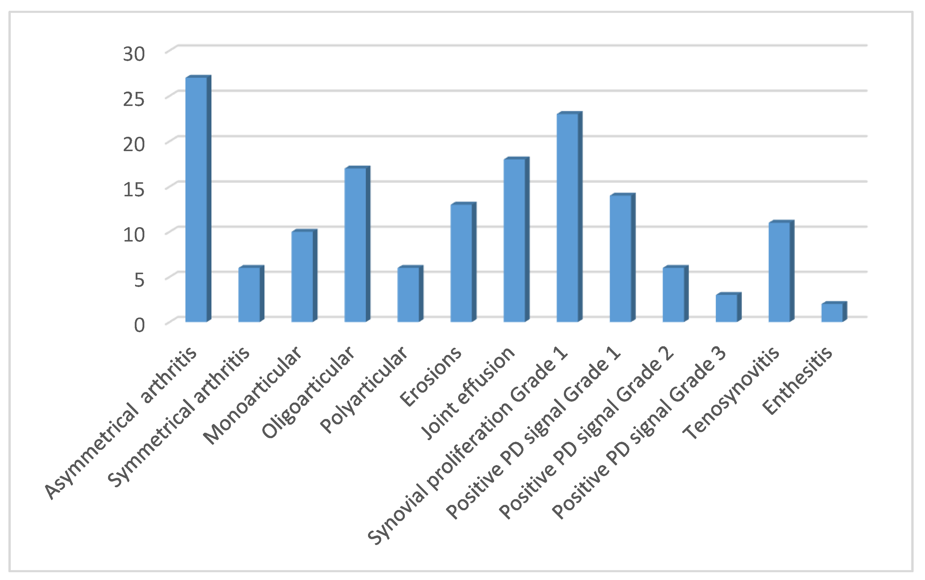
Figure 1. Patients (n) with positive US finding in the group of 72 HLA-B * 35 SpA patients. Abbreviations: US: ultrasound; SpA: spondyloarthritis, PD: power Doppler.
Life 11 00524 g001
Table 1. Demographic and baseline characteristics in 72 HLA-B*35 positive patients.
Age, yearsMedian (IQR)56 (51–65)
Femalen (%)61 (85)
Disease duration, months Median (IQR)14 (9–19)
ESR (mm/h)Median (min–max)10 (1–60)
ESR ≥ 29 mm/hn (%)11(15)
CRP (mg/L)Median(min–max)3(0.3–42)
CRP ≥ 5 mg/Ln (%)15 (21)
Joint pain and swellingn(%)57(79)
Tendon pain and swellingn (%)14 (19)
Dactylitisn(%)3 (4)
ACPAn (%)2 (3)
RFn (%)3 (4)
ANAn (%)
NSAIDsn (%)65 (90)
csDMARDsn (%)30 (42)
CSsn (%)15 (21)
bDMARDSn (%)3 (4)
Abbreviations: ESR: erythrocyte sedimentation rate; CRP: C-reactive protein; ACPA: anti-citrullinated protein antibody; RF: rheumatoid factor; ANA: anti-nuclear antibody; NSAID: non-steroidal anti-inflammatory drug; csDMARD: conventional synthetic disease-modifying anti-rheumatic drugs; CS: corticosteroids; bDMARD: biological disease-modifying anti-rheumatic drugs.
Table 2. Measures of disease activity and physical functioning in patients with and without a positive US finding.
72 HLA-B*35 Positive Patients
Positive US
(n = 33)
Negative US
(n = 39)
p-Value Difference
(95% CI)
ESR (mm/h)Median (IQR)
(min–max)
9 (5–15)
(1–60)
10 (5–18)
(1–52)
0.743
CRP (mg/L)Median (IQR)
(min–max)
4 (1.8–7)
(0.8–42)
2.9 (1–3.7)
(0.3–15)
0.039 1.1 (−0.38–2.6)
DAS28ESRMedian (IQR)
(min–max)
4.3 (3.1–4.8)
(1.8–5.8)
3.5 (2.5–4.1)
(0.4–5.7)
0.039 0.8 (−0.1–1.72)
DAS28CRPMedian (IQR)
(min–max)
4.1 (3.4–4.6)
(2.3–5.8)
3.4 (2.4–3.9)
(1.2–4.9)
0.002 0.7 (−0.03–1.31)
HAQMedian (IQR)
(min–max)
1.25 (1–1.7)
(0–2.5)
1.1 (0.9–1.4)
(0.4–2.3)
0.124
Mann–Whitney U test.
Table 3. The association between acute phase reactants and positive US findings.
72 HLA-B*35 Positive Patients
Positive US
(n = 33)
Negative US
(n = 39)
p-Value *OR(95%CI)
p-Value **
ESR (≥29 mmHg)n (%)4 (12)5 (13)0.9550.76 (0.19–3)
0.691
CRP (≥5 mg/L)n (%)9(27)6(15)0.3442.1 (0.65–6.6)
0.221
* χ2 test; ** logistic regression.
Publisher’s Note: MDPI stays neutral with regard to jurisdictional claims in published maps and institutional affiliations.