Review

Zebrafish Vascular Mural Cell Biology: Recent Advances, Development, and Functions

Department of Molecular Pathophysiology, Institute of Advanced Medical Sciences, Nippon Medical School, Tokyo 113 8602, Japan
*
Author to whom correspondence should be addressed.
Life 2021, 11(10), 1041; https://doi.org/10.3390/life11101041
Submission received: 6 September 2021 / Revised: 29 September 2021 / Accepted: 29 September 2021 / Published: 3 October 2021

Abstract

:
Recruitment of mural cells to the vascular wall is essential for forming the vasculature as well as maintaining proper vascular functions. In recent years, zebrafish genetic tools for mural cell biology have improved substantially. Fluorescently labeled zebrafish mural cell reporter lines enable us to study, with higher spatiotemporal resolution than ever, the processes of mural cell development from their progenitors. Furthermore, recent phenotypic analysis of platelet-derived growth factor beta mutant zebrafish revealed well-conserved organotypic mural cell development and functions in vertebrates with the unique features of zebrafish. However, comprehensive reviews of zebrafish mural cells are lacking. Therefore, herein, we highlight recent advances in zebrafish mural cell tools. We also summarize the fundamental features of zebrafish mural cell development, especially at early stages, and functions.

1. Introduction

Blood vessels are mainly composed of vascular endothelial cells, that make up the luminal surface, and vascular mural cells, that cover the endothelial cells from the abluminal side. The recruitment of mural cells to the vascular wall plays an important role in angiogenesis, acting in an organ-dependent manner, as well as subsequent vascular stabilization, and functions. Mural cells are divided into two main types: pericytes and vascular smooth muscle cells (VSMCs) [1,2]. Pericytes mainly cover capillaries, whereas VSMCs cover relatively large vessels from arteries to small arterial vessels and veins. At the transition site between VSMCs and pericytes, pericytes are further categorized into several subtypes depending on their morphology or gene expression along the vascular tree [3]. However, recent single-cell RNA sequences of mouse brain mural cells revealed that pericytes form the single homogeneous population distinguished from VSMC clusters and indicated the lower genetic continuity between VSMCs and pericytes in the brain [2]. This single-cell RNA sequencing analysis also revealed several pericyte selective genes such as Abcc9 and Kcnj8 [2]; however, these genes expressions are present not only in pericytes, but also in VSMCs, and may even be absent in the pericytes of some organs [2,4]. A precise definition distinguishing these two cell types is needed, but thus far, clear and simple criteria for pericytes, which could be applied to all organs, have not been established [2,4]. Given the difficulty of establishing definitions and/or appropriate terminology, we use the term “mural cells” in this review, referring especially to those observed in early developmental stages, rather than using the terms “pericyte” or “VSMC” unless well-defined or used in the original reference sources.
The zebrafish is a suitable model animal for studying cardiovascular system development [5]. Zebrafish embryos are fertilized and develop externally, they are transparent, and can easily be manipulated genetically. Therefore, many transgenic zebrafish lines have been established for visualizing processes or intracellular signaling during vascular development [5,6,7,8,9,10]. These tools have dramatically expanded our knowledge of the processes underlying vasculogenesis and angiogenesis including sprouting, lumenization, and pruning. However, transgenic zebrafish reporters for visualizing all types of mural cells have not been available to investigators, hampering our understanding of the complex processes underlying mural cell development and subsequent vascular maturation steps. It is noteworthy, however, that several mural cell reporter lines have been developed in the last decade in our and other labs (Table 1), allowing us to achieve a better understanding of mural cell development and functions [11,12,13,14,15,16,17,18,19,20,21,22,23,24,25,26,27,28].
To the best of our knowledge, although there are many excellent reviews on other systems [29,30,31,32], there have been no comprehensive reviews focusing specifically on zebrafish mural cell development and functions thus far. Therefore, to facilitate optimal usage of the zebrafish as a model for mural cell biology, this review summarizes the fundamental processes of mural cell development, focusing especially on the early developmental stages, their regulatory mechanisms, and functions in zebrafish.

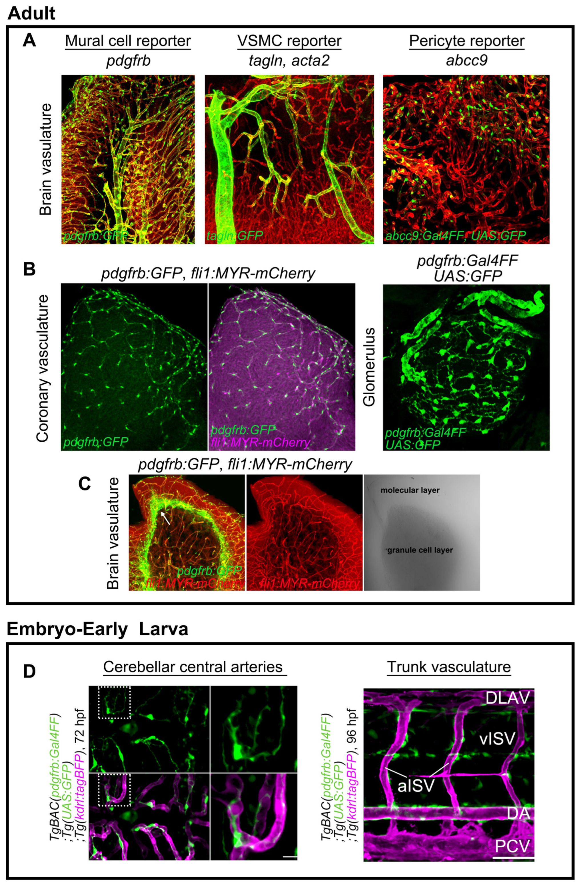
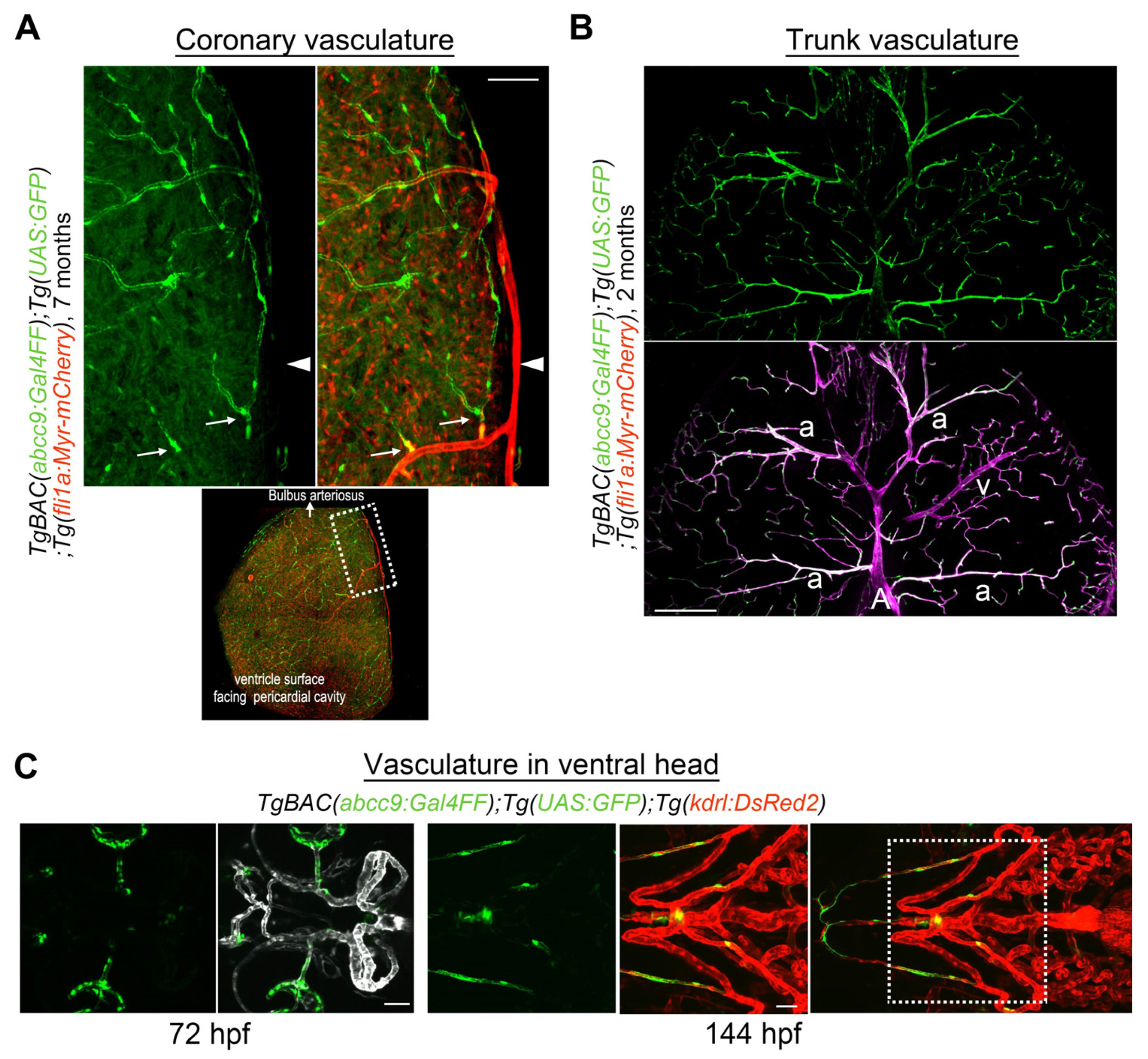
2. Genetic Tools for Mural Cell Biology in Zebrafish

Zebrafish VSMC, a type of mural cells, were first described by Santoro et al. in a 2009 study utilizing an antibody for sm22a-b (tagln) and transmission electron microscopy [33]. In line with their observations, two transgenic zebrafish lines, Tg(sm22a-b:GFP) [18] and Tg(acta2:GFP) [23], were developed and shown to allow visualization GFP (green fluorescent protein)-positive mural cells suggestive of VSMCs in large-caliber vessels such as the dorsal or ventral aorta, but not in relatively small-caliber trunk vessels or cranial vessels, during embryonic stages. This prompted investigators to question the presence of pericytes in zebrafish [23]. Wang et al., on the other hand, succeeded in visualizing pericytes in brain vessels by performing fluorescence in situ hybridization for platelet-derived growth factor receptor beta (pdgfrb) [34]. In the trunk regions, pdgfrb-positive mural cells were detected not only in the ventral portion of the dorsal aorta, but also in the intersegmental vessels (ISVs). These results support the suitability of pdgfrb for visualizing all types of mural cells in zebrafish. Consistently, transgenic zebrafish carrying a modified BAC clone containing the pdgfrb promoter labeled all mural cells including pericytes and VSMCs in the organs, from the early to the adult stages (Figure 1A,B,D) [11,12]. It is noteworthy that mural cell precursors are weakly positive for pdgfrb and pdgfrb reporter lines, thereby enabling us to trace the specification process into the mural cell lineage from their pdgfrblow precursors [12,28]. In addition to VSMC-selective and pan mural cell reporter lines, a pericyte-selective reporter for abcc9, which is a pericyte-selective gene found in the mouse brain, has been established (Figure 1A) [2,28]. We showed abcc9 to be the earliest and most selective marker for mural cells, especially in the brain, and to become selective for pericytes during development (Figure 1A) [2,28]. In addition to the brain, abcc9 reporter expression is found in coronary vascular pericytes (Figure 2A). In contrast, abcc9 becomes positive in the trunk (arteriolar) VSMC (Figure 2B). These observations indicate organotypic differences among mural cells in zebrafish, as reported in mice [4]. These zebrafish mural cell reporters also indicate that fundamental gene expression profiles of mural cell types in the organs are conserved between zebrafish and mice [2]. Utilizing these novel genetic tools (Table 1), we can now easily address mural cell developmental processes from the early to the adult stages, including specification into mural cells, one of the major advantages of zebrafish as compared to other animal models.
Non-vascular-associated strong pdgfrb reporter expression is observable in the adult brain, including the stem cell niche area located at the cerebellum interface between the granule cell and molecular layers [35] (Figure 1D). PDGFRβ is reportedly expressed by neural stem cells in the adult mouse subventricular zone [36] as well as by human placental mesenchymal stem cells [37]. Thus, in addition to the established vascular biology, pdgfrb reporter zebrafish are also useful in a broad range of research fields.

3. Mural Cell Development in Zebrafish

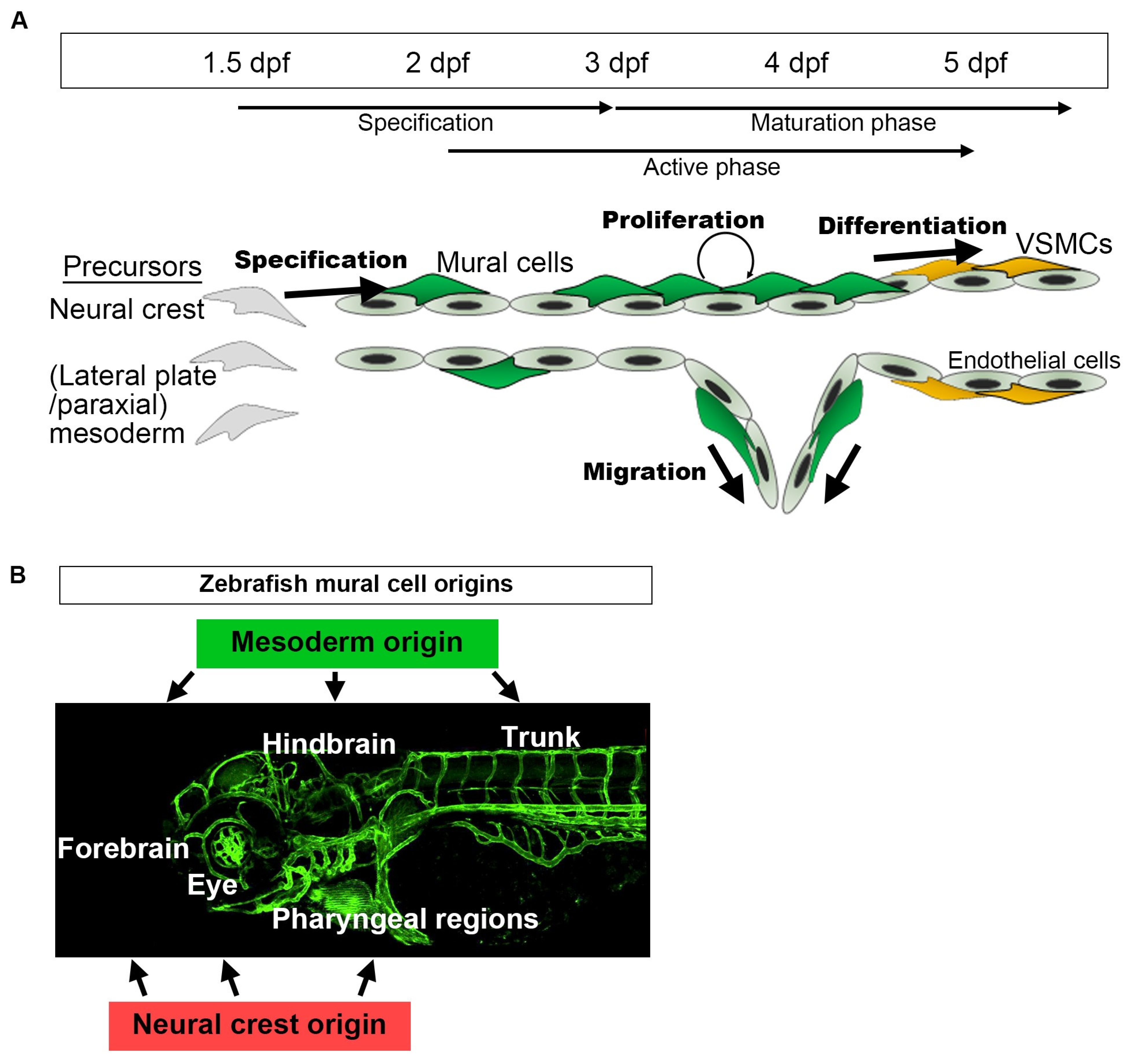
3.1. Overview of Mural Cell Development during the Early Developmental Stages in Zebrafish

We will first summarize the basic developmental time course of zebrafish mural cells, mainly in the trunk and brain vasculature, and then move on to individual tissues and organs. pdgfrb-positive cells are initially observed at approximately 10 h post-fertilization (hpf), and these pdgfrb-positive mural cell precursors are broadly distributed in the trunk or cerebral base by 24–48 hpf [12]. Among them, those located in proximity to arteries are specified to the mural cell lineage during the period spanning approximately 36–72 hpf [28] (Figure 3A). Increased pdgfrb expression or the induction of abcc9 is the hallmark of specification into the mural cell lineage [28]. However, most of the mural cells beneath the dorsal aorta and approximately half of those in ISVs are negative for abcc9 by 5 days post-fertilization (dpf) [28], suggesting differences in mural cell characteristics from the first step of development onward. After specification, mural cells actively proliferate and migrate toward the arteries not covered with mural cells, and almost all of the brain and trunk arteries have been covered by mural cells by 4–5 dpf. Simultaneously, mural cells around large-caliber vessels such as those comprising the Circle of Willis and the dorsal aorta start to express tagln or acta2 and appear to acquire VSMC properties after 3 dpf. At this early age, VSMC markers were reportedly detectable in only a subset of the mural cells in ISV and were barely detected in those of central arteries (CtAs) before 5 dpf [28]. The mural cells lacking VSMC markers show pericyte features morphologically (protruded round cell body and extension of long thin processes longitudinally along vessels) and genetically (positive for the pericyte marker abcc9). However, whether these cells are actually pericytes or still undergoing maturation needs to be confirmed. During the early developmental stages, although the primordial hindbrain channel (PHBC) has few, most of veins lack mural cell coverage. As yet, little is known about how and when zebrafish veins acquire mural cell coverage. Lineage tracing employing the Cre/lox-system and genetic intervention leading to the loss of specific cell derivatives revealed mural cells in the trunk, forebrain, and hindbrain to be derived from the mesoderm, whereas those in the forebrain, hyaloid vessel, and pharyngeal regions are from the neural crest (Figure 3B) [12].

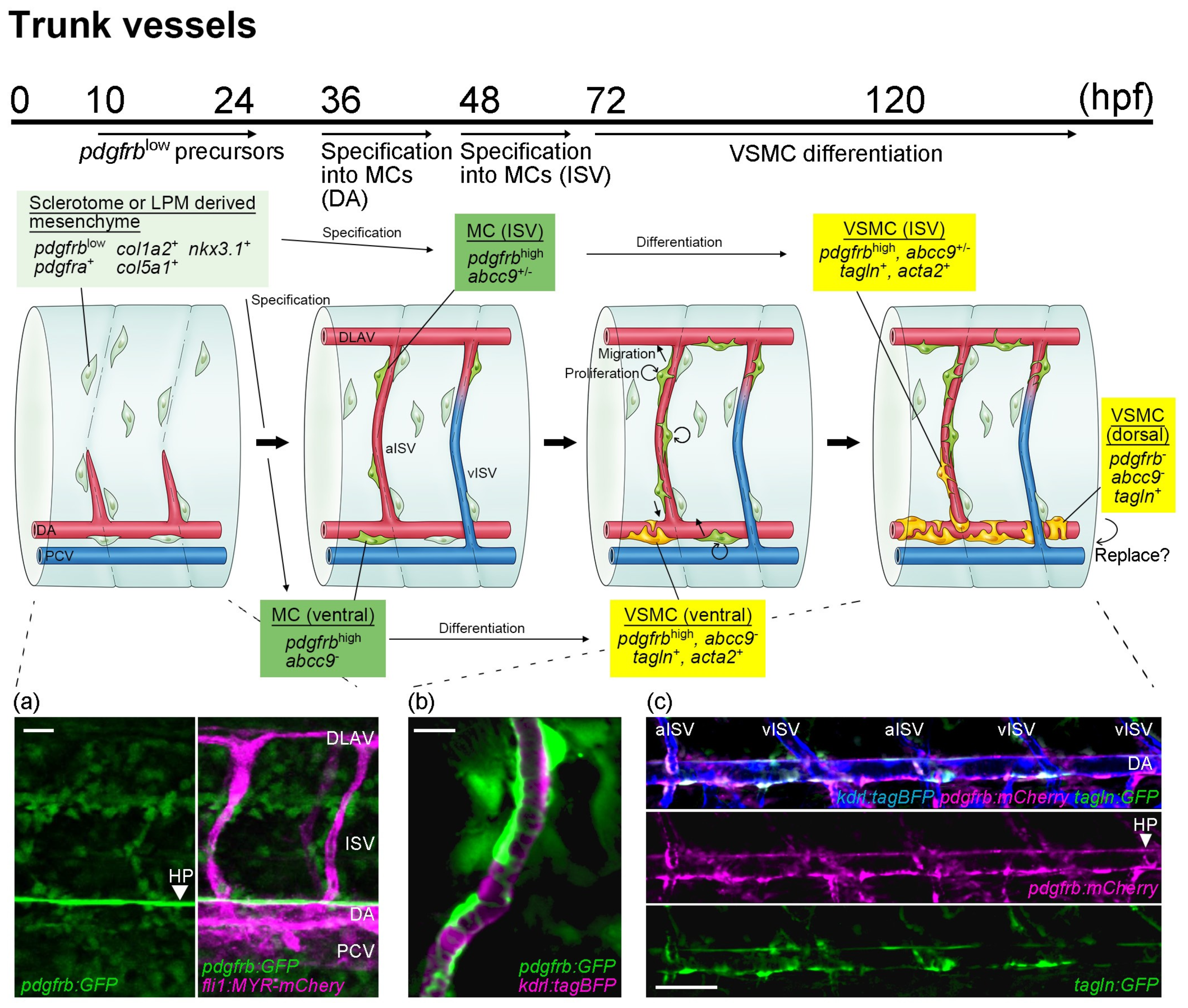
3.2. Mural Cell Development in Axial Vessels in the Trunk

Trunk mural cells are derived from sclerotomes and the lateral plate mesoderm. Time-lapse imaging performed by Rajan et al. clearly showed that nkx3.1-positive sclerotomes and sclerotome-derived notochord-associated cells disperse around the trunk region, and that some are distributed along the ISVs by 48 hpf [38]. These sclerotome-derived mesenchymal cells in the vicinity of arterial endothelial cells in ISVs subsequently become mural cells (Figure 4). In addition to a low level of pdgfrb expression, these sclerotome-derived cells are positive for pdgfra and extracellular matrix genes, col1a2 and col5a1, suggesting these cells to have fibroblast characteristics [38]. Considering the onset of mural cell emergence in ISV after 48 hpf [12,28], the specification into mural cells from sclerotome-derived fibroblasts takes place after the alignment of progenitors around arterial vessels. Given how well distributed those precursors are around ISVs [38], there might be signals actively recruiting pdgfrblow mesenchymal precursors to ISV walls (or somite boundaries) rather than stochastic phenomena. Among perivascular pdgfrblow cells, only a few become mural cells. Therefore, mural cells increase their number and their vascular coverage via subsequent proliferation and migration. After the important specification period (36–72 hpf) in the trunk, the perivascular pdgfrblow mesenchyme not specified to become mural cells shows decreased pdgfrb expression [28]. Whether there is communication between mural cells and perivascular fibroblasts during their development remains unknown.
The mural cells beneath the dorsal aorta can be seen after approximately 36 hpf, earlier than ISV mural cells. Subsequently, these dorsal aorta mural cells start the process of entirely covering the dorsal aorta and after 3 dpf they express VSMC markers, acta2 [23] and/or tagln [20], indicating differentiation into VSMC (Figure 4). Although half of ISV mural cells are negative for abcc9 before 5 dpf, nearly all mural cells beneath the dorsal aorta are consistently negative for abcc9 when they first become detectable [28]. Thus, dorsal aorta mural cells appear to be programed to be VSMC from the beginning of the specification process. In addition to the ventral side, tagln-positive cells are observed on the dorsal side of the dorsal aorta [20], becoming prominent a few days after the emergence of mural cells at the ventral side. Different from those emerging on the ventral side, most tagln-positive cells on the dorsal side are negative for pdgfrb reporter expression [12]. Therefore, distinct types of VSMCs might be present on the dorsal and ventral sides of the dorsal aorta. Consistent with this possibility, the lateral plate mesoderm-derived VSMCs in mice reportedly cover the ventral side of the descending dorsal aorta in the early stage of aortic development, whereas the VSMCs covering the dorsal side are from paraxial mesoderm-derived somites [39]. Eventually, all dorsal aorta VSMCs are replaced by somite-derived VSMCs [39]. The emergence of ISV mural cells was strongly suppressed when the production of paraxial mesoderm derivatives was inhibited by tbx6 morpholino oligonucleotide (unpublished data), whereas that of mural cells beneath the dorsal aorta was suppressed when paraxial and lateral plate mesoderm derivative productions were both inhibited [12]. Therefore, ISV mural cells are mainly derived from paraxial mesoderm and dorsal aorta mural cells are from both paraxial and lateral plate mesoderm, observations consistent with the results of previous studies [20,38]. To date, the reasons for VSMC on the dorsal aorta arising from different sources and whether there are differences in their roles remain unknown. As reviewed by Sato [40], endothelial cells in the dorsal aorta show dorsoventral differences in their origins. Using quail-chick chimera analysis, Pardanaud et al. demonstrated that the dorsal portion of the dorsal aorta is composed of somite-derived endothelial cells and splanchnic mesoderm-derived endothelial cells at the ventral portion of the dorsal aorta [41]. This study was further extended, and it was shown that the hemogenic ventral wall of the dorsal aorta is replaced by non-hemogenic somite-derived endothelium in chicks [42]. In zebrafish, the dorsoventral difference within the dorsal aorta is well documented, especially during hemogenic endothelial cell development [43,44]. Therefore, dorsoventral differences in the dorsal aorta may affect mural cell emergence and the subsequent characteristics of these cells, in addition to explaining the difference in origins. However, there is no evidence of a dorsoventral endothelial cell lineage difference in zebrafish as reported in chicks. Rather, this is likely to reflect local differences in signaling [45,46,47], as shown in mice [48]. Interestingly, pdgfrb signaling reportedly promotes hematopoiesis in the ventral part of the dorsal aorta in zebrafish [49,50]. Therefore, the dorsoventral differences in VSMC might contribute to the efficient progress of hematopoiesis. Future analysis of ventral and dorsal VSMC gene profiles is anticipated to provide better understanding of their physiological roles and the well-programed events of vascular development.

3.3. Mural Cell Development in the Brain

Embryonic brain mural cells are derived from the pdgfrblow mesenchyme distributed in the cerebral base at the level of the basilar artery (BA), just above the anterior endodermal cell sheet, or around the choroidal vascular plexus (CVP) [12] (Figure 5A). Similarly to axial vessels, the pdgfrblow mesenchyme located in the vicinity of endothelial cells is specified to the mural cell lineage after approximately 36 hpf (Figure 5A). These mural cells were observed to emerge at the cerebral base or CVP, then proliferate, and after CtAs had connected to these vessels and blood flow had begun, migrate to CtAs along the vessels. When they migrate, these cells preferentially extend their processes along inter-endothelial junctions [12]. Interestingly, they form scaffold-like structures in the extending processes at the inter-endothelial cell junctions and 90% of mural cells were observed to move forward by relocating their cell bodies to this node, as if they had jumped from the original point to this node [12]. When the cells migrate to the next position, mural cell division frequently takes place simultaneously and leaves one mural cell at the original position while supplying the additional mural cell to the uncovered new region, thereby expanding vascular coverage. However, whether this is also the case when a mural cell co-migrates with endothelial tip cells during sprouting angiogenesis is unknown. Once angiogenesis in the brain has stabilized at around 5 dpf, mural cell migration and proliferation are also arrested [12], indicating the behaviors of endothelial and mural cells to be synchronized. This correlation between mural cells and endothelial proliferation is reportedly observed during wound healing angiogenesis in the skin of adult zebrafish [51].
The mural cells emerging around the CVP migrate toward anterior mesencephalic central arteries (AMCtAs) and the middle mesencephalic central artery (MMCtA) along the connecting vessels between the CVP and AMCtA/MMCtA (Figure 5B). Mural cells, in contrast, emerging on the basal communicating artery (BCA), primarily migrate to the AMCtA and MMCtA, the posterior communicating segment (PCS) to the MMCtA, and the BA to CCtAs (Figure 5B). Mural cells emerging around the CVP are derived from the neural crest, whereas those around the Circle of Willis and BA are from the mesoderm; therefore, zebrafish hindbrain mural cells are mainly of mesoderm origin in both the forebrain and the intermediate region (Figure 5B) [12]. These distinct brain region populations persist in the adult brain [12]. Thus, the comparison of gene expressions between mural cells derived from the neural crest and those from the mesoderm in the zebrafish brain provides clues to elucidating the mechanisms regulating the organotypicity of mural cells (“organ environment” vs. “origin”).
In the embryonic brain, abcc9 is the earliest and most selective marker for specified mural cells. Soon after the specification into mural cells, abcc9 reporter expression is induced in nearly all of the mural cells in the brain [28] and shows typical pericyte-like morphology (Figure 5A). Subsequently, mural cells at the Circle of Willis and BA transform morphologically and show a shape typical of VSMC (Figure 5A). In accordance with the morphological changes, these mural cells start to express tagln and acta2 after approximately 72 hpf [28,31], suggesting differentiation into VSMCs. Before reaching the Circle of Willis and BA, mural cell expressions of acta2 and tagln are induced around the caudal division of the internal carotid (CaDI) [52], whereas those of mural cells around CtAs begin later. VSMC marker expression in AMCtA- or CCtA-covering mural cells starts with the cells located close to the branching point from the BCA or BA, respectively, after 5–6 dpf [53]. Thus, VSMC marker expression gradually turns toward the downstream vessels (CaDI → Circle of Willis → CtAs) during development, which may indicate that mechanical forces produced by blood flow facilitate differentiation into VSMCs, given the importance of blood flow in VSMC development in zebrafish, as described below [19,24]. Blood flow was not essential, however, for specification into mural cells and the de novo emergence in zebrafish trunk and brain vessels [12]. Brain mural cells, especially around the CtAs, initially show typical pericyte morphology and a gene signature characterized by abcc9, but later differentiate into VSMCs around large-caliber vessels, which is different from the dorsal aorta mural cells. Contractile VSMCs reportedly have low migratory and proliferation ability [54]. Considering the active migration and proliferation of mural cells during embryonic stages to entirely cover brain vessels, this mural cell development in the brain passing through a pericyte-like cell type stage before the differentiating into VSMC appears to be both reasonable and beneficial. This system, probably not only in the brain but also in other organs and tissues, may allow the spatio-temporal flexibility of VSMC coverage in accordance with vascular development. It would be interesting and potentially worthwhile to investigate the gene expressions related to cell motility and the proliferation of both cell types. As yet, we do not know whether these pericyte-like mural cells observed during early embryonic stages are identical to what we consider to be (mature) pericytes or immature progenitors of pericyte and VSMCs. Detailed gene expression analysis during the course of development may answer this question.

3.4. Mural Cell Development in the Ventral Head

Mural cells are observed around the ventral aorta from approximately 66 dpf, and these cells arise mainly from a foxc1b+ neural crest derivative [23,24]. Mural cells in the ventral aorta and aortic arches become positive for acta2 but negative for abcc9 soon after their initial appearance (Figure 2C), suggesting that the mode of differentiation is similar to that of VSMC observed in the dorsal aorta. Interestingly, most ventral aorta mural cells are also negative for pdgfrb [24], as observed in the dorsal part of the dorsal aorta.

3.5. Mural Cell Development in the Kidney

Glomerular mesangial cells, known to be a type of mural cell, are clearly visualized in pdgfrb reporter zebrafish (Figure 1A). Glomerular development in the zebrafish pronephros starts after 34 hpf and vascularization in the glomerulus becomes visible after 2 dpf [55]. However, the precise timing of mesangial cell emergence and the mechanisms underlying zebrafish mesangial development, including its origin, remain unknown [55].

3.6. Mural Cell Development during Regeneration

Observation of adult zebrafish skin tissue over a few months demonstrated that neither endothelial cells nor pericytes divided or migrated [51]. This suggests that endothelial cells and pericytes are in a quiescent state in normal skin capillaries, thereby maintaining a stable vascular structure. However, endothelial cells were rapidly activated by wounding, and angiogenesis was induced in non-injured blood vessels. During angiogenesis in skin wound healing, pericytes also increased in number by proliferating at a rate similar to that of endothelial cells and migrated to the leading edge [51]. According to the termination of neovascularization (active angiogenesis process), the number of endothelial cells and pericytes started to decrease gradually and returned to the pre-injury level over the course of a few months, indicating the synchronized regulation of endothelial cell and pericyte dynamics. The source of mural cells in the regenerated vessels was analyzed in a fin regeneration model [15]. Tracking photo-converted cells unexpectedly revealed that pre-existing mural cells are not the source of those for repaired vessels [15]. In the zebrafish fin, mural cells originate from pdgfrb-positive cuboidal-shaped cells around arterial vessels running through the center of the fin ray, in regenerating vessels but also in normal development. Furthermore, Pdgfrb signaling turned out to be essential for the de novo emergence of mural cells from cuboidal-shaped cells during the process of regeneration. However, whether mural cells in the regenerating vessels are always supplied by de novo emergence during the repair process at the other sites is unknown.

4. Molecular Mechanisms Underlying Mural Cell Development

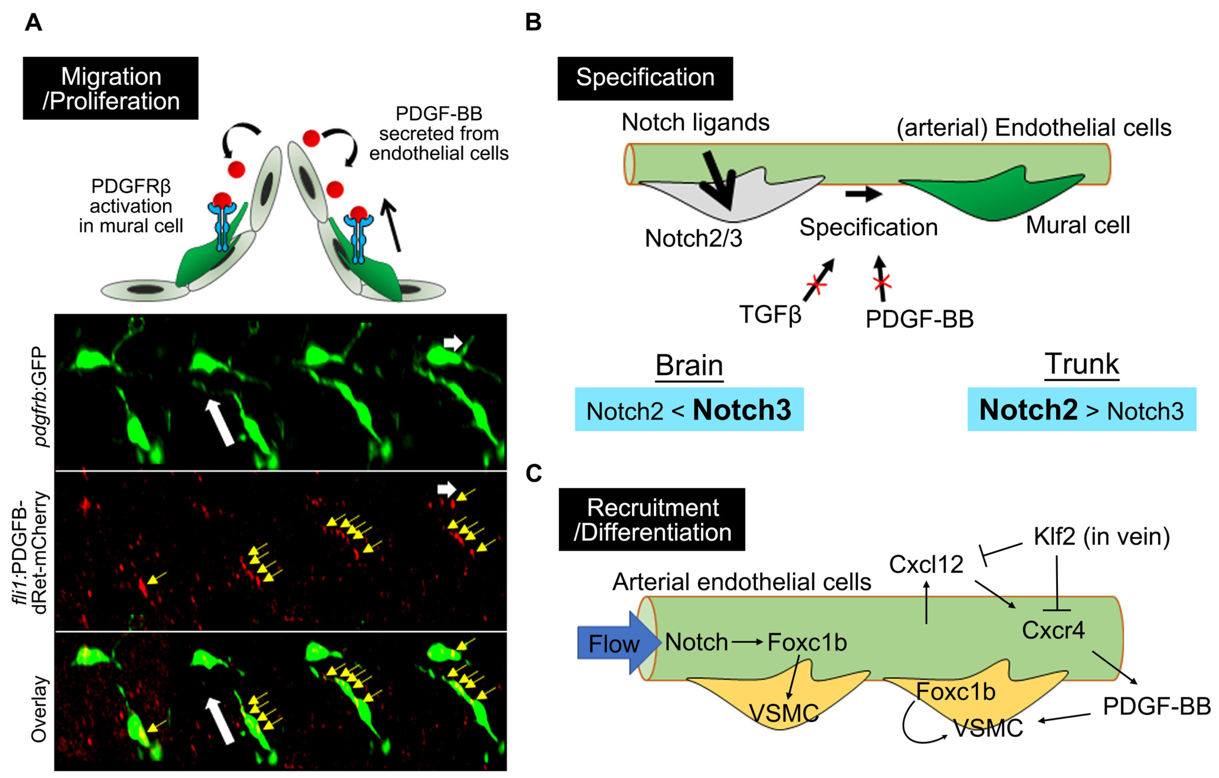
4.1. PDGFRβ-Mediated Signaling in Mural Cell Development

Our recent study utilizing pdgfb and pdgfrb mutant zebrafish revealed that the organotypic requirement of PDGFB–PDGFRβ signaling for mural cell development recognized in studies of mice is similar to that in zebrafish, suggesting a highly conserved role for PDGFB–PDGFRβ signaling in mural cell recruitment in vertebrate species [53]. As noted above, mural cells expand their vascular coverage via migration and proliferation. It is known that PDGF-BB released from endothelial cells activates PDGFRβ expressed on mural cells, thereby promoting mural cell migration/recruitment to the vascular wall and their proliferation [56]. pdgfb or pdgfrb mutant zebrafish analysis confirmed that PDGFB–PDGFRβ signaling is not essential for mural cell specification, although is indispensable for subsequent migration and proliferation [53,56,57]. In vivo live imaging in zebrafish showed that the uptake of PDGF-B released from endothelial cells into the mural cell processes extended toward the direction of migration (Figure 6A), which further supports the proposed model that PDGF-B functions as a chemoattractant for mural cells via PDGFRβ [56]. In zebrafish, the Pdgfb ligand-encoding pdgfb gene is duplicated, i.e., to pdgfba and pdgfbb, both of which are enriched in endothelial cells. pdgfba;pdgfbb double-mutants showed additive mural cell loss, although with a greater contribution from pdgfba than pdgfbb, suggesting that pdgfba and pdgfbb function cooperatively [53]. The higher contribution of pdgfba than pdgfbb probably reflects their expression levels in endothelial cells [53]. A recent study also showed that Cxcr4 activation in arterial endothelial cells induces Pdgfb production, whereas this pathway is blocked by Klf2 in venous endothelial cells, thereby recruiting mural cells to the dorsal aorta, but not to veins [58]. However, it is not clear whether the de novo formation of mural cells around the dorsal aorta is affected or differentiation into VSMC from the mural cell lineage is inhibited because analyses were performed using tagln and acta2 VSMC reporters, in which the first pdgfrb-positive mural cells to emerge were not labeled.
pdgfb and pdgfrb mutant analyses indicated this signaling to be indispensable for the de novo formation of mural cells via specification from the perivascular mesenchyme in the trunk and brain vessels [12,53]. However, there is a discrepancy between the pdgfrb mutant phenotype and previous zebrafish studies regarding trunk mural cells and vascular development, as discussed in detail in a recent publication [53]. Briefly, pdgfrb mutants display normal vascular development throughout the body and VSMC coverage beneath the dorsal aorta during the early stages of development. However, inhibition of Pdgfrb signaling by the ubiquitous over-expression of a dominant negative form of Pdgfrb [20], morpholino [59], or an inhibitor [59] resulted in defective ISV formation [59] or decreased VSMC coverage of the dorsal aorta [20]. Considering the possibility that dominant negative proteins can interfere with related molecules and that non-sense-mediated decay of the pdgfrb transcript may upregulate compensatory paralogous genes [60], this discrepancy raises the possibility of the existence of redundant or compensatory pathways for mural cell development [60,61]. One candidate for such a function is the related PDGFRα receptor, encoded by the pdgfra gene. However, pdgfra;pdgfrb double-mutant larvae at 4 dpf showed neither trunk vascular defects nor any significant reduction in the VSMC coverage of the dorsal aorta as compared to the wild type [53], although some pdgfra mutant larvae without flow showed VSMC loss, apparently reflecting an essential role in VSMC development [19]. These results suggest that pdgfra and pdgfrb are dispensable for initial development of the trunk vasculature and VSMCs on the dorsal aorta at this stage. Therefore, the discrepancy might not be explained by the compensatory role of Pdgfra, acting as a substitute for Pdgfrb, in mural cell development; further studies are needed to elucidate the mechanisms responsible for the observed differences.

4.2. Notch Signaling in Mural Cell Development

The molecular mechanisms underlying mural cell development, particularly the specification step, have yet to be clarified. In efforts to understand the molecular mechanisms regulating mural cell specification, zebrafish serve as a useful model because in vivo processes can easily be traced from precursors. It was found that mural cell emergence was fully inhibited by treatment during the specification period (36–72 hpf) with γ-secretase inhibitors, i.e., DAPT and LY411575, that block Notch signaling [28]. Furthermore, mural cell emergence is completely prevented in embryos depleted of both notch2 and notch3, which were found to be expressed in mural cells [28]. Suppression of either notch2 or notch3 expression did not completely inhibit mural cells, suggesting that these two genes function redundantly during specification into mural cells (Figure 6B). Timelapse imaging of zebrafish Notch activity reporters clearly demonstrated Notch activation in mural cells during specification [28]. pdgfrblow mesenchymal cells in which Notch activity is “ON” show increased pdgfrb expression, which is one of the hallmarks of specification, and extend their processes along the endothelial cells to assure the formation of close contacts, which would be a typical morphological feature of mural cells. However, the cells without Notch activation do not show such changes and pdgfrb expression is further reduced. These results indicate that pdgfrblow mesenchymal cells are specified to mural cells through the activation of Notch2 and Notch3 signaling, probably via their ligands expressed in arterial endothelial cells, as shown by a mouse study [62,63]. Therefore, lower expression of Notch ligands in venous endothelial cells [62,63,64] might explain the absence of mural cell coverage in veins during early developmental stages. Zebrafish mural cells arise from the mesoderm and neural crest [12], but Notch2 and Notch3 signaling are essential for mural cell specification regardless of their origin [28]. However, Notch3 functions predominantly in brain mural cells and Notch2 in trunk mural cells, suggesting that the properties of mural cells differ depending on the site of action starting in the very early stage of development. Chen et al. demonstrated blood-flow-induced Notch activation in arterial endothelial, i.e., not only in mural cells, to be important for the recruitment of acta2- or tagln-positive VSMC to the dorsal aorta [19]. However, it is not clear whether this Notch activation regulates the de novo formation of mural cells or subsequent differentiation into VSMC. A prior study found that Notch3 morphants showed cranial hemorrhage, considered to be attributable to a reduction in brain mural cells [34]. However, neither cranial hemorrhage nor edema is induced, even in the absence of mural cells in the CtAs, as reported in pdgfrb mutants [12,53]. This suggests that cranial hemorrhage in embryos lacking Notch3 might be induced by defects in endothelial cells because Notch3 also functions in these cells [64].

4.3. Forkhead Box Transcription Factors

Forkhead box domain transcription factors such as Foxc1 [65] and FoxF2 [66] are expressed by pericytes in the mouse brain and regulate pericyte proliferation and blood–brain barrier (BBB) formation. These genes are also important for zebrafish mural cell development [19,24,25,67]. Duplicated foxc genes in zebrafish, foxc1a and foxc1b, and genetic deletion or suppression by morpholinos of these genes results in impaired VSMC coverage of the ventral artery [24]. foxc1b is expressed in neural-crest-derived progenitors and positively regulates subsequent differentiation into VSMCs in the ventral portion of the vessel [24]. foxc1a is important for foxc1b expression, such that the foxc1a mutant shows a stronger effect than the foxc1b mutant on VSMC differentiation around the ventral artery [24]. On the other hand, Chen et al. revealed that foxc1b induced by flow-mediated Notch activation in arterial endothelial cells promotes VSMC recruitment, whereas foxc1a suppression in endothelial cells exerted no major effects on VSMC coverage in the dorsal aorta [19]. Hence, foxc1 functions in both mural cells and endothelial cells to promote VSMC development (Figure 6C). The zebrafish foxf2 gene is also duplicated, as foxf2a and foxf2b, and both genes are expressed in brain mural cells as well as endothelial cells [67]. Interestingly, foxf2b mutants show reduced pdgfrb-positive pericytes and acta2-positive VSMC in the zebrafish brain [67]. A murine study revealed that FoxF2 is important for PDGFRβ expression [66]. There are thus several possible mechanisms by which foxf2b regulates mural cell development in zebrafish, i.e., migration and proliferation driven by Pdgfrb, specification to the mural cell lineage, and even subsequent differentiation processes, whether cell-autonomously or non-cell-autonomously, via endothelial modulation.

4.4. TGFβ Signaling

Tumor growth factor β (TGFβ) signaling is also among the well-known factors involved in mural cell differentiation, survival, and proliferation, both via the TGFβ and activin branches of the pathway [68,69,70,71]. However, TGFβ signaling turned out to largely be dispensable for zebrafish brain and trunk mural cell development during the early developmental stages, at least through 6 dpf [28]. Neither pharmacological inhibition of TGFβ signaling nor the depletion of alk1 was associated with major problems in mural cell specification. Additionally, a transgenic zebrafish reporter that monitors the activation of TGFβ or activin/nodal signaling revealed activation of this pathway in endothelial cells, but there was no induction in either brain or trunk mural cells in the early stages (unpublished data). Although reductions in mural cell number were observed on brain vessels and ISVs following treatment with SB431542 or alk1 morpholino, this might represent indirect effects. Similarly, alk5 mutants exhibit defective VSMC development in the cardiac outflow tract, but this is considered to be attributable to secondary effects of perturbed endothelial TGFβ signaling [72].

5. Function of Mural Cells in Zebrafish

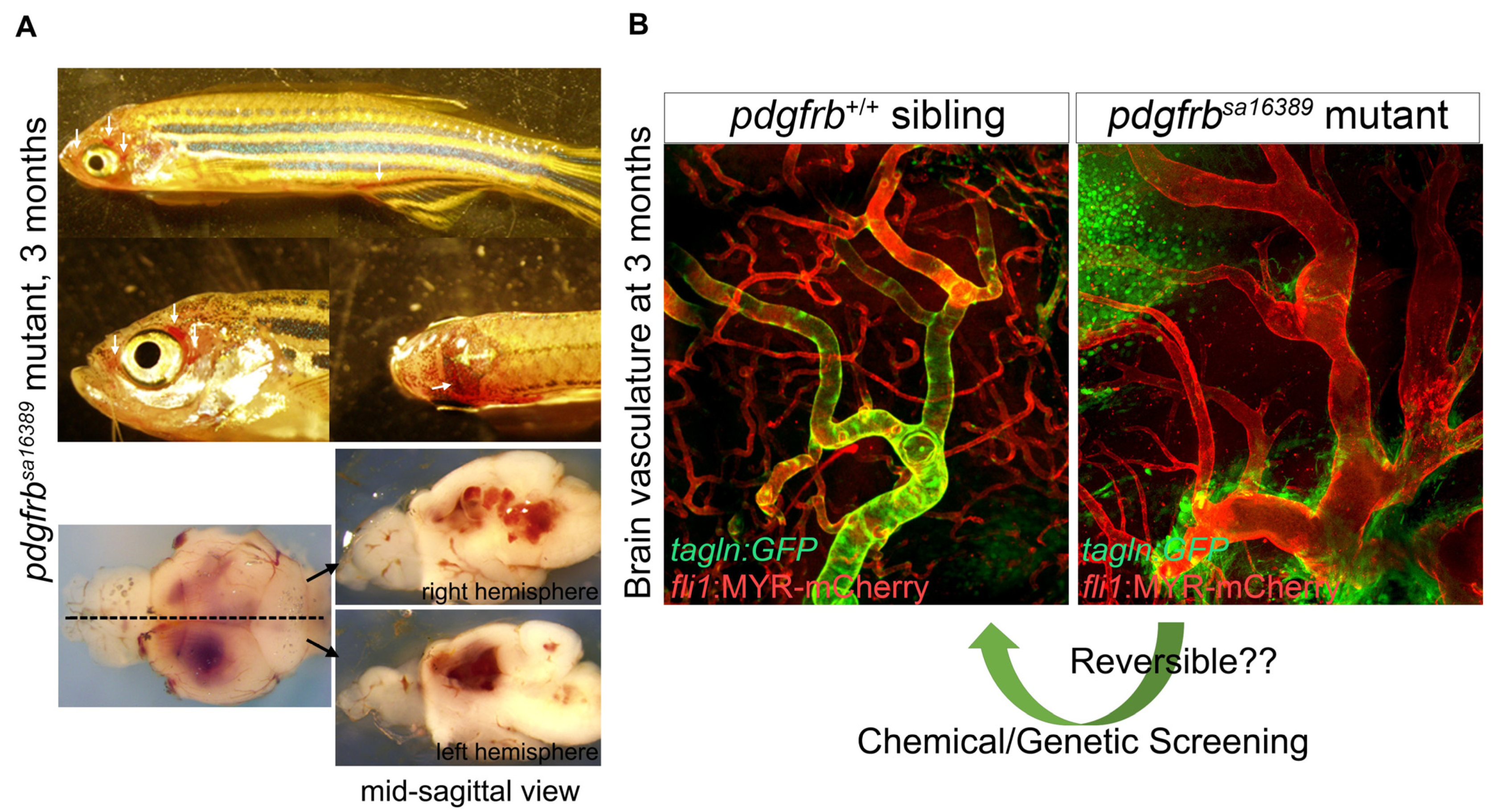
5.1. Observations Made in the Phenotyping Portion of pdgfrb Mutant Zebrafish Studies

Phenotyping studies of Pdgfb and Pdgfrb mutant mice lacking pericyte coverage have made major contributions to elucidating the functions of pericytes. Recently, we carried out extensive phenotypic analyses of pdgfrb mutant zebrafish organs from the early embryonic through adult stages (see details in Ando et al., 2021) [53]. Although pdgfrb mutant zebrafish do not have developmental defects during the early stages, they later exhibit severe vascular pathology (Figure 7). We found that the organ-specific differences in the sensitivity of mural cell recruitment to loss of pdgfrb in zebrafish correspond to those previously reported for Pdgfb and Pdgfrb mutant mice, suggesting a high degree of evolutionary conservation of these processes in vertebrates. In marked contrast to mice, however, zebrafish pdgfrb null mutants reach adulthood despite extensive cerebral vascular anomalies and hemorrhage (Figure 7A), offering unique opportunities to model cerebrovascular pathology and test therapeutic strategies. Taking advantage of the tractability of zebrafish for chemical/genetic screening and the resistance of this model to early death in the absence of pericytes, pdgfrb mutant zebrafish may prove useful in discovering and testing drugs for treating cerebrovascular as well as neurovascular diseases (Figure 7B).

5.2. BBB

Brain vessels acquire a specialized barrier property, the aforementioned BBB, to limit the passage of substances between blood and the parenchyma. The BBB is established by an endothelial cell sheet and surrounds other cell types, such as pericytes and astrocytes. Several independent studies have shown that zebrafish develop a BBB [73,74,75,76,77]. Imaging of reporter expression for glut1 (earliest BBB marker) or plvap (which correlates negatively with BBB maturation) indicates that BBB formation starts when cerebral angiogenesis begins at 2 dpf [76]. Evaluation of BBB integrity by tracer leakage from the circulation into the brain parenchyma has further demonstrated that the BBB becomes sufficiently mature by 5 dpf [73], consistent with the timing of the formation of the fundamental cerebral vascular structure and the completion of mural cell coverage. Cerebral vascular leakage through transcytosis is high during early stages and is suppressed by 5 dpf, reflecting the induction of mfsd2aa expression in endothelial cells [73]. Consistent with the results of a mouse study, mfsd2aa mutant zebrafish persistently showed elevated brain vasculature permeability through adulthood [73], indicating the conserved role of Mfsd2a in the suppression of transcytosis, which is important for BBB integrity. It remains unknown, however, as to whether zebrafish pericytes positively control this Mfsd2a expression, as suggested by the mouse studies. In contrast to morpholino-based analysis [78], mfsd2aa mutant zebrafish did not exhibit a hemorrhagic phenotype in the brain [73] such as that observed in the Mfsd2a mutant mouse [79,80]. Mutants or morphants of notch3 [34], foxc1 [25], or pitx2 [25] reportedly exhibit hemorrhage in the brain, which presumably results from defective mural cell development. However, to our surprise, loss of mural cell recruitment to CtAs in pdgfrb mutant zebrafish did not lead to vascular malformations or any apparent signs of hemorrhage during early developmental stages [53], indicating that endothelial cells can produce basic barrier integrity without pericytes in zebrafish. Therefore, the aforementioned hemorrhagic phenotype in mutant embryos may not be due to impaired mural cell recruitment, although whether the BBB is fully mature at embryonic stages without pericytes is an issue which merits more detailed investigation. pdgfrb mutant zebrafish began developing severe vascular malformations and hemorrhage in the brain at around one month of age. Why these defects arise so much later in zebrafish, despite similar mural cell loss at an early age, is unknown. As discussed in our recent report [53], differences in blood pressure or oxygen supply may explain these unexpected findings. At a later age, the vascular defects in zebrafish may progress showing courses similar to those in mouse embryos.
Astroglial cells also contribute to BBB integrity. Astrocyte endfeet wrap around the vascular wall within the central nervous system, and endothelial cells develop into the BBB cooperatively with astrocytes. Whether zebrafish possess astrocytes similar to those of mammals has been questioned, because instead, zebrafish develop a type of glial cells with radial morphology often termed radial glia, which are labeled by glial fibrillary acidic protein (GFAP) [74,81,82]. However, a recent analysis revealed that these GFAP-positive cells have gene signatures and functions similar to those of mammalian astrocytes [81]. Furthermore, despite lacking the classical stellate astrocyte morphology seen in mammals, the endfeet of GFAP-positive cells envelop the vascular wall in the adult olfactory bulb (Figure 8), possibly indicating that zebrafish develop neurovascular units as in mammals [82]. However, extension of GFAP-positive cell endfeet to the vessels in other brain regions was not prominent in the early stages, including 5 dpf, when the BBB matures into the adult structure (unpublished data) [74]. This might be attributable to the unsuitability of GFAP for labelling astroglial cells associated with endothelial cells. Therefore, future analyses are required to elucidate the role of astroglial cells in zebrafish BBB formation.

5.3. Regulation of Vascular Tone

Zebrafish blood circulation starts on the dorsal aorta at ~24 hpf. After circulation begins, mural cells emerge beneath the dorsal aorta independently of blood flow and then differentiate into VSMCs in a blood-flow-dependent manner. Recruitment of mural cells is important for basement membrane organization around the dorsal aorta [20]. In the absence of mural cell coverage, basement membrane components such as collagen Ⅳ and fibronectin around the dorsal aorta are drastically reduced, which leads to dilation of the dorsal aorta and increased elasticity of the vascular wall [20]. In this setting, mRNA expression levels of collagen Ⅳ and fibronectin in endothelial cells are similar but fragmentation of these proteins is increased. Therefore, mural cells are important for basement membrane stabilization in addition to producing vascular membrane proteins. Acquisition and alteration of the vasoactivity of cerebral pericytes and VSMC during the early stages of development have been investigated [52]. At 4 dpf, soon after their emergence, VSMCs can induce vascular constriction in response to vasoconstrictors, whereas the vasodilation capacity of VSMC is not yet fully developed. Eventually, VSMCs become vasoactive, capable of both constriction and relaxation, by 6 dpf. On the other hand, capillary pericytes have the ability to regulate the vascular diameter in response to vasoconstrictors and vasodilators at 4 dpf, but lose this capacity by 6 dpf. The timing of VSMC coverage of trunk vessels, heralding the start of vascular diameter regulation via their constriction and relaxation, remains uncertain, but may progress as observed in the brain.

6. Closing Remarks

The importance of pericytes and VSMC in vascular formation and functions is well recognized, although the precise mechanisms governing the development of these cells under in vivo conditions remain as yet incompletely understood. In the 2010s, there were major advancements in our fundamental understanding of mural cell biology in zebrafish. A series of recent analyses revealed that the basic molecular mechanisms regulating mural cell development, as represented by PDGFRβ signaling, and the roles of mural cells are well conserved between zebrafish and mammals. Thus, the zebrafish is a powerful model organism for addressing mural cell development, dynamics, and functions. Combining mural cell reporters with techniques such as fluorescent biosensors, the Cre/lox-mediated lineage tracing system, and omics analysis, further understanding of the mechanisms governing mural cells develop will be achieved in the future. Additionally, mural cell reporters are useful for investigating cell-to-cell communication with endothelial cells, fibroblasts, and possibly other as yet unknown interacting cell types. Considering the relationships of developmental abnormalities affecting mural cells with cerebral disease, future extensive analyses utilizing zebrafish as a model to identify candidate genes functions for mural cell development will also yield better understanding of the processes of pathogenesis and provide insights for devising new clinical treatments.

Author Contributions

Conceptualization, K.A.; investigation, K.A. and T.I.; data curation, K.A.; writing—original draft preparation, K.A.; writing—review and editing, T.I. and S.F.; supervision, S.F.; funding acquisition, K.A. All authors have read and agreed to the published version of the manuscript.

Funding

This research was funded by Grants-in-Aid for Young Scientists (Start-up) from the Japan Society for the Promotion of Science, grant number 26893336; by Overseas Research Fellowships from the Japan Society for the Promotion of Science, grant number N/A; by the Ministry of Health, Japan Science and Technology Agency for Act-X, grant number JPMJAX1912; by the Kanae Foundation for the Promotion of Medical Science, grant number N/A.

Institutional Review Board Statement

Not applicable.

Informed Consent Statement

Not applicable.

Data Availability Statement

Not applicable.

Acknowledgments

We thank all previous and current laboratory members for their support. We thank N. Mochizuki and C. Betsholtz for their aid in performing zebrafish mural cell studies. The glomerulus mesangial cell image was generously provided by L. Ebarashi.

Conflicts of Interest

The authors declare no conflict of interest.

References

  1. Armulik, A.; Genové, G.; Betsholtz, C. Pericytes: Developmental, physiological, and pathological perspectives, problems, and promises. Develop. Cell 2011, 21, 193–215. [Google Scholar] [CrossRef] [Green Version]
  2. Vanlandewijck, M.; He, L.; Mäe, M.A.; Andrae, J.; Ando, K.; Del Gaudio, F.; Nahar, K.; Lebouvier, T.; Laviña, B.; Gouveia, L. A molecular atlas of cell types and zonation in the brain vasculature. Nature 2018, 554, 475. [Google Scholar] [CrossRef] [Green Version]
  3. Uemura, M.T.; Maki, T.; Ihara, M.; Lee, V.M.Y.; Trojanowski, J.Q. Brain Microvascular Pericytes in Vascular Cognitive Impairment and Dementia. Front. Aging Neurosci. 2020, 12, 80. [Google Scholar] [CrossRef] [Green Version]
  4. Muhl, L.; Genové, G.; Leptidis, S.; Liu, J.; He, L.; Mocci, G.; Sun, Y.; Gustafsson, S.; Buyandelger, B.; Chivukula, I.V.; et al. Single-cell analysis uncovers fibroblast heterogeneity and criteria for fibroblast and mural cell identification and discrimination. Nature Commun. 2020, 11, 3953. [Google Scholar] [CrossRef]
  5. Fukuhara, S.; Fukui, H.; Wakayama, Y.; Ando, K.; Nakajima, H.; Mochizuki, N. Looking back and moving forward: Recent advances in understanding of cardiovascular development by imaging of zebrafish. Develop. Growth Differ. 2015, 57, 333–340. [Google Scholar] [CrossRef] [Green Version]
  6. Gebala, V.; Collins, R.; Geudens, I.; Phng, L.-K.; Gerhardt, H. Blood flow drives lumen formation by inverse membrane blebbing during angiogenesis in vivo. Nat. Cell Biol. 2016, 18, 443–450. [Google Scholar] [CrossRef] [PubMed] [Green Version]
  7. Kashiwada, T.; Fukuhara, S.; Terai, K.; Tanaka, T.; Wakayama, Y.; Ando, K.; Nakajima, H.; Fukui, H.; Yuge, S.; Saito, Y. β-catenin-dependent transcription is central to Bmp-mediated formation of venous vessels. Development 2015, 142, 497–509. [Google Scholar] [CrossRef] [Green Version]
  8. Lawson, N.D.; Weinstein, B.M. In vivo imaging of embryonic vascular development using transgenic zebrafish. Develop. Biol. 2002, 248, 307–318. [Google Scholar] [CrossRef] [PubMed] [Green Version]
  9. Okuda, K.S.; Hogan, B.M. Endothelial Cell Dynamics in Vascular Development: Insights From Live-Imaging in Zebrafish. Front. Physiol. 2020, 11, 842. [Google Scholar] [CrossRef] [PubMed]
  10. Yokota, Y.; Nakajima, H.; Wakayama, Y.; Muto, A.; Kawakami, K.; Fukuhara, S.; Mochizuki, N. Endothelial Ca2+ oscillations reflect VEGFR signaling-regulated angiogenic capacity in vivo. eLife 2015, 4, e08817. [Google Scholar] [CrossRef]
  11. Vanhollebeke, B.; Stone, O.A.; Bostaille, N.; Cho, C.; Zhou, Y.; Maquet, E.; Gauquier, A.; Cabochette, P.; Fukuhara, S.; Mochizuki, N.; et al. Tip cell-specific requirement for an atypical Gpr124- and Reck-dependent Wnt/beta-catenin pathway during brain angiogenesis. eLife 2015, 4, e06489. [Google Scholar] [CrossRef] [PubMed]
  12. Ando, K.; Fukuhara, S.; Izumi, N.; Nakajima, H.; Fukui, H.; Kelsh, R.N.; Mochizuki, N. Clarification of mural cell coverage of vascular endothelial cells by live imaging of zebrafish. Development 2016, 143, 1328–1339. [Google Scholar] [CrossRef] [PubMed] [Green Version]
  13. Bower, N.I.; Koltowska, K.; Pichol-Thievend, C.; Virshup, I.; Paterson, S.; Lagendijk, A.K.; Wang, W.; Lindsey, B.W.; Bent, S.J.; Baek, S.; et al. Mural lymphatic endothelial cells regulate meningeal angiogenesis in the zebrafish. Nat. Neurosci. 2017, 20, 774–783. [Google Scholar] [CrossRef] [PubMed]
  14. Rider, S.A.; Christian, H.C.; Mullins, L.J.; Howarth, A.R.; MacRae, C.A.; Mullins, J.J. Zebrafish mesonephric renin cells are functionally conserved and comprise two distinct morphological populations. Am. J. Physiol. Renal Physiol. 2017, 312, F778–F790. [Google Scholar] [CrossRef] [PubMed] [Green Version]
  15. Leonard, E.V.; Figueroa, R.J.; Bussmann, J.; Lawson, N.D.; Amigo, J.D.; Siekmann, A.F. Regenerating vascular mural cells in zebrafish fin blood vessels are not derived from pre-existing ones and differentially require pdgfrb signaling for their development. bioRxiv 2021. [Google Scholar] [CrossRef]
  16. Tsata, V.; Möllmert, S.; Schweitzer, C.; Kolb, J.; Möckel, C.; Böhm, B.; Rosso, G.; Lange, C.; Lesche, M.; Hammer, J.; et al. A switch in pdgfrb+ cell-derived ECM composition prevents inhibitory scarring and promotes axon regeneration in the zebrafish spinal cord. Develop. Cell 2021, 56, 509–524. [Google Scholar] [CrossRef]
  17. Xu, B.; Zhang, Y.; Du, X.-F.; Li, J.; Zi, H.-X.; Bu, J.-W.; Yan, Y.; Han, H.; Du, J.-L. Neurons secrete miR-132-containing exosomes to regulate brain vascular integrity. Cell Res. 2017, 27, 882–897. [Google Scholar] [CrossRef] [PubMed]
  18. Seiler, C.; Abrams, J.; Pack, M. Characterization of zebrafish intestinal smooth muscle development using a novel sm22α-b promoter. Develop. Dyn. 2010, 239, 2806–2812. [Google Scholar] [CrossRef] [Green Version]
  19. Chen, X.; Gays, D.; Milia, C.; Santoro, M.M. Cilia Control Vascular Mural Cell Recruitment in Vertebrates. Cell Rep. 2017, 18, 1033–1047. [Google Scholar] [CrossRef] [Green Version]
  20. Stratman, A.N.; Pezoa, S.A.; Farrelly, O.M.; Castranova, D.; Dye, L.E.; Butler, M.G.; Sidik, H.; Talbot, W.S.; Weinstein, B.M. Interactions between mural cells and endothelial cells stabilize the developing zebrafish dorsal aorta. Development 2017, 144, 115–127. [Google Scholar] [CrossRef] [Green Version]
  21. Elworthy, S.; Savage, A.M.; Wilkinson, R.N.; Malicki, J.J.; Chico, T.J.A. The role of endothelial cilia in postembryonic vascular development. Develop. Dyn. 2019, 248, 410–425. [Google Scholar] [CrossRef]
  22. Chhabria, K.; Vouros, A.; Gray, C.; MacDonald, R.B.; Jiang, Z.; Wilkinson, R.N.; Plant, K.; Vasilaki, E.; Howarth, C.; Chico, T.J.A. Sodium nitroprusside prevents the detrimental effects of glucose on the neurovascular unit and behaviour in zebrafish. Dis. Models Mech. 2019, 12, dmm039867. [Google Scholar] [CrossRef] [Green Version]
  23. Whitesell, T.R.; Kennedy, R.M.; Carter, A.D.; Rollins, E.L.; Georgijevic, S.; Santoro, M.M.; Childs, S.J. An α-smooth muscle actin (acta2/αsma) zebrafish transgenic line marking vascular mural cells and visceral smooth muscle cells. PLoS ONE 2014, 9, e90590. [Google Scholar] [CrossRef] [Green Version]
  24. Whitesell, T.R.; Chrystal, P.W.; Ryu, J.-R.; Munsie, N.; Grosse, A.; French, C.R.; Workentine, M.L.; Li, R.; Zhu, L.J.; Waskiewicz, A.; et al. foxc1 is required for embryonic head vascular smooth muscle differentiation in zebrafish. Dev. Biol. 2019, 453, 34–47. [Google Scholar] [CrossRef] [PubMed]
  25. French, C.R.; Seshadri, S.; Destefano, A.L.; Fornage, M.; Arnold, C.R.; Gage, P.J.; Skarie, J.M.; Dobyns, W.B.; Millen, K.J.; Liu, T. Mutation of FOXC1 and PITX2 induces cerebral small-vessel disease. J. Clin. Investig. 2014, 124, 4877–4881. [Google Scholar] [CrossRef] [PubMed] [Green Version]
  26. Qiu, J.; Fan, X.; Wang, Y.; Jin, H.; Song, Y.; Han, Y.; Huang, S.; Meng, Y.; Tang, F.; Meng, A. Embryonic hematopoiesis in vertebrate somites gives rise to definitive hematopoietic stem cells. J. Mol. Cell Biol. 2016, 8, 288–301. [Google Scholar] [CrossRef] [PubMed]
  27. Miesfeld, J.B.; Gestri, G.; Clark, B.S.; Flinn, M.A.; Poole, R.J.; Bader, J.R.; Besharse, J.C.; Wilson, S.W.; Link, B.A. Yap and Taz regulate retinal pigment epithelial cell fate. Development 2015, 142, 3021–3032. [Google Scholar] [PubMed] [Green Version]
  28. Ando, K.; Wang, W.; Peng, D.; Chiba, A.; Lagendijk, A.K.; Barske, L.; Crump, J.G.; Stainier, D.Y.R.; Lendahl, U.; Koltowska, K.; et al. Peri-arterial specification of vascular mural cells from naïve mesenchyme requires Notch signaling. Development 2019, 146, dev165589. [Google Scholar] [CrossRef] [Green Version]
  29. Berthiaume, A.-A.; Hartmann, D.A.; Majesky, M.W.; Bhat, N.R.; Shih, A.Y. Pericyte Structural Remodeling in Cerebrovascular Health and Homeostasis. Front. Aging Neurosci. 2018, 10, 210. [Google Scholar] [CrossRef]
  30. Holm, A.; Heumann, T.; Augustin, H.G. Microvascular Mural Cell Organotypic Heterogeneity and Functional Plasticity. Trends Cell Biol. 2018, 28, 302–316. [Google Scholar] [CrossRef] [PubMed]
  31. Lendahl, U.; Nilsson, P.; Betsholtz, C. Emerging links between cerebrovascular and neurodegenerative diseases-a special role for pericytes. EMBO Rep. 2019, 20, e48070. [Google Scholar] [CrossRef]
  32. Yamazaki, T.; Mukouyama, Y.-S. Tissue Specific Origin, Development, and Pathological Perspectives of Pericytes. Front. Cardiovasc. Med. 2018, 5, 78. [Google Scholar] [CrossRef] [Green Version]
  33. Santoro, M.M.; Pesce, G.; Stainier, D.Y. Characterization of vascular mural cells during zebrafish development. Mech. Dev. 2009, 126, 638–649. [Google Scholar] [CrossRef]
  34. Wang, Y.; Pan, L.; Moens, C.B.; Appel, B. Notch3 establishes brain vascular integrity by regulating pericyte number. Development 2014, 141, 307–317. [Google Scholar] [CrossRef] [Green Version]
  35. Kaslin, J.; Ganz, J.; Geffarth, M.; Grandel, H.; Hans, S.; Brand, M. Stem cells in the adult zebrafish cerebellum: Initiation and maintenance of a novel stem cell niche. J. Neurosci. 2009, 29, 6142–6153. [Google Scholar] [CrossRef] [PubMed] [Green Version]
  36. Delgado, A.C.; Maldonado-Soto, A.R.; Silva-Vargas, V.; Mizrak, D.; von Känel, T.; Tan, K.R.; Paul, A.; Madar, A.; Cuervo, H.; Kitajewski, J.; et al. Release of stem cells from quiescence reveals gliogenic domains in the adult mouse brain. Science 2021, 372, 1205–1209. [Google Scholar] [CrossRef]
  37. Wang, S.; Mo, M.; Wang, J.; Sadia, S.; Shi, B.; Fu, X.; Yu, L.; Tredget, E.E.; Wu, Y. Platelet-derived growth factor receptor beta identifies mesenchymal stem cells with enhanced engraftment to tissue injury and pro-angiogenic property. Cell. Mol. Life Sci. 2018, 75, 547–561. [Google Scholar] [CrossRef]
  38. Rajan, A.M.; Ma, R.C.; Kocha, K.M.; Zhang, D.J.; Huang, P. Dual function of perivascular fibroblasts in vascular stabilization in zebrafish. PLOS Genet. 2020, 16, e1008800. [Google Scholar] [CrossRef] [PubMed]
  39. Wasteson, P.; Johansson, B.R.; Jukkola, T.; Breuer, S.; Akyürek, L.M.; Partanen, J.; Lindahl, P. Developmental origin of smooth muscle cells in the descending aorta in mice. Development 2008, 135, 1823–1832. [Google Scholar] [CrossRef] [PubMed] [Green Version]
  40. Sato, Y. Dorsal aorta formation: Separate origins, lateral-to-medial migration, and remodeling. Dev. Growth Differ. 2013, 55, 113–129. [Google Scholar] [CrossRef] [PubMed] [Green Version]
  41. Pardanaud, L.; Luton, D.; Prigent, M.; Bourcheix, L.M.; Catala, M.; Dieterlen-Lievre, F. Two distinct endothelial lineages in ontogeny, one of them related to hemopoiesis. Development 1996, 122, 1363–1371. [Google Scholar] [CrossRef]
  42. Pouget, C.; Gautier, R.; Teillet, M.A.; Jaffredo, T. Somite-derived cells replace ventral aortic hemangioblasts and provide aortic smooth muscle cells of the trunk. Development 2006, 133, 1013–1022. [Google Scholar] [CrossRef] [Green Version]
  43. Perlin, J.R.; Robertson, A.L.; Zon, L.I. Efforts to enhance blood stem cell engraftment: Recent insights from zebrafish hematopoiesis. J. Exp. Med. 2017, 214, 2817–2827. [Google Scholar] [CrossRef] [Green Version]
  44. Rho, S.S.; Kobayashi, I.; Oguri-Nakamura, E.; Ando, K.; Fujiwara, M.; Kamimura, N.; Hirata, H.; Iida, A.; Iwai, Y.; Mochizuki, N.; et al. Rap1b Promotes Notch-Signal-Mediated Hematopoietic Stem Cell Development by Enhancing Integrin-Mediated Cell Adhesion. Dev. Cell 2019, 49, 681–696. [Google Scholar] [CrossRef]
  45. Nguyen, P.D.; Hollway, G.E.; Sonntag, C.; Miles, L.B.; Hall, T.E.; Berger, S.; Fernandez, K.J.; Gurevich, D.B.; Cole, N.J.; Alaei, S.; et al. Haematopoietic stem cell induction by somite-derived endothelial cells controlled by meox1. Nature 2014, 512, 314–318. [Google Scholar] [CrossRef] [PubMed]
  46. Pouget, C.; Peterkin, T.; Simões, F.C.; Lee, Y.; Traver, D.; Patient, R. FGF signalling restricts haematopoietic stem cell specification via modulation of the BMP pathway. Nat. Commun. 2014, 5, 5588. [Google Scholar] [CrossRef] [PubMed] [Green Version]
  47. Wilkinson, R.N.; Pouget, C.; Gering, M.; Russell, A.J.; Davies, S.G.; Kimelman, D.; Patient, R. Hedgehog and Bmp polarize hematopoietic stem cell emergence in the zebrafish dorsal aorta. Dev. Cell 2009, 16, 909–916. [Google Scholar] [CrossRef] [Green Version]
  48. Souilhol, C.; Gonneau, C.; Lendinez, J.G.; Batsivari, A.; Rybtsov, S.; Wilson, H.; Morgado-Palacin, L.; Hills, D.; Taoudi, S.; Antonchuk, J.; et al. Inductive interactions mediated by interplay of asymmetric signalling underlie development of adult haematopoietic stem cells. Nat. Commun. 2016, 7, 10784. [Google Scholar] [CrossRef] [Green Version]
  49. Damm, E.W.; Clements, W.K. Pdgf signalling guides neural crest contribution to the haematopoietic stem cell specification niche. Nat. Cell Biol. 2017, 19, 457–467. [Google Scholar] [CrossRef] [PubMed] [Green Version]
  50. Lim, S.E.; Esain, V.; Kwan, W.; Theodore, L.N.; Cortes, M.; Frost, I.M.; Liu, S.Y.; North, T.E. HIF1α-induced PDGFRβ signaling promotes developmental HSC production via IL-6 activation. Exp. Hematol. 2017, 46, 83–95. [Google Scholar] [CrossRef] [PubMed] [Green Version]
  51. Noishiki, C.; Yuge, S.; Ando, K.; Wakayama, Y.; Mochizuki, N.; Ogawa, R.; Fukuhara, S. Live imaging of angiogenesis during cutaneous wound healing in adult zebrafish. Angiogenesis 2019, 22, 341–354. [Google Scholar] [CrossRef]
  52. Bahrami, N.; Childs, S.J. Development of vascular regulation in the zebrafish embryo. Development 2020, 147, dev183061. [Google Scholar] [CrossRef]
  53. Ando, K.; Shih, Y.H.; Ebarasi, L.; Grosse, A.; Portman, D.; Chiba, A.; Mattonet, K.; Gerri, C.; Stainier, D.Y.R.; Mochizuki, N.; et al. Conserved and context-dependent roles for pdgfrb signaling during zebrafish vascular mural cell development. Dev. Biol 2021. [Google Scholar] [CrossRef] [PubMed]
  54. Wanjare, M.; Kusuma, S.; Gerecht, S. Defining differences among perivascular cells derived from human pluripotent stem cells. Stem Cell Rep. 2014, 2, 561–575. [Google Scholar] [CrossRef] [Green Version]
  55. Ichimura, K.; Bubenshchikova, E.; Powell, R.; Fukuyo, Y.; Nakamura, T.; Tran, U.; Oda, S.; Tanaka, M.; Wessely, O.; Kurihara, H.; et al. A Comparative Analysis of Glomerulus Development in the Pronephros of Medaka and Zebrafish. PLoS ONE 2012, 7, e45286. [Google Scholar] [CrossRef] [Green Version]
  56. Armulik, A.; Abramsson, A.; Betsholtz, C. Endothelial/pericyte interactions. Circ. Res. 2005, 97, 512–523. [Google Scholar] [CrossRef] [Green Version]
  57. Hellstrom, M.; Lindahl, P.; Abramsson, A.; Betsholtz, C. Role of PDGF-B and PDGFR-beta in recruitment of vascular smooth muscle cells and pericytes during embryonic blood vessel formation in the mouse. Development 1999, 126, 3047–3055. [Google Scholar] [CrossRef] [PubMed]
  58. Stratman, A.N.; Burns, M.C.; Farrelly, O.M.; Davis, A.E.; Li, W.; Pham, V.N.; Castranova, D.; Yano, J.J.; Goddard, L.M.; Nguyen, O.; et al. Chemokine mediated signalling within arteries promotes vascular smooth muscle cell recruitment. Commun. Biol. 2020, 3, 734. [Google Scholar] [CrossRef]
  59. Wiens, K.M.; Lee, H.L.; Shimada, H.; Metcalf, A.E.; Chao, M.Y.; Lien, C.L. Platelet-derived growth factor receptor beta is critical for zebrafish intersegmental vessel formation. PLoS ONE 2010, 5, e11324. [Google Scholar] [CrossRef] [Green Version]
  60. El-Brolosy, M.A.; Kontarakis, Z.; Rossi, A.; Kuenne, C.; Gunther, S.; Fukuda, N.; Kikhi, K.; Boezio, G.L.M.; Takacs, C.M.; Lai, S.L.; et al. Genetic compensation triggered by mutant mRNA degradation. Nature 2019, 568, 193–197. [Google Scholar] [CrossRef] [PubMed]
  61. El-Brolosy, M.A.; Stainier, D.Y. Genetic compensation: A phenomenon in search of mechanisms. PLoS Genet. 2017, 13, 7. [Google Scholar] [CrossRef] [Green Version]
  62. Liu, H.; Kennard, S.; Lilly, B. NOTCH3 expression is induced in mural cells through an autoregulatory loop that requires endothelial-expressed JAGGED1. Circ. Res. 2009, 104, 466–475. [Google Scholar] [CrossRef] [Green Version]
  63. Villa, N.; Walker, L.; Lindsell, C.E.; Gasson, J.; Iruela-Arispe, M.L.; Weinmaster, G. Vascular expression of Notch pathway receptors and ligands is restricted to arterial vessels. Mech Dev. 2001, 108, 161–164. [Google Scholar] [CrossRef]
  64. Quillien, A.; Moore, J.C.; Shin, M.; Siekmann, A.F.; Smith, T.; Pan, L.; Moens, C.B.; Parsons, M.J.; Lawson, N.D. Distinct Notch signaling outputs pattern the developing arterial system. Development 2014, 141, 1544–1552. [Google Scholar] [CrossRef] [PubMed] [Green Version]
  65. Siegenthaler, J.A.; Choe, Y.; Patterson, K.P.; Hsieh, I.; Li, D.; Jaminet, S.-C.; Daneman, R.; Kume, T.; Huang, E.J.; Pleasure, S.J. Foxc1 is required by pericytes during fetal brain angiogenesis. Biol. Open 2013, 2, 647–659. [Google Scholar] [CrossRef] [Green Version]
  66. Reyahi, A.; Nik, A.M.; Ghiami, M.; Gritli-Linde, A.; Pontén, F.; Johansson, B.R.; Carlsson, P. Foxf2 Is Required for Brain Pericyte Differentiation and Development and Maintenance of the Blood-Brain Barrier. Dev. Cell 2015, 34, 19–32. [Google Scholar] [CrossRef] [Green Version]
  67. Chauhan, G.; Arnold, C.R.; Chu, A.Y.; Fornage, M.; Reyahi, A.; Bis, J.C.; Havulinna, A.S.; Sargurupremraj, M.; Smith, A.V.; Adams, H.H.H.; et al. Identification of additional risk loci for stroke and small vessel disease: A meta-analysis of genome-wide association studies. Lancet Neurol. 2016, 15, 695–707. [Google Scholar] [CrossRef] [Green Version]
  68. Chen, W.; Guo, Y.; Walker, E.J.; Shen, F.; Jun, K.; Oh, S.P.; Degos, V.; Lawton, M.T.; Tihan, T.; Davalos, D.; et al. Reduced mural cell coverage and impaired vessel integrity after angiogenic stimulation in the Alk1-deficient brain. Arterioscler. Thromb. Vasc. Biol. 2013, 33, 305–310. [Google Scholar] [CrossRef] [Green Version]
  69. Garg, N.; Khunger, M.; Gupta, A.; Kumar, N. Optimal management of hereditary hemorrhagic telangiectasia. J. Blood Med. 2014, 5, 191–206. [Google Scholar]
  70. Tang, Y.; Urs, S.; Boucher, J.; Bernaiche, T.; Venkatesh, D.; Spicer, D.B.; Vary, C.P.H.; Liaw, L. Notch and Transforming Growth Factor-β (TGFβ) Signaling Pathways Cooperatively Regulate Vascular Smooth Muscle Cell Differentiation. J. Biol. Chem. 2010, 285, 17556–17563. [Google Scholar] [CrossRef] [PubMed] [Green Version]
  71. Yamazaki, T.; Nalbandian, A.; Uchida, Y.; Li, W.; Arnold, T.D.; Kubota, Y.; Yamamoto, S.; Ema, M.; Mukouyama, Y.S. Tissue Myeloid Progenitors Differentiate into Pericytes through TGF-β Signaling in Developing Skin Vasculature. Cell Rep. 2017, 18, 2991–3004. [Google Scholar] [CrossRef]
  72. Boezio, G.L.M.; Bensimon-Brito, A.; Piesker, J.; Guenther, S.; Helker, C.S.M.; Stainier, D.Y.R. Endothelial TGF-β signaling instructs smooth muscle cell development in the cardiac outflow tract. eLife 2020, 9, e57603. [Google Scholar] [CrossRef]
  73. O’Brown, N.M.; Megason, S.G.; Gu, C. Suppression of transcytosis regulates zebrafish blood-brain barrier function. eLife 2019, 8, e47326. [Google Scholar] [CrossRef]
  74. O’Brown, N.M.; Pfau, S.J.; Gu, C. Bridging barriers: A comparative look at the blood-brain barrier across organisms. Genes Dev. 2018, 32, 466–478. [Google Scholar] [CrossRef] [Green Version]
  75. Quiñonez-Silvero, C.; Hübner, K.; Herzog, W. Development of the brain vasculature and the blood-brain barrier in zebrafish. Dev. Biol. 2020, 457, 181–190. [Google Scholar] [CrossRef]
  76. Umans, R.A.; Henson, H.E.; Mu, F.; Parupalli, C.; Ju, B.; Peters, J.L.; Lanham, K.A.; Plavicki, J.S.; Taylor, M.R. CNS angiogenesis and barriergenesis occur simultaneously. Dev. Biol. 2017, 425, 101–108. [Google Scholar] [CrossRef] [PubMed]
  77. Xie, J.; Farage, E.; Sugimoto, M.; Anand-Apte, B. A novel transgenic zebrafish model for blood-brain and blood-retinal barrier development. BMC Develop. Biol. 2010, 10, 76. [Google Scholar] [CrossRef] [PubMed] [Green Version]
  78. Guemez-Gamboa, A.; Nguyen, L.N.; Yang, H.; Zaki, M.S.; Kara, M.; Ben-Omran, T.; Akizu, N.; Rosti, R.O.; Rosti, B.; Scott, E.; et al. Inactivating mutations in MFSD2A, required for omega-3 fatty acid transport in brain, cause a lethal microcephaly syndrome. Nat. Genet. 2015, 47, 809–813. [Google Scholar] [CrossRef] [PubMed] [Green Version]
  79. Andreone, B.J.; Chow, B.W.; Tata, A.; Lacoste, B.; Ben-Zvi, A.; Bullock, K.; Deik, A.A.; Ginty, D.D.; Clish, C.B.; Gu, C. Blood-Brain Barrier Permeability Is Regulated by Lipid Transport-Dependent Suppression of Caveolae-Mediated Transcytosis. Neuron 2017, 94, 581–594. [Google Scholar] [CrossRef] [Green Version]
  80. Ben-Zvi, A.; Lacoste, B.; Kur, E.; Andreone, B.J.; Mayshar, Y.; Yan, H.; Gu, C. Mfsd2a is critical for the formation and function of the blood–brain barrier. Nature 2014, 509, 507–511. [Google Scholar] [CrossRef] [Green Version]
  81. Nagai, J.; Yu, X.; Papouin, T.; Cheong, E.; Freeman, M.R.; Monk, K.R.; Hastings, M.H.; Haydon, P.G.; Rowitch, D.; Shaham, S.; et al. Behaviorally consequential astrocytic regulation of neural circuits. Neuron 2021, 109, 576–596. [Google Scholar] [CrossRef]
  82. Scheib, J.; Byrd-Jacobs, C. Zebrafish Astroglial Morphology in the Olfactory Bulb Is Altered With Repetitive Peripheral Damage. Front. Neuroanat. 2020, 14, 4. [Google Scholar] [CrossRef] [PubMed]
Figure 1. Transgenic zebrafish fluorescent reporter lines for mural cells. (A) Expressions of TgBAC(pdgfrb:GFP)ncv22Tg (left), TgBAC(tagln:EGFP)ncv25Tg (center), and TgBAC(abcc9:GAL4FF)ncv34Tg (right) reporters in zebrafish adult brain. Vessels are labeled with Tg(fli1:MYR-mCherry)ncv1Tg or Tg(kdrl:DsRed2)pd27 (red). All types of mural cells are labeled with pdgfrb. VSMCs can be labeled with tagln or acta2 reporter. Brain pericytes are selectively labeled with the abcc9 reporter [2]. See also Table 1. (B) pdgfrb reporter labels mural cells in coronary vessels and glomerulus mesangial cells. (C) Non-vascular-associated pdgfrb expression (green) at the interface between the molecular and granule cell layers. Images are transverse sectional views of the adult zebrafish cerebellum. (D) pdgfrb reporter (green) labels mural cells in the brain and trunk from the early stages of development. Scale bars: 50 μm, 20 μm (enlarged images).
Life 11 01041 g001
Figure 2. abcc9 reporter expression in the zebrafish. (A) Expression of TgBAC(abcc9:GAL4FF)ncv34Tg (green) is selective in capillary pericytes in coronary vessels. abcc9 reporter expression is negative around arteries (arrowhead) but starts after the appearance of several branches (arrows). Dotted area on the ventricle surface facing the pericardial cavity is enlarged at the top. (B) Transverse sectional view of the trunk vessels. In contrast to the brain (Figure 1A) and coronary vessels (Figure 2A), abcc9 is positive in arteriolar VSMC (a), while still being negative in arterial VSMC covering the primary artery (A) or the dorsal aorta (not shown in this image). Interestingly, the expression of abcc9 is lower in veins (v) than in arterioles or capillaries, which is also in clear contrast to the brain vasculature, suggesting that abcc9 expression reflects organotypic differences among mural cells. (C) Observation of the TgBAC(abcc9:GAL4FF)ncv34Tg reporter (green) around the vasculature in the ventral head at 72 hpf (left) or 144 hpf (right) indicates that mural cells covering the ventral artery and aortic arches are negative for abcc9, which is similar to the findings in the dorsal aorta. Dotted area in the right image is enlarged on the left. Scale bars: 50 μm (A), 200 μm (B), 30 μm (C, 72 hpf), 50 μm (C, 144 hpf).
Life 11 01041 g002
Figure 3. A schematic presentation of the developmental time course of zebrafish mural cells mainly in the brain and trunk. (A) Neural crest or mesoderm-derived mural cell precursors located in the vicinity of (arterial) endothelial cells are destined for the mural cell lineage during the period 36–72 hpf. Subsequently, such mural cells actively proliferate and migrate to cover arterial vessels. This proliferation and migration of mural cells appears to take place when active angiogenesis is induced (active phase). Along with the establishment of a vascular hierarchy, mural cells on larger caliber vessels start to differentiate into VSMCs after approximately 72 hpf (maturation phase). Upon formation of the vascular system, mural cell proliferation, migration, and differentiation take place. Mural cells, other than those on the dorsal/ventral aorta, resemble pericytes when they first appear, especially in the brain. However, whether they are identical to pericytes or still undergoing maturation (progenitor) to differentiate into VSMCs or pericytes remains unknown. (B) Mural cells in the hindbrain and trunk are of mesoderm origin, and those in pharyngeal regions and eyes are from the neural crest. The anterior part of the brain (forebrain) contains mural cells derived from both the mesoderm and the neural crest [12].
Life 11 01041 g003
Figure 4. A schematic presentation of mural cell development in trunk vessels during early developmental stages. Potential pdgfrblow mural cell precursors (light green) are detectable from 10 hpf and are distributed throughout the trunk by 24 hpf, preferentially along the somite boundary where ISV is formed (a). Then, specification from pdgfrblow precursors located in the vicinity of arterial endothelial cells into mural cells (green) starts after approximately 36 hpf beneath the dorsal aorta and 48 hpf on the ISVs. Subsequently, the proliferation and migration of mural cells are both induced in a PDGFRβ signaling-dependent manner. The arterial ISV (aISV) (b), the dorsal aorta (c), and the dorsal portion of the venous ISV (vISV) are well covered by mural cells by 96 hpf. Mural cells beneath the dorsal aorta start to wrap around the dorsal aorta and express VSMC markers such as acta2 and tagln at approximately 72 hpf. Later, these VSMC markers become positive in some ISV mural cells. In most mural cells differentiating into VSMC, acta2 expression is induced earlier than that of tagln. Images obtained in the vicinity of the dorsal aorta of TgBAC(pdgfrb:mCherry) ncv23Tg;TgBAC(tagln:GFP)ncv25Tg;Tg(kdrl:tagBFP)mu293Tg at 96 hpf are shown (b). Interestingly, the expression of tagln;GFP in mural cells increases when these cells are combined with pdgfrb reporter lines. Mural cells that are tagln-positive but negative for pdgfrb become apparent on the dorsal side of the dorsal aorta, mostly at approximately 96 hpf. However, whether VSMCs on the dorsal aorta consist of both VSMC sources or are replaced by mural cells emerging from the dorsal side, as observed in the mouse, remains to be determined. Scale bars: 20 μm (a,b), 50 μm (c).
Life 11 01041 g004
Figure 5. (A) A schematic of mural cell development in cranial vessels during early development from 10 hpf and are distributed at the cerebral base and around the CVP by 24 hpf. Then, specification from pdgfrblow precursors located in the vicinity of arterial endothelial cells on the BA, PCS, BCA, or CVP into mural cells (green) starts at approximately 36 hpf (a). Specification into mural cells from precursors is also slightly induced around primordial hindbrain channels (PHBCs). After the connection of CtAs to CVP, BCA, PCS, or BA and the start of blood circulation, mural cells emerging at the cerebral base migrate to CtAs. When these cells migrate, cell division frequently take place to entirely cover CtAs (b). Time-lapse imaging of TgBAC(abcc9:Gal4FF)ncv34Tg;Tg(UAS:GFP);Tg(5xUAS:MYR-mCherry)ncv504Tg;Tg(kdrl:tagBFP)mu293Tg shows morphological changes from pericyte-like cells to VSMCs around the PCS (c). Mural cell bodies and plasma membranes are highlighted by GFP (green) and MYR-mCherry (magenta), respectively. This VSMC-like morphological change in the cerebral base precedes the expression of VSMC markers such as acta2 or tagln. This is not always the case in CtA mural cells, however. (B) Mural cells appearing around the CVP are derived from the neural crest, whereas those emerging around the BCA, PCS, and BA have a mesodermal origin. Reflecting the connection routes of vessels and the features of mural cell migration along the vessels, mural cells around the CVP migrate toward AMCtAs (blue) and MMCtAs (yellow); those appearing at the BCA to AMCtAs and MMCtAs; and those emerging around the PCS or BA toward CCtAs (aqua). Therefore, mural cells in the zebrafish forebrain, in the hindbrain, or in the middle tend to become neural crest, mesoderm, or mixed-origin tissues, respectively. Scale bar: 20 μm (c). Choroidal vascular plexus (CVP). Basal communicating artery (BCA). Posterior communicating segment (PCS). Basilar artery (BA). Primordial hindbrain channel (PHBC).
Life 11 01041 g005
Figure 6. Molecular mechanisms underlying zebrafish mural cell development. (A) PDGF-BB/PDGFRβ signaling is essential for zebrafish mural cell migration and proliferation. The timelapse imaging of brain mural cells during the embryonic stage, at the bottom, shows the uptake of mCherry-fused PDGF-B into the mural cell processes (yellow arrows) extending toward the direction of migration. White arrows indicate the direction of mural cell migration. mCherry-fused PDGF-B depleted of the retention motif was originally expressed by endothelial cells using the fli1 promoter. This observation fits the proposed model in which PDGF-BB secreted from endothelial cells activates PDGFRβ expressed in mural cells to attract these cells to the vascular wall and the leading front. (B) Notch2/3 are both indispensable for mural cell specification, with a preference for Notch3 in the brain and Notch2 in the trunk. TGFβ or PDGF-BB is not essential for specification into mural cells, at least during early developmental stages, in the brain or trunk vessels of zebrafish. (C) Arterial endothelial cells induce VSMC recruitment or VSMC differentiation via Foxc1b or in a PDGF-BB-dependent manner. Cell-autonomous Foxc1b function, in VSMC developing from progenitors, has also been reported.
Life 11 01041 g006
Figure 7. Abnormal vascular formation in pdgfrb mutant. (A) pdgfrbsa16389 mutants have no apparent vascular abnormalities, edema, or signs of hemorrhage during early developmental stages [12,53]. They start to show clear vascular abnormalities, however, after 1 month of age. After the juvenile stage, they have signs of bleeding (arrows) and the associated prominent phenotype in brain vessels. Images of brains dissected from the fish shown on the top are presented in the lower panels. (B) Comparison of the brain vasculature of wild-type (left) and pdgfrbsa16389 mutant (right) mice, showing the same anatomical region, confirms the capillary network reduction and dilation of arteries. Coverage with VSMC visualized by TgBAC(tagln:EGFP)ncv25Tg reporter (green) found in the wild type is absent in the pdgfrb mutant. Regardless of how severe vascular defects are, pdgfrb mutant zebrafish can reach adulthood and produce viable offspring, which is a unique feature of zebrafish and indicates the utility of the pdgfrb mutant zebrafish for chemical or genetic screening, allowing the discovery of treatments for vascular anomalies such as aneurysms. Scale bars: 50 μm (B). Fluorescent images are from [53].
Life 11 01041 g007
Figure 8. Distribution of astroglia in the zebrafish olfactory bulb. GFAP:GFP reporter (green) indicates vascular coverage by astroglia in the adult zebrafish olfactory bulb. Dotted areas are enlarged at the bottom. Scale bars: 30 μm, 20 μm (enlarged images).
Life 11 01041 g008
Table 1. Transgenic zebrafish lines for mural cells.
Regulatory Regions/PromoterTypes of Labeled Mural CellsExpressed GeneName/ConstructOriginal ReferenceZFIN IDNote
pdgfrbAll mural cells (Pericyte, VSMC)mCitrinTgBAC(pdgfrb:Citrine)s1010Vanhollebeke et al., 2015 [11]ZDB-TGCONSTRCT-150911-2BAC clone; CH73-289D6. Inserted position of Citrin is different from that of GFP in TgBAC(pdgfrb:EGFP)ncv22Tg
GFPTgBAC(pdgfrb:EGFP)ncv22TgAndo et al., 2016 [12]ZDB-TGCONSTRCT-160609-1BAC clone; CH1073-606I16
GFPTgBAC(pdgfrb:EGFP)uq15bhTgBower et al., 2017 [13]ZDB-ALT-180306-11BAC clone; CH1073-606I16
GFPTg(pdgfrb:EGFP)ue302TgRider et al., 2017 [14]ZDB-TGCONSTRCT-170830-1Use 7.16-kbp pdgfrb promoter region
mCherryTgBAC(pdgfrb:mCherry)ncv23TgAndo et al., 2016 [12]ZDB-ALT-160609-2BAC clone; CH1073-606I16
H2B-dendraTg(pdgfrb:H2B-dendra)mu158Leonard et al., 2021 [15]Not assigned yetBAC clone; CH1073-606I16
TetA-2A-AmCyanTg(pdgfrb:TETA-2A-AmCyan)mps7TgTsata et al., 2021 [16]ZDB-ALT-200915-4CRISPR-Cas9-mediated targeted knock-in of CreERT2 at pdgfrb coding locus
Gal4FFTgBAC(pdgfrb:GAL4FF)ncv24TgAndo et al., 2016 [12]ZDB-ALT-160609-3BAC clone; CH1073-606I16
Gal4-VP16Ki(pdgfrβ:Gal4)Xu et al., 2017 [17]ZDB-ALT-171120-1CRISPR/Cas9-mediated targeted knock-in of Gal4-VP16 at pdgfrb coding locus
CreERT2Tg(pdgfrb:CreERT2)mps6TgTsata et al., 2021 [16]ZDB-ALT-200911-6CRISPR-Cas9-mediated targeted knock-in of CreERT2 at pdgfrb coding locus
taglnVSMCGFPTg(tagln:GFP)p151Seiler et al., 2010 [18]ZDB-ALT-101123-2Use tagln promoter region containing ECR5
GFPTgBAC(tagln:EGFP)ncv25TgAndo et al., 2016 [12]ZDB-ALT-160609-4BAC clone; CH1073-307D13
Caax-EGFPTg(tagln:CAAX-EGFP)uto37TgChen et al., 2017 [19]ZDB-ALT-170323-2Use 2 kbp tagln promoter region
NLS-EGFP-2A-CFP-FTASETg(tagln:NLS-EGFP-2A-CFP-FTASE)y450TgStratman et al., 2017 [20]ZDB-TGCONSTRCT-170227-2NA
mCherryTg(tagln:mCherry)sh441TgElworthy et al., 2019 [21]ZDB-TGCONSTRCT-191111-1Use tagln promoter region containing ECR5
NLS-mCherryTg(tagln:NLS-mCherry)sh480TgChhabria et al., 2019 [22]ZDB-TGCONSTRCT-201222-1NA
ECR-GAL4Tg(tagln:ECR-GAL4)y449TgStratman et al., 2017 [20]ZDB-TGCONSTRCT-170227-1NA
acta2VSMCGFPTg(acta2:GFP)ca7TgWhitesell et al., 2014 [23]ZDB-TGCONSTRCT-120508-1Use 2.4-kbp acta2 promoter region
GFPTgBAC(acta2:EGFP)uq17bhBower et al., 2017 [13]ZDB-TGCONSTRCT-180306-9BAC clone; DKEY-256C3
mCherryTg(acta2:mCherry)uto5TgChen et al., 2017 [19]ZDB-TGCONSTRCT-170323-1Use acta2 promoter region
mCherryTg(acta2:mCherry)ca8TgWhitesell et al., 2014 [23]ZDB-TGCONSTRCT-120508-2Use 2.4 kbp acta2 promoter region
Gal4FFTg(acta2:GAL4FF,myl7:EGFP)ca62TgWhitesell et al., 2019 [24]ZDB-TGCONSTRCT-200102-2Use 2.4 kbp acta2 promoter region
foxc1bVSMCGFPTg(foxc1b:EGFP)mw44TgFrench et al., 2014 [25]ZDB-TGCONSTRCT-150312-6Use 5 kbp foxc1b promoter region
EOSTg(foxc1b:Eos)tsu2013TgQiu et al., 2016 [26]ZDB-TGCONSTRCT-161212-5Use 3.6 kbp foxc1b promoter region
Gal4-VP16Tg(-5foxc1b:GAL4-VP16)mw72TgMiesfeld et al., 2015 [27]ZDB-TGCONSTRCT-151218-7NA
Gal4FFTg(foxc1b:GAL4FF,myl7:EGFP)Whitesell et al., 2019 [24]ZDB-TGCONSTRCT-200102-3Use 5 kbp foxc1b promoter region
myh11aVSMCYFPTg(myh11a:YFP)mu125Leonard et al., 2021 [15]Not assigned yetBAC clone; CH73-223E22
abcc9Pericyte, (VSMC in the trunk)Gal4FFTgBAC(abcc9:GAL4FF)ncv34TgAndo et al., 2019 [28]ZDB-TGCONSTRCT-210412-1BAC clone; CH211-58C15. IRES-Gal4FF fragment is inserted at the c-tail of abcc9 (ENSDART00000079987), which resembles SUR2B isoform. Construction method is described in Vanlandewijck et al., 2018 [2]. abcc9 reporter become selective for pericyte in brain or coronary vessels, but arteriolar VSMCs are also labeled in the trunk.
Publisher’s Note: MDPI stays neutral with regard to jurisdictional claims in published maps and institutional affiliations.

