Article

Extrusion-Based Bioprinting in a Cost-Effective Bioprinter

by
Jones Joseph Jebaraj Dharmaraj
1,
Rajesh Jesudoss Hynes Navasingh
1,2,*,
Grzegorz Krolczyk
2 and
Shenbaga Velu Pitchumani
3
1
Department of Mechanical Engineering, Mepco Schlenk Engineering College, Sivakasi 626005, Tamil Nadu, India
2
Faculty of Mechanical Engineering, Opole University of Technology, 76 Proszkowska St., 45-758 Opole, Poland
3
School of Mechanical Engineering, Vellore Institute of Technology, Chennai 600127, Tamil Nadu, India
*
Author to whom correspondence should be addressed.
Machines 2024, 12(8), 518; https://doi.org/10.3390/machines12080518
Submission received: 25 June 2024 / Revised: 19 July 2024 / Accepted: 27 July 2024 / Published: 30 July 2024
(This article belongs to the Section Advanced Manufacturing)

Abstract

:
Three-dimensional (3D) bioprinting has emerged as a revolutionary approach in the life sciences, combining multiple disciplines such as computer engineering, materials science, robotics, and biomedical engineering. This innovative technology enables the production of cellular constructs using bio-inks, and differs from conventional 3D printing by incorporating living cells. The present work addresses the conversion of a commercial thermoplastic 3D printer into a low-cost bioprinter. The modification addresses the challenges of the high cost of commercial bioprinters, limited adaptability, and specialized personnel requirements. This modification uses an extrusion-based bioprinting method that is particularly popular in research due to its viscosity tolerance and versatility. The individual steps, including replacing the extruder with a syringe pump, rebuilding the electronic motherboard, and configuring the firmware, are explained in detail. The work aims at providing access to bioprinting technology so that laboratories with modest resources can take advantage of the immense potential of this technology. This modification resulted in improved resolution, allowing submicron movements, which is comparable to some of the commercially available bioprinters. The accuracy of the modified printer was validated using hydrogel bioprinting tests, suggesting that it is suitable for broader applications in regenerative medicine.

1. Introduction

Three-dimensional bioprinting is a recent development in the additive manufacturing domain and is a multidisciplinary study subject that combines several fields of study such as biomedical engineering, materials engineering, robotics, electronics, and mechanical engineering to develop novel applications in life sciences such as tissue engineering and regenerative medicine [1]. The differentiating factor between bioprinting and mainstream 3D printing is the usage of live cells that are either deposited on a prefabricated three-dimensional scaffold or seeded in a viscous medium and deposited on the substrate at a later stage [2,3]. The materials used in bioprinting processes are called bio-inks. The constructs created using the technique of bioprinting can have a wide range of applications ranging from organ-on-a-chip [4], patient-specific organoids for drug development [5], disease model development [6], cancer and tumor modeling [7,8], and personalized tissue patches for organ transplantation [9,10,11]. Due to the layer-by-layer deposition of bio-inks into three-dimensional structures, their growth closely resembles an in vivo environment compared to the currently used two-dimensional cell culture environment [12,13]. The 3D bioprinter offers the users the freedom to select different biomaterials based on the requirement and various patterns of deposition and allows the deposition of various types of cells. Even stem cells, such as embryonic stem cells [14], induced pluripotent stem cells [15,16], animal mesenchymal stromal cells [17,18], neural stem cells [19], hematopoietic stem cells [20], etc., can be deposited with the bioprinting process, and such cells deposited in vitro displayed cell differentiation. Therefore, this process has attracted recent attention from the research community in the field of regenerative medicine. Further, the development in the technology has led to the replacement of animal testing with in vitro research in certain cases [21]. In addition, 3D bioprinting systems also have the potential to revolutionize drug development by providing better in vitro system models [22].
It is clear from the above discussion that access to this cutting-edge technology could bring a lot of benefits to humanity and aid research in many fields. However, the inhibitive factors for such a development are the high cost of the commercially available bioprinters and consumables with the initial cost of a commercial bioprinter starting at around USD 100,000, limited customizability, and the requirement of trained personnel for operation. To avoid this shortfall, several researchers have attempted to develop their own bioprinter.
To the authors’ knowledge, the earliest such development was carried out in 2015 by Mielczarek et al. They used a custom extrusion-based system to extrude agarose, cellulose, silicone beads, and gelatin material [23]. The same year, Wang et al. used an Acer HD6510BD beam projector system with vat photopolymerization technique to bioprint Alginate and GelMA with mouse embryonic fibroblast cells [24]. In 2016, efforts have be put forth by Goldstein et al. and Reid et al., separately, to extrude chondrocyte cells in collagen/alginate with CaSO4 crosslinker and induced pluripotent stem cells with Geltrix medium [25,26]. Neuroblastoma cells in Chitosan/Gelatin medium were extruded by Roehm et al. in a Maker’s Tool Works motion platform [27]. HEK293 cells and mouse embryonic stem cells were extruded by Bessler et al. using a custom screw-based extrusion in a Prusa i3,RepRap firmware printer [28]. Similarly, a cost-effective Anet A8 machine was used by Kahl et al. to bioprint Alginate/Gelatin hydrogel with HEK293 cells [29]. A combination of Inkjet and extrusion-based technologies was attempted by Yenilmez et al. to bioprint Alginate and GelMA with mouse embryonic fibroblast cells [30]. They used a commercially available high-precision Konmison 2020B CNC stage with Marlin firmware. In 2020, Sanz Garcia et al. attempted to print Gelatin and Sodium Alginate with human adipose-derived mesenchymal stem cells in a custom printhead [31]. The same year, Sodium Alginate-gelatin medium containing HEK 293T and HeLa Kyoto cells was bioprinted in an Anet-A8 machine by Ioannidis et al. [9]. A FlashForge Finder machine was modified by Tashman et al. to make collagen scaffolds [32]. Researchers have also used a robotic arm for the bioprinting process. Lei et al. have used such a system to print Sodium Alginate/Hydroxyapatite/Carboxy Methyl Cellulose (CMC), PEGDA, with mouse fibroblast cells [33]. An Anycubic Photon S LCD printer was modified by Breideband et al. in 2022 to bioprint GelMA/PEGDA containing cholangiocarcinoma organoids [34]. A Kentstrapper Verve extrusion printer was modified by D’Atanasio et al. to bioprint Cellink Laminink 411 with the human medulloblastoma and endothelial cell line [35].
A general bioprinting process starts with the formulation of bioink, by mixing live cells and the polymer medium. The polymer can be anything with desirable biomechanical characteristics. The mixture is then loaded into the bioprinter. The CAD model of the required shape should be created as a digital representation. The widely used format for this task is *.stl. These files are loaded into an appropriate software for slicing, that is, layer-by-layer planning, as per the machine requirements. The output is often sent as a G-code, which is the instruction for the bioprinter mainboard to interpret and fabricate. The firmware (Marlin-2.1.2), which is the software loaded into the microcontroller of the printer mainboard performs the work of interpretation. Figure 1 shows the general sequence of actions in a bioprinting process.
There are four main types of bioprinting systems [36]. These are extrusion-based bioprinting [37], vat polymerization-based bioprinting [38,39], laser-based bioprinting [40], and inkjet-based bioprinting [41]. These processes are illustrated in Figure 2. Extrusion-based bioprinting systems typically use piston-based [42], screw-based [43], or pneumatic-based processes to force the bio-ink out of a nozzle. Vat polymerization processes use light-based crosslinking of monomers in the bio-ink either using laser photocuring or using a DLP-based system. Laser-induced bioprinting processes use a laser-absorbent film on which the cells to be printed are coated as a thin film. When the high-intensity laser beam strikes the biofilm, the absorbent layer is ablated, causing forward transfer of the cells [44,45]. Inkjet bioprinting systems typically use piezoelectric [46], thermal, or electrostatic [47] micro injector nozzles as used in the conventional inkjet-based printing process to spray bio-ink that hardens either by solvent evaporation or by photocrosslinking [48].
Another important research aspect of a bioprinting system is the formulation of bio-inks. The characteristics of the bio-ink are dependent on the type of the bioprinting system [49,50,51]. Extrusion-based bioprinting systems can use various types of bio-inks, and have a wide range of viscosities [52,53,54,55]. The rheological properties of the vat polymerization technique and inkjet bioprinting systems are usually in the lower end in the order of a few hundred mPa·s. [56,57,58,59,60]. Also, laser-based, bio-ink, and piezoelectric include other important properties such as biocompatibility, printability, biodegradability, mechanical integrity, support for cell growth and division, and perfusion [14,61,62,63,64].
Among the bioprinting methodologies, extrusion-based bioprinting has been widely used by researchers, due to its tolerance of the viscosity of the bio-ink [65], ability to support different kinds of bio-inks and the wider availability of components, compared to other bioprinting methods. Most of the researchers who have developed customized versions of their bioprinter have used extrusion-based bioprinter designs for the same reason [32]. Most of the parts of a commercially available thermoplastic 3D printer can be repurposed for an extrusion-based bioprinter [66]. In this article, the authors present a method to convert a commercially available 3D printer designed for fused deposition modeling into a cost-effective bioprinter (<USD 1500).

2. Materials and Methods

It was decided to modify a commercially available thermoplastic printer onto a bioprinter due to higher repeatability and reproducibility, lesser tendency to warp, and better dimensional positioning accuracy. A sub-USD 1000 thermoplastic printer, readily available in the market, was selected to be modified as a bioprinter. The criteria for the base model are a larger build volume with the ability to accommodate standard tissue-culture well plates on the printer, and enclosed construction, which allows easy sterilization control of biological agents when the machine is used for bioprinting.
The architecture of the parts inside the thermoplastic printer, as shown in Figure 3, is essential for understanding the process of converting a commercial 3D printer into a bioprinter. The parts available in a commercially available thermoplastic 3D printer can be architecturally classified into the following categories: (1) the frame, to enclose the printer and provide structural support to the parts; (2) the 3D movement mechanism, consisting of stepper motors, lead screws, guideways, pulleys, and belts enabling the movement of the printhead; (3) the thermoplastic printhead and extruder, usually called the hot end, that heats the plastic and extrudes it in the required fashion; (4) the electronics and support accessories, that control and interface the printer with the NC code supplied to it; and (5) the firmware, which is the software code, which resides in the electronic storage and provides kinematics-based control of the electronics. The architecture of a thermoplastic based 3D printer is shown below in Figure 3. The geometric layout used for the development of the bioprinter is shown in Figure 4.
The plastic parts needed for the conversion of the 3D printer into a bioprinter are made using the fused deposition modeling (FDM) technique from Acrylonitrile Butadiene Styrene (ABS) filament spools. NEMA17 stepper motors, locally sourced through online seller, robokits.com are used for the extrusion of the bio-ink medium in the syringe pump. The lead screw for the syringe pump is 25 mm long, 8 mm in diameter, with a 1.25 mm pitch trapezoidal-profile metric-thread screw made of stainless steel with a compatible brass nut. The guideway is made of two smooth rods 6 mm in diameter and is made of stainless steel material. Linear bearings (LM6UU) are supported on the linear rails.

3. Development and Testing of Customized Bioprinter

A series of procedures must be performed to transform a standard 3D printer into a useful bioprinter. Initially, the parts that are designed to extrude heated plastic must be replaced with parts to extrude the bio-ink. This is often carried out with a syringe pump. This step is usually followed by modifications in the electronics to make the syringe pump work as per the requirements. Then, the parameters in the firmware, which is the software embedded in the microcontroller, need to be changed to suit both the hardware and the electronics. Later, other functional additions like the HEPA filter for filtration of particles and a UV lamp for sterilization can be added, and the corresponding alterations can be carried out as required.

3.1. Replacement of Extruder with the Syringe Pump

The process of converting a 3D printer into a bioprinter begins with the replacement of the extruder. To achieve this, the thermoplastic printer was thoroughly examined, and it was found that the easiest method of modification was to remove the extruder assembly from the X-axis carriage and add the modified syringe pump and its housing in the X carriage.
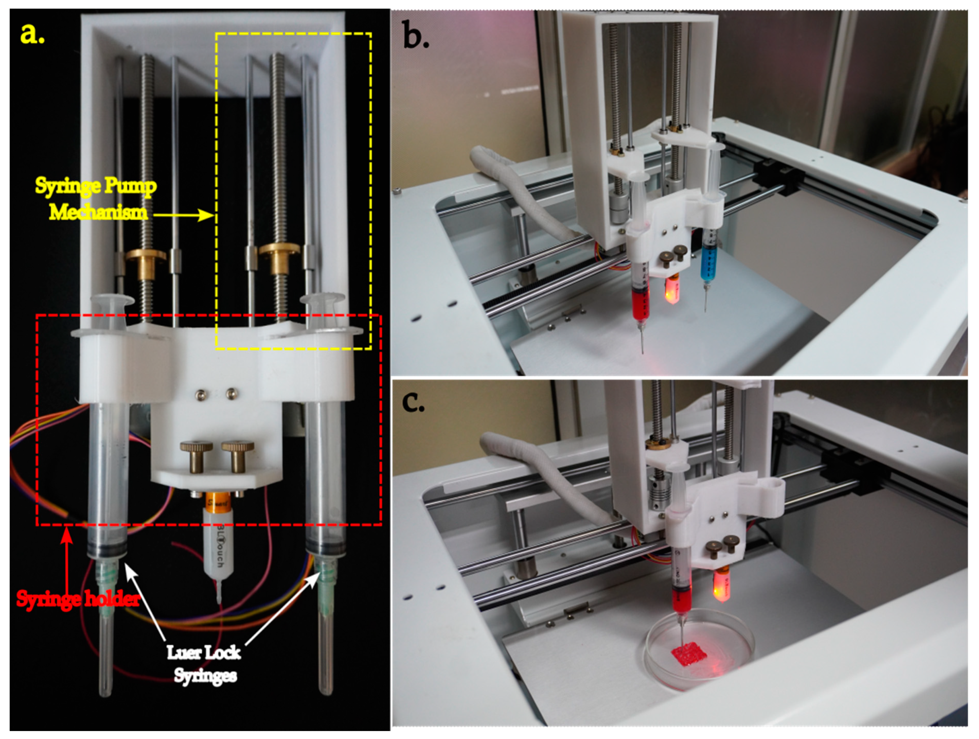
A syringe pump was designed and 3D printed using an FDM machine (Bambu Lab X1 Carbon, Shanghai, China, bought through WOL3D, Coimbatore) in ABS. The syringe pump consists of two assemblies, viz., the syringe holder and the syringe plunger mechanism. Both the syringe holder and the syringe plunger mechanism are attached separately to the X carriage of the printer, as shown in Figure 5. The syringe holder is capable of holding a maximum of two 10 mL syringes at once. The Z-axis probe is also connected to the syringe holder.
Each syringe plunger mechanism consists of a NEMA17 stepper motor mounted below the frame. The motion is then transferred to the lead screw, using a coupler. The syringe yoke is mounted on the lead screw and is supported by two smooth rods. When the stepper motor is operated, the syringe yoke can move linearly, upwards and downwards. The yoke can be coupled to the plunger of the syringe. This coupling causes the piston of the syringe to apply force to the material loaded in the syringe, thereby causing extrusion.

3.2. Conversion of the Electronic Mainboard

After the completion of the mechanical conversion, the electronics of the printer need to be modified. This step ensures extended customizability to future modifications. The base model is shipped with a custom-made motherboard, based on ATmega2560, Atmel, San Jose, CA, USA, which is an 8-bit microcontroller. With the board, only two syringe-pump assemblies can be added. However, considering the future expandability of the bioprinter, the board was swapped with BigTreeTech Octopus V1.1, which is not only a 32-bit board, but also has the support necessary to add five syringe-pump assemblies, a built-in WiFi interface, CAN- and SPI-based communications, and support for up to four additional heaters, to name a few of the desired features.
Similarly, the stepper-motor drivers were also upgraded to high-resolution TMC2209, which support 256 micro steps, are extremely silent in driving the stepper motors and can perform sensor-less homing with the StallGuard™ technology. Therefore, the end-stop sensors in the X and Y axes were removed and the board was configured appropriately.
To provide a sterile environment during the bioprinting process, a laminar flow hood setup, which recirculates the air through a HEPA filter, is placed on the top side of the printer. The air is taken in from the bottom part of the printer and released over the build plate as a laminar stream. This setup, in conjunction with the UV sterilization lamp, is started 30 min before the bioprinting process to ensure sterility of the chamber.

3.3. Configuration of the Firmware

The firmware is the software that resides inside the microcontroller of the mainboard. A vast majority of the commercially available open-sourced 3D printers use Marlin as firmware due to its stability, popularity, frequent bug fixes, and quicker introduction of new features. Marlin is also compatible with the majority of the mainboards, including the Octopus V1.1, which is used as the mainboard in this conversion. Therefore, the firmware is selected to be Marlin. The firmware is downloaded from the Marlin homepage, and before the firmware is uploaded onto the board, several changes in accordance with the hardware used need to be made in the configuration file. They are as follows.
The drivers for the X, Y, Z, and extruder axes need to be specified. By default, the driver type is A4988. Since we have used TMC2209 drivers, that needs to be specifically mentioned in the configuration file. All other motor options can be commented out.
  • #define X_DRIVER_TYPE TMC2209
  • #define Y_DRIVER_TYPE TMC2209
  • #define Z_DRIVER_TYPE TMC2209
  • #define E0_DRIVER_TYPE TMC2209→//Driver for the first extruder
  • #define E1_DRIVER_TYPE TMC2209→//Driver for the second extruder
Similarly, since we are using two extruders, it must be specified in the extruder settings section.
  • #define EXTRUDERS 2
The safety section is used for the prevention of cold extrusion and overtemperature. Those settings can be turned off, since the extrusion is often performed at room temperature in a bioprinter. Here, a very minimum temperature of 20 °C is given as a prevention temperature.
  • #define PREVENT_COLD_EXTRUSION
  • #define EXTRUDE_MINTEMP 20
As an alternative, a dummy thermistor, with a value greater than the cold extrusion temperature can also be provided in the firmware.
The settings were also changed in the Configuration_adv.h file. To enable sensorless homing, the following change needs to be made in the abovementioned file.
  • #define SENSORLESS_HOMING
  • #define X_STALL_SENSITIVITY 100
  • #define Y_STALL_SENSITIVITY 100
  • #define Z_STALL_SENSITIVITY 100
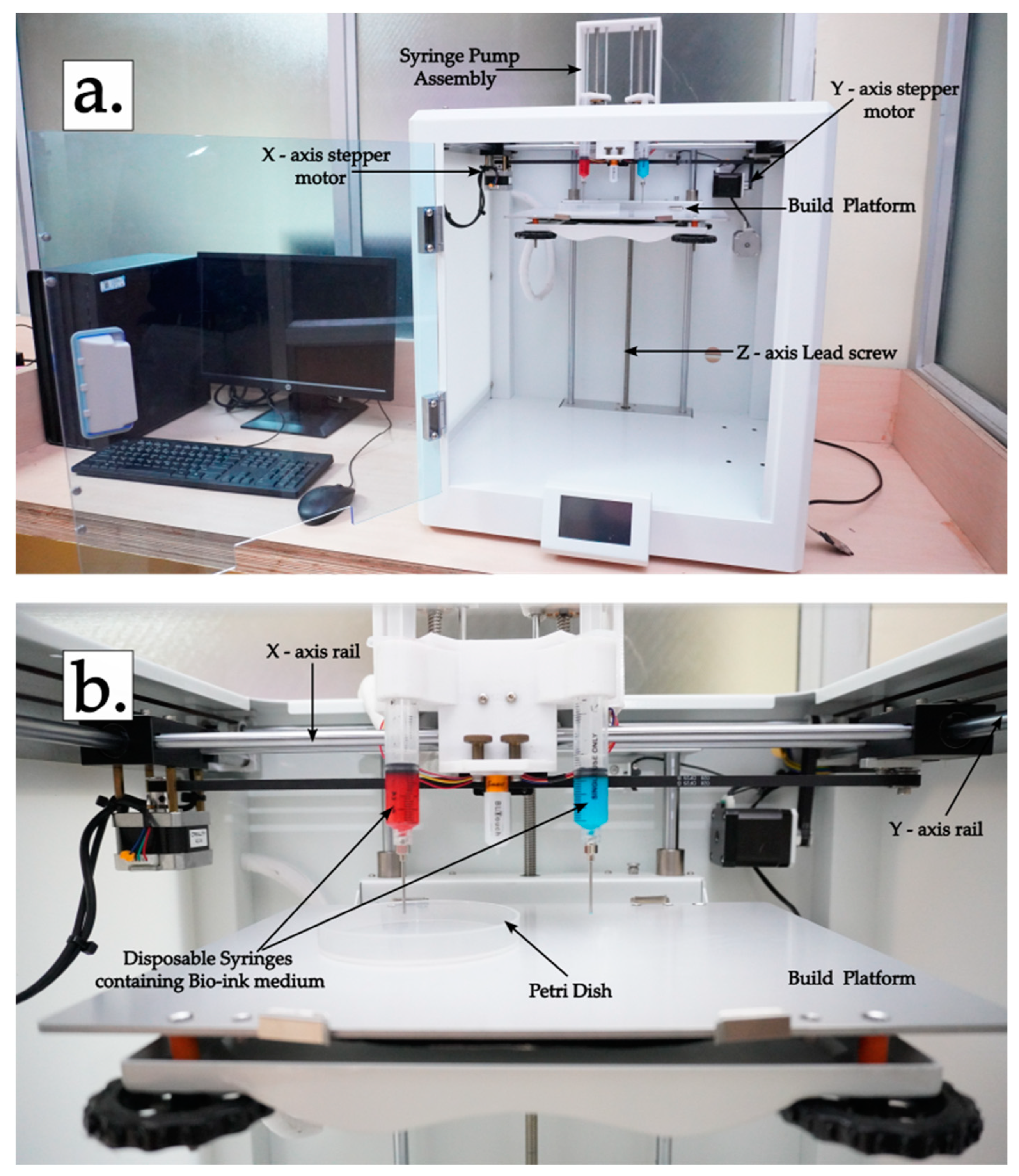
With all these settings, the firmware is cross-compiled on a personal computer and uploaded to the main board. With the upload, the conversion of the bioprinter is complete. The completed bioprinter is shown in Figure 6.

3.4. Testing of the Bioprinter

After the fabrication of the bioprinter, tests were performed to ensure suitability. First, the maximum extent of movement of the mechanism was found out after the modification. The maximum range in the X, Y, and Z axes is found to be 250 mm × 220 mm × 250 mm, yielding a total build volume of 13,750 cm3.
Resolution is an important characteristic of a mechanical system. It is the least amount of movement that can be achieved on any axis. It is dictated by the movement systems, stepper-motor specification, and microstepping configuration. The inverse of the steps/mm which is specified in the firmware can be used to find the resolution. For a belt-driven axis, which in our case means the X and Y axes, the steps/mm value can be calculated by
steps/mm = (steps/rotation × microsteps)/(belt pitch × pulley teeth)
For a stepper motor with 200 steps per rotation, and with a microstepping configuration of 256 microsteps per step, a 2 mm belt pitch, and a GT2 timing pulley of 20 teeth, the number of steps per mm is 1280 and the achievable printer resolution is 0.78 μm in the X and Y axes. Similarly, for the Z axis, which is driven by the lead screw mechanism, the steps/mm is
steps/mm = (steps/rotation×microsteps)/(screw pitch × number of screw starts)
As in the previous case, the number of steps/rotation of the stepper motor is 200, and the micro-step setting is 256. The screw pitch is 2 mm for a two-start screw. Therefore, the number of steps per mm is 12,800 and the resolution in the Z direction is 0.08 μm. The reported resolutions of a few of the commonly available bioprinters and thermoplastic printers are reported in Table 1.
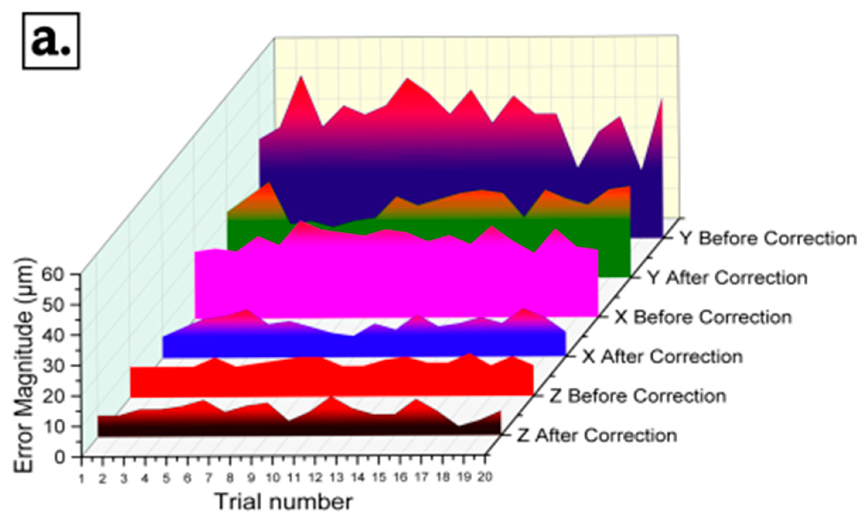
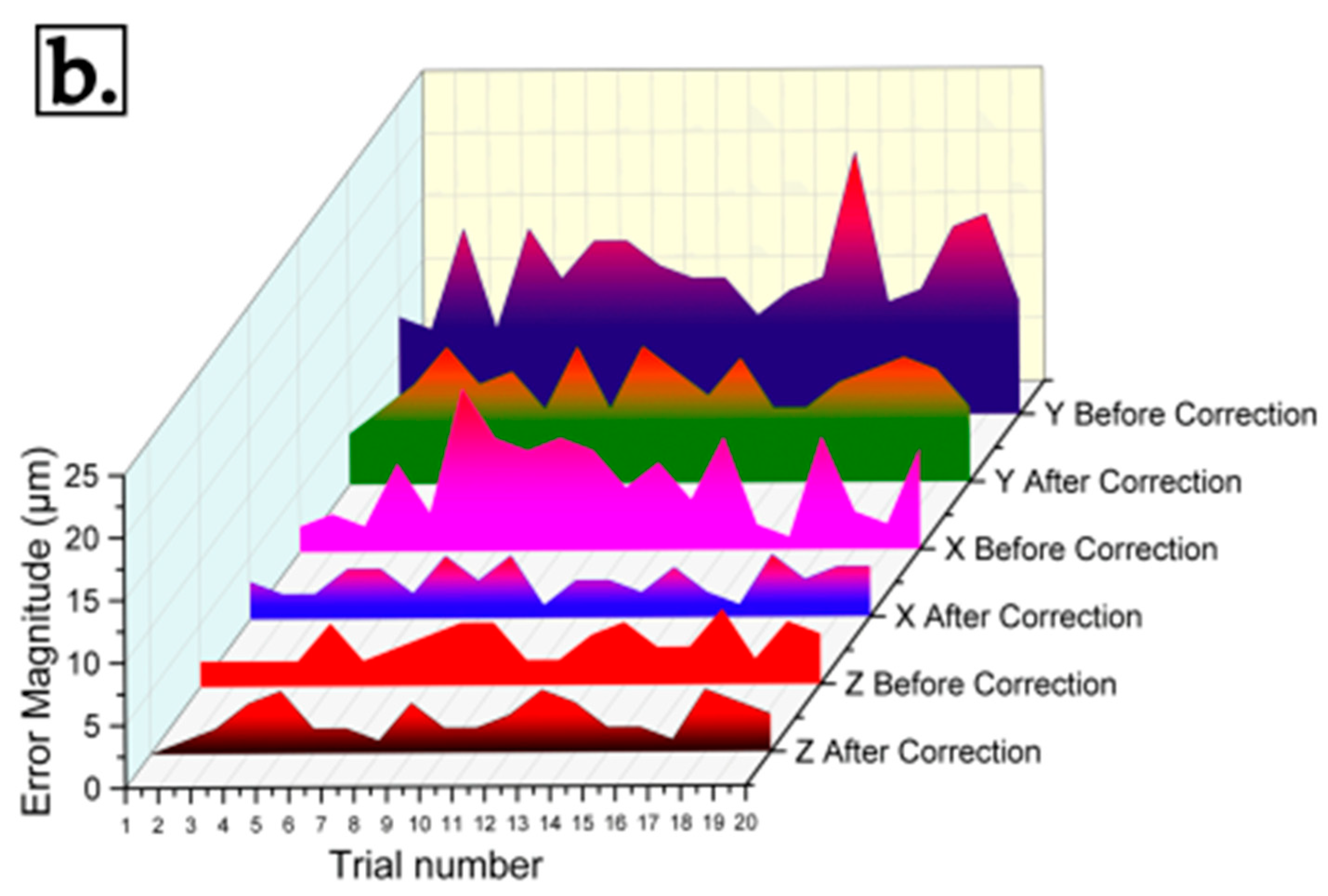
Both positional error and repeatability measurements were performed on all three axes. The positional error refers to the deviation between the intended position of the printer’s nozzle and its actual position during the printing process. Positional error is the difference between actual movement and estimated movement. Repeatability is the ability of the machine to reach the same point after moving to some other point. A mechanical dial indicator (2109S-10, Mitutoyo, Kawasaki, Japan) with a precision of 0.001 mm and a range of 25 mm was used to perform these measurements. The dial indicator was aligned parallel to each axis and supported using a magnetic base. To perform positional-error measurement, the axis is incrementally moved using the printer controls, and brought into contact with the probe of the dial indicator. The zero position is reset, and the axes are moved in 1 mm increments for a total of 20 mm. The difference between the actual value moved and the distance to be moved is taken as the error. The average positional error is determined as 26.4 μm for the X-axis, 40.21 μm for the Y-axis, and 11 μm for the Z-axis, and therefore a correction is included in the steps-per-mm setting, as mentioned in the literature [32]. The values of correction included in steps/mm are 1313, 1331, and 12,941 in the X, Y, and Z axes, respectively, to account for the skipped steps. Similarly, to perform the repeatability measurements, the printer is initially homed in all three axes. The dial indicator is set up as mentioned earlier in a convenient location. The axes are moved to come in contact with the dial indicator to around the median position of the range of the dial indicator and zeros are reset. The position of the printhead in the corresponding axis is noted and the axes are backed off by random distances to minimize systematic error. Using the G-code interface, with absolute referencing, the printer is moved to the set position using the linear interpolation command, G01. The over-travel or the under-travel specified by the dial gauge is taken as the error. This process is repeated several times to obtain an average value of repeatability. Figure 7 shows the measurement of positional error and repeatability, while Table 2 shows the average values obtained. After all of the above-mentioned procedures, a hydrogel was extruded in the modified 3D printer and it was demonstrated to successfully extrude the hydrogel in the desired shape. Figure 8 shows the plot variation of the positional error and repeatability.

4. Discussion

Several researchers around the world have reported their work on developing a bioprinter [9,28,29,30,32,33,35,67,68]. Our work revolves around the modification of a commercially available thermoplastic printer into a functional, extrusion-based, customizable bioprinter. The work differs from all previously reported works in the following areas. In this work, we report a bioprinter which, after modification, is capable of achieving sub-micron movement resolutions, as reported in Table 2. The improvement in resolution is mainly due to the usage of the stepper-motor driver (TMC2209, Trinamic, Hamburg, Germany) with higher levels of microstepping (256× compared to 16×). This, however, increases the work of the microcontroller by 16 times in controlling the stepper motor. Therefore, a powerful microcontroller is desired. In accordance with these needs, BigTreeTech Octopus Pro V1.1 mainboard (USD 50) with STM32F429ZE, which is a 32-bit microcontroller, 180 MHz CPU with ARM Cortex-M4 was selected. Additional reasons for the selection are the ability to use up to five extruders, which expands the space for further modification, the ability to supply a higher voltage to the stepper motors with a reduced current, keeping the motors cool and avoiding skipped steps, and the capability of adding additional accessories. The original mainboard, based on AtMega 2560, which is an 8-bit microcontroller, is swapped with the above-mentioned board.
A syringe pump mechanism is designed especially for the bioprinter and fabricated with ABS plastic. To perform the conversion, the existing extruder assembly was replaced with the syringe pump, and the electronic mainboard was swapped and configured as required. Necessary configurations were carried out in the Marlin firmware and the code was uploaded to the board.
The resolution of the printer is enhanced by the modification of the printer, and is in line with the reported resolutions of the commercial bioprinters, as discussed earlier. The resolution obtained is thought to be more than sufficient to perform bioprinting. However, if an additional resolution is required in the X and Y axes, it can be achieved by using a pulley with more teeth or by completely changing the belt-driven axes into screw-driven axes. Such a modification could improve the resolution by one order of magnitude. The measured values of positional error and repeatability of the customized bioprinter were also found to be within acceptable limits. It is also worth noting that, at a measurement precision of 1 μm when using the dial indicator, even the roughness of the contacting surface with the probe can have a profound effect on the measured values of repeatability since the contact point would disengage and re-engage during the process of measurement. On the other hand, for the measurement of positional error, since there is no re-engagement of the probe contact point during the entire period of measurement, no considerable effects were observed.
The process parameters that affect the extrusion-based bioprinting process were studied in detail by Tian et al. [69]. One of the most important output characteristics of a bioprinting process is printability, which is ability to extrude the bio-ink into high shape-fidelity prints and is mainly dependent upon the rheological characteristics of the bio-ink. It has been reported that the higher the viscosity of bio-ink, the better the printability. However, if the viscosity is higher than an optimal limit, the extrusion back pressure could cause pressure-related apoptosis of the cells in the bio-ink medium. Therefore, a shear thinning bioink is preferred. It has been reported in several studies in the literature that the cell density of the bio-ink also affects printability. Other parameters that affect printability are the bulk modulus, stiffness of the bioprinted scaffold, temperature of print, and crosslinking mechanics
The second important output of a bioprinting process is the cell viability after bioprinting, and it is dependent on many other factors, including rheology. Biocompatibility, which is the ability of the bio-ink medium to work in consonance with cells without causing cytotoxicity is another causative factor that affects the cell viability. Also, biodegradability, which is the ability of the bio-ink medium to slowly disintegrate as the cells proliferate and take up their space, is also an important input parameter. Other parameters that influence the cell viability include porosity of the fabricated scaffold and permeability, for nutrient and gas exchange.
Another parameter of the bioprinting process is the print resolution and shape fidelity. As in the case of well-established additive manufacturing techniques, it is dependent on the type of bioprinting. Laser-based processes tend to have the highest resolution among all bioprinting processes, allowing up to single-cell resolutions, followed by photopolymerization and inkjet bioprinting. Extrusion-based bioprinting tends to have the least resolution and shape fidelity, owing to the nozzle diameter and higher viscosity. A smaller nozzle diameter leads to better shape fidelity, but leads to higher wall shear stresses and back pressure, causing reduced cell viability.
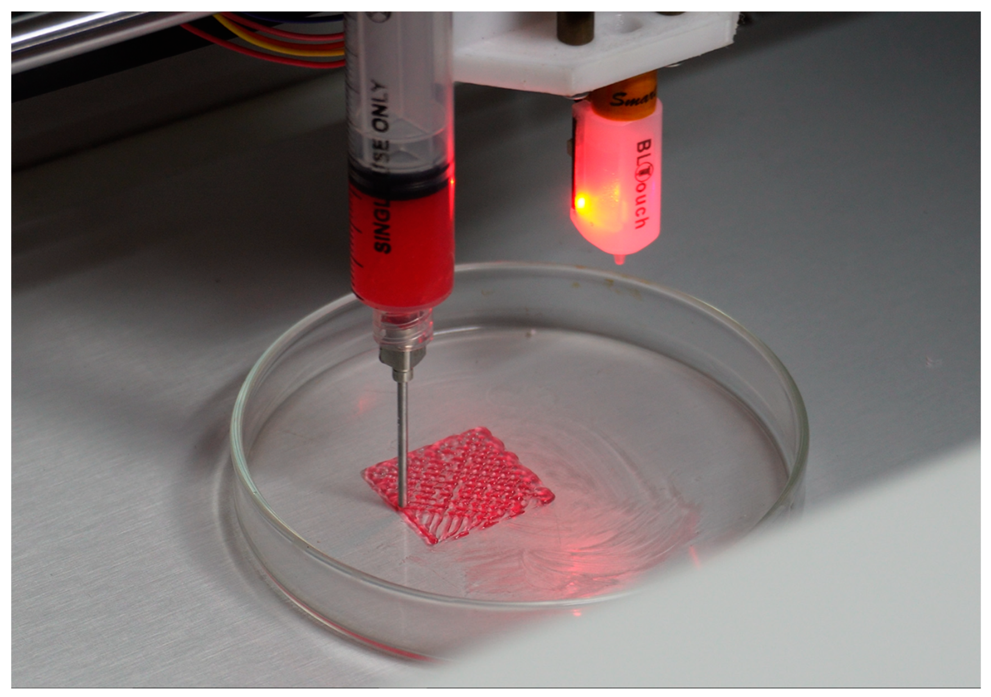
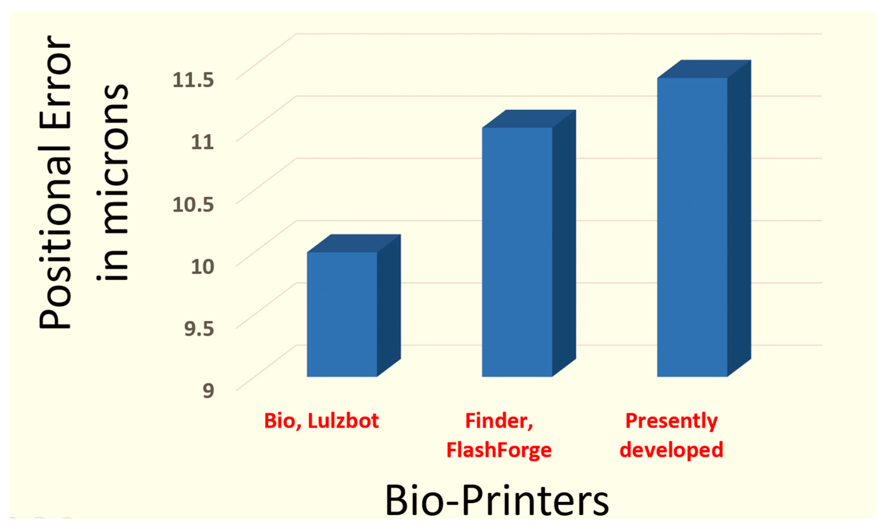
The fabricated custom bioprinter was validated by printing a polymer hydrogel in the setup. Figure 9 shows the hydrogel extruded by the bioprinter. The print resolutions are comparable to the results produced by commercially available bioprinting systems. The comparisons of positional error and repeatability is shown in Figure 10 and Figure 11.
The positioning errors of several printers are shown in Figure 10. The positioning inaccuracies (measured in microns) for the Bio, Lulzbot, Finder, FlashForge, and the currently under-development printers are shown visually in the chart. At 10 microns, the Bio, Lulzbot printer model has the least positional inaccuracy. This suggests that it can print with great precision. With a positional inaccuracy of only 11 microns, the Finder, FlashForge printer is marginally less accurate than the Bio, Lulzbot model, but it is still quite accurate. The positioning inaccuracy of the recently created printer is 11.4 microns. Despite being the largest positional error of the three models that are shown, this discrepancy nevertheless falls within a comparable range, demonstrating competitive precision.
The repeatability in microns of three distinct bio-printers—Bio, Lulzbot; Finder, FlashForge; and the currently in-development printer—is compared in the bar chart (Figure 11). The printer’s capacity to repeatedly return to the same spot under the same circumstances is referred to as repeatability. Better repeatability is indicated by lower values. Finder, FlashForge, has the lowest value, of 2.5 microns, and the best repeatability. The current printer exhibits a moderate level of performance, with a repeatability value of 3.1 microns. At 5 microns, Bio, Lulzbot has the highest repeatability value.
In the future, additional printheads having varying capabilities like a coaxial print head, which can form alginate structures in a calcium ionic solution, pneumatic print heads, micro-extrusion printheads, temperature-controlled print heads, integrated photocuring printheads, etc., can be added for additional functionality. Venturi-effect-based mixing of the cells and medium during extrusion is also a possibility, since such a process could reduce the pressure-related apoptosis of cells during printing, which is currently a problem in extrusion-based systems. A design for the quick loading and releasing of the print heads will also be a desirable feature. Also, the ability to perform FRESH bioprinting, as cited in several research articles, can enhance the usability of the printer. Integration with imaging systems can also provide better control of the printing parameters. On the firmware side, Klipper is a newly developed firmware that is making strides in the field of DIY additive-manufacturing machines. The advantage of Klipper over Marlin is that the interpretation of G-codes happens in a dedicated single-board computer, like Raspberry Pi, embedded inside the machine. Due to the offloading of work to the SBC, the mainboard can perform several tasks without crashing. Such systems can be added as a customization when multiple print heads are used. These future upgrades can add value to the bioprinter and enable researchers to perform a variety of experiments.

5. Conclusions

Converting a commercial thermoplastic 3D printer into a bioprinter provides a viable and cost-effective solution for researchers and laboratories working on a limited budget. This research highlights the feasibility of this approach as a cost-effective solution and provides an opportunity for laboratories and researchers working on a limited budget to access state-of-the-art bioprinting capabilities.
  • With adjustments made, the adapted printer can now print with incredibly high precision—resolutions of less than one micron. This level of accuracy is essential for complex bioprinting applications.
  • The developed printer exhibits performance comparable to high-end, more-expensive commercial bioprinters, thanks to modifications made to an easily accessible machine and the use of open-sourced firmware.
  • The ability to increase bioprinting’s accessibility is among the work’s most important ramifications. Expensive prices frequently serve as obstacles, but, by making changes to current equipment, we can remove these obstacles and democratize access.
  • The use of Venturi-effect-based cell and medium mixing during extrusion may be investigated in the future. This procedure might be able to solve the present problem of pressure-induced cell death during printing, which is common in extrusion systems. Increasing operating efficiency could be achieved by another design enhancement that makes it easier for print heads to load and release quickly. Furthermore, as other study publications have indicated, including the FRESH bioprinting method may greatly enhance the printer’s performance.
A significant advancement in bioprinting could be concentrating on integrating imaging systems, which would provide more precise control over print conditions. Due to its capacity to interpret G-codes via an integrated single-board computer, such as a Raspberry Pi, Klipper firmware may also provide significant software benefits when compared to the current Marlin firmware. This offloading makes sure that the mainboard can manage several jobs at once, particularly when using several print heads. These improvements are intended to increase the bioprinter’s worth and expand the range of experiments that may be carried out by researchers.

Author Contributions

Software, J.J.J.D. and S.V.P.; Validation, R.J.H.N.; Investigation, J.J.J.D.; Writing—review & editing, G.K. and S.V.P.; Visualization, G.K.; Supervision, R.J.H.N.; Project administration, R.J.H.N. All authors have read and agreed to the published version of the manuscript.

Funding

This research received no external funding.

Data Availability Statement

Data are contained within the article.

Acknowledgments

Author J.J.J.D. acknowledges the support of ACIC-Mepco Innovative Foundation, Mepco Schlenk Engineering College Campus, Sivakasi, Tamil Nadu, India. Author R.J.H.N. acknowledges the support of Ulam NAWA Postdoctoral Fellowship of the Polish National Agency for Academic Exchange programme, Contract No Agreement no. BPN/ULM/2022/1/00133/U/00001.

Conflicts of Interest

The authors declare no conflict of interest.

References

  1. Aljohani, W.; Ullah, M.W.; Zhang, X.; Yang, G. Bioprinting and Its Applications in Tissue Engineering and Regenerative Medicine. Int. J. Biol. Macromol. 2018, 107, 261–275. [Google Scholar] [CrossRef]
  2. Murphy, S.V.; Atala, A. 3D Bioprinting of Tissues and Organs. Nat. Biotechnol. 2014, 32, 773–785. [Google Scholar] [CrossRef]
  3. Mobaraki, M.; Ghaffari, M.; Yazdanpanah, A.; Luo, Y.; Mills, D.K. Bioinks and Bioprinting: A Focused Review. Bioprinting 2020, 18, e00080. [Google Scholar] [CrossRef]
  4. Yu, F.; Choudhury, D. Microfluidic Bioprinting for Organ-on-a-Chip Models. Drug Discov. Today 2019, 24, 1248–1257. [Google Scholar] [CrossRef] [PubMed]
  5. Antill-O’Brien, N.; Bourke, J.; O’Connell, C.D. Layer-By-Layer: The Case for 3D Bioprinting Neurons to Create Patient-Specific Epilepsy Models. Materials 2019, 12, 3218. [Google Scholar] [CrossRef]
  6. Ma, H.; Xing, F.; Yu, P.; Xu, J.; Wu, X.; Luo, R.; Xiang, Z.; Maria Rommens, P.; Duan, X.; Ritz, U. Integrated Design and Fabrication Strategies Based on Bioprinting for Skeletal Muscle Regeneration: Current Status and Future Perspectives. Mater. Des. 2023, 225, 111591. [Google Scholar] [CrossRef]
  7. Banda Sánchez, C.; Cubo Mateo, N.; Saldaña, L.; Valdivieso, A.; Earl, J.; González Gómez, I.; Rodríguez-Lorenzo, L.M. Selection and Optimization of a Bioink Based on PANC-1- Plasma/Alginate/Methylcellulose for Pancreatic Tumour Modelling. Polymers 2023, 15, 3196. [Google Scholar] [CrossRef]
  8. Moore, C.A.; Siddiqui, Z.; Carney, G.J.; Naaldijk, Y.; Guiro, K.; Ferrer, A.I.; Sherman, L.S.; Guvendiren, M.; Kumar, V.A.; Rameshwar, P. A 3D Bioprinted Material That Recapitulates the Perivascular Bone Marrow Structure for Sustained Hematopoietic and Cancer Models. Polymers 2021, 13, 480. [Google Scholar] [CrossRef]
  9. Ioannidis, K.; Danalatos, R.I.; Champeris Tsaniras, S.; Kaplani, K.; Lokka, G.; Kanellou, A.; Papachristou, D.J.; Bokias, G.; Lygerou, Z.; Taraviras, S. A Custom Ultra-Low-Cost 3D Bioprinter Supports Cell Growth and Differentiation. Front. Bioeng. Biotechnol. 2020, 8, 1279. [Google Scholar] [CrossRef]
  10. Noor, N.; Shapira, A.; Edri, R.; Gal, I.; Wertheim, L.; Dvir, T. 3D Printing of Personalized Thick and Perfusable Cardiac Patches and Hearts. Adv. Sci. 2019, 6, 1900344. [Google Scholar] [CrossRef]
  11. Li, L.; Lu, P.; Liu, Y.; Yang, J.; Li, S. Three-Dimensional-Bioprinted Bioactive Glass/Cellulose Composite Scaffolds with Porous Structure towards Bone Tissue Engineering. Polymers 2023, 15, 2226. [Google Scholar] [CrossRef] [PubMed]
  12. Duval, K.; Grover, H.; Han, L.-H.; Mou, Y.; Pegoraro, A.F.; Fredberg, J.; Chen, Z. Modeling Physiological Events in 2D vs. 3D Cell Culture. Physiology 2017, 32, 266–277. [Google Scholar] [CrossRef] [PubMed]
  13. Scognamiglio, C.; Soloperto, A.; Ruocco, G.; Cidonio, G. Bioprinting Stem Cells: Building Physiological Tissues One Cell at a Time. Am. J. Physiol. Cell Physiol. 2020, 319, C465–C480. [Google Scholar] [CrossRef] [PubMed]
  14. Ouyang, L.; Yao, R.; Zhao, Y.; Sun, W. Effect of Bioink Properties on Printability and Cell Viability for 3D Bioplotting of Embryonic Stem Cells. Biofabrication 2016, 8, 035020. [Google Scholar] [CrossRef] [PubMed]
  15. Gu, Q.; Tomaskovic-Crook, E.; Wallace, G.G.; Crook, J.M. 3D Bioprinting Human Induced Pluripotent Stem Cell Constructs for In Situ Cell Proliferation and Successive Multilineage Differentiation. Adv. Healthc. Mater. 2017, 6, 1700175. [Google Scholar] [CrossRef] [PubMed]
  16. Koch, L.; Deiwick, A.; Franke, A.; Schwanke, K.; Haverich, A.; Zweigerdt, R.; Chichkov, B. Laser Bioprinting of Human Induced Pluripotent Stem Cells—The Effect of Printing and Biomaterials on Cell Survival, Pluripotency, and Differentiation. Biofabrication 2018, 10, 035005. [Google Scholar] [CrossRef] [PubMed]
  17. Bae, S.-W.; Lee, K.-W.; Park, J.-H.; Lee, J.; Jung, C.-R.; Yu, J.; Kim, H.-Y.; Kim, D.-H. 3D Bioprinted Artificial Trachea with Epithelial Cells and Chondrogenic-Differentiated Bone Marrow-Derived Mesenchymal Stem Cells. Int. J. Mol. Sci. 2018, 19, 1624. [Google Scholar] [CrossRef] [PubMed]
  18. Liang, L.; Li, Z.; Yao, B.; Enhe, J.; Song, W.; Zhang, C.; Zhu, P.; Huang, S. Extrusion Bioprinting of Cellular Aggregates Improves Mesenchymal Stem Cell Proliferation and Differentiation. Biomater. Adv. 2023, 149, 213369. [Google Scholar] [CrossRef]
  19. Hsieh, F.-Y.; Lin, H.-H.; Hsu, S. 3D Bioprinting of Neural Stem Cell-Laden Thermoresponsive Biodegradable Polyurethane Hydrogel and Potential in Central Nervous System Repair. Biomaterials 2015, 71, 48–57. [Google Scholar] [CrossRef]
  20. Tao, J.; Zhu, S.; Liao, X.; Wang, Y.; Zhou, N.; Li, Z.; Wan, H.; Tang, Y.; Yang, S.; Du, T.; et al. DLP-Based Bioprinting of Void-Forming Hydrogels for Enhanced Stem-Cell-Mediated Bone Regeneration. Mater. Today Bio 2022, 17, 100487. [Google Scholar] [CrossRef]
  21. Yun, Y.E.; Jung, Y.J.; Choi, Y.J.; Choi, J.S.; Cho, Y.W. Artificial Skin Models for Animal-Free Testing. J. Pharm. Investig. 2018, 48, 215–223. [Google Scholar] [CrossRef]
  22. Frankowski, J.; Kurzątkowska, M.; Sobczak, M.; Piotrowska, U. Utilization of 3D Bioprinting Technology in Creating Human Tissue and Organoid Models for Preclinical Drug Research—State-of-the-Art. Int. J. Pharm. 2023, 644, 123313. [Google Scholar] [CrossRef] [PubMed]
  23. Mielczarek, J.; Gazdowicz, G.; Kramarz, J.; Łątka, P.; Krzykawski, M.; Miroszewski, A.; Pieczarko, P.; Szczelina, R.; Warchoł, P.; Wróbel, S. A Prototype of a 3D Bioprinter. Solid State Phenom. 2015, 237, 221–226. [Google Scholar] [CrossRef]
  24. Wang, Z.; Abdulla, R.; Parker, B.; Samanipour, R.; Ghosh, S.; Kim, K. A Simple and High-Resolution Stereolithography-Based 3D Bioprinting System Using Visible Light Crosslinkable Bioinks. Biofabrication 2015, 7, 045009. [Google Scholar] [CrossRef] [PubMed]
  25. Goldstein, T.A.; Epstein, C.J.; Schwartz, J.; Krush, A.; Lagalante, D.J.; Mercadante, K.P.; Zeltsman, D.; Smith, L.P.; Grande, D.A. Feasibility of Bioprinting with a Modified Desktop 3D Printer. Tissue Eng. Part C Methods 2016, 22, 1071–1076. [Google Scholar] [CrossRef]
  26. Reid, J.A.; Mollica, P.A.; Johnson, G.D.; Ogle, R.C.; Bruno, R.D.; Sachs, P.C. Accessible Bioprinting: Adaptation of a Low-Cost 3D-Printer for Precise Cell Placement and Stem Cell Differentiation. Biofabrication 2016, 8, 025017. [Google Scholar] [CrossRef]
  27. Roehm, K.D.; Madihally, S.V. Bioprinted Chitosan-Gelatin Thermosensitive Hydrogels Using an Inexpensive 3D Printer. Biofabrication 2017, 10, 015002. [Google Scholar] [CrossRef] [PubMed]
  28. Bessler, N.; Ogiermann, D.; Buchholz, M.-B.; Santel, A.; Heidenreich, J.; Ahmmed, R.; Zaehres, H.; Brand-Saberi, B. Nydus One Syringe Extruder (NOSE): A Prusa I3 3D Printer Conversion for Bioprinting Applications Utilizing the FRESH-Method. HardwareX 2019, 6, e00069. [Google Scholar] [CrossRef]
  29. Kahl, M.; Gertig, M.; Hoyer, P.; Friedrich, O.; Gilbert, D.F. Ultra-Low-Cost 3D Bioprinting: Modification and Application of an Off-the-Shelf Desktop 3D-Printer for Biofabrication. Front. Bioeng. Biotechnol. 2019, 7, 184. [Google Scholar] [CrossRef]
  30. Yenilmez, B.; Temirel, M.; Knowlton, S.; Lepowsky, E.; Tasoglu, S. Development and Characterization of a Low-Cost 3D Bioprinter. Bioprinting 2019, 13, e00044. [Google Scholar] [CrossRef]
  31. Sanz-Garcia, A.; Sodupe-Ortega, E.; Pernía-Espinoza, A.; Shimizu, T.; Escobedo-Lucea, C. A Versatile Open-Source Printhead for Low-Cost 3D Microextrusion-Based Bioprinting. Polymers 2020, 12, 2346. [Google Scholar] [CrossRef] [PubMed]
  32. Tashman, J.W.; Shiwarski, D.J.; Feinberg, A.W. Development of a High-Performance Open-Source 3D Bioprinter. Sci. Rep. 2022, 12, 22652. [Google Scholar] [CrossRef] [PubMed]
  33. Lei, I.M.; Sheng, Y.; Lei, C.L.; Leow, C.; Huang, Y.Y.S. A Hackable, Multi-Functional, and Modular Extrusion 3D Printer for Soft Materials. Sci. Rep. 2022, 12, 12294. [Google Scholar] [CrossRef] [PubMed]
  34. Breideband, L.; Wächtershäuser, K.N.; Hafa, L.; Wieland, K.; Frangakis, A.S.; Stelzer, E.H.K.; Pampaloni, F. Upgrading a Consumer Stereolithographic 3D Printer to Produce a Physiologically Relevant Model with Human Liver Cancer Organoids. Adv. Mater. Technol. 2022, 7, 2200029. [Google Scholar] [CrossRef]
  35. D’Atanasio, P.; Fiaschini, N.; Rinaldi, A.; Zambotti, A.; Cantini, L.; Mancuso, M.; Antonelli, F. Design and Implementation of an Accessible 3D Bioprinter: Benchmarking the Performance of a Home-Made Bioprinter against a Professional Bioprinter. Appl. Sci. 2023, 13, 10213. [Google Scholar] [CrossRef]
  36. Yilmaz, B.; Al Rashid, A.; Mou, Y.A.; Evis, Z.; Koç, M. Bioprinting: A Review of Processes, Materials and Applications. Bioprinting 2021, 23, e00148. [Google Scholar] [CrossRef]
  37. Betancourt, N.; Chen, X. Review of Extrusion-Based Multi-Material Bioprinting Processes. Bioprinting 2022, 25, e00189. [Google Scholar] [CrossRef]
  38. Ng, W.L.; Lee, J.M.; Zhou, M.; Chen, Y.-W.; Lee, K.-X.A.; Yeong, W.Y.; Shen, Y.-F. Vat Polymerization-Based Bioprinting—Process, Materials, Applications and Regulatory Challenges. Biofabrication 2020, 12, 022001. [Google Scholar] [CrossRef] [PubMed]
  39. Li, Y.; Zhang, X.; Zhang, X.; Zhang, Y.; Hou, D. Recent Progress of the Vat Photopolymerization Technique in Tissue Engineering: A Brief Review of Mechanisms, Methods, Materials, and Applications. Polymers 2023, 15, 3940. [Google Scholar] [CrossRef]
  40. Dou, C.; Perez, V.; Qu, J.; Tsin, A.; Xu, B.; Li, J. A State-of-the-Art Review of Laser-Assisted Bioprinting and Its Future Research Trends. ChemBioEng Rev. 2021, 8, 517–534. [Google Scholar] [CrossRef]
  41. Li, J.; Wu, C.; Chu, P.K.; Gelinsky, M. 3D Printing of Hydrogels: Rational Design Strategies and Emerging Biomedical Applications. Mater. Sci. Eng. R Rep. 2020, 140, 100543. [Google Scholar] [CrossRef]
  42. Ning, L.; Chen, X. A Brief Review of Extrusion-based Tissue Scaffold Bio-printing. Biotechnol. J. 2017, 12, 1600671. [Google Scholar] [CrossRef] [PubMed]
  43. Ning, L.; Yang, B.; Mohabatpour, F.; Betancourt, N.; Sarker, M.; Papagerakis, P.; Chen, X. Process-Induced Cell Damage: Pneumatic versus Screw-Driven Bioprinting. Biofabrication 2020, 12, 025011. [Google Scholar] [CrossRef] [PubMed]
  44. Fernández-Pradas, J.M.; Colina, M.; Serra, P.; Domínguez, J.; Morenza, J.L. Laser-Induced Forward Transfer of Biomolecules. Thin Solid Film. 2004, 453–454, 27–30. [Google Scholar] [CrossRef]
  45. Keriquel, V.; Oliveira, H.; Rémy, M.; Ziane, S.; Delmond, S.; Rousseau, B.; Rey, S.; Catros, S.; Amédée, J.; Guillemot, F.; et al. In Situ Printing of Mesenchymal Stromal Cells, by Laser-Assisted Bioprinting, for in Vivo Bone Regeneration Applications. Sci. Rep. 2017, 7, 1778. [Google Scholar] [CrossRef] [PubMed]
  46. Wijshoff, H. The Dynamics of the Piezo Inkjet Printhead Operation. Phys. Rep. 2010, 491, 77–177. [Google Scholar] [CrossRef]
  47. Kamisuki, S.; Hagata, T.; Tezuka, C.; Nose, Y.; Fujii, M.; Atobe, M. A Low Power, Small, Electrostatically-Driven Commercial Inkjet Head. In Proceedings of the MEMS 98. IEEE. Eleventh Annual International Workshop on Micro Electro Mechanical Systems. An Investigation of Micro Structures, Sensors, Actuators, Machines and Systems, Cat. No.98CH36176, Heidelberg, Germany, 25–29 January 1998; IEEE: Piscataway, NJ, USA, 1998; pp. 63–68. [Google Scholar]
  48. Kim, J.D.; Choi, J.S.; Kim, B.S.; Chan Choi, Y.; Cho, Y.W. Piezoelectric Inkjet Printing of Polymers: Stem Cell Patterning on Polymer Substrates. Polymer 2010, 51, 2147–2154. [Google Scholar] [CrossRef]
  49. Goodarzi Hosseinabadi, H.; Dogan, E.; Miri, A.K.; Ionov, L. Digital Light Processing Bioprinting Advances for Microtissue Models. ACS Biomater. Sci. Eng. 2022, 8, 1381–1395. [Google Scholar] [CrossRef] [PubMed]
  50. Lim, W.; Kim, G.J.; Kim, H.W.; Lee, J.; Zhang, X.; Kang, M.G.; Seo, J.W.; Cha, J.M.; Park, H.J.; Lee, M.-Y.; et al. Kappa-Carrageenan-Based Dual Crosslinkable Bioink for Extrusion Type Bioprinting. Polymers 2020, 12, 2377. [Google Scholar] [CrossRef]
  51. Khoshnood, N.; Zamanian, A. A Comprehensive Review on Scaffold-Free Bioinks for Bioprinting. Bioprinting 2020, 19, e00088. [Google Scholar] [CrossRef]
  52. Paxton, N.; Smolan, W.; Böck, T.; Melchels, F.; Groll, J.; Jungst, T. Proposal to Assess Printability of Bioinks for Extrusion-Based Bioprinting and Evaluation of Rheological Properties Governing Bioprintability. Biofabrication 2017, 9, 044107. [Google Scholar] [CrossRef] [PubMed]
  53. Pepelnjak, T.; Stojšić, J.; Sevšek, L.; Movrin, D.; Milutinović, M. Influence of Process Parameters on the Characteristics of Additively Manufactured Parts Made from Advanced Biopolymers. Polymers 2023, 15, 716. [Google Scholar] [CrossRef] [PubMed]
  54. Suntornnond, R.; Tan, E.; An, J.; Chua, C. A Mathematical Model on the Resolution of Extrusion Bioprinting for the Development of New Bioinks. Materials 2016, 9, 756. [Google Scholar] [CrossRef] [PubMed]
  55. Kyle, S.; Jessop, Z.M.; Al-Sabah, A.; Whitaker, I.S. ‘Printability’ of Candidate Biomaterials for Extrusion Based 3D Printing: State-of-the-Art. Adv. Healthc. Mater. 2017, 6, 1700264. [Google Scholar] [CrossRef] [PubMed]
  56. Lee, J.M.; Suen, S.K.Q.; Ng, W.L.; Ma, W.C.; Yeong, W.Y. Bioprinting of Collagen: Considerations, Potentials, and Applications. Macromol. Biosci. 2021, 21, e2000280. [Google Scholar] [CrossRef] [PubMed]
  57. Li, W.; Mille, L.S.; Robledo, J.A.; Uribe, T.; Huerta, V.; Zhang, Y.S. Recent Advances in Formulating and Processing Biomaterial Inks for Vat Polymerization-Based 3D Printing. Adv. Healthc. Mater. 2020, 9, e2000156. [Google Scholar] [CrossRef] [PubMed]
  58. Sameni, F.; Ozkan, B.; Zarezadeh, H.; Karmel, S.; Engstrøm, D.S.; Sabet, E. Hot Lithography Vat Photopolymerisation 3D Printing: Vat Temperature vs. Mixture Design. Polymers 2022, 14, 2988. [Google Scholar] [CrossRef] [PubMed]
  59. Dababneh, A.B.; Ozbolat, I.T. Bioprinting Technology: A Current State-of-the-Art Review. J. Manuf. Sci. Eng. 2014, 136, 061016. [Google Scholar] [CrossRef]
  60. Hölzl, K.; Lin, S.; Tytgat, L.; Van Vlierberghe, S.; Gu, L.; Ovsianikov, A. Bioink Properties before, during and after 3D Bioprinting. Biofabrication 2016, 8, 032002. [Google Scholar] [CrossRef]
  61. Chopin-Doroteo, M.; Mandujano-Tinoco, E.A.; Krötzsch, E. Tailoring of the Rheological Properties of Bioinks to Improve Bioprinting and Bioassembly for Tissue Replacement. Biochim. Biophys. Acta (BBA)-Gen. Subj. 2021, 1865, 129782. [Google Scholar] [CrossRef]
  62. Fatimi, A.; Okoro, O.V.; Podstawczyk, D.; Siminska-Stanny, J.; Shavandi, A. Natural Hydrogel-Based Bio-Inks for 3D Bioprinting in Tissue Engineering: A Review. Gels 2022, 8, 179. [Google Scholar] [CrossRef]
  63. Damiati, L.A.; El-Yaagoubi, M.; Damiati, S.A.; Kodzius, R.; Sefat, F.; Damiati, S. Role of Polymers in Microfluidic Devices. Polymers 2022, 14, 5132. [Google Scholar] [CrossRef]
  64. Chimene, D.; Kaunas, R.; Gaharwar, A.K. Hydrogel Bioink Reinforcement for Additive Manufacturing: A Focused Review of Emerging Strategies. Adv. Mater. 2020, 32, e1902026. [Google Scholar] [CrossRef]
  65. Ozbolat, I.T.; Moncal, K.K.; Gudapati, H. Evaluation of Bioprinter Technologies. Addit. Manuf. 2017, 13, 179–200. [Google Scholar] [CrossRef]
  66. Koch, F.; Thaden, O.; Tröndle, K.; Zengerle, R.; Zimmermann, S.; Koltay, P. Open-Source Hybrid 3D-Bioprinter for Simultaneous Printing of Thermoplastics and Hydrogels. HardwareX 2021, 10, e00230. [Google Scholar] [CrossRef]
  67. Lobo, D.A.; Ginestra, P. Cell Bioprinting: The 3D-BioplotterTM Case. Materials 2019, 12, 4005. [Google Scholar] [CrossRef]
  68. Krige, A.; Haluška, J.; Rova, U.; Christakopoulos, P. Design and Implementation of a Low Cost Bio-Printer Modification, Allowing for Switching between Plastic and Gel Extrusion. HardwareX 2021, 9, e00186. [Google Scholar] [CrossRef]
  69. Tian, S.; Zhao, H.; Lewinski, N. Key Parameters and Applications of Extrusion-Based Bioprinting. Bioprinting 2021, 23, e00156. [Google Scholar] [CrossRef]
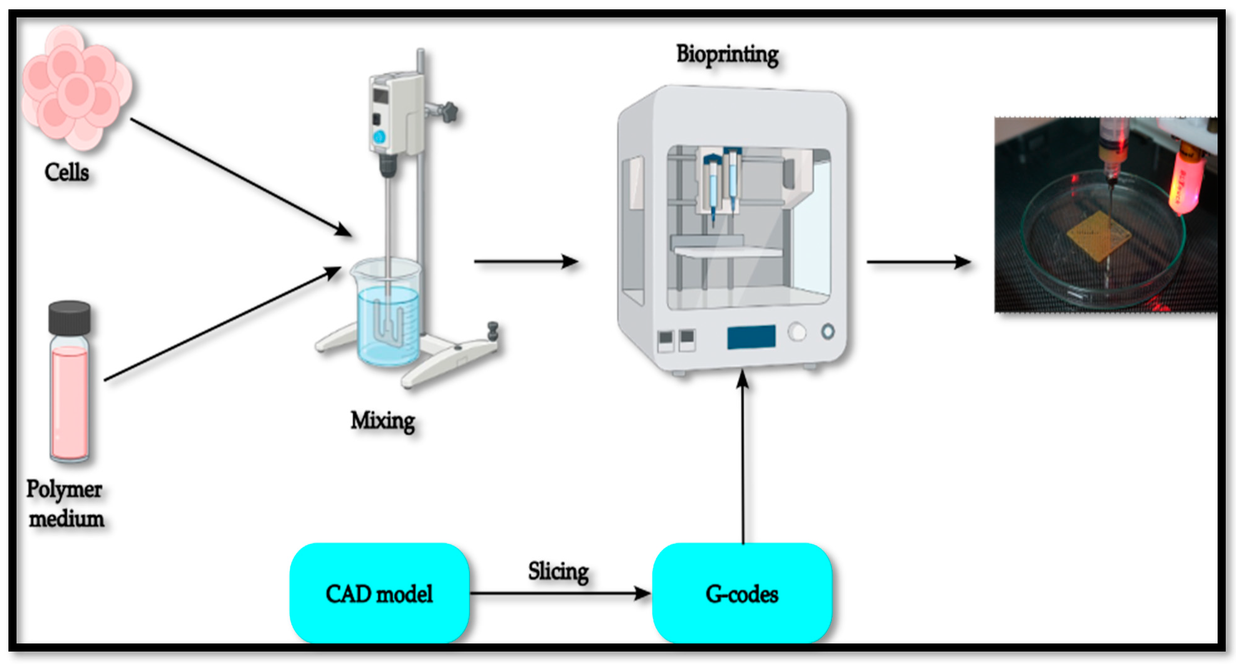
Figure 1. General bioprinting process.
Machines 12 00518 g001
Figure 2. Various Bioprinting methodologies.
Machines 12 00518 g002
Figure 3. The general architecture of a thermoplastic 3D printer.
Machines 12 00518 g003
Figure 4. Rough geometric layout of the bioprinter.
Machines 12 00518 g004
Figure 5. (a) Fabricated syringe pump with the syringe holder and the syringe pump mechanism; (b) assembly of syringe pump in the customized printer; (c) trial print using the syringe pump.
Machines 12 00518 g005
Figure 6. (a) Completed bioprinter; (b) close-up view of the screw-pump extrusion printhead.
Machines 12 00518 g006
Figure 7. Precision check of the bioprinter using a dial indicator.
Machines 12 00518 g007
Figure 8. (a) Positional error measurement. (b) Repeatability measurement.
Machines 12 00518 g008aMachines 12 00518 g008b
Figure 9. Testing of the 3D bioprinter with hydrogel.
Machines 12 00518 g009
Figure 10. Comparison of positional error with the commercial printers.
Machines 12 00518 g010
Figure 11. Comparison of repeatability with the commercial printers.
Machines 12 00518 g011
Table 1. Resolutions of commercially available bioprinters and thermoplastic printers.
PrinterManufacturerResolution in the X and Y Direction (μm)Resolution in the Z Direction (μm)
3D BioplotterEnvisionTEC11
Allevi 2Allevi 3D51
BioLulzbot105
BioXCellink11
FinderFlashForge112.5
Customized Bioprinter-0.780.08
Table 2. Average positional error and repeatability.
AxisPositional Error (μm)Repeatability (μm)
Before CorrectionAfter CorrectionBefore CorrectionAfter Correction
X26.411.25.73.1
Y40.224.711.78
Z11.08.43.42.7
Disclaimer/Publisher’s Note: The statements, opinions and data contained in all publications are solely those of the individual author(s) and contributor(s) and not of MDPI and/or the editor(s). MDPI and/or the editor(s) disclaim responsibility for any injury to people or property resulting from any ideas, methods, instructions or products referred to in the content.