Review

A Review of Boron-Bearing Minerals (Excluding Tourmaline) in the Adirondack Region of New York State

by
David G. Bailey
1,*,
Marian V. Lupulescu
2,
Robert S. Darling
3,
Jared W. Singer
4 and
Steven C. Chamberlain
5
1
Geosciences Department, Hamilton College, Clinton, NY 13323, USA
2
New York State Museum, Research and Collections, Albany, NY 12230, USA
3
Department of Geology, State University of New York, College at Cortland, Cortland, NY 13045, USA
4
Earth & Environmental Sciences, Rensselaer Polytechnic Institute, Troy, NY 12180, USA
5
Center for Mineralogy, New York State Museum, Albany, NY 12230, USA
*
Author to whom correspondence should be addressed.
Minerals 2019, 9(10), 644; https://doi.org/10.3390/min9100644
Submission received: 23 August 2019 / Revised: 10 October 2019 / Accepted: 12 October 2019 / Published: 22 October 2019
(This article belongs to the Special Issue Minerals of the Southern Grenville Province)

Abstract

:
Boron is a biologically important element, but its distribution in the natural environment and its behavior during many geological processes is not fully understood. In most metamorphic and igneous environments, boron is incorporated into minerals of the tourmaline supergroup. In high-grade metamorphic terranes like that of the Adirondack region of northern New York State, uncommon rock compositions combined with unusual and variable geologic conditions resulted in the formation of many additional boron-bearing minerals. This paper reviews the occurrences and geological settings of twelve relatively uncommon boron-bearing minerals in the southern Grenville Province of upstate New York and provides new chemical and Raman spectral data for seven of these minerals. The boron minerals range from relatively simple metal borates (e.g., vonsenite), to chemically complex borosilicates (e.g., prismatine), to a relatively rare borosilicate-carbonate (e.g., harkerite). Some are of primary igneous origin, while others are formed by a variety of prograde and retrograde metamorphic processes or by metasomatic/hydrothermal processes. Most of the boron minerals are formed within, or adjacent to, metasedimentary lithologies that surround the anorthositic massifs of the central Adirondacks. The metasedimentary rocks are thought to be the source of most of the boron, although additional boron isotope studies are needed to confirm this and to constrain the mechanisms of the formation of these unusual minerals.

1. Introduction

Boron is an unusual element that is essential for life [1] and has many important technical applications, from rocket-fuel igniter to antiseptic eye drops. Because of its small ionic radius, boron is an element not found in most of the common rock-forming minerals, and few geologists are familiar with the distribution of boron in the Earth, or how boron behaves in geological processes. Because most borate compounds are water-soluble, borate minerals are most common in evaporite deposits and in clay-bearing sediments [2]. In metamorphic and igneous rocks, most boron is incorporated into tourmaline because of its stability over a wide range of pressures, temperatures, and bulk rock compositions [3]. In metamorphic rocks, other boron-bearing minerals are uncommon and form only in lithologies of unusual composition, or under unusual metamorphic conditions [3]. Recent studies [4,5,6] have documented the complexity and diversity of boron-bearing minerals, and how both of these have increased over the Earth’s history.
Spectacular, well-formed crystals of danburite and tourmaline from northern New York have been widely known and collected for over 150 years [7,8,9]. Over the past three decades, with the advance of microanalytical techniques, many more boron-bearing minerals have been identified throughout the Adirondack region of upstate New York. These minerals range from relatively simple metal borates (e.g., vonsenite), to chemically complex borosilicates (e.g., prismatine), to a relatively rare borosilicate–carbonate (e.g., harkerite). To date, 12 boron-bearing minerals (excluding tourmaline) have been reported from this region. The goal of this paper is to draw attention to the diversity of boron-bearing minerals in northern New York, to provide a review of their occurrences and geological contexts, and to provide new chemical and Raman spectral data for seven of these unusual minerals.

2. Materials and Methods

Much of the information presented in this paper is from previously published sources, which are cited. Seven of the 12 boron minerals were provided by the New York State Museum in Albany, NY, for additional study. New chemical analyses for these minerals were obtained by electron microprobe (EMP) and laser ablation, inductively-coupled, mass spectrometry (LA-ICPMS) at Rensselaer Polytechnic Institute (RPI), Troy, NY, USA. Individual mineral grains were epoxy mounted, polished using 1 μm alumina powder followed by colloidal silica, and carbon-coated for electron probe microanalysis using the CAMECA SX-100 at RPI. Typical major elements were measured, including Na, Mg, Al, Si, K, Ca, Ti, Mn, and Fe using a 20 nA beam current at 15 kV accelerating voltage. For stillwellite, major elements (REE, Si) and Fe were measured at 15 kV and with a 10 nA beam current. Sm and Gd were measured using the Lβ emission line, and Pr was measured using the Lα line and corrected for the La peak overlap. Trace elements in stillwellite (S, P, Cl, Ca, Y, Pb, Th, and U) were measured using a 60 nA beam current. Peak and background acquisition times were 30 s each, for each element (except Ti at 60 s each). Oxygen was calculated based on cation stoichiometry, and boron (oxide) was calculated by difference. For datolite, boron oxide was determined by difference after assuming stoichiometry of 1 mole (OH) per mole SiO2. Matrix corrections were applied using the Pouchou and Pichoir algorithm in the CAMECA Peaksight Software (version 5.1, CAMECA, Gennevilliers, France). Each specimen was analyzed with 3 replicates.
The same polished mineral mounts were analyzed for trace elements by a laser ablation (Photon Machines Analyte 193) inductively coupled plasma mass spectrometer (Varian 820MS), also at RPI. The ablation duration was 30 s at a 7 Hz repetition rate with a fluence of ~6 J/cm2. The spot size was 80 μm square, except for dumortierite, which utilized a 30 μm circle. Background acquisition was ~30 s allowing ~15 s for washout between the peak and background integrations. Each specimen was analyzed with 3 replicates, and helium carrier gas transported the aerosol to the ICPMS. The plasma was run in hot-plasma mode with no collision gases. Data were processed in the Iolite software package by the Trace Elements IS (Internal Standard) data reduction scheme using NIST 610 and USGS GSD-1g as possible reference materials. Data were computed iteratively with either reference material applied as the primary reference and recomputed with various possible internal standards, including Mg, Al, Si, Ca, and Fe. For danburite, Si and Ca were used as an internal standard and both NIST 610 and USGS GSD-1g were used as reference glasses. For datolite, dumortierite, prismatine, and serendibite, Si and Al were used as internal standards and both NIST 610 and USGS GSD-1g were used as reference glasses. For vonsenite, Mg, Al, and Fe were used as internal standards and only USGS GSD-1g was used as a reference glass. For each mineral phase, 3 spatial replicates were averaged. NIST and USGS glass analyses were processed in the same manner as ‘secondary’ standards, and reasonable values were obtained.
Raman spectra were obtained using a Thermo-Fisher Almega XR spectrometer at Hamilton College. Spectra were collected on polished, unoriented mineral surfaces excited with a 532 nm laser at 80% power and using a 100 μm slit aperture.

3. Boron Minerals in the Adirondack Mountains

In addition to tourmaline supergroup minerals [10], twelve boron-bearing minerals have been reported from the Adirondack region of northern New York (Table 1; Figure 1). Precise locations are being withheld to prevent uncontrolled collecting of these minerals, but are available upon request for most localities for the purposes of scientific research.
The Adirondack Mountains in New York are divided into the Adirondack Highlands and Adirondack Lowlands by a zone of deformation known as the Colton–Carthage shear zone (CCsz) [11,12] (Figure 1). The Adirondack Highlands are the southernmost extension of the Mesoproterozoic Grenville Province, a region underlain by granulite facies meta-igneous and meta-sedimentary rocks. The Highlands were multiply deformed during two major orogenic events: The Shawinigan Orogeny (~1165 Ma), and the Ottawan Phase of the Grenville Orogeny (~1050 Ma) [13]. Peak metamorphism in the Highlands occurred during the Ottawan Phase of the Grenville Orogeny. Approximately half of the investigated boron mineral occurrences are within the Adirondack Highlands, some hosted by meta-sedimentary lithologies, and others by igneous or meta-igneous lithologies.
The Adirondack Lowlands are characterized by amphibolite facies, supracrustal rocks of the Grenville Supergroup that were deformed and metamorphosed during the Shawinigan Orogeny (Figure 1) [14]. The Lowlands do not display features of the younger Ottawan Orogenic Phase, and it has been suggested that they are part of an extensive orogenic lid encompassing much of the southeastern edge of the Grenville Province [15]. Approximately half of the investigated boron mineral occurrences are within, or adjacent to, the Adirondack Lowlands, and all are hosted by meta-sedimentary lithologies.

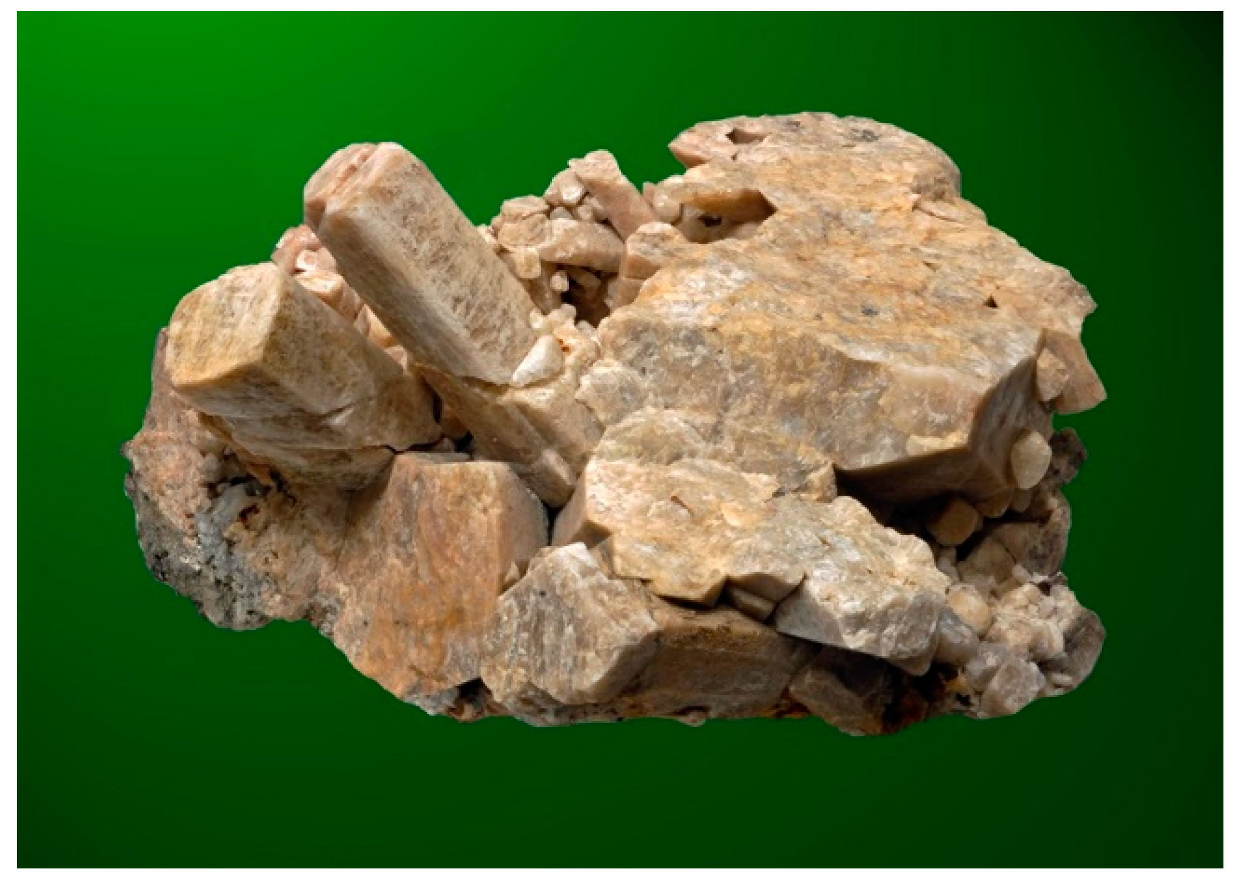
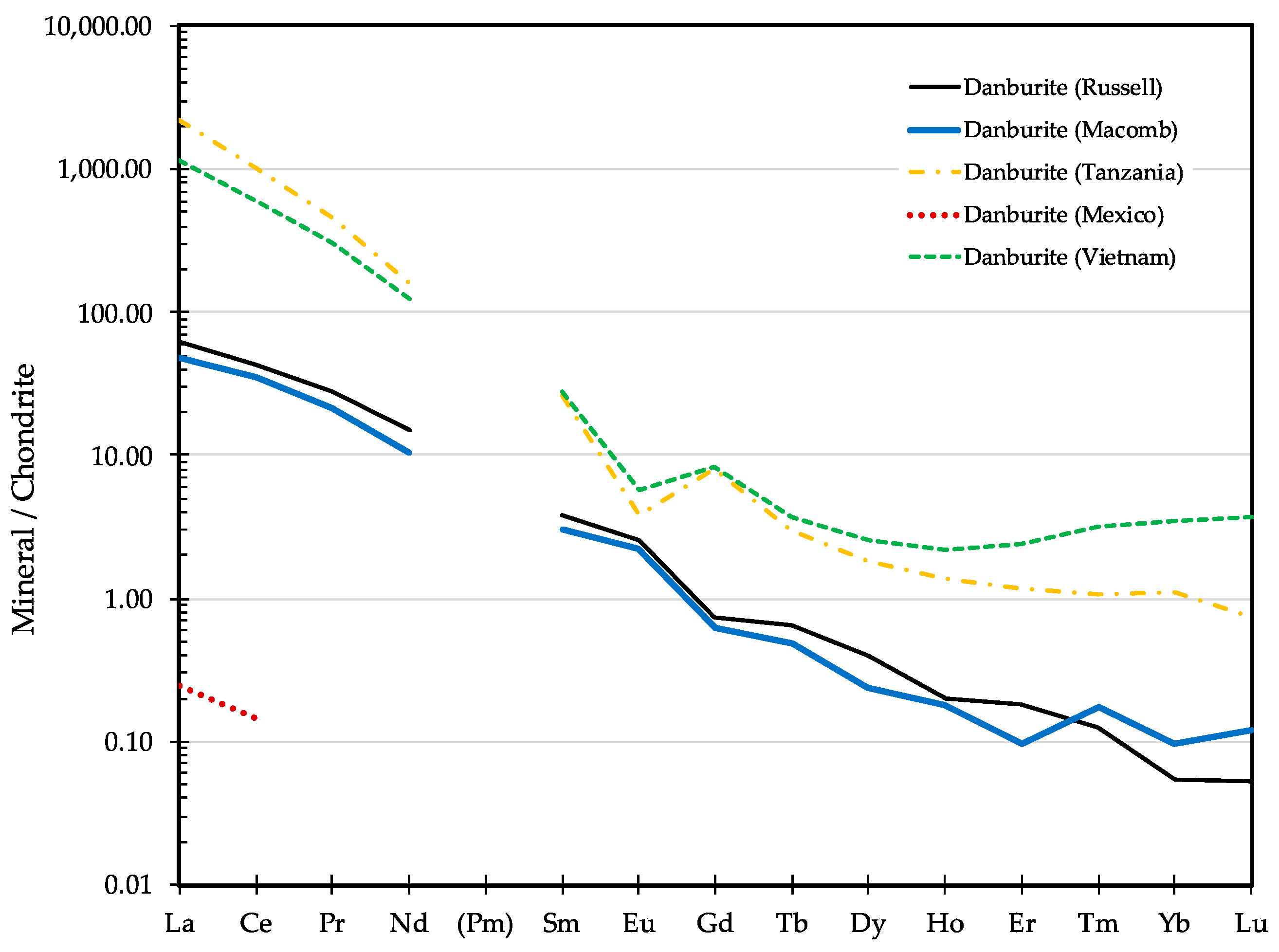
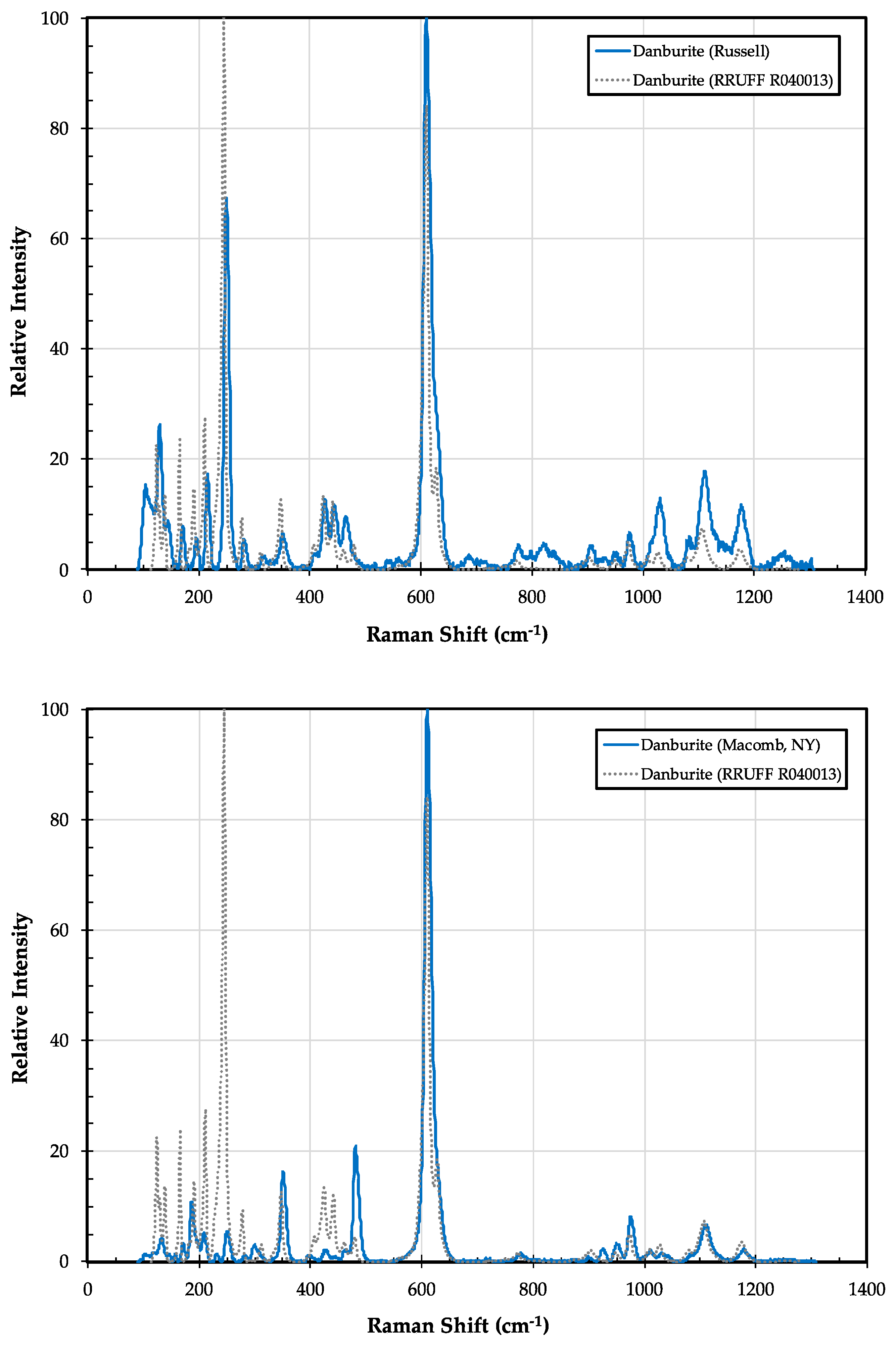
3.1. Danburite CaB2Si2O8

3.1.1. Danburite History and Geologic Setting

Danburite is a calcium borosilicate associated with skarns and high-grade metamorphosed calc-silicates. Fairly large crystals (up to 10 cm long) were first collected in the town of Russell in the Adirondack Lowlands by Chester D. Nims in the late 1870s, and identified by Brush and Dana [16] (Figure 2). Good descriptions and overviews of the mineral assemblage at the Russell danburite occurrence can be found in Clark [17] and in Chamberlain, Lupulescu, Bailey and Carlin [7]. Minor danburite was reported to occur at the McLear pegmatite in the town of Dekalb [18], although this has not been confirmed by more recent studies [19,20]. A third locality was discovered in 2014 in the town of Macomb [21].
At the original Russell locality, prismatic danburite crystals occur in elongate calc-silicate pods within the Grenville marble. These pods are 10 s of meters in length, and consist of danburite intergrown with quartz, diopside, calcite, phlogopite, tourmaline, and minor pyrite [16]. These coarse-grained pods or veins occur within a granular, medium-grained (0.25 to 2 mm), diopside-danburite-calcite unit. A recent examination of samples from the classic locality, and from three adjacent sub-localities, identified 17 associated minerals: Primary minerals include diopside, marialite, microcline, phlogopite, titanite, quartz, calcite, apatite, pyrite, and chalcopyrite; late-stage secondary minerals include tremolite, talc, quartz, allanite-(Ce), bornite, chalcocite, and datolite (as microscopic crystals in fractures within, and on, danburite) [7]. The granular diopside-danburite lithology is a prograde metamorphic assemblage that, in this region, reached temperatures of 650–700 °C, and pressures of 6–7 kbars [22] during the peak of the Shawinigan Orogeny (ca. 1160 Ma). Grew [3] considers this to be the highest temperature of danburite formation reported to date.
The recently discovered danburite occurrence in the town of Macomb is approximately 25 km northwest of the Russell locality. Here, a small (~5 m long) zone of danburite mineralization occurs along the contact of a small pegmatite intrusion with the Grenville marble [21]. Crystals of danburite up to 12.5 cm long are associated with albite, calcite, diopside, dravite, marialite, quartz, phlogopite, and minor tremolite, graphite, pyrite, titanite, and zircon [21]. While mineralogically similar to the classic site in Russell, here, the danburite is clearly part of a skarn formed by the intrusion of a small granitic pegmatite. The age of the Macomb pegmatite and skarn formation has recently been dated at 1168 ± 2.8 Ma [23].

3.1.2. Danburite Properties

Despite many of the crystals having rough and rounded crystal faces, Brush and Dana did a detailed crystallographic analysis of the danburite crystals from Russell. The crystals tend to be elongated, with prominent {110} and {120} rhombic prisms, with a total of 23 distinct crystal forms identified [16]. The danburite crystals from Macomb exhibit a similar morphology but tend to be less elongated and flattened perpendicular to [100] [21].
Danburite crystals from the original locality in Russell, NY, have been analyzed by wet chemistry and by electron microprobe (Table 2). Both analyses revealed that the crystals are relatively pure, with only minor amounts of Al, Fe, and Mg. The relatively high concentration of Al2O3 reported in the wet chemical analysis was likely due to the impurities included in the bulk sample, although limited substitution of Al3+ and H+ for Si4+ is possible [24].
LA-ICP-MS analysis revealed low concentrations of almost all trace elements in both the Russell and Macomb danburites, with the exception of Sr (826 and 713 ppm, respectively) (Table 3). Both danburites also have a strong preference for incorporating light REE and exhibit steep chondrite-normalized profiles (Figure 3). Compared to danburites from other world localities, the Adirondack danburites have overall REE concentrations lower than the danburites from Tanzania or Vietnam, but significantly higher than the danburites from Charcas, Mexico (Figure 3).
Raman spectra on danburite from the classic locality in Russell and from the recently discovered locality in Macomb are similar, and most of the peaks correspond to those observed in gem-quality danburite from Charcas, Mexico (Figure 4); the lack of a strong peak at ~250 cm−1 in the Macomb danburite is a notable difference that might be due to crystal orientation. The Raman peaks observed in both samples are consistent with those observed by Best et al. [33], with peaks between 800 and 1100 cm−1 assigned to Si–O–B and B–O–B bond stretching modes, the large peak around 620 cm−1 assigned to B–O–Si bond bending, and the 120 to 350 cm−1 peaks largely assigned to Ca translations and rotations of B2O and Si2O7 structural units, respectively. As noted by Best [33], assignment of bands to stretching, bending, and lattice modes in danburite is problematic because of the framework structure, and that breaking the danburite lattice into [Si2O7]6−, [B2O]4+ and Ca2+ ions is physically unrealistic, yet it results in good predictions of the numbers of bands for each symmetry species.

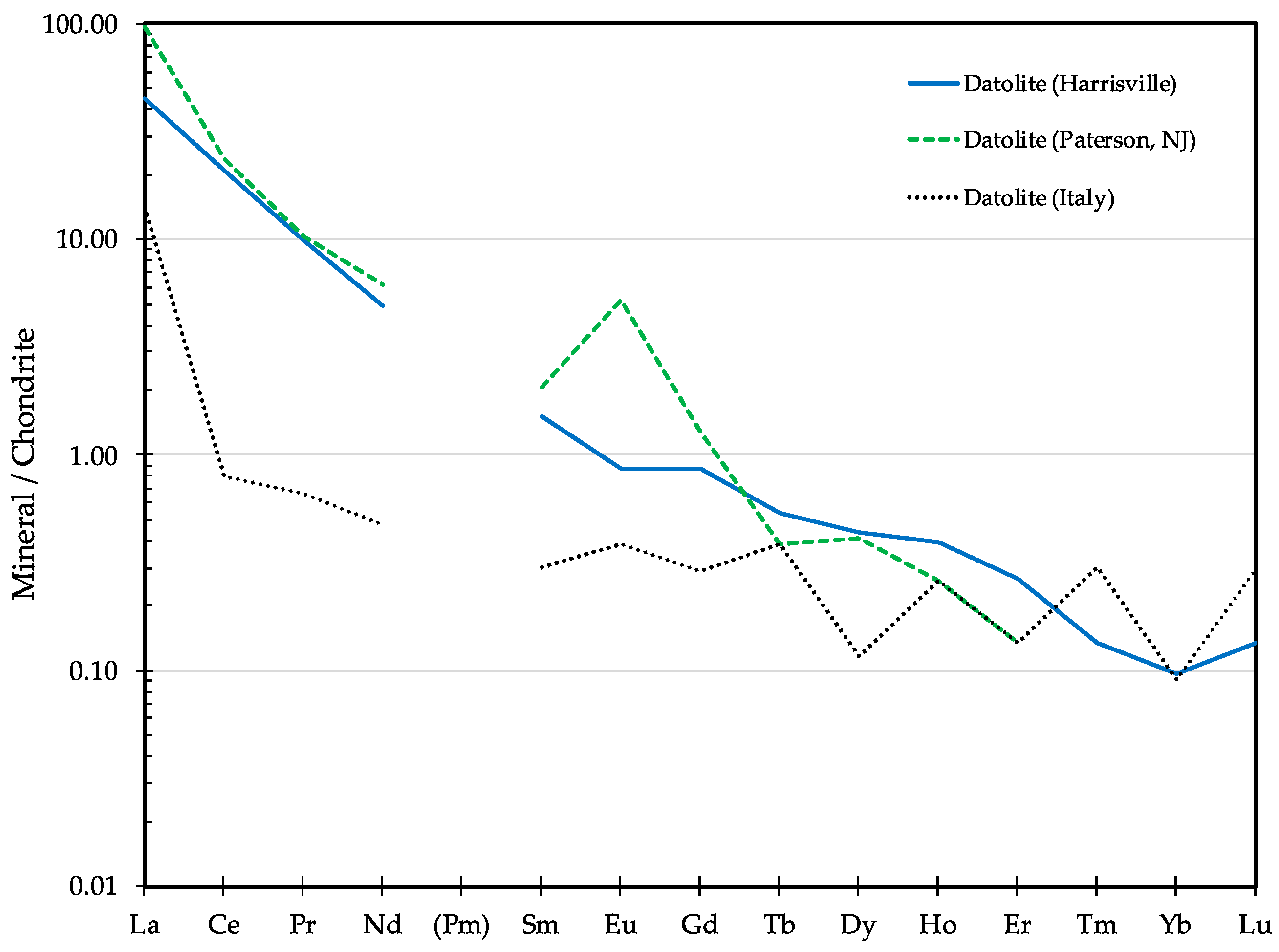
3.2. Datolite CaB(SiO4)(OH)

3.2.1. Datolite History and Geologic Setting

Datolite is a calcium borosilicate commonly associated with zeolites in hydrothermally altered mafic volcanic rocks. While beautiful, well-formed, translucent crystals of datolite have been collected from southern New York and New Jersey for over 200 years [35], well-formed crystals are rare in the Adirondack region, even though datolite has been reported at six different locations (Figure 1).
Datolite was first reported at Schroon Lake in the eastern Adirondacks, where it was found as microscopic crystals in cavities in a metagabbro along with crystals of chabazite [36]. Datolite also occurs in association with the danburite at Russell and Macomb (described above), but only as fine coatings on, or fracture fillings within, the earlier danburite crystals [3,7]. Datolite was also reported to occur at the gem diopside locality in Dekalb [37], but more recent studies have failed to confirm its presence at this site [38]. It was also reported as a rare mineral at the Woodcock mine in the Fowler talc belt [39], but this occurrence has similarly not been confirmed.
Relatively large (up to 5 mm) granular aggregates of datolite occur at the Valentine wollastonite deposit in Harrisville, NY. The datolite occurs in association with a large number of calc-silicate minerals, including, but not limited to: Wollastonite, diopside, hedenbergite, andradite, quartz, calcite, prehnite, apophyllite, pectolite, fluorapatite, brewsterite-Ba, and celadonite [40] (Figure 5).
In all of the Adirondack occurrences, datolite is a late-stage, low-temperature, hydrothermal mineral. This is supported by phase equilibria in the system CaO–B2O3–SiO2–H2O–CO2, which indicates that at relatively low temperatures, datolite is favored over danburite in the presence of H2O-rich fluids [3]. At all of the localities in the Adirondacks, datolite is a secondary mineral forming from the breakdown of preexisting high-grade borosilicate minerals (e.g., danburite and/or tourmaline). The source of the boron for the datolite at the Valentine mine is unclear since no boron-bearing minerals have been identified in the any of the pre-existing skarn lithologies [40,41].

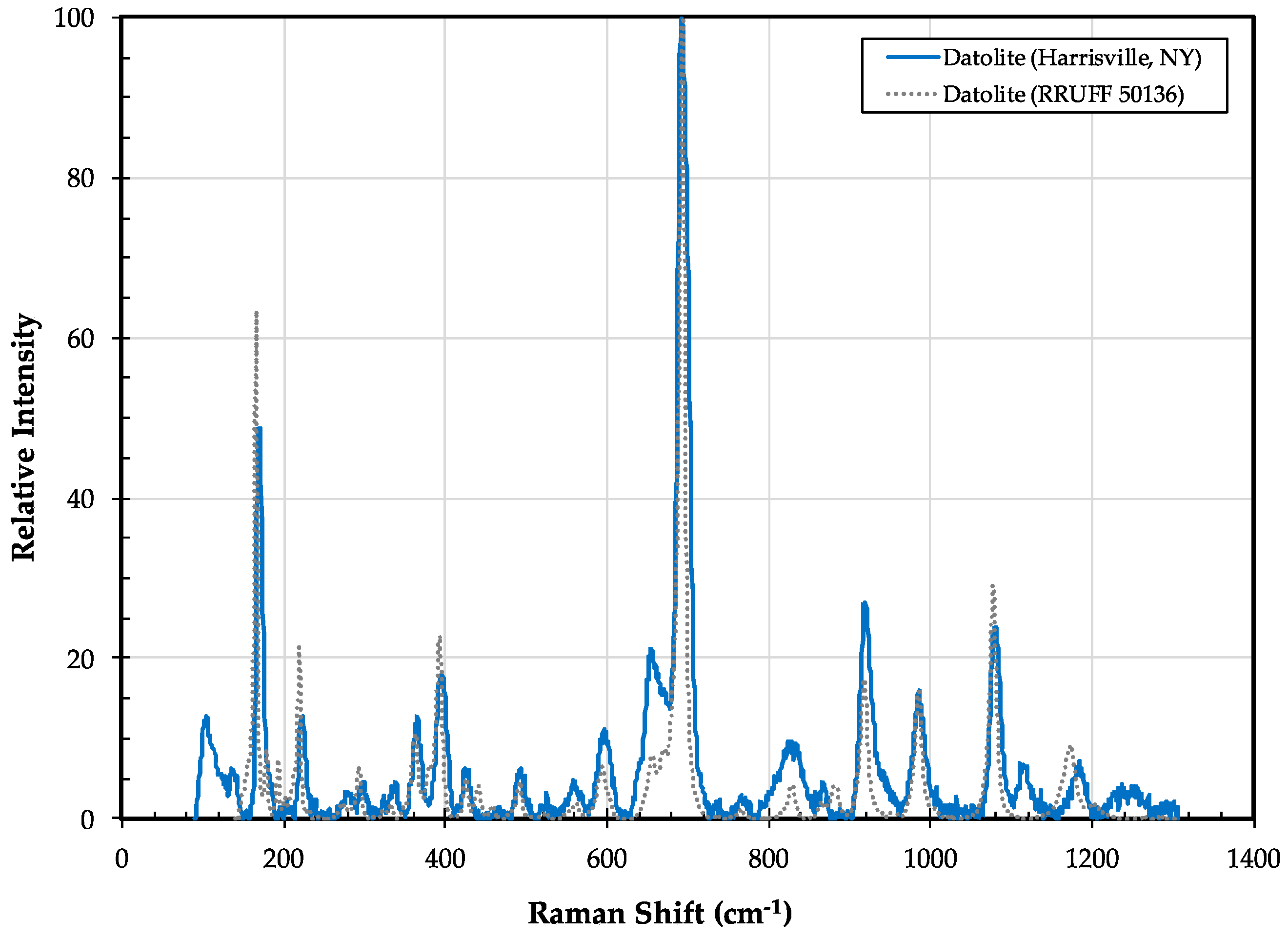
3.2.2. Datolite Properties

Datolite from most of the Adirondack localities is extremely fine-grained and occurs as overgrowths on earlier calc-silicate minerals. At the Valentine wollastonite mine, the crystals are larger (up to 1 cm), typically transparent, pale greenish-yellow, wedge-shaped, and striated [40].
Chemically, datolite from the Valentine mine in Harrisville is essentially stochiometric, with only minor impurities of iron and potassium (Table 2). It contains extremely low concentrations (<1 ppm) of most trace elements, with the exception of Sr and the light REE (3–18 ppm). Overall, REE concentrations are similar to those observed in datolites from the altered basalts of northern NJ, although they lack the positive europium anomaly; REE concentrations are significantly higher than those seen in the altered ophiolitic rocks of the northern Apennines in Italy (Figure 6).
Raman spectra on datolite from the Valentine mine are nearly identical to spectra of datolite from the calc-silicate skarn at Charcas, Mexico (Figure 7). Frost et al. [42] attributed the peaks at ~917 cm−1 and 985 cm−1 to symmetric and antisymmetric B–O stretching, respectively, and the peaks at ~1080 cm−1 and ~1170 cm−1 to symmetric and antisymmetric stretching of tetrahedral Si–O bonds, respectively. The large peak around 700 cm−1 is assigned to symmetric stretching of tetrahedral B–O bonds and the large peak at ~165 cm−1 is suggested to be an O–Ca–O bending mode.

3.3. Dumortierite AlAl6BSi3O18

3.3.1. Dumortierite History and Geologic Setting

Dumortierite is an uncommon, Al-rich, borosilicate typically found in granitic pegmatites or in high-grade metapelitic rocks. Nice specimens of dumortierite have been known from southern New York pegmatites for over 100 years [45], but only recently has dumortierite been reported from pegmatites in the Adirondacks [46,47].
In the eastern Adirondacks, small (<2 mm long) needles of blue dumortierite (Figure 8) were found at the margin of the Batchellerville pegmatite, where it is in contact with a biotite gneiss. The dumortierite is associated with quartz, albite, and microcline (present as anti-perthite lamellae) and minor apatite [46].
At the Benson Iron Mines in Star Lake, dumortierite also occurs as small needles at the contact of small pegmatitic veins intruding a mafic gneiss. The purple-blue, acicular crystals (up to 3 mm long), are associated with quartz and sillimanite. In some pegmatite bodies, dumortierite is included within gem-quality feldspar and has a narrow tourmaline envelope [47].
In the central Adirondacks, dumortierite has also been reported from a small granitic pegmatite on Ledge mountain, northwest of Indian Lake, where it is associated with quartz and chlorite [48].

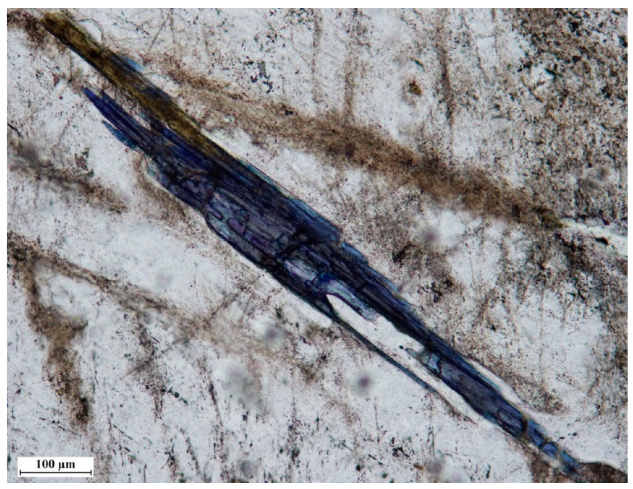
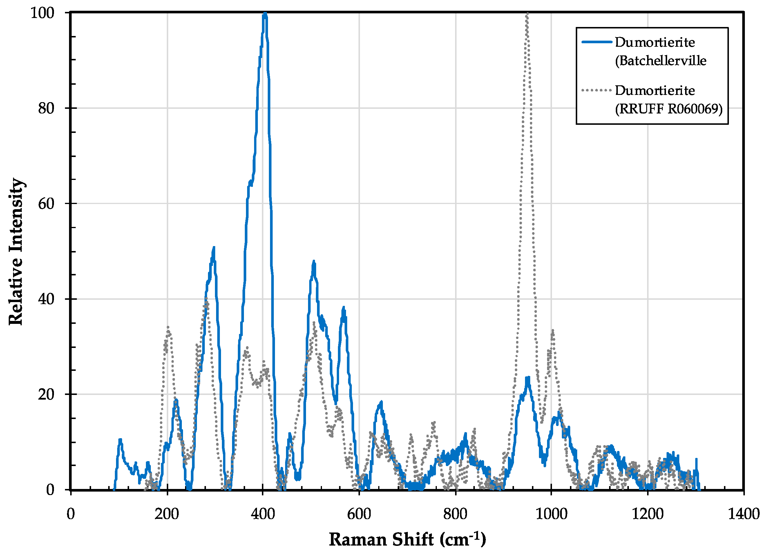
3.3.2. Dumortierite Properties

At all three localities in the Adirondacks, dumortierite occurs as relatively small, acicular, blue crystals along the margins of small granitic pegmatites. Major element compositions of dumortierite from Batchellerville (Table 2) and from Ledge Mountain [48] are very similar. Mineral formulas calculated using the method of Grew [3] yield similar and nearly ideal stochiometric formulas:
Batchellerville:
(Al.86Mg.15)(Al5.94Fe.06)6(Si2.88Al.11Ti.01)3BO3(O,OH,F)2O13
                          Ledge Mountain [48], (Ti not analyzed)
(Al.87Mg.14)(Al5.89Fe.11)6(Si2.87Al.13)3BO3(O,OH,F)2O13
Most trace element concentrations in dumortierites from the Benson Mines and Batchellerville are quite low (<10 ppm), and many, including most of the REE, are below detection limits (Table 3). Notable exceptions are the semi-metals As, Sb, and Bi, with concentrations up to 4000 ppm, and the high-field strength elements Ta, Nb, and Zr, with concentrations up to 200 ppm. Pieczka et al. reported [49,50] that Ta and Nb substitute for aluminum in the Al1 site of dumortierite as part of the solid solution with holtite, and As and Sb substitute for Si as seen in szklaryite.
A Raman spectrum on the Batchellerville dumortierite (Figure 9) is a fairly good match to the Raman spectrum of dumortierite from Dehesa, CA, although notable differences in peak intensities and slight shifts in peak wavenumbers are seen. Differences in crystal orientation might explain these differences.

3.4. Grandidierite MgAl3O2(BO3)(SiO4)

3.4.1. Grandidierite History and Geologic Setting

Grandidierite is a relatively uncommon borosilicate despite forming in a fairly wide range of geologic environments, including granitic pegmatites [51,52], pelitic migmatites [53,54], granulite facies leucogneisses [55,56], upper amphibolite to lower granulite facies calc-silicate lithologies [3,25,29], and pyrometasomatized pelitic xenoliths in plutonic and volcanic rocks [57,58].
In the Adirondacks, grandidierite is found only at two localities; Johnsburg in the Adirondack Highlands, and Russell in the Adirondack Lowlands (Figure 1). In both, the grandidierite occurs as small, anhedral grains apparently forming from the breakdown of serendibite (described below) by the reaction [29]:
serendibite + diopside + Na2O + H2O + CO2 = grandidierite + pargasite + calcite + B2O3
Conditions of grandidierite formation are interpreted to have been lower granulite facies at Johnsburg, and upper amphibolite facies at Russell [25].

3.4.2. Grandidierite Properties

Grandidierite is pleochroic blue-green in a thin section and occurs as irregular patchy grains within, or surrounding, the larger serendibite crystals [29]. The only chemical analysis of grandidierite from the Adirondacks [25] indicates that it is very Mg-rich and Fe-poor (Table 2). Samples for additional analysis were unable to be located.

3.5. Harkerite Ca12Mg4Al(BO3)3(SiO4)4(CO3)5·H2O

3.5.1. Harkerite History and Geologic Setting

Harkerite is a rare borosilicate carbonate typically found in high-temperature, low pressure, calc-silicate skarns [3,59]. Harkerite was first identified at Cascade Slide in the Adirondack Highlands by Jaffe, reported by Baillieul in 1976 [60], and confirmed in subsequent studies [26,61]. Material suitable for additional analysis was unable to be located.
Cascade Slide is an unusual occurrence of medium- to coarse-grained marble in the Adirondack Highlands surrounded by anorthosite (Figure 1). Baillieul noted that the harkerite occurred as indistinct, opaque, white grains up to 3 mm in a calc-silicate marble, where it commonly formed irregular rims around diopside and monticellite grains; it could be easily identified by its red-orange fluorescence under short-wave ultraviolet radiation [60]. Commonly associated minerals include: Calcite, monticellite, diopside, and spinel; less commonly observed minerals include forsterite, garnet, magnetite, and barite [26].
The occurrence of harkerite at Cascade Slide is unusual in that it occurs in a region of granulite facies metamorphism, where peak metamorphic conditions are thought to have been 7.4 ± 1 kbar and 750 ± 30 °C [61]. Possible explanations for the presence of harkerite in high-pressure metamorphic rocks include: 1) Stabilization at high pressures due to extremely low CO2 activities, or 2) localized preservation of earlier, low-pressure mineral assemblages [3]. Recent oxygen isotope and geochronology studies in the Adirondacks support the latter interpretation, with relatively shallow emplacement of the large anorthosite massifs resulting in localized skarn formation at ~1155 Ma, followed by high-pressure granulite facies metamorphism at ~1050 Ma [62,63]. Harkerite may have been preserved at these high pressures due to the near-absence of a fluid phase during subsequent granulite facies metamorphism [26].

3.5.2. Harkerite Properties

The harkerite at Cascade Slide occurs as small (typically < 3 mm), irregular patches in a medium-grained, monticellite-diopside-marble; usually with blue calcite [26,60]. Optically, the harkerite grains exhibit very low, first-order interference colors, and in some instances, fine polysynthetic twinning, which may have been introduced during the transformation from an isometric to a trigonal structure [26].
Powder X-ray diffraction [60] and Fourier transform infrared (FTIR) spectroscopy both yield patterns nearly identical to those on harkerite from the type locality in Skye, UK [26].
Chemical analyses of harkerite grains from Cascade slide (Table 2) have higher silicon, and lower boron contents than harkerite from the type locality, presumably due to the substitution (BO3)4 = AlSi4O15(OH) [26]. While no trace element analysis has been performed on the Cascade Slide harkerite, on the basis of charge balance and site occupancy considerations, Grew et al. suspect significant substitution of Sr, Y, and/or REE [26].

3.6. Kornerupine/Prismatine (□,Mg,Fe2+)(Mg,Fe2+)2(Al,Mg,Fe2+,Fe3+)7(Si,Al)4(Si,Al,B)O21(OH,F)/(□,Mg,Fe2+)(Mg,Fe2+)2(Al,Mg,Fe2+,Fe3+)7(Si,Al)4(B,Si,Al)O21(OH,F)

3.6.1. Kornerupine/Prismatine History and Geologic Setting

These minerals are described here in the same section because they vary only in the amount of boron in the crystal structure. The two minerals, kornerupine, and prismatine form the kornerupine mineral group, which contains 0.0 to 1.0 formula units B (per 21.5 oxygens) [64,65]. Kornerupine contains between 0.0 and 0.5 formula units B, whereas prismatine contains 0.5 to 1.0 formula units. Because boron is not commonly determined by electron microprobe methods, any specimen of the kornerupine mineral group where the B content is unknown should be considered “kornerupine” (sensu lato) [64].
In the Adirondacks, kornerupine is reported from the Warrensburg area in the southeast [66,67,68,69], and prismatine is reported from the Moose River area in the west [27] (Figure 1, locations 14, 15, and 16). Unlike the prismatine located in the Moose River area, the boron content of the Warrensburg locality has not been reported, thus it is referred to as kornerupine in this report consistent with the original publications of Farrar [66,67,68,69].
Kornerupine from the Warrensburg area occurs in Al-rich ultramafic rocks (comprising forsterite, bronzite, tschermakite, spinel, and phlogopite) associated with an anorthosite sill [67]. The kornerupine occurs as a late phase along with pyrope, corundum, and sapphirine [67]. Prismatine from the Moose River area, in the western Adirondacks, forms spectacularly developed, dark green, euhedral crystals (Figure 10) measuring up to 10 cm or more along the c-axis. In surface exposures, prismatine forms radiating sprays in coarse-grained feldspathic lenses that are generally parallel to gneissic foliation. In these lenses, the prismatine is arranged randomly, but the longest and best-developed crystals are formed parallel to the foliation plane. The euhedral shape and the random arrangement are inferred to be the result of crystallization from a melt [27]. A proposed melt forming reaction is [27]:
tourmaline + sillimanite + biotite + cordierite → prismatine + rutile + melt
Here, the prismatine (and rutile) are the peritectic phases. Figure 10 (bottom) shows prismatine surrounding a grain of hercynite, suggesting that hercynite may have been a reactant phase as well. Figure 11 shows some of the abundant rutile generated by the above reaction. Interestingly, the rutile shows what appears to be exsolution lamellae of ilmenite. Peak metamorphic conditions in this region are estimated to have reached ~850 ± 20 °C and 6.0 ± 0.6 kbar [27,70].

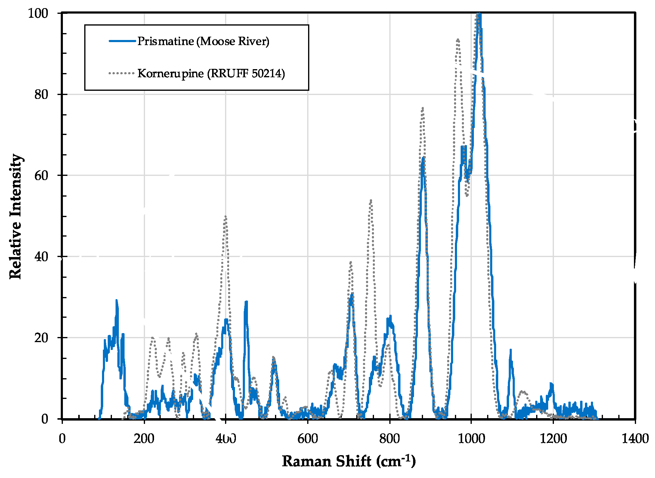
3.6.2. Kornerupine/Prismatine Properties

Crystals of the kornerupine–prismatine series are all orthorhombic, with space group Cmcm [71]. The crystal structure is complex, with three tetrahedral sites (T(1) through T(3)), five distinct octahedral sites (M(1) through M(5)), and one 8-coordination, distorted, cubic site (X); boron, when present, is completely ordered in the T(3) site [71]. As its name implies, prismatine tends to form highly prismatic grains with the {110} form dominating in euhedral specimens [64]. Prismatine from the Moose River area is no exception as prismatic faces parallel to the c-axis are well developed. Calculated interfacial angles between the {110} prism faces are about 81° and 99° based on unit cell dimensions of a, b listed in Table 2 of [64]. Significantly, it was these interfacial angles that first hinted that the elongated, dark-colored grains were not an amphibole (56° and 124°) or a pyroxene (87° and 93°). The identification as prismatine was later determined by powder X-ray diffraction, electron microprobe, and SIMS analysis [27].
Table 2 lists two electron microprobe analyses of prismatine, one is an average of two analysis from Table 3 (sample MR-99-11) of [27] and a new analysis reported here. Both show similar Mg abundances, but the Al2O3 content of the new analysis is somewhat higher (by ~2.5 wt. %), as is the FeO content (by 0.37 wt. %), but SiO2 is slightly lower (by 0.86 wt. %). Boron abundance was not determined in the new analysis, but the calculated amount of B2O3 is 2.20 wt. %. The averaged SIMS analysis from [27] yields 0.76 formula units of B, confirming the prismatine identification.
Trace element data for the Moose River prismatine are listed in Table 3. The prismatine is relatively enriched in first row transition elements Sc, V, Cr, Co, Ni, Zn, Ga. These cations are inferred to occupy the five (6-fold) M sites and possibly the one (8-fold) X site in prismatine [71]. Rare earth element and Y concentrations are generally low (and below detection limits for Sm and Eu), but the prismatine is relatively enriched in the heavy rare earth elements (HREE), as shown in Figure 12. Because of their relatively large ionic radius, these elements are inferred to occupy only the single (8-fold) X site in prismatine [71]. This HREE enrichment (and light REE exclusion) in prismatine (Figure 12) is similar to that of garnet, which frequently incorporates HREE into its three (8-fold) X sites. A number of substitution vectors for Y and REE have been proposed for garnet [72], and we suspect a similar number to operate in prismatine. However, the total amount of REE in prismatine is likely to be low as it has only one 8-fold site per 21.5 oxygens [64,71] compared to garnet’s three 8-fold sites per 12 oxygens.
Figure 13 shows the Raman spectrum of the Moose River prismatine compared to a kornerupine specimen from Sri Lanka (RRUFF sample R050214). The Sri Lankan sample has a calculated B formula unit value of 0.59, thus it could be prismatine. Significant Moose River prismatine peak positions occur at 326, 333, 401, 450, 515, 707, 762, 802, 882, 978 and 1015 cm−1. All of the peak positions that are >700 cm−1 fall within the range of values listed in Table 1 of [73]. Frost et al. [74], attributed the peaks between 925 and 1085 cm-1 in kornerupine to Si–O–Si stretching vibrations, and the peaks they observed at 923 and 947 cm−1 to symmetrical stretching vibrations of trigonal boron. This assignment to stretching vibrations of trigonal boron is, however, problematic considering that detailed studies of the crystal chemistry of kornerupine and prismatine have shown that boron is completely ordered at the tetrahedral T(3) site [65,71]. In addition, we did not observe any distinct peaks at these wavenumbers in the Moose River prismatine. The pairs of strong peaks at ~965 and ~1010 cm−1, and ~700 and ~750 cm−1 are characteristic of kornerupine and prismatine regardless of boron content [73]. Some peak positions are shifted slightly compared to those of the Sri Lankan sample (Figure 13) and to those listed in Table 1 of [73], which are attributed to slight differences in major element chemistry. Variation in peak intensity is attributed to crystal orientation relative to the incident laser. Significantly, the strong peaks at 802 and 882 cm−1 confirm the presence of boron [73] in the Moose River prismatine.

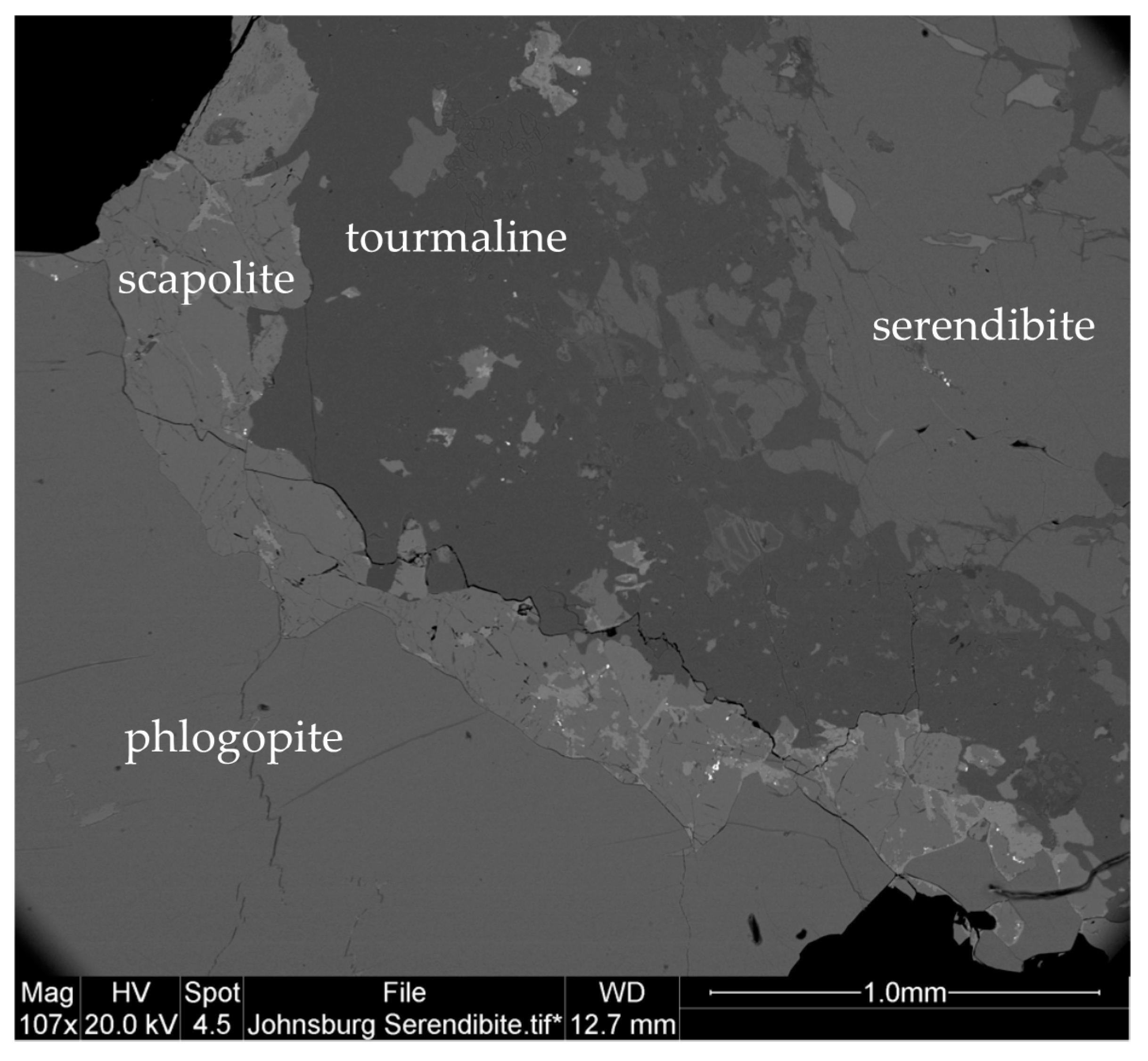
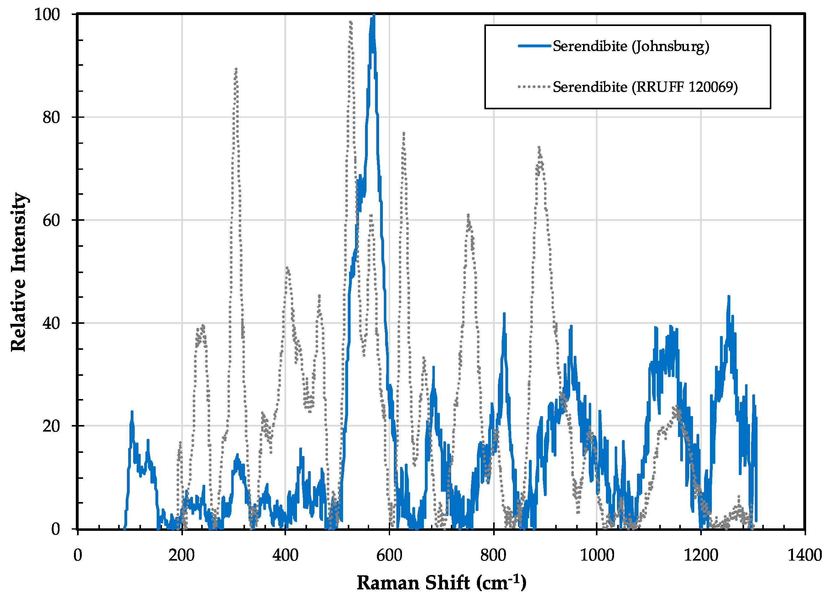
3.7. Serendibite Ca4[Mg6Al6]O4[Si6B3Al3O36]

3.7.1. Serendibite History and Geologic Setting

Serendibite is a rare borosilicate mineral with fewer than 20 distinct world localities [5]. The serendibite in the Adirondack Highlands near Johnsburg, NY, first reported in 1932, was the second recognized world locality [28]. Serendibite was later found in the Adirondack Lowlands in a drill core of the St. Joe Resources Company in the town of Russell, NY [25]. Serendibite is characteristically found in upper amphibolite to granulite facies, calc-silicate skarns [3,25].
The serendibite at Johnsburg is blue to gray-blue and occurs as massive, granular aggregates in small (10s cm wide) pods and lenses interpreted to be a skarn that formed along the contact of a potassium feldspar-rich meta-igneous lithology and a medium-grained, dolomite-bearing marble [29]. Major phases closely associated with the serendibite are diopside, phlogopite, scapolite, plagioclase feldspar, and tremolite [28]. Other phases noted by Larsen and Schaller [28] include orthoclase, tourmaline, spinel, chlorite, serpentine, sericite, and talc. Grew et al. [29] identified pargasite, sinhalite, grandidierite, and borian sapphirine; additional phases we identified by SEM/EDS include zircon, thorianite, and barite.
Serendibite grains at Johnsburg are up to 1 cm in diameter, anhedral, and occasionally exhibit symplectic intergrowths with diopside [29]. Many grains are rimmed by tourmaline (dravite), scapolite, phlogopite, and/or calcite (Figure 14). Less common are rims or overgrowths of pargasite, grandidierite, sinhalite, and/or borian sapphirine [29]. According to Grew [3], the equilibrium mineral assemblage at Johnsburg was serendibite + diopside + phlogopite + scapolite + sinhalite, which presumably formed under the peak metamorphic conditions in the region of 720–740 °C, and 6.5–8 kbar [75]. Most of the complex overgrowths are interpreted to be retrograde breakdown products of the primary serendibite [29].
The only other known occurrence of serendibite in the Adirondacks was encountered in a drill core in the town of Russell in the Adirondack Lowlands. A serendibite-diopside rock occurred over a 13–20 cm interval at a depth of 17.7 m, in drill hole 1872, of the St. Joe Resources Company [25]. Here the deep blue serendibite is associated with apatite, calcite, pargasite, and fine-grained, secondary phyllosilicates (sericite and/or chlorite?); less common are scapolite, tourmaline, grandidierite, phlogopite, sinhalite(?), and spinel [25]. Similar to the serendibite at Johnsburg, grains of up to 1 cm are usually overgrown and replaced by multiple phases [25]. Grew et al. [25] proposed a three-stage history for the Russell serendibite lithology: (1) A primary metamorphic assemblage of serendibite + diopside ± scapolite ± apatite; (2) secondary assemblages of serendibite with tourmaline, grandidierite, pargasite, spinel, ± phlogopite; and (3) a late replacement of many phases by fine-grained phyllosilicates. Inferred conditions for the formation of serendibite at Russell are 660–750 °C and 6.7–7.4 kbar, slightly lower than the granulite facies conditions at Johnsburg [25]. The lack of any spatially associated igneous/meta-igneous lithology is also a notable difference with the Johnsburg locality; here the serendibite appears to have formed directly from high-grade metamorphism of boron-rich calcareous sediments.

3.7.2. Serendibite Properties

In both Adirondack localities, serendibite occurs as anhedral, pale to deep blue grains in a granular calc-silicate lithology. In thin section, grains exhibit high positive relief, are very pale blue and faintly pleochroic, and display prominent polysynthetic twinning on {011}.
Chemically, the two Adirondack serendibites are very similar in composition (Table 2) and are relatively Mg and Na-rich, and Fe-poor compared to the serendibites from other localities [25]. Grew et al. [25] noted that much of the observed chemical variation in the Adirondack serendibites could be explained by either the Tschermaks substitution (Mg + Si = 2 Al), or the coupled substitution (Na + Si = Ca + Al). We report here the first trace element analyses of the Johnsburg serendibite (Table 3). Grains contain low concentrations of most trace elements, with the exception of Ga (99 ppm) and Y (59 ppm), both of which are likely substituting for Al in the five Al-dominant octahedral sites [76]. Rare earth element concentrations are also quite low, with concentrations around 10X chondrite, although with a slight preference for the heavy REE and a pronounced negative Eu anomaly (Figure 12).
A Raman spectrum of the Johnsburg serendibite (Figure 15) shows multiple broad, relatively low-intensity peaks that only weakly correspond to peaks observed in serendibite from Burma; additional analyses of multiple grains in known crystallographic orientation are needed to fully characterize the nature of bonding in the Johnsburg serendibite.

3.8. Sinhalite MgAl(BO4)

3.8.1. Sinhalite History and Geologic Setting

Sinhalite is a rare magnesium aluminum borate found in high-grade calc-silicate lithologies. It was first recognized as a mineral in 1952 [77], and shortly thereafter, it was identified in association with the Johnsburg serendibite (described above) [78]. It also has been tentatively identified in association with the serendibite from Russell, NY [25].
At Johnsburg, the sinhalite forms anhedral grains up to 1 cm. It is typically in contact with scapolite and/or diopside, and in places it is enclosed by serendibite or tourmaline [29]. It is part of the equilibrium skarn assemblage serendibite + diopside + phlogopite + scapolite + sinhalite that formed at granulite facies conditions of ~720–740 °C, and 6.5–8 kbar [3,29].

3.8.2. Sinhalite Properties

The Johnsburg sinhalite has not been well-characterized, and material for additional analysis was unable to be located. Schaller and Hildebrand [78] reported some fundamental optical properties and powder X-ray diffraction data for the Johnsburg sinhalite, and Grew et al. [29] provided two electron microprobe analyses of the Johnsburg sinhalite (average of their analyses provided in Table 2).
Interestingly, because sinhalite is isostructural with forsterite [79], it has been suggested that a coupled solid solution mechanism of B + Al = Mg + Si may exist between the two minerals [78]. Grew, however, showed that B-bearing olivines contain little Al and argued against this mechanism [3]. Sinhalite appears to be a distinct borate mineral with little to no solid solution with the silicate olivine group. Its paragenetic relationship to the multiple phases in the Johnsburg serendibite-diopside skarn is not well-established.

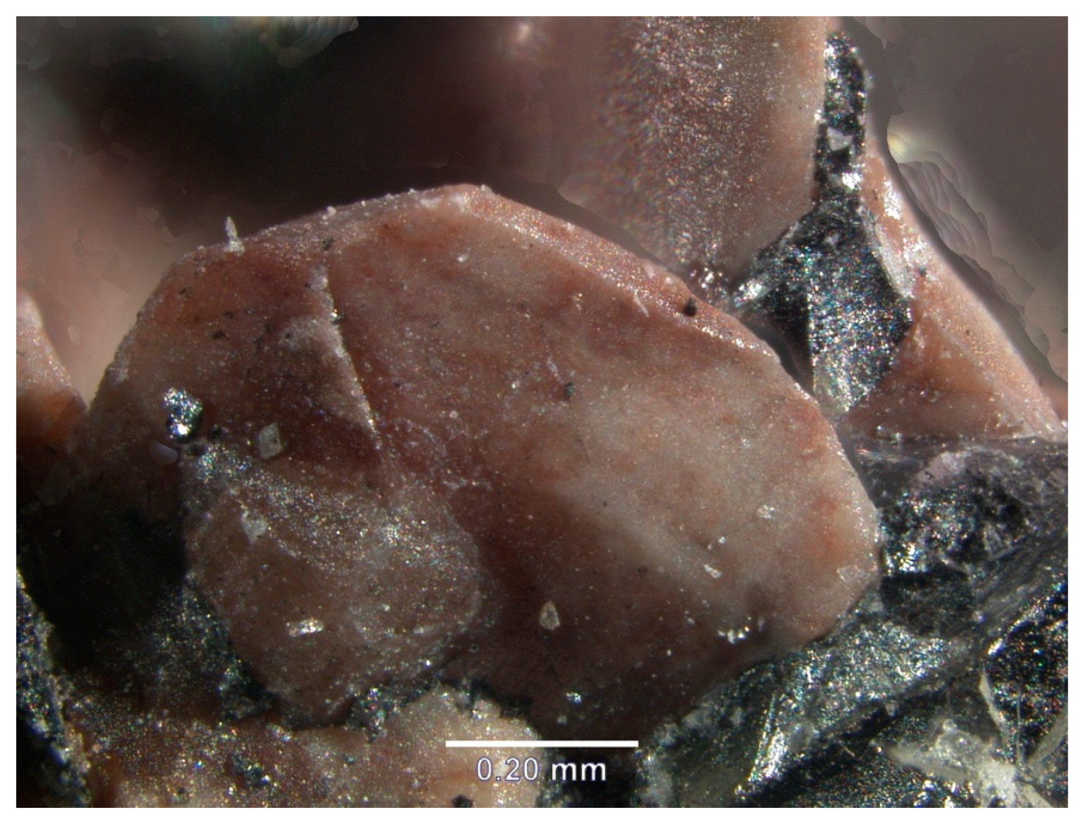
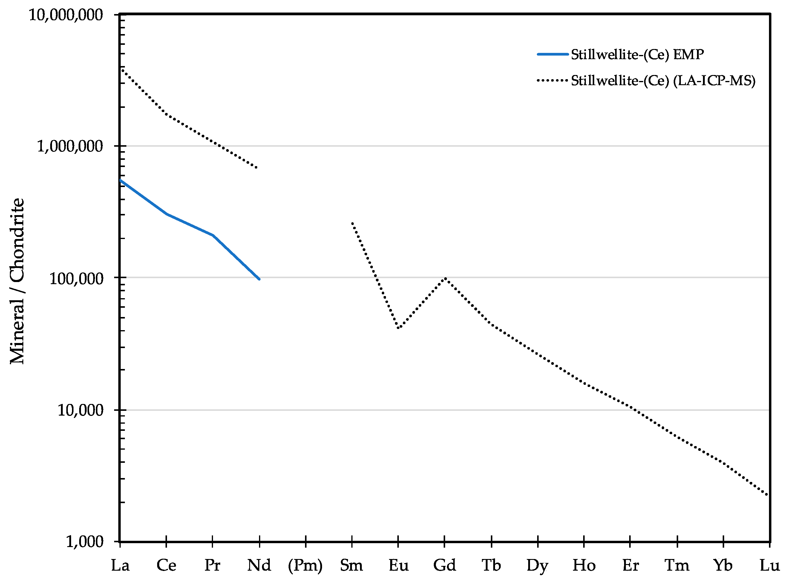
3.9. Stillwellite-(Ce) CeBSiO5

3.9.1. Stillwellite-(Ce) History and Geologic Setting

Stillwellite-(Ce) is an uncommon, rare-earth-element borosilicate typically found as a late-stage metasomatic phase in metamorphosed calcareous sediments [80], or as a late hydrothermal phase in alkaline intrusions [81,82]. In New York State, the only known occurrence of stillwellite-(Ce) is in the iron ore deposits of Mineville in the eastern Adirondack Highlands (Figure 1). It was first identified in 1979 in a sample collected from the “Old Bed” orebody in the Adirondack Mine of the Republic Steel Company; the sample was collected near a fault at the minus 2100-feet level in the mine [83]. The presence of stillwellite-(Ce) was confirmed on the basis of both X-ray diffraction and semi-quantitative spectrographic chemical analysis [83]. More recent chemical analyses have confirmed the presence of stillwellite-(Ce) at Mineville [84], but because of the lack of available in-situ samples with carefully documented textural and mineralogical associations, the conditions under which these grains formed is unclear. In addition, the source of B to form stillwellite-(Ce) in these magnetite ore bodies is not obvious because there are no spatially associated meta-carbonates or alkaline intrusions.

3.9.2. Stillwellite-(Ce) Properties

At Mineville, stillwellite-(Ce) occurs in association with fluorapatite and magnetite as 1 to 2 mm wide, tabular crystals with a waxy luster, and a pink to reddish color (Figure 16). Backscatter electron images reveal that monazite often rims and fills fractures within the stillwellite-(Ce) grains and that many of the grains are inhomogeneous and variably altered. Because of this, obtaining consistent and accurate compositional data was challenging. After mounting and imaging many grains, a few homogeneous cores with relatively bright backscatter intensities yielded consistent and essentially stochiometric major element compositions by electron microprobe (reported in Table 2). Chemically, the stillwellite-(Ce) is essentially (REE)BSiO5, with only minor substitution of Ca and Th.
Unfortunately, we were unable to analyze these same spots by LA-ICPMS, and, therefore, are unable to report accurate trace element data for the Mineville stillwellite-(Ce). However, preliminary LA-ICPMS data indicate that while stillwellite-(Ce) has a strong preference for the light REE, all of the REE are present in relatively high concentrations (Figure 17). Y, Th, and U, which are presumably substituting for the REE, are also present in moderate to high concentrations (1000 s to 10,000 s ppm). Ga and the semi-metals Ge and As are also present in moderate concentrations (~1000 s ppm) and are presumably substituting for silicon.
The Raman spectrum for Mineville stillwellite-(Ce) is similar, but not identical, to that of stillwellite-(Ce) from Tajikstan (Figure 18). The complete lack of a major peak at ~ 475 cm−1 and the presence of a strong peak at ~1025 cm−1 may be artifacts of orientation, but suggest there may also be significant differences in the crystal chemistry of the two samples.

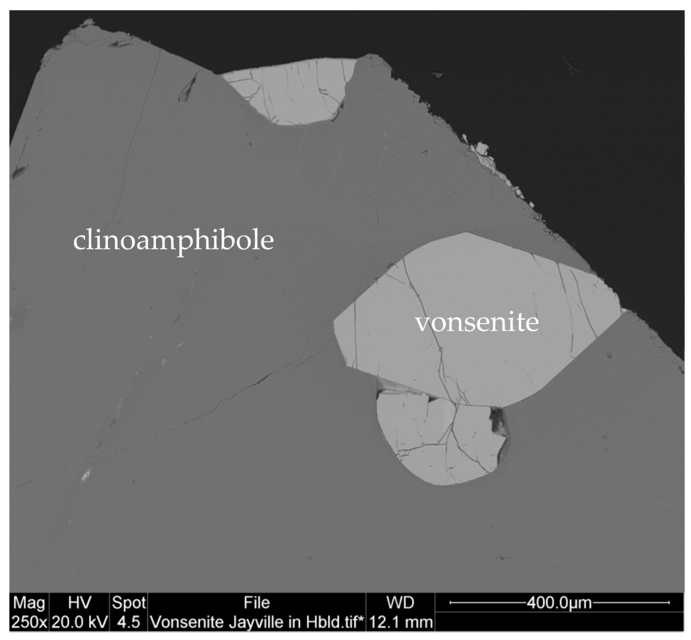
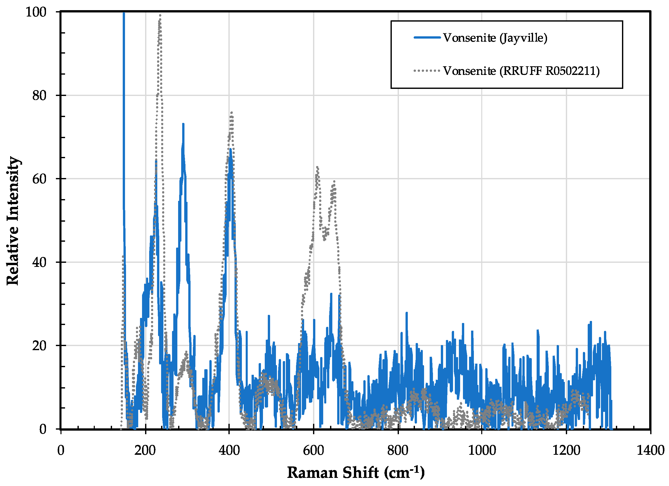
3.10. Vonsenite Fe2+2Fe3+(BO3)O2

3.10.1. Vonsenite History and Geologic Setting

Vonsenite is a relatively rare iron borate of the ludwigite-vonsenite series. In 1947, B. F. Leonard found a metallic mineral with unusual optical properties in the ore from a drill core at the Jayville iron deposit, and tentatively identified it as ilvaite [30,85]. In 1950, Henderson, from Princeton University, prepared the mineral for spectrographic analysis and X-ray diffraction and realized that the mineral was not ilvaite, but he was unable to identify it [30]. In 1951, Axelrod and Fletcher, from the U.S. Geological Survey, identified the mineral as vonsenite [30]. One specimen of vonsenite was also found at the Clifton iron deposit, approximately 20 km northeast of the Jayville deposit [30]. While the Jayville vonsenite was described in a subsequent U.S.G.S. report in 1964 [86], only two recent studies have been done on the ore-forming processes involved in generating the vonsenite-bearing rocks [87,88].
At Jayville, vonsenite occurs as fine-grained (0.1 to 2.0 mm) gray to black granular aggregates or stubby prismatic crystals up to 5.5 cm in length. It is primarily associated with magnetite, clinoamphibole (pargasite), and biotite (Figure 19). Additional minerals reported in association with the vonsenite-magnetite ore include orthopyroxene, clinopyroxene, scapolite, and fluorite, and less commonly, quartz, zircon, chlorite, talc, titanite, hematite, goethite, pyrite, and chalcopyrite [30,87]. Hall, Johnson and Rosner [87] report vonsenite partially replacing magnetite in the ore body, which is laced throughout by thin veins of vonsenite.
The magnetite-vonsenite ores at Jayville were originally interpreted as a pyrometasomatic skarn deposit, where lenses and bodies of calcareous sedimentary rocks were enclosed and metasomatized by granitic intrusions (now granitic gneisses) [30]. Johnson, Chapman, and Valder [88] argued for a similar origin, where the magnetite-vonsenite ore bodies were formed by metasomatic alteration of calc-silicate gneisses due to the intrusion of the Lyon Mountain granite during the Ottawan orogeny. Lupelescu et al. [89] reached a similar conclusion, and using U/Pb zircon geochronology, dated the intrusive and ore-forming event to ca. 1040 Ma. However, Hall, Johnson, and Rosner [87] argued for a more complex ore-forming process involving a multi-stage series of fluid infiltration events. They present evidence for vonsenite formation by a boron-rich metasomatic event 70–90 Ma after the peak of Ottawan metamorphism and granite emplacement, and at greenschist facies metamorphic conditions.

3.10.2. Vonsenite Properties

In hand specimens, vonsenite is black, submetallic, and difficult to distinguish from the associated magnetite. In transmitted light, vonsenite is opaque; in reflected light, it exhibits very strong pleochroism [30]. Prismatic crystals from Jayville exhibit the following crystal forms: {001}, {010}, {100}, {3.10.0}, {250}, {120}, and {320}, and X-ray diffraction analysis yielded an orthorhombic unit cell with dimensions of a = 9.47 Å, b = 12.31 Å, and c = 3.07 Å [30,90].
Vonsenite is the Fe-rich end-member of the ludwigite group, an isostructural group of orthorhombic metal borates. While substitution of Mg2+, Mn2+, and Ni2+ for Fe2+ can be extensive, the borate at Jayville is near end-member vonsenite (Table 2). Spectrographic analysis of a bulk sample in the 1950s identified trace concentrations of: Sn, Pb, Zn, Ti, Cu, Cr, Zr, Ba, Ca, Ag, Co, and Ni [30]. Modern LA-ICPMS analysis of an unaltered, single crystal revealed significant amounts of Sn (1827 ppm), Zn (436 ppm), Cd (93 ppm), and Zr (52 ppm); all other trace elements were under 20 ppm, and the REE were all at concentrations well-below chondrite (Table 3).
The Jayville vonsenite is only weakly Raman-active, yielding a relatively poor spectrum with three distinct peaks at ~226, 290, and 409 cm−1 (Figure 20). Only two of these match peaks seen in vonsenite from Spain, and the relatively large, broad peak at ~610 cm−1 in the Spanish vonsenite was not detected. Because the samples are compositionally very similar [91], the spectral differences may largely be due to differences in crystal orientation. The intense Raman peaks observed by Frost et al. [92] in vonsenite at 997 cm−1 and at 1059 cm−1, which they attributed to B–O stretching vibrational modes, are not seen in either the Jayville or Spanish vonsenite samples.

3.11. Warwickite (Mg,Ti,Fe,Cr,Al)2O(BO3)

Warwickite History and Geologic Setting

Warwickite is a rare Mg-Ti-Fe borate found in high-grade, Mg-rich marbles (as it is at its type locality in Warwick, NY) [93], or in unusual B-rich skarns [94]. In 2013, while studying samples from the Edwards mining district in the New York State Museum collection, one of the authors (Lupulescu) came across some small, bright green crystals that were unusual in the context of the associated mineral assemblage of anhydrite, calcite, dolomite, spinel, and pyrite [31]. These were analyzed by electron microprobe and found to be essentially Fe-free warwickite [31] (Table 1). Since that time, Lupulescu has found similar (although light brown in color) crystals in a sample from the Balmat zinc mines. Both samples have been studied, and a paper describing their occurrence and crystal chemistry is currently in review.
Both warwickite occurrences are within the amphibolite-grade, Upper Marble unit of the Grenville Marble in the Adirondack Lowlands. While tourmaline is the common boron-bearing mineral throughout this unit, localized Mg-rich, B-rich, and Al-poor lithologies appear to have stabilized warwickite preferentially over tourmaline.

4. Summary and Discussion

The Adirondack region of upstate New York is home to a wide range of rock types that formed over ~500 Ma of Earth’s history in the Meso- and Neoproterozoic. The entire region experienced multiple orogenic events, with the last major event in the Adirondack Lowlands being the Shawinigan Orogeny (~1165 Ma), when rocks experienced upper amphibolite facies metamorphic conditions, and in the Highlands the Ottawan Orogeny (~1050 Ma) when the rocks experienced granulite facies metamorphic conditions. Despite the widespread abundance of tourmaline in nearly all metasedimentary rocks of the Adirondack region, and the compositional diversity of tourmaline in these rocks [95,96,97,98,99], numerous igneous and tectonic events along with a wide range of parent lithologies provided the necessary conditions for generating the unusual boron-bearing minerals described in this paper. While the origins of all of these minerals are not completely understood, a summary of the current models for the formation of these minerals follows:
  • Primary igneous origin
    (a)
    Granitic pegmatites
    • Dumortierite (Ledge Mountain, Benson Mines, Batchellerville)
    (b)
    Anatectic melts
    • Prismatine (Moose River)
  • 2. Regional metamorphic origin
    (a)
    Al and Mg-rich ultramafic, metaigneous rocks
    • Kornerupine (Warrensburg)
    (b)
    Al-poor, Mg-rich metasedimentary rocks
    • Warwickite (Edwards, Balmat)
    (c)
    Ca and B-rich metasedimentary rocks
    • Danburite (Russell)
    • Serendibite (Russell)
  • Skarn/pyrometasomatic origin
      • Danburite (Macomb)
      • Datolite (Valentine Mine)
      • Harkerite (Cascade Slide)
      • Serendibite (Johnsburg)
      • Sinhalite (Johnsburg)
      • (?) Vonsenite (Jayville)
  • Retrograde/metasomatic origin
      • Datolite (Russell, Macomb)
      • Grandidierite (Johnsburg, Russell)
      • Stillwellite-(Ce) (Mineville)
      • (?) Vonsenite (Jayville)
While some of these minerals formed in a single geologic environment (e.g., dumortierite and harkerite), others appear to have formed in a number of different environments (e.g., danburite and datolite). Most of the minerals, however, formed within high-grade, late-Proterozoic metasedimentary lithologies that surround the High Peaks region of the Adirondack Highlands. Only one mineral (harkerite) formed in the central Adirondack Highlands, a region dominated by anorthosite massifs and related meta-igneous lithologies. However, even in this instance, the harkerite formed within a calc-silicate metasedimentary xenolith in the Marcy anorthosite.
In the Adirondack Lowlands, most of the B-bearing minerals formed within, or adjacent to, calcareous metasedimentary rocks with evaporitic affinities; these are likely to be the primary source of boron for these minerals. In the Adirondack Highlands, the B-bearing minerals occur in a wider range of lithologies (e.g., Fe deposits in Jayville to metamorphosed ultramafic rocks at Warrensburg), and the source of the boron in each of these occurrences is less clear. Preliminary work done on the boron isotopic composition of tourmalines in the Adirondacks was able to distinguish between boron of sedimentary and igneous origin [100]; future boron isotope studies on the minerals described in this report will help constrain the source of boron and the geological processes by which they formed.
Over the past 100 years, the number of B-bearing minerals identified in the Adirondack region has gradually increased, and it is almost a certainty that more will be discovered in the future. Identifying and studying these minerals improves our understanding of the complex geologic history of the southern Grenville Province, and improves our understanding of the geochemistry of boron.

Author Contributions

M.V.L., S.C.C. and D.G.B. were responsible for the conceptualization of this research project; D.G.B., M.V.L. and J.W.S. were responsible for developing the analytical methodology; D.G.B., M.V.L. and R.S.D. contributed to data compilation and curation; D.G.B., R.S.D., and M.V.L. did the writing for the original draft; D.G.B., J.W.S., M.V.L., R.S.D. and S.C.C. contributed to analyzing and interpreting the data; all of the authors contributed to the review and editing of the manuscript.

Funding

This research received no external funding, but was supported by internal funds provided by Hamilton College and the New York State Museum.

Acknowledgments

The authors thank the New York State Museum for providing samples for this study. We also thank Ken Bart, Director of the Microscopy and Imaging Facility, Hamilton College, Clinton, New York for help with SEM imaging, and Jeff Chiarenzelli at St. Lawrence University, Canton, New York for preparing the sample location map. We thank two anonymous reviewers who provided very thoughtful and constructive comments, greatly improving the manuscript. Finally, DB would like to thank Nick Foit, an inspiring teacher responsible for sparking his interest in mineralogy.

Conflicts of Interest

The authors declare no conflict of interest. The funders had no role in the design of the study; in the collection, analyses, or interpretation of data; in the writing of the manuscript, or in the decision to publish the results.

References

  1. Uluisik, I.; Karakaya, H.C.; Koc, A. The importance of boron in biological systems. J. Trace Elem. Med. Biol. 2018, 45, 156–162. [Google Scholar] [CrossRef] [PubMed]
  2. Leeman, W.P.; Sisson, V.B. Geochemistry of boron and its implications for crustal and mantle processes. Rev. Mineral. 1996, 33, 645–707. [Google Scholar]
  3. Grew, E.S. Borosilicates (exclusive of tourmaline) and boron in rock-forming minerals in metamorphic environments. Rev. Mineral. 1996, 33, 387–502. [Google Scholar]
  4. Grew, E.S. Boron; from cosmic scarcity to 300 minerals. Elements 2017, 13, 225–229. [Google Scholar] [CrossRef]
  5. Grew, E.S.; Hystad, G.; Hazen, R.M.; Golden, J.; Krivovichev, S.V.; Gorelova, L.A. How many boron minerals occur in Earth’s upper crust? Am. Mineral. 2017, 102, 1573–1587. [Google Scholar] [CrossRef]
  6. Grew, E.S.; Krivovichev, S.V.; Hazen, R.M.; Hystad, G. Evolution of structural complexity in boron minerals. Can. Mineral. 2016, 54, 125–143. [Google Scholar] [CrossRef]
  7. Chamberlain, S.C.; Lupulescu, M.; Bailey, D.G.; Carlin, D.M., Jr. Classic danburite locality near Russell, St. Lawrence County, New York; new collecting and new research. Rocks Miner. 2011, 86, 175–176. [Google Scholar]
  8. Chamberlain, S.C.; Robinson, G.W.; Walter, M.R.; Chiarenzelli, J.R.; Lupulescu, M.V.; Bailey, D.G. Collector’s Guide to the Black Tourmaline of Pierrepont, New York; Schiffer Publishing, Ltd.: Atglen, PA, USA, 2016; p. 128. [Google Scholar]
  9. Dana, J.D. A System of Mineralogy; Including an Extended Treatise on Crystallography; with an Appendix Containing the Application of Mathematics to Crystallographic Investigation, and a Mineralogical Bibliography; Durrie, Peck, Herrick, and Noyes: New Haven, CT, USA, 1837; p. 571. [Google Scholar]
  10. Bosi, F. Tourmaline crystal chemistry. Am. Mineral. 2018, 103, 298–306. [Google Scholar] [CrossRef]
  11. Streepey, M.M.; Johnson, E.L.; Mezger, K.; van der Pluijm, B.A. Early history of the Carthage-Colton shear zone, Grenville Province, Northwest Adirondacks, New York (U.S.A.). J. Geol. 2001, 109, 479–492. [Google Scholar] [CrossRef]
  12. Isachsen, Y.W.; Wright, S.F.; Geraghty, E.P. Extent and Character of the Carthage-Colton Mylonite Zone, Northwest Adirondacks, New York; 02781670; U. S. Nuclear Regulatory Commission, Office of Nuclear Regulatory Research: Washington, DC, USA, 1981; p. 105.
  13. Chiarenzelli, J.; Selleck, B. Bedrock geology of the Adirondack Region. Adirond. J. Environ. Stud. 2016, 21, 19–42. [Google Scholar]
  14. Chiarenzelli, J.; Kratzmann, D.; Selleck, B.; deLorraine, W. Age and provenance of Grenville Supergroup rocks, Trans-Adirondack Baisn, constrained by detrital zircons. Geology 2015, 43, 183–186. [Google Scholar] [CrossRef]
  15. Rivers, T. Upper-crustal orogenic lid and mid-crustal core complexes; signature of a collapsed orogenic plateau in the hinterland of the Grenville Province. Can. J. Earth Sci. 2012, 49, 1–42. [Google Scholar] [CrossRef]
  16. Brush, G.J.; Dana, E.S. On crystallized danburite from Russell, Saint Lawrence County, New York. Am. J. Sci. 1880, 20, 111–118. [Google Scholar] [CrossRef]
  17. Clark, W. Danburite locality near Russell, New York. Rocks Miner. 1949, 24, 36–37. [Google Scholar]
  18. Jensen, D.E. Minerals of New York State; Ward Press: Rochester, NY, USA, 1978; p. 220. [Google Scholar]
  19. Butts, B.D.; Kelson, C.R. A mineralogical and geochemical investigation of a granitic pegmatite near Dekalb Junction, St. Lawrence County, New York. Abstr. Programs Geol. Soc. Am. 2012, 44, 113. [Google Scholar]
  20. Munschauer, R.W., II; Bailey, D.G. A mineralogical and historical study of the McLear Pegmatite, Dekalb Junction, New York. Rocks Miner. 2010, 85, 464. [Google Scholar]
  21. Sutherland, A.; Sutherland, S.; Robinson, G.W.; Lupulescu, M.; Bailey, D.G.; Chamberlain, S.C. New danburite locality discovered in the town of Macomb, St. Lawrence County, New York. Rocks Miner. 2017, 92, 180–184. [Google Scholar] [CrossRef]
  22. Edwards, R.L.; Essene, E.J. Pressure, temperature and C-O-H fluid fugacities across the amphibolite-granulite transition, NW Adirondack Mountains, NY. J. Petrol. 1988, 29, 39–72. [Google Scholar] [CrossRef]
  23. Chiarenzelli, J.; Lupulescu, M.; Robinson, G.; Bailey, D.; Singer, J. Age and origin of silicocarbonate pegmatites of the Adirondack region. Minerals 2019, 9, 508. [Google Scholar] [CrossRef]
  24. Beran, A. OH groups in nominally anhydrous framework structures: An infrared spectroscopic investigation of danburite and labradorite. Phys. Chem. Miner. 1987, 14, 441–445. [Google Scholar] [CrossRef]
  25. Grew, E.S.; Yates, M.G.; Delorraine, W.F. Serendibite from the Northwest Adirondack Lowlands, in Russell, New York, USA. Mineral. Mag. 1990, 54, 133–136. [Google Scholar] [CrossRef]
  26. Grew, E.S.; Yates, M.G.; Adams, P.M.; Kirkby, R.; Wiedenbeck, M. Harkerite and associated minerals in marble and skarn from Crestmore Quarry, Riverside County, California and Cascade Slide, Adirondack Mountains, New York. Can. Mineral. 1999, 37, 277–296. [Google Scholar]
  27. Darling, R.S.; Florence, F.P.; Lester, G.W.; Whitney, P.R. Petrogenesis of prismatine-bearing metapelitic gneisses along the Moose River, west-central Adirondacks, New York. Mem. Geol. Soc. Am. 2004, 197, 325–336. [Google Scholar]
  28. Larsen, E.S.; Schaller, W.T. Serendibite from Warren County, New York, and its paragenesis. Am. Mineral. 1932, 17, 457–465. [Google Scholar]
  29. Grew, E.S.; Yates, M.G.; Swihart, G.H.; Moore, P.B.; Marquez, N. The paragenesis of serendibite at Johnsburg, New York, USA; an example of boron enrichment in the granulite facies. In Progress in Metamorphic and Magmatic Petrology; Perchuk, L.L., Ed.; Cambridge University Press: Cambridge, UK, 1991; pp. 247–285. [Google Scholar]
  30. Leonard, B.F., III; Vlisidis, A.C. Vonsenite at the Jayville magnetite deposit, Saint Lawrence County, New York. Am. Mineral. 1961, 46, 786–811. [Google Scholar]
  31. Lupulescu, M.V.; Rowe, R.; Bailey, D.; Hawkins, M. Chernikovite, Fe-free warwickite, and dissakisite-(Ce) from New York State. Rocks Miner. 2014, 89, 542. [Google Scholar]
  32. Huong, L.T.-T.; Otter, L.M.; Forster, M.W.; Hauzenberger, C.A.; Krenn, K.; Alard, O.; Macholdt, D.S.; Weis, U.; Stoll, B.; Jochum, K.P. Femtosecond laser ablation ICP mass spectrometry and CNHS elemental analyzer reveal trace element characteristics of Danburite from Mexico, Tanzania, and Vietnam. Minerals 2018, 8, 234. [Google Scholar] [CrossRef]
  33. Best, S.P.; Clark, R.J.H.; Hayward, C.L.; Withnall, R. Polarized single-crystal Raman spectroscopy of danburite, CaB2Si2O8. J. Raman Spectrosc. 1994, 25, 557–563. [Google Scholar] [CrossRef]
  34. Lafuente, B.; Downs, R.T.; Yang, H.; Stone, N. The power of databases: The RRUFF project. In Highlights in Mineralogical Crystallography; Armbruster, T., Danisi, R.M., Eds.; W. De Gruyter: Berlin, Germany, 2015; pp. 1–30. [Google Scholar]
  35. Torrey, J. Analysis of datolite from Patterson, New Jersey. Am. J. Sci. Arts 1820, 2, 369. [Google Scholar]
  36. Rowley, E.B. Rare-earth pegmatite discovered in Adirondack Mountain area, Essex County, New York; Pt. 1. Rocks Miner. 1962, 37, 341–347. [Google Scholar] [CrossRef]
  37. Newland, D.H.; Cushing, H.P. Geology of the Gouverneur Quadrangle. Bull. N. Y. State Mus. Sci. Serv. 1925, 259, 122. [Google Scholar]
  38. Robinson, G.W. Famous mineral localities; De Kalb, New York. Mineral. Rec. 1990, 21, 535–541. [Google Scholar]
  39. Engel, A.E.J. The Precambrian Geology and Talc Deposits of the Balmat-Edwards District, Northwest Adirondack Mountains, New York; USGS Open-File Report #62–43; US Geological Survey: Reston, VA, USA, 1962.
  40. Chamberlain, S.C.; King, V.T.; Cooke, D.; Robinson, G.W.; Holt, W. Minerals of the Gouverneur Talc Company No. 4 quarry (Valentine Deposit); Town of Diana, Lewis County, New York. Rocks Miner. 1999, 74, 236–249. [Google Scholar] [CrossRef]
  41. Gerdes, M.L.; Valley, J.W. Fluid flow and mass transport at the Valentine wollastonite deposit, Adirondack Mountains, New York State. J. Metamorph. Geol. 1994, 12, 589–608. [Google Scholar] [CrossRef]
  42. Frost, R.L.; Xi, Y.; Scholz, R.; Lima, R.M.F.; Horta, L.F.C.; Lopez, A. Thermal analysis and vibrational spectroscopic characterization of the borosilicate mineral datolite—CaBSiO4(OH). Spectrochim. Acta Part A Mol. Biomol. Spectrosc. 2013, 115, 376–381. [Google Scholar] [CrossRef] [PubMed]
  43. Zaccarini, F.; Morales-Ruano, S.; Scacchetti, M.; Garuti, G.; Heide, K. Investigation of datolite (CaB[SiO4/(OH)]) from basalts in the Northern Apennines ophiolites (Italy); genetic implications. Chem. Erde Geochem. 2008, 68, 265–277. [Google Scholar] [CrossRef]
  44. Pan, Y.; Fleet, M.E. Intrinsic and external controls on the incorporation of rare-earth elements in calc-silicate minerals. Can. Mineral. 1996, 34, 147–159. [Google Scholar]
  45. Schaller, W.T. Dumortierite. Am. J. Sci. 1905, 19, 211–224. [Google Scholar] [CrossRef]
  46. Seadler, A.R.; Lupulescu, M.V.; Bailey, D.G. Pegmatites of New York State: The Batchellerville Pegmatite. In New York State Geological Association, 84th Annual Meeting, Field Trip Guidebook; New York State Geological Association: Clinton, NY, USA, 2012; Volume 84. [Google Scholar]
  47. Lupulescu, M.V.; Bailey, D.G.; Hawkins, M.; Carl, J.D.; Chiarenzelli, J.R. The Benson Mines, St. Lawrence County, New York. Rocks Miner. 2014, 89, 118–131. [Google Scholar] [CrossRef]
  48. Gervais, S.M.; Metzger, E.P. Preliminary analysis of borosilicate minerals in pegmatitic leucosomes within aluminous granulites at Ledge Mountain, Central Adirondack Highlands, New York. In American Geophysical Union, Fall Meeting; American Geophysical Union: San Francisco, CA, USA, 2011; Volume V31F-2594. [Google Scholar]
  49. Pieczka, A.; Evans, R.J.; Grew, E.S.; Groat, L.A.; Ma, C.; Rossman, G.R. The dumortierite supergroup; I, A new nomenclature for the dumortierite and holtite groups. Mineral. Mag. 2013, 77, 2825–2839. [Google Scholar] [CrossRef]
  50. Pieczka, A.; Evans, R.J.; Grew, E.S.; Groat, L.A.; Ma, C.; Rossman, G.R. The dumortierite supergroup; II, Three new minerals from the Szklary Pegmatite, SW Poland; Nioboholtite, (Nb0.6 0.4)Al6BSi3O18, titanoholtite, (Ti0.75 0.25)Al6BSi3O18, and szklaryite, Al6BAs33+O15. Mineral. Mag. 2013, 77, 2841–2856. [Google Scholar] [CrossRef]
  51. Grew, E.S. Boron and beryllium minerals in granulite-facies pegmatites and implications of beryllium pegmatites for the origin and evolution of the Archean Napier Complex of East Antarctica. Mem. Natl. Inst. Polar Res. Spec. Issue 1998, 53, 74–92. [Google Scholar]
  52. Huijsmans, J.P.P.; Barton, M.; van Bergen, M.J. A pegmatite containing Fe-rich grandidierite, Ti-rich dumortierite and tourmaline from the Precambrian, high-grade metamorphic complex of Rogaland, S.W. Norway. Neues Jahrb. Fuer Mineral. Abh. 1982, 143, 249–261. [Google Scholar]
  53. Carmichael, D.M. Genesis of grandidierite and kornerupine in metapelites by anatectic dehydration of tourmaline. Abstr. Programs Geol. Soc. Am. 1994, 26, 448–449. [Google Scholar]
  54. Nicollet, C. Occurrences of grandidierite, serendibite and tourmaline near Ihosy, Southern Madagascar. Mineral. Mag. 1990, 54, 131–133. [Google Scholar] [CrossRef]
  55. Carson, C.J.; Hand, M.; Dirks, P.H.G.M. Stable coexistence of grandidierite and kornerupine during medium pressure granulite facies metamorphism. Mineral. Mag. 1995, 59, 327–339. [Google Scholar] [CrossRef]
  56. MacGregor, J.R. Boron Isotopic Study of the Borosilicates Tourmaline, Prismatine and Grandidierite in Granulite Facies Paragneisses, from the Larsemann Hills, Prydz Bay, East Antarctica. Master’s Thesis, University of Maine, Orono, ME, USA, 2012. [Google Scholar]
  57. Herd, R.K.; Windley, B.F.; Ackermand, D. Grandidierite from a pelitic xenolith in the Haddo House Complex, NE Scotland. Mineral. Mag. 1984, 48, 401–406. [Google Scholar]
  58. van Bergen, M.J. Grandidierite from aluminous metasedimentary xenoliths within acid volcanics, a first record in Italy. Mineral. Mag. 1980, 43, 651–658. [Google Scholar] [CrossRef] [Green Version]
  59. Barbieri, M.; Cozzupoli, D.; Federico, M.; Fornaseri, M.; Merlino, S.; Orlandi, P.; Tolomeo, L. Harkerite from the Alban Hills, Italy. Lithos 1977, 10, 133–141. [Google Scholar] [CrossRef]
  60. Baillieul, T.A. The Cascade slide; a mineralogical investigation of a calc-silicate body on Cascade Mountain, Town of Keene, Essex County, New York. Master’s Thesis, University of Massachusetts at Amherst, Amherst, MA, USA, 1976. [Google Scholar]
  61. Valley, J.W.; Essene, E.J. Akermanite in the Cascade Slide xenolith and its significance for regional metamorphism in the Adirondacks. Contrib. Mineral. Petrol. 1980, 74, 143–152. [Google Scholar] [CrossRef] [Green Version]
  62. Peck, W.H.; Selleck, B.W.; Valley, J.W.; Spicuzza, M.J.; Taylor, A.T. Emplacement and metamorphism of the Marcy Anorthosite; new constraints from geochronology and oxygen isotopes. Abstr. Programs Geol. Soc. Am. 2017, 49, #106-2. [Google Scholar]
  63. Valley, J.W. The conundrum at Cascade Slide; a petrologic/stable isotopic answer. Pap. Proc. Gen. Meet. Int. Mineral. Assoc. 1986, 253. [Google Scholar]
  64. Grew, E.S.; Cooper, M.A.; Hawthorne, F.C. Prismatine; revalidation for boron-rich compositions in the kornerupine group. Mineral. Mag. 1996, 60, 483–491. [Google Scholar] [CrossRef]
  65. Hawthorne, F.C.; Cooper, M.A.; Grew, E.S. The crystal chemistry of the kornerupine-prismatine series; III, Chemical relations. Can. Mineral. 2009, 47, 275–296. [Google Scholar] [CrossRef]
  66. Farrar, S.S. Mg-Al-B-rich facies associated with the Moon Mountain metanorthosite sill, southeastern Adirondacks, NY. Abstr. Programs Geol. Soc. Am. 1995, 27, 42–43. [Google Scholar]
  67. Farrar, S.S. Mg-Al-rich Adirondack Grenville ultramafics; anything similar in Southern Appalachians? Abstr. Programs Geol. Soc. Am. 2007, 39, 27. [Google Scholar]
  68. Farrar, S.S.; Babcock, L.G. A sapphirine+ kornerupine-bearing hornblende spinel peridotite associated with Adirondack anorthositic sill. Abstr. Programs Geol. Soc. Am. 1993, 25, 265. [Google Scholar]
  69. Farrar, S.S.; Miller, J.M. Southeastern Adirondack anorthositic sills and dikes with included ultramafic lenses. Abstr. Programs Geol. Soc. Am. 2005, 37, 22. [Google Scholar]
  70. Storm, L.C.; Spear, F.S. Application of the titanium-in-quartz thermometer to pelitic migmatites from the Adirondack Highlands, New York. J. Metamorph. Geol. 2009, 27, 479–494. [Google Scholar] [CrossRef]
  71. Cooper, M.A.; Hawthorne, F.C.; Grew, E.S. The crystal chemistry of the kornerupine-prismatine series; I, Crystal structure and site populations. Can. Mineral. 2009, 47, 233–262. [Google Scholar] [CrossRef]
  72. Carlson, W.D.; Gale, J.D.; Wright, K. Incorporation of Y and REEs in aluminosilicate garnet: Energetics from atomic simulation. Am. Mineral. 2014, 99, 1022–1034. [Google Scholar] [CrossRef]
  73. Wopenka, B.; Freeman, J.J.; Grew, E.S. Raman spectroscopic identification of B-free and B-rich kornerupine (prismatine). Am. Mineral. 1999, 84, 550–554. [Google Scholar] [CrossRef]
  74. Frost, R.L.; Lopes, A.; Xi, Y.; Scholz, R. A vibrational spectroscopic study of the silicate mineral kornerupine. Spectrosc. Lett. 2015, 48, 487–491. [Google Scholar] [CrossRef]
  75. Bohlen, S.R.; Valley, J.W.; Essene, E.J. Metamorphism in the Adirondacks; I, Petrology, pressure and temperature. J. Petrol. 1985, 26, 971–992. [Google Scholar] [CrossRef]
  76. Grice, J.D.; Belley, P.M.; Fayek, M. Serendibite, a complex borosilicate mineral from Pontiac, Quebec; description, chemical composition, and crystallographic data. Can. Mineral. 2014, 52, 1–14. [Google Scholar] [CrossRef]
  77. Claringbull, G.F.; Hey, M.H. Sinhalite (MgAlBO4), a new mineral. Mineral. Mag. 1952, 29, 841–849. [Google Scholar]
  78. Schaller, W.T.; Hildebrand, F.A. A second occurrence of the mineral sinhalite (2MgO·Al2O3·B2O3). Am. Mineral. 1955, 40, 1022–1031. [Google Scholar]
  79. Hayward, C.L.; Angel, R.J.; Ross, N.L. The structural redetermination and crystal chemistry of sinhalite, MgAlBO4. Eur. J. Mineral. 1994, 6, 313–321. [Google Scholar] [CrossRef]
  80. Edwards, A.B.; McAndrew, J. Stillwellite, a New Rare Earth Mineral from the Mary Kathleen Lease, Mount Isa District, Queensland; Commonwealth Scientific and Industrial Research Organization: Melbourne East, Australia, 1955; p. 3. [Google Scholar]
  81. Grice, J.D.; Gault, R.A. Johnsenite-(Cd), a new member of the eudialyte group from Mont Saint-Hilaire, Quebec, Canada. Can. Mineral. 2006, 44, 105–116. [Google Scholar] [CrossRef]
  82. Hirtopanu, P.; Andersen, C.J.; Fairhurst, J.R.; Jakab, G. Allanite-(Ce) and its associations, from the Ditrau alkaline intrusive massif, East Carpathians, Romania. Proc. Rom. Acad. Ser. B Chem. Life Sci. Geosci. 2013, 15, 59–74. [Google Scholar]
  83. Mei, L.; Larson, R.R.; Loferski, P.J.; Klemic, H. Analyses and Description of a Concentrate of Stillwellite from Mineville, Essex County, New York; OF 79-847; U. S. Geological Survey: Reston, VA, USA, 1979; p. 9.
  84. Lupulescu, M.V.; Pyle, J.M. The Fe-P-REE deposit at Mineville, Essex Co., NY; manifestations of Precambrian and Mesozoic fluid infiltration events. Abstr. Programs Geol. Soc. Am. 2005, 37, 4. [Google Scholar]
  85. Leonard, B.F., III; Vlisidis, A.C. Vonsenite from Saint Lawrence County, northwest Adirondacks, New York. Am. Mineral. 1960, 45, 439–442. [Google Scholar]
  86. Buddington, A.F.; Leonard, B.F. Ore Deposits of the Saint Lawrence County Magnetite District, Northwest Adirondacks, New York; P 0377; U. S. Geological Survey: Reston, VA, USA, 1964; p. 259. [Google Scholar]
  87. Hall, T.; Johnson, E.L.; Rosner, M. Analysis of the role and timing of fluid flow during ore synthesis and vonsenite crystallization in the Jayville iron deposit, NW Adirondack Mountains, NYS. Abstr. Programs Geol. Soc. Am. 2013, 45, 726. [Google Scholar]
  88. Johnson, E.L.; Chapman, D.; Valder, J. Fluid driven metamorphism along the Carthage-Colton mylonite zone; the role of late granite intrusion, and regional fluid flow during the waning stages of the Ottawan Orogeny; Adirondack Mountians, NYS. Abstr. Programs Geol. Soc. Am. 2005, 37, 387. [Google Scholar]
  89. Lupulescu, M.V.; Chiarenzelli, J.R.; Selleck, B.; McLelland, J.M.; Regan, S.P. LA-MC-ICP-MS U/Pb zircon geochronology of the Grenville-age iron deposits of New York. Abstr. Programs Geol. Soc. Am. 2016, 48, 31. [Google Scholar]
  90. Bonner, E.P.T.; Hughes, J.; Lupulescu, M.V.; Chiarenzelli, J. Vonsenite, 2FeO.FeBO3, a high-temperature mineral occurring in contact metamorphic deposits; crystal structure at room and liquid nitrogen temperatures. Green Mt. Geol. 2016, 43, 19. [Google Scholar]
  91. Lopez Ruiz, J.; Salvador Salvador, P. Chemical and crystallographic data for vonsenite from Burguillos del Cerro, Badajoz, Spain. Am. Mineral. 1971, 56, 2149–2151. [Google Scholar]
  92. Frost, R.L.; Scholz, R.; Lopez, A.; Xi, Y.; Belotti, F.M. Infrared and Raman Spectroscopic Characterization of the Borate Mineral Vonsenite. Spectrosc. Lett. 2014, 47, 512–517. [Google Scholar] [CrossRef] [Green Version]
  93. Shepard, C.U. Notice of warwickite, a new mineral species. Am. J. Sci. 1838, 34, 313–315. [Google Scholar]
  94. Aleksandrov, S.M.; Troneva, M.A. Geochemistry of titanium and its modes of occurrence in metasomatically altered rocks at skarn deposits. Geochem. Int. 2003, 41, 21–37. [Google Scholar]
  95. Cotkin, S.J. Manganiferous dravite-uvite tourmaline, fluorine distribution, and talc formation, Northwest Adirondacks. Neues Jahrb. Fuer Mineral. Mon. 1989, 1989, 201–211. [Google Scholar]
  96. Dalton, C.T. Geochemical Characterization and Petrologic Implications of Grenville-Aged Tourmalines from the Adirondack Lowlands, St. Lawrence County, New York. Master’s Thesis, University of Akron, Akron, OH, USA, 2003. [Google Scholar]
  97. Lupulescu, M.; Rakovan, J. A new occurrence of tourmaline with tetrahedrally coordinated boron; rossmanite and associated minerals from Newcomb, Essex County, New York. Rocks Miner. 2009, 84, 366. [Google Scholar]
  98. Lupulescu, M.V. Tourmaline-group minerals in Grenville rocks from New York; relationship between composition and the geological setting. Abstr. Programs Geol. Soc. Am. 2007, 39, 41. [Google Scholar]
  99. Lupulescu, M.V.; Chiarenzelli, J.R. Geochemistry of tourmaline from some Adirondacks locations; indicator of the host environment. Guideb. N. Y. State Geol. Assoc. Meet. 2014, 86, 56–70. [Google Scholar]
  100. Swihart, G.H.; Moore, P.B. A reconnaissance of the boron isotopic composition of tourmaline. Geochim. Cosmochim. Acta 1989, 53, 911–916. [Google Scholar] [CrossRef]
Figure 1. Locations of the boron-bearing minerals in the Adirondack Region of northern New York. AL = Adirondack Lowlands; AH = Adirondack Highlands; SA = Southern Adirondacks; CCsz = Carthage-Colton shear zone. (Map prepared by J. Chiarenzelli).
Minerals 09 00644 g001
Figure 2. Danburite crystals from the original Russell locality collected by C. D. Nims. Specimen 26 cm across; Oren Root Collection, #HC120 at the New York State Museum. Stephen Nightingale photo; previously described in [7].
Minerals 09 00644 g002
Figure 3. Chondrite-normalized rare earth element profiles of danburite. Russell and Macomb profiles are averages of three spot analyses (this study). Tanzania, Mexico, and Vietnam danburite profiles are averages of two analyses each from reference [32].
Minerals 09 00644 g003
Figure 4. Raman spectra of danburite from: (top) Russell, NY, and (bottom) Macomb, NY. Both compared to the Raman spectrum of danburite from Charcas, Mexico [34]. All spectra collected on unoriented samples using a 532 nm laser and background corrected.
Minerals 09 00644 g004
Figure 5. Backscatter SEM image of datolite intergrown with wollastonite and diopside; wollastonite is being replaced by an unidentified, fibrous, Ca–Na silicate, possibly pectolite. Sample from the Valentine wollastonite mine, Harrisville, NY.
Minerals 09 00644 g005
Figure 6. Chondrite-normalized rare earth element profiles of datolite. Harrisville Valentine property datolite profile is an average of three spot analyses (this study). Italy datolite profile is an average of seven analyses from reference [43]; Patterson, NJ analysis from reference [44].
Minerals 09 00644 g006
Figure 7. Raman spectrum of datolite from Valentine wollastonite mine, Harrisville, NY, showed in comparison to that of datolite from Charcas, Mexico [34]. Both spectra collected on unoriented samples using a 532 nm laser, and background corrected.
Minerals 09 00644 g007
Figure 8. Elongate, blue, dumortierite crystal in Batchellerville pegmatite intergrown with quartz (clear), and albite (dusty brown alteration along fractures). Sample BV-6; PPL.
Minerals 09 00644 g008
Figure 9. Raman spectrum of dumortierite from Batchellerville, NY, showed in comparison to that of dumortierite from Dehesa, CA [34]. Both spectra collected on unoriented samples using a 532 nm laser, and background corrected.
Minerals 09 00644 g009
Figure 10. Transmitted, plane-polarized light images of prismatine from Moose River: (top) Euhedral prismatine exhibiting {110} cleavage; (bottom) prismatine overgrowing hercynite. Other phases present in both images include microcline microperthite and intermediate plagioclase feldspar (both colorless), ilmenite (opaque), and rutile (dark brown to nearly opaque), and minor biotite (yellow-brown).
Minerals 09 00644 g010
Figure 11. Backscatter SEM image of Moose River prismatine with inclusions of zircon and rutile with exsolved ilmenite lamellae.
Minerals 09 00644 g011
Figure 12. Chondrite-normalized REE profiles of Moose River prismatine and Johnsburg serendibite; an average of three spot analyses each.
Minerals 09 00644 g012
Figure 13. Raman spectrum of prismatine from Moose River, NY, shown in comparison to that of kornerupine from Sri Lanka [34]. Both spectra collected on unoriented samples using a 532 nm laser, and background corrected.
Minerals 09 00644 g013
Figure 14. Backscatter SEM image of Johnsburg serendibite being replaced by tourmaline and scapolite.
Minerals 09 00644 g014
Figure 15. Raman spectrum of serendibite from Johnsburg, NY, showed in comparison to that of serendibite from Mogok, Burma [34]. Both spectra collected on unoriented samples using a 532 nm laser, and background corrected.
Minerals 09 00644 g015
Figure 16. Synchroscope image of tabular stillwellite-(Ce) crystals on magnetite from Mineville, NY.
Minerals 09 00644 g016
Figure 17. Chondrite-normalized REE profile of stillwellite-(Ce) from Mineville, NY. Blue line—single electron microprobe spot analysis of unaltered core; black dotted line—an average of three randomly selected LA-ICPMS spot analyses. EMP data are believed to accurately reflect the REE concentrations in the unaltered grains. LA-ICPMS concentrations are systematically in error, possibly by an order of magnitude, but are shown to illustrate the inferred shape of the overall REE profile.
Minerals 09 00644 g017
Figure 18. Raman spectrum of stillwellite-(Ce) from Mineville, NY showed in comparison to that of stillwellite-(Ce) from Dara-i-Pioz, Tajikistan [34]. Both spectra collected on unoriented samples using a 532 nm laser, and background corrected.
Minerals 09 00644 g018
Figure 19. Backscatter SEM image of a polished section of vonsenite-bearing iron ore from Jayville, NY. (Black background is epoxy mount).
Minerals 09 00644 g019
Figure 20. Raman spectrum of vonsenite from Jayville, NY, showed in comparison to that of vonsenite from the Monchi mine, Spain [34]. Both spectra collected on unoriented samples using a 532 nm laser, and background corrected.
Minerals 09 00644 g020
Table 1. List of boron-bearing minerals reported from northern New York, along with their chemical formulas and locations. Map # refers to location number on Figure 1.
Sample IDMap #Locality NameTownCounty
Danburite            CaB2Si2O8
DA-11Old Van Buskirk FarmRussellSt. Lawrence
DA-22Downing Farm/Hickory LakeMacombSt. Lawrence
DA-33McLear PegmatiteDekalbSt. Lawrence
Datolite            CaB(SiO4)(OH)
DT-11Valentine MineDianaLewis
DT-22Old Van Buskirk FarmRussellSt. Lawrence
DT-34Downing Farm/Hickory LakeMacombSt. Lawrence
DT-45Woodcock MineFowlerSt. Lawrence
DT-56McLear PegmatiteDekalbSt. Lawrence
DT-67Schroon LakeSchroon LakeWarren
Dumortierite            AlAl6BSi3O18
DU-18Batchellerville PegmatiteEdinburgSaratoga
DU-29Benson MinesStar LakeSt. Lawrence
DU-310Ledge MountainIndian LakeHamilton
Grandidierite            MgAl3O2(BO3)(SiO4)
G-111Armstrong FarmJohnsburgWarren
G-212St. Joe Drill CoreRussellSt. Lawrence
Harkerite            Ca12Mg4Al(BO3)3(SiO4)4(CO3)5.H2O
H-113Cascade SlideKeeneEssex
Kornerupine            (□,Mg,Fe2+)(Mg,Fe2+)2(Al,Mg,Fe2+,Fe3+)7(Si,Al)4(Si,Al,B)O21(OH,F)
K-114WarrensburgWarrensburgWarren
Prismatine            (□,Mg,Fe2+)(Mg,Fe2+)2(Al,Mg,Fe2+,Fe3+)7(Si,Al)4(B,Si,Al)O21(OH,F)
P-115Moose River 1LyonsdaleLewis
P-216Moose River 2LyonsdaleLewis
Serendibite            Ca4(Mg6Al6)O4(Si6B3Al3O36)
SB-111Armstrong FarmJohnsburgWarren
SB-212St. Joe Drill CoreRussellSt. Lawrence
Sinhalite            MgAl(BO4)
SH-111Armstrong FarmJohnsburgWarren
SH-212St. Joe Drill CoreRussellSt. Lawrence
Stillwellite-(Ce)            CeBSiO5
ST-117Mineville Iron MineMinevilleEssex
Vonsenite            Fe2+2Fe3+(BO3)O2
V-118Jayville MineJayvilleSt. Lawrence
V-219Clifton MineCliftonSt. Lawrence
Warwickite            (Mg,Ti,Fe,Cr,Al)2O(BO3)
W-120BalmatFowlerSt. Lawrence
W-221Edwards MineEdwardsSt. Lawrence
Table 2. Major element analyses of boron-bearing minerals from the Adirondacks (in weight percent). All averages of three spot analyses by electron microprobe at RPI with the exception of: (a) Wet chemical analysis of Russell danburite [16]; (b) electron microprobe analysis of grandidierite from Russell [25]; (c) electron microprobe and ion microprobe analysis of harkerite [26]; (d) average of two electron microprobe and ion microprobe analysis of prismatine sample MR-99-11 from Moose River [27], (e) average of two wet chemical analysis of Johnsburg serendibite [28], (f) average of two electron microprobe analysis of Russell serendibite [25], (g) electron microprobe analysis of sinhalite from Johnsburg [29], (h) wet chemical analysis of Jayville vonsenite [30], and (i) electron microprobe analysis of Edwards warwickite [31]. EMP = electron microprobe; SIMS = secondary ion mass spectrometry; nd = not detected; blank cells = not analyzed; numbers in blue italics are calculated by analytical difference; the analytical difference in datolite partitioned between H2O and B2O3 assuming one mole (OH) per formula unit. For all EMP analyses, total iron is reported as either all ferric (Fe2O3T) or all ferrous iron (FeO *); for the two wet chemical analyses, iron is reported as analyzed.
MineralDanburiteDanburiteDanburiteDatoliteDumortieriteGrandidieriteHarkeritePrismatinePrismatine
Source (a) (b)(c)(d)
Map ID1124812131616
LocalityRussellRussellMacombHarrisvilleBatcheller.RussellCascade Sl.Moose R.Moose R.
MethodEMPWetEMPEMPEMPEMPEMP/SIMSEMP/SIMSEMP
SiO249.1948.2349.1537.1531.4320.8115.9030.6029.74
TiO2nd ndnd0.080.03 0.240.24
Al2O30.020.470.03nd63.9052.032.9240.9343.48
Fe2O3T tr 0.97
FeO *0.01 0.010.02 1.140.769.6510.02
MnOnd ndndnd0.06 0.050.05
MgO0.01 ndnd0.7513.0810.8314.4914.28
CaO24.1823.2424.2736.43nd0.0744.000.030.04
Na2Ond ndndnd0.01 0.040.03
K2O0.01 0.010.010.010.05 0.02
La2O3
Ce2O3
Pr2O3
Nd2O3
Sm2O3
Cr2O3
V2O3
Y2O3
ZrO2
ThO2
B2O326.5826.9326.5320.842.867.435.873.542.10
Li2O 0.21
CO2 14.95
H2O 5.55 0.90
Cl 1.24
TOTAL100.0098.87100.00100.00100.0094.7197.3799.78100.00
MineralSerendibiteSerendibiteSerendibiteSinhaliteStillwell.VonseniteVonseniteWarwickite
Source (e)(f)(g) (h)(i)
Map ID1111121117181821
LocalityJohnsburgJohnsburgRussellJohnsburgMinevilleJayvilleJayvilleEdwards
MethodEMPWetEMPEMPEMPEMPWetEMP
SiO225.4926.3024.700.0122.670.02
TiO20.01 0.03nd 0.060.1321.89
Al2O338.9434.0534.9041.59 0.381.486.96
Fe2O3T 30.14
FeO *1.002.762.021.05nd86.3654.040.01
MnO0.06 0.070.02 0.460.280.14
MgO15.5415.4415.4130.64 1.420.5643.63
CaO13.6813.3014.31nd0.03nd
Na2O1.31 0.57 nd
K2O0.01 nd 0.01
La2O3 21.55
Ce2O3 31.13
Pr2O3 2.99
Nd2O3 7.19
Sm2O3 0.38
Cr2O3 0.91
V2O3
Y2O3 0.01
ZrO2 0.76
ThO2 0.09
B2O33.968.377.4427.6813.9211.2913.3725.70
Li2O
CO2
H2O
Cl 0.04
TOTAL100.00100.2299.45100.99100.00100.00100.00100.00
Table 3. Trace element analyses of boron-bearing minerals from the Adirondacks (in parts per million by weight). All new 2019 analyses by LA-ICP-MS at RPI, with the exception of the Benson Mines dumortierite that was analyzed in 2015 at the same laboratory for a limited set of elements. Blank = not analyzed; nd = not detected.
MineralDanburiteDanburiteDatoliteDumort.Dumort.PrismatineSerendibiteVonsenite
Map ID124810161118
LocalityRussellMacombHarrisvilleBatchellervilleBensonMoose R.JohnsburgJayville
MethodLA-ICP-MSLA-ICP-MSLA-ICP-MSLA-ICP-MSLA-ICP-MSLA-ICP-MSLA-ICP-MSLA-ICP-MS
Sc0.480.460.309.304.51214.51.8
V0.620.680.59nd7.6538716
Crndndndnd0.1247nd2.7
Condndndnd 39.73.714.0
Nindnd0.33nd 24.63.85.4
Zn0.28ndnd18.0 75330436
Gand0.211.24127 859910.5
Ge0.91.61.819 2.42.30.6
Asnd0.4nd3462363nd1.8nd
Sbnd0.05nd314070ndndnd
Bind0.02nd25.5133ndndnd
Sr826713110.04 0.114.50.04
Y0.490.240.92nd 10590.014
Zr0.008nd0.013164521952.1
Nb0.0010.0050.1120673.90.180.042.7
Mo0.0260.004nd7.7 0.0110.0143.4
Cdndndnd0.6 0.420.3293
Inndndndnd 0.540.0318
Sn0.180.110.237.0 2.382.101827
Ba0.320.100.02nd 1.211.110.17
La2116150.3 0.0020.6460.004
Ce3730180.267 0.0063.4220.013
Pr3.362.621.21nd 0.0010.8640.005
Nd9.36.73.1nd 0.0125.570.013
Sm0.770.610.30nd nd3.748nd
Eu0.1940.1700.0670.031 nd0.1140.004
Gd0.200.170.240.110 0.1316.580.003
Tb0.0340.0250.0280.029 0.0821.3750.002
Dy0.1340.0820.1480.054 0.9438.800.004
Ho0.01500.01380.0298nd 0.3541.7830.003
Er0.0410.0220.0600.034 1.6785.000.003
Tm0.00420.00570.0044nd 0.4060.6880.001
Yb0.01200.02100.0214nd 3.9334.6600.045
Lu0.00180.00410.0046nd 0.8000.6940.023
Hf0.00230.00210.00382.1 0.5150.9791.90
Ta0.00250.00170.008253.110.80.0070.0240.433
W0.00460.01060.705.4 0.0190.0200.003
Pb0.220.160.384.5 0.4640.6670.053
Th0.0030.0140.740.5 nd5.40.005
U0.00030.00224.50.5 0.0362.50.009

