Article

The Effects of Amino Acids on the Polymorphs and Magnesium Content of Calcium–Magnesium Carbonate Minerals

College of Geography and Tourism, Anhui Normal University, Wuhu 241002, China
*
Author to whom correspondence should be addressed.
Minerals 2025, 15(7), 763; https://doi.org/10.3390/min15070763
Submission received: 16 June 2025 / Revised: 18 July 2025 / Accepted: 19 July 2025 / Published: 21 July 2025
(This article belongs to the Section Biomineralization and Biominerals)

Abstract

Calcium–magnesium (Ca–Mg) carbonates are among the most widely distributed carbonates in the Earth’s surface environment, and their formation mechanisms are of great significance for revealing geological environmental changes and carbon sequestration processes. In this study, the gas diffusion method was employed with L-glutamic acid, L-glycine, and L-lysine as nucleation templates for carbonate minerals to systematically investigate their regulatory effects on the mineralization of Ca–Mg carbonates. The results demonstrated that L-glycine, with the shortest length, was more conducive to forming aragonite, whereas acidic L-glutamic acid, which contains more carboxyl groups, was more beneficial for the structural stability of aragonite. The morphology of the Ca-Mg carbonate minerals became more diverse and promoted the formation of spherical and massive mineral aggregates under the action of amino acids. Moreover, the amino acids significantly increased the MgCO3 content in Mg calcite (L-glutamic acid: 10.86% > L-glycine: 7.91% > L-lysine: 6.63%). The acidic L-glutamic acid likely promotes the dehydration and incorporation of Mg2+ into the Mg calcite lattice through the preferential adsorption of Mg2+ via its side-chain carboxyl groups. This study shows how amino acid functional groups influence Ca–Mg carbonate mineralization and provides insights into biogenic Mg-rich mineral origins and advanced mineral material synthesis.

Graphical Abstract

1. Introduction

Ca–Mg carbonate minerals are widely distributed in surface environments on Earth, including marine and lake sediments, soil, rocks, and living organisms [1,2]. They are among the most abundant mineral types in the surface environment and are widely involved in key geological processes, such as the early diagenesis of marine, lake, and river sediments, the regulation of the chemical evolution of the water environment, and the formation of travertine deposits in caves [3]. As the largest carbon reservoir on the Earth’s surface, carbonate rocks containing Ca–Mg carbonate minerals play a crucial role in the global carbon cycle and the evolution of life by regulating the migration and transformation of carbon, in the lithosphere, hydrosphere, and atmosphere [4]. Furthermore, Ca–Mg carbonate minerals and rocks, as key industrial raw materials, have significant application value in fields such as building materials, chemical manufacturing, and environmental remediation [5]. Therefore, they have become the focus of global attention in recent decades. Analyzing the sedimentary mechanisms of Ca–Mg carbonate minerals is scientifically important for revealing geological environmental changes and carbon sequestration processes and synthesizing biomimetic mineral materials.
The Ca–Mg carbonate minerals include vaterite, aragonite, calcite, calcium carbonate monohydrate, dolomite, huntite, and magnesite. Among these, calcite and dolomite are the most common minerals. Calcite is an essential component in many organisms and plays an important role in the geological environment. Many marine organisms (e.g., sea snakes, oysters, foraminifera, and shellfish) use calcite to form exoskeletons or shells [6]. Dolomite is primarily found in ancient rock strata (such as Precambrian and Paleozoic strata). However, it is almost impossible to synthesize dolomite in modern marine environments or precipitate dolomite abiotically under normal temperature and pressure conditions in laboratories. Its scarcity in the modern ocean (the “dolomite problem”) has long remained unresolved [7]. Researchers suggest that Mg-rich carbonate minerals, particularly high-Mg calcite and huntite, may be precursor phases of dolomite [8]. Exploring the influence mechanism of the polymorphs and MgCO3 content of Ca–Mg carbonate minerals will provide ideas for magnesium incorporation in Mg-rich carbonates, which is beneficial for solving the classic “dolomite problem.” Furthermore, the unique polymorphs and Mg content of these minerals contribute to their excellent mechanical properties, demonstrating their great potential for material applications. For instance, Mg calcite is frequently used in construction due to its impressive compressive strength and stability [9]. Mg calcite can effectively enhance the load-bearing capacity and stability of soil, thereby preventing problems such as foundation settlement and landslides. Additionally, Mg calcite is an effective adsorbent in water treatment processes, where it helps remove pollutants and improve water quality [10]. Therefore, research on the influence mechanism of polymorphs and Mg content of Ca–Mg carbonate minerals will also provide a scientific theoretical basis for the synthesis of excellent mineral materials.
An increasing number of studies have demonstrated that microorganisms play a significant role in the formation of Ca–Mg carbonate minerals [11,12]. Organic molecules secreted by microorganisms, such as polysaccharides, amino acids, and lipids, play important roles in the formation of Ca–Mg carbonate minerals [13]. Research has revealed that polymorphs of Ca–Mg carbonate minerals formed with organic molecules secreted by microorganisms as nucleation templates are related to the properties of the organic substances, including heterogeneity, electrical charge, and molecular structure [14]. Moreover, the secretion of extracellular organic molecules by microorganisms can modulate the physicochemical properties of the surrounding microenvironment, including ion concentration, pH, and saturation, which creates favorable conditions for the deposition of Ca–Mg carbonate minerals [12]. Furthermore, these organic molecular interactions directly affect the evolution of mineral types and morphologies [15]. However, the mechanism by which organic molecules affect the polymorphs and Mg content of Ca–Mg carbonate minerals is unclear.
Numerous studies have demonstrated that sugars, alcohols, and amino acids containing functional groups, such as -COOH, -OH, and -NH3, presumably provide nucleation sites for the crystallization of Ca–Mg carbonate minerals [16,17,18]. Moreover, these soluble biological macromolecules can form diverse regulatory templates by changing the type of residue, connection positions, or rotation in their secondary structure. Therefore, in this study, acidic amino acids (L-glutamic acid), neutral amino acids (L-glycine), and basic amino acids (L-lysine) were used as regulatory templates for the mineralization of Ca–Mg carbonates to explore their effects on the polymorphs and Mg content of Ca–Mg carbonate minerals.

2. Materials and Methods

2.1. Selection and Properties of Amino Acids

In this study, L-glycine, L-lysine, and L-glutamic acid (Shanghai yuanye Bio-Technology Co., Ltd., Shanghai, China; AR, 99%) were selected as representative amino acids of neutral, basic, and acidic character, respectively, to perform biomimetic mineralization experiments on Ca–Mg carbonates. The differences in the specific properties of the three amino acids are compared in Table 1.

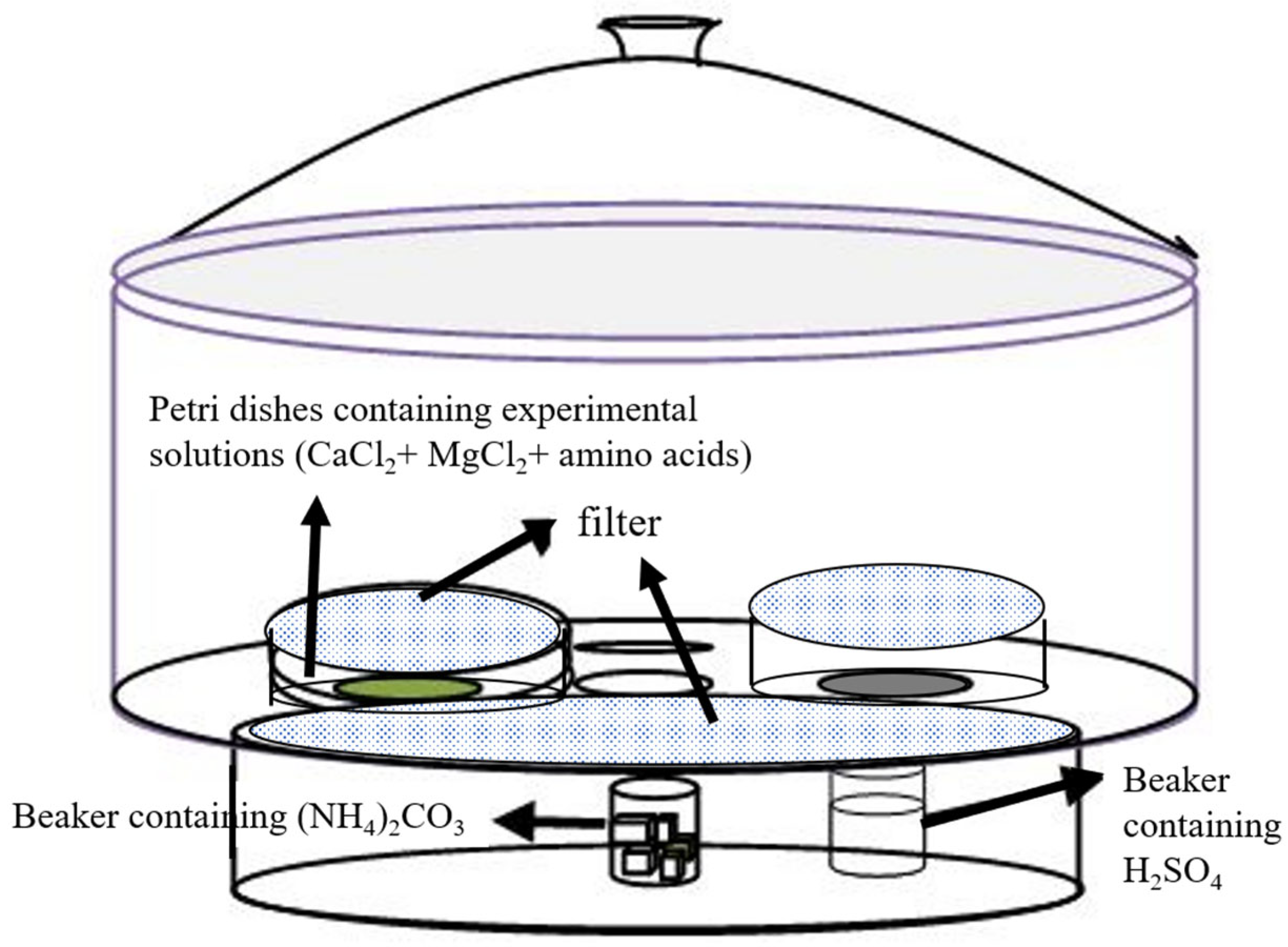
2.2. Biomimetic Mineralization Experiments of Ca–Mg Carbonates

A gas diffusion method for ammonium carbonate decomposition was employed to accomplish biomimetic mineralization [19]. The general setup of the reaction chamber is illustrated in Figure 1. Crystallization reactions occur in a closed container; therefore, a 10 L desiccator was used in this study. Ammonia and CO2 diffuse from the lower section into the upper space, where a reaction occurs. In this study, Petri dishes containing 25 mL experimental solutions were placed in the upper space and sealed with a 0.22 μm cellulose acetate filter. The 25 mL experimental solutions contained 0.01 M CaCl2, 0.01 M MgCl2, and 1.5 mg solid amino acid powder, and their initial pH was adjusted to 5.5. Two beakers containing 10 g of ammonium carbonate and 100 mL of concentrated sulfuric acid (to absorb NH3) were placed at the bottom of the desiccator. Subsequently, aseptic static cultures were established, and the Petri dishes were observed at specific intervals. Finally, the reaction solution and sediment in the Petri dishes were transferred to a sterilized centrifuge tube, centrifuged (5000 rpm, 10 min), rinsed with deionized water, air-dried, and subjected to mineralogical and morphological analyses. All experiments were repeated twice.

2.3. Observation and Analysis

The pH of the solutions was measured using a pHS-3C pH meter (precision: 0.01). The mineralogical compositions of the precipitates were determined by X-ray diffraction (XRD; Rigaku D/MAX-B, Tokyo, Japan). The samples were prepared by dropping the dried precipitate onto glass slides. The measurement parameters were as follows: Cu Kα radiation, 35 kV tube voltage, 20 mA current, scanning rate of 2°/min, step size of 0.02°, and scanning range of 20–60° (2θ). The FT-IR (Nicolet 380, Thermo Fisher Scientific Inc., Waltham, MA, USA) experiment was conducted by using the potassium bromide method in a scanning range of 400–4000 cm−1 with a resolution of 4 cm−1. The morphologies of the mineral products were observed using a field-emission scanning electron microscope (FESEM; Zeiss, Oberkochen, Germany) equipped with an energy-dispersive X-ray spectrometer (EDS). Sample preparation involved washing the precipitates three times with distilled water, soaking them in absolute ethanol, and depositing them onto SEM stubs. After air-drying, the samples were sputter-coated with ~8 nm gold film and imaged at 20 kV and 60 μA. The precipitated minerals were ground in an agate mortar and sieved to obtain uniform grains, and their thermal decomposition characteristics were analyzed using a thermal analyzer (TGA/DSC1/1600LF, METTLER TOLEDO Co., Zurich, Switzerland) from 20 to 800 °C at a constant heating rate (20 °C min−1) with pure nitrogen as a protective gas to prevent oxidation.
When the precipitated minerals contained calcite and aragonite, the proportions of aragonite and calcite were calculated using the following formulas [20]:
XA = 3.157 IA(221)/(IC(104) + 3.157 IA(221))
XC = 1 − XA
where XA and XC represent the proportions of aragonite and calcite minerals (%), respectively. IA (221) and IC(104) are the diffraction intensities of the (221) and (104) crystal planes of aragonite and calcite, respectively.
The Mg content of calcite is often expressed as a molar fraction of MgCO3. When the molar fraction of MgCO3 in the calcite was less than 4 mol%, it was considered to be Mg-containing calcite or low-Mg calcite; when the MgCO3 content in the calcite was between 4 and 35 mol%, it was high-Mg calcite; and when the MgCO3 content in the calcite was between 36 and 52 mol%, it was Ca–dolomite or dolomite. The molar fraction of MgCO3 in the calcite was calculated using the empirical formula proposed by Lumsden [21]:
MgCO3 (mol%) = −333.33d + 1011.99
where d denotes the distance (nm) of the crystal face (104 nm) of the Mg calcite family.

3. Results

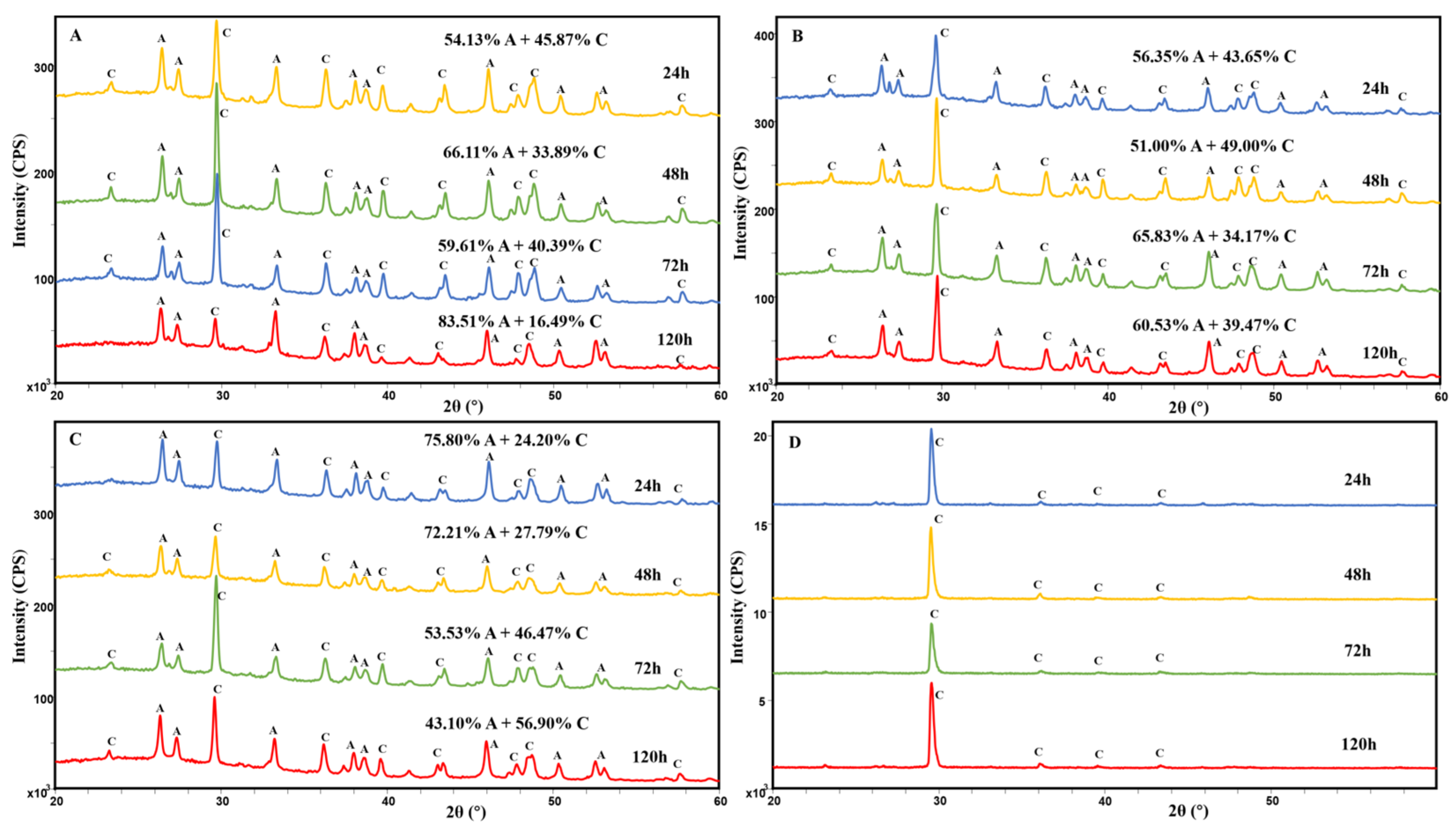
3.1. XRD and FT-IR Analyses of Ca-Mg Carbonate

The XRD pattern (Figure 2) illustrates that the Ca–Mg carbonate minerals formed throughout the mineralization experiment period (from 24 to 120 h) were Mg calcite (d104 ≈ 3.00, d11-3 ≈ 2.28, d11-6 ≈ 1.85) and aragonite (d111 ≈ 3.39, d012 ≈ 2.70, d221 ≈ 1.97) regardless of which amino acid was used. However, the Ca–Mg carbonate minerals formed throughout the mineralization experiments without amino acids were single calcite, and no aragonite was precipitated. Calculating the proportion of aragonite in the deposited Ca–Mg carbonate minerals revealed that under the action of L-glycine, the proportions of aragonite in the deposited Ca–Mg carbonate minerals at 24, 48, 72, and 120 h were 54.13, 66.11, 59.61, and 83.51%, respectively. Under the action of L-glutamic acid, the proportions of aragonite in the sedimentary Ca–Mg carbonate minerals at 24, 48, 72, and 120 h were 56.35, 51.00, 65.83, and 60.53%, respectively. Under the action of L-lysine, the proportions of aragonite in the sedimentary Ca-Mg carbonate minerals at 24, 48, 72, and 120 h were 75.80, 72.21, 53.53, and 43.10%, respectively. Characteristic peaks of the minerals were obtained from the FTIR spectra (Figure 3). It can be observed from Figure 3 that the peaks at 712, 1403, and 2532 cm−1 are the characteristic peaks of calcite, and the peaks at 699, 853, and 1082 cm−1 prove the existence of aragonite, which is consistent with the results of the XRD analyses.

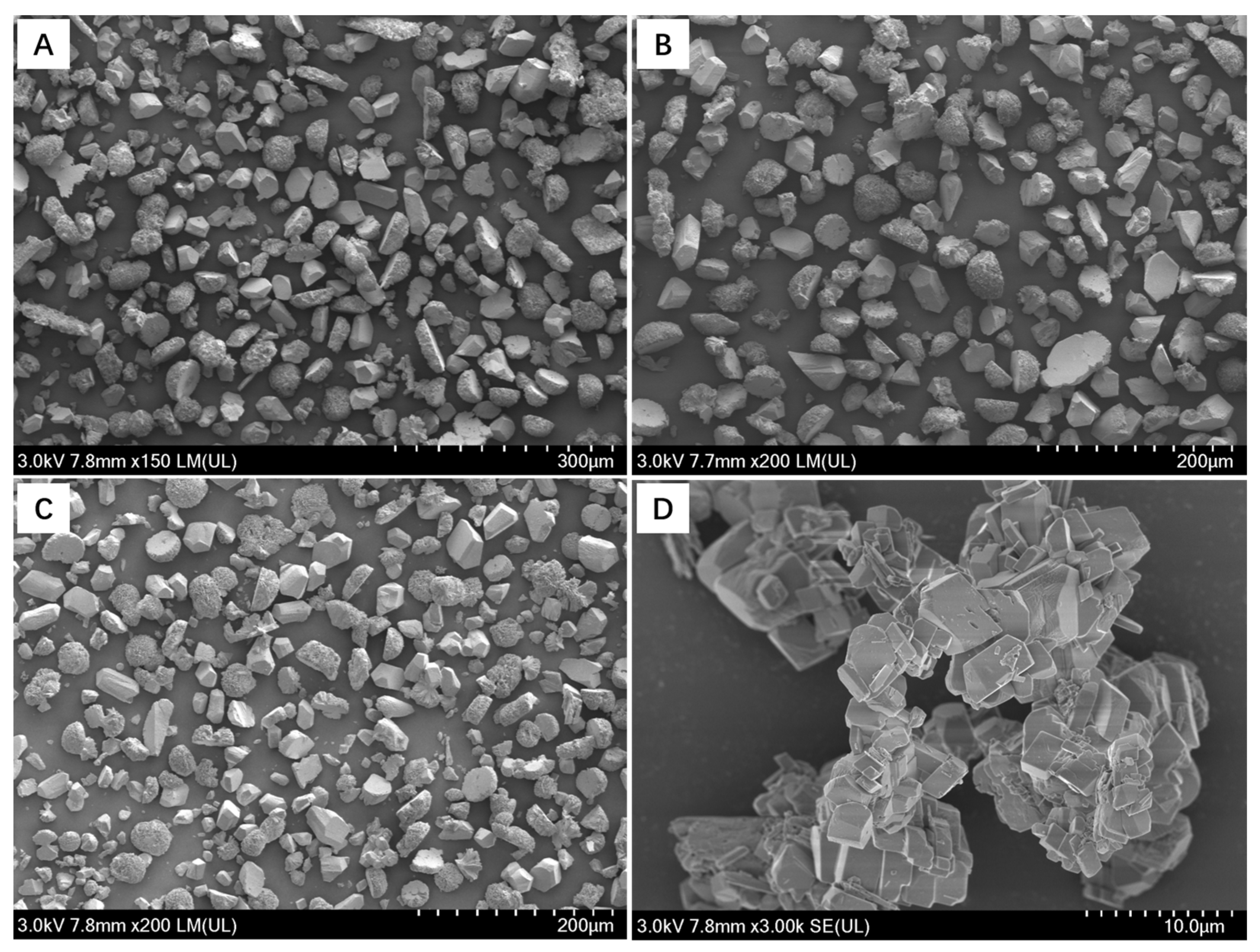
3.2. Morphological Changes of Ca-Mg Carbonate

In the biomimetic mineralization experiment of Ca–Mg carbonates involving different amino acids, the morphologies of the Ca–Mg carbonate minerals formed at 120 h were observed using FE-SEM. The particle size of Ca–Mg carbonate minerals formed under the action of amino acids was between 10 and 100 μm, presenting the morphology of prismatic, spherical, and irregular mineral aggregates (Figure 4). The particle size of the formed Ca–Mg carbonate minerals in the control experiment without amino acids ranged between 1 and 10 μm and presented the morphology of relatively typical rhombohedral mineral aggregates.

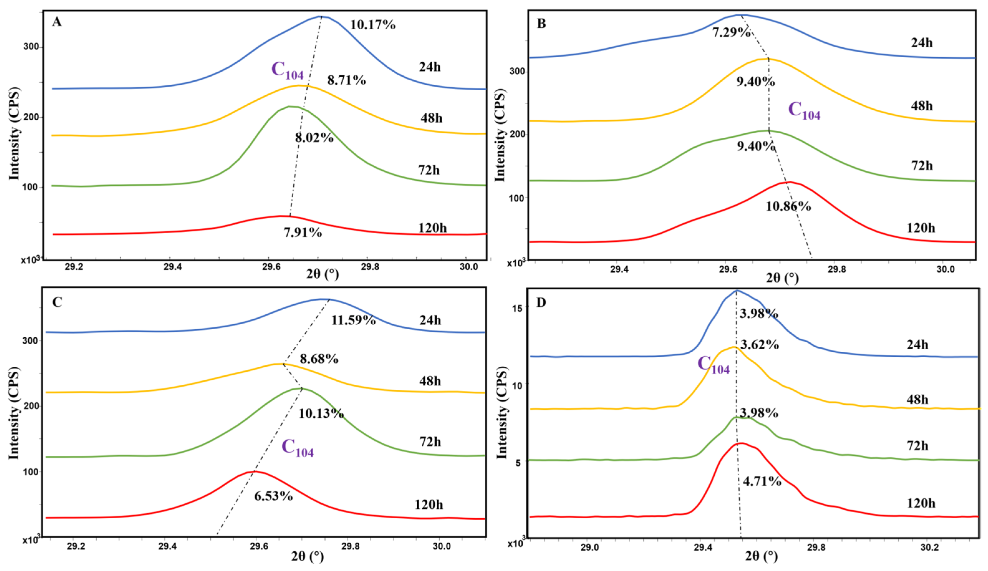
3.3. Magnesium Content in Calcite

Figure 5 illustrates the calculation of the Mg content (molar fraction of MgCO3) in calcite induced by L-glycine in sequence, which was 10.17% (24 h), 8.71% (48 h), 8.02% (72 h), and 7.91% (120 h). The Mg content of the calcite formed under the action of L-glutamic acid gradually increased from 7.29% at 24 h to 10.86% at 120 h. The Mg content of the calcite formed in the presence of L-lysine decreased from 11.59% at 24 h to 6.53% at 120 h. However, in the control experiment without amino acids, the Mg content of the calcite was maintained at approximately 4%.

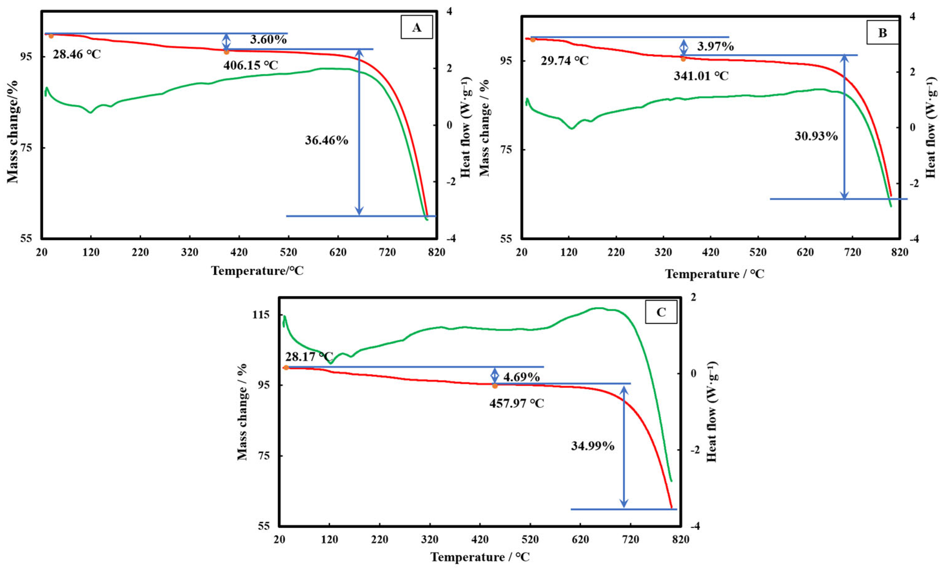
3.4. Thermal Decomposition Characteristics of the Different Precipitates

A thermogravimetric differential scanning calorimetry (TG-DSC) analysis of the precipitated minerals was performed to verify the influence of amino acids on the structural stability of aragonite. As illustrated in Figure 6, the decomposition temperature in the first stage was 28–458 °C, and the mass loss rate was approximately 4%, indicating that approximately 4% of amino acid organic molecules and a small amount of water molecules were adsorbed or incorporated into the mineral. The weight-loss temperature during the second stage of thermal decomposition was 341–800 °C. At this stage, aragonite is transformed into calcite, and the minerals gradually decompose to release CO2. The mass loss rate was approximately 35%, which is lower than the theoretical value of 44% (in the system of pure calcium carbonate). Obviously, the precipitated aragonite and Mg calcite were more stable in the amino acid systems, and some minerals required higher temperatures (>800 °C) for transformation and decomposition. Thus, the TG-DSC curves of Ca–Mg carbonate minerals induced by different amino acids still showed a downward trend at 800 °C and too small mass loss owing to the incomplete decomposition of minerals below 800 °C. Furthermore, the initial temperatures of thermal decomposition for minerals induced by L-lysine, L-glycine, and L-glutamic acid were 341, 406, and 458 °C, respectively. The mineral phase stability of aragonite induced by glutamic acid was better than that of L-glycine and L-lysine, which is consistent with the variation law of the proportion of aragonite in the mineral phase composition throughout the previous mineralization cycle (Figure 2).

4. Discussion

4.1. Control of Reaction Liquid and Mineral Precipitation

In the biomimetic mineralization experiment of Ca–Mg carbonates, the continuous dissolution of (NH4)2CO3 released large amounts of CO2 and NH3 (Reaction (1)), and the pH increased (Figure S1) owing to the dissolution of NH3 in the reaction liquid and the release of OH- (Reaction (2)). Meanwhile, the continuous dissolution of CO2 in the alkaline solution increased the CO32- concentration in the solution (Reaction (3)). Ultimately, mineral precipitation occurred quickly when supersaturation was attained because the concentrations of cations and anions in the reaction solution increased rapidly (Reaction (4)).
(NH4)2CO3 → 2NH3 + CO2 + H2O
NH3 + H2O → NH4+ + OH-
CO2 + H2O → CO32- + 2H+
xCa2+ + (1 − x) Mg2+ + CO32- → CaxMg1−xCO3 (0 ≤ x ≤ 1)
Compared to the control experiment, the Ca–Mg carbonate minerals mainly underwent heterogeneous nucleation in the biomimetic mineralization system involving amino acid molecules [22]. Heterogeneous nucleation enables crystals to form preferentially on the surfaces of amino acid molecules by reducing the nucleation energy barrier rather than spontaneous nucleation in solution (homogeneous nucleation) [23]. Amino acids carry different charges in aqueous solutions, where the pH is not equal to their isoelectric point (pl), thereby affecting the nucleation of Ca–Mg carbonate minerals [24]. When the pH of the aqueous solution is greater than the pl value of the amino acids, the amino acids (L-glycine and L-glutamic acid) release protons and acquire negative charges. The negatively charged amino acids attract Ca2+ and Mg2+ ions, increasing the local ion concentration and leading to local supersaturation, thereby promoting the nucleation of Ca–Mg carbonate minerals. When the pH of the aqueous solution was lower than the pH value of the amino acid, the amino acid (L-lysine) was protonated and acquired a positive charge. Similarly, positively charged amino acids attract CO32− ions, causing the solution to reach a local supersaturated state, thereby promoting the nucleation of Ca–Mg carbonate minerals [25].

4.2. The Effect of Amino Acids on Polymorphs of Ca–Mg Carbonates

In addition to calcite, metastable aragonite was formed under the action of amino acid molecules, compared with the control experiment. This result implies that amino acid molecules are conducive to aragonite formation. According to the proportion of aragonite in the precipitated minerals under the action of different amino acid molecules, the proportion of L-glycine aragonite increased steadily, reaching a maximum of 83.51% with the extension of mineralization time. The effect of L-glutamic acid on the proportion of sedimentary minerals was relatively stable, with approximately 58.43% aragonite in the precipitates. Under the influence of L-lysine, the proportion of aragonite in the sedimentary minerals gradually decreased to 43.10%. Compared with L-lysine and L-glutamic acid, L-glycine is more conducive to the formation of aragonite. Glycine is the simplest amino acid and has a single hydrogen atom (–H) as its side chain group; therefore, the length of the side chain can be regarded as approximately 0. The ionic radius of Ca2+ is approximately 0.174 nm [26]. According to the template theory analysis (Table 2), glycine is more likely to form ligand templates with coordination numbers of 8 or 12 with Ca2+ [27]. Therefore, the amount of aragonite with a coordination number of 9 in the mineralization products induced by glycine was significantly higher than that induced by glutamic acid and lysine.
The results of TG-DSC analysis of the precipitated minerals further confirmed the influence of amino acids on the structural stability of aragonite (Figure 6). Amino acid molecules are conducive to the formation and stability of aragonite and inhibit the phase transition process of its transformation into calcite. The orthogonal lattice parameters of aragonite match relatively well with the molecular arrangement of amino acids, thereby preferentially inducing aragonite nucleation rather than the thermodynamically more stable trigonal calcite [28]. However, the adsorption of amino acid molecules on the surface of aragonite can reduce its surface energy and delay its phase transition kinetics [29]. Amino acids combine with Ca2+ and Mg2+ through their carboxyl functional groups to form a protective layer on the surface of aragonite, preventing its structural reorganization into calcite.
Compared to the control experiment, the morphology of the Ca–Mg carbonate minerals formed under the action of amino acid molecules was more diverse. It has been established that many fibrous and needle-like mineral aggregates formed with amino acids are likely to be relatively stable aragonite growing along the C-axis direction, as supported by the XRD (Figure 2) and EDS (Figure 7) analysis results. This finding is consistent with previous studies, which demonstrated that spherical and needle-like aragonite forms with the participation of amino acids [30]. Amino acid molecules also had a significant influence on calcite morphology. Amino acids can guide the orderly arrangement of mineral ions through specific functional groups or structures, match the lattice parameters of calcite minerals, and promote the preferred growth of specific crystal planes [31]. Second, the geometric arrangement of the amino acid side chains may match the lattice spacing of calcite minerals (for example, the carboxylic acid group spacing is similar to the Ca2+ spacing in calcite), inducing epitaxial growth and thereby causing the morphological evolution of calcite [32]. Furthermore, the Mg contents of the calcite mineral phases formed under the action of different amino acids varied significantly. Mg2+ replaced Ca2+ in the calcite mineral lattice, resulting in the accumulation of lattice strain and inducing changes in particle size and morphology (Figure 7).

4.3. The Effect of Amino Acids on Mg Content in Calcite

As illustrated in Figure 5, the Mg content in the calcite mineral phase induced by amino acids was significantly higher than that in the control experiment (without amino acid participation). Furthermore, the Mg content of the calcite formed by the action of different amino acid molecules varied significantly. As illustrated in Figure 5, the Mg content in the calcite (120 h) induced by the acidic amino acid (L-glutamic acid) was higher than that in the calcite formed under the action of neutral L-glycine and basic L-lysine. Characteristic peaks of the minerals and organic functional groups were obtained from the FTIR spectra (Figure 3). In addition to the characteristic peaks of Ca-Mg carbonate minerals induced by amino acids, certain peaks representing other organic functional groups present in/on the minerals also exist, such as O–H and N–H stretching (2925 cm−1) and C=O stretching (1787 cm−1; 1636 cm−1) illustrated in Figure 3, which indicates that these organic functional groups of amino acids have a close relationship with the nucleation and growth of the minerals, especially the influence of the carboxyl group on Mg content in calcite.
The carboxyl group of L-glutamic acid carries a negative charge in an alkaline solution and preferentially adsorbs Mg2+, which has a higher charge density (compared to Ca2+, Mg2+ has a smaller ionic radius and a higher charge density). This selective adsorption enables more Mg2+ to be enriched in the local microenvironment of the mineral, thereby increasing the possibility of Mg2+ entering the calcite lattice [33]. Indeed, Mg2+ has a stronger hydration ability than Ca2+ and is prone to form hydrated Mg2+ in a water environment, making it difficult for Mg2+ to enter the calcite lattice [34]. It may only partially dehydrate when Mg2+ is involved in the growth of calcite, and the residual hydration spheres doped with Mg2+ inhibit the further incorporation of surface Mg2+ into the calcite lattice [35]. However, the numerous carboxyl groups in acidic amino acids (L-glutamic acid) tend to form complexes with Mg2+, thereby reducing the stability of hydrated Mg2+ and promoting dehydration. Therefore, L-glutamic acid causes more Mg2+ to enter the calcite lattice and replace Ca2+, which accelerates the incorporation of Mg2+ at the growth interface of the calcite crystals.
As the ionic radius of Mg2+ is significantly smaller than that of Ca2+, the gradual substitution of Mg2+ for Ca2+ in the calcite lattice results in lattice distortion [36]. Acidic amino acids can stabilize the distorted calcite lattice by forming complexes with Mg and adhering to the crystal surface, thereby reducing the calcite surface energy. Therefore, more Mg2+ in the solution could replace Ca2+, forming relatively thermodynamically stable calcite. Furthermore, it has been reported that biomolecules rich in carboxyl groups in biological minerals (for example, L-glutamic acid) can temporarily stabilize the amorphous precursor phase (such as amorphous calcium carbonate), delaying its transformation to calcite. Therefore, more Mg2+ was integrated into the final calcite during the reorganization process from amorphous calcium carbonate to calcite [37]. In conclusion, amino acid molecules, especially acidic amino acids, can induce the formation and stability of Mg-rich calcite minerals through multiple mechanisms, such as ion selectivity, coordination stability, and thermodynamic regulation. This process has important applications in geology, environmental science, and biomimetic material synthesis.

5. Conclusions

In this study, acidic (L-glutamic acid), neutral (L-glycine), and basic (L-lysine) amino acids were selected to systematically explore their effects on the mineral composition, morphology, and Mg2+ incorporation efficiency of Ca–Mg carbonates using the gas diffusion method. The results demonstrated that L-glycine, with the shortest side chain, was more conducive to forming aragonite, whereas acidic L-glutamic acid, containing more carboxyl groups, was more beneficial to the structural stability of aragonite. The morphology of the Ca-Mg carbonate minerals became more diverse and promoted the formation of spherical and massive mineral aggregates under the action of amino acids. Acidic amino acids (L-glutamic acid) preferentially adsorb Mg2+ through the carboxyl group and promote its dehydration, which increases the MgCO3 content in calcite to 10.86%. Research has revealed the mechanism by which biological macromolecules directly regulate mineral formation through ion-selective adsorption or incorporation induced by functional groups, offering new insights into the biogenesis of Ca–Mg carbonate minerals and the design of biomimetic mineralized materials.

Supplementary Materials

The following supporting information can be downloaded at https://www.mdpi.com/article/10.3390/min15070763/s1: Figure S1: Temporal evolution of pH during biomimetic mineralization with amino acids.

Author Contributions

Data curation, C.Z.; funding acquisition, C.Z.; methodology, C.Z. and Y.J.; resources, C.Z.; writing—original draft, C.Z. and S.Q. All authors have read and agreed to the published version of the manuscript.

Funding

This work was supported by the Natural Science Foundation of Jiangsu Province (BK20200564), the Natural Science Research Project of the Anhui Educational Committee (KJ2021A0103), the Science and Technology Plan Project of Wuhu (2022jc12), and the Ph.D. Research Start-up Fund (762157).

Data Availability Statement

The original contributions presented in this study are included in the article/Supplementary Material. Further inquiries can be directed to the corresponding author.

Conflicts of Interest

The authors declare no conflicts of interest.

References

  1. Morse, J.W.; Arvidson, R.S.; Lüttge, A. Calcium carbonate formation and dissolution. Chem. Rev. 2007, 107, 342–381. [Google Scholar] [CrossRef] [PubMed]
  2. Liao, R.; Chen, W.; Liu, P.; Wang, L.; Xia, Q.; Yang, X.; He, Y.; Guo, Q. Mineral transformation, element transport and hydrological impact in weathering at the Bingling Temple Grottoes: Implications for weathering in alkaline environments in NW China. Catena 2024, 239, 107966. [Google Scholar] [CrossRef]
  3. Hanken, N.M.; Bjørlykke, K.; Nielsen, J.K. Carbonate sediments. In Petroleum Geoscience: From Sedimentary Environments to Rock Physics; Springer Science & Business Media: Berlin/Heidelberg, Germany, 2015; pp. 151–216. [Google Scholar]
  4. Santos, H.S.; Nguyen, H.; Venâncio, F.; Ramteke, D.; Zevenhoven, R.; Kinnunen, P. Mechanisms of Mg carbonates precipitation and implications for CO2 capture and utilization/storage. Inorg. Chem. Front. 2023, 10, 2507–2546. [Google Scholar] [CrossRef]
  5. Qin, W.; Wang, C.Y.; Ma, Y.X.; Shen, M.J.; Li, J.; Jiao, K.; Tay, F.R.; Niu, L.N. Microbe-mediated extracellular and intracellular mineralization: Environmental, industrial, and biotechnological applications. Adv. Mater. 2020, 32, 1907833. [Google Scholar] [CrossRef] [PubMed]
  6. Huang, J.; Zhang, R. The mineralization of molluscan shells: Some unsolved problems and special considerations. Front. Mar. Sci. 2022, 9, 874534. [Google Scholar] [CrossRef]
  7. Wen, Y.; Sánchez-Román, M.; Li, Y.; Wang, C.; Han, Z.; Zhang, L.; Gao, Y. Nucleation and stabilization of Eocene dolomite in evaporative lacustrine deposits from central Tibetan plateau. Sedimentology 2020, 67, 3333–3354. [Google Scholar] [CrossRef]
  8. Stanienda-Pilecki, K.J. Crystals structures of carbonate phases with Mg in Triassic rocks, mineral formation and transitions. Sci. Rep. 2023, 13, 18759. [Google Scholar] [CrossRef] [PubMed]
  9. Rakhimova, N. Calcium and/or magnesium carbonate and carbonate-bearing rocks in the development of alkali-activated cements—A review. Constr. Build. Mater. 2022, 325, 126742. [Google Scholar] [CrossRef]
  10. Amare, A.; Kassa, Y.; Lemma, B.; Bhaskarwar, A.N.; Mullu, T.; Tibebe, D. Optimised phosphate adsorption using a synergistic calcite-dolomite mix: A novel approach for water treatment. Chem. Ecol. 2025, 41, 668–693. [Google Scholar] [CrossRef]
  11. Anbu, P.; Kang, C.H.; Shin, Y.J.; So, J.S. Formations of calcium carbonate minerals by bacteria and its multiple applications. Springerplus 2016, 5, 250. [Google Scholar] [CrossRef] [PubMed]
  12. Hao, Z.; Su, Y.; Liu, S.; Zhang, X. Roles of bacteria and extracellular polymeric substance in calcium carbonate formation: Insights from the effects of calcium source and deposition rate on nucleation. Biochem. Eng. J. 2024, 202, 109160. [Google Scholar] [CrossRef]
  13. Zhang, C.; Li, F.; Lyu, J.; Yao, Y. Biomimetic mineralization of Ca-Mg carbonates: Relevance to microbial cells and extracellular polymeric substances. Microsc. Microanal. 2023, 29, 665–674. [Google Scholar] [CrossRef] [PubMed]
  14. Robles-Fernández, A.; Areias, C.; Daffonchio, D.; Vahrenkamp, V.C.; Sánchez-Román, M. The role of microorganisms in the nucleation of carbonates, environmental implications and applications. Minerals 2022, 12, 1562. [Google Scholar] [CrossRef]
  15. Kleber, M.; Bourg, I.C.; Coward, E.K.; Hansel, C.M.; Myneni, S.C.; Nunan, N. Dynamic interactions at the mineral–organic matter interface. Nat. Rev. Earth Environ. 2021, 2, 402–421. [Google Scholar] [CrossRef]
  16. Rao, A.; Berg, J.K.; Kellermeier, M.; Gebauer, D. Sweet on biomineralization: Effects of carbohydrates on the early stages of calcium carbonate crystallization. Eur. J. Mineral. 2014, 26, 537–552. [Google Scholar] [CrossRef]
  17. Roberts, J.A.; Kenward, P.A.; Fowle, D.A.; Goldstein, R.H.; González, L.A.; Moore, D.S. Surface chemistry allows for abiotic precipitation of dolomite at low temperature. Proc. Natl. Acad. Sci. USA 2013, 110, 14540–14545. [Google Scholar] [CrossRef] [PubMed]
  18. Kenward, P.A.; Fowle, D.A.; Goldstein, R.H.; Ueshima, M.; González, L.A.; Roberts, J.A. Ordered low-temperature dolomite mediated by carboxyl-group density of microbial cell walls. AAPG Bull. 2013, 97, 2113–2125. [Google Scholar] [CrossRef]
  19. Lian, B.; Hu, Q.; Chen, J.; Ji, J.; Teng, H.H. Carbonate biomineralization induced by soil bacterium Bacillus megaterium. Geochim. Cosmochim. Acta 2006, 70, 5522–5535. [Google Scholar] [CrossRef]
  20. Kontoyannis, C.G.; Vagenas, N.V. Calcium carbonate phase analysis using XRD and FT-Raman spectroscopy. Analyst 2000, 125, 251–255. [Google Scholar] [CrossRef]
  21. Lumsden, D.N. Discrepancy between thin-section and X-ray estimates of dolomite in limestone. J. Sediment. Res. 1979, 49, 429–435. [Google Scholar]
  22. Štajner, L.; Kontrec, J.; Džakula, B.N.; Maltar-Strmečki, N.; Plodinec, M.; Lyons, D.M.; Kralj, D. The effect of different amino acids on spontaneous precipitation of calcium carbonate polymorphs. J. Cryst. Growth 2018, 486, 71–81. [Google Scholar] [CrossRef]
  23. Lyu, J.; Li, F.; Zhang, C.; Gower, L.; Wasman, S.; Sun, J.; Yang, G.; Chen, J.; Gu, L.; Tang, X.; et al. From the inside out: Elemental compositions and mineral phases provide insights into bacterial calcification. Chem. Geol. 2021, 559, 119974. [Google Scholar] [CrossRef]
  24. Hernández-Hernández, A.; Rodríguez-Navarro, A.B.; Gómez-Morales, J.; Jiménez-López, C.; Nys, Y.; García-Ruiz, J.M. Influence of model globular proteins with different isoelectric points on the precipitation of calcium carbonate. Cryst. Growth Des. 2008, 8, 1495–1502. [Google Scholar] [CrossRef]
  25. Guo, Y.; Wang, F.; Zhang, J.; Yang, L.; Shi, X.; Fang, Q.; Ma, X. Biomimetic synthesis of calcium carbonate with different morphologies under the direction of different amino acids. Res. Chem. Intermed. 2013, 39, 2407–2415. [Google Scholar] [CrossRef]
  26. Cao-Luu, N.H.; Luong, H.V.; Nguyen, T.V.; Nguyen-Thi, B.T.; Pham, D.T.; Pham, N.C.; Ho, M.H. Curcumin-Loaded Co-Axial Electrospun Chitosan/Polyvinyl Alcohol/Calcium Chloride Nanofibrous Membranes for Wound Healing Enhancement. ChemistrySelect 2024, 9, e202402644. [Google Scholar] [CrossRef]
  27. Niu, Y.Q.; Liu, J.H.; Aymonier, C.; Fermani, S.; Kralj, D.; Falini, G.; Zhou, C.H. Calcium carbonate: Controlled synthesis, surface functionalization, and nanostructured materials. Chem. Soc. Rev. 2022, 51, 7883–7943. [Google Scholar] [CrossRef] [PubMed]
  28. Gardella, G.; Castillo Alvarez, M.C.; Presslee, S.; Finch, A.A.; Penkman, K.; Kroger, R.; Clog, M.; Allison, N. Contrasting the Effects of Aspartic Acid and Glycine in Free Amino Acid and Peptide Forms on the Growth Rate, Morphology, Composition, and Structure of Synthetic Aragonites. Cryst. Growth Des. 2024, 24, 9379–9390. [Google Scholar] [CrossRef] [PubMed]
  29. Fu, G.; Qiu, S.R.; Orme, C.A.; Morse, D.E.; De Yoreo, J.J. Acceleration of calcite kinetics by abalone nacre proteins. Adv. Mater. 2005, 17, 2678–2683. [Google Scholar] [CrossRef]
  30. Ryu, M.; Kim, H.; Lim, M.; You, K.; Ahn, J. Comparison of dissolution and surface reactions between calcite and aragonite in L-glutamic and L-aspartic acid solutions. Molecules 2010, 15, 258–269. [Google Scholar] [CrossRef] [PubMed]
  31. Borukhin, S.; Bloch, L.; Radlauer, T.; Hill, A.H.; Fitch, A.N.; Pokroy, B. Screening the incorporation of amino acids into an inorganic crystalline host: The case of calcite. Adv. Funct. Mater. 2012, 22, 4216–4224. [Google Scholar] [CrossRef]
  32. Sommerdijk, N.A.; With, G.D. Biomimetic CaCO3 mineralization using designer molecules and interfaces. Chem. Rev. 2008, 108, 4499–4550. [Google Scholar] [CrossRef] [PubMed]
  33. Shi, Y.; Yu, L.; Chen, S.; He, Y.; Yang, X.; Duan, L.; Cai, J. Effects of L-glutamic acid, N, N-diacetic acid as chelating agent on acidification of carbonate reservoirs in acidic environments. J. Nat. Gas Sci. Eng. 2020, 82, 103494. [Google Scholar] [CrossRef]
  34. Zhang, C.; Li, F.; Yang, K.; Zhou, J. The Formation of Calcium–Magnesium Carbonate Minerals Induced by Curvibacter sp. HJ-1 under Different Mg/Ca Molar Ratios. Minerals 2024, 14, 606. [Google Scholar] [CrossRef]
  35. Purgstaller, B.; Mavromatis, V.; Immenhauser, A.; Dietzel, M. Transformation of Mg-bearing amorphous calcium carbonate to Mg-calcite–In situ monitoring. Geochim. Cosmochim. Acta 2016, 174, 180–195. [Google Scholar] [CrossRef]
  36. Astilleros, J.M.; Fernández-Díaz, L.; Putnis, A. The role of magnesium in the growth of calcite: An AFM study. Chem. Geol. 2010, 271, 52–58. [Google Scholar] [CrossRef]
  37. Long, X.; Ma, Y.; Qi, L. Biogenic and synthetic high magnesium calcite—A review. J. Struct. Biol. 2014, 185, 1–14. [Google Scholar] [CrossRef] [PubMed]
Figure 1. Schematic of biomimetic mineralization experiments of Ca–Mg carbonates.
Minerals 15 00763 g001
Figure 2. XRD patterns of the precipitates collected during biomimetic mineralization with and without amino acids ((A): L-glycine; (B): L-glutamic acid; (C): L-lysine; (D): CK).
Minerals 15 00763 g002
Figure 3. FTIR analysis of Ca–Mg carbonate minerals induced by amino acids.
Minerals 15 00763 g003
Figure 4. FESEM images of the precipitated minerals formed at 120 h in biomimetic mineralization with and without amino acids ((A): L-glycine; (B): L-glutamic acid; (C): L-lysine; (D): CK).
Minerals 15 00763 g004
Figure 5. Mg content in calcite formed in biomimetic mineralization with and without amino acids ((A): L-glycine; (B): L-glutamic acid; (C): L-lysine; (D): CK).
Minerals 15 00763 g005
Figure 6. The TG-DSC curves of Ca–Mg carbonate minerals induced by different amino acids ((A): L-glycine; (B): L-lysine; (C): L-glutamic acid).
Minerals 15 00763 g006
Figure 7. SEM and EDS spectra of Ca–Mg carbonate minerals formed at 120 h in biomimetic mineralization with glutamic acid ((A)—spherical aggregates; (B)—prismatic aggregates; (A1,B1) are enlarged images of (A) and (B), respectively; (A2,B2) are EDS spectra of (A) and (B), respectively).
Minerals 15 00763 g007
Table 1. Physicochemical properties of the selected amino acids.
L-GlycineL-LysineL-Glutamic Acid
Isoelectric Point (pl)5.979.743.22
Charge state (Physiological pH)NeutralPositively charged (basic)Negatively charged (acidic)
Side Chain Structure-H-(CH2)4NH2-(CH2)2COOH
Table 2. Relationship between the radius ratio of positive/negative ions and the coordination number of crystals.
R+/R0.155–0.2250.225–0.4140.414–0.7320.732–1≥1
Coordination number346812
Note: R+ represents positive ions, and R represents negative ions.
Disclaimer/Publisher’s Note: The statements, opinions and data contained in all publications are solely those of the individual author(s) and contributor(s) and not of MDPI and/or the editor(s). MDPI and/or the editor(s) disclaim responsibility for any injury to people or property resulting from any ideas, methods, instructions or products referred to in the content.

