
Crystal Structure Features, Spectroscopic Characteristics and Thermal Conversions of Sulfur-Bearing Groups: New Natural Commensurately Modulated Haüyne Analogue, Na6Ca2−x(Si6Al6O24)(SO42−,HS−,S2●−,S4,S3●−,S52−)2−y


 ,
,  , and
, and
Abstract
1. Introduction
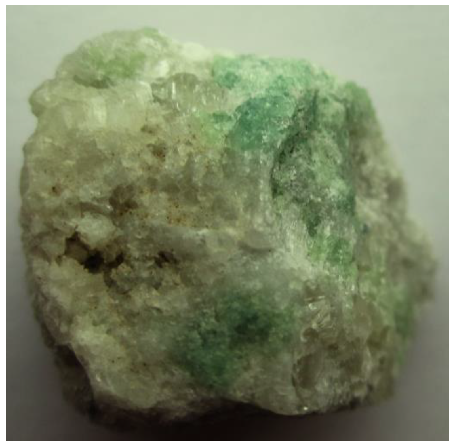
2. Studied Sample
3. Methods
4. Results
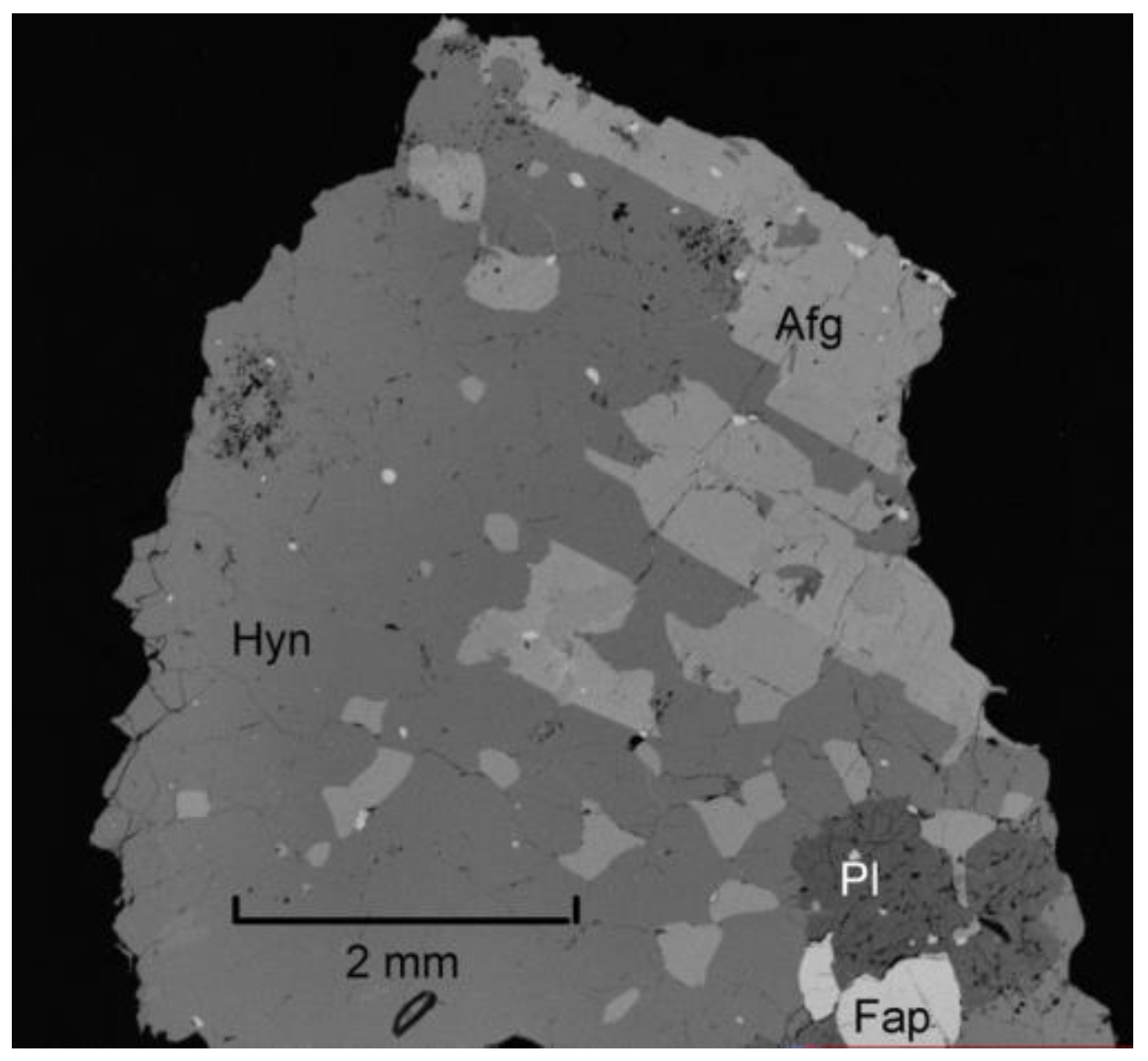
4.1. Chemical Composition
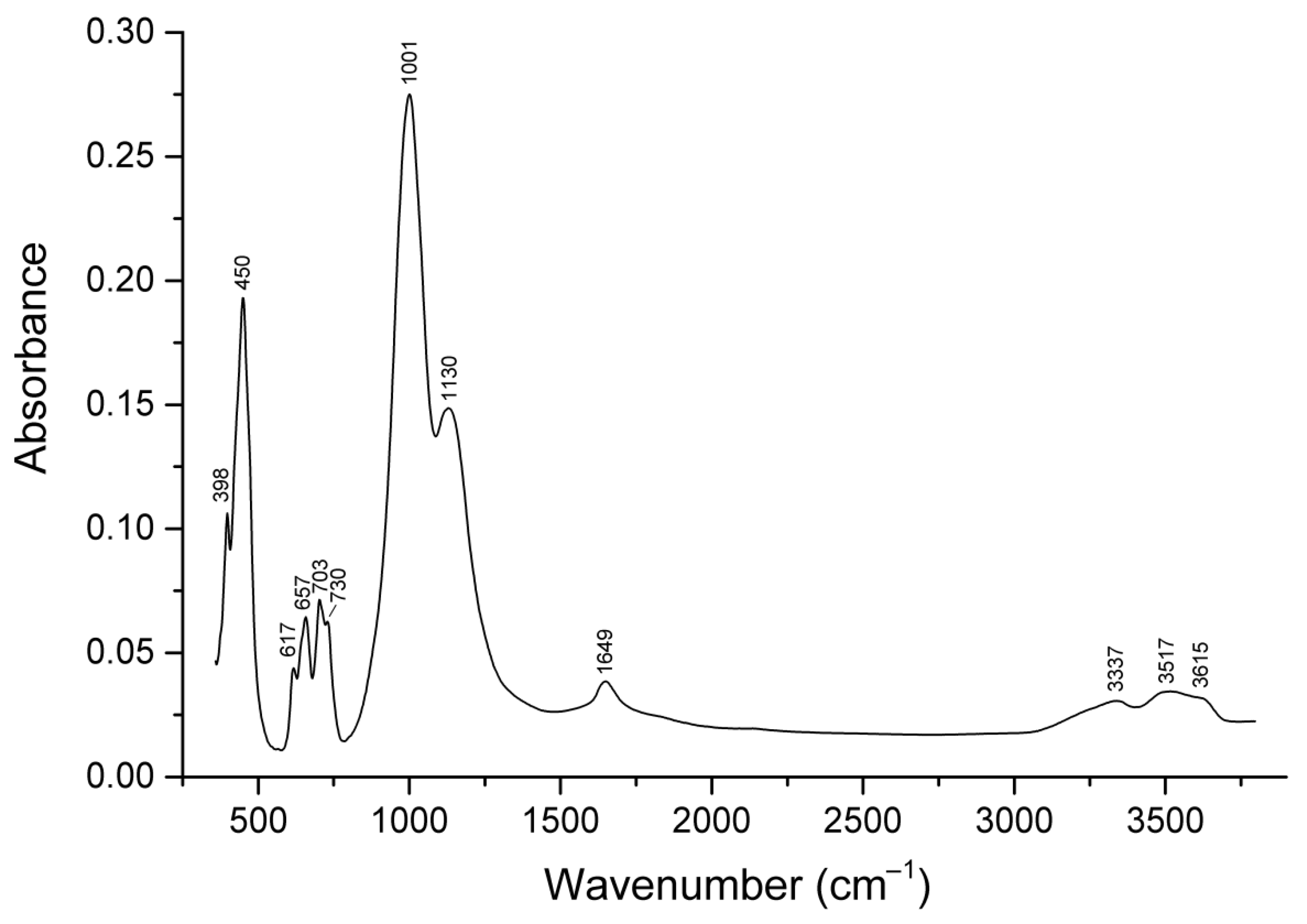
4.2. Infrared Spectroscopy
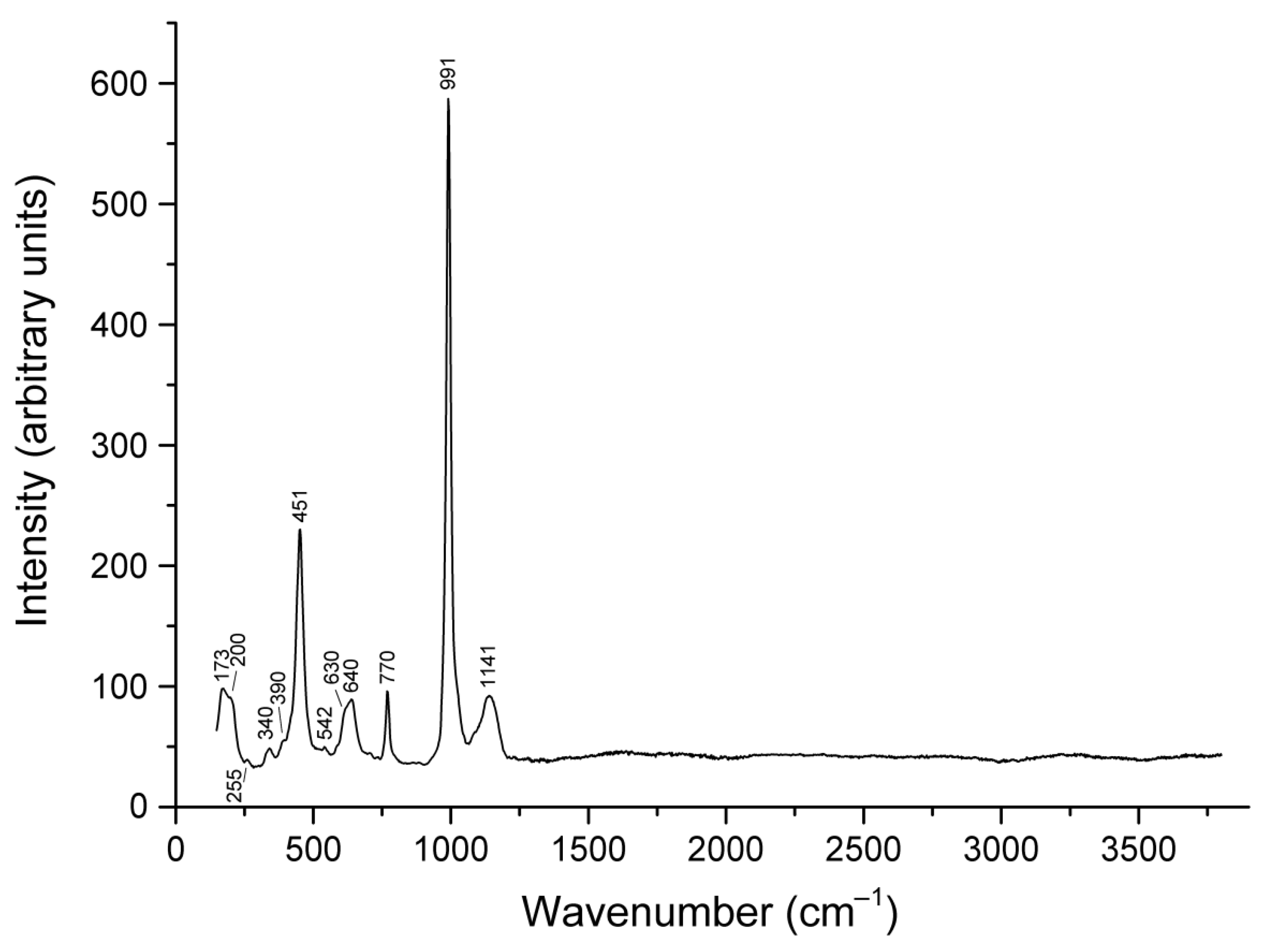
4.3. Raman Spectroscopy
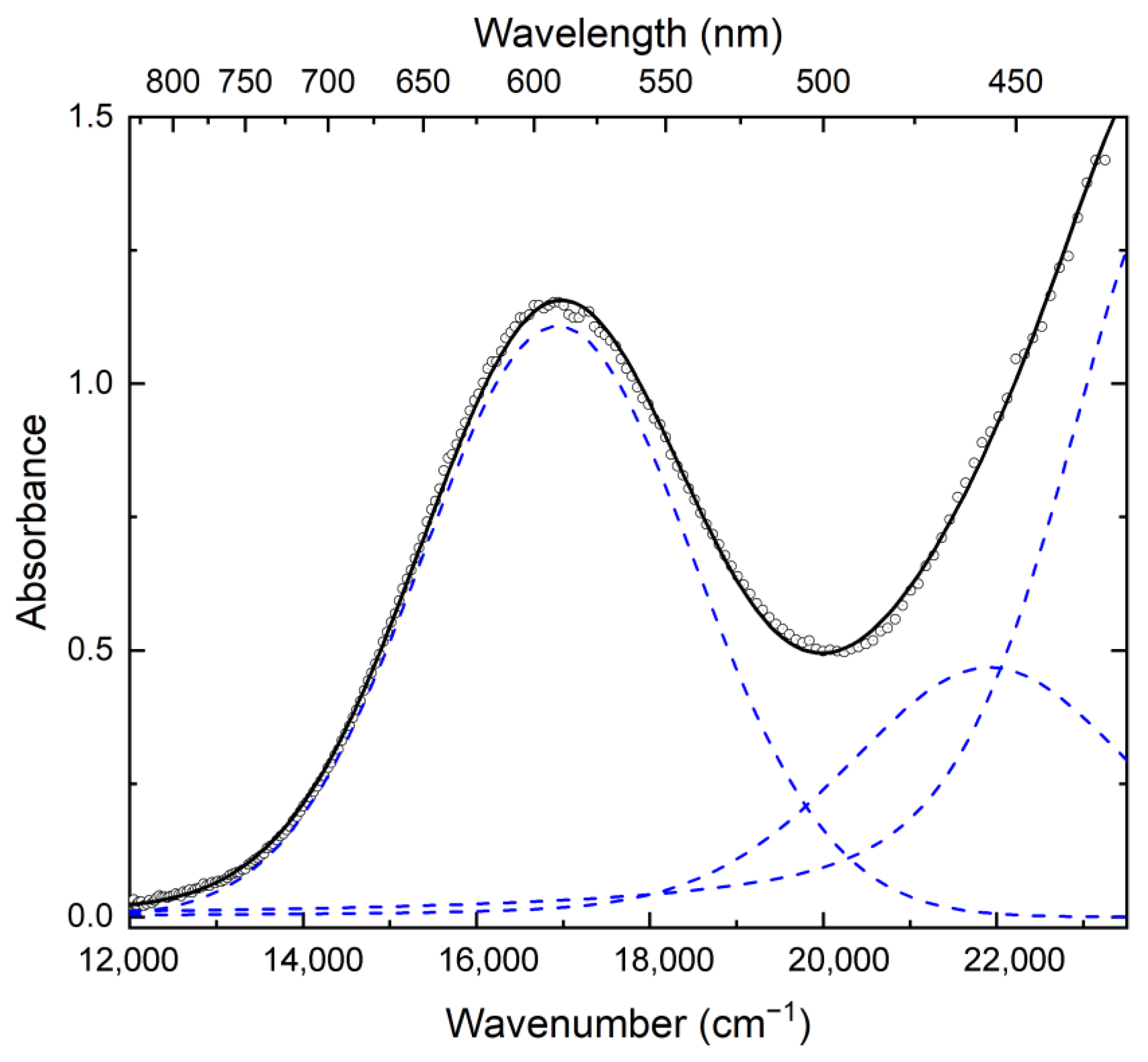
4.4. UV–Vis–near IR Absorption Spectroscopy
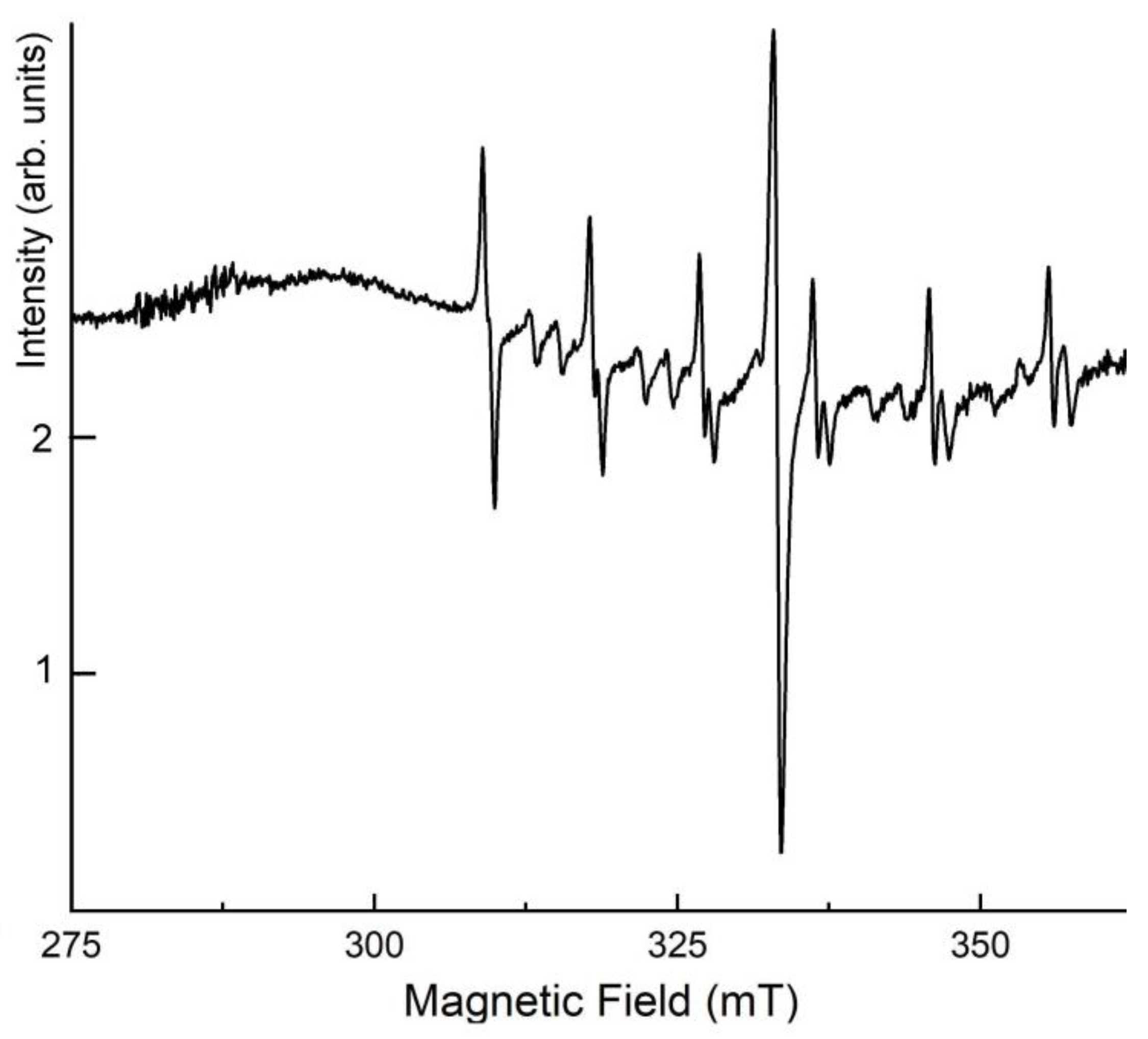
4.5. ESR Spectroscopy
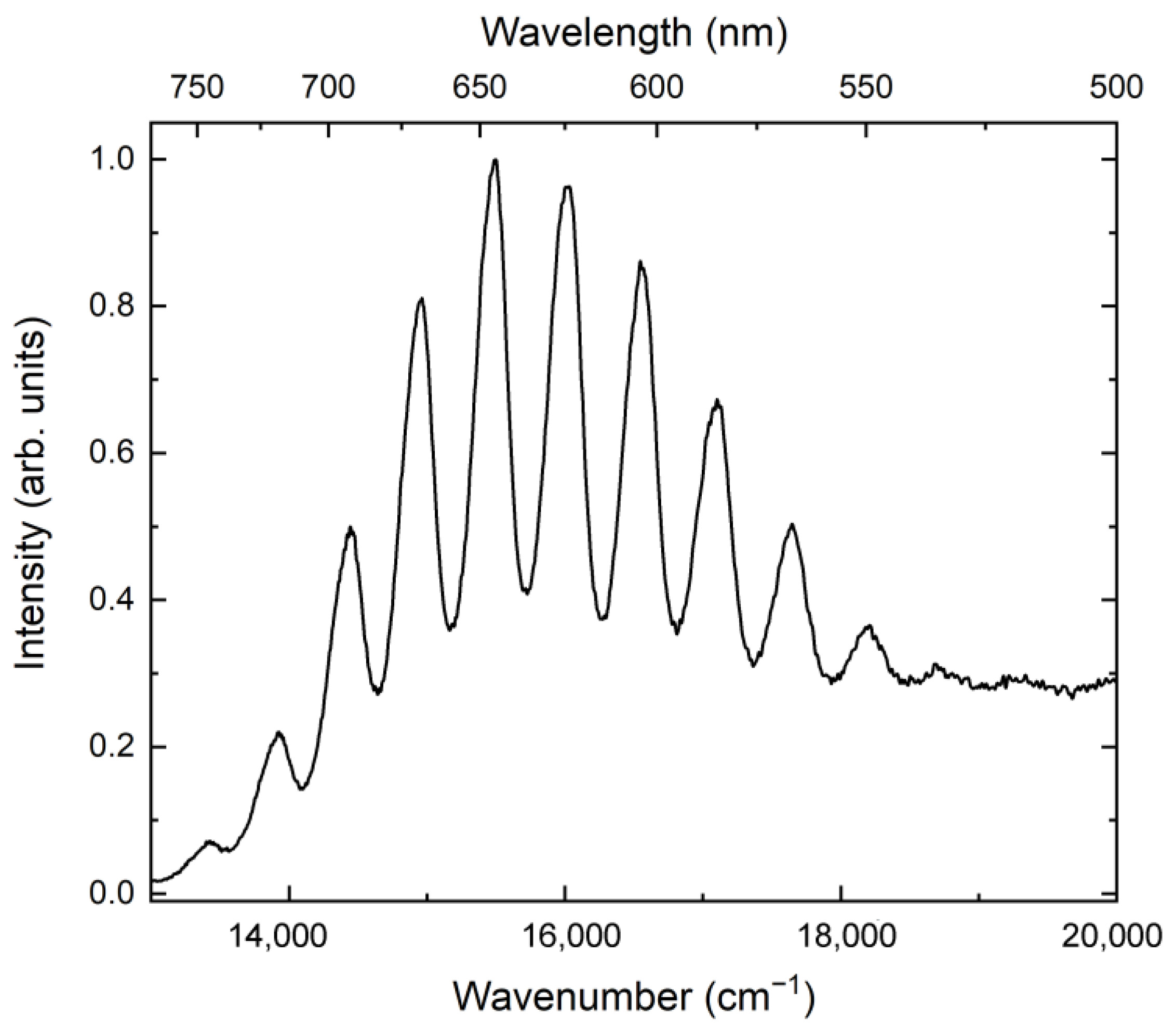
4.6. Luminescence Spectroscopy
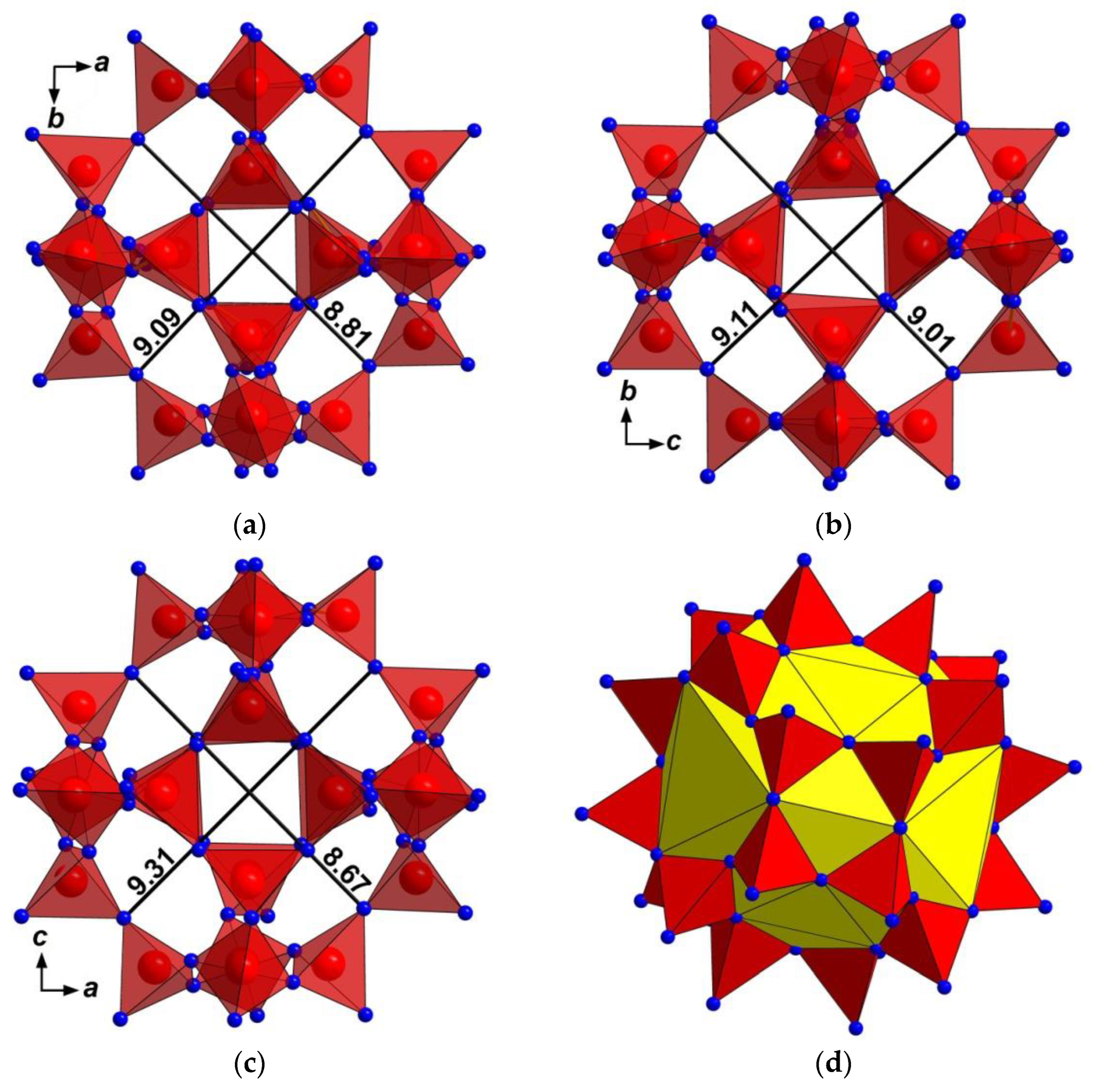
4.7. Structural Features
4.8. Thermal Conversions
5. Discussion
5.1. Extra-Framework Components
5.2. Structure Modulations
5.3. Mechanisms of Thermal Conversions and Their Geochemical Importance
6. Conclusions and Implications
Supplementary Materials
Author Contributions
Funding
Data Availability Statement
Acknowledgments
Conflicts of Interest
References
- Chukanov, N.V.; Aksenov, S.M. Crystal chemistry, chemical diversity and physical properties of microporous sodalite-type materials: A review. Intern. J. Molec. Sci. 2024, 25, 10218. [Google Scholar] [CrossRef]
- Sapozhnikov, A.N.; Tauson, V.L.; Lipko, S.V.; Shendrik, R.Y.; Levitskii, V.I.; Suvorova, L.F.; Chukanov, N.V.; Vigasina, M.F. On the crystal chemistry of sulfur-rich lazurite, ideally Na7Ca(Al6Si6O24)(SO4)(S3)−·nH2O. Amer. Miner. 2021, 106, 226–234. [Google Scholar] [CrossRef]
- Chukanov, N.V.; Shendrik, R.Y.; Vigasina, M.F.; Pekov, I.V.; Sapozhnikov, A.N.; Shcherbakov, V.D.; Varlamov, D.A. Crystal chemistry, isomorphism, and thermal conversions of extra-framework components in sodalite-group minerals. Minerals 2022, 12, 887. [Google Scholar] [CrossRef]
- Chukanov, N.V.; Vigasina, M.F.; Shendrik, R.Y.; Varlamov, D.A.; Pekov, I.V.; Zubkova, N.V. Nature and isomorphism of extra-framework components in cancrinite- and sodalite-related minerals: New data. Minerals 2022, 12, 729. [Google Scholar] [CrossRef]
- Chukanov, N.V.; Shchipalkina, N.V.; Shendrik, R.Y.; Vigasina, M.F.; Tauson, V.L.; Lipko, S.V.; Varlamov, D.A.; Shcherbakov, V.D.; Sapozhnikov, A.N.; Kasatkin, A.V.; et al. Isomorphism and mutual transformations of S-bearing components in feldspathoids with microporous structures. Minerals 2022, 12, 1456. [Google Scholar] [CrossRef]
- Bolotina, N.B.; Sapozhnikov, A.N.; Chukanov, N.V.; Vigasina, M.F. Structure modulations and symmetry of lazurite-related sodalite-group minerals. Crystals 2023, 13, 768. [Google Scholar] [CrossRef]
- Oxford Diffraction CrysAlisPro Software System; v. 1.171.42.49; Rigaku Corporation: Oxford, UK, 2022.
- Sheldrick, G.M. Crystal structure refinement with SHELXL. Acta Crystallogr. 2015, C71, 3–8. [Google Scholar] [CrossRef]
- Frisch, M.J.; Trucks, G.W.; Schlegel, H.B.; Scuseria, G.E.; Robb, M.A.; Cheeseman, J.R.; Scalmani, G.; Barone, V.; Mennucci, B.; Petersson, G.A.; et al. Gaussian 09, Revision D.01; Gaussian, Inc.: Wallingford, CT, USA, 2009. [Google Scholar]
- Dubessy, J.; Boiron, M.-C.; Moissette, A.; Monnion, C.; Sretenskaya, N. Determination of water, hydrates and pH in fluid inclusions by micro-Raman spectrometry. Eur. J. Min. 1992, 4, 885–894. [Google Scholar] [CrossRef]
- Eckert, B.; Steudel, F. Molecular spectra of sulfur molecules and solid sulfur allotropes. Top. Curr. Chem. 2003, 231, 31–97. [Google Scholar] [CrossRef]
- Wong, M.W.; Steudel, R. Structure and spectra of tetrasulfur S4—An ab initio MO study. Chem. Phys. Lett. 2003, 379, 162–169. [Google Scholar] [CrossRef]
- Ling, Z.C.; Wang, A.; Jolliff, B.L. Mineralogy and geochemistry of four lunar soils by laser-Raman study. Icarus 2011, 211, 101–113. [Google Scholar] [CrossRef]
- Hettmann, K.; Wenzel, T.; Marks, M.; Markl, G. The sulfur speciation in S-bearing minerals: New constraints by a combination of electron microprobe analysis and DFT calculations with special reference to sodalite-group minerals. Amer. Miner. 2012, 97, 1653–1661. [Google Scholar] [CrossRef]
- Steudel, R.; Chivers, T. The role of polysulfide dianions and radical anions in the chemical, physical and biological sciences, including sulfur-based batteries. Chem. Soc. Rev. 2019, 48, 3279–3319. [Google Scholar] [CrossRef]
- Rejmak, P. Computational refinement of the puzzling red tetrasulfur chromophore in ultramarine pigments. Phys. Chem. Chem. Phys. 2020, 22, 22684–22698. [Google Scholar] [CrossRef]
- Caggiani, M.C.; Mangone, A.; Aquafredda, P. Blue coloured haüyne from Mt. Vulture (Italy) volcanic rocks: SEM-EDS and Raman investigation of natural and heated crystals. J. Raman Spectrosc. 2022, 53, 956–968. [Google Scholar] [CrossRef]
- Chivers, T.; Oakley, R.T. Structures and spectroscopic properties of polysulfide radical anions: A theoretical perspective. Molecules 2023, 28, 5654. [Google Scholar] [CrossRef]
- Rolfe, J. Emission spectra of S2−, Se2−, and SeS− ions in KI Crystals. J. Chem. Phys. 1968, 49, 4193–4197. [Google Scholar] [CrossRef]
- Kowalak, S.; Jankowska, A.; Zeidler, S.; Wiećkowski, A.B. Sulfur radicals embedded in various cages of ultramarine analogs prepared from zeolites. J. Solid State Chem. 2007, 180, 1119–1124. [Google Scholar] [CrossRef]
- Sapozhnikov, A.N.; Bolotina, N.B.; Chukanov, N.V.; Shendrik, R.Y.; Kaneva, E.V.; Vigasina, M.F.; Ivanova, L.A.; Tauson, V.L.; Lipko, S.V. Slyudyankaite, Na28Ca4(Si24Al24O96)(SO4)6(S6)1/3(CO2)·2H2O, a new sodalite group mineral from the Malo-Bystrinskoe lazurite deposit, Baikal Lake area, Russia. Amer. Miner. 2023, 108, 1805–1817. [Google Scholar] [CrossRef]
- Bolotina, N.B.; Chukanov, N.V.; Sapozhnikov, A.N.; Zubkova, N.V.; Pekov, I.V.; Varlamov, D.A.; Vigasina, M.F.; Bulakh, M.O.; Yapaskurt, V.O.; Ksenofontov, D.A. Vladimirivanovite revised: General crystal chemistry and isomorphous substitutions of extra-framework species. Minerals 2024, 14, 883. [Google Scholar] [CrossRef]
- Sapozhnikov, A.N.; Tauson, V.L.; Lipko, S.V.; Danilov, B.S.; Chukanov, N.V. On the temperature conditions of formation of pink S4-containing haüyne from the Malo-Bystrinskoe lazurite deposit (East Siberia, Russia). Zap. Russ. Miner. Soc. 2025, 154, 107–117. [Google Scholar] [CrossRef]


| Haüyne-45Å | Afghanite | |||
|---|---|---|---|---|
| Constituent | Mean Over 11 Spot Analyses | Standard Deviation | Mean Over 4 Spot Analyses | Standard Deviation |
| Na2O | 17.67 | 0.40 | 14.42 | 0.17 |
| K2O | 0.18 | 0.09 | 0.71 | 0.09 |
| CaO | 7.90 | 0.15 | 12.82 | 0.26 |
| Al2O3 | 27.59 | 0.46 | 27.04 | 0.37 |
| Fe2O3 | 0.03 | 0.05 | 0.05 | 0.04 |
| SiO2 | 33.80 | 0.54 | 32.15 | 0.40 |
| SO3 | 13.28 | 0.36 | 11.21 | 0.14 |
| Cl− | 0.42 | 0.09 | 4.71 | 0.14 |
| H2O | 1.04 | - | No data | - |
| –O=Cl− | –0.09 | - | –1.06 | - |
| Total | 101.82 | - | 101.95 | - |
| Formula coefficients calculated on 12 (Si+Al+Fe) atoms | ||||
| Na | 6.20 | 0.13 | 5.24 | 0.08 |
| K | 0.04 | 0.02 | 0.17 | 0.02 |
| Ca | 1.53 | 0.02 | 2.57 | 0.04 |
| Al | 5.88 | 0.07 | 5.97 | 0.03 |
| Fe | 0.00 | 0.01 | 0.01 | 0.01 |
| Si | 6.12 | 0.07 | 6.03 | 0.03 |
| S | 1.80 | 0.04 | 1.58 | 0.04 |
| Cl | 0.13 | 0.03 | 1.50 | 0.05 |
| H2O | 0.63 | - | - | - |
| Haüyne-45Å | Afghanite | Assignment |
|---|---|---|
| Raman Shift (cm−1) | ||
| 156 | 173, 200 sh | Librations and translations of extra-framework anions |
| 255 w | 255 w | Bending vibrations of S3●− (the ν2 mode) cyclic S52− and/or gauche-S52−-2 |
| 280 w | - | gauche-S52−-2 or S4●− bending mode |
| - | 340, 390 | cis-S4 mixed ν3 and ν4 modes |
| 444 s | 451 s | Bands of stretching vibrations of cis-S4 overlapping with the band of framework bending vibrations |
| 546 s | 542 w | Overlapping bands corresponding to S2●− stretching vibrations and S3●− symmetric stretching (ν1) mode |
| 590 w | gauche-S52−-2 stretching mode | |
| 615 | 630 sh | S2●− combination (stretching + libration) mode, stretching vibrations of gauche-S52−-2, framework vibrations and SO42− bending F2(ν4) mode |
| 640 | - | cis-S4 symmetric stretching mode (ν3) |
| 708 | - | gauche-S52−-2 combination mode |
| - | 770 | Mixed vibrations of the framework |
| 800 | - | S3●− combination mode (ν1 + ν2) |
| 989 s | 991s | SO42− symmetric stretching vibrations [A1(ν1) mode] |
| 1140 | 1141 | SO42− asymmetric stretching vibrations [F2(ν3) mode], possibly, overlapping with S2●− overtone (2×ν1) |
| 1370 w | - | S3●− combination mode (2ν1 + ν2) |
| 1637 | - | S3●− overtone (3′ν1) |
| 2577 | - | H2S stretching mode |
| 3305 | - | O–H stretching vibrations |
| Cage Number | Dimensions of the Cages (Å) | Volume of the 36-Fold Polyhedron (Å3) | ||
|---|---|---|---|---|
| ab | bc | ac | ||
| Sod 1 | 8.71 9.21 | 9.00 9.22 | 8.81 8.96 | 396.78 |
| Sod 2 | 8.85 9.22 | 8.92 9.06 | 8.94 9.09 | 389.37 |
| Sod 3 | 9.23 9.35 | 9.00 9.17 | 9.03 9.07 | 417.32 |
| Sod 4 | 8.81 9.09 | 9.01 9.11 | 8.67 9.31 | 387.45 |
| Sod 5 | 8.83 9.17 | 8.97 9.12 | 8.76 9.24 | 399.32 |
| Sod 6 | 8.63 9.05 | 8.69 9.01 | 8.92 9.14 | 382.37 |
| Sod 7 | 8.77 9.16 | 8.94 9.27 | 8.92 9.05 | 391.96 |
| Sod 8 | 8.86 8.89 | 9.02 9.21 | 8.87 9.13 | 382.03 |
| Sod 9 | 8.77 9.03 | 8.96 9.00 | 8.69 9.22 | 394.22 |
| Sod 10 | 8.73 9.25 | 8.89 9.13 | 8.68 9.24 | 393.31 |
| Sod 11 | 8.83 9.39 | 9.05 9.15 | 8.99 9.01 | 383.43 |
| Sod 12 | 8.86 9.07 | 8.93 9.04 | 9.09 9.13 | 386.63 |
| Sod 13 | 8.57 9.36 | 8.84 8.99 | 8.69 9.16 | 394.56 |
| Sod 14 | 8.60 9.10 | 8.63 9.32 | 8.64 9.23 | 382.34 |
| Sod 15 | 8.86 9.06 | 8.82 9.16 | 8.83 9.02 | 386.92 |
| Sod 16 | 8.70 9.17 | 8.78 9.23 | 8.89 9.25 | 389.12 |
| Sod 17 | 8.71 9.08 | 8.84 8.98 | 8.72 9.24 | 389.93 |
| Sod 18 | 8.63 9.28 | 8.67 9.22 | 8.64 9.25 | 380.77 |
| Sod 19 | 8.95 9.05 | 9.03 9.07 | 8.97 9.11 | 380.41 |
| Sod 20 | 8.82 9.19 | 8.93 9.14 | 8.79 9.12 | 391.59 |
| Sod 21 | 8.68 9.14 | 8.66 9.03 | 8.59 9.28 | 383.69 |
| Sod 22 | 8.80 9.14 | 9.00 9.09 | 8.86 9.15 | 389.53 |
| Sod 23 | 8.99 9.03 | 8.72 9.24 | 9.00 9.07 | 383.71 |
| Sod 24 | 8.96 9.04 | 8.78 9.00 | 8.88 9.02 | 377.41 |
| Sod 25 | 8.89 9.09 | 8.62 9.22 | 8.97 9.07 | 389.84 |
| A row along the a axis | ||||
| Sod 1a | 8.60 9.39 | 8.57 9.27 | 8.81 9.17 | 384.87 |
| Sod 2a | 8.57 9.36 | 8.84 8.99 | 8.69 9.16 | 394.55 |
| Sod 3a | 8.75 9.12 | 8.78 9.03 | 8.70 9.28 | 385.75 |
| Sod 4a | 8.84 9.07 | 8.96 9.00 | 8.98 9.04 | 377.96 |
| Sod 5a | 9.01 9.02 | 8.85 9.22 | 8.95 9.11 | 381.87 |
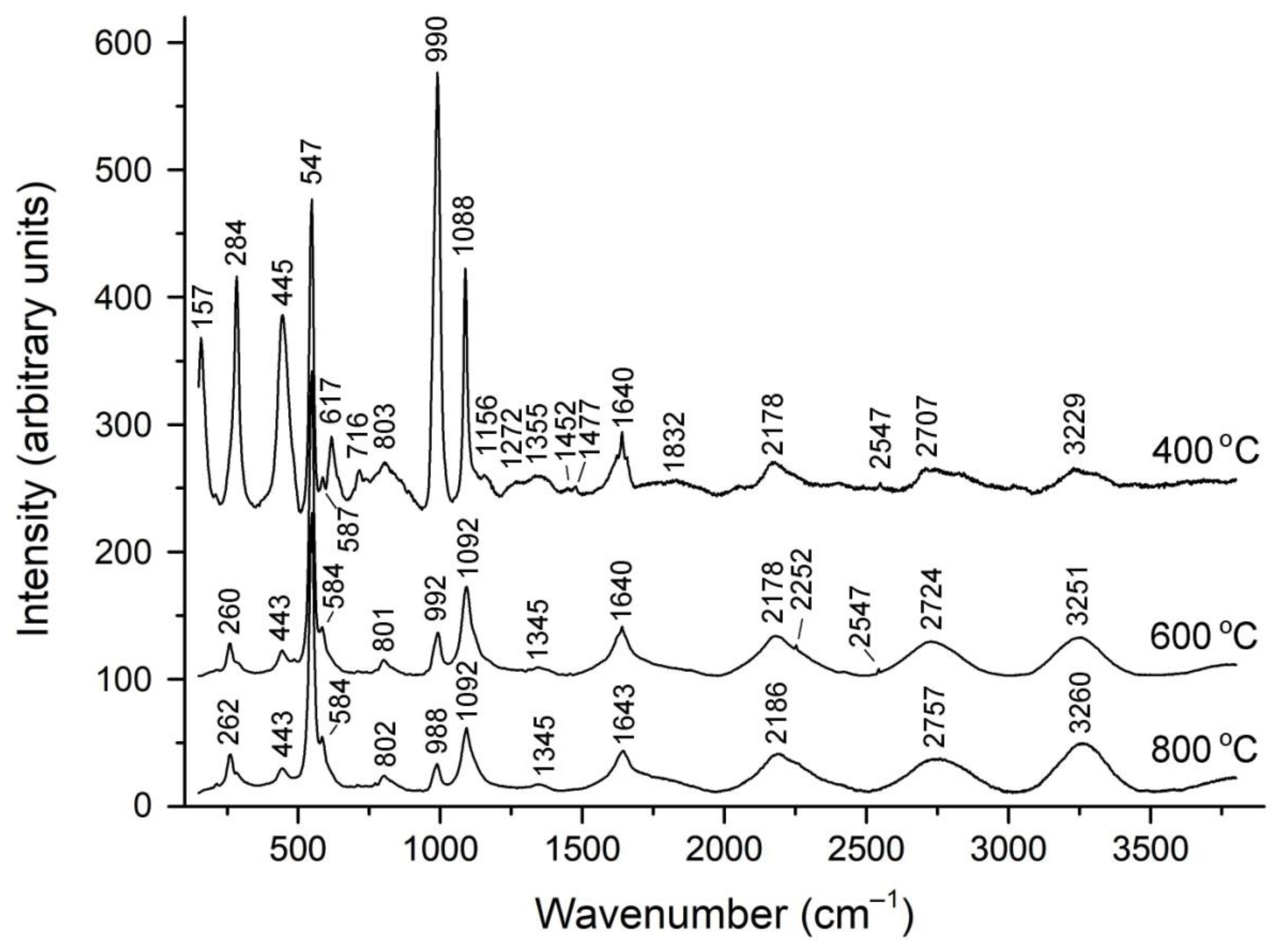
| 400 °C | 600 °C | 800 °C | Assignment |
|---|---|---|---|
| 157 s | - | - | Librations and translations of extra-framework anions |
| - | 260 | 262 | Bending vibrations of S3●− (the ν2 mode) cyclic S52− and/or gauche-S52−-2 |
| 284 s | - | - | gauche-S52−-2 or S4●− bending mode |
| 445 s | 443 | 443 | Bands of stretching vibrations of cis-S4 overlapping with the band of framework bending vibrations |
| 547 s | 547s | 548s | Overlapping bands corresponding to S2●− stretching vibrations and S3●− symmetric stretching (ν1) mode |
| 587 w | 584 w | 584 w | gauche-S52−-2 stretching mode |
| 617 | - | - | S2●− combination (stretching + libration) mode, stretching vibrations of gauche-S52−-2, framework vibrations and/or SO42− bending F2(ν4) mode |
| 716 w | - | - | gauche-S52−-2 combination mode |
| 803 | 801 w | 802 w | S3●− combination mode (ν1 + ν2) |
| 990 s | 992 | 988 | SO42− symmetric stretching vibrations [A1(ν1) mode] |
| 1088 | 1092 | 1092 | S3●− overtone (2′ ν1) |
| 1156 w | - | - | SO42− asymmetric stretching vibrations [F2(ν3) mode], possibly, overlapping with S2●− overtone (2 × ν1) |
| 1355 w | 1345 w | 1345 w | S3●− combination mode (2ν1 + ν2) |
| 1640 | 1640 | 1643 | S3●− overtone (3′ ν1) |
| 2547 | 2547 | - | HS− stretching mode |
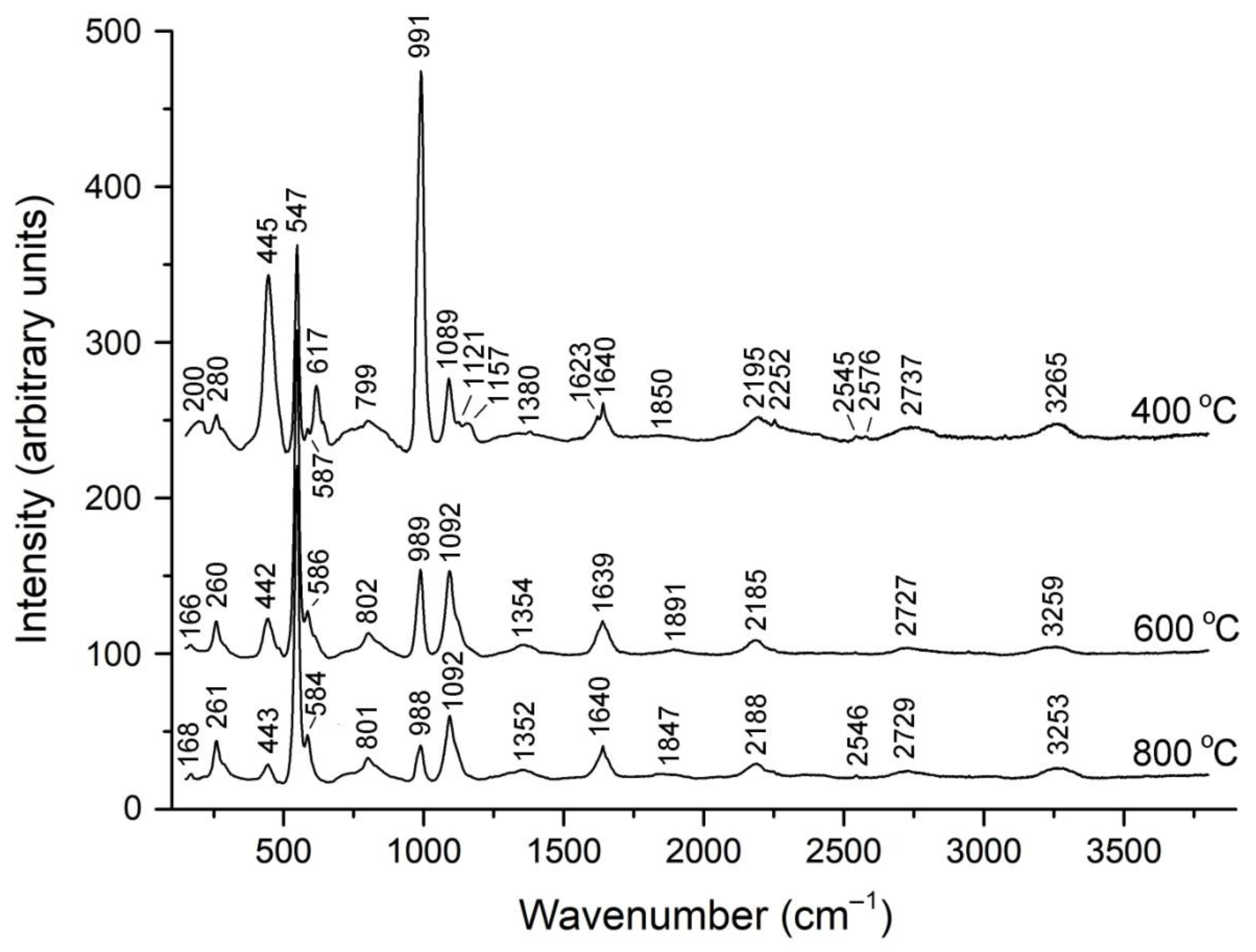
| 400 °C | 600 °C | 800 °C | Assignment |
|---|---|---|---|
| 200 w | 166 w | 168 w | Librations and translations of extra-framework anions |
| - | 260 | 261 | Bending vibrations of S3●− (the ν2 mode) cyclic S52− and/or gauche-S52−-2 |
| 280 | - | - | gauche-S52−-2 or S4●− bending mode |
| 445 s | 442 | 443 | Bands of stretching vibrations of cis-S4 overlapping with the band of framework bending vibrations |
| 547 s | 547 s | 547 s | Overlapping bands corresponding to S2●− stretching vibrations and S3●− symmetric stretching (ν1) mode |
| 587w | 586w | 584w | gauche-S52−-2 stretching mode |
| 617 | - | - | S2●− combination (stretching + libration) mode, stretching vibrations of gauche-S52−-2, framework vibrations and/or SO42− bending F2(ν4) mode |
| 799 | 802 | 801 | S3●− combination mode (ν1 + ν2) |
| 991 s | 989 | 988 | SO42− symmetric stretching vibrations [A1(ν1) mode] |
| 1089 | 1092s | 1092s | S3●− overtone (2′ ν1) |
| 1121 w, 1157 w | - | - | SO42− asymmetric stretching vibrations [F2(ν3) mode], possibly, overlapping with S2●− overtone (2 × ν1) |
| (1380 w) | 1354 w | 1352 w | S3●− combination mode (2ν1 + ν2) |
| 1640 | 1639 | 1640 | S3●− overtone (3′ ν1) |
| 2195 | 2185 | 2188 | S3●− overtone (4′ ν1) |
| 2545 w | - | 2546 w | HS− stretching mode |
| 2737 | 2727 | 2729 | S3●− overtone (5′ ν1) |
| 3265 | 3259 | 3253 | S3●− overtone (6′ ν1) |
Disclaimer/Publisher’s Note: The statements, opinions and data contained in all publications are solely those of the individual author(s) and contributor(s) and not of MDPI and/or the editor(s). MDPI and/or the editor(s) disclaim responsibility for any injury to people or property resulting from any ideas, methods, instructions or products referred to in the content. |
© 2025 by the authors. Licensee MDPI, Basel, Switzerland. This article is an open access article distributed under the terms and conditions of the Creative Commons Attribution (CC BY) license (https://creativecommons.org/licenses/by/4.0/).
Share and Cite
Chukanov, N.V.; Zubkova, N.V.; Shendrik, R.Y.; Sapozhnikov, A.N.; Pekov, I.V.; Vigasina, M.F.; Chervonnaya, N.A.; Varlamov, D.A.; Bolotina, N.B.; Ksenofontov, D.A.; et al. Crystal Structure Features, Spectroscopic Characteristics and Thermal Conversions of Sulfur-Bearing Groups: New Natural Commensurately Modulated Haüyne Analogue, Na6Ca2−x(Si6Al6O24)(SO42−,HS−,S2●−,S4,S3●−,S52−)2−y. Minerals 2025, 15, 709. https://doi.org/10.3390/min15070709
Chukanov NV, Zubkova NV, Shendrik RY, Sapozhnikov AN, Pekov IV, Vigasina MF, Chervonnaya NA, Varlamov DA, Bolotina NB, Ksenofontov DA, et al. Crystal Structure Features, Spectroscopic Characteristics and Thermal Conversions of Sulfur-Bearing Groups: New Natural Commensurately Modulated Haüyne Analogue, Na6Ca2−x(Si6Al6O24)(SO42−,HS−,S2●−,S4,S3●−,S52−)2−y. Minerals. 2025; 15(7):709. https://doi.org/10.3390/min15070709
Chicago/Turabian StyleChukanov, Nikita V., Natalia V. Zubkova, Roman Yu. Shendrik, Anatoly N. Sapozhnikov, Igor V. Pekov, Marina F. Vigasina, Nadezhda A. Chervonnaya, Dmitry A. Varlamov, Nadezhda B. Bolotina, Dmitry A. Ksenofontov, and et al. 2025. "Crystal Structure Features, Spectroscopic Characteristics and Thermal Conversions of Sulfur-Bearing Groups: New Natural Commensurately Modulated Haüyne Analogue, Na6Ca2−x(Si6Al6O24)(SO42−,HS−,S2●−,S4,S3●−,S52−)2−y" Minerals 15, no. 7: 709. https://doi.org/10.3390/min15070709
APA StyleChukanov, N. V., Zubkova, N. V., Shendrik, R. Y., Sapozhnikov, A. N., Pekov, I. V., Vigasina, M. F., Chervonnaya, N. A., Varlamov, D. A., Bolotina, N. B., Ksenofontov, D. A., & Pushcharovsky, D. Y. (2025). Crystal Structure Features, Spectroscopic Characteristics and Thermal Conversions of Sulfur-Bearing Groups: New Natural Commensurately Modulated Haüyne Analogue, Na6Ca2−x(Si6Al6O24)(SO42−,HS−,S2●−,S4,S3●−,S52−)2−y. Minerals, 15(7), 709. https://doi.org/10.3390/min15070709