Article

Mineralogy of Petrified Wood from Costa Rica

by
George E. Mustoe
1,*,
Guillermo E. Alvarado
2 and
Armando J. Palacios
3
1
Geology Department, Western Washington University, Bellingham, WA 98225, USA
2
Centro de Investigaciones en Ciencias Geológicas, Universidad de Costa Rica, San José 10101, Costa Rica
3
Departamento de Investigación de Ciencias Naturales, Universidad Adventista de Centroamérica (UNADECA), Alajuela 138-4050, Costa Rica
*
Author to whom correspondence should be addressed.
Minerals 2025, 15(5), 497; https://doi.org/10.3390/min15050497
Submission received: 30 March 2025 / Revised: 27 April 2025 / Accepted: 2 May 2025 / Published: 7 May 2025
(This article belongs to the Special Issue Mineralogy and Geochemistry of Fossils)

Abstract

:
Costa Rica is located along the narrow isthmus that connected South America to North America beginning in the mid-Cenozoic. The exchange of vertebrates between the two continents has received considerable study, but paleobotanical aspects are less known. The Pacific coast “ring of fire” volcanoes produced abundant hyaloclastic material that provided a source of silica for wood petrifaction, and the tropical forests contained diverse taxa. This combination resulted in the preservation of petrified wood at many sites in Costa Rica. Fossil wood ranges in age from Lower Miocene to Middle Pleistocene, but Miocene specimens are the most common. Our research involved the study of 54 specimens, with the goal of determining their mineral compositions and interpreting the fossilization processes. Data came from thin-section optical microscopy, SEM images, and X-ray diffraction. Two specimens were found to be mineralized with calcite, but most of the woods contained crystalline quartz and/or opal-CT. The preservation of anatomical detail is highly variable. Some specimens show evidence of decay or structural deformation that preceded mineralization, but other woods have well-preserved cell structures. This preliminary study demonstrates the abundance and botanical diversity of fossil wood in Costa Rica, hopefully opening a door into future studies that will consider the taxonomy and evolutionary aspects of the country’s fossil forests.

Graphical Abstract

1. Introduction

Fossil and subfossil woods are relatively abundant in Costa Rica, ranging in age from Miocene to Quaternary. These fossils have received little scientific attention; most reports simply mention petrified wood as an example of fossilization [1,2,3,4]. Some Costa Rican fossil woods have bright colors caused by the presence of trace metals. Petrified wood has long been prized by amateur collectors, and specimens have also been commercially exploited in less detail. Popular interest is evidenced using fossil wood for jewelry and ornamental purposes, with specimens displayed in homes, hotels, and parks.
Costa Rican petrified wood potentially provides information for understanding the paleoenvironment, paleoclimate, and paleogeography of the region. This report provides the first investigation that focus on the petrographic and taphonomic aspects of petrified wood from Costa Rica, using specimens from sites that span the length of the nation. The goal of our research study is to provide an analysis of the mineralogy of the fossil wood, setting the stage for future studies.

1.1. Geologic Setting

Costa Rica is part of the Central America terrestrial bridge that connects North America and South America, forming a natural barrier between the Pacific Ocean and the Caribbean Sea (Figure 1) This southern part of the Panama isthmus is of relatively young geologic age, created as a result of the oblique subduction of the former Farallón plate that, during the Lower Miocene, broke into the Cocos and Nazca plates, which were subducted below the Caribbean plate. For that reason, Costa Rica is characterized by complex geology and physiography. The geology has been mapped and described by several authors [5,6,7,8,9,10,11,12,13].
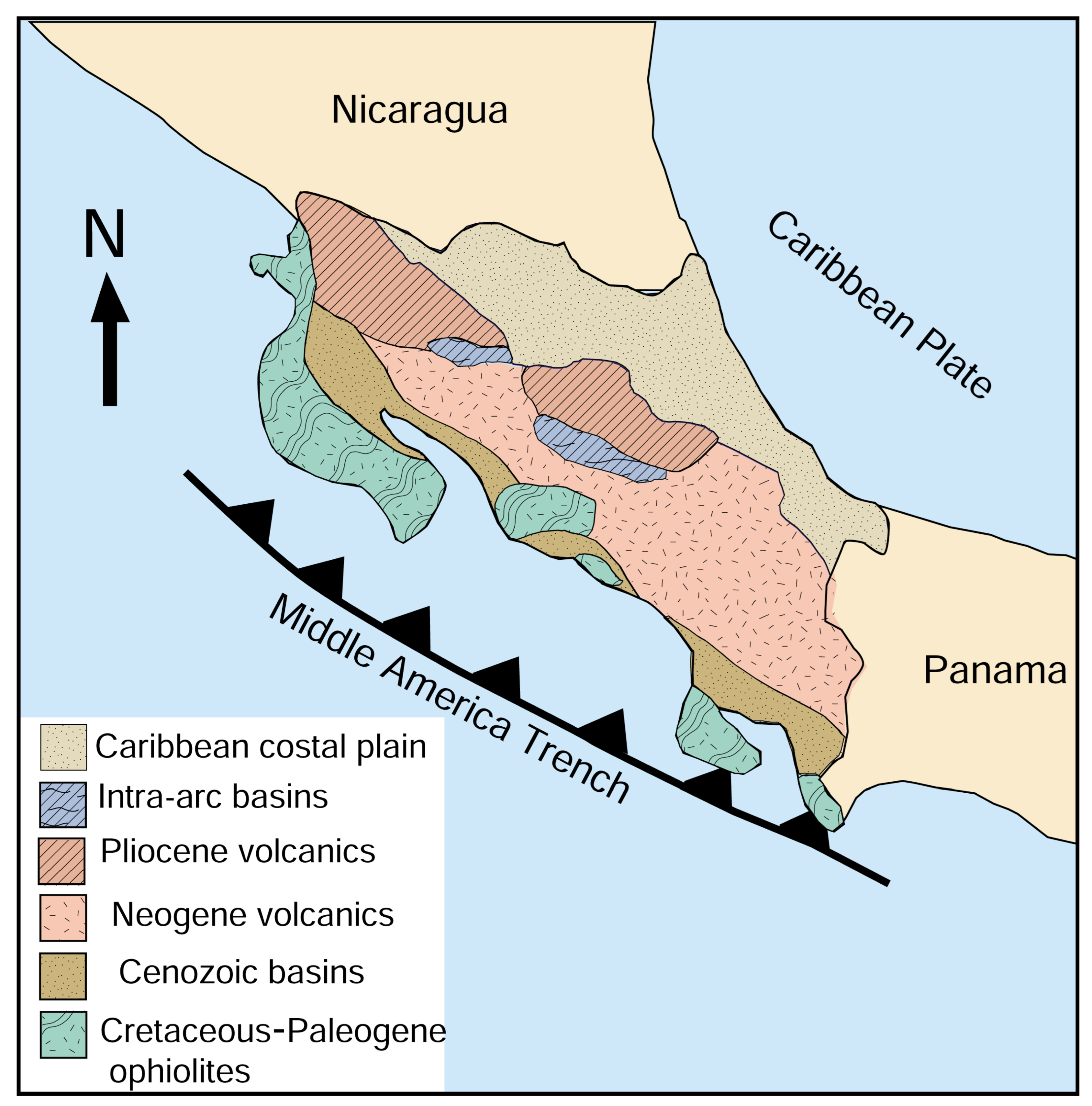
Costa Rica is characterized by complex geology (Figure 2). The oldest rocks are Mesozoic ophiolites (basalts, diabases, peridotites, rare picrites, and komatiites), and related sedimentary rocks (radiolarites, limestones, and hyaloclastic sediments) of Jurassic to Eoceneage. Late Cretaceous (Campanian) subalkaline volcanism and Cenozoic subalkaline volcanism produced volcanic islands and ranges that were created and destroyed several times. Younger deposits include deep sea marine sediments (shales and sandstones) and shallower marine sediments of Late Cretaceous to Pliocene age. These sediments include shales, sandstones, conglomerates, and limestone. Coetaneous fluvial and paralic volcaniclastic sediments became abundant beginning in the Neogene, when volcanic activity was a dominant geologic process in the islands of the ancestral archipelago. This deposition continued throughout the remainder of the Quaternary. The present physiography was largely shaped by Neogene and Quaternary volcanism [10,13].
This geologic setting is a favorable environment for the preservation of fossil wood. Neogene volcanism produced calderas with adjacent forested slopes. Most silicate minerals have low solubility, and Si in groundwater comes from two principal sources: volcanic glass and the weathering of feldspar [14,15]. In Costa Rica, felsic to intermediate volcanic rocks were a ready source for dissolved silica, preserving wood in two possible environments: trees or woody debris were buried by airfall tephra and pyroclastic flow deposits, or wood was fluvially transported to become driftwood along the margins of lakes or in shallow marine deposits near the continental coast. Taphonomy suggests that the dominant form of wood petrifaction involved transported wood fragments, explaining the relative scarcity of fossilized logs preserved in the growth position. The rapid burial of wood hindered microbial degradation. The durability of fossil wood allowed specimens to withstand fluvial transport, and good specimens can be found along rivers and in coastal cliffs (Figure 3).

1.2. Paleobiology of Central America

The separation of North and South America at ~525 Ma caused plants and animals of the two continents to have differing evolutionary histories. During the mid-Cenozoic, the Central American archipelago comprised a dispersed chain of small islands situated between the Pacific Ocean and the Caribbean Sea and the Pacific Ocean. Miocene tectonism resulted in an uplift that eventually created an isthmus between North and South America (Figure 4). A detailed history of this process can be found in [16]. The isthmus is known to have been in place by ~3 Ma (Late Pliocene/Early Pleistocene), but vertebrate fossils suggest that an emerging peninsula was present during the Miocene [17,18].
The development of a continuous land bridge led to the exchange of taxa between North and South America, a phenomenon referred to as the Great American Interchange. This episode is best known for the migration of animals. In North America, the opossum, armadillo, and porcupine all trace back to ancestors that came across the land bridge from South America. Likewise, bears, cats, dogs, horses, llamas, and raccoons all made the trek south across the isthmus. Plants were also part of the interchange. Migration rates for plants were different from animals that were capable of walking, flying, or swimming. However, plant rage expansion was directly related to animal migration because of the important role that vertebrates play in the transport of seeds. Published research includes three classic papers [19,20,21].

1.3. History of Paleobotany Research in Central America

The study of fossil plants in Central America dates back to the pioneering work of U.S. Geological Survey paleobotanist Edward S. Berry. His studies focused on fossil plants from Panama [22,23,24] (1914, 1919, 1921, 1928) but included Costa Rica [25] and the surrounding region [26] (1942). Later, paleobotany research focused on the paleoflora of Panama, reflecting geological discoveries that originated with the excavation of the Panama Canal [27,28,29,30,31,32]. Petrified wood from Guatemala was reported by Mustoe and Eberl [33].
The first mention of petrified wood in Costa Rica came from the southeastern part of San José (capital of Costa Rica) and from localities at Caldera and Pozo Azul de Candelaria [1,2]. There was a surprising silence in the mention of petrified wood until 1970, when Madrigal [3] reported isolated fragments of fossil wood south of Mata de Limón. This report was followed by cursory mentions of fossil wood [8,34,35,36]. Most of these reports lack detailed descriptions and/or precise locations.
Beginning in the 1980s, paleobotanical research in Costa Rica became focused on subfossil pollen diatoms and charcoal fragments collected from soils, lake sediments, and peat bogs, which span an age range from Late Pleistocene to Holocene [37,38,39,40,41]. These studies revealed that plant species migrated up and down mountain slopes in response to cyclic climate change. Other studies reported plant fossils preserved in shale, but again, this fossil record mostly comes from the Late Pleistocene [42,43]. Taxonomic descriptions are typically restricted to a single species [44,45,46,47,48,49,50,51]. These studies provide a fascinating look at environmental change that was largely controlled by climate fluctuations. A limitation is that these interpretations are applicable only to Pleistocene and Holocene phenomena.
The study of Costa Rican fossil wood potentially offers a means for studying botanical evolution and environmental change as far back as the Miocene. The goal of our research is to find diverse localities where fossil wood is preserved, to examine the mineralogy of this wood in the hope of understanding fossilization processes, and to evaluate the preservation of anatomic detail that could one day be useful for taxonomic studies and paleoenvironmental interpretation. Our research strategy is preliminary in nature, but it is work that will hopefully open a door to future investigations. This research has already begun, as evidenced by the recent publication of a study that described the taxonomy of five Miocene wood specimens, one each from locations in central and northern Costa Rica [52]. The challenge of interpreting the geographic dispersal and taxonomy of ancient plant communities is evidenced by the diversity of modern plant communities in Costa Rica, which include approximately 670 genera of trees and shrubs, distributed within 124 families that contain about 1800 species [53]. Another source [54] recognized 192 families of woody plants, with 860 indigenous genera and 181 introduced taxa. By any metric, the paleobotanical history of Costa Rica’s tropical forests will be a daunting task.

2. Methods and Materials

The 53 fossil woods specimens used in this study came from diverse locations (Figure 3, Table S1), but there are three principal localities: Salinas village near Caldera (central Costa Rica), Guapinol de la Cruz (northern Costa Rica), and San Vito de Coto Brus (southern Cost Rica). All of these sites are located on the Pacific flank of the central mountain axis. The Salinas–Caldera–Tivives localities contains Lower Miocene tuffaceous reddish to greenish fluvial and estuarine sandstones and pyroclastic deposits of the Mata de Limón Member [8,9,55]. The Guapinol locality occurs near an old volcanic caldera associated with the Monteverde volcanism of the Upper Pliocene to Lower Pleistocene age [56]. San Vito samples came from the contact of Late Miocene shallow estuarine/deltaic sediments (siltstones and sandstones with conglomerates), called the San Gerardo unit, that overlies the San Vito unit (volcanic debris avalanche and fluvial deposits) of the Lower Pleistocene age [5,55,57,58]. All fossils were surface collected near the outcrops. Specimens were selected to represent a range of colors and textures, recording the GPS location for each specimen. Age estimates are based on the geologic setting and regional stratigraphy.
Analytical methods included the measurement of density via hydrostatic weighing. Density values are useful for evaluating mineral composition; woods mineralized with opal commonly have densities of 1.9–21 g/cm3, compared to 2.3–2.6 g/cm3 for woods mineralized with chalcedony or quartz [59]. However, these density values may vary for woods that contain porosities or those that are only partially mineralized. Mineral identifications were, therefore, confirmed using a combination of data: XRD, SEM/EDS, and optical mineralogy.
Microscopy included low-power reflected light images obtained with a Bausch & Lomb StereoZoom binocular microscope (New York Microscope Company, Hicksville, NY, USA, https://microscopecentral.com, accessed on 7 January 2025). Thin-section images were made using two microscope systems: (1) a Zeiss petrographic microscope (Carl Zeiss Microscopy, White Plains, NY, USA) equipped with a Hayear 500B 5-megapixel CMOS camera (Shenzen Hayear Electronics Ltd., Shenzen, China. https://hayear.com, accessed on 24 February 2025) and (2) an Annlov digital microscope (https://www.tomlov.com (accessed on 23 February 2025)) equipped with a 16-megapixel camera (https://www.annlov.net/products/ (accessed on 16 January 2025)).
Thin sections were made at a standard 30-micron thickness, using Wards-Ingrahm equipment (Wards Science, Rochester, NY, USA. https://www.wardsci.com, accessed on 19 April 2025). System Three General Purpose Epoxy Resin was used for cementing rock blocks to frosted glass slides and for attaching coverslips (System Three, Seattle, WA, USA; https://www.systemthree.com, accessed on 19 April 2025).
SEM images came from a Tescan Vega III scanning electron microscope (Tescan, Brno, Czech Republic) that includes an Oxford energy-dispersive X-ray detector running AzTek 4.0 software. Freshly fractured specimens were mounted on 1 cm diameter aluminum stubs using epoxy adhesive and sputter-coated with Pd to provide electrical conductivity. SEM conditions were a 10 KV beam voltage, with the beam diameters and working distance optimized for each photomicrograph. The most common photographic conditions were a 9 mm working distance with a 90-s acquisition time.
X-ray diffraction patterns were made on packed powdered specimens using a Rigaku Miniflex diffractometer running SmartLab II software (Rigaku Corp., Tokyo, Japan). Samples were analyzed using Ni-filtered Cu-α radiation at 40 KV, 20 Ma over a 2-theta range of 5–55°.

3. Results

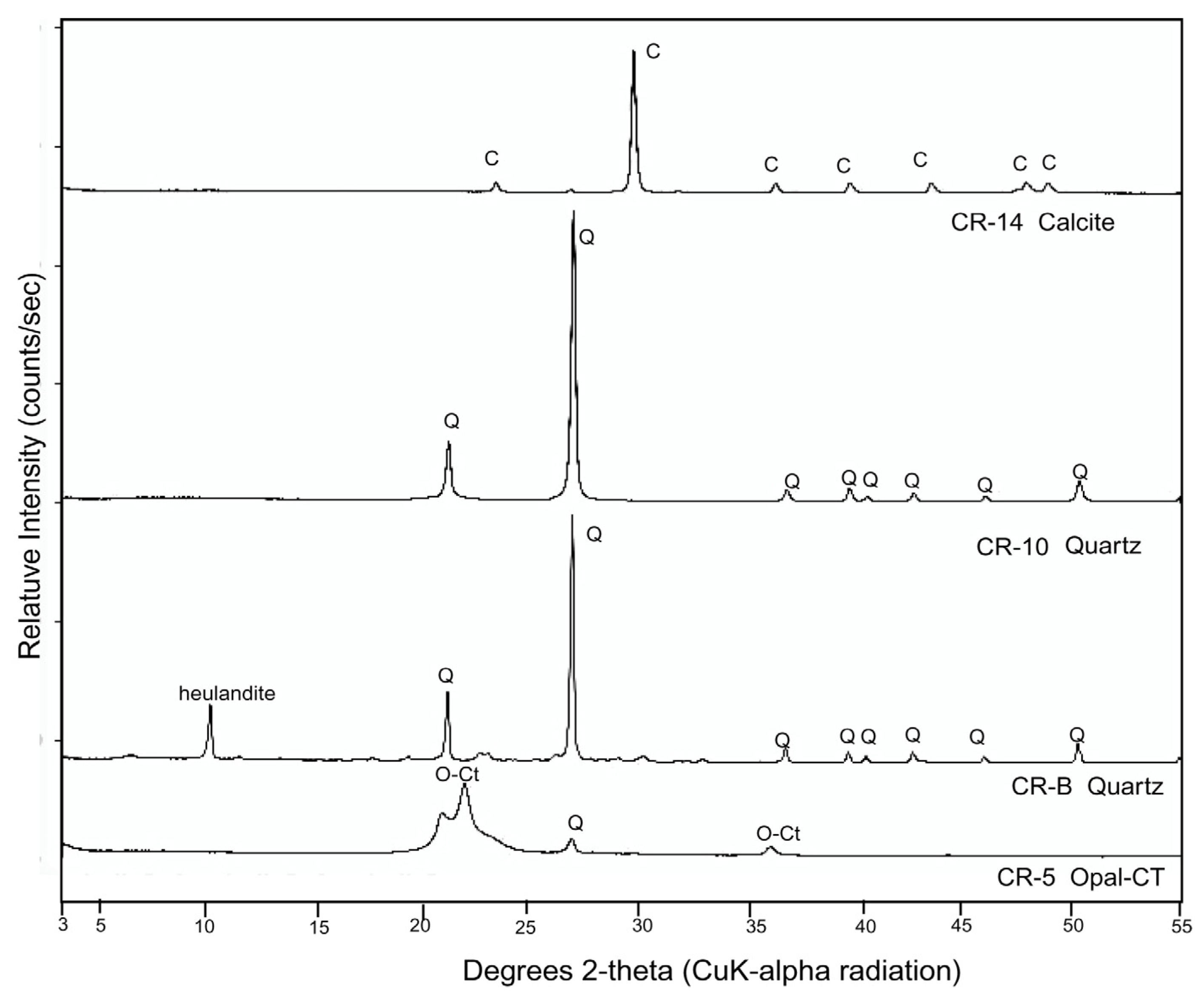
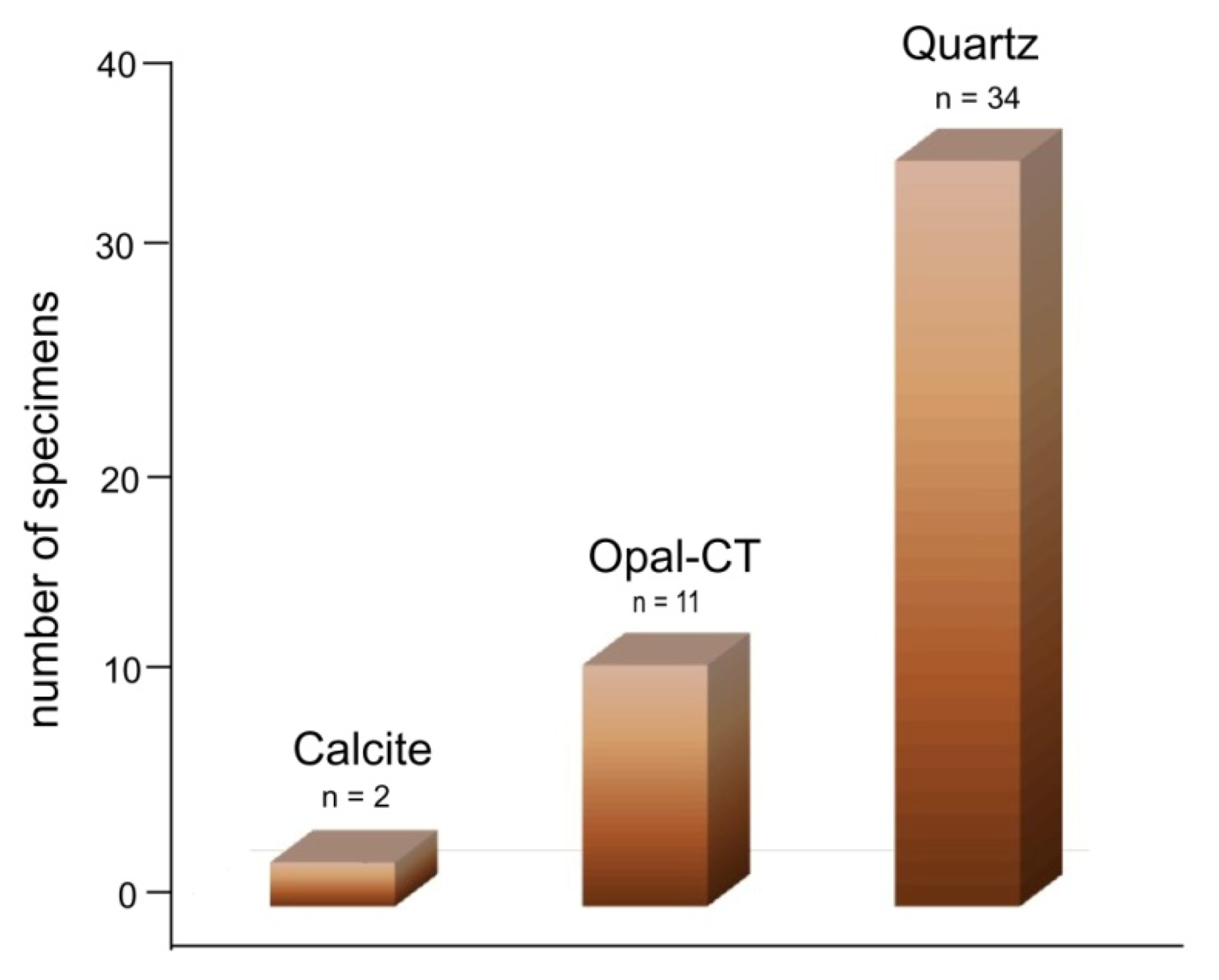
Fossil woods from Costa Rica localities show considerable variation in their mineral compositions. The samples typically show distinctive compositions in XRD patterns (Figure 5). Silicified woods are composed of opal-CT, microcrystalline quartz, and megacrystalline quartz. Specimens of each composition type are described in detail in an accessory file. The compositions are summarized in Figure 6.

3.1. Wood Fossilization Processes

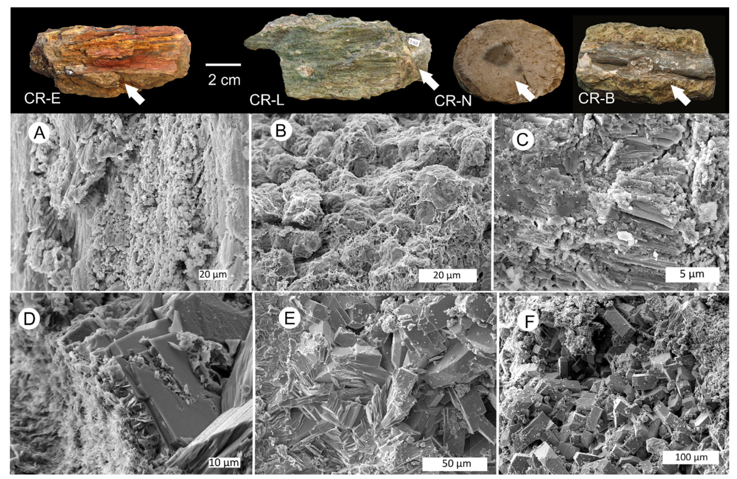
Fossil woods from Costa Rica have a wide range of mineral compositions. Intermediate to felsic volcaniclastic rocks provided a source of dissolved silica. This diagenesis is evidenced by SEM images of the matrix that encloses silicified wood specimens. Diagenetic characteristics include the devitrification of volcanic glass and paragenesis of zeolites. The presence of intact phenocrysts of plagioclase suggests that Si was released from volcanic glass rather than the degradation of feldspar (Figure 7).

3.2. Evidence of Decay

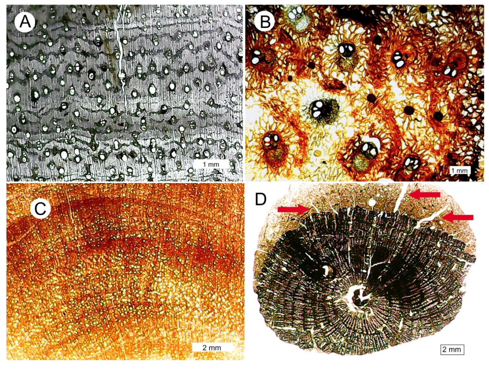
Anatomical preservation varies among individual specimens. Some samples preserve excellent cell structures (Figure 8). Wood color variations have not been studied for Costa Rican specimens, but previous research suggests that petrified wood colors are commonly caused by trace levels of metallic elements, particularly iron. A wide range of colors can be produced by variations in the abundance and oxidation state of Fe. Brown and black colors may result from the presence of relict organic matter [60].
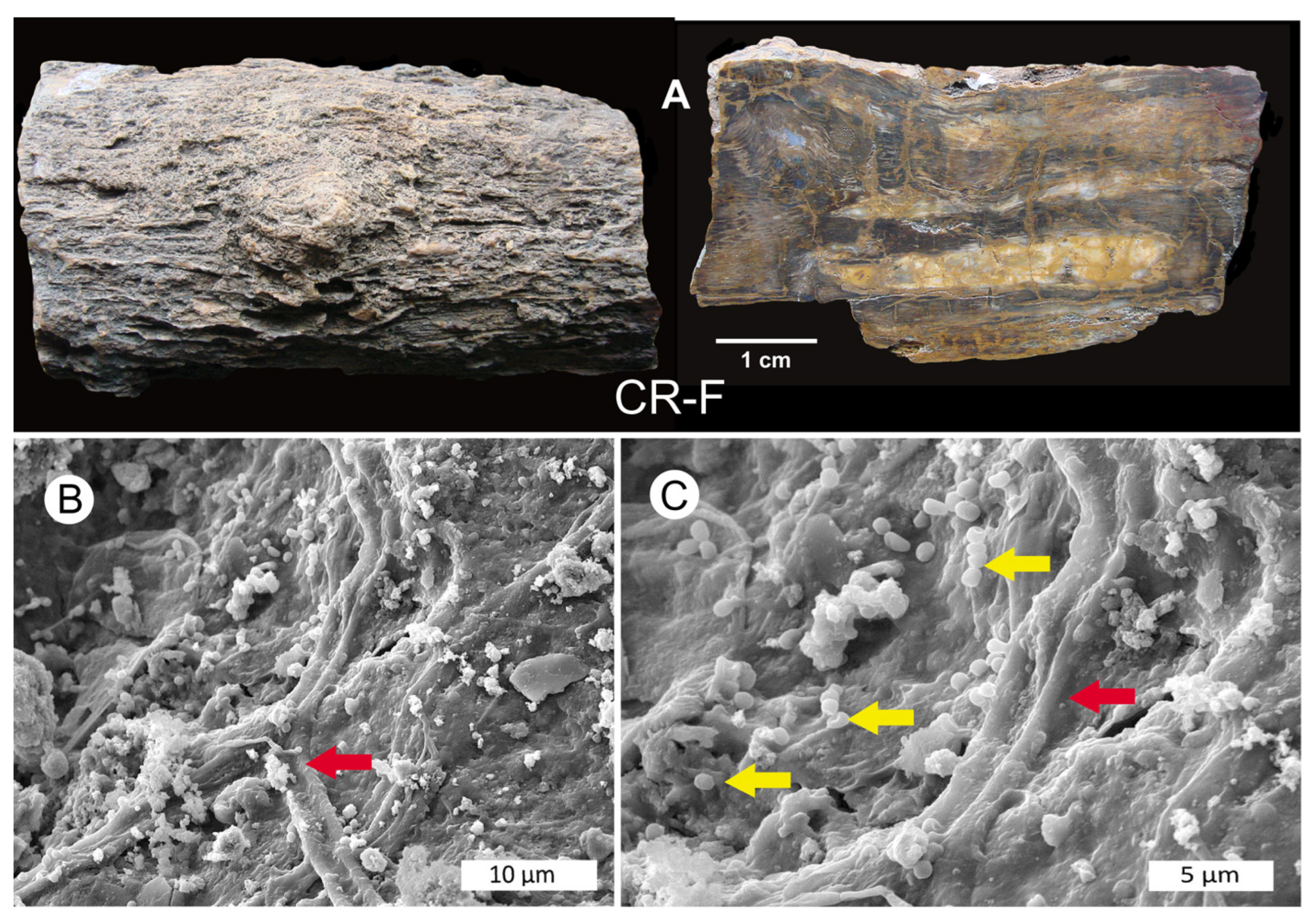
Some Costa Rican specimens show evidence of decay prior to the onset of petrifaction (Figure 9). Other woods were deformed or fragmented prior to petrifaction (Figure 10).

3.3. Silicified Woods: Microcrystalline Quartz

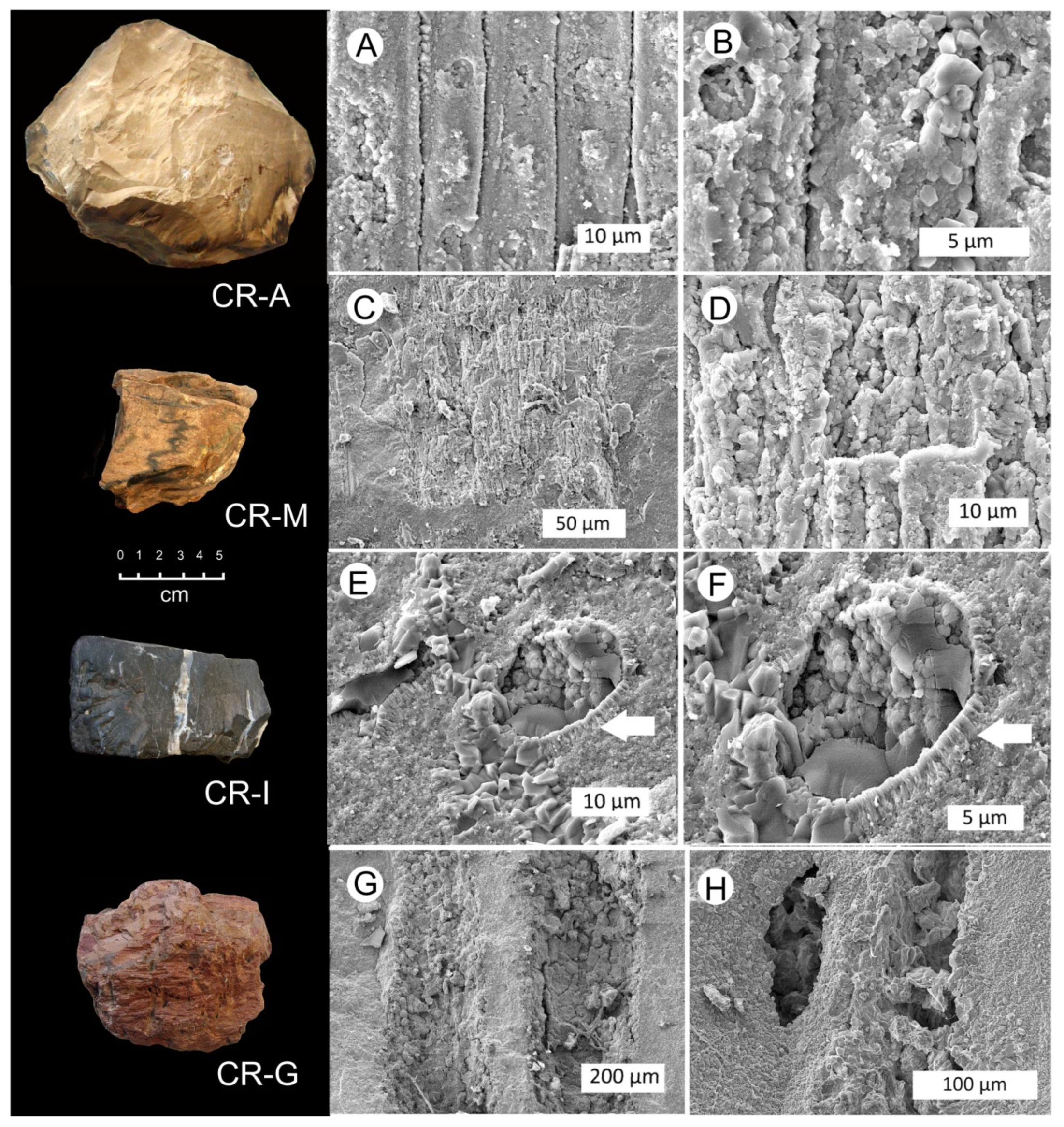
The most common form of wood petrifaction in Costa Rica is the result of the replacement of tissue quartz, both as microgranular crystals and chalcedony. Quartz formation occurs when dissolved silica concentrations are relatively low, with a precipitation rate that allows time for the development of well-ordered lattices. SEM images commonly show the microcrystallinity. These images may also show topographic aspects of anatomical features (Figure 11).
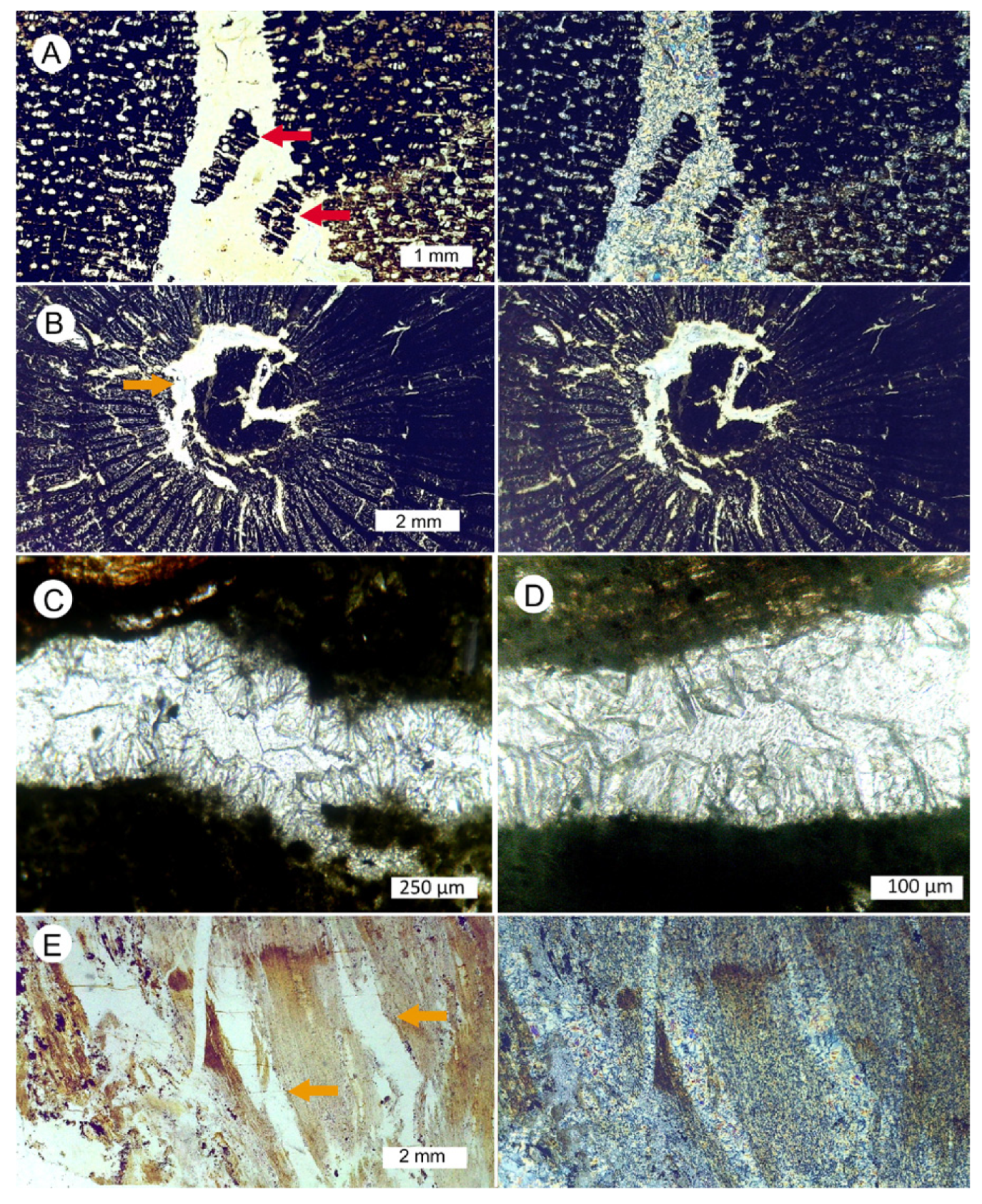
Transmitted light views of petrographic thin sections provide the identifications of fossil woods where void spaces have been filled with microcrystalline quartz (Figure 12). These features are indicative of multiple stages of silica precipitation, where cellular tissues were silicified, followed by coarser-textured quartz forming later in spaces that remained open.

3.4. Silicified Wood: Megacrystalline Quartz

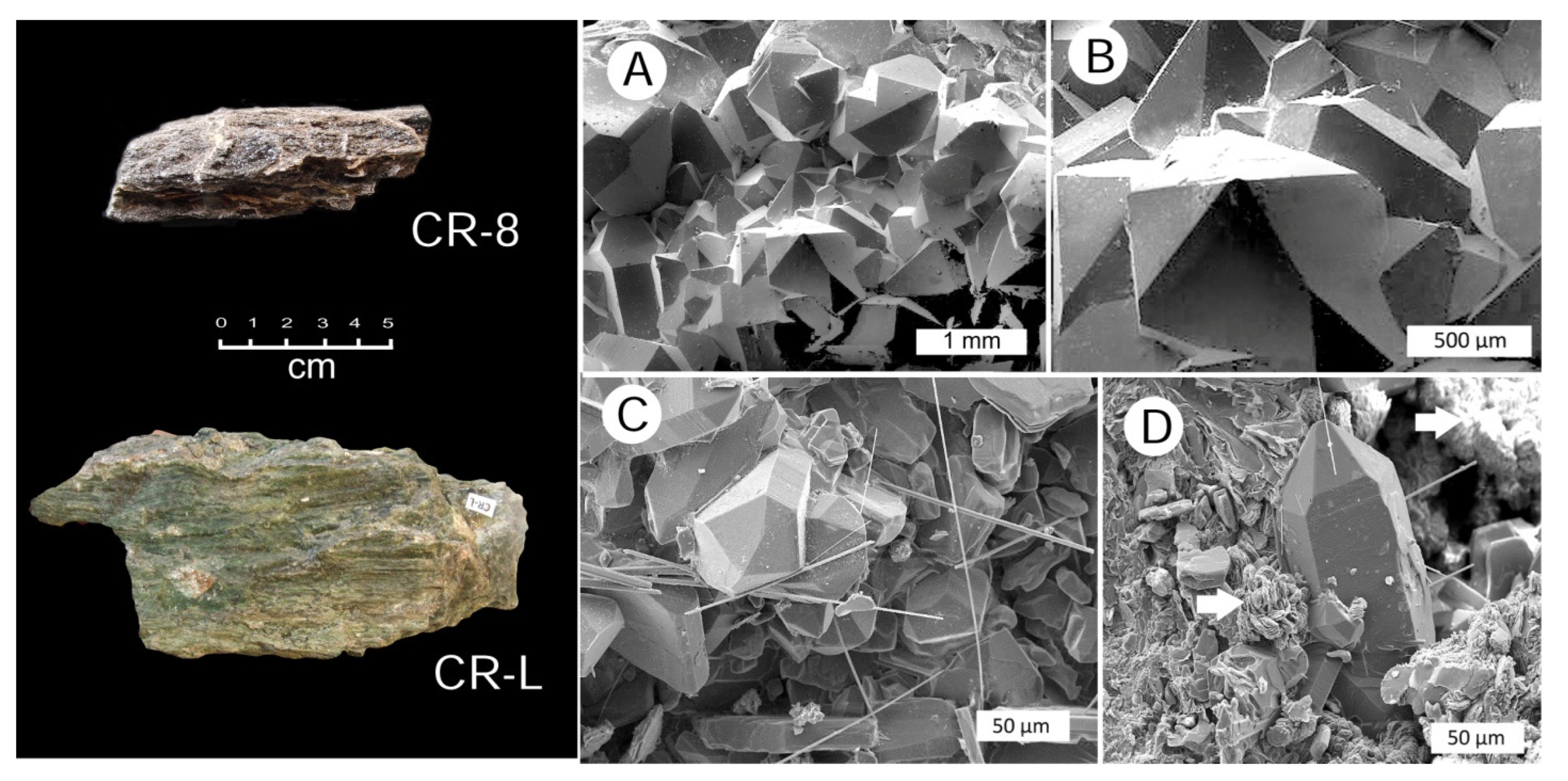
A few Costa Rican fossil woods contain megacrystalline quartz crystals (Figure 13). In some specimens, euhedral quartz crystals are the predominant constituent. In other specimens, quartz crystals occur in open spaces (e.g., conductive vessels or rot pockets), representing a late stage in a mineralization sequence that occurred in multiple episodes.

3.5. Chalcedony Mineralization

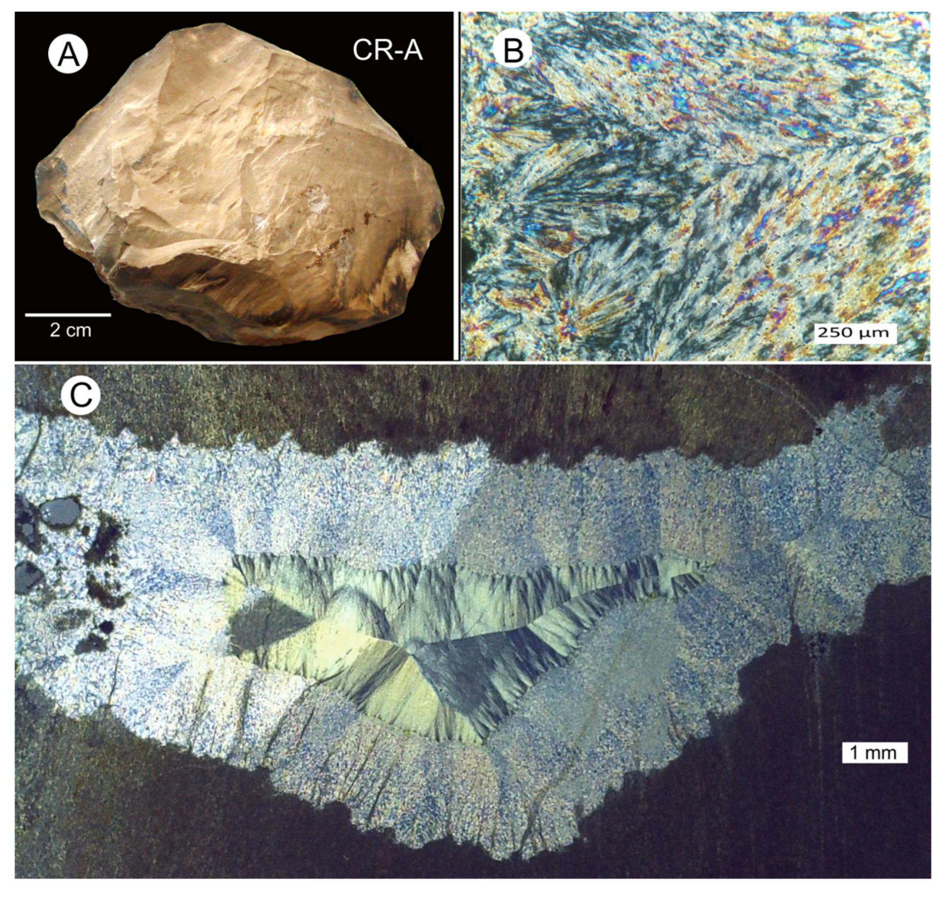
Chalcedony is a form of quartz that has a fibrous morphology (Figure 14). The chalcedony group comprises three structural types. Length-fast and length-slow chalcedony can be recognized by optical properties under polarized light. Moganite (a monoclinic form of hydrous SiO2) can be recognized via X-ray diffraction and infrared spectroscopy [61]. However, in our investigation, we have not attempted to identify individual members of the chalcedony family because our focus has been to determine whether or not petrified woods preserve anatomical details that would permit future taxonomic identifications.

3.6. Silicified Wood: Opal-CT

The mineralization of wood with opal occurs when high concentrations of dissolved silica result in rapid precipitation rates, when silica molecules lack the time needed to form well-ordered lattices. Wood opal can have two forms. Opal-A is an amorphous form of hydrous silica that consists of very small spherical masses (lepispheres). Most opal wood is composed of opal-CT, a material that has incipient crystal structure representing the structural transition of amorphous opal to crystobalite/tridymite. Less commonly, the composition is opal-C, which has an incipient cristobalite structure [62].
These hydrous silica polymorphs can be identified from X-ray diffraction patterns, thin-section petrography, and SEM images. The lack of crystallinity causes opal-A to show only a broad, low XRD pattern. Likewise, in thin sections, opal-A is isotropic under polarized light, while opal-C and opal-CT have weak birefringence. Under SEM examination, opal-A commonly appears as smooth-surfaced microspheres. In contrast, opal-C and opal-CT commonly consist of hemispherical shapes that have a distinctive “cornflake” texture [62].
In our study, opal wood from Costa Rican localities consisted of opal-CT. Photomicrographs of five specimens are shown in Figure 15 and Figure 16.

3.7. Opal and Quartz

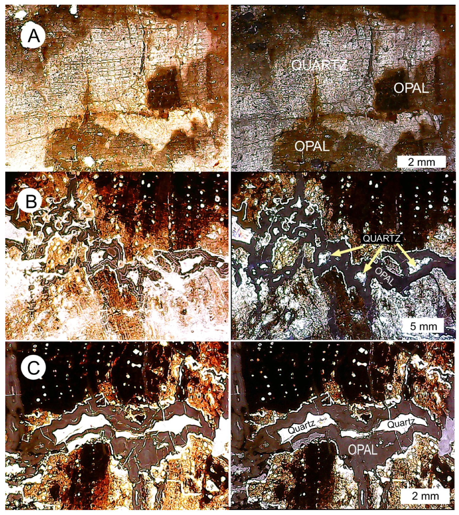
A few specimens show intermixtures of quartz and opal-CT, as evidenced by transmitted light images of thin sections. Zonation patterns visible in specimen CR-29 suggest the possibility of diagenetic transformation (Figure 17A). For specimen CR-23, the precipitation of opal occurred during a late stage of mineralization when groundwater containing dissolved silica entered a complex fracture zone. The walls of the fracture are coated with a very thin layer of clear quartz, which is overlain by a thick layer of opal-CT. Open central areas became sites for second-stage quartz precipitation (Figure 17B,C). The angular outline of the fracture zone is evidence that the wood had been petrified prior to fracture.

3.8. Calcite Wood

After silica, calcium carbonate is the most common agent for wood petrifaction, but known examples are relatively rare. Indeed, woods mineralized with calcite have previously been reported from only 16 global locations [63], originating under a variety of geological conditions, with ages ranging from Jurassic to Pleistocene [64,65].
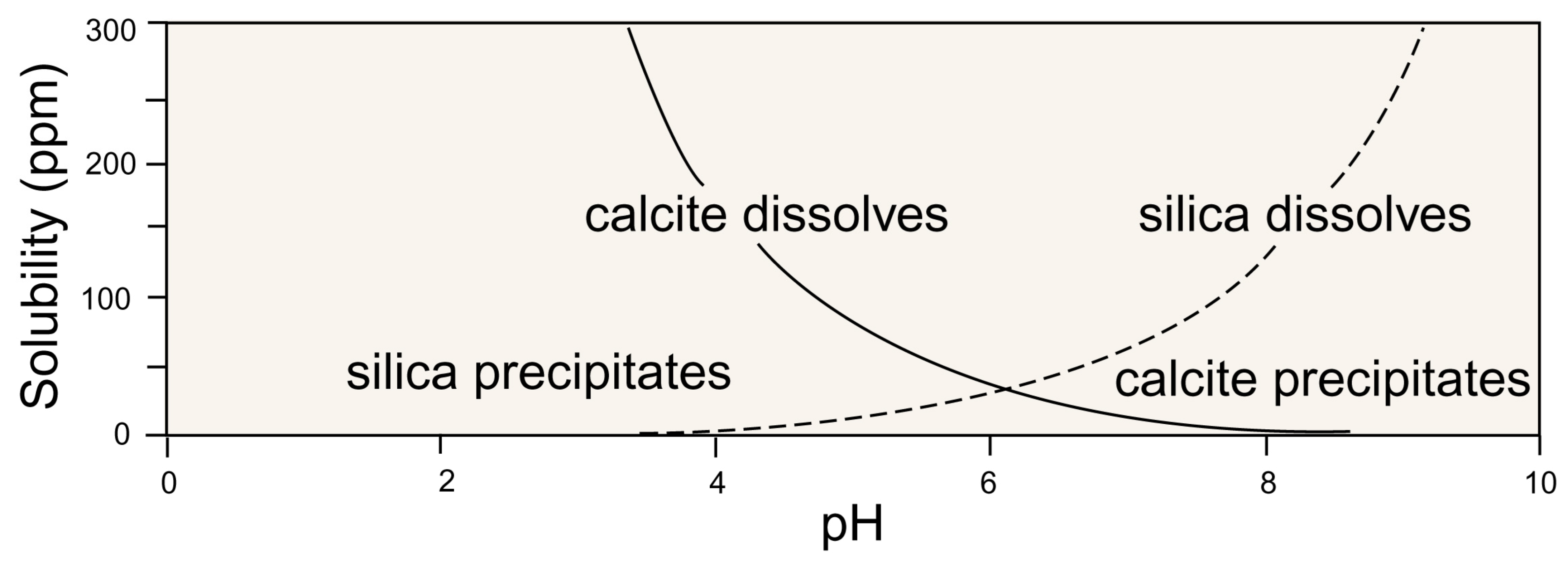
Several factors result in calcite mineralization of wood. Dissolved calcium needs to be at saturation levels, but this element can be present in groundwater in any region that contains limestone or other calcareous rocks. Wood petrified with calcite may occur in situations where there is a paucity of dissolved Si, but the predominant factor is the pH. Silicification is driven by the solubility of Si under basic conditions, with precipitation occurring when the pH becomes acidic (Figure 18). Calcite mineralization involves inverse condition, with acidity favoring Ca solubility and precipitation occurring at a basic pH.
The differing geochemical conditions between silicification and calcification have a bearing on the phenomenon of permineralization, which is defined as intact cellular tissue that becomes entombed within mineral matter. Contrary to longstanding commentary on wood petrifaction, most silicified only a small percentage of relict organic matter [66]. In contrast, the more gentle pH conditions that accompany calcite precipitation are favorable for the preservation of cellular material, and it is common for calcite-mineralized woods to be examples of true permineralization [65,66].
Figure 18. Stability diagram for silica and calcite. These geochemical limitations are evident in Costa Rican samples. Silicified woods do not contain calcite, and calcite-mineralized woods do not contain quartz. Data adapted from [67,68].
Minerals 15 00497 g018
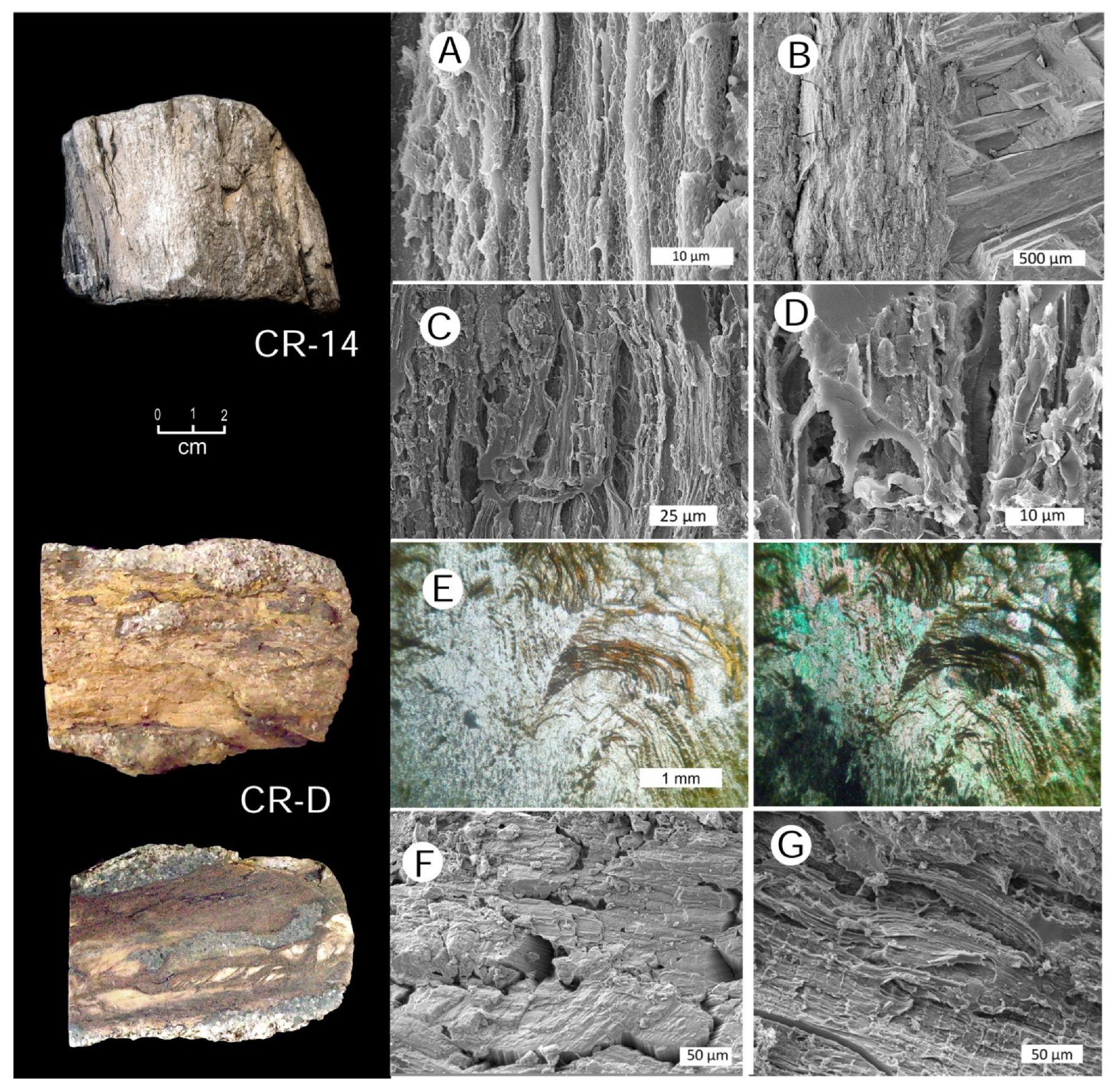
Examples of calcite mineralized woods from Costa Rica are shown in Figure 19.

3.9. Pyrite as an Accessory Mineral in Fossil Wood

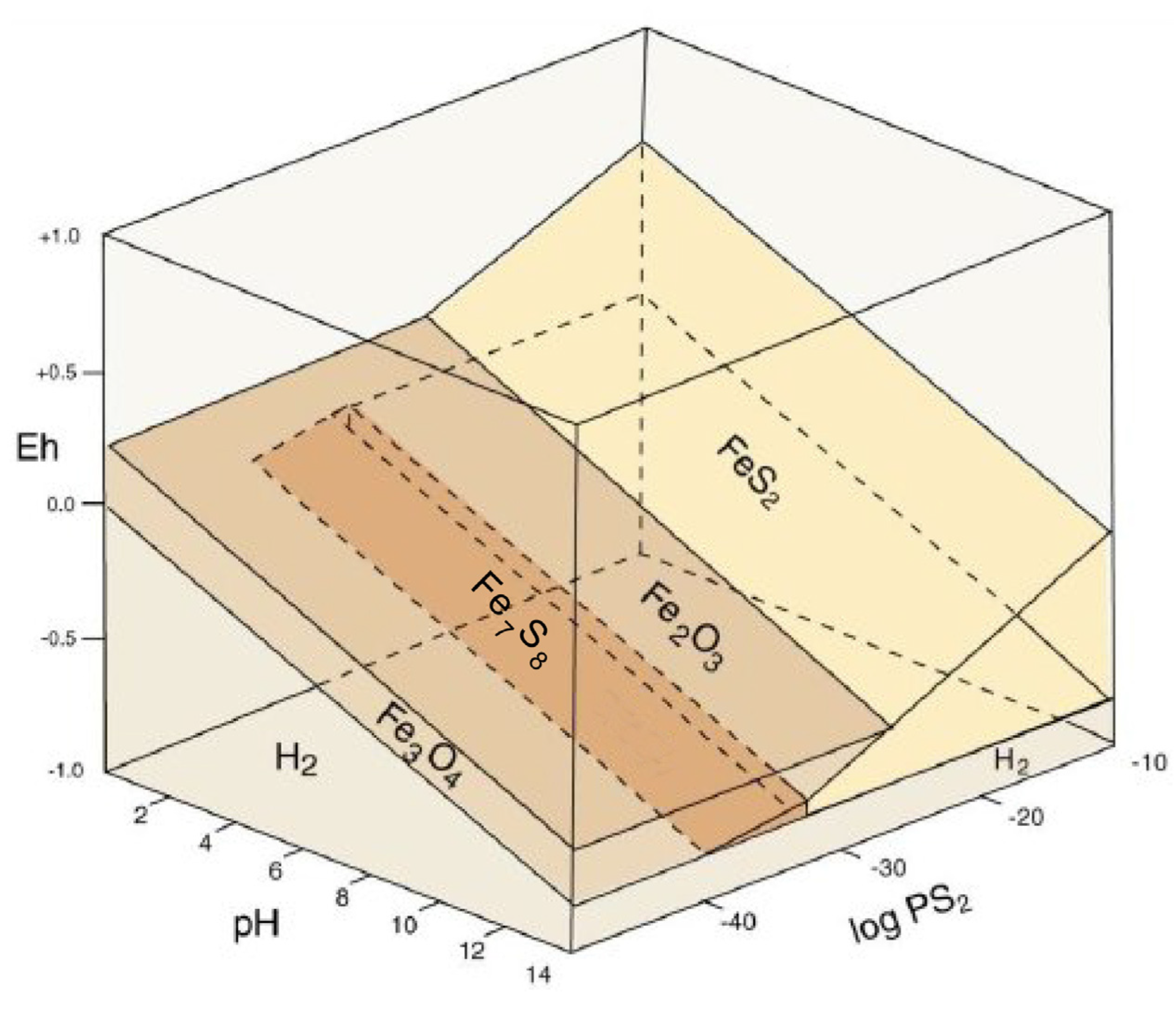
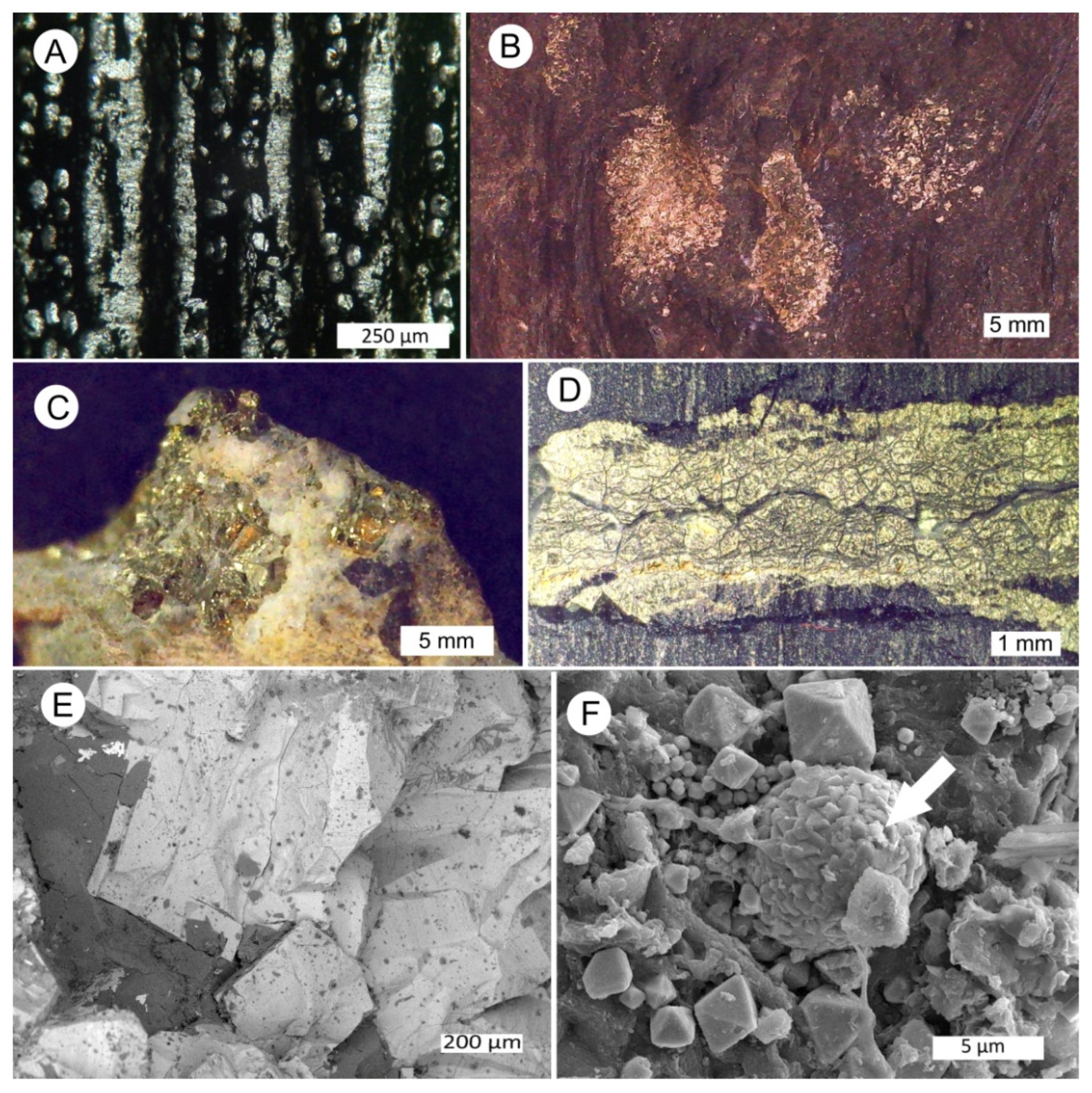
The precipitation of pyrite requires the availability of dissolved iron and sulfur and appropriate pH and Eh conditions. The decomposition of plant tissue commonly produces reducing conditions, and pyrite may be found in fossil wood [60,61,62,63,64,65,66,67,68,69,70,71,72]. This is especially likely in Costa Rican wood because the volcanic deposits and anoxic sediments are sources of sulfur. The abundance of iron is evidenced by the widespread presence of lateritic soil, where deep weathering caused the removal of more soluble elements to create an excess of iron and aluminum [73]. Under reducing conditions, this iron may become soluble. Pyrite may precipitate over a wide pH range (Figure 20), explaining why the mineral may occur as an accessory mineral in both silicified and calcified wood (Figure 21).

3.10. Preservation of Relict Tissue

Wood that consists of original organic matter or altered variations is sometimes described as “carbonized wood”, but this terminology is ambiguous. There are three mechanisms that can cause ancient wood to remain unmineralized. One possibility is the burial wood in impermeable sediments, where mineral-bearing groundwater is unable to penetrate. This “mummified wood” typically occurs when wood is buried in a stratum of dense clay. Coalification is another mechanism, where organic constituents of plant tissues undergo metamorphic reactions in response the heat and pressure. The resulting material is arbitrarily categorized by “coal rank”, e.g., lignite, sub-bituminous, bituminous, and anthracite. Lignite (also known as brown coal) may preserve anatomical detail, but higher rank coals generally contain plant remains that have little, if any, structural fidelity. The third possibility is the conversion of wood to charcoal as a result of anaerobic heating. Although charcoalified wood may consist of pure carbon, its anatomical detail is commonly very good. Tissues composed of reduced carbon (charcoal and most coal) is likely to be resistant to silicification because that mineralization is commonly initiated by organic templating, where silicic acid molecules form hydrogen with functional groups in the cellulosic cell walls of buried wood. This bonding does not occur when the tissues have been thermally converted to carbonaceous alteration products. These processes were summarized by Mustoe [62].
The preservation of relict tissue in Costa Rica may have originated from more than one mechanism. The dark color of some specimens (Figure 22) is suggestive of localized charring that may have developed from contact with hot volcanic materials, and as discussed earlier in this report, the calcification of wood may be in the form of permineralization, where cell walls may remain intact within wood that was infiltrated with CaCO3. This wood may retain a dark brown color, but wood with blackened areas commonly represents charcoalification.

4. Conclusions

Costa Rica has a multitude of petrified wood occurrences that originated in association with Neogene to Middle Quaternary volcanism. This fossil wood is potentially important as source of information regarding ancient environments, the distribution of trees and shrubs, and the migration of the plant taxa during the formation of the Central American land bridge. Our investigation is a preliminary study of the mineralization mechanisms and the degree of anatomical preservation. The latter characteristic is important for future taxonomic research.
At each locality, the fossilization of wood depended on a variety of physical and chemical parameters. The requirements were the availability of dissolved elements at concentrations that exceed the saturation point in an environment, with pH and Eh conditions that favored precipitation. Buried wood commonly retains hollow cells that allow for the permeation of mineral-bearing groundwater, but mineralization can only occur if the sediment that encloses the wood has permeability. Costa Rica volcanism had conditions that commonly met the above requirements. Pyroclastic materials and hyaloclastic alluvial sediments provided sources for dissolved silica. This solubilization was enhanced by the warm, moist tropical/subtropical climate.
The precipitation of minerals within buried wood was facilitated by the release of organic reducing agents during the degradation of tissue. Wood petrifaction may represent localized Eh and pH gradients, which explains why the mineralogical composition of buried wood may be different compared to the composition of the adjacent matrix. For example, wood may become mineralized because the pore spaces in the enclosing sediment initially remain open. This permeability allows for the entrance of mineral-bearing groundwater. Initial silicification may result from the affinity of cellulosic cell walls for silicic acid molecules, with mineralization continuing in successive events to produce complete silicification. Under different Eh and pH conditions, woods may be mineralized with calcite instead of silica. These fossilization processes are important for determining the degree of anatomical preservation. In general, wood petrifaction involves rate-competitive processes, where tissue is being degraded at the same time as it is becoming mineralized. If the rate of wood decomposition is rapid, cellular detail is not likely to be preserved. In extreme cases (e.g., wood buried by lava flows), the result may be a cast that preserves only the external shape. Conversely, a slow rate of wood degradation may mean that the wood becomes coalified rather than petrified. Our study of 53 specimens revealed a diversity of mineral compositions, with their anatomical preservation ranging from excellent to very poor. These results suggest that future taxonomic studies will require the careful selection of specimens and a search for localities that offer favorable geologic conditions.
Finding optimum collecting sites requires geological expertise because the occurrence of petrified wood specimens involves a multitude of factors. Although it may sometimes be possible to observe fossil wood in situ (e.g., limbs or logs preserved within pyroclastic fall deposits), most specimens are found at surface levels, where weathering and erosion released the fossil wood from the less-durable matrix. This process potentially allows for specimens to be transported by streams and rivers to downstream locations. Likewise, longshore processes may laterally move petrified wood once fluvially transported specimens arrive at the coast.
In summary, despite a paucity of past research, petrified wood occurs in abundance in Costa Rica. These specimens potentially provide a record of Neogene plant communities that span a temporal range that begins with the Miocene appearance of the Central American land bridge. This fossil record has the potential to widen our paleobotanical knowledge beyond the data that are obtainable from foliage fossils and palynology, which mostly provide information from the Pleistocene and Holocene, when the land bridge was well established. The complexity of this task is evidenced by the molecular clock data obtained from extant taxa, which suggest that 58 unambiguous migration events occurred across the Isthmus of Panama, determined from 16 phylogenetic events for plants and 27 for animals [75].

Supplementary Materials

The following supporting information can be downloaded at: https://www.mdpi.com/article/10.3390/min15050497/s1; Table S1: Descriptions of Individual Costa Rica Specimens.

Author Contributions

G.E.M. and G.E.A. cooperated in the design of the research and the writing of the manuscript. G.E.A. and A.J.P. were responsible for collecting specimens and field investigations. G.E.M. performed analytical work using facilities at Western Washington University. All authors have read and agreed to the published version of the manuscript.

Funding

This research involved no external funding.

Data Availability Statement

The specimens described in this report are archived at the Geology Department, Western Washington University, Bellingham, WA 98225, USA.

Acknowledgments

The fossil woods specimens are part of a project at Costa Rica University, project titled “B9705 Conservation and Research of the Paleobiological Heritage of Costa Rica”. Access to WWU analytical facilities was facilitated by Kyle Mikkelson (Advanced Materials Science & Engineering Center), Mike Kraft (University Instrument Center), and Ben Paulson (Geology Department).

Conflicts of Interest

The authors declare no conflicts of interest.

References

  1. Alfaro, A. Comprobaciones geológicas. Boletín Del Fom. 1911, 1, 123–131. [Google Scholar]
  2. Alfaro, A. Rocas sedimentarias de Costa Rica. Boletín Del Fom. 1913, 3, 853–861. [Google Scholar]
  3. Madrigal, R. Geología del Mapa Básico “Barranca”, Costa Rica, Informes Técnicos y Notas Geológicas; Ciudad Univeritaria “Rodorigo Facio”: San José, Costa Rica, 1970; 55p. [Google Scholar]
  4. Alfaro, A. Caracterización petrográfica y geoquímica de las rocas ígneas en el sector Pacífico de la cordillera de Talamanca, Costa Rica. Licentiure Thesis, Universidad de Costa Rica, San José, Costa Rica, 2017. [Google Scholar]
  5. Alvarado, G.E.; Bonilla-Mata, A.; Cárdenes-Sandí, G. A Synopsis of the Geology, Geotectonic, and Geological History of Costa Rica. In Landscapes and Landforms of Costa Rica; Quesada-Román, E., Ed.; Springer: Berlin/Heidelberg, Germany, 2024; pp. 3–34. [Google Scholar]
  6. Denyer, P.; Alvarado, G.E. Desarrollo y evolución de la geología. In Geología de Costa Rica; Denyer, P., Kussmaul, S., Eds.; Editorial Tecnólógica de Costa Rica: Cartago, Costa Rica, 2000; pp. 185–218. [Google Scholar]
  7. Weyl, R. Geology of Central America, 2nd ed.; Gebruder-Borntraeger: Berlin, Germany, 1980. [Google Scholar]
  8. Laurito, C.A. Sinópsis de la estratigrafía de la cuenca Barranca-Herradura y el promontorio Herradura. Provincia de Puntarenas, Costa Rica. Brenesia 1988, 29, 21–32. [Google Scholar]
  9. Denyer, P.; Aguilar, T.; Alvarado, G.E. Geología y estratigrafía de la hoja Barranca, Costa Rica. Rev. Geol. Amér. Central 2003, 29, 105–125. [Google Scholar] [CrossRef]
  10. Alvarado, G.E.; Dengo, C.; Martens, U.; Bundschuh, J.; Aguilar, T.; Bonis, S.B. Stratigraphy and geologic history. In Central America: Geology, Resources and Hazards; Bundschuh, J., Alvarado, G.E., Eds.; Taylor and Francis: London, UK, 2007; pp. 345–394. [Google Scholar]
  11. Denyer, P.; Alvarado, G.E. Mapa Geológico de Costa Rica 2007(1: 400 000); Librería Francesa S.A: San Jóse, Costa Rica, 2007. [Google Scholar]
  12. Denyer, P.; Aguilar, T.; Alvarado, G.E.; Chavarría, M.M.; Herrera, P. Mapa geológico de la hoja Bahía Salinas. Escala 1:50 000. In Perspectiva Geológica del Noroeste de Costa Rica: Historia, Evolución y Cartografía; Denyer, P., Ed.; EUCR-CICG: San Jóse, Costa Rica, 2019. [Google Scholar]
  13. Alvarado, G.E.; Cárdenes, G. Geology, tectonics, and geomorphology of Costa Rica: A natural history approach. In Costa Rica Ecosystems; Kappelle, M., Ed.; University Chicago Press: Chicago, IL, USA, 2017; pp. 30–61. [Google Scholar]
  14. Murata, K.J. Volcanic ash as a source of silica for the silicification of wood. Am. J. Sci. 1940, 238, 586–596. [Google Scholar] [CrossRef]
  15. Matysová, P.; Rössler, R.; Götze, J.; Leichmann, J.; Forbes, G.; Taylor, E.I.; Dakala, J.; Grygar, T. Alluvial and volcanic pathways to silicified plant stems (Upper Carboniferous-Triassic) and their taphonomic and paleoenvironmental meaning. Palaeogeogr. Palaeoclimatol. Palaeoecol. 2010, 292, 127–143. [Google Scholar] [CrossRef]
  16. O’Dea, O.; Lessios, H.A.; Coates, A.G.; Eytan, R.J.; Resrepo-Moreno, S.A.; Cine, A.L.; Collins, L.S.; de Queiroz, A.; Farris, D.W.; Norris, R.D.; et al. Formation of the Isthmus of Panama. Sci. Adv. 2016, 2, e1600883. [Google Scholar] [CrossRef]
  17. Lucas, S.G. Vertebrate paleontology in Central America 30 years of progress. Rev. Geológica De América 2014, 30, 139–155. [Google Scholar]
  18. Lucas, S.G.; Alvarado, G.E. Vertebrate paleoentology in Central America: A narrative and analytical history. In History of Geoscience Celebrating 50 Years of INHIGEO; Mayer, W., Clary, R.M., Azuela, L.F., Mota, T.S., Wolkowicz, S., Eds.; Geological Society of London: London, UK, 2017; pp. 155–169. [Google Scholar]
  19. Janzen, D.H. Herbivores and the number of tree species in tropical forests. Am. Nat. 1970, 104, 502–528. [Google Scholar] [CrossRef]
  20. Janzen, D.H.; Martin, P.S. The fruit the gompotheres ate. Science 1982, 215, 19–27. [Google Scholar] [CrossRef]
  21. Herrera, C.M. Vertebrate frugivores and their interaction with invertebrate fruit predators: Evidence from the Costa Rican dry forest. Oikos 1989, 54, 185–288. [Google Scholar] [CrossRef]
  22. Berry, E.W. Fossil plants in the Panama Canal Zone. Science 1914, 39, 357. [Google Scholar] [CrossRef]
  23. Berry, E.W. The fossil higher plants from the Canal Zone. U.S. Natl. Mus. Bull. 1919, 33, 15–44. [Google Scholar]
  24. Berry, E.W. A palm fruit from the Miocene of western Panama. J. Wash. Acad. Sci. 1928, 18, 455–457. [Google Scholar]
  25. Berry, E.W. Tertiary fossil plants from Costa Rica. Proc. United States Natl. Mus. 1921, 59, 169–185. [Google Scholar] [CrossRef]
  26. Berry, E.W. Mesozoic and Cenozoic plants of South America, Central America, and the Antilles. In Proceedings of the Eighth American Scientific Conference, Washington, DC, USA, 10–18 May 1940; 1942; Volume 4, p. 3650373. [Google Scholar]
  27. Stern, W.L.; Eyde, R.H. Fossil forests of Ocú Panama. Science 1963, 140, 2014. [Google Scholar] [CrossRef]
  28. Reyes, O.R.; Falcon-Lang, H.; Gasson, P.; Collinson, M.; Jaramillo, C. Fossil woods (Malvaceae) from the lower Miocene (early to mid-Burdigalian) part of the Cucaracha Formation of Panama (Central America) and their biogeographic implications. Rev. Palaeobot. Palynol. 2014, 209, 11–14. [Google Scholar] [CrossRef]
  29. Judd, N.; Nelson, C.W. A liana from the lower Miocene of Panama and the fossil record of Connaraceae. Am. J. Bot. 2017, 104, 1–9. [Google Scholar] [CrossRef]
  30. Judd, N.; Dunham, J.I. Fossil woods from the Cenozoic of Panama (Azuero Peninsula) reveal an ancient neotropical rainforest. IAWA J. 2017, 38, 366–411. [Google Scholar] [CrossRef]
  31. McFadden, B.J.; Jones, D.S.; Judd, N.A.; Moreno-Bernai, J.W.; Morgan, G.S.; Poretell, R.W.; Perez, V.J.; Moran, S.M.; Wood, A.R. Integrated chronology flora and faunas and paleoecology of the Alajuela Formation, Late Miocene of Panama. PLoS ONE 2017, 104, 1–9. [Google Scholar] [CrossRef]
  32. Rodriquez-Reyes, O.; Gasson, P.; Thornton, C.; Falcon-Lang, H.; Judd, N.A. Panascleroticoxylon crytstllosa gen. et. sp. nov.: A new Miocene malpghialean tree from Panama. IAWA J. 2017, 38, 437–455. [Google Scholar] [CrossRef]
  33. Mustoe, G.E.; Eberl, M. New discovery of Neogene fossil forests in Guatemala. Geosciences 2020, 10, 49. [Google Scholar] [CrossRef]
  34. Laurito, C. Los proboscídeos fósiles de Costa Rica y sucontexto en la América Central. Vínculos 1988, 14, 29–58. [Google Scholar]
  35. Amann, H. Randmarine und terrestrische Ablagerungsräume des neogenen Inselbogensystems in Costa Rica (Mittelamerika). Profil 1993, 4, 1–161. [Google Scholar]
  36. Rojas, M. Geología de la hoja Monterrey—3247 I escala 1:50 000; Depart. Investigación, Dirección de Geología y Minas: Minae, San Jóse, Costa Rica, 2019. [Google Scholar]
  37. Hooghiemstra, H.; Cleef, A.M.; Noldus, G.; Kappelle, M. Upper Quaternary vegetation dynamics and palaeoclimatology of the La Chonta bog area (Cordillera de Talmanca, Costa Rica). J. Quat. Sci. 1992, 7, 205–225. [Google Scholar] [CrossRef]
  38. Islebe, G.A.; Hooghiemstra, H. Vegetation and climate history of montane Costa Rica since the Last Glacial. Quat. Sci. 1997, 16, 589–604. [Google Scholar] [CrossRef]
  39. Islebe, G.A.; Hooghiemstra, H.; van’t Veer, R. Historia holocénica de la vegetación y del niveldeagua endos tuberas de la cordillera de Talmanca, Costa Rica. In Páramos d Costa Rica; Kapelle, M., Horn, S., Eds.; Instituto Nacional de Biodiveridad, INBio, Santo Domingo de Heredia: Santo Domingo, Costa Rica, 2005; pp. 337–352. [Google Scholar]
  40. Kappelle, M.; Haberyan, K.A.; Horn, S.P. Algas fósiles y recientes de los páramos de Costa Rica. In Páramos de Costa Rica; Kapelle, M., Horn, S., Eds.; Instituto Nacional de Biodiveridad, INBio: Santo Domingo de Herdia, Costa Rica, 2005; pp. 331–341. [Google Scholar]
  41. Horn, S.P. Late Quaternary lake and swamp sediments: Rocorders of climate and environment. In Central America: Geology, Resources and Hazards; Bundschuh, J., Alvarado, G.E., Eds.; Taylor & Francis: London, UK, 2007; pp. 423–441. [Google Scholar]
  42. Kesel, R.H. Quaternary history of the Rio General valley, Costa Rica. Natl. Geogr. Soc. Res. Rep. 1983, 15, 339–358. [Google Scholar]
  43. Pérez, E.A. Comparación de la flora fósil del pleistoceno, con la flora actual en la localidad de la Palmera de San Carlos, Provincia del Alajuela. Escuela la de Ciencia Exactas y Naturales. Programa de licenciatura en Manejo de Recursos Naturals, Universidad Estatal a Distancia, San José, Costa Rica, 2001; 126p. [Google Scholar]
  44. Pérez, E.A.; Laurito, C.A. Quercus corrugata Hooker (Fagaceae) como un indicatorpaleoclimático del Pleistoceno del Costa Rica. Rev. Geológica De América Central 2003, 28, 83–90. [Google Scholar]
  45. Pérez, E.A. Presencia de Juglans olanchana Standley & L.O. Williams (Juglandaceae) en el territorio costrarricenes durante el Pleistoceno. Rev. Geológica De América Central 2003, 28, 77–81. [Google Scholar]
  46. Gómez, L.D. A first report of a fossil fern-like Pteropsida from Costa Rica. Rev. Biol. Trop. 1970, 16, 255–258. [Google Scholar]
  47. Gómez, L.D. Palmacitesberry anum, a new palm fossil from the Costa Rican Tertiary. Rev. Biol. Trop. 1971, 19, 121–132. [Google Scholar]
  48. Gómez, L.D. Keratophyllum bromeliodes (Bromeliaceae), nov. gen. et sp., del Terciario Medio de Costa Rica. Rev. Biol. Trop. 1972, 20, 221–229. [Google Scholar]
  49. Gómez, L.D. Criptonemiales calcáreas fossils en las terciarias de Patarrá. Rev. Biol. Trop. 1973, 21, 107–110. [Google Scholar]
  50. Gómez, L.D. Ficus padifolia H.B.K. en la diatomita pliocena/pleistocena de la Formación Bagaces, Guanacaste, Costa Rica. Veröff. Uberseemuseum Brem. Reihe A 1974, 4, 141–148. [Google Scholar]
  51. Gómez, L.D. Preliminary note on a fossil Equisetum from Costa Rica. Fern. Gaz. 1978, 11, 401–404. [Google Scholar]
  52. Cevallos-Ferriz, S.R.S.; Alvarado, G.E.; Huerta-Vergara, A.R.; Cárdenes, G.; Ramírez, T.D.; Bonilla, N.W.; Gusmán, E. Fossil woods from the Miocene of Costa Rica and Nicaragua: Insights into the Neotropical floral puzzle. J. South Am. Earth Sci. 2024, 144, 1–14. [Google Scholar] [CrossRef]
  53. Fournier, L.A.; Jiménez, A.; Salas, S.; Marin, F. Las familias y géneros de árboles y arbustos de Costa Rica. Rev. Biol. Trop. 1966, 14, 317–328. [Google Scholar]
  54. Gómez-Laurito, J.; Fournier, L.A. Las Familias y Los Generos de Plantas Leñosas de Costa Rica. Available online: https://tropicalstudies.org/rbt/attachments/volumes/vol14-2/11-Fournier-Arboles.pdf (accessed on 20 April 2025).
  55. Alvarado, G.E.; Gans, P.B. Síntesis geocronológica del magmatismo, metamorfismo y metalogenia de Costa Rica, América Central. Rev. Geol. Amér. Central 2012, 46, 7–122. [Google Scholar] [CrossRef]
  56. Denyer, P.; Montero, W.; Aguilar, T.; Herrera, P.; Alvarado, G.E.; Barrantes, M.; y Chavarría, M.M. Mapa Geológico de la Hoja Carrillo Norte (Escala 1:50 000); Editorial Universidad de Costa Rica: San José, Costa Rica, 2019. [Google Scholar]
  57. Tournon, J.; Alvarado, G.E. Mapa Geológico de Costa Rica. Folleto Explicativo, 1: 500,000; Tecnológica de Costa Rica: Cartago, Costa Rica, 1997. [Google Scholar]
  58. Lucas, S.G.; Sequeira, C.; Chesnel, V.; Rodríguez, D.; Alvarado, G.E.; Méndez, J.C.; Vargas, A.; Vargas, C.; Ruiz, G. Boid snake fossils from the Neogene of Costa Rica. Rev. Geológica De América Cent. 2025, 72, 1–11. [Google Scholar]
  59. Mustoe, G.E. Density and loss on ignition as indicators of the fossilization of silicified wood. IAWA J. 2016, 37, 98–111. [Google Scholar] [CrossRef]
  60. Mustoe, G.E.; Acosta, M. Origin of petrified wood color. Geosciences 2016, 6, 25. [Google Scholar] [CrossRef]
  61. The Quartz Page. Available online: www.quartzpage.de (accessed on 7 January 2025).
  62. Mustoe, G.E. Silicification of wood: An overview. Minerals 2023, 13, 206. [Google Scholar] [CrossRef]
  63. Rasbury, E.T.; Hatton, K.; Cox, S.E.; Sousa, F.J.; Hemming, S.R.; Steponaitis, E.; Saslaw, M.; Rowan, C.M.; Boyd, M.D.; Gaiku, M.; et al. Carbonate mineralized wood from Oligocene-Miocene Deposits in the Turkana Basin. Turkana Miocene Project Publication #6. 2025; in press. [Google Scholar]
  64. Mustoe, G.E.; Beard, G. Calcite-mineralized fossil wood from Vancouver Island, British Columbia, Canada. Geosciences 2021, 11, 38. [Google Scholar] [CrossRef]
  65. Mustoe, G.E. Mineralogy of non-silicified wood. Geosciences 2018, 8, 85. [Google Scholar] [CrossRef]
  66. Mustoe, G.E. Wood petrifaction: A new view of permineralization and replacement. Geosciences 2017, 7, 119. [Google Scholar] [CrossRef]
  67. Utami, W.S.; Herdianita, N.R.; Atmaja, R.W. The effect of temperature and pH on the formation of silica scaling at Dieng Geothermal Field, central Java, Indonesia. In Proceedings of the Thirty-Ninth Workshop on Geothermal Field Engineering Stanford University, Stanford, CA, USA, 24–26 February 2014. [Google Scholar]
  68. Nicholson, K. Geothermal Fluids: Chemistry and Exploration Techniques; Springer: Berlin, Germany, 1968; 263p. [Google Scholar]
  69. Garcia-Guinea, J.; Martinez-Frias, A.; Harffy, M. Cell-hosted pyrite framboids in fossil woods. Naturwissenschaft 1988, 85, 78–81. [Google Scholar] [CrossRef]
  70. Tibbs, S.L.; Briggs, D.E.G.; Prossl, K.F. Pyritization of plant fossils from the Devonian Hunsrücck Slate of Germany. Palaeontol. Z. 2003, 77, 241–246. [Google Scholar] [CrossRef]
  71. Kendrick, P.; Edwards, D. Anatomy of the Lower Devonian Gosslingia breconenis Herad based on pyritized axes with some comments on the permineralizaton process. Bot. J. Linnean Soc. 1988, 97, 95–123. [Google Scholar] [CrossRef]
  72. Grimes, S.T.; Davies, K.L.; Butler, I.B.; Brock, F.; Edwards, D.; Rickard, D.; Briggs, D.E.G.; Parkes, R.J. Understanding fossilization: Experimental pyritization of plants. Geology 2001, 29, 123–126. [Google Scholar] [CrossRef]
  73. Camacho-Umaña, M.E.; Quesada-Román, A.; Villatro-Sánchez, M.; Alemán-Montes, B.; Mata, R.; Henríquez-Henríquez, C.; Céspedes-Rivera, J.; Alvarado, A. Highly weathered landscapes of Costa Rica. In Landscapes and Landforms of Costa Rica; Quesada-Román, A., Ed.; Springer: Cham, Switzerland, 2024; pp. 431–451. [Google Scholar]
  74. Garrels, R.M.; Christ, C.L. Solutions, Minerals and Equilibria; Harper and Row: New York, NY, USA; John Weatherhill, Inc.: Tokyo, Japan, 1965. [Google Scholar]
  75. Cody, S.; Richardson, J.E.; Pennington, R.T. The great American biotic exchange revisited. Ecography 2010, 33, 326–332. Available online: https://www.nature.com/articles/s41598-020-58575-6 (accessed on 3 May 2024). [CrossRef]
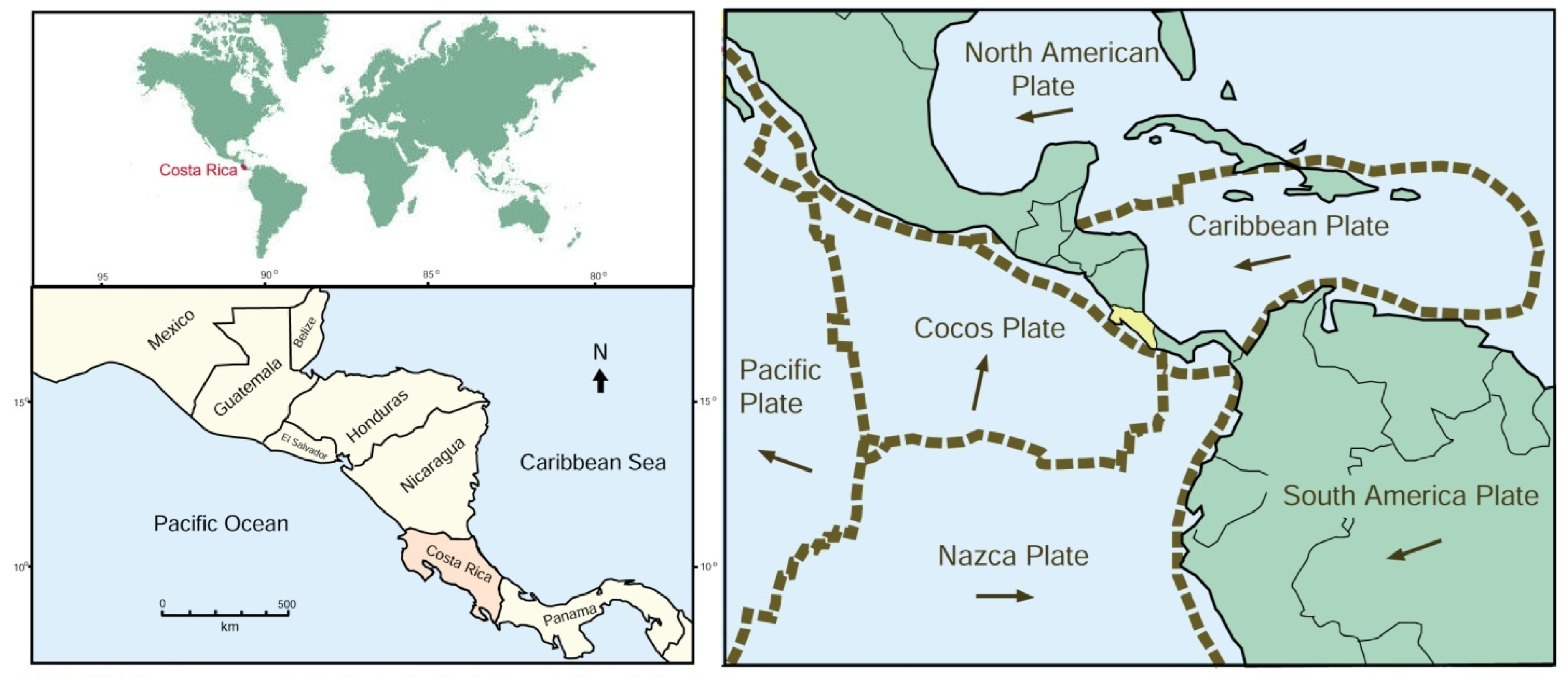
Figure 1. Geographic and tectonic setting of Central America.
Minerals 15 00497 g001
Figure 2. Simplified geology map of Costa Rica. Adapted from [5,11].
Minerals 15 00497 g002
Figure 3. Fossil wood localities for specimens studied in our research are shown as red circles.
Minerals 15 00497 g003
Figure 4. Geologic evolution of Central America. (A) Early Cenozoic dispersal of islands. (B) Miocene volcanism let to expansion of land areas. (C) By the late Miocene, an uplift had produced a continuous peninsula. Adapted from [13].
Minerals 15 00497 g004
Figure 5. XRD patterns for Costa Rican wood containing opal-CT, quartz, and calcite. Specimen CR-B contains heulandites, a zeolite family mineral produced by diagenetic alteration of the tephra matrix. CR-5 is opalized wood that contains a trace of quartz. Samples CR-10 (quartz) and CR-14 (calcite) are monomineralic.
Minerals 15 00497 g005
Figure 6. Compositions of fossilized woods used in this investigation.
Minerals 15 00497 g006
Figure 7. SEM images showing diverse mineral compositions, with identifications based on XRD and SEM/EDS data. The upper photos show specimens with arrows marking areas of matrix. (A) Tephra on surface of silicified wood specimen CR-E. (B) Matrix shows alteration of tephra to clay mineral, specimen CR-L. (C) Tephra matrix containing tabular fragments of silicified wood, specimen CR-N. (D) Fossil wood tuffaceous matrix containing unaltered plagioclase minerals in microcrystalline clay that was formed via the devitrification of volcanic glass. (E) Plagioclase phenocrysts in tuffaceous matrix. (F) Blocky heulandites crystals in tuffaceous material. Images (DF) are from specimen CR-B.
Minerals 15 00497 g007
Figure 8. Thin-section views of fossil woods that preserve anatomical details. (A) Opalized wood in transverse orientation, specimenCR-5. (B) Palmoxylon wood in transverse orientation, specimen CR-10. (C) Transverse image of silicified wood showing annual rings, specimen CR-28. (D) Transverse view of silicified wood. Contact with tuffaceous matrix wood is visible in the upper part of the photo. Small quartz-filled fractures (marked with arrows) show the entry of silica-bearing groundwater. Specimen CR-30.
Minerals 15 00497 g008
Figure 9. Silicified wood preserving evidence of fungal decay. (A) External view at left, with sawn surface at left, specimen CR-F. (B) SEM image shows silicified fungal hyphae (red arrows). (C) Higher-magnification image reveals many spherical objects that are presumed to be fungal spores (yellow arrows).
Minerals 15 00497 g009
Figure 10. Quartz-mineralized wood showing deformation textures. (A) Wood texture shows that plastic deformation occurred before the buried wood became mineralized, (B) Other areas of the same thin section show fragmentation of the brown-colored wood, with intervening spaces filled with volcaniclastic sediment. Specimen CR-F.
Minerals 15 00497 g010
Figure 11. Specimen photos and SEM images of woods mineralized with microcrystalline quartz. (A,B) Radial views of wood showing cells that preserve intervessel pits, specimen CR-A. (C,D) Radial views showing wood cells replaced by quartz, specimen CR-M. Image (C) shows a region where cells are well preserved, but adjacent areas are rather featureless. In image (D), cells have been replaced by microcrystalline quartz, with intercellular spaces remaining unmineralized. (E,F) Transverse views showing a single vessel that is partly filled with quartz crystals, with the cell wall replaced by chalcedony (marked with arrows). Specimen CR-I. (G,H) Radial views of two adjacent vessels that contain linings of crystalline quartz, with the central lumen remaining open. Specimen CR-G.
Minerals 15 00497 g011
Figure 12. Thin-section photomicrographs showing microcrystalline quartz filling open spaces. For each pair, the photo on the left is ordinary transmitted light; the right image is the polarized light view. (A) Fracture zone encloses fragments of silicified wood (marked with red arrows) that were detached from adjacent tissue. Specimen CR-M, transverse orientation. (B) Transverse view of specimen CR-30. Shrinkage of the wood prior to fossilization produced a crescent-shaped void (shown by orange arrow) that was later filled with quartz. (C,D) High-magnification views of quartz-filled fracture, CR-30. (E) Longitudinal view of silicified wood that contained parallel fractures (marked with orange arrows) that became sites for quartz precipitation. Specimen CR-F.
Minerals 15 00497 g012
Figure 13. Fossil woods CR-8 and CR-L contain macrocrystalline quartz. (A,B) SEM views of euhedral quartz crystals that replace wood tissue, CR-8. (C,D) Specimen CR-L preserves silicified wood fragments within a hyaloclastic matrix. Silicified wood fibers are preserved as thin needles. Volcanic glass was diagenetically altered to crinkly microcrystalline masses of clay, marked with arrows in image (D).
Minerals 15 00497 g013
Figure 14. Chalcedony mineralization in specimen CR-A. (A) Specimen before cutting. (B) Polarized light view of thin section, showing radiating chalcedony fibers. (C) Low-magnification transmitted light view reveals a fracture where walls are thickly coated with microgranular quartz, with a central zone that contains radiating masses of chalcedony.
Minerals 15 00497 g014
Figure 15. Opalized wood from Costa Rica. (A) Transmitted light image of a thin section of specimen CR-6, in the transverse orientation, showing anatomical preservation (B) SEM photo of CR-6, radial orientation, revealing horizontal ray cells. (C,D) Oblique radial view of specimen CR-6.Cell walls have been replaced by vitreous opal-CT, and cell lumens are loosely filled with opal-CT microspheres. (EG) SEM images of specimen CR-18, longitudinal orientation, showing botryoidal masses of opal-CT. Images (FH) show a void space that contains relatively large hemispherical masses of opal-CT that display incipient crystallization.
Minerals 15 00497 g015
Figure 16. Additional opalized wood specimens. (A) Specimen CR-19, radial orientation. SEM image shows three adjacent cells that have been replaced by opal-CT. (B) Higher-magnification view of the same specimen reveals the porous texture of an outer cell wall. (C,D) Specimen CR-H, transverse orientation SEM image depicts a single cell that is filled with porous opal-CT. (E) Specimen photos. (F) Transverse thin section of opalized Palmoxylon wood, specimen CR-K, that displays the preservation of anatomical detail. Image on the left is ordinary transmitted light; the right image shows the weak birefringence characteristic of opal-CT. (G,H) SEM images of CR-K wood. Image (G) is a radial orientation view of three adjacent cells. Image (H) has an oblique transverse orientation. Individual cells can be seen to be filled with solid opal-CT.
Minerals 15 00497 g016
Figure 17. Thin-section images of fossil woods that contain both quartz and opal-CT. (A) Specimen CR-29 contains irregular zones of quartz and opal-CT. Under ordinary transmitted light (left image), opalized zones have brown color, in contrast to the pale color of quartz. Polarized light illumination (right image) shows the weak birefringence of the opal and the brighter birefringence of quartz. (B,C) Specimen CR-23 contains a complex fracture zone perpendicular to the long axes of silicified wood cells. The fracture zone is lined by a thin quartz layer, overlain by a thick zone of opal-CT; a central zone contains transparent quartz. This texture is evidence that fracture-filling occurred in three episodes.
Minerals 15 00497 g017
Figure 19. Calcite-mineralized wood. (AD) SEM images of specimen CR-14 in a radial orientation. (A) Cell walls are replaced by relatively solid opal-CT, in contrast to the porous opal-T that fills cell lumens. (B) Low-magnification SEM photo shows opalized wood in contact with platy calcite crystals. (C,D) The porous nature of opal-CT is evident in these SEM images of the fractured surface of a single cell (EG), specimen CRE-D. (E) Thin section of specimen, transverse orientation. Ordinary transmitted light image on left shows relict wood fibers enclosed in calcite. Image on the left is the same view under polarized light, displaying bright birefringence colors of calcite in contrast to the low birefringence of the wood fibers. (F) SEM image of platy calcite matrix. (G) Calcite-mineralized wood shows evidence of decomposition prior to mineralization.
Minerals 15 00497 g019
Figure 20. Stability fields of common iron minerals at atmospheric pressure: hematite (Fe2O3), pyrrhotite (Fe7S8), magnetite (Fe3O4), and pyrite (FeS2). Adapted from [74].
Minerals 15 00497 g020
Figure 21. Fossil woods that contain pyrite inclusions. (A) Longitudinal view of silicified wood, where vessels and cell lumen are filled with pyrite specimen CR-34. (B) Longitudinal view of carbonized wood that contains masses of microcrystalline pyrite, specimen CR-IV. (C) Radial pyrite crystals in white quartz, specimen CR-21). (D) Pyrite filling a fracture in-silicified wood, specimen CR-21. (E) Backscattered-electron SEM image of cubic pyrite crystals in quartz, specimen CR-21. (F) Secondary-electron SEM image of calcite-mineralized wood containing a pyrite framboid (marked with arrow) and many octahedral pyrite crystals. Specimen CR-D.
Minerals 15 00497 g021
Figure 22. Fossil wood containing relict organic matter. Specimen CR-K is opalized Palmoxylon, where dark areas are tissues (marked with arrows) that remain unmineralized. (A,B) are SEM images that show the fibrous wood and adjacent matrix (marked M).
Minerals 15 00497 g022
Disclaimer/Publisher’s Note: The statements, opinions and data contained in all publications are solely those of the individual author(s) and contributor(s) and not of MDPI and/or the editor(s). MDPI and/or the editor(s) disclaim responsibility for any injury to people or property resulting from any ideas, methods, instructions or products referred to in the content.