Article

Study on the Oxidation Inhibition of Pyrite by 2-Mercaptobenzimidazole in the Presence of Acidithiobacillus ferrooxidans

1
School of Minerals Processing & Bioengineering, Central South University, Changsha 410083, China
2
Key Laboratory of Biohydrometallurgy of Ministry of Education, Central South University, Changsha 410083, China
*
Author to whom correspondence should be addressed.
Minerals 2025, 15(5), 487; https://doi.org/10.3390/min15050487
Submission received: 25 March 2025 / Revised: 29 April 2025 / Accepted: 30 April 2025 / Published: 6 May 2025
(This article belongs to the Section Environmental Mineralogy and Biogeochemistry)

Abstract

:
Acid mine drainage (AMD) is the result of the oxidation of pyrite and other sulfide ores, and the catalytic action of microorganisms accelerates the generation of AMD. In this paper, the interaction between 2-mercaptobenzimidazole (MBI) and pyrite in the presence of Acidithiobacillus ferrooxidans (At. ferrooxidans) was studied. The results of leaching experiments showed that when the dosage of MBI was more than or equal to 6 g/kg, the oxidation of pyrite was obviously inhibited, and the inhibition rate of 9 g/kg MBI was the best, reaching 97.1%. Electrochemical tests show that when the amount of MBI exceeds 16.8 mg, the pyrite surface treated with MBI will show good oxidation corrosion resistance, and the corrosion resistance will be enhanced with the increase in MBI dosage. Subsequently, the influence of MBI on bacterial growth was ruled out by experiments, and the surface of passivated pyrite was observed and characterized. The results showed that MBI could chelate with iron on the surface of pyrite through -C=N and -SH in the molecule, which enhanced the hydrophobicity of pyrite, thus reducing the contact between pyrite and the external environment and effectively inhibiting the oxidation of pyrite.

1. Introduction

The rapid development of China’s industry is inseparable from the consumption of mineral resources, and the continuous mining and mining of mineral resources for a long time has led to a series of problems, including the production of acid mine drainage (hereafter AMD) [1,2]. The main source of AMD is waste rock and tailings, which are obtained from metal sulfide minerals after mining activities. These mine solid wastes mainly contain pyrite, which can be rapidly oxidized when exposed to oxygen and water in the natural environment. Dissolved iron ions and sulfuric acid enter the water body, and many associated minerals will also release heavy metal ions during this process [3,4]. In the end, it will produce a low-pH wastewater rich in heavy metal ions and sulfates, which will seriously harm the soil environment and water resources in the surrounding area, lead to the deterioration of water quality and the degradation of vegetation and aquatic organisms, and cause great harm to local flora and fauna and human health [5,6]. Without attention and governance, its effects can last for decades [7]. This is also an urgent global problem to be solved in environmental protection and the restoration of mine areas [8].
In order to reduce or inhibit the production of AMD, in the past few decades, there are many research reports on how to control AMD pollution, which can be divided into two kinds; one is to treat and repair wastewater, such as with physical technology [9,10,11], the chemical neutralization and precipitation method [12,13], the microbial restoration method [14], or the constructed wetland method [15,16]. This method of treating AMD has its limitations. First of all, the generation and impact of AMD are long-lasting, and the treatment of wastewater cannot avoid long-term and high-cost maintenance and management [17,18,19]. Moreover, the storage of AMD has high risks. It will bring immeasurable economic losses and ecological damage to society [20]. Therefore, it is no longer the first choice to control the pollution of AMD, from this point of view. The other is to prevent and inhibit the production of AMD at the source. Obviously, pyrite, as the most common sulfide waste ore, is the main source of AMD, so researchers will focus on how to inhibit the oxidation of pyrite. Compared with the previous method, the treatment of AMD from the source is safer and more environmentally friendly, and more conducive to long-term development. At present, people start from the pyrite oxidation mechanism, first by preventing or reducing the oxidation factors in the external environment (oxygen and water) and ore contact to achieve the inhibition of the pyrite oxidation rate. The main methods studied under this idea are the isolated covering method [21,22,23] and the surface passivation method [3,24,25]. Second, microorganisms play a catalytic role in the process of pyrite oxidation, which can obtain energy by converting Fe2+ to Fe3+ [26]. If these leaching bacteria are suppressed or killed, the pyrite oxidation rate can be greatly slowed down, which is the sterilization and bacteriostasis method [27]. Among them, the surface passivation method is a promising method to prevent AMD, which needs to find a substance that can adhere to the surface of pyrite, and this substance can play a role in isolating oxygen and water. There has been much research on the control of pyrite oxidation by surface passivation, and there are also various kinds of coatings that can be applied to the surface of pyrite by this method, including inorganic coatings, represented by phosphate [28,29], silicate [30] and ferric hydroxide [31]. Organic coatings are represented by lignin, humic acid [32,33], sodium oleate [34], phospholipid [35,36,37] and triethylene tetramine (TETA) [38,39]. The coating can be produced by carrier microencapsulation technology, that is, the metal oxide or hydroxide coating formed on the surface of pyrite by the action of some organic carriers, such as catechol [40,41]. Additionally, organosilane coating is produced by coating pyrite with organosilane derivatives such as methyl trimethoxysilane (MTMOS) [42], γ-mercaptopropyl trimethoxysilane [3] and 3-amino-propyl trimethoxysilane–methyl trimethoxysilane (APS-MTMS) [43]. Although the passivation layers formed by various methods inhibit the oxidation of pyrite to varying degrees, they cannot be applied in mine sites because of their own shortcomings.
2-Mercaptobenzimidazole (hereafter MBI) is an azole compound containing a sulfhydryl group, which is widely used to protect low-carbon steel, copper and various alloys [44,45]. It is a very typical corrosion inhibitor, and it is also regarded as an efficient collector of pyrite in the field of flotation [46], and it can effectively adsorb pyrite in an acidic environment with pH = 2–4, making its surface hydrophobic [47]. These applications are all because MBI can chelate with metal cations such as Fe, Cu, Zn and Pb [45], and its adsorption on metal or mineral surface is more stable than that of some ionic compounds and covalent metal salt compounds. Therefore, from the above properties, we can guess that MBI may have the effect of slowing down or inhibiting pyrite oxidation. However, up to now, there have been basically no studies on 2-mercaptobenzimidazole in inhibiting the oxidation of pyrite.
In this paper, taking MBI as the main research object, the effect of MBI on the oxidation behavior of pyrite under the action of Acidithiobacillus ferrooxidans (hereafter At. ferrooxidans) was studied through a leaching experiment, and the electrochemical method; a scanning electron microscope (SEM), Fourier transform infrared (FTIR) spectroscopy, X-ray photoelectron spectroscopy (XPS), a contact angle test and other analytical techniques were used to verify the interaction mechanism between MBI and pyrite. It is believed that this study can provide a practical and reliable choice for the oxidation inhibition of pyrite and the treatment of AMD.

2. Materials and Methods

2.1. Materials and Strains

The pyrite used in this experiment was obtained from a mine in Hubei Province. The samples were crushed, ground and screened through a 200-mesh sieve, and finally the powder samples below 200 mesh were stored in a sealed plastic bag with desiccant. The strain At. ferrooxidans used in this study was provided by the Key Laboratory of Biohydrometallurgy, Ministry of Education, Central South University. In order to improve the activity of the strain when leaching ore, the strain was acclimated with pyrite before the experiment. The medium used to cultivate strains was basal 9K medium [48]. MBI was purchased from Shanghai McLean Biochemical Technology Co., Ltd. (Shanghai, China), and its purity was 98%.

2.2. Oxidation Resistance Test

2.2.1. Biological Oxidation Resistance Test

The experiment was conducted in the form of shaking flask leaching. The specific procedure was as follows: Five 250 mL Erlenmeyer flasks were prepared, each containing 100 mL of 9K medium and 2.0 g of pyrite. The initial pH of the solution was adjusted to 2.0. Three flasks were supplemented with 3 g/kg, 6 g/kg and 9 g/kg MBI, respectively (“g/kg” denotes the mass ratio of MBI to pyrite), which were used as experimental groups. Each of these flasks was inoculated with At. ferrooxidans at an initial concentration of 1×10⁷ cells/mL. The remaining two flasks were used as blank controls; one was inoculated with bacteria but without MBI addition, while 3 g/kg MBI without bacterial inoculation was added to the other. All flasks were then placed in a temperature-controlled incubator set at 30 °C and 170 rpm for 27 days. Daily sampling was performed to determine the total iron concentration and monitor its pH, redox potential and bacterial concentration. The total iron concentration was determined by the 1,10-phenanthroline method [49], and the bacterial concentration was counted by microscope with a blood cell counting plate. All experiments were carried out in triplicate under the same conditions.

2.2.2. Electrochemical Test

In this study, a conventional three-electrode system was used, in which a pyrite carbon paste electrode, a graphite electrode and a saturated silver/silver chloride electrode were used as a working electrode, counter electrode and reference electrode, respectively. The electrolyte solution was 0.1 M sodium sulfate adjusted to pH 2 using sulfuric acid. The pyrite electrode passivation procedure was conducted as follows: First, a tripod was placed in a beaker, with the pyrite positioned above the tripod and a magnetic stir bar placed beneath. Next, dilute sulfuric acid (pH 2) was poured into the beaker until complete immersion of the pyrite, followed by the addition of varying MBI dosages. Finally, the beaker was placed on a magnetic stirrer and agitated for 20 min; when finished, the samples were retrieved and surface-dried using filter paper. Non-passivated pyrite electrodes served as the control group. The electrochemical measurements were performed using a CHI760E electrochemical workstation, and the electrochemical methods used include the Tafel curve (TAFEL) and electrochemical impedance spectroscopy (EIS). All potential values reported herein are referenced to the saturated Ag/AgCl electrode.

2.3. Antimicrobial Activity Test of MBI

The effect of MBI on the growth of At. ferrooxidans was evaluated by observing the growth of bacteria. The specific steps were as follows: Four 250 mL Erlenmeyer flasks were prepared, each containing 100 mL 9K medium and 4.47 g FeSO4·7H2O. The initial pH of the solutions was adjusted to 2.0. Three flasks were supplemented with 6 mg, 12 mg and 18 mg MBI, respectively, serving as experimental groups. These three groups were used as the experimental groups, while the remaining MBI-free flask was designated as the blank control. All flasks were inoculated with At. ferrooxidans at an initial concentration of 1 × 10⁷ cells/mL and incubated in a temperature-controlled incubator at 30 °C at 170 rpm. The pH value, ORP value, total iron concentration and bacterial concentration in the solution were determined regularly. The methods for determining total iron concentration and bacteria concentration were consistent with those described in Section 2.2.1. All experiments were carried out in triplicate under the same conditions.

2.4. Pyrite Passivation Treatment Steps

Characterization test: A 100 mL aliquot of deionized water was measured and poured into a beaker, and its pH was adjusted to 2 using 50 % sulfuric acid. Subsequently, 18 mg MBI was added into the beaker and dispersed via ultrasonication. Following this, 2 g pyrite powder was added into the solution, which was then stirred for 20 min. Finally, the sample was filtered and dried.
Contact angle measurement: Four pyrite cubes (approximately 2×2×2 cm) were prepared. Two opposing flat surfaces of each cube were polished to smoothness using silicon carbide sandpaper. Three beakers were then prepared, each containing 100 mL of deionized water, and their pH was adjusted to 2 using 50 % sulfuric acid. Subsequently, 5 mg, 10 mg and 20 mg MBI were added to the three beakers, respectively, and dispersed via ultrasonication. After completing the above preparations, a glass tripod was placed in each beaker. The polished pyrite cubes were positioned on the tripods, ensuring complete immersion in the solutions. A magnetic stir bar was placed beneath each tripod. The beakers were then placed on a magnetic stirrer and agitated for 20 min. Upon completion of stirring, the samples were retrieved and surface-dried using filter paper.

2.5. Analysis Method

The morphology of the pyrite surface after MBI passivation was observed by a scanning electron microscope (SEM, TESCAN MIRA3 LMH, Brno, Czech Republic), and the contact angle of the sample surface was measured by the drop method of a contact angle goniometer. The functional groups in the samples were analyzed by a Fourier transform infrared (FTIR) spectrometer (Thermo Scientific Nicolet iS50, Waltham, MA, USA) in the wavelength range of 400–4000 cm−1. The X-ray photoelectron spectra (XPS) of passivated and unpassivated pyrite samples were obtained by a Thermo Fisher NEXSA equipped with a monochromatic Al kα (1486.6 eV) X-ray source.

3. Results and Discussion

3.1. Effect of MBI on Pyrite Oxidation Under the Action of At. ferrooxidans

In the process of AMD formation, microorganisms can accelerate the oxidation of ferrous iron, which greatly increases the speed of AMD production [50]. When the pH value is low, the ferrous oxidation ability of dissolved oxygen is weakened, Fe3+ becomes the main oxidant, and the catalysis of microorganisms becomes the main reason for AMD’s continuous production [51]. Therefore, it is more practical to study the oxidation of pyrite by MBI under the action of microorganisms. Figure 1 shows the oxidation of pyrite under different MBI dosages. The pH of each group decreases gradually with time, because pyrite will produce H+ and SO42− during the oxidation process [52]. As can be seen in Figure 1a, the pH in the bacterial group with 3 g/kg MBI and the bacterial group without MBI obviously decreases, indicating severe pyrite oxidation in these systems. By the final day of the experiment, their pH values reached about 1.3 and 1.4, respectively. On the contrary, the bacterial group with 9 g/kg MBI and the sterile group with 3 g/kg MBI had the lowest oxidation degrees, because their pH decreased at the slowest rate. By comparing the sterile group of 3 g/kg MBI with the bacterial group of 3 g/kg MBI, it can be found that the presence of bacteria can greatly accelerate the oxidation of pyrite. Secondly, the pH of the bacterial group with 9 g/kg MBI decreased more slowly than that of the bacterial group with 6 g/kg MBI, indicating that the oxidation rate of pyrite will slow down with the increase in MBI. Notably, the bacterial group with 3 g/kg MBI exhibits a significantly faster pH decline rate compared to the control group without MBI, indicating that the addition of a small amount of MBI in the presence of bacteria may promote the oxidation of pyrite. In addition, the bacterial group with 3 g/kg MBI, the bacterial group without MBI and the bacterial group with 6 g/kg MBI exhibit identical trends in oxidation–reduction potential (ORP): a gradual initial increase followed by a rapid surge to 570 mV, after which the ORP stabilizes near this value. However, the time it took for the ORP of the three groups to reach the plateau was different. The group with 3 g/kg MBI achieved stabilization earliest, followed by the group without MBI, and the group with 6 g/kg MBI displayed the slowest progression to the plateau phase. However, the ORP of the bacterial group with 9 g/kg MBI and the sterile group with 3 g/kg MBI did not increase significantly. Since the ORP reflects Fe3+/Fe2+, the increase in ORP indicates the increase in Fe3+ concentration [53,54], so it can also indicate that the pyrite oxidation degree is higher in the bacterial group with 3 g/kg MBI and the bacterial group without MBI (Figure 1b). Figure 1c shows the variations of free bacteria concentration in the bacterial group. The free bacteria in the 3 g/kg MBI group and MBI-free group showed logarithmic growth on the 15th day, and the highest bacterial concentrations reached 3 × 10⁸ and 3.8 × 10⁸ cells/mL, respectively, while the bacterial concentrations in the 6 g/kg MBI group and 9 g/kg MBI group remained at about 5 × 10⁷ cells/mL. This difference may occur because the dissolution of pyrite is hindered, and the bacteria cannot obtain enough energy from a small amount of Fe2+, resulting in slow bacterial growth [55]. In terms of iron ion dissolution, the total iron dissolution of the bacterial group with 3 g/kg MBI was the highest, reaching 49%, which indicates that the pyrite oxidation in this group is the severest, and this confirms the speculation that a small amount of MBI may accelerate pyrite oxidation in the bacterial system [56]. The total iron dissolution of the bacterial group without MBI was 35%, and the total iron dissolutions of the bacterial groups with 6 g/kg MBI and 9 g/kg MBI and the sterile group with 3 g/kg MBI were all much lower than that of the bacterial group without MBI. Among them, the total iron dissolution of the bacterial group with 9 g/kg MBI was only 2.9%, and the inhibition degree reached 97.1%, which shows that an appropriate amount of MBI can effectively inhibit the oxidation behavior of pyrite, thus greatly reducing the generation of AMD.

3.2. Effect of MBI on the Growth of At. ferrooxidans

In order to further reveal the mechanism of MBI inhibiting pyrite oxidation in the presence of At. ferrooxidans, experiments were set up to explore the effect of MBI on the growth of At. ferrooxidans. During the experimental period, pH went through a process of increasing first and then decreasing, because the oxidation of Fe2+ at the initial stage of the experiment is an acid-consuming process (Equation (1)) [57]. With the increase in time, the concentration of Fe3+ in the solution increased, and a hydrolysis reaction occurred; jarosite precipitate was generated in the flask, and the pH value decreased again (Equation (2)) [58,59]. The change in pH value in the group without MBI and with 6 mg MBI was basically the same, rising to 2.5 around 36 h, and then gradually decreasing. However, the speed of pH rising to the highest point in the groups with 12 mg MBI and 18 mg MBI was slightly slower, and the time points of pH rising to the highest point in the two groups were 48 h and 72 h, respectively (Figure 2a), indicating that it takes time for bacterial growth to adapt to MBI, which makes the Fe2+ oxidation experience in the two groups longer. By monitoring the bacterial concentration in each group, it was found that the bacteria in the groups without MBI and with 6 mg MBI entered the logarithmic phase at 24 h, reaching the stationary phase at 48 h and 72 h, respectively. Subsequently, bacterial concentrations ceased to increase significantly in both groups, while the bacteria in the groups with 12 mg MBI and 18 mg MBI began to increase rapidly at 48 h (Figure 2c). In addition, the groups supplemented with 12 mg and 18 mg MBI exhibited delayed increases in ORP and Fe2+ oxidation degree compared to the former two groups, which also reflects that MBI has a slight inhibitory effect on the growth of At. ferrooxidans. Afterwards, the growth of bacteria returned to normal (Figure 2b,d).
Fe2+ + 1/4O2 + H+→Fe3+ + 1/2H2O
3Fe3+ + K+ + 2SO42− + 6H2O→KFe3(SO4)2(OH)6 + 6H+

3.3. Electrochemical Measurement

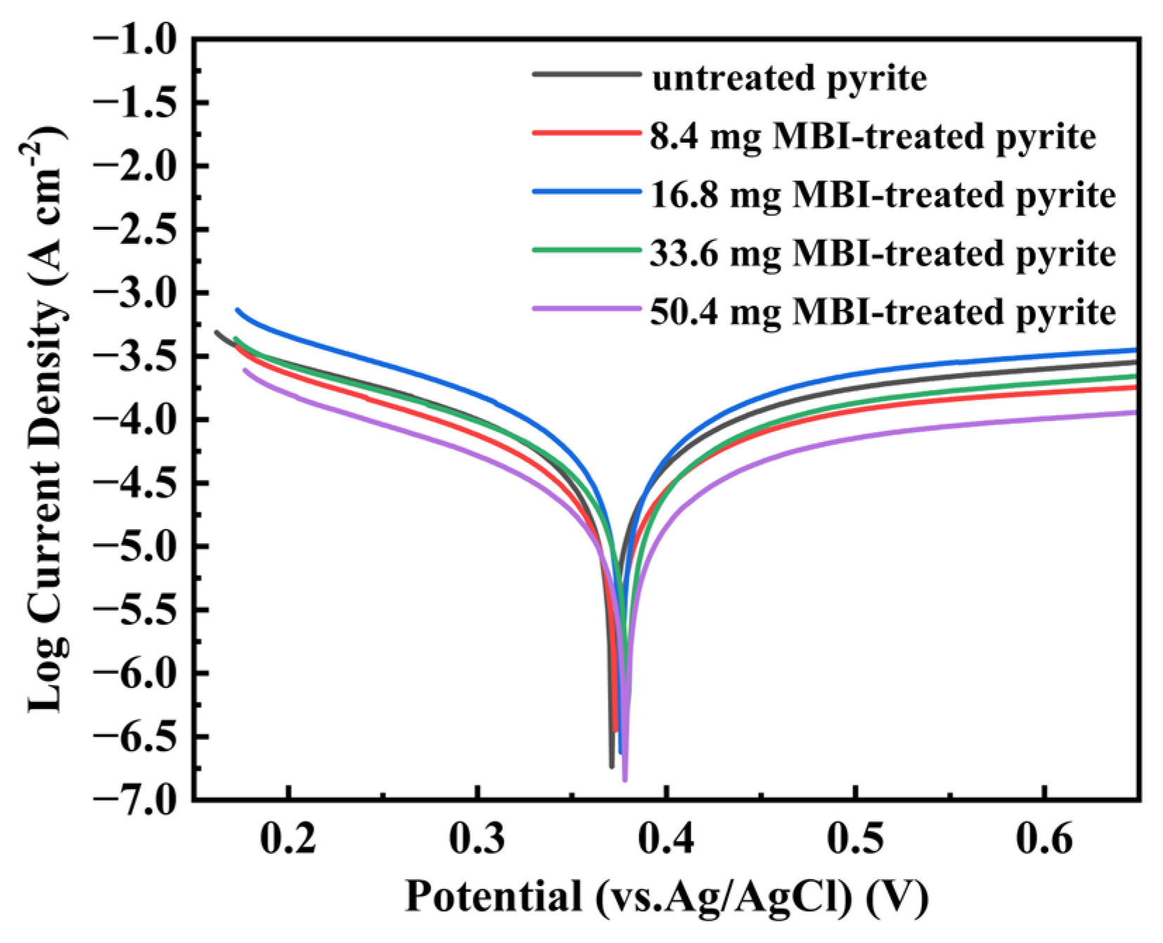
Figure 3 displays the Tafel polarization curves of raw pyrite electrodes and MBI-treated pyrite electrodes. The corrosion potential and corrosion current obtained by the Tafel extrapolation method are summarized in Table 1. Usually, the sample with a higher corrosion potential, Ecorr, and lower corrosion current, Icorr, exhibited stronger oxidation resistance. As shown in Figure 3 and Table 1, the pyrite electrodes treated with 16.8 mg, 33.6 mg, and 50.4 mg MBI exhibited higher Ecorr values and lower Icorr values compared to untreated pyrite, indicating that the oxidation resistance of these treated pyrite electrodes was stronger than that of raw pyrite. Furthermore, the oxidation resistance improved with increasing MBI dosage. Notably, while the 8.4 mg MBI-treated pyrite showed a higher Ecorr than untreated pyrite, its Icorr was significantly greater, suggesting that low MBI dosages fail to enhance oxidation resistance and may even exert adverse effects.
Electrochemical impedance spectra of the raw pyrite and MBI-treated pyrite electrodes are presented in the form of a Nyquist diagram (Figure 4) and can be fitted by an equivalent circuit model (Figure 5). In Figure 5, Rs stands for electrolyte solution resistance, Rct stands for charge transfer resistance, CPE1 stands for electric double layer capacitance at the interface between electrode and electrolyte, and W stands for Warburg impedance. Due to the excessively high film resistance, the electron transfer between pyrite and external substances was impeded. The migration of iron vacancies and oxygen vacancies was significantly restricted, so the impedance spectrum in the low-frequency part shows Warburg impedance [60,61]. In the high-frequency region, it shows a capacitive reactance arc, and its radius is related to charge transfer resistance, Rct. The larger the radius, the greater the charge transfer resistance, Rct [62], indicating that the material has better corrosion resistance. However, the impedance spectrum of the pyrite electrode treated with 50.4 mg MBI is much larger than that of other groups in the high-frequency region, and the radius of the semicircle in the high-frequency region also increases with the increase in MBI dosage (Figure 4), which shows that MBI has a passivation effect on pyrite, and the passivation ability is proportional to the dosage. In addition, the high-frequency semicircle of the pyrite electrode treated with 8.4 mg MBI is smaller than that of the raw pyrite electrode, indicating that a small amount of MBI will not have a passivation effect, but may aggravate pyrite oxidation due to pitting corrosion [63,64].

3.4. Mechanism of Inhibiting Pyrite Oxidation by MBI Passivation Layer

3.4.1. SEM Analysis

The difference in pyrite surface morphology before and after MBI passivation was observed by an SEM. As shown in Figure 6, the surface of raw pyrite is relatively smooth and flat, with some tiny mineral particles adhering to it (Figure 6a,b). In contrast, a large number of filaments can be observed on the surface of pyrite passivated by MBI (Figure 6c). By comparing the images in a smaller scale (1 μm), it can be clearly seen that there are strip-like coverings on the surface of pyrite passivated by MBI, and a large number of strip-like objects are arranged in disorder, which makes its surface rough and reduces the contact between the pyrite and the external environment (Figure 6b,d). Therefore, it can be concluded from the above results that a large amount of MBI can be densely attached to the surface of pyrite, thus covering the pyrite and preventing it from oxidizing.

3.4.2. Contact Angle Test

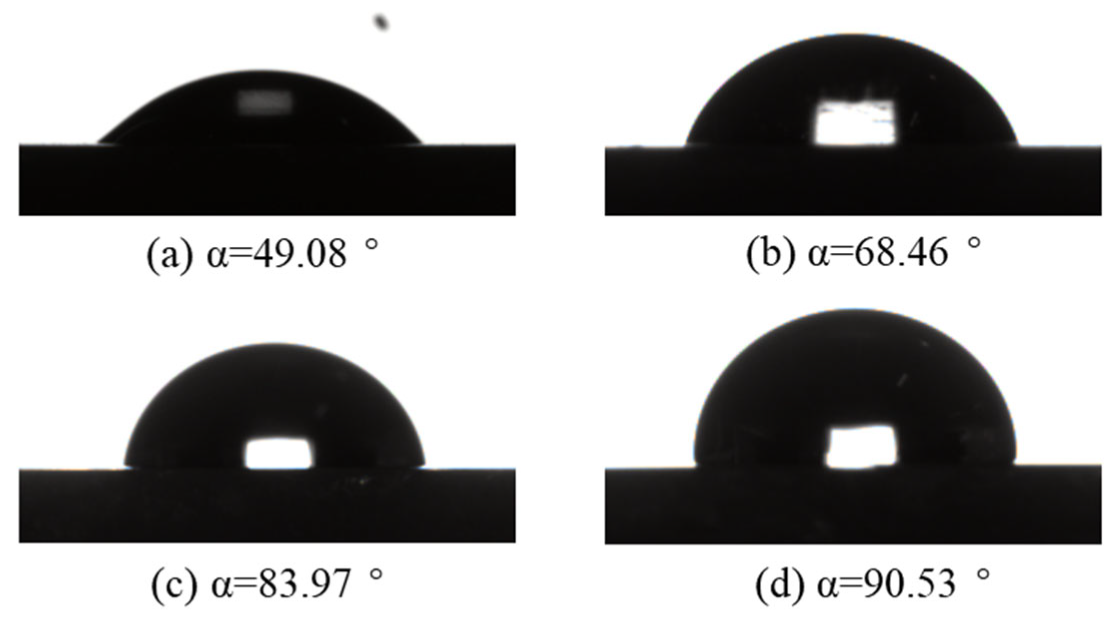
The contact angle of the pyrite surface was measured to explore the influence of the adsorption of pyrite by MBI on the hydrophilicity and hydrophobicity of the pyrite surface. It can be seen from Figure 7a–d that the hydrophilicity and hydrophobicity of the pyrite surface have also changed after passivation with different amounts of MBI. The contact angle of raw pyrite without passivation is 49.08°, while the contact angles of pyrite passivated with 5 mg MBI, 10 mg MBI and 20 mg MBI have increased to 68.46°, 83.97° and 90.53°, respectively, indicating that MBI can make the surface of pyrite hydrophobic. Moreover, as the amount of MBI increases, the hydrophobicity of the pyrite surface becomes stronger and stronger. This is because the benzene ring has certain hydrophobicity [65], and the enhancement of hydrophobicity can reduce the contact between the pyrite surface and the external liquid environment. Combined with the results of the SEM analysis, the adsorption of MBI on pyrite can isolate pyrite from external oxidation factors, weaken the oxidation of pyrite by oxygen and water, and enhance the oxidation resistance of pyrite.

3.4.3. FTIR Analysis

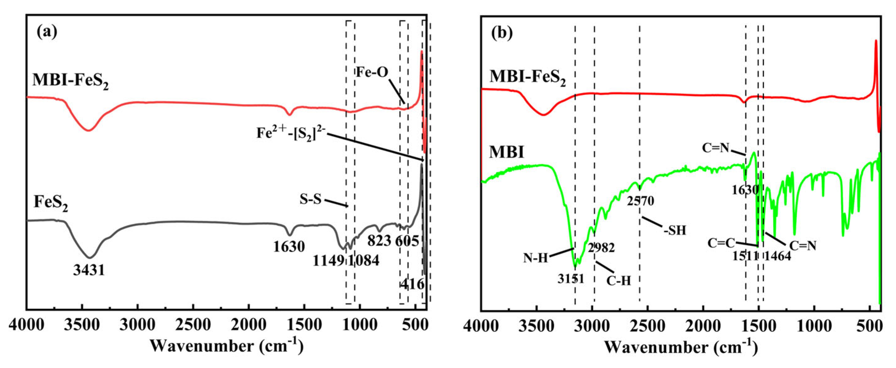
In order to explore the interaction mechanism between MBI and the pyrite surface, FTIR analysis was carried out on MBI, raw pyrite and pyrite passivated by MBI. Figure 8a shows the infrared spectrum of pyrite before and after passivation with MBI. In the spectrum of raw pyrite, the peak at 416 cm−1 is caused by the stretching vibration of Fe2+-[S2]2−, while the peak at 1084 cm−1 is the result of S-S stretching vibration. Both peaks are characteristic peaks of pyrite [25,66], and the adsorption band at 605 cm−1 belongs to Fe-O bond, which may be related to the partial oxidation of the pyrite surface [67]. In contrast, the spectra of pyrite passivated by MBI show that the peak intensities of 416 cm−1 and 1084 cm−1 are lower than those of raw pyrite, which indicates that MBI interacts with pyrite and is adsorbed on pyrite, and the bonds involved in this process are mainly Fe2+-[S2]2− and S-S bonds. In addition, the FT-IR spectra of MBI and passivated pyrite are compared (Figure 8b). The peaks at 3151 cm−1 and 2982 cm−1 in the FTIR spectra of MBI are attributed to the tensile vibration of N-H and C-H, respectively. Notably, the tensile vibration of -C=N at 1464 cm−1 and the tensile vibration of -SH at 2570 cm−1 disappear after the interaction between MBI and pyrite, which also shows that MBI reacts with pyrite by molecular -C=N and -SH [46]. Therefore, the above conclusions can be taken together to show that MBI can be adsorbed on the surface of pyrite mainly through the chelation of -C=N and -SH groups with Fe2+.

3.4.4. XPS Analysis

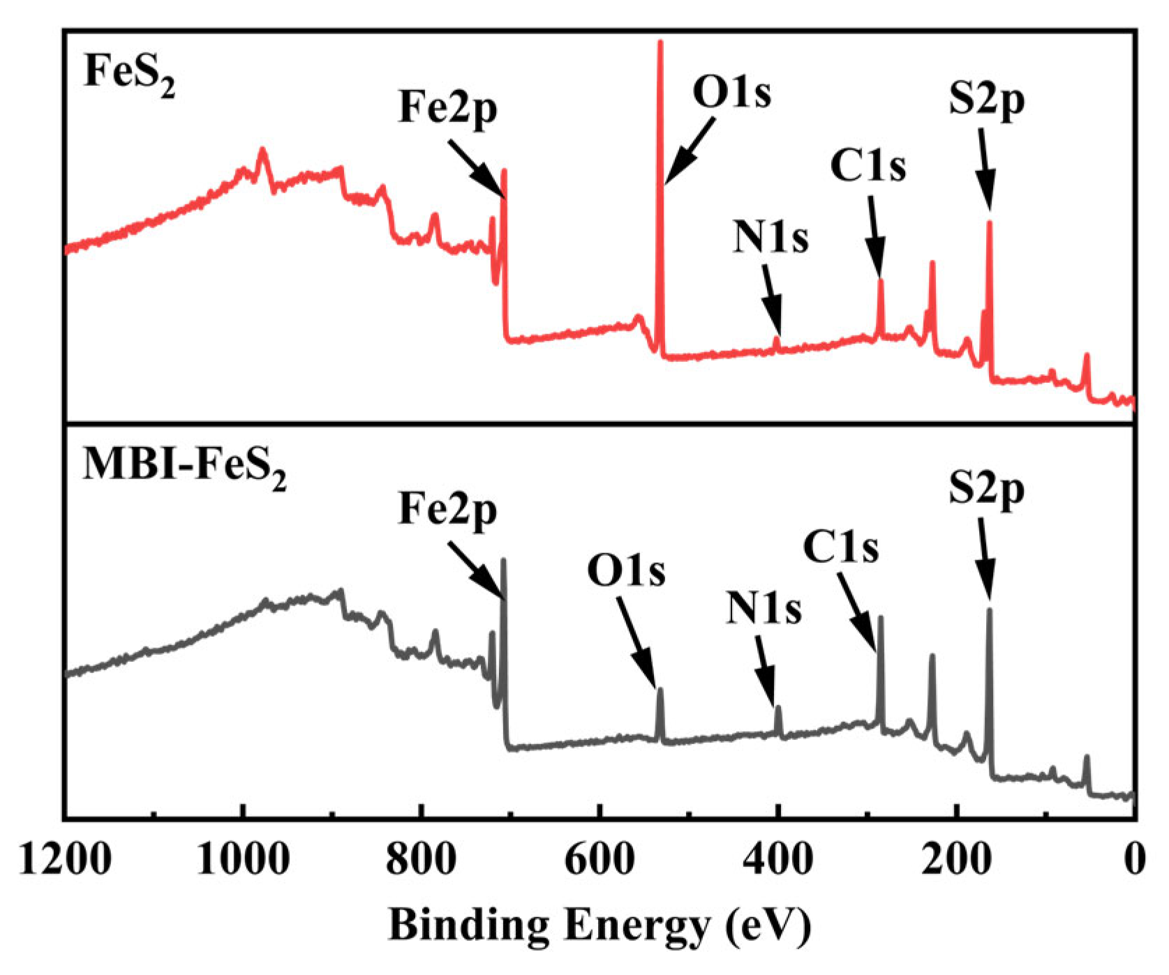
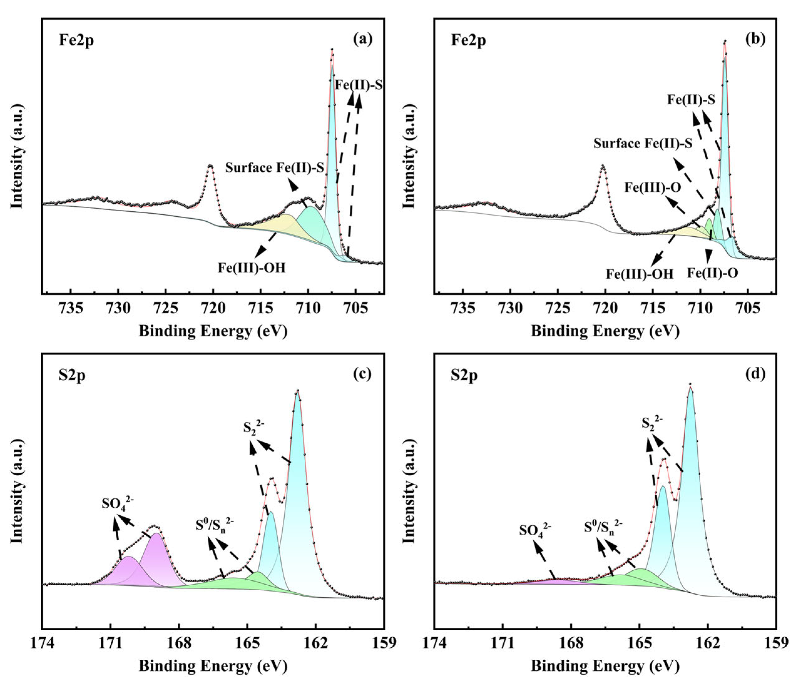
In order to further explore the mechanism of MBI inhibiting pyrite oxidation, the raw pyrite and pyrite passivated by MBI were analyzed by XPS. From the XPS spectrum (Figure 9), it can be seen that the characteristic signals of C1s and N1s on pyrite treated by MBI are stronger than those on raw pyrite, indicating that MBI, containing C and N elements, interacts with pyrite [68]. The increase in the Fe peak also shows that MBI is adsorbed on the mineral surface [47]. Figure 10 shows the Fe2p and S2p of the original pyrite and the treated pyrite. In Figure 10a and b, Fe (II)-S in pyrite is represented by a narrow peak with a binding energy of 707.4 eV, which is also an important characteristic peak of pyrite [69]. The characteristic peaks with binding energies at 709.1 and 712.3 eV belong to Fe (II)-S and Fe (III)-OH on the surface of pyrite, respectively [70,71]. Compared with raw pyrite, the characteristic peak signals of passivated pyrite samples are weakened, and the corresponding binding energy is also slightly reduced, indicating that MBI chelates with Fe (II) and Fe (III) on the pyrite surface. This is also consistent with the conclusion of the FTIR analysis. The S2p of pyrite samples is shown in Figure 10c,d, and the characteristic peaks of raw pyrite at 169.1 eV and 170.2 eV belong to sulfate, which indicates that there is partial oxidation on the surface of pyrite, but after the interaction between MBI and pyrite, the characteristic peak of sulfate decreases (Figure 10d), which is due to the competitive adsorption of MBI on pyrite, resulting in the separation of SO42− from its original position [72]. The peaks at 162.8 eV and 163.9 eV are assigned to disulfide (S22−), while those at 165.2 eV and 165.8 eV correspond to elemental sulfur/polysulfide (S0/Sn2−) [73,74]. Sulfide (S22−) mainly comes from S atoms near the surface of pyrite [75], and elemental sulfur/polysulfide (S0/Sn2−) is produced by surface oxidation of pyrite [76]. Comparing the peaks of pyrite before and after MBI passivation, there is no obvious difference, indicating that the role of MBI and pyrite does not involve sulfur in pyrite.

3.4.5. Mechanism of MBI Inhibiting Pyrite Oxidation

Based on the analysis results of SEM, FTIR and XPS, the mechanism of inhibiting pyrite oxidation by MBI under the action of At. ferrooxidans is proposed in Figure 11. In the acidic environment with pH equal to 2, Fe3+ is greatly increased because At. ferrooxidans can accelerate the oxidation of Fe2+. In the presence of oxidants such as Fe3+ and O2, the oxidation of pyrite will start at the surface, and the oxidation rate will be greatly improved. After the addition of MBI, MBI can slightly inhibit the growth of At. ferrooxidans. In addition, -C=N and -SH in MBI molecules will chelate with Fe2+ and Fe3+ on the surface of pyrite, and a large amount of MBI will be adsorbed on the surface of pyrite, forming a hydrophobic film layer, which can reduce the possibility of contact between pyrite and dissolved Fe3+, O2 and bacteria, and at the same time reduce the number of oxidation sites on the surface of pyrite [74], thus inhibiting pyrite oxidation and improving pyrite oxidation resistance.

4. Conclusions

In this paper, the inhibition of pyrite oxidation by MBI was studied in the acidic environment of At. ferrooxidans. The specific conclusions are as follows:
(1)
In the presence of At. ferrooxidans, 3 g/kg MBI cannot effectively inhibit pyrite oxidation; on the contrary, it can promote pyrite oxidation. When the dosage is more than or equal to 6 g/kg, MBI can effectively inhibit the oxidation of pyrite, and 9 g/kg MBI has the best inhibition effect, with the inhibition rate reaching 97.1%.
(2)
MBI can inhibit the growth of At. ferrooxidans. With the increase in MBI dosage, the growth of At. ferrooxidans was temporarily inhibited, and then returned to normal in a short time. Therefore, the bacteriostatic effect of MBI may be one of the reasons for the inhibition of pyrite oxidation.
(3)
When the dosage of MBI is greater than or equal to 16.8 mg, the pyrite electrode will show oxidation resistance, and its oxidation resistance will be enhanced with the increase in MBI dosage.
(4)
The contact angle test shows that the adhesion of MBI to the pyrite surface will enhance the hydrophobicity of the pyrite surface, and its hydrophobicity will also increase with the increase in MBI dosage.
(5)
The -C=N and -SH groups in MBI can chelate with Fe2+ and Fe3+ on the surface of pyrite, so a large amount of MBI is adsorbed on pyrite, which on the one hand strengthens the hydrophobicity of pyrite, and on the other hand leads to the reduction in oxidation sites on pyrite.
Based on the above conclusions, MBI can effectively inhibit or slow down the oxidation of pyrite. However, the optimal dosage, stability and practical application potential of MBI have not been explored in this paper, and future experiments will focus on these points. This research is expected to become an effective method for AMD control and governance at present, and will also provide a new idea for the innovation of methods.

Author Contributions

Conceptualization, Y.Z. (Yansheng Zhang); methodology, J.H.; validation, J.H. and X.L.; formal analysis, J.H. and Y.Z. (Yeyang Zhou); investigation, J.H. and J.Y.; resources, Y.Z. (Yansheng Zhang); data curation, J.H. and X.W.; writing, J.H.; supervision, B.L.; funding acquisition, Y.Z. (Yansheng Zhang). All authors have read and agreed to the published version of the manuscript.

Funding

This research was funded by the National Natural Science Foundation of China (grant number: 52474313), the National Natural Science Foundation of China (grant number: 51974363) and the National Key Research and Development Program of China (grant number: 2022YFC2105303).

Data Availability Statement

Data are contained within the article.

Conflicts of Interest

The authors declare no conflicts of interest.

References

  1. Hughes, T.A.; Gray, N.F. Co-treatment of acid mine drainage with municipal wastewater: Performance evaluation. Environ. Sci. Pollut. Res. 2013, 20, 7863–7877. [Google Scholar] [CrossRef] [PubMed]
  2. Tong, L.; Fan, R.; Yang, S.; Li, C. Development and Status of the Treatment Technology for Acid Mine Drainage. Min. Metall. Explor. 2021, 38, 315–327. [Google Scholar] [CrossRef]
  3. Ouyang, Y.; Liu, Y.; Zhu, R.; Ge, F.; Xu, T.; Luo, Z.; Liang, L. Pyrite oxidation inhibition by organosilane coatings for acid mine drainage control. Miner. Eng. 2015, 72, 57–64. [Google Scholar] [CrossRef]
  4. Masindi, V.; Foteinis, S.; Renforth, P.; Ndiritu, J.; Maree, J.P.; Tekere, M.; Chatzisymeon, E. Challenges and avenues for acid mine drainage treatment, beneficiation, and valorisation in circular economy: A review. Ecol. Eng. 2022, 183, 106740. [Google Scholar] [CrossRef]
  5. Elamraoui, L.; Elghali, A.; Fashae, O.A.; Benzaazoua, M. Harnessing phosphate limestone waste as a cost-effective solution for acid mine drainage treatment. Sci. Total Environ. 2024, 949, 175188. [Google Scholar] [CrossRef]
  6. Wang, S.; Zhao, Y.; Li, S. Pyrite oxidation inhibition by hydrophobic films for acid mine drainage control at the source. Physicochem. Probl. Miner. Process. 2019, 55, 1132–1140. [Google Scholar]
  7. Mosai, A.K.; Ndlovu, G.; Tutu, H. Improving acid mine drainage treatment by combining treatment technologies: A review. Sci. Total Environ. 2024, 919, 170806. [Google Scholar] [CrossRef]
  8. Sheoran, A.S.; Sheoran, V. Heavy metal removal mechanism of acid mine drainage in wetlands: A critical review. Miner. Eng. 2006, 19, 105–116. [Google Scholar] [CrossRef]
  9. Menzel, K.; Barros, L.; García, A.; Ruby-Figueroa, R.; Estay, H. Metal sulfide precipitation coupled with membrane filtration process for recovering copper from acid mine drainage. Sep. Purif. Technol. 2021, 270, 118721. [Google Scholar] [CrossRef]
  10. Silva, G.C.; Bertoli, A.C.; Duarte, H.A.; Ladeira, A.C.Q. Recovery of rare earth elements from sulfate-rich acid mine water: Looking through the keyhole the exchange reaction for cationic resin. J. Environ. Chem. Eng. 2022, 10, 108715. [Google Scholar] [CrossRef]
  11. León-Venegas, E.; Vilches-Arenas, L.F.; Fernández-Baco, C.; Arroyo-Torralvo, F. Potential for water and metal recovery from acid mine drainage by combining hybrid membrane processes with selective metal precipitation. Resour. Conserv. Recycl. 2023, 188, 106629. [Google Scholar] [CrossRef]
  12. Pereira, T.C.B.; Dos Santos, K.B.; Lautert-Dutra, W.; de Souza Teodoro, L.; de Almeida, V.O.; Weiler, J.; Bogo, M.R. Acid mine drainage (AMD) treatment by neutralization: Evaluation of physical-chemical performance and ecotoxicological effects on zebrafish (Danio rerio) development. Chemosphere 2020, 253, 126665. [Google Scholar] [CrossRef] [PubMed]
  13. Johnson, D.B.; Hallberg, K.B. Acid mine drainage remediation options: A review. Sci. Total Environ. 2005, 338, 3–14. [Google Scholar] [CrossRef] [PubMed]
  14. Neculita, C.M.; Zagury, G.J.; Bussière, B. Passive treatment of acid mine drainage in bioreactors using sulfate-reducing bacteria: Critical review and research needs. J. Environ. Qual. 2007, 36, 1–16. [Google Scholar] [CrossRef]
  15. Wang, H.; Zhang, M.; Xue, J.; Lv, Q.; Yang, J.; Han, X. Performance and microbial response in a multi-stage constructed wetland microcosm co-treating acid mine drainage and domestic wastewater. J. Environ. Chem. Eng. 2021, 9, 106786. [Google Scholar] [CrossRef]
  16. Woulds, C.; Ngwenya, B.T. Geochemical processes governing the performance of a constructed wetland treating acid mine drainage, Central Scotland. Appl. Geochem. 2004, 19, 1773–1783. [Google Scholar] [CrossRef]
  17. Sahoo, P.K.; Kim, K.; Equeenuddin, S.M.; Powell, M.A. Current approaches for mitigating acid mine drainage. Rev. Environ. Contam. Toxicol. Vol. 2013, 226, 1–32. [Google Scholar]
  18. Park, I.; Tabelin, C.B.; Jeon, S.; Li, X.; Seno, K.; Ito, M.; Hiroyoshi, N. A review of recent strategies for acid mine drainage prevention and mine tailings recycling. Chemosphere 2019, 219, 588–606. [Google Scholar] [CrossRef]
  19. Skousen, J.G.; Ziemkiewicz, P.F.; McDonald, L.M. Acid mine drainage formation, control and treatment: Approaches and strategies. Extr. Ind. Soc. 2019, 6, 241–249. [Google Scholar] [CrossRef]
  20. Rubinos, D.A.; Jerez, Ó.; Forghani, G.; Edraki, M.; Kelm, U. Geochemical stability of potentially toxic elements in porphyry copper-mine tailings from Chile as linked to ecological and human health risks assessment. Environ. Sci. Pollut. Res. 2021, 28, 57499–57529. [Google Scholar] [CrossRef]
  21. Peppas, A.; Komnitsas, K.A.; Halikia, I. Use of organic covers for acid mine drainage control. Miner. Eng. 2000, 13, 563–574. [Google Scholar] [CrossRef]
  22. Sephton, M.G.; Webb, J.A.; McKnight, S. Applications of Portland cement blended with fly ash and acid mine drainage treatment sludge to control acid mine drainage generation from waste rocks. Appl. Geochem. 2019, 103, 1–14. [Google Scholar] [CrossRef]
  23. Jia, Y.; Maurice, C.; Öhlander, B. Effect of the alkaline industrial residues fly ash, green liquor dregs, and lime mud on mine tailings oxidation when used as covering material. Environ. Earth Sci. 2014, 72, 319–334. [Google Scholar] [CrossRef]
  24. Georgopoulou, Z.J.; Fytas, K.; Soto, H.; Evangelou, B. Feasibility and cost of creating an iron-phosphate coating on pyrrhotite to prevent oxidation. Environ. Geol. 1996, 28, 61–69. [Google Scholar] [CrossRef]
  25. Zheng, K.; Li, H.; Xu, L.; Li, S.; Wang, L.; Wen, X.; Liu, Q. The influence of humic acids on the weathering of pyrite: Electrochemical mechanism and environmental implications. Environ. Pollut. 2019, 251, 738–745. [Google Scholar] [CrossRef]
  26. Leathen, W.W.; Braley Sr, S.A.; McIntyre, L.D. The Role of Bacteria in the Formation of Acid from Certain Sulfuritic Constituents Associated with Bituminous Coal: I. Thiobacillus thiooxidans. Appl. Microbiol. 1953, 1, 61–64. [Google Scholar] [CrossRef]
  27. Frattini, C.J.; Leduc, L.G.; Ferroni, G.D. Strain variability and the effects of organic compounds on the growth of the chemolithotrophic bacterium Thiobacillus ferrooxidans. Antonie Van Leeuwenhoek 2000, 77, 57–64. [Google Scholar] [CrossRef]
  28. Elsetinow, A.R.; Schoonen, M.A.; Strongin, D.R. Aqueous geochemical and surface science investigation of the effect of phosphate on pyrite oxidation. Environ. Sci. Technol. 2001, 35, 2252–2257. [Google Scholar] [CrossRef]
  29. Nyavor, K.; Egiebor, N.O. Control of pyrite oxidation by phosphate coating. Sci. Total Environ. 1995, 162, 225–237. [Google Scholar] [CrossRef]
  30. Fan, R.; Short, M.D.; Zeng, S.J.; Qian, G.; Li, J.; Schumann, R.C. The formation of silicate-stabilized passivating layers on pyrite for reduced acid rock drainage. Environ. Sci. Technol. 2017, 51, 11317–11325. [Google Scholar] [CrossRef]
  31. Nicholson, R.V.; Gillham, R.W.; Reardon, E.J. Pyrite oxidation in carbonate-buffered solution: 2. Rate control by oxide coatings. Geochim. Et Cosmochim. Acta 1990, 54, 395–402. [Google Scholar] [CrossRef]
  32. Gao, B.; Han, Z.; Cheng, H.; Zhou, H.; Wang, Y.; Chen, Z. Treating waste with waste: Lignin acting as both an effective bactericide and passivator to prevent acid mine drainage formation at the source. Sci. Total Environ. 2024, 927, 172162. [Google Scholar] [CrossRef] [PubMed]
  33. Ačai, P.; Sorrenti, E.; Gorner, T.; Polakovič, M.; Kongolo, M.; de Donato, P. Pyrite passivation by humic acid investigated by inverse liquid chromatography. Colloids Surf. A Physicochem. Eng. Asp. 2009, 337, 39–46. [Google Scholar] [CrossRef]
  34. Jiang, C.L.; Wang, X.H.; Parekh, B.K. Effect of sodium oleate on inhibiting pyrite oxidation. Int. J. Miner. Process. 2000, 58, 305–318. [Google Scholar] [CrossRef]
  35. Kim, J.; Kim, G.; Cremer, P.S. Investigations of water structure at the solid/liquid interface in the presence of supported lipid bilayers by vibrational sum frequency spectroscopy. Langmuir 2001, 17, 7255–7260. [Google Scholar] [CrossRef]
  36. Pierre Louis, A.M.; Yu, H.; Shumlas, S.L.; Van Aken, B.; Schoonen, M.A.; Strongin, D.R. Effect of phospholipid on pyrite oxidation and microbial communities under simulated acid mine drainage (AMD) conditions. Environ. Sci. Technol. 2015, 49, 7701–7708. [Google Scholar] [CrossRef]
  37. Zhang, X.; Borda, M.J.; Schoonen, M.A.; Strongin, D.R. Adsorption of phospholipids on pyrite and their effect on surface oxidation. Langmuir 2003, 19, 8787–8792. [Google Scholar] [CrossRef]
  38. Liu, Y.; Dang, Z.; Xu, Y.; Xu, T. Pyrite passivation by triethylenetetramine: An electrochemical study. J. Anal. Methods Chem. 2013, 2013, 387124. [Google Scholar] [CrossRef]
  39. Cai, M.F.; Dang, Z.; Chen, Y.W.; Belzile, N. The passivation of pyrrhotite by surface coating. Chemosphere 2005, 61, 659–667. [Google Scholar] [CrossRef]
  40. Satur, J.; Hiroyoshi, N.; Tsunekawa, M.; Ito, M.; Okamoto, H. Carrier-microencapsulation for preventing pyrite oxidation. Int. J. Miner. Process. 2007, 83, 116–124. [Google Scholar] [CrossRef]
  41. Thakur Jha, R.K.; Satur, J.; Hiroyoshi, N.; Ito, M.; Tsunekawa, M. Suppression of pyrite oxidation by carrier microencapsulation using silicon and catechol. Miner. Process. Extr. Metall. Rev. 2012, 33, 89–98. [Google Scholar] [CrossRef]
  42. Khummalai, N.; Boonamnuayvitaya, V. Suppression of arsenopyrite surface oxidation by sol-gel coatings. J. Biosci. Bioeng. 2005, 99, 277–284. [Google Scholar] [CrossRef] [PubMed]
  43. Dong, Y.; Liu, Z.; Liu, W.; Lin, H. A new organosilane passivation agent prepared at ambient temperatures to inhibit pyrite oxidation for acid mine drainage control. J. Environ. Manag. 2022, 320, 115835. [Google Scholar] [CrossRef] [PubMed]
  44. Mahdavian, M.; Ashhari, S. Corrosion inhibition performance of 2-mercaptobenzimidazole and 2-mercaptobenzoxazole compounds for protection of mild steel in hydrochloric acid solution. Electrochim. Acta 2010, 55, 1720–1724. [Google Scholar] [CrossRef]
  45. Wu, X.; Wiame, F.; Maurice, V.; Marcus, P. 2-Mercaptobenzimidazole films formed at ultra-low pressure on copper: Adsorption, thermal stability and corrosion inhibition performance. Appl. Surf. Sci. 2020, 527, 146814. [Google Scholar] [CrossRef]
  46. Chai, W.; Huang, Y.; Peng, W.; Han, G.; Cao, Y.; Liu, J. Enhanced separation of pyrite from high-sulfur bauxite using 2-mercaptobenzimidazole as chelate collector: Flotation optimization and interaction mechanisms. Miner. Eng. 2018, 129, 93–101. [Google Scholar] [CrossRef]
  47. Zheng, M.; Peng, T.; Wang, Y.; Luo, L.; Shao, W.; Gao, J.; Lu, Q. Flotability of galena and pyrite using 2-mercaptobenzimidazole as a chelating agent: Adsorption characteristics and flotation mechanisms. Powder Technol. 2021, 393, 449–460. [Google Scholar] [CrossRef]
  48. Jadhav, U.; Hocheng, H. Recovery of phosphorus from source separated urine by Acidithiobacillus ferrooxidans culture supernatant. Ecol. Eng. 2016, 92, 90–96. [Google Scholar] [CrossRef]
  49. Caldwell, D.H.; Adams, R.B. Colorimetric determination of iron in water with o-phenanthroline. J. Am. Water Work. Assoc. 1946, 38, 727–730. [Google Scholar] [CrossRef]
  50. Ogbughalu, O.T.; Vasileiadis, S.; Schumann, R.C.; Gerson, A.R.; Li, J.; Smart, R.S.C.; Short, M.D. Role of microbial diversity for sustainable pyrite oxidation control in acid and metalliferous drainage prevention. J. Hazard. Mater. 2020, 393, 122338. [Google Scholar] [CrossRef]
  51. Tabelin, C.B.; Corpuz, R.D.; Igarashi, T.; Villacorte-Tabelin, M.; Alorro, R.D.; Yoo, K. Acid mine drainage formation and arsenic mobility under strongly acidic conditions: Importance of soluble phases, iron oxyhydroxides/oxides and nature of oxidation layer on pyrite. J. Hazard. Mater. 2020, 399, 122844. [Google Scholar] [CrossRef] [PubMed]
  52. Xia, S.; Song, Z.; Zhao, X.; Li, J. Review of the recent advances in the prevention, treatment, and resource recovery of acid mine wastewater discharged in coal mines. J. Water Process Eng. 2023, 52, 103555. [Google Scholar] [CrossRef]
  53. Ye, J.; Wang, S.; Zhang, P.; Nabi, M.; Tao, X.; Zhang, H.; Liu, Y. L-cysteine addition enhances microbial surface oxidation of coal inorganic sulfur: Complexation of cysteine and pyrite, inhibition of jarosite formation, environmental effects. Environ. Res. 2020, 187, 109705. [Google Scholar] [CrossRef] [PubMed]
  54. Liu, W.; Sayem, A.K.; Perez, J.P.; Hornback, S.; Owusu-Fordjour, E.Y.; Yang, X. Mechanism investigation of food waste compost as a source of passivation agents for inhibiting pyrite oxidation. J. Environ. Chem. Eng. 2024, 12, 113465. [Google Scholar] [CrossRef]
  55. Meruane, G.; Vargas, T. Bacterial oxidation of ferrous iron by Acidithiobacillus ferrooxidans in the pH range 2.5–7.0. Hydrometallurgy 2003, 71, 149–158. [Google Scholar] [CrossRef]
  56. Rojas-Chapana, J.A.; Tributsch, H. Biochemistry of sulfur extraction in bio-corrosion of pyrite by Thiobacillus ferrooxidans. Hydrometallurgy 2001, 59, 291–300. [Google Scholar] [CrossRef]
  57. Chandra, A.P.; Gerson, A.R. The mechanisms of pyrite oxidation and leaching: A fundamental perspective. Surf. Sci. Rep. 2010, 65, 293–315. [Google Scholar] [CrossRef]
  58. Daoud, J.; Karamanev, D.J.M.E. Formation of jarosite during Fe2+ oxidation by Acidithiobacillus ferrooxidans. Miner. Eng. 2006, 19, 960–967. [Google Scholar] [CrossRef]
  59. Piervandi, Z.; Darban, A.K.; Mousavi, S.M.; Abdollahy, M.; Asadollahfardi, G.; Funari, V. Effect of biogenic jarosite on the bio-immobilization of toxic elements from sulfide tailings. Chemosphere 2020, 258, 127288. [Google Scholar] [CrossRef]
  60. Fattah-Alhosseini, A.; Taheri Shoja, S.; Heydari Zebardast, B.; Mohamadian Samim, P. An electrochemical impedance spectroscopic study of the passive state on AISI 304 stainless steel. Int. J. Electrochem. 2011, 2011, 152143. [Google Scholar] [CrossRef]
  61. Wu, S.; Ning, Y.; Xie, H.; Tian, H.; Lv, J.; Liu, B. Study on the galvanic corrosion behavior of copper-nickel/titanium alloys under simulated seawater environment. J. Solid State Electrochem. 2024, 28, 2315–2329. [Google Scholar] [CrossRef]
  62. Walter, G.W. A review of impedance plot methods used for corrosion performance analysis of painted metals. Corros. Sci. 1986, 26, 681–703. [Google Scholar] [CrossRef]
  63. Meng, X.; Chen, X.; Wu, M.; Zhao, Y.; Fan, Y. Effect of Hydrostatic Pressure on Electrochemical Behavior of X100 Steel in NaHCO3+NaCl Solution. J. Chin. Soc. Corros. Prot. 2016, 36, 219–224. [Google Scholar]
  64. Macdonald, D.D. The point defect model for the passive state. J. Electrochem. Soc. 1992, 139, 3434. [Google Scholar] [CrossRef]
  65. Graziano, G. Hydrophobicity of benzene. Biophys. Chem. 1999, 82, 69–79. [Google Scholar] [CrossRef]
  66. Reyes-Bozo, L.; Escudey, M.; Vyhmeister, E.; Higueras, P.; Godoy-Faúndez, A.; Salazar, J.L. Adsorption of biosolids and their main components on chalcopyrite, molybdenite and pyrite: Zeta potential and FTIR spectroscopy studies. Miner. Eng. 2015, 78, 128–135. [Google Scholar] [CrossRef]
  67. Liu, Y.; Hu, X.; Xu, Y. PropS-SH/SiO2 nanocomposite coatings for pyrite oxidation inhibition to control acid mine drainage at the source. J. Hazard. Mater. 2017, 338, 313–322. [Google Scholar] [CrossRef]
  68. Li, D.; Liu, C.; Liu, Y.; Chen, X.; Wu, W.; Li, F. Tannic acid as an eco-friendly natural passivator for the inhibition of pyrite oxidation to prevent acid mine drainage at the source. Appl. Surf. Sci. 2022, 591, 153172. [Google Scholar] [CrossRef]
  69. Descostes, M.; Mercier, F.; Thromat, N.; Beaucaire, C.; Gautier-Soyer, M. Use of XPS in the determination of chemical environment and oxidation state of iron and sulfur samples: Constitution of a data basis in binding energies for Fe and S reference compounds and applications to the evidence of surface species of an oxidized pyrite in a carbonate medium. Appl. Surf. Sci. 2000, 165, 288–302. [Google Scholar]
  70. Mu, Y.; Li, L.; Peng, Y. Surface properties of fractured and polished pyrite in relation to flotation. Miner. Eng. 2017, 101, 10–19. [Google Scholar] [CrossRef]
  71. Hong, M.; Wang, J.; Yang, B.; Liu, Y.; Sun, X.; Li, L. Inhibition of pyrite oxidation through forming biogenic K-jarosite coatings to prevent acid mine drainage production. Water Res. 2024, 252, 121221. [Google Scholar] [CrossRef] [PubMed]
  72. Schaufuß, A.G.; Nesbitt, H.W.; Kartio, I.; Laajalehto, K.; Bancroft, G.M.; Szargan, R. Incipient oxidation of fractured pyrite surfaces in air. J. Electron Spectrosc. Relat. Phenom. 1998, 96, 69–82. [Google Scholar] [CrossRef]
  73. Niu, X.; Chen, J.; Li, Y.; Xia, L.; Li, L.; Sun, H.; Ruan, R. Correlation of surface oxidation with xanthate adsorption and pyrite flotation. Appl. Surf. Sci. 2019, 495, 143411. [Google Scholar] [CrossRef]
  74. Zhang, P.; Yuan, S.; Liao, P. Mechanisms of hydroxyl radical production from abiotic oxidation of pyrite under acidic conditions. Geochim. Et Cosmochim. Acta 2016, 172, 444–457. [Google Scholar] [CrossRef]
  75. Nesbitt, H.W.; Bancroft, G.M.; Pratt, A.R.; Scaini, M.J. Sulfur and iron surface states on fractured pyrite surfaces. Am. Mineral. 1998, 83, 1067–1076. [Google Scholar] [CrossRef]
  76. Zhang, G.; Ye, G.; Liu, D. Adsorption study of fenugreek gum onto pyrite surface: Implications for chalcopyrite-pyrite flotation separation. Colloids Surf. A Physicochem. Eng. Asp. 2023, 677, 132404. [Google Scholar] [CrossRef]
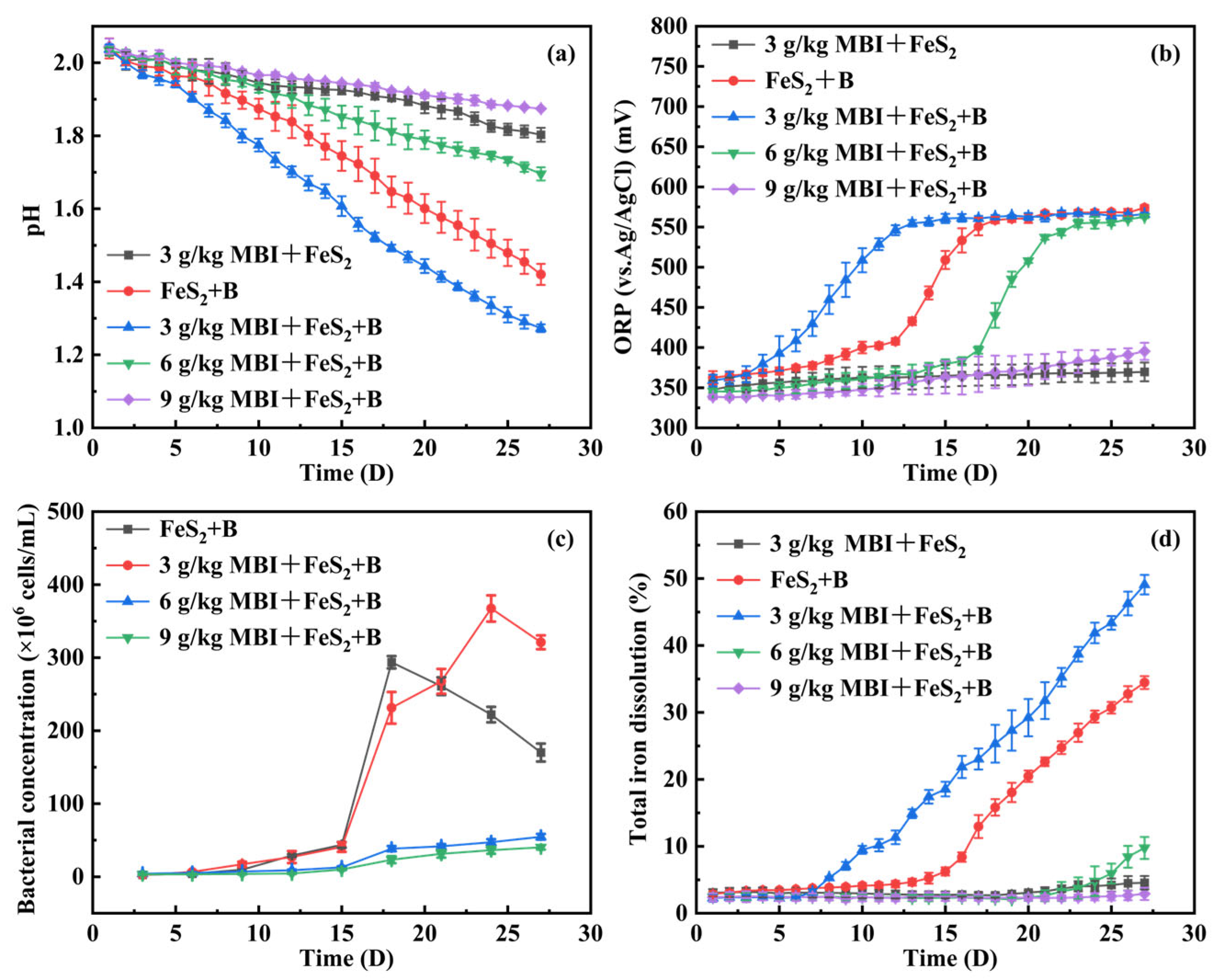
Figure 1. Variations of solution pH (a), ORP (vs. Ag/AgCl) (b), bacterial concentration (c) and total iron dissolution (d) during pyrite leaching experiments with different MBI dosages (The amount of pyrite: 2 g, initial pH: 2, initial bacterial concentration: 1.0 × 10⁷ cells/mL. In the figure, “MBI” denotes 2-mercaptobenzimidazole, “B” denotes bacteria, which refers to At. ferrooxidans here).
Minerals 15 00487 g001
Figure 2. Variations in solution pH (a), ORP (vs. Ag/AgCl) (b), bacterial concentration (c) and degree of Fe2+ oxidation (d) during growth experiment of At. ferrooxidans with different MBI dosages (Initial pH: 2, initial bacterial concentration: 1.0 × 10⁷ cells/mL).
Minerals 15 00487 g002
Figure 3. Tafel polarization curves of untreated pyrite electrode and MBI-treated pyrite electrodes in 0.1 M Na2SO4 solution (electrolyte pH = 2).
Minerals 15 00487 g003
Figure 4. Nyquist diagram of EIS data obtained from untreated pyrite electrode and MBI-treated pyrite electrodes in 0.1 M Na2SO4 solution (electrolyte pH = 2; frequency range: 100,000 to 0.01 Hz).
Minerals 15 00487 g004
Figure 5. Equivalent circuit proposed by fitting EIS data obtained from untreated pyrite electrode and MBI-treated pyrite electrodes.
Minerals 15 00487 g005
Figure 6. SEM images of raw pyrite (a,b) and pyrite passivated by MBI (c,d).
Minerals 15 00487 g006
Figure 7. Static water contact angles of pyrite after passivation with different MBI dosages: (a) raw pyrite; (b) 5 mg MBI; (c) 10 mg MBI; (d) 20 mg MBI.
Minerals 15 00487 g007
Figure 8. FTIR spectra of pyrite before and after passivation (a) and FTIR spectra of MBI and passivated pyrite (b).
Minerals 15 00487 g008
Figure 9. XPS full spectra of raw pyrite and pyrite passivated by MBI.
Minerals 15 00487 g009
Figure 10. XPS spectra of Fe2p peaks of raw pyrite (a) and pyrite passivated by MBI (b); S2p peaks of raw pyrite (c) and pyrite passivated by MBI (d).
Minerals 15 00487 g010
Figure 11. MBI chelates with Fe2+ and Fe3+ on the surface of pyrite through -C=N and -SH in the molecule, thereby attaching to the surface of pyrite. The formed passivation layer isolates pyrite from the external environment and ultimately inhibits the oxidation of pyrite. The mechanism for the inhibition of pyrite oxidation by MBI is shown in Figure 11.
Minerals 15 00487 g011
Table 1. Corrosion potentials and corrosion current densities of untreated pyrite electrode and MBI-treated pyrite electrodes.
ElectrodeEcorr (vs. Ag/AgCl) (mV)Icorr (μA cm−2)
Untreated pyrite3716.803
8.4 mg MBI-treated pyrite3739.358
16.8 mg MBI-treated pyrite3765.708
33.6 mg MBI-treated pyrite3794.872
50.4 mg MBI-treated pyrite3782.952
Disclaimer/Publisher’s Note: The statements, opinions and data contained in all publications are solely those of the individual author(s) and contributor(s) and not of MDPI and/or the editor(s). MDPI and/or the editor(s) disclaim responsibility for any injury to people or property resulting from any ideas, methods, instructions or products referred to in the content.