ResUNet: Application of Deep Learning in Quantitative Characterization of 3D Structures in Iron Ore Pellets


Abstract
1. Introduction
2. Summary of Previous Research
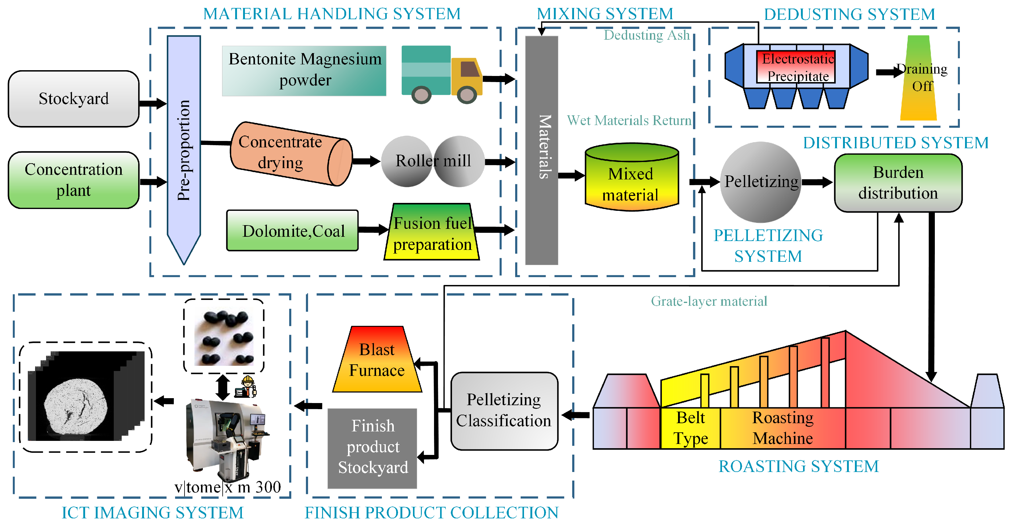
3. Materials and Methods
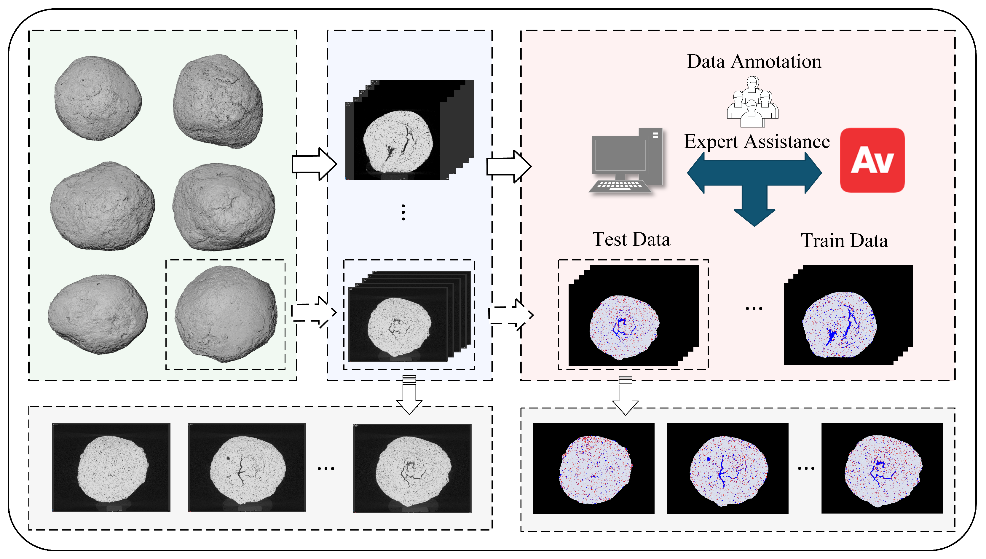
3.1. Data Preparation
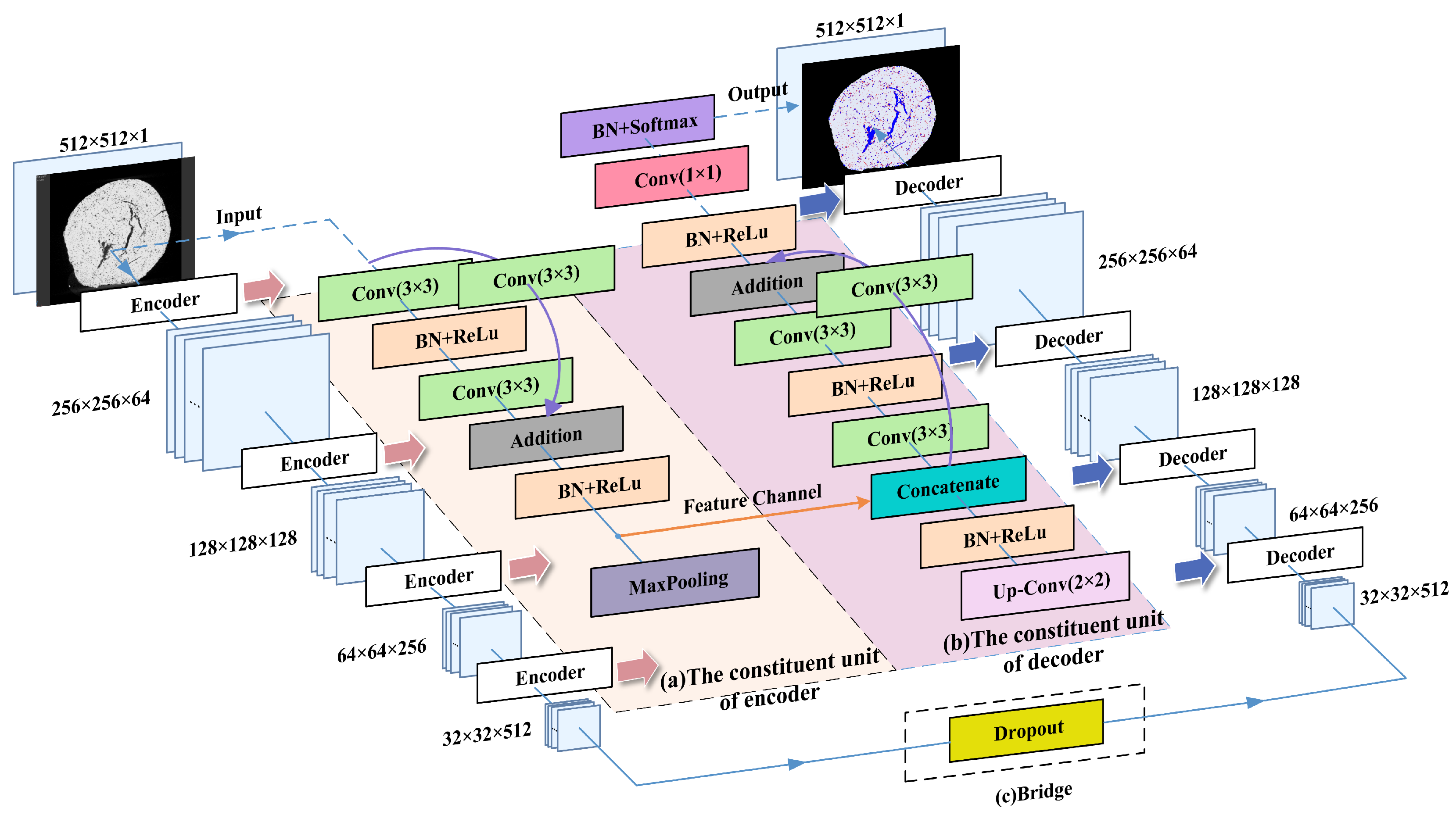
3.2. ResUNet Network Structure
3.2.1. Encoder
3.2.2. Bridge
3.2.3. Decoder
3.3. Image Augmentation
3.4. Image Normalization
3.5. Experimental Details
3.6. Evaluation Metrics
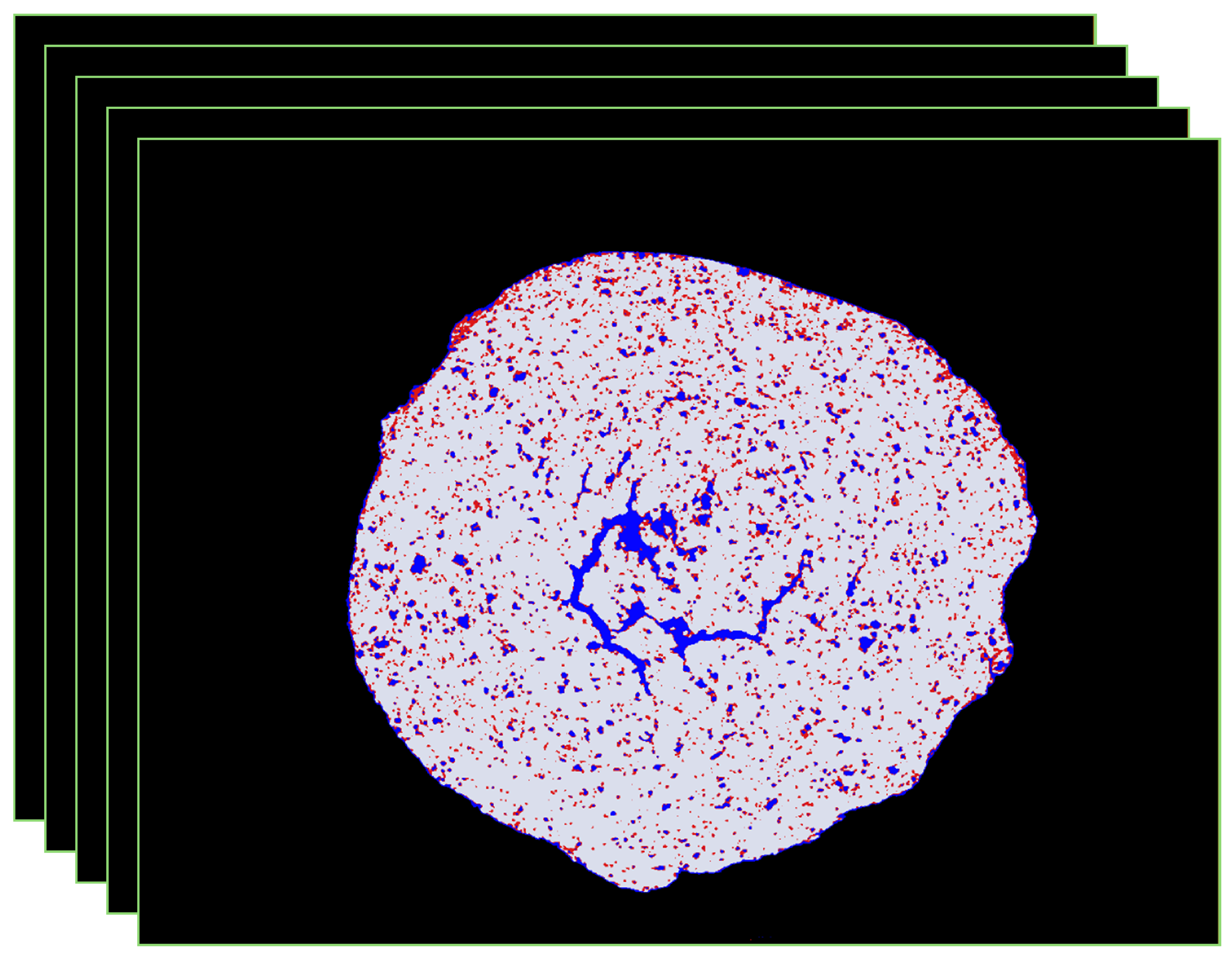
4. Results
4.1. Model Evaluation
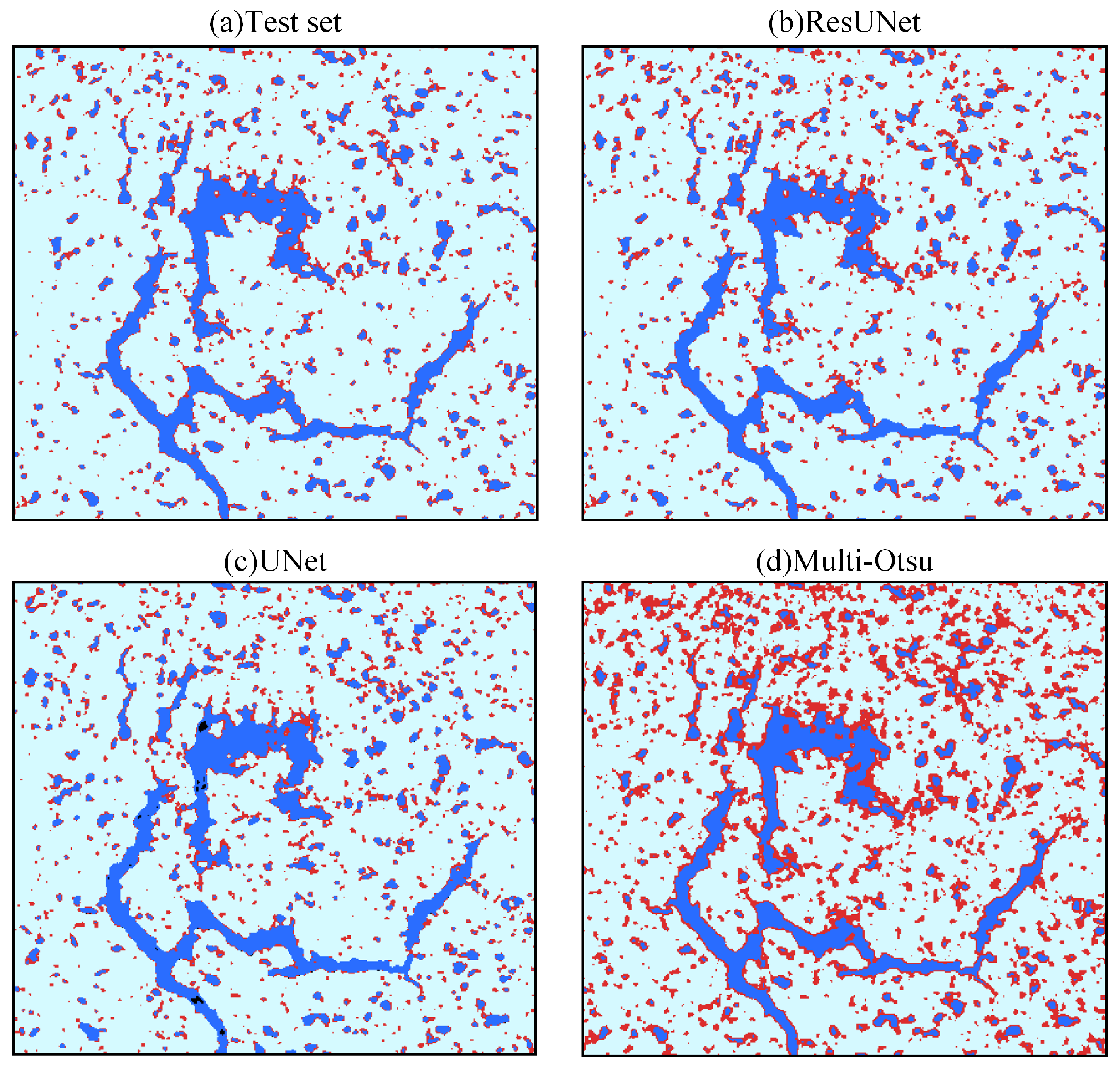
4.2. Comparative Experiments and Analysis of Segmentation Methods
4.2.1. Multi-Otsu Thresholding Method
4.2.2. Basic U-Net Architecture
4.2.3. Experimental Results and Analysis
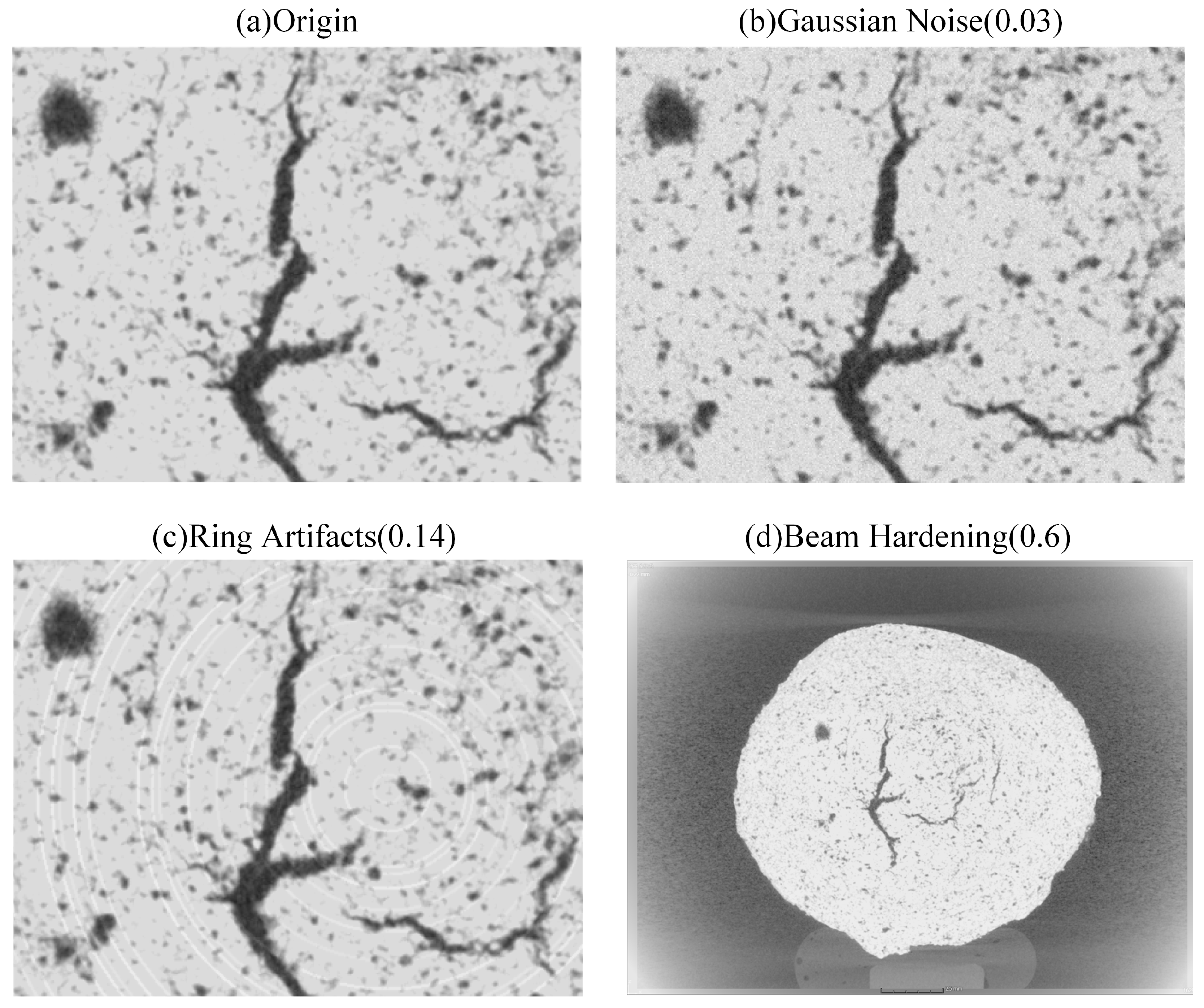
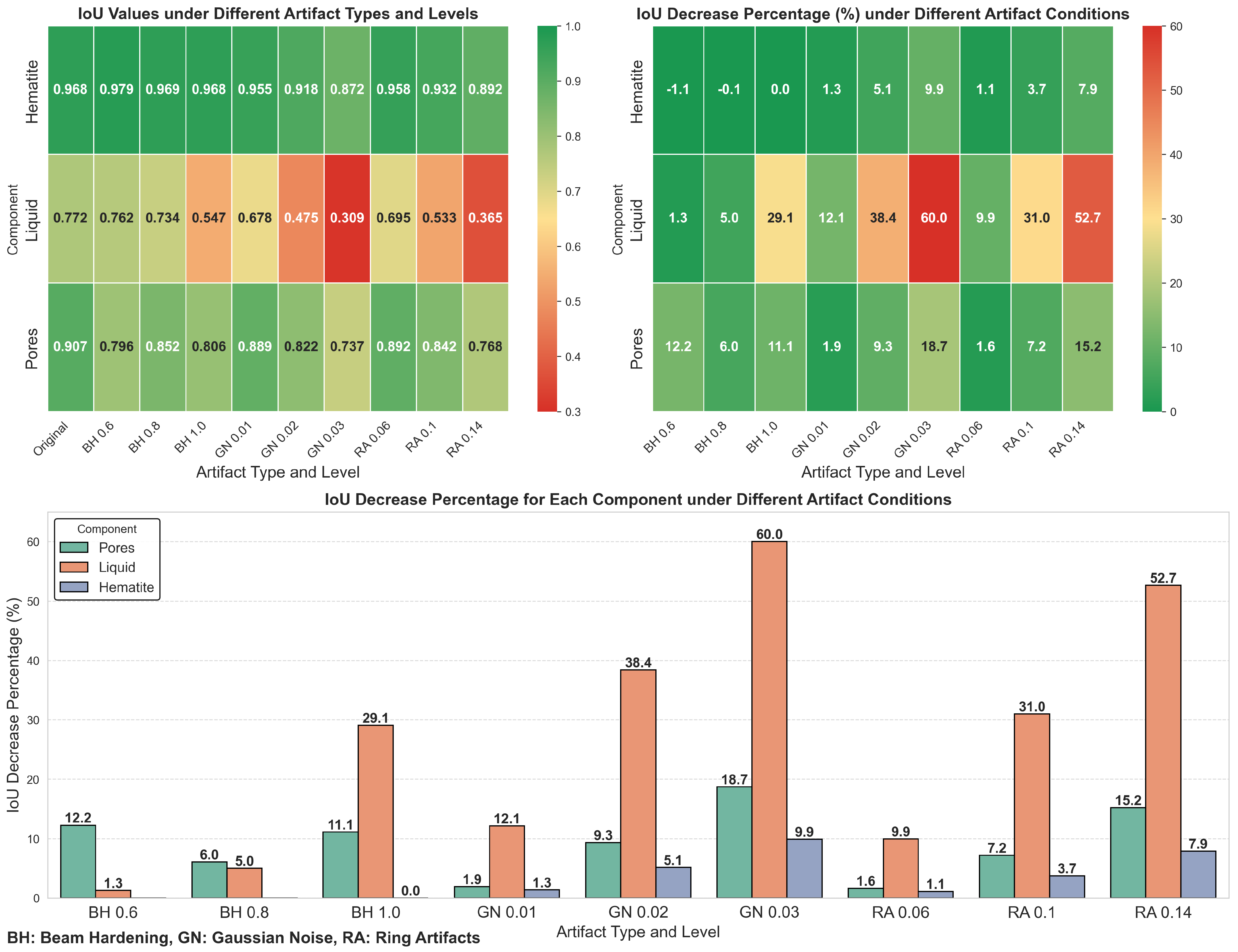
4.3. Model Robustness Analysis
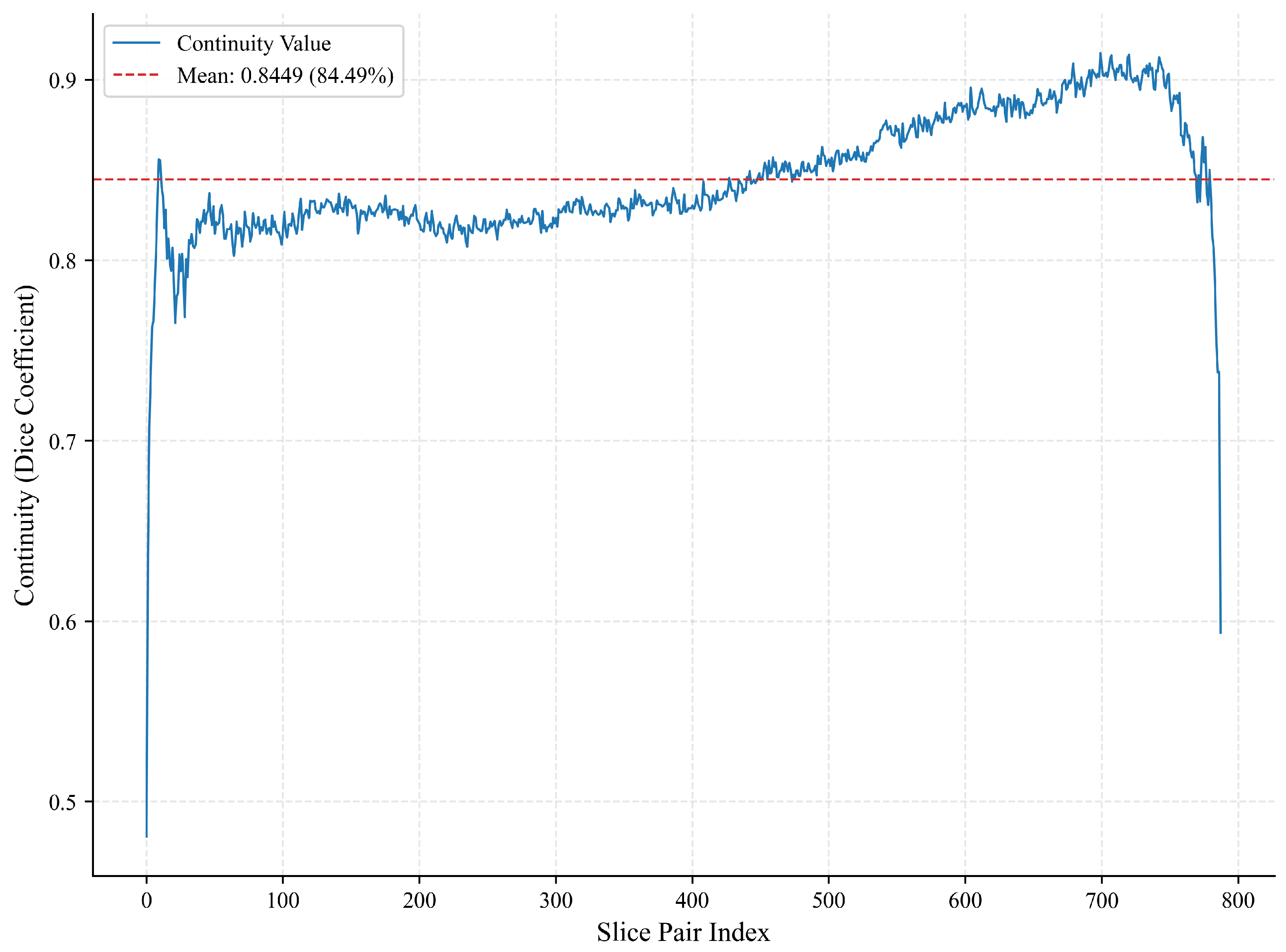
4.4. Reliability Analysis of Phase Identification
4.5. Model Application
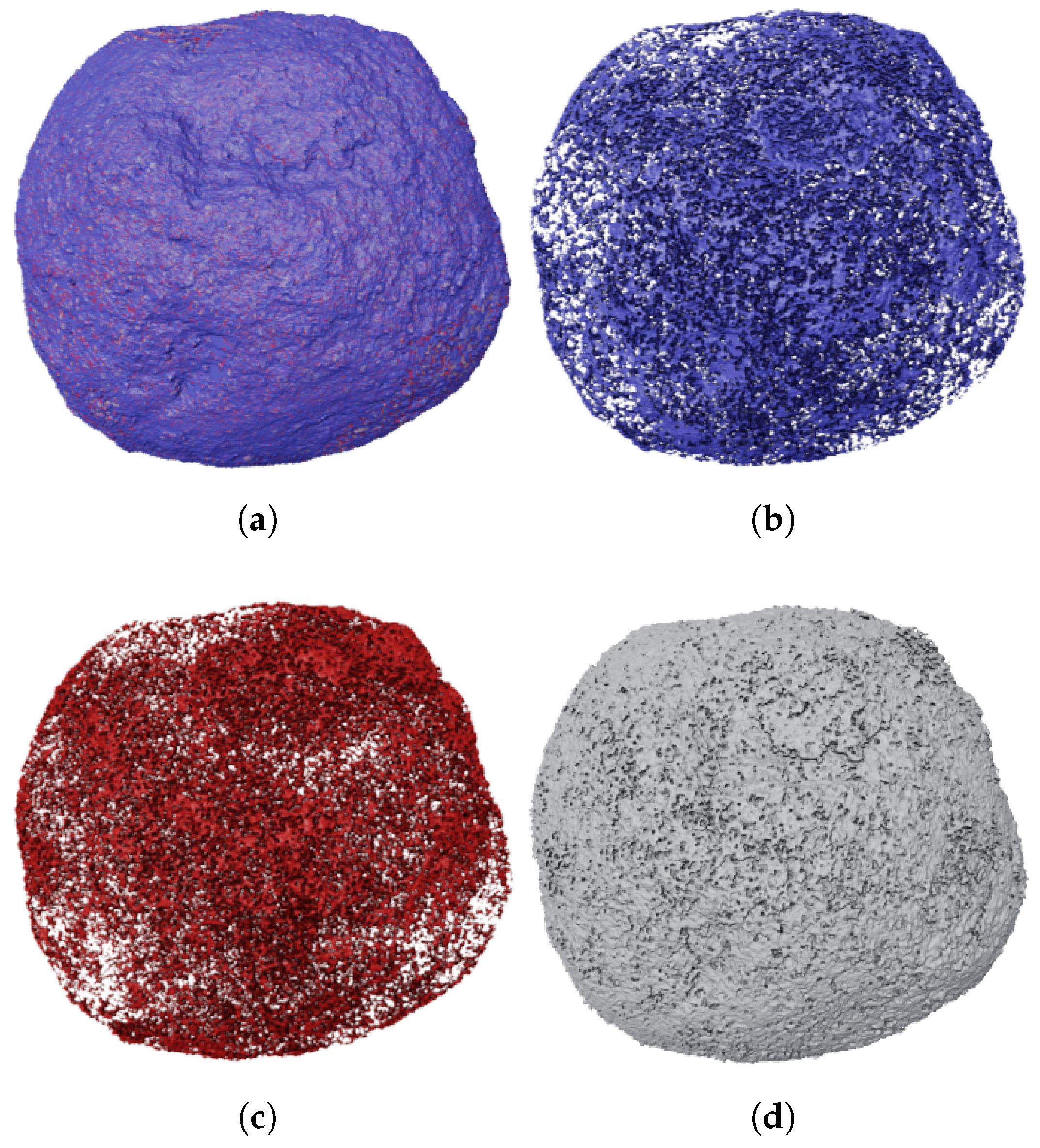
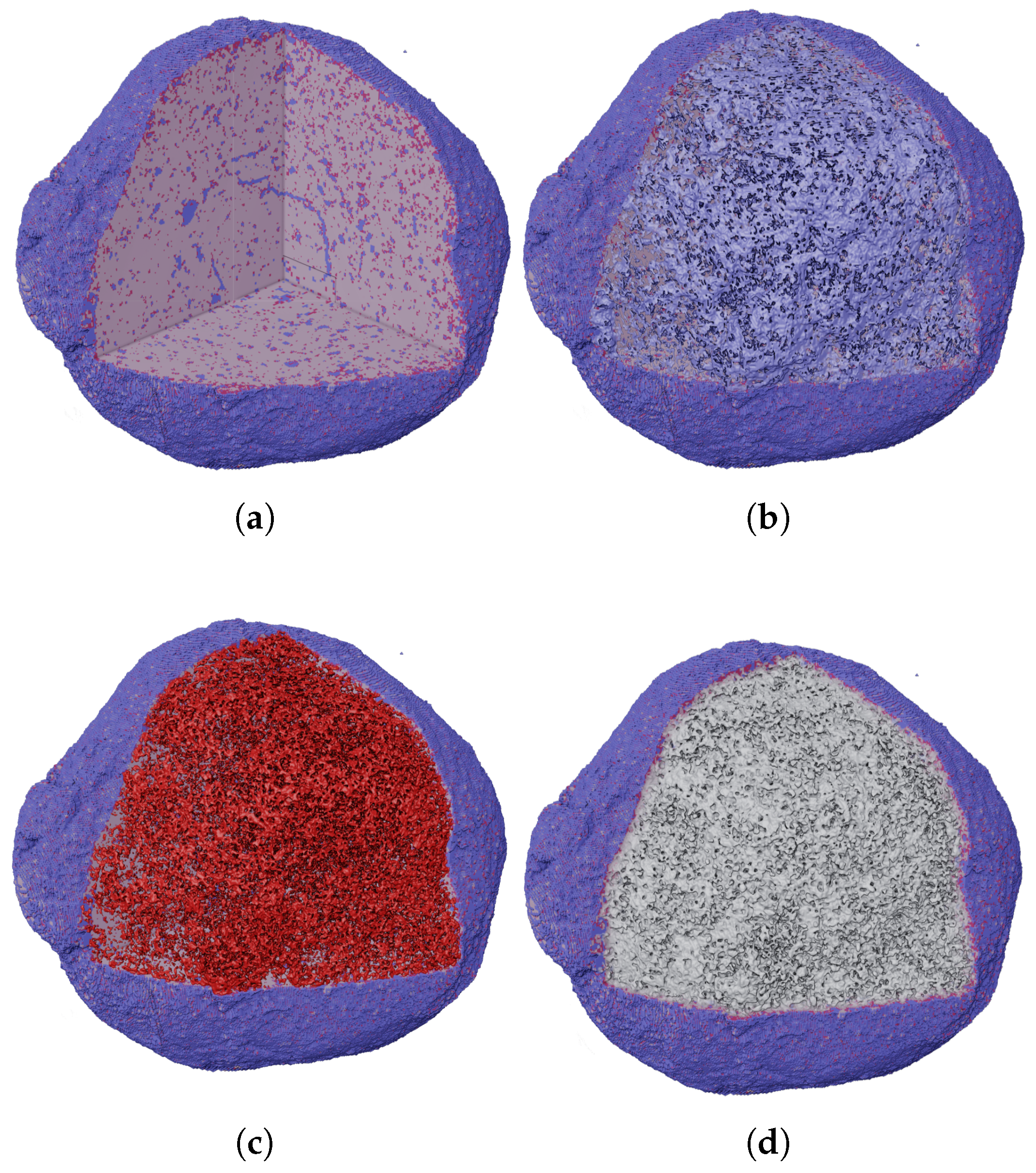
4.5.1. Three-Dimensional Reconstruction
4.5.2. Quantitative Analysis
Volume Fraction
Fractal Dimension
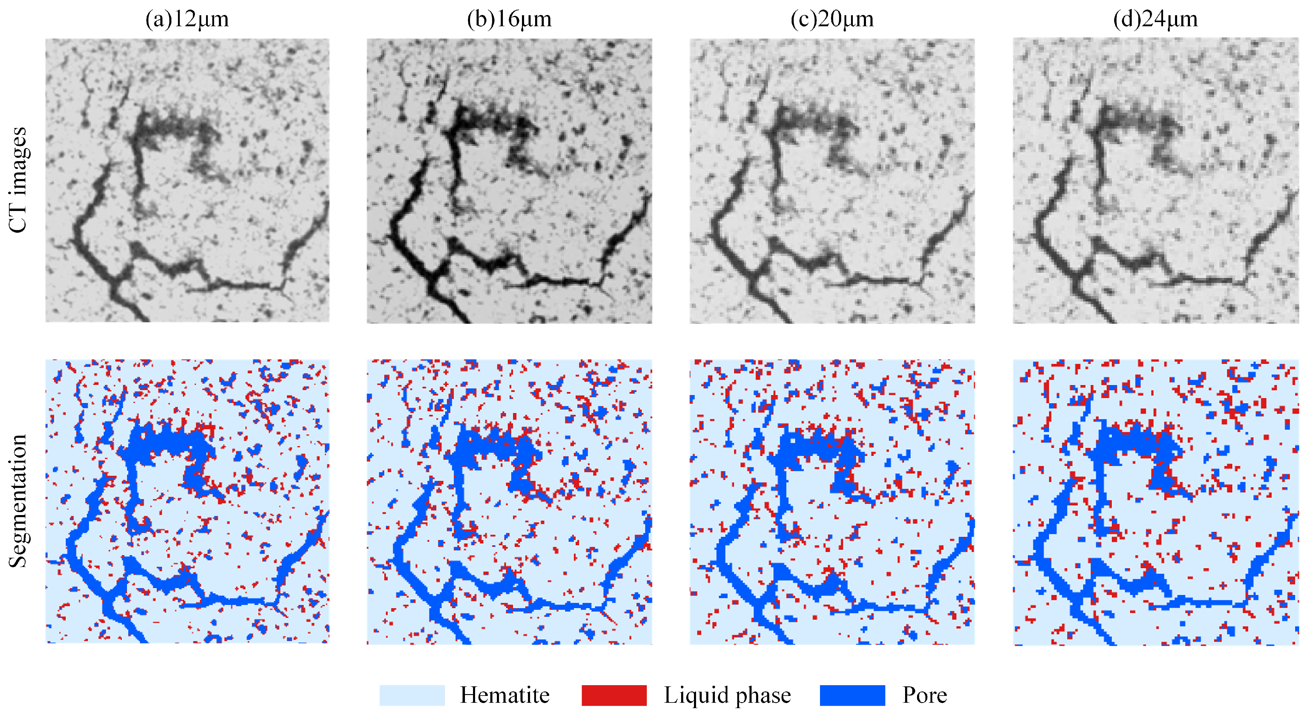
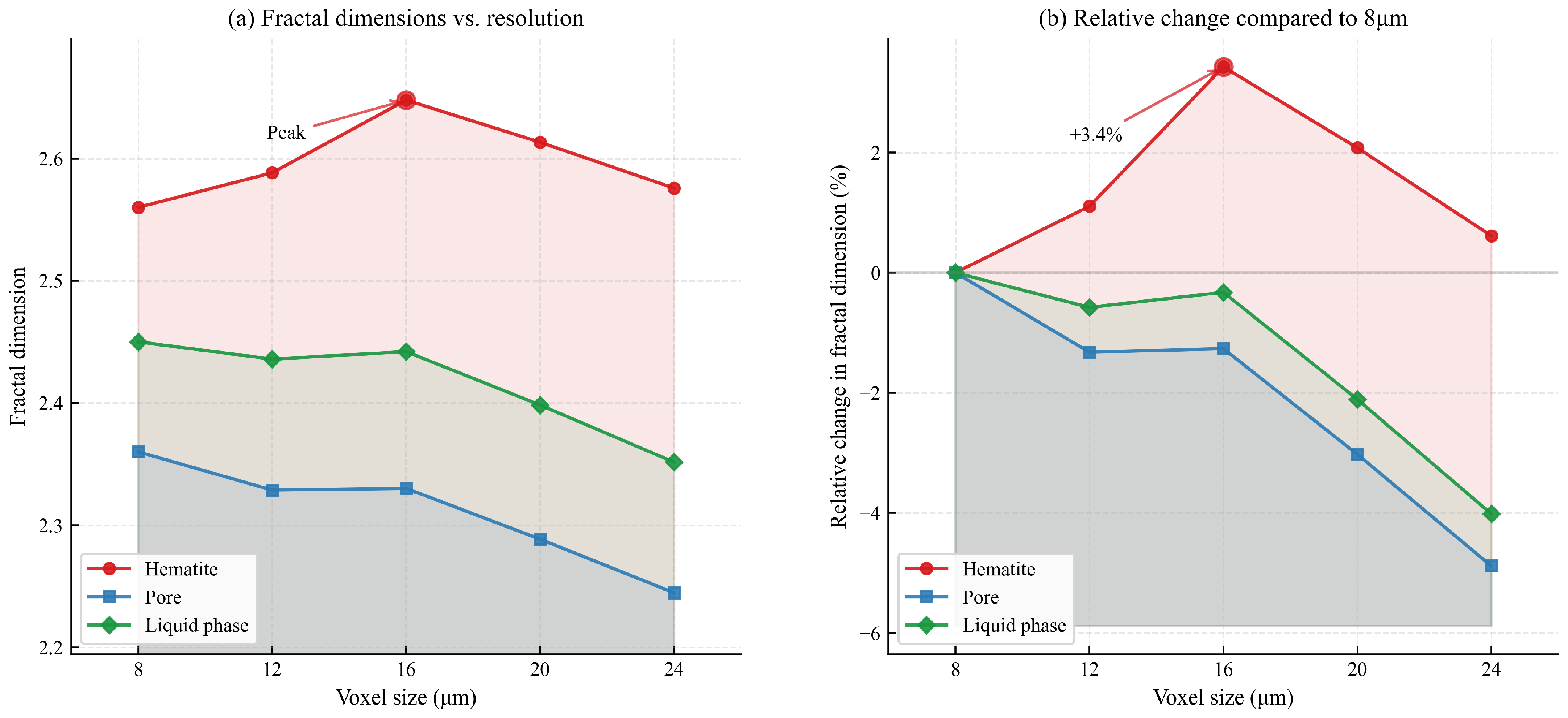
4.6. Effect of Spatial Resolution on Segmentation Results
5. Conclusions
Supplementary Materials
Author Contributions
Funding
Data Availability Statement
Acknowledgments
Conflicts of Interest
References
- de Moraes, S.L.; de Lima, J.R.B.; Ribeiro, T.R. Iron Ore Pelletizing Process: An Overview. In Iron Ores and Iron Oxide Materials; Shatokha, V., Ed.; IntechOpen: Rijeka, Croatia, 2018. [Google Scholar] [CrossRef]
- Boechat, F.O.; da Rocha, L.T.; de Carvalho, R.M.; Jung, S.M.; Tavares, L.M. Amenability of Reduced Iron Ore Pellets to Mechanical Degradation. ISIJ Int. 2018, 58, 1028–1033. [Google Scholar] [CrossRef]
- Sarda, A.; Nithesh, N.; Krishna, N.; Shrivastva, T.; Prabhu, A. Study and Implementation of Quality Improvement Techniques to Improve the Consistency in Cold Crushing Strength of Iron Ore Pellets. Asian J. Eng. Appl. Technol. 2014, 3, 10–15. [Google Scholar] [CrossRef]
- Latif, G.; Bouchard, K.; Maitre, J.; Back, A.; Bédard, L.P. Deep-Learning-Based Automatic Mineral Grain Segmentation and Recognition. Minerals 2022, 12, 455. [Google Scholar] [CrossRef]
- Wang, Y.D.; Shabaninejad, M.; Armstrong, R.T.; Mostaghimi, P. Deep neural networks for improving physical accuracy of 2D and 3D multi-mineral segmentation of rock micro-CT images. Appl. Soft Comput. 2021, 104, 107185. [Google Scholar] [CrossRef]
- Wang, Y.D.; Blunt, M.J.; Armstrong, R.T.; Mostaghimi, P. Deep learning in pore scale imaging and modeling. Earth-Sci. Rev. 2021, 215, 103555. [Google Scholar] [CrossRef]
- Filippo, M.P.; da Fonseca Martins Gomes, O.; da Costa, G.A.O.P.; Mota, G.L.A. Deep learning semantic segmentation of opaque and non-opaque minerals from epoxy resin in reflected light microscopy images. Miner. Eng. 2021, 170, 107007. [Google Scholar] [CrossRef]
- Li, C.; Wang, D.; Kong, L. Application of Machine Learning Techniques in Mineral Classification for Scanning Electron Microscopy—Energy Dispersive X-Ray Spectroscopy (SEM-EDS) Images. J. Pet. Sci. Eng. 2021, 200, 108178. [Google Scholar] [CrossRef]
- Nellros, F.; Thurley, M. Automated image analysis of iron-ore pellet structure using optical microscopy. Miner. Eng. 2011, 24, 1525–1531. [Google Scholar] [CrossRef]
- Dwarapudi, S.; Ranjan, M. Influence of Oxide and Silicate Melt Phases on the RDI of Iron Ore Pellets Suitable for Shaft Furnace of Direct Reduction Process. ISIJ Int. 2010, 50, 1581–1589. [Google Scholar] [CrossRef]
- Graça, L.M.; Lagoeiro, L.E.; Lima, R.M.F.; Barbosa, P.F.; Machado, M.M. Effect of the Morphological Types in Grinding of Iron-Ore Products. Miner. Process. Extr. Metall. Rev. 2015, 36, 324–331. [Google Scholar] [CrossRef]
- Donskoi, E.; Poliakov, A.; Manuel, J.R.; Peterson, M.; Hapugoda, S. Novel developments in optical image analysis for iron ore, sinter and coke characterisation. Appl. Earth Sci. 2015, 124, 227–244. [Google Scholar] [CrossRef]
- Guntoro, P.I.; Tiu, G.; Ghorbani, Y.; Lund, C.; Rosenkranz, J. Application of machine learning techniques in mineral phase segmentation for X-ray microcomputed tomography (µCT) data. Miner. Eng. 2019, 142, 105882. [Google Scholar] [CrossRef]
- Chen, L.; Bentley, P.; Mori, K.; Misawa, K.; Fujiwara, M.; Rueckert, D. DRINet for Medical Image Segmentation. IEEE Trans. Med Imaging 2018, 37, 2453–2462. [Google Scholar] [CrossRef] [PubMed]
- Ronneberger, O.; Fischer, P.; Brox, T. U-Net: Convolutional Networks for Biomedical Image Segmentation. In Proceedings of the Medical Image Computing and Computer-Assisted Intervention—MICCAI 2015; Navab, N., Hornegger, J., Wells, W.M., Frangi, A.F., Eds.; Springer: Cham, Switzerland, 2015; pp. 234–241. [Google Scholar] [CrossRef]
- Michen, M.; Rehak, M.; Haßler, U. Active deep learning for segmentation of industrial CT data. Tm-Tech. Mess. 2023, 90, 500–511. [Google Scholar] [CrossRef]
- Meizheng, G.; Qiong, L. Research on Dolomite Mineral Facies Segmentation Algorithm Based on Improved UNet Network Structure. In Proceedings of the 2023 42nd Chinese Control Conference (CCC), Tianjin, China, 24–26 July 2023; pp. 7817–7822. [Google Scholar] [CrossRef]
- Sun, W.; Brown, S.B.; Leach, R.K. An Overview of Industrial X-Ray Computed Tomography; NPL Report ENG 32; National Physical Laboratory: Teddington, UK, 2012. [Google Scholar]
- De Chiffre, L.; Carmignato, S.; Kruth, J.P.; Schmitt, R.; Weckenmann, A. Industrial applications of computed tomography. CIRP Ann. 2014, 63, 655–677. [Google Scholar] [CrossRef]
- Dutta, S.; Prakash, P.; Matthews, C.G. Impact of data augmentation techniques on a deep learning based medical imaging task. In Proceedings of the Medical Imaging 2020: Imaging Informatics for Healthcare, Research, and Applications; Chen, P.H., Deserno, T.M., Eds.; International Society for Optics and Photonics, SPIE: Bellingham, WA, USA, 2020; Volume 11318, p. 113180M. [Google Scholar] [CrossRef]
- Umadevi, T.; Kumar, P.; Lobo, N.F.; Prabhu, M.; Mahapatra, P.; Ranjan, M. Influence of Pellet Basicity (CaO/SiO2) on Iron Ore Pellet Properties and Microstructure. ISIJ Int. 2011, 51, 14–20. [Google Scholar] [CrossRef]
- Ioffe, S.; Szegedy, C. Batch Normalization: Accelerating Deep Network Training by Reducing Internal Covariate Shift. arXiv 2015, arXiv:1502.03167. [Google Scholar]
- He, K.; Zhang, X.; Ren, S.; Sun, J. Deep Residual Learning for Image Recognition. In Proceedings of the 2016 IEEE Conference on Computer Vision and Pattern Recognition (CVPR), Las Vegas, NV, USA, 27–30 June 2016; pp. 770–778. [Google Scholar] [CrossRef]
- Garbin, C.; Zhu, X.; Marques, O. Dropout vs. batch normalization: An empirical study of their impact to deep learning. Multimed. Tools Appl. 2020, 79, 12777–12815. [Google Scholar] [CrossRef]
- Taha, A.A.; Hanbury, A. Metrics for evaluating 3D medical image segmentation: Analysis, selection, and tool. BMC Med. Imaging 2015, 15, 29. [Google Scholar] [CrossRef]
- Zou, K.H.; Warfield, S.K.; Bharatha, A.; Tempany, C.M.; Kaus, M.R.; Haker, S.J.; Wells, W.M.; Jolesz, F.A.; Kikinis, R. Statistical validation of image segmentation quality based on a spatial overlap index1: Scientific reports. Acad. Radiol. 2004, 11, 178–189. [Google Scholar] [CrossRef]
- An, W.; Wang, C.; Zhang, H.; Bi, Z. Measuring the Formal Complexity of Architectural Curved Surfaces Based on 3D Box-Counting Dimension. Nexus Netw. J. Archit. Math. 2022, 24, 753–766. [Google Scholar] [CrossRef]


| Block | Conv Layer | Filter | Output Size | |
|---|---|---|---|---|
| Input | 512 × 512 × 1 | |||
| Encoder | Block1 | Conv1–3 | [3 × 3, 64] | 256 × 256 × 64 |
| Block2 | Conv4–6 | [3 × 3, 128] | 128 × 128 × 128 | |
| Block3 | Conv7–9 | [3 × 3, 256] | 64 × 64 × 256 | |
| Block4 | Conv10–12 | [3 × 3, 512] | 32 × 32 × 512 | |
| Bridge | Block5 | Conv13–15 | [3 × 3, 1024] | 32 × 32 × 1024 |
| Decoder | Block6 | Conv16–19 | [2 × 2, 512] | 64 × 64 × 512 |
| [3 × 3, 512] | ||||
| Block7 | Conv20–23 | [2 × 2, 256] | 128 × 128 × 256 | |
| [3 × 3, 256] | ||||
| Block8 | Conv24–27 | [2 × 2, 128] | 256 × 256 × 128 | |
| [3 × 3, 128] | ||||
| Block9 | Conv28–31 | [2 × 2, 64] | 512 × 512 × 64 | |
| [3 × 3, 64] | ||||
| Output | Conv32 | [1 × 1, 4] | 512 × 512 × 4 |
| Hyperparameter | Value |
|---|---|
| Epoch | 1500 |
| BatchSize | 24 |
| Learning Rate | 0.001 |
| Recall | Precision | F1 | IoU | |
|---|---|---|---|---|
| Pores | 0.9415 | 0.9607 | 0.9510 | 0.9065 |
| Liquid phase | 0.7497 | 0.9519 | 0.8387 | 0.7718 |
| Hematite | 0.9701 | 0.9977 | 0.9837 | 0.9679 |
| Metric | Value |
|---|---|
| M | 84.49% |
| Mdn | 83.32% |
| SD | 3.63% |
| Skewness | −1.75 |
| Kurtosis | 16.01 |
| Pores | Liquid Phase | Hematite |
|---|---|---|
| 9.599% | 12.534% | 77.867% |
| Pores | Liquid Phase | Hematite |
|---|---|---|
| 2.36 | 2.45 | 2.56 |
Disclaimer/Publisher’s Note: The statements, opinions and data contained in all publications are solely those of the individual author(s) and contributor(s) and not of MDPI and/or the editor(s). MDPI and/or the editor(s) disclaim responsibility for any injury to people or property resulting from any ideas, methods, instructions or products referred to in the content. |
© 2025 by the authors. Licensee MDPI, Basel, Switzerland. This article is an open access article distributed under the terms and conditions of the Creative Commons Attribution (CC BY) license (https://creativecommons.org/licenses/by/4.0/).
Share and Cite
Huang, Y.; Liu, W.; Mi, Z.; Wu, X.; Yang, A.; Li, J. ResUNet: Application of Deep Learning in Quantitative Characterization of 3D Structures in Iron Ore Pellets. Minerals 2025, 15, 460. https://doi.org/10.3390/min15050460
Huang Y, Liu W, Mi Z, Wu X, Yang A, Li J. ResUNet: Application of Deep Learning in Quantitative Characterization of 3D Structures in Iron Ore Pellets. Minerals. 2025; 15(5):460. https://doi.org/10.3390/min15050460
Chicago/Turabian StyleHuang, Yanqi, Weixing Liu, Zekai Mi, Xuezhi Wu, Aimin Yang, and Jie Li. 2025. "ResUNet: Application of Deep Learning in Quantitative Characterization of 3D Structures in Iron Ore Pellets" Minerals 15, no. 5: 460. https://doi.org/10.3390/min15050460
APA StyleHuang, Y., Liu, W., Mi, Z., Wu, X., Yang, A., & Li, J. (2025). ResUNet: Application of Deep Learning in Quantitative Characterization of 3D Structures in Iron Ore Pellets. Minerals, 15(5), 460. https://doi.org/10.3390/min15050460