Article

Synthesis and Characterization of Na-X Zeolite Using a Natural Opaline Diatomite Rock from SE Spain

1
Dipartimento di Scienze, Università degli Studi “G. D’Annunzio”, via dei Vestini 30, 66013 Chieti, Italy
2
Department de Mineralogia, Petrologia i Geologia Aplicada, Universitat de Barcelona, 08028 Barcelona, Spain
*
Author to whom correspondence should be addressed.
Minerals 2025, 15(3), 238; https://doi.org/10.3390/min15030238
Submission received: 23 January 2025 / Revised: 19 February 2025 / Accepted: 24 February 2025 / Published: 26 February 2025

Abstract

:
A natural diatomite was used to hydrothermally synthesise Na-X zeolite. Albacete diatomite from southern Spain (mainly constituted by opaline silica) was chemically treated with HCl and NaOH to obtain sodium silicate, a reagent necessary for zeolitic synthesis. The experimental synthesis protocol was performed at 75 °C by mixing the obtained silicatic solution with chemical reagents represented by soda and alumina. Na-X zeolite begins to appear after only 1.5 h and reaches its crystallisation climax at 11 h. Hydroxisodalite appears at 40 h. Synthesized phases were subjected to chemical-crystallographical and mineralogical-textural characterisation. The thermal behaviour and infrared response have also been investigated. The purity of the synthesized zeolite, verified through quantitative phase analysis using the combined Rietveld and reference intensity ratio methods, opens the way to a possible industrial transfer of the experimental procedure.

Graphical Abstract

1. Introduction

Zeolites are a family of about 250 synthetics and natural minerals, whose structure is basically made of a three-dimensional network of Al–Si tetrahedra arranged to form channels containing water and exchangeable alkaline or alkaline earth cations. In general, zeolites are associated with multiple technological and industrial applications such as cation exchange [1], in gas purification and separation [2], in the hydration of Portland cement [3], as matrices for pigments [4], among others.
Research into zeolites is of great industrial interest and their use is extended to several new technological applications. The group of low silica zeolites, such as Na-X, is successfully involved in ion exchange [5], adsorption [6], in gas and liquid [7] separation, dehydration of organic liquids [8], carbon dioxide capture [9], encapsulation materials for textile applications [10], as fuel cell electrolytes, [11,12], catalysis [13], heavy metal removal by sorption [14,15], among others. Na-X zeolite also plays an important role in catalysis due to its rigid and stable structure and large void spaces. In fact, the regular opening aperture of about 8 Å of these faujasite-type zeolites is large enough to accommodate the typical large molecules commonly found in gas oil during catalytic cracking and refining operations [16].
Zeolites are generally synthesized in hydrothermal environments by mixing silicatic and aluminatic solutions. Crystallization passes through the previous formation of a gel phase [17]. Usually, experimental protocols start using expensive commercial sources of silica and alumina. A continuous trend in zeolitic synthesis is the research into alternative and less expensive natural materials and cheaper industrial procedures. Therefore, there is a great economic potential for using natural substitutes for chemical reagents in zeolite synthesis.
There is a lot of literature that faces the experimental syntheses of zeolites from natural materials. Among these natural precursors, the most common are clay minerals like kaolinite [18,19,20,21,22], smectite [23], bentonite [24], attapulgite [25], and halloysite [26], but also from industrial waste as aluminosilicate residue coming from lithium extraction from spodumene [27] and rice husk ash [28,29].
Some literature reports zeolitic synthesis from tripolaceous and Diatomite rocks [30,31,32,33,34,35,36,37]. Sanhueza et al. [31,32,33] report on the synthesis of high silica zeolites from diatomites. In particular, Sanhueza et al. [31] show that the use of Diatomite rocks in the synthesis of mordenite is more economical with respect to the use of kaolin because the silica skeletons of diatoms over kaolinites are in an amorphous state. Therefore, it is not necessary to expend energy in thermal activation of the raw material like in synthesis processes involving metakaolin [38].
Sanhueza et al. [32] synthesised ZSM-5 from diatomite, exploring the effect of adding diethanolamine as a template. The same authors [33] achieved the synthesis of mesoporous materials from a diatomite in the presence of the surfactant cetyltrimethylammonium bromide (CTAB) at 110 °C and for a reaction time of 48 h.
A few works deal with the synthesis of low-silica sodium zeolites from diatomites [34,35,36,37]. Among them, Chaisena et al. [35] report the synthesis from activated diatomite by treating it with H2SO4 at 100 °C for 24 h and then calcining at 1100 °C for 5 h, and Na-P1, analcime, cancrinite and hydroxisodalite were created. Also, Rangsriwatananon et al. [39] report on the synthesis of low-silica sodium zeolites (analcime, cancrinite, Na-P1 and hydroxisodalite) from modified diatomites (6 M H2SO4 refluxed at 100 °C for 24 h followed by calcination at 1100 °C for 5 h). Du et al. [40] synthesize P zeolite at 90 °C from a diatomite treated with sodium hexametaphosphate to open pores and favour the remotion of the clay component.
Our previous work [34] represents the sole attempt at the synthesis of Na-X zeolite from diatomite; it illustrated a synthesis protocol based on the formation of Na-X zeolite from natural tripolaceous rocks and volcanic rocks. Diatomite undergoes HCl and NaOH chemical treatments to separate the silicatic solution necessary as a synthesis reagent; Na-X was found at 18 h at 80°. Surprisingly, as far as we know no other work reports the synthesis of this zeolite from diatomite in the last 20 years.
This paper aims to synthesize the low-silica Na-X zeolite through a simple and economical experimental protocol, which reduces at a minimum the treatments on raw diatomite, i.e., it avoids the very expensive activation of the diatomite. Moreover, an attempt at an economical optimization of the industrial protocols proposed in our previous work [34] is aimed for, that is by reducing temperatures and reaction times. Finally, a further and fundamental improvement of our past protocol is conducted here for the first time through the quantification of the purity of the synthesis powders. This quantification is nowadays mandatory for a possible industrial transfer of any experimental procedure, as industry requires powders pure at least at 90%.

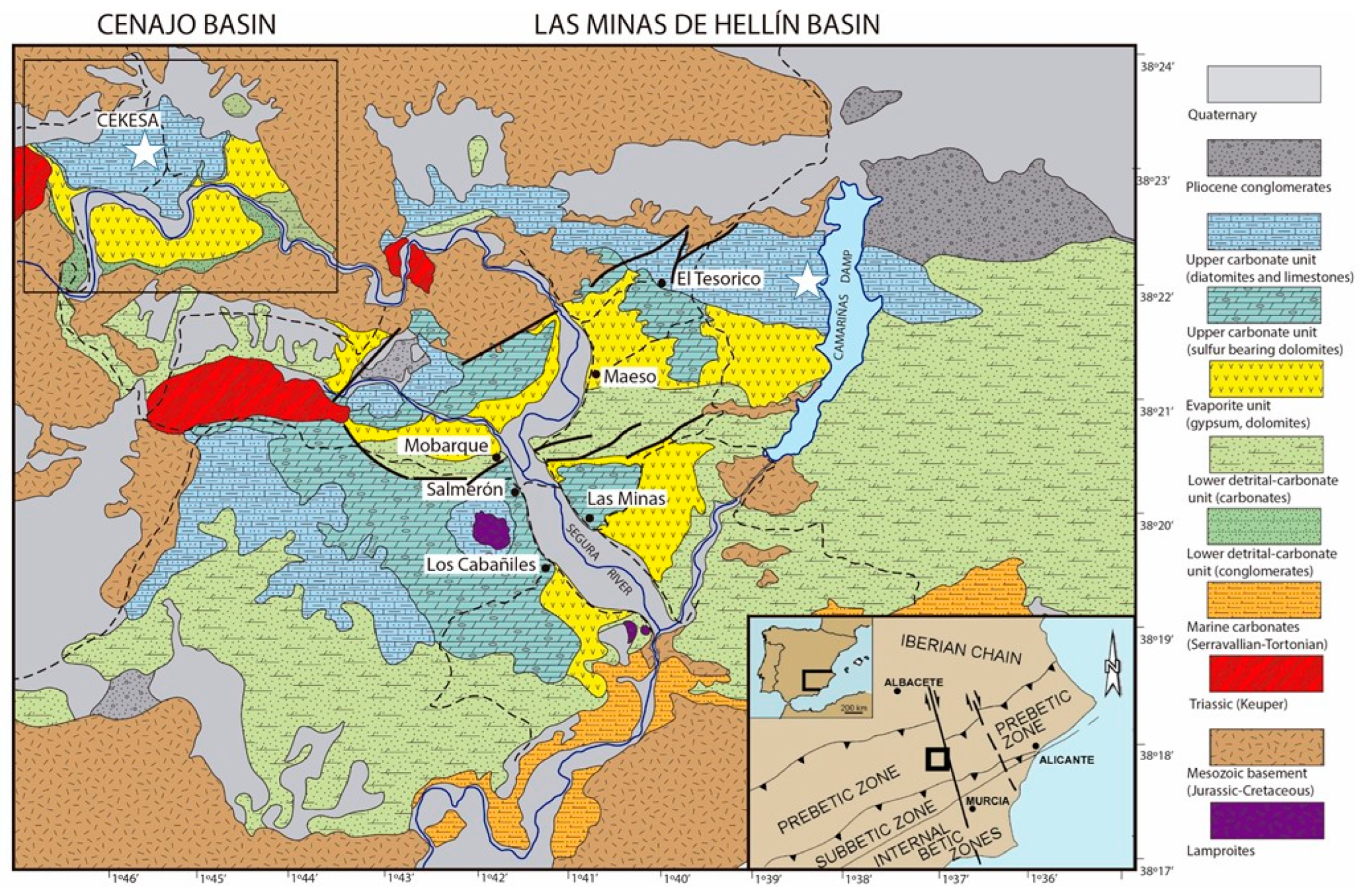
2. Diatomite Rocks: The “Albacete Diatomite”

Diatomite rocks are mainly opaline in composition and originate from the accumulation of skeletal remnants of several organisms (mainly diatoms but also other silica-rich biological remnants like radiolarian and sponges) that fix silica dissolved at very low levels in natural waters [34].
The physical and chemical properties of these rocks, i.e., high granulometric homogeneity, high porosity and low crystallinity, allow them to be used and applied in a lot of fields as high quality filters, as sorbents, as abrasives, in the cement industry (pozzolanic modalities), in the manufacture of explosives, in the food industry (quality filter of beer, wine, etc.), in cosmetics, and also as raw materials in the synthesis of zeolitic minerals or other silicates, among others.
Therefore, diatomite is a raw material of extreme versatility, and its economic value is only hampered with respect to other similar materials (i.e., the expandable ones like volcanic perlite or some clays), by transportation costs that determine the maximum economic distance; however, most forms of diatomite can be shipped and still remain competitive with alternative materials. It thus can be considered as a regional-scale resource. Spain was once the fifth largest world producer (around 110,000 t/year [41,42,43,44,45], and maintained good levels of production (around 50,000 t/year in mean along the last two decades, data from Spanish administration, Estadística Minera de España, MITECO [46]) and has huge reserves of this product. Now (data available up to 2022) following a sharp increase in the last years, the production rose to circa 90,000 t/year and Spain is the 9th largest world producer (3.5%–4% of world production) and the 2nd in importance after Denmark in the European Union, and the 1st in high-grade silica diatomite in this EU economic area. All the Spanish production is concentrated in the Albacete province. There are three main measures of diatomite quality, taking into account the silica content: the lowest (35%–50% SiO2) is mainly used as cement additive, the medium (40%–65% SiO2) is employed in other uses (i.e., fertilizer industries, filters, etc) and the highest quality is applied in more added-value uses (i.e., catalysis media). The diatomites are used as found or otherwise thermally activated at high temperature. Consequently, there is great potential for this raw material in SE Spain if a new technological use is developed locally. In fact, several researchers conducted recent studies on its potential use in the synthesis of wollastonite [47] for the ceramic industry or the pozzolanity of the high-quality silica-rich products [48].
Spanish diatomite deposits outcrop largely in the south-eastern area of the Iberian Peninsula in the foreland (Prebetic zone) of the Betic Orogen. These sedimentary rocks were deposited in the Neogene marine basins of the provinces of Alicante, Murcia, Almeria and in the Neogene lacustrine basins of Albacete province. It is in this last province where the mined deposits are concentrated. They consist of open pit quarries with homogeneous white oxidised deposits a few dozen meters thick at the surface. Other large potential deposits (the so-called “albarizas” in the Jerez de la Frontera region, which constitute most of substratum of the famous sherry vineyards) are well known in the Guadalquivir basin and have been sporadically mined in the past.
The diatomite samples analysed here come from the SE area of Albacete Province and come from different quarries: Alarcon Palacios/Ceville Pit (near Elche de la Sierra); Cekesa Pit (NW Las Minas); and Camarilla Pit (NE Las Minas) (Figure 1). Each of these quarries is placed in an independent former lacustrine basin. The Late Miocene coastal lacustrine successions (Tortonian to Messinian in age) in these basins unconformable overlie Mesozoic terrigenous and carbonate basement units or Middle Miocene marine carbonates. It is made up of three sedimentary units: (1) a lower detrital-carbonate unit, (2) an evaporite unit, and (3) an upper carbonate unit. The upper carbonate unit (up to 300 m thick) consists of alternating carbonates (dominant at the bottom of the succession, and sulphur-rich) and diatomite-rich beds. The middle part contains the mined decametric-thick diatomaceous beds. The succession ends with siliciclastic-carbonate lacustrine deposits ([49] and references therein).
Diatomite samples appear as porous and easily friable rocks made of decimetric white and grey layers (Figure 2); heterometric greenish agglomerates (clay minerals), whose diameter varies between a few millimetres to 2 cm, are occasionally present. Some stratigraphic levels show massive centimetre-sized nodules of opal. In most of outcrops and beds of the silica-rich studied samples here, the content of carbonate is low and sulphates, native sulphur and halides are merely anecdotic.

3. Methods

Diatomite samples were ground and carefully treated at 130 °C in pyrex recipients for 48 h prior to any other manipulation. The microtextural analysis of Albacete diatomite was carried out on samples mounted on metal stubs covered with carbon conductive biadhesive labels after overnight drying at 40 °C. Then, samples were observed by a scanning electron microscope (SEM), FEI QUANTA-250, coupled with SDD EDS detectors (Thermo Fisher UltraDry—Pathfinder Alpine) (ThermoFisher Scientific, Dartford, UK). Operating conditions were 15 kV and a range of variation of 9.8 to 10.1 mm in window conditions, following the procedure as explained in [21]. In addition, a strategy of double coating (graphite plus vaporised Au) in the sample treatment was chosen. Observation of the SEM images (Figure 3) confirms the presence of diatoms and a good preservation of the opaline assemblage.
The diatomite rock mineralogy was determined by X-ray powder diffraction (XRPD) through a Siemens D5000 (Bruker-Siemens, Billerica, MA, USA), using the following operative conditions: CuKα radiation (1.518), 40 kV, 40 mA, 2–45° scanning interval, step size 0.020° 2θ and counting time of 0.8 sec per step. XRPD analyses conducted on a certain number of samples from the deposit revealed a mineralogical composition mainly made of amorphous opaline silica (highly variable from sample to sample), quartz, accessory montmorillonite, and minor amounts of carbonates; some samples also contain accessory chlorite and K-micas. In order to evaluate the carbonatic content, all diatomite samples were subjected to calcimetric analysis. After some preliminary tests, we focused our attention on the AP3 sample (from the former Alarcon Palacios Pit), whose composition is made of opaline silica, quartz, calcite and montmorillonite. In particular, it is characterised by the highest content in opaline silica (visible as an amorphous bulge between 17 and 25 2θ in the XRPD spectra) and the lowest amount of carbonatic phases with respect to the other diatomite samples (Figure 4); calcimetric analysis reveals values of 8% CaCO3 for this sample.

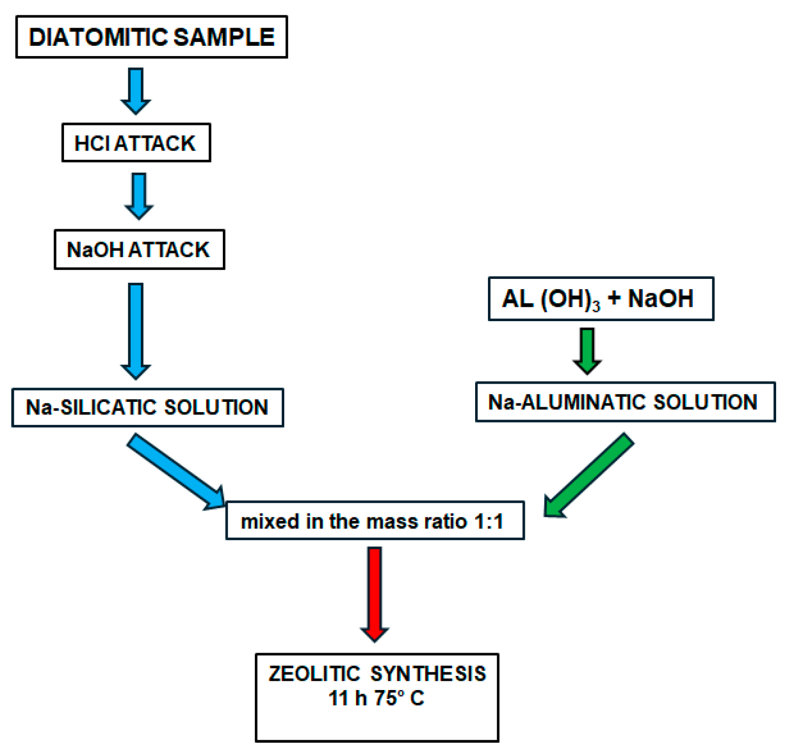
Preparation of Reagents from Natural Rocks and Zeolitic Synthesis

Figure 5 illustrates the experimental protocol executed to synthesize Na-X zeolite from the diatomite. It involved the preparation, mixing and successive hydrothermal treatment at 75 °C of a Na–alumo–-silicatic solution.
  • Silicatic solution
The AP3 diatomite sample was treated to separate the “siliceous organic” fraction. A first attack with HCl (10 mL) on the ground and powdered raw material (85 gr) separated the carbonate fraction via solution and other impurities such as clorurate compounds. The XRPD analysis showed that the recovered remnant solid fraction that remained on the filter (after solid–liquid separation) contained only montmorillonite, quartz and amorphous silica (Figure 6).
The skeletal fossil fraction, mainly constituted by amorphous silica, was separated by adding the diatomite sample (about 77 gr after the HCl attack) to 660 cc of a NaOH solution (3 M). This system was accurately mixed with a magnetic stirrer for two hours and was finally put in a Teflon container and heated in an oven at 80 °C for 24 h. At the end of this treatment, filtration of the solution separated the silicatic solution (1 Na2O − 1 SiO2 − 100 H2O) from the remnant solid and insoluble fraction (made of clay minerals and quartz). Chemical analysis of the obtained solution revealed values for Al, Fe, K and Ti lower than 0.54 ppm, 2 ppm of Mg and 2.2 ppm of Ca.
At the end of the chemical treatments, the AP3 diatomite rock composition resulted in 8 wt% of carbonates, 6 wt% of insoluble fraction (montmorillonite and quartz) and 86 wt% of remnants of diatoms and sponges.
  • Aluminatic solution
35 g of Al(OH)3 (65%) were mixed with 400 cc of NaOH (3 M). The obtained solution was then put in an oven at 100 °C for an hour and resulted in 0.6 Al2O3 − 1.2 Na2O − 100 H2O.
  • Zeolitic synthesis
Synthesis was conducted in a stainless-steel Teflon-lined reactor at autogenous pressure by mixing 100 cc of aluminatic solution with 100 cc of silicatic solution, with a resulting composition for the initial mixture of 2.2 Na2O − 0.6 Al2O3 − 1 SiO2 − 200 H2O. The system was thoroughly mixed for two hours with a magnetic stirrer to favour homogenisation of the just formed gel phase. It was then put in an oven at 75 °C.
Synthetic products were periodically sampled from the reactors, filtered from the solution, thoroughly washed with distilled water and dried in an oven at 40 °C for a day. Then they were mineralogically characterised by XRPD analysis.
Identification of zeolites and relative peak assignment were performed with reference to the following JCPDS codes: 00-038-0237 (Milton, 1959) for zeolite Na-X; 00-041-0009 (Felsche et al., 1986) for HS. Both the crystalline and amorphous phases in the synthesis powders were estimated using quantitative phase analysis (QPA) applying the combined Rietveld and reference intensity ratio (RIR) methods; corundum NIST 676a was added to each sample, amounting to 10% (according to the strategy proposed by Novembre et al.) [20] and the powder mixtures were homogenized by hand-grinding in a mortar. Data for the QPA refinement were collected in the angular range 5–110° 2 theta with steps of 0.02° and 10 s step−1, a divergence slit of 0.5° and a receiving slit of 0.1 mm. Data were processed with the GSAS software 3.0 [50] and the graphical interface [51], starting with the structural models by Olson [52] for Na-X, and Felsche et al. [53] for hydroxysodalite (HS). We refined the XRPD data with the following procedure: background parameters, zero shift, cell parameters and peak profiles as in [54].
Structural variations at high temperatures were conducted with a diffractometer Siemens (Bruker–Siemens, Billerica, MA, USA), D500 with geometry Bragg–Brentano, Cu Kα (1.518 Å), 30 KV, 30 mA, with position sensitive detector Braun PSD-50 m and monochromator Ni filter and high temperature camera ANTON PARA HTK-10.
The microtextural analysis of individual synthesised phases was performed as described above for diatomite samples.
Chemical analysis of synthesised zeolites was conducted by inductively coupled plasma (ICP-AES) emission spectrometry PerkinElmer (Waltham, MA, USA) Optima 8300. through previous Na2O2 + NaOH fusion of samples in Pt meltpots and subsequent acid solubilisation. Each sample was fused in duplicate and meltpots were cleaned each time by alkaline fusion plus an acid bath. Standard solutions were prepared each time. The standards for ICP-AES were prepared by traditional wet methods from a certified commercial traceable mist and the operating conditions were fixed as in [55]. Considering the high content of the light element Na2O, which precludes analytical interference, its composition was independently measured by X-ray Fluorescence by means of a Sequential X-ray WDXRF, Panalytical (Malvern, UK), Axios PW 4400/40 sequential spectrophotometer at Centres Cientifics i Tecnològics de la Universitat de Barcelona (CCiT-UB). The analysis strategy included the production of 3 fused pearls (original, duplicate and cleaning pearl in a Pt crucible and collector Pt dish, using LiI as a viscosity corrector) at a ratio dilution sample/lithium tetraborate 1/20. The precision and accuracy of the ICP-AES and XRF analyses were monitored using reference materials issued by the Geological Survey of Japan [56] analysed as unknown samples. See [57] for method details, analytical precision and accuracy.
The specific surface of selected synthetic phases was determined by the BET (Brunauer–Emmett–Teller) method from the data of the adsorbed volume of N2 with a Micromeritics (Norcross, GA, USA) ASAP2010 instrument that operates from 10 to 127 kPa, following the procedure as explained in Novembre et al. [34]. The density was determined by He-picnometry using an AccuPyc 1330 pycnometer. Steel standards were also provided by Micromeritics.
Thermal analyses (thermogravimetry (TG) and differential thermal analysis (DTA)) were carried out on the synthesised zeolites by means of a Mettler (Columbus, OH, USA) TGA/SDTA851e instrument (operating conditions were a 10°/min step from 30 to 1100 °C, 50 mL/min nitrogen and using an approximate sample weight of 10 mg in an Al2O3 crucible) assisted with the commercial software provided by the producer.
Some selected synthetic zeolites were studied by infrared spectrometry (IR) by means of a spectrometer Varian (Palo Alto, CA, USA) 1000 Scimitar served by a separator of KBr. A SiC filament was used as the source of IR radiation. Powder pressed pellets were prepared with a KBr/sample mass ratio of 0.5, and a pressure undergone prior to the determination of 10 t/cm2. The spectra were processed with the program OMNIC 3.1°.

4. Results and Discussion

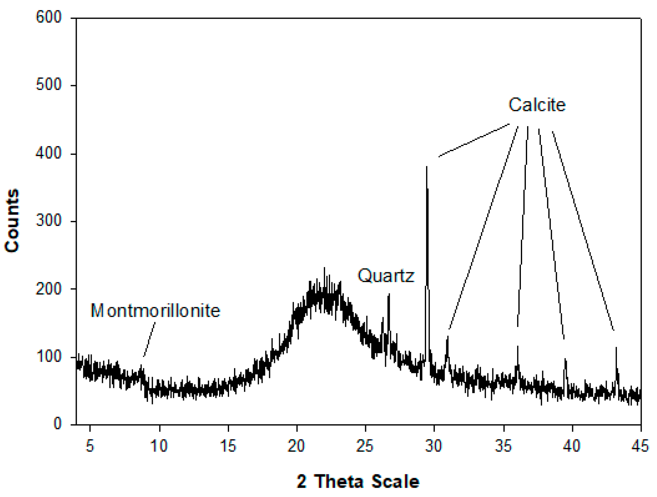
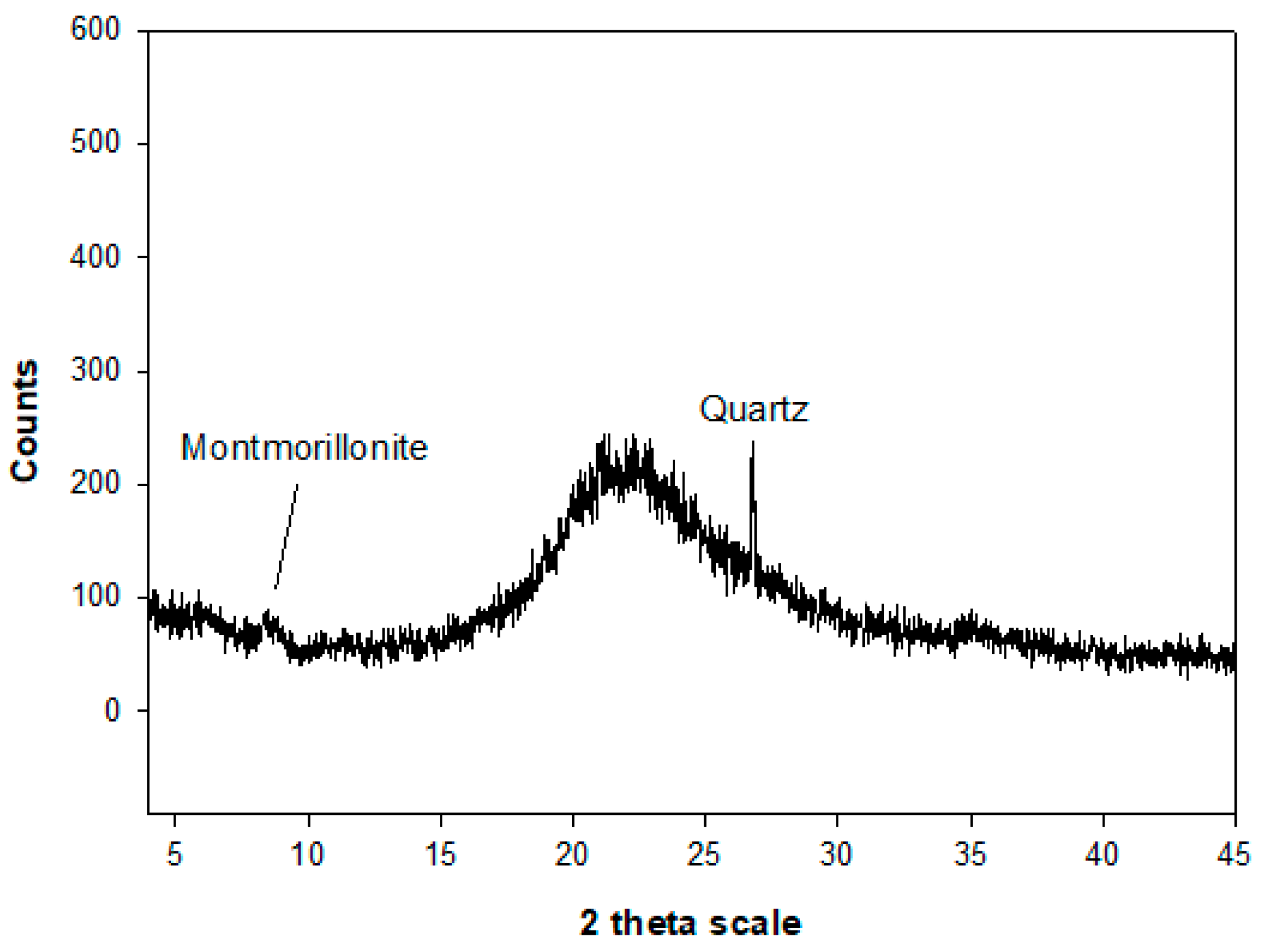
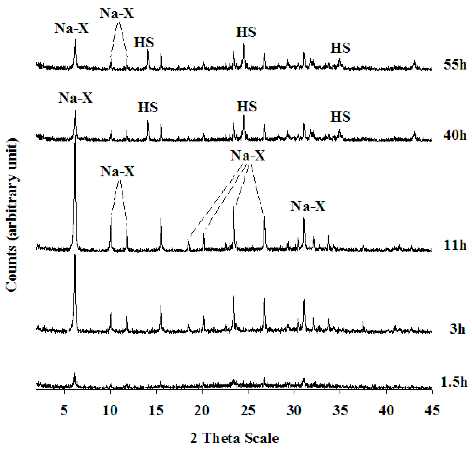
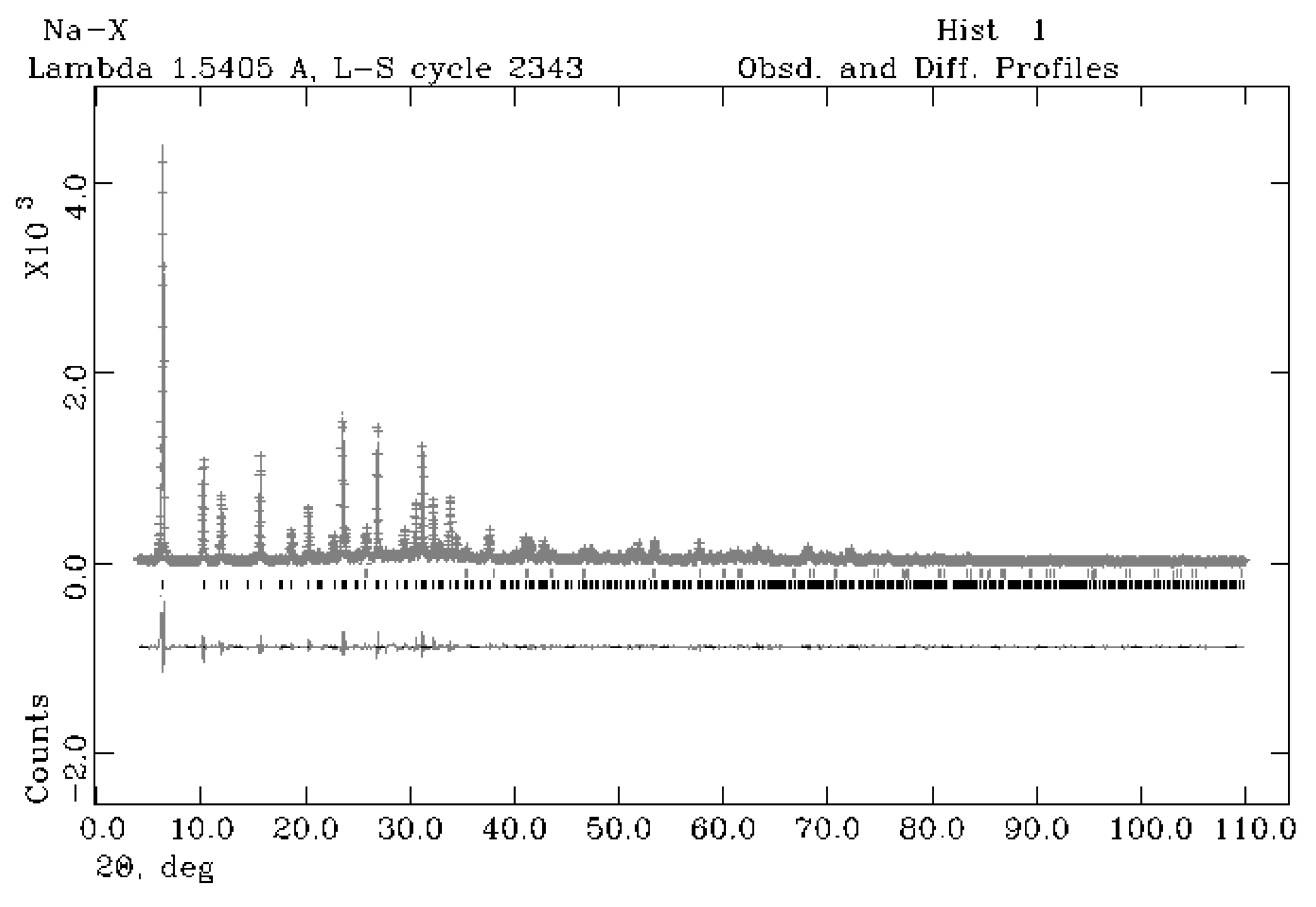
4.1. Mineralogical, Crystallographical and Chemical Characterisation of Na-X Zeolite

Na-X starts to crystallise at about 1.5 h (Figure 7). The intensity in peak heights increases over time and the crystallisation climax is reached at about 11 h. HS starts to crystallise at about 40 h.
The sample at 11 h was considered for a more detailed chemical characterisation, resulting in only Na-X. The ICP-AES analysis of this sample indicates a Si/Si+Al molar ratio of 0.56 (Table 1). This is consistent with data for the commercial zeolite Na-X 13 (UOP), which has a ratio of 56%. This value also confirms data from Stamires [58], which indicates that the Si/Si+Al ratio can vary from 52% to 73% in the X, Y and faujasite series.
Table 2 reports the results of the Rietveld refinement for sample at 11 h of the experimental run with cell parameters for the zeolite Na-X, refined with a cubic symmetry, spatial group Fd-3, a0 = 24.97(32) Å. There is good agreement with the values obtained by Stamires [58], who found that the a0 parameter varies in the X, Y and Faujasite series from 24.98 to 24.64 Å.
In Figure 8, the observed and calculated profiles and difference plot for zeolite Na-X with tick marks at the positions of the Bragg peaks are reported for the sample at 11 h of synthesis run. Results of the QPA analysis are illustrated in Table 2, resulting in 92.6% of a zeolitic phase achieved at 11 h. The Rwp (weighted profile residual factor), Rp (profile residual factor) and CHI2 (squared ratio between Rwp and Rp) parameters, used to verify the Rietveld refinement quality, are reported in Table 2 indicating a very good result.
The chemical formula of zeolite Na-X was calculated based on the chemical analysis reported in Table 1 in Na92.33Si99.91Al91.25O384·485H2O.

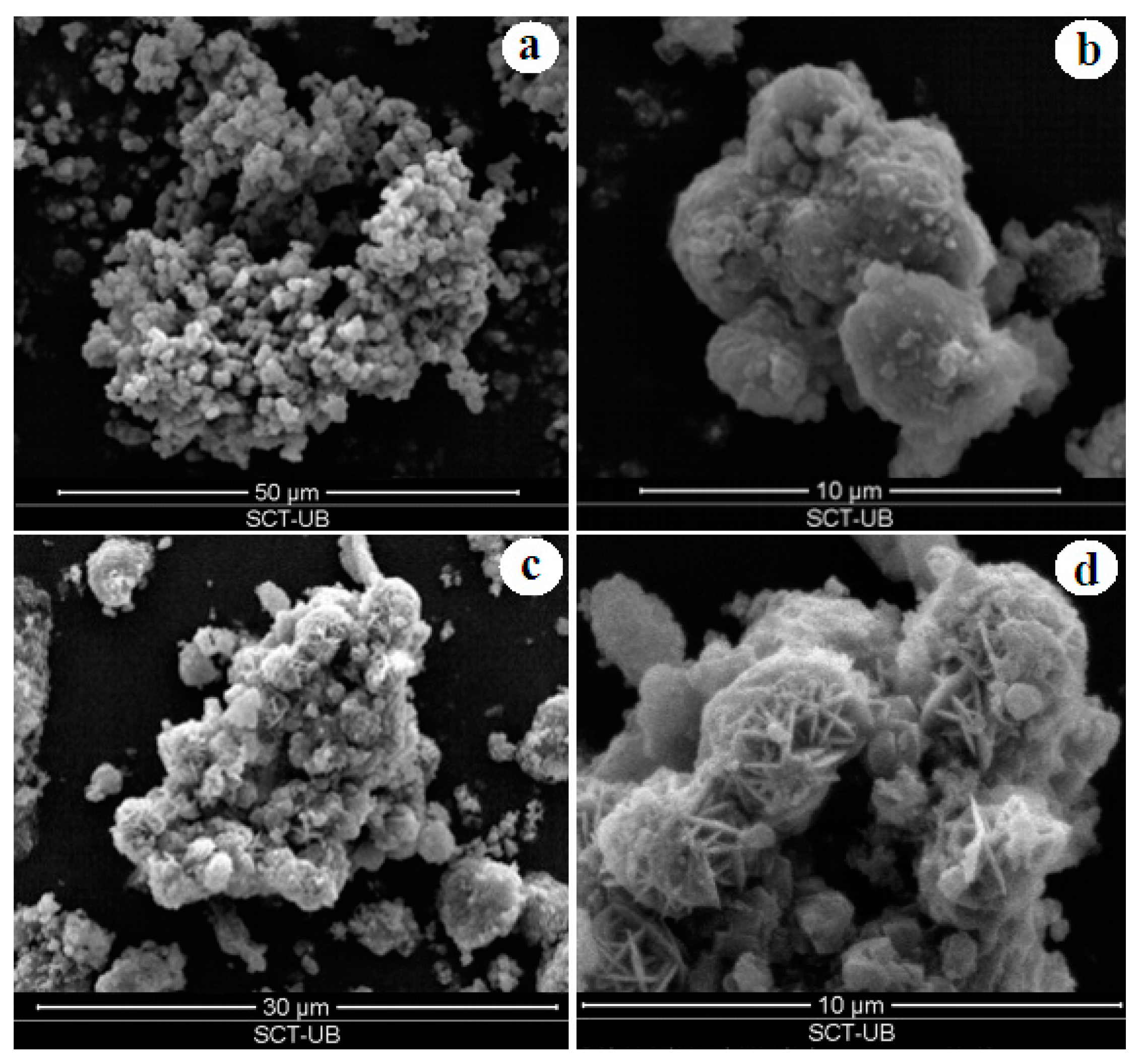
4.2. Textural and Physical Characterisation

Textural characterisation of the synthesised zeolites is illustrated in Figure 9. A SEM image of the sample at 3 h provides evidence of the incipit in the crystalline growth of the Na-X zeolite from the gel system (Figure 9a). The climax in Na-X crystallisation is reached in the sample after 11 h of the synthetic run, as testified by the gradual increase in crystallinity and grain size (Figure 9b). The Na-X zeolite exhibits aggregates characterized by rounded-shaped crystals whose dimensions reach about 4 microns after 11 h. Evidence of a new synthetic phase (HS) in crystal aggregates of lepispheric morphology is seen after 40 h (Figure 9c); an increase in HS grain size (about 4 microns) is registered in the 55h sample (Figure 9d).
Physical characterisation of the sample at 11 h shows a specific surface of 354.19 m2/g; the value is consistent with data on naturally derived Na-X zeolites [25]. A density value was found of 1.91 gr/cm3, which is also in the range of the published data for this zeolite [14,29] and similar to the certified density of a commercial zeolite (1.950 gr/cm3, Na-X-13X UOP zeolite).

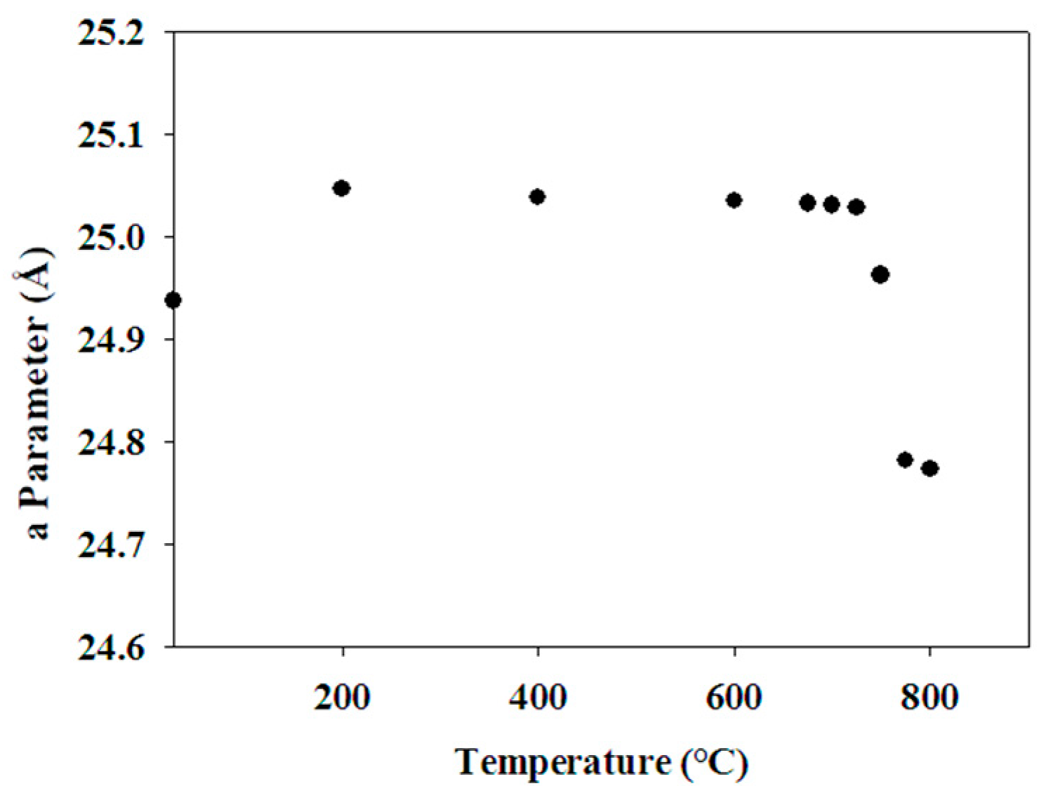
4.3. Thermal Behaviour

The thermal behavior of the sample at 11 h reveals continuous water loss between 100 °C and 900 °C (Figure 10). In particular, water loss referred to the super cage and the sodalite cage is seen between 100 and 400 °C. Near to 960 °C, there is an exothermic peak that corresponds to nepheline crystallisation. The TG curve reveals a weight loss of 2.50 mg corresponding to 24.4% of water.
The XRD study at high temperatures was used to analyse the thermal modifications of the cell lattice of the synthesised Na-X zeolite. In the sample at 11 h, zeolitic breakdown begins at 800 °C and nepheline crystallisation starts at 850 °C (Figure 11). Thus 800 °C marks the stability limits of the synthesised Na-X zeolite.
The a0 parameter modifications due to water loss with increase in temperature were applied to the sample at 11 h (Figure 12). A slight lattice expansion at 200 °C can be related to vapour water pressure. Then a0 cell value seems to rest to a constant value till about 750 °C. After this a sharp contraction of a0 parameter value is testified till 800 °C, when the Na-X structure breakdown begins.

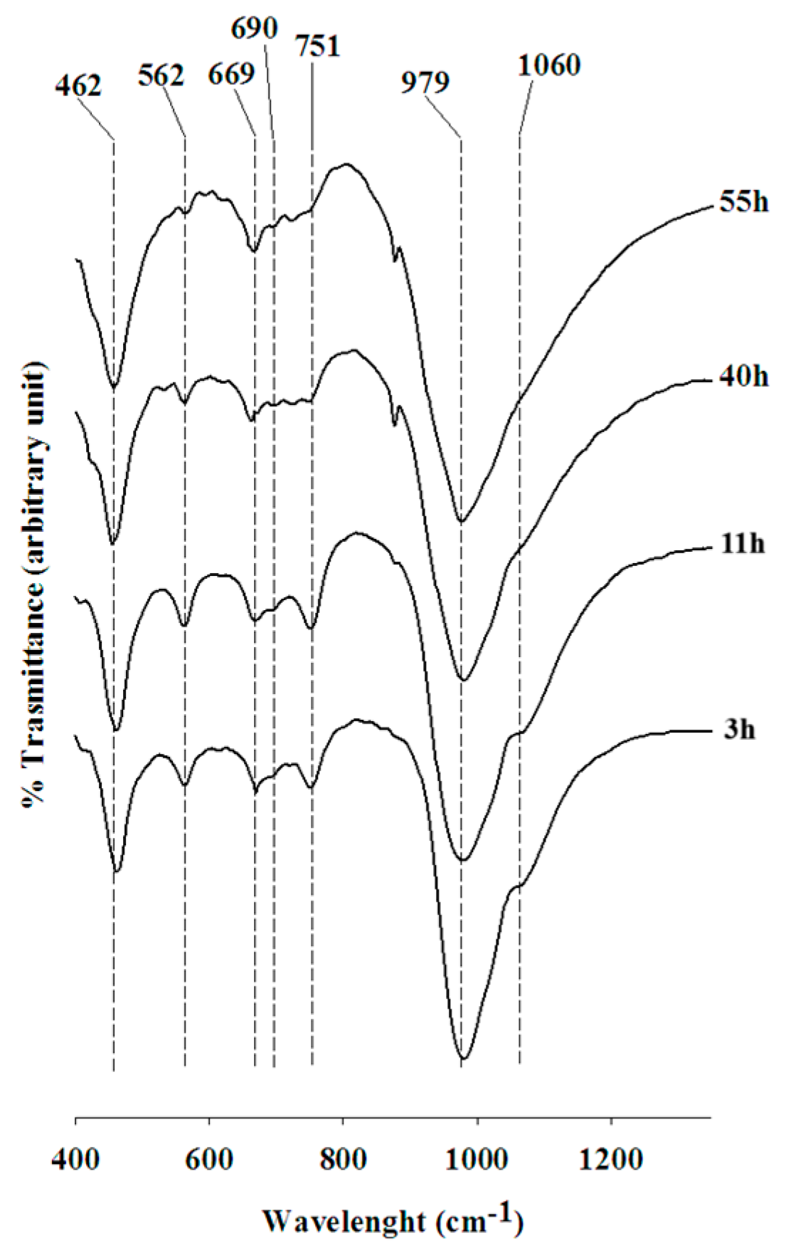
4.4. IR Spectroscopy

Figure 13 shows the results of the IR analyses performed on the experimental run. The values of the absorption bands found here for the Na-X phase are compiled in Table 3 together with known data for Na-X [34,59].
The differences in composition of the synthesised products evidenced by the XRPD spectra are also testified by the IR curves. The IR bands, in fact, progressively underwent changes over time during the crystallisation run.
The first two samples at 3 and 11 h correspond to Na-X, while the sample at 40 and 55 h refers to Na-X + HS (Figure 13). A substantial change in the morphology of the IR adsorption bands of the samples at 3 and 11 h is evidenced when compared to the sample at 40 and 55 h. Wavelength values (cm−1) of the adsorption bands of Na-X are indicated in Figure 13 and discussed below.
In the region between 1250 and 950 cm−1, related to the inner bonds of the tetrahedral asymmetric stretching zone and the asymmetric stretching of external bonds in the tetrahedral zone [54], the main band is located at 979 cm−1. This band is asymmetric due to the overlap with another peak at 1060 cm−1. In the spectral zone 750–650 cm−1, relative to the symmetric stretch region, there are three poorly defined bands at 751, 669 and 690 cm−1. In the spectrum region of 650 to 500 cm−1, corresponding to the vibration zone of the double-ring bonds, there is a band at 562 cm−1. In the sector 500–420 cm−1, related to the deformation vibrations of the O-T-O bond [54], there is evidence of a band at 462 cm−1. The position of all bands is in good agreement with bibliographic data (Table 3).

5. Conclusions

A natural, relatively cheap and abundant raw geologic material, Albacete diatomite, is exploited here in the synthesis of Na-X zeolite. The experimental protocol presented concerns the extraction of biogenic amorphous silica from the diatomite rock and its subsequent use as a chemical reagent in zeolitic synthesis. The procedure is fast and quite simple compared to the methods usually applied by the industry, which require more expensive chemical reagents. In detail, the diatomite is subjected to an attack with HCl followed by a treatment with NaOH in order to separate a silicatic solution; this is mixed in a 1:1 ratio with an aluminatic solution prepared from chemical reagents. The synthesis from the solution is performed under hydrothermal conditions at 75 °C.
Na-X zeolite begins to appear after only 1.5 h from the beginning of the experimental run and it reaches its crystallisation climax at 11 h. Then substitution by another phase, the HS phase, begins at about 40 h. The range of existence of the zeolite as a pure and isolated phase is quite wide, from 11 to about 40 h, when the appearance of HS occurs. Crystals at 11 h shows dimensions around 5 microns, a specific surface of 354.19 m2/g, a density of 1.91 gr/cm3, a Si/Si+Al molar ratio of 0.56, a chemical formula of Na92.33Si99.91Al91.25O384·485H2O with 24.4% of water. The thermal and infrared behaviours are comparable to those reported for commercial Na-X. The a0 cell value, calculated in 24.97(32) Å seems to rest at a constant value until about 750° C, when the structure breaks down due to the transformation in nepheline.
When comparing the procedure with our previous attempt [34] referring to the synthesis of Na-X zeolite from an analogue starting material (tripolaceous italian rock), temperature of synthesis, time relative to the first appearance and crystallization climax for Na-X indicate a kinetic improvement and consequently a better result in terms of economy of the procedure. In particular, the synthesis temperature drops by 5 degrees, the time to the first appearance is shortened by 4.5 h and the climax of crystallization occurs 7 h early. Finally, a further and crucial improvement of our past protocol consists in the quantification of the purity of the zeolite. The QPA analysis gives a purity of 92.6% so an industrial adaptation of this protocol seems to be highly plausible.

Author Contributions

Conceptualization, D.N. and D.G.; Data curation, D.N. and D.G.; Investigation, D.N. and D.G.; Methodology, D.N. and D.G.; validation, D.N. and D.G.; formal analysis, D.N. and D.G.; investigation, D.N. and D.G.; resources, D.N. and D.G.; data curation, D.N. and D.G.; Supervision, D.N. and D.G.; Writing—original draft, D.N.; Writing—review and editing, D.G. All authors have read and agreed to the published version of the manuscript.

Funding

This research received funding from Spanish projects CGL2011-28022 and PID 2020-118999GB-100.

Data Availability Statement

The original contributions presented in the study are included in the. article: further inquiries can be directed to the corresponding author.

Acknowledgments

The authors greatly acknowledge the technical staff at Universitat of Barcelona (CCiT-UB) and Chieti for their help during the development of the work.

Conflicts of Interest

The authors declare no conflicts of interest.

References

  1. Beiersdorfer, R.E.; Ming, D.W.; Galindo, C. Solubility and cation exchange properties of zeoponic substrates. Microp. Mesopor. Mat. 2003, 61, 231–247. [Google Scholar] [CrossRef]
  2. Ackley, M.W.; Rege, S.U.; Saxena, H. Application of natural zeolites in the purification and separation of gases. Microp. Mesopor. Mat. 2003, 61, 25–42. [Google Scholar] [CrossRef]
  3. Perraki, T.; Kakali, G.; Kontoleon, F. The effect of natural zeolites on the early hydration of Portland cement. Microp. Mesopor. Mat. 2003, 61, 205–212. [Google Scholar] [CrossRef]
  4. Kowalak, S.; Jankowska, A. Application of zeolites as matrices for pigments. Microp. Mesopor. Mat. 2003, 61, 213–222. [Google Scholar] [CrossRef]
  5. Chang, H.L.; Shih, W.H. Synthesis of Zeolites A and X from Fly Ashes and TheirIon-Exchange Behavior with Cobalt Ions. Ind. Eng. Chem. Res. 2000, 39, 4185–4191. [Google Scholar] [CrossRef]
  6. Gesikiewicz-Puchalska, A.; Zgrzebnicki, M.; Michalkiewicz, B.; Kałamaga, A.; Narkiewicz, U.; Morawski, A.W.; Wrobel, R. Changes in Porous Parameters of the Ion Exchanged X Zeolite and Their Effect on CO2 Adsorption. Molecules 2021, 26, 7520. [Google Scholar] [CrossRef]
  7. Medykowska, M.; Wi´sniewska, M.; Chibowski, S. Fly Ash-Based Na-X Zeolite Application in Separation Process of Bovine Serum Albumin from Aqueous Solution in the Presence of Organic Substances with Anionic Character. Materials 2023, 16, 5201. [Google Scholar] [CrossRef]
  8. Zhou, H.; Korelskiy, D.; Leppajarvi, T.; Grahn, M.; Tanskanen, J.; Hedlund, J. Ultrathin zeolite X membranes for pervaporation dehydration of ethanol. J. Membr. Sci. 2012, 399, 106–111. [Google Scholar] [CrossRef]
  9. Li, X.S.; Remias, J.E.; Neathery, J.K.; Liu, K.L. NF/RO faujasite zeolite membrane- ammonia absorption solvent hybrid system for potential post-combustion CO2 capture application. J. Membr. Sci. 2011, 366, 220–228. [Google Scholar] [CrossRef]
  10. Perez, E.; Martin, L.; Rubio, C.; Urieta, J.S.; Piera, E.; Caballero, M.A.; Tellez, C.; Coronas, J. Encapsulation of alpha-Tocopheryl acetate into Zeolite Y for textile application. Ind. Eng. Chem. Res. 2010, 49, 8495–8500. [Google Scholar] [CrossRef]
  11. Zhang, Z.H.; Desilets, F.; Felice, V.; Mecheri, B.; Licoccia, S.; Tavares, A.C. On the proton conductivity of Nafion-Faujasite composite membranes for low temperature direct methanol fuel cells. J. Power Sources 2011, 196, 9176–9187. [Google Scholar] [CrossRef]
  12. Felice, V.; Tavares, A.C. Faujasite zeolites as solid electrolyte for low temperature fuel cells. Solid State Ion. 2011, 194, 53–61. [Google Scholar] [CrossRef]
  13. Kulawong, S.; Youngjan, S.; Khemthong, P.; Chanlek, N.; Wittayakun, J.; Osakoo, N. Magnesium Impregnated on NaX Zeolite Synthesized from Cogon Grass Silica for Fast Production of ructose via Microwave-Assisted Catalytic Glucose Isomerization. Catalysts 2021, 11, 981. [Google Scholar] [CrossRef]
  14. Baši´c, A.; Penga, Ž.; Penga, J.; Kuzmani´c, N.; Svilovi´c, S. Zeolite NaX Mass and Propeller Agitator Speed Impact on Copper Ions Sorption. Processes 2023, 11, 264. [Google Scholar] [CrossRef]
  15. Ezzeddine, Z.; Batonneau-Gener, I.; Pouilloux, Y.; Hamad, H.; Saad, Z. Synthetic Nax Zeolite as a Very Efficient Heavy Metals Sorbent in Batch and Dynamic Conditions. Colloids Interfaces 2018, 2, 22. [Google Scholar] [CrossRef]
  16. Wang, C.; Zhou, J.; Wang, Y.; Yang, M.; Li, Y.; Meng, C. Synthesis of zeolite X from low-grade bauxite. J. Chem. Technol. Biotechnol. 2013, 88, 1350–1357. [Google Scholar] [CrossRef]
  17. Breck, D.W. Zeolite molecular sieves. In Structure, Chemistry and Use; Robert E. Krieger Publishing Company: Malabar, FL, USA, 1984; p. 771. [Google Scholar]
  18. Aznar, A.J.; La Iglesia, A. Obtención de zeolitas a partir de arcillas aluminosas españolas. Boletín Geol. Y Min. 1985, 96, 541–549. [Google Scholar]
  19. Sanhueza, V.; Kelm, U.; Cid, R. Synthesis of molecular sieves from Chilean kaolinites: I. synthesis of NaA type zeolites. J. Chem. Technol. Biotechnol. 1999, 74, 358–363. [Google Scholar] [CrossRef]
  20. Novembre, D.; Gimeno, D.; Del Vecchio, A. Improvement in the synthesis conditions and studying the physicochemical properties of the zeolite Li-A (BW) obtained from a kaolinitic rock. Sci. Rep. 2020, 10, 5715–5723. [Google Scholar] [CrossRef]
  21. Novembre, D.; Gimeno, D.; Del Vecchio, A. Synthesis and characterization of Na-P1 (GIS) zeolite using a kaolinitic rock. Sci. Rep. 2021, 11, 4872–4883. [Google Scholar] [CrossRef]
  22. Novembre, D.; Gimeno, D. Synthesis and characterization of analcime (ANA) zeolite using a kaolinitic rock. Sci. Rep. 2021, 11, 13373–13382. [Google Scholar] [CrossRef]
  23. Abdmeziem-Hamoudi, K.; Siffert, B. Synthesis of molecular sieve zeolites from a smectite- type clay material. Appl. Clay Sci. 1989, 4, 1–9. [Google Scholar] [CrossRef]
  24. De Lucas, A.; Uguina, M.A.; Covian, I.; Rodriguez, L. Synthesis of 13X zeolite from calcined kaolin and sodium silicates for use in detergents. Ind. Eng. Chem. Res. 1992, 31, 2134–2140. [Google Scholar] [CrossRef]
  25. Feng, M.; Shi, Z.; Tong, Y.; Zhang, K. Facile Synthesis of Zeolite NaX from Natural Attapulgite Clay for Pb2+ Adsorption. Chemistry 2024, 6, 1217–1229. [Google Scholar] [CrossRef]
  26. Gualtieri, A.F. Synthesis of sodium zeolites from a natural halloysite. Phys. Chem. Miner. 2001, 28, 719–728. [Google Scholar] [CrossRef]
  27. Ibsaine, F.; Azizi, D.; Dionne, J.; Tran, L.H.; Coudert, L.; Pasquier, L.-C.; Blais, J.-F. Conversion of Aluminosilicate Residue Generated from Lithium Extraction Process to NaX Zeolite. Minerals 2023, 13, 1467. [Google Scholar] [CrossRef]
  28. Novembre, D.; Gimeno, D.; Marinangeli, L.; Tangari, A.C.; Rosatelli, G.; Ciulla, M.; di Profio, P. Rice Husk as Raw Material in Synthesis of NaA (LTA) Zeolite. Molecules 2024, 29, 4396. [Google Scholar] [CrossRef]
  29. Novembre, D.; Gimeno, D.; Marinangeli, L.; Tangari, A.C.; Rosatelli, G.; Ciulla, M.; di Profio, P. Synthesis and Characterization of Na-P1 (GIS) Zeolite Using Rice Husk. Molecules 2024, 29, 5596. [Google Scholar] [CrossRef]
  30. Takaaki, W.; Mioko, H.; Keiko, K.; Hiroji, I.; Osamu, T.; Kazuhiko, I.; Takashi, N.; Downs, R.T.; Rakovan, J.F. Zeolite synthesis from paper ash at low temperature (90 °C) with addition of diatomite. J. Hazard. Mater. 2006, B132, 244–252. [Google Scholar]
  31. Sanhueza, V.; Kelm, U.; Cid, R. Synthesis of mordenite from diatomite: A case of zeolite synthesis from natural material. J. Chem. Technol. Biotechnol. 2003, 78, 485–488. [Google Scholar] [CrossRef]
  32. Sanhueza, V.; Kelm, U.; Cid, R.; Lòpez-Escobar, L. Syntesis of ZSM-5 from diatomite: A case of zeolite syntesis from a natural material. J. Chem. Technol. Biotechnol. 2004, 79, 686–690. [Google Scholar] [CrossRef]
  33. Sanhueza, V.; Lòpez-Escobar, L.; Kelm, U.; Cid, R. Syntesis of a mesoporous material from two natural sources. J. Chem. Technol. Biotechnol. 2006, 81, 614–617. [Google Scholar] [CrossRef]
  34. Novembre, D.; Di Sabatino, B.; Gimeno, D.; Garcia Valles, M.; Martinez-Manent, S. Synthesis of Na-X zeolites from tripolaceous deposits (Crotone, Italy) and volcanic zeolitized rocks (Vico Volcano, Italy). Microp. Mesop. Mat. 2004, 75, 1–11. [Google Scholar] [CrossRef]
  35. Chaisena, A.; Rangsriwatananon, K. Syntesis of sodium zeolites from natural and modified diatomite. Mat. Lett. 2005, 59, 1474–1479. [Google Scholar] [CrossRef]
  36. Novembre, D.; Pace, C.; Gimeno, D. Syntheses and characterization of zeolites K-F and W type using a diatomite precursor. Mineral. Mag. 2014, 78, 1209–1225. [Google Scholar] [CrossRef]
  37. Vinaches, P.; da Silva Filho, S.H.; Souza, I.M.S.; Pergher, S.B.C. Diatomite incorporation in zeolite Fu-1. Mat. Lett. 2021, 293, 129697–129700. [Google Scholar] [CrossRef]
  38. Chandrasekar, S.; Pramada, P.N. Investigation on the synthesis of zeolite Na-X from Kerala kaolin. J. Por. Mat. 1999, 6, 283–297. [Google Scholar] [CrossRef]
  39. Rangsriwatananon, K.; Chaisena, A.; Thongkasam, C. Thermal and acid treatment on natural raw diatomite influencing in the synthesis of sodium zeolites. J. Porous Mater. 2008, 15, 499–505. [Google Scholar] [CrossRef]
  40. Du, Y.; Shia, S.; Daib, H. Water-bathing synthesis of high-surface-area zeolite P from diatomite. Particuology 2011, 9, 174–178. [Google Scholar] [CrossRef]
  41. Calvo, J.P. Los yacimientos de diatomita de España. Boletín Geológico y Minero. 1981, 92, 272–284. [Google Scholar]
  42. Calvo, J.P.; Elizaga, E. Diatomite deposits in Southeastern Spain: Geological and economical aspects. Ann. Inst. Geol. Publ. Hung. 1987, 70, 537–543. [Google Scholar]
  43. Griffiths, J. Spain minerals: Mixed fortunes. Ind. Miner. 1991, 285, 23–47. [Google Scholar]
  44. Regueiro, M.; Calvo, J.P.; Elizaga, E.; Calderón, V. Spanish diatomite: Geology and economics. Ind. Miner. 1993, 306, 57–67. [Google Scholar]
  45. Regueiro, M.; Calvo, J.P.; Elizaga, E.; Calderón, V. Diatomite deposits in Spain: An overview. In Proceedings of the VI International Flint Symposium, Abstracts Book, 93–99 ITGE, Madrid, Spain, 17–19 October 1991. [Google Scholar]
  46. Estadística Minera de España, Ministerio Para la Transición Ecológica y el Reto Demográfico. Available online: https://www.miteco.gob.es/content/dam/miteco/es/energia/files-1/mineria/Estadistica/DatosBibliotecaConsumer/2022/Estad%C3%ADstica%20Minera%20de%20Espa%C3%B1a%202022.pdf (accessed on 15 January 2025).
  47. Regueiro, M.; García-Ten, J.; Alonso-Jiménez, A. Synthesis of wollastonite from diatomite-rich marls and its potential in ceramic uses. Bol. Soc. Esp. Ceram Vidrio 2022, 61, 585–594. [Google Scholar] [CrossRef]
  48. Costafreda, J.L.; Martín, D.A.; Astudillo, B.; Presa, L.; Para, J.L.; Sanjuán, M.A. Diatomites from the Iberian Peninsula as Pozzolans. Materials 2023, 16, 3883. [Google Scholar] [CrossRef]
  49. García-Veigas, J.; Gimeno, D.; Pineda, V.; Cendón, D.I.; Sánchez-Román, M.; Artiaga, D.; Bembibre, G. The elemental sulfur ore deposit of Salmerón, Las Minas de Hellín basin. Late Miocene, SE Spain. Bol. Geol. Minero 2022, 133, 135–161. [Google Scholar] [CrossRef]
  50. Larson, A.C.; Von Dreele, R.B. GSAS: General Structure Analysis System. Document Laur 86-748; Los Alamos National Laboratory: Los Alamos, NM, USA, 1997. [Google Scholar]
  51. Toby, B.H. EXPGUI, a Graphical User Interface for GSAS. J. Appl. Crystallogr. 2001, 34, 210–213. [Google Scholar] [CrossRef]
  52. Olson, D.H. The crystal structure of dehydrated NaX. Zeolites 1995, 15, 439–443. [Google Scholar] [CrossRef]
  53. Felsche, J.; Luger, S.; Baerlocher, C. Crystal structures of the hydro-sodalite Na6[AlSiO4]6 8H2O and of the anhydrous sodalite Na6[AlSiO4]6. Zeolites 1986, 5, 367–372. [Google Scholar] [CrossRef]
  54. Novembre, D.; Gimeno, D.; d’Alessandro, N.; Tonucci, L. Hydrothermal synthesis and characterization of kalsilite by using a kaolinitic rock from Sardinia, Italy, and its application in the production of biodiesel. Mineral. Mag. 2018, 82, 961–973. [Google Scholar] [CrossRef]
  55. Fernandez-Turiel, J.L.; Gimeno, D.; Valero, F.; Rodríguez, J.J.; Carnicero, M.; Rodríguez, J.J. Factors influencing the quality of a surface water supply system: The Ter river, northeastern Spain. Fres. Environ. Bull. 2003, 12, 67–75. [Google Scholar]
  56. Imai, N.; Terashima, S.; Itoh, S.; Ando, A. 1994 compilation values for GSJ reference samples, Igneous Rocks series. Geochem. J. 1995, 29, 91–95. [Google Scholar] [CrossRef]
  57. Aulinas, M.; Gimeno, D.; Fernandez-Turiel, J.L.; Perez-Torrado, F.J.; Rodriguez-Gonzalez, A.; Gasperini, D. The Plio-Quaternary magmatic feeding system beneath Gran Canaria (Canary Islands, Spain): Constraints from thermobarometric studies. J. Geol. Soc. 2010, 167, 785–801. [Google Scholar] [CrossRef]
  58. Stamires, D.N. Properties of the Zeolite, Faujasite, Substitutional Series: A Review with New Data. Clays Clay Min. 1973, 21, 379–389. [Google Scholar] [CrossRef]
  59. Flaningen, E.M.; Khatami, H.A.; Szymanski, H.A. Infrared structural study of zeolite frameworks. Mol. Sieve Zeolites 1971, 16, 201–229. [Google Scholar]
Figure 1. Geological map with the location of geological raw material in Albacete province, SE Spain (after García-Veigas et al. [49] modified). White stars correspond respectively to the active open pits of CEKESA and Pantano de Camarillas (Minas del Volcán S.A). The formerly active Alarcón Palacios pit is placed in the same lacustrine upper unit 23 km WNW of CEKESA and 5 km NE from Elche de la Sierra village.
Minerals 15 00238 g001
Figure 2. Macroscopic image of Albacete diatomite rock.
Minerals 15 00238 g002
Figure 3. Scanning electron microscope images of Albacete Diatomite AP3 sample.
Minerals 15 00238 g003
Figure 4. XRPD diffraction pattern of AP3 sample.
Minerals 15 00238 g004
Figure 5. Scheme of the experimental procedure for the synthesis of Na-X zeolite from diatomite.
Minerals 15 00238 g005
Figure 6. Diatomite after HCl treatment.
Minerals 15 00238 g006
Figure 7. XRD sequence of the synthetic run.
Minerals 15 00238 g007
Figure 8. Rietveld refinement plot: Observed (+) and calculated profiles and difference plot for Na-X zeolite (11 h) and corundum NIST 676a with tick marks at the position of the Bragg peaks. From the bottom: Na-X zeolite, corundum NIST 676a.
Minerals 15 00238 g008
Figure 9. SEM images of zeolitic crystals synthesized (a): 3 h; (b): 11 h; (c): 40 h; (d): 55 h.
Minerals 15 00238 g009
Figure 10. DTA-TG curves of sample at 11 h.
Minerals 15 00238 g010
Figure 11. XRPD patterns of sample at 11 h at different temperatures. Z-X: Na-X zeolite; Nef: Nepheline.
Minerals 15 00238 g011
Figure 12. Evolution of cell parameters with temperature for sample at 11 h.
Minerals 15 00238 g012
Figure 13. IR spectra of the synthetic run.
Minerals 15 00238 g013
Table 1. Chemical characterization (weight %) of products of synthesis run (11 h). The standard deviation values are reported in brackets and are calculated for 5 analyses. Values for MgO, MnO, TiO2 and P2O5 oxides are under detected limits. (*): Na-X 13X-UOP.
11 hNa-X (*)
SiO216.80 (0.37)1.36
Al2O313.02 (0.18)12.77
Fe2O30.02 (0.01)/
CaO0.13 (0.01)/
Na2O8 (0.09)11.27
K2O0.03 (0.05)/
Si/Al1.11.28
Si/Si+Al0.520.56
Table 2. Results of the QPA analyses conducted on sample at 11 h.
Sample + 10%
Corundum Nist 676a
75 °C–11 h
Rwp0.12
Rp0.08
CHI22.33
space group Na-XFd-3
a (Å)24.97 (32)
Na-X92.6 (14)
% amorphous7.4 (18)
Table 3. Symmetric and asymmetric stretch, double rings and T-O bend for sample at 11 h. (*) Flaningen et al. [54]; (**) Novembre et al. [34]. M: medium; S: strong; W: weak.
ZeoliteAsymmetric StretchSymmetric StrechDoulble Rings
Na-X (11 h)1060 (M)-979-(S)751 (M)-669 (W)-690 (M)562 (M)
Na-X (*)1060 (M)-971 (S)746 (M)-668 (W)-690 (M)560 (M)
Na-X (**)1065 (M)-982 (S)753 (M)-671 (W)-692 (M)566 (M)
Disclaimer/Publisher’s Note: The statements, opinions and data contained in all publications are solely those of the individual author(s) and contributor(s) and not of MDPI and/or the editor(s). MDPI and/or the editor(s) disclaim responsibility for any injury to people or property resulting from any ideas, methods, instructions or products referred to in the content.

