Article

Enhanced Antibiotic Removal via Adsorption–Photocatalysis Using a ZnO–TiO2–Halloysite Nanocomposite

by
Jairo R. Marques
1,
Rodrigo P. Feitosa
1,
Idglan S. de Lima
1,
Luis H. Oliveira
1,
Edson C. Silva-Filho
1,
Francisco Franco
2,
Juan A. Cecilia
2,3,* and
Josy A. Osajima
1,*
1
Interdisciplinary Laboratory of Advanced Materials (LIMAV), Federal University of Piauí, Teresina 64049-550, PI, Brazil
2
Department of Inorganic Chemistry, Crystallography and Mineralogy, University of Málaga, 29071 Malaga, Spain
3
Interuniversity Institute for Biorefinery Research (I3B), University of Málaga, 29071 Malaga, Spain
*
Authors to whom correspondence should be addressed.
Minerals 2025, 15(12), 1253; https://doi.org/10.3390/min15121253 (registering DOI)
Submission received: 30 September 2025 / Revised: 11 November 2025 / Accepted: 19 November 2025 / Published: 26 November 2025
(This article belongs to the Special Issue Organo-Clays: Preparation, Characterization and Applications)

Abstract

A nanocomposite combining the photocatalytic activity of ZnO and TiO2 with the adsorption capacity of halloysite was developed for the degradation of ciprofloxacin hydrochloride (CIP). Characterization was performed by UV-Vis diffuse reflectance spectrophotometry, X-ray fluorescence, X-ray diffraction (XRD), X-ray photoelectron spectroscopy (XPS), and scanning electron microscopy. The results revealed uniform dispersion of ZnO and TiO2 particles on the halloysite surface and the formation of heterojunctions, contributing to efficient adsorption and photocatalytic degradation. XRD and XPS analyses confirmed the presence of Ti4+ in the anatase phase, supporting the high photocatalytic potential of the synthesized samples. Photodegradation tests of CIP (30 mg L−1) showed that the 5Zn-Ti-Hal sample achieved the highest removal efficiency (71.45%), with a predominance of photocatalysis (42.57%) over adsorption (28.58%). Bioassays demonstrated a significant antibacterial effect against Staphylococcus aureus (50.35% inhibitory effect) and no toxicity to Artemia salina (100% survival). These results indicate that ZnO–TiO2–halloysite nanocomposites are a promising green technology for aquatic remediation, offering efficient CIP degradation, antibiotic inactivation, and environmental safety.

1. Introduction

Pollutants in aquatic environments, such as antibiotic drugs, are known for their toxicity and resistance to degradation, requiring effective treatments for their remediation in nature [1,2,3]. Ciprofloxacin hydrochloride (CIP), one of the antibiotics with the highest concentrations in surface waters [4], attracts attention due to the problem of antibacterial resistance [4,5]. According to Hu et al. [5], CIP resistance genes can be widely detected in the hydrological network, where many bacteria, including Escherichia coli, Salmonella enterica, Staphylococcus aureus, and Klebsiella pneumoniae, are identified as resistant to CIP. In this situation, heterogeneous photocatalysis has proven to be an effective option for eliminating CIP in aqueous solutions [4,6,7,8,9].
Heterogeneous photocatalysis is an efficient green technology method for environmental applications such as water treatment and air purification, which uses light to activate heterogeneous photocatalysts through mainly the generation of reactive oxygen species (ROS) to trigger oxidation and reduction processes for the degradation of pollutants, such as pesticides, polychlorinated biphenyls, endocrine disruptors, dyes, and pharmaceuticals, among others [3,7,10]. In this context, heterogeneous photocatalysis using semiconductor materials combined with clay minerals is a promising alternative for treating organic pollutants in aquatic environments [1,3,6,10,11].
Among semiconductors, the metal oxides titanium dioxide (TiO2) and zinc oxide (ZnO) have wide application in photocatalytic activities in aquatic environments [1,3,6,10,11,12,13] due to their low cost, material abundance, excellent photochemical properties, catalytic efficiency, and environmental compatibility [3,11,13,14]. However, TiO2 and ZnO, due to limitations such as low adsorption capacity [2,3,11,14,15,16], large band gap (ZnO = 3.37 eV and TiO2 = 3.2 eV) [3,10,11,15,17], and a tendency to agglomerate [2,3,10,11]. For this reason, they are often immobilized onto support materials to enhance pollutant removal efficiency [2,3,6,11,12,14].
In recent years, alternative strategies have been developed to overcome these limitations by coupling semiconductors. For example, Rosa et al. [18] with TiO2 coupled to biochar obtained 93% removal for wastewater treatment. Studies have already demonstrated that TiO2 in the presence of ZnO has its photocatalytic activity increased [3,12]. The combination of semiconductor materials with clay minerals, such as halloysite, can help with these limitations [1,2,3,6,10,11,16,17]. Halloysite clay has already been shown to be an excellent option for supporting and improving the photodegradation capacity of these materials, while enhancing the ability to remove pollutants through its adsorption capacity [1,2,3,6,10,11,16,17].
Halloysite, a clay mineral with the general chemical formula Al2Si2O5(OH)4·nH2O (nH2O is intercalated water), chemically similar to kaolinite, with a tubular shape and empty lumen, where the tube has a submicron outer diameter of 50–200 nm and the lumen has a diameter of 5–30 nm and a length of 0.5–2 µm [11,19]. The structure of halloysite tubes is composed of curly sheets with an outer surface of Si-O tetrahedra and an inner surface of Al-OH octahedra [16], with water molecules intercalated between adjacent layers [19]. Therefore, due to their morphological characteristics, availability, environmental friendliness, difference in charge distribution between the inner and outer walls, the presence of –OH groups inside and exposed dangling bonds outside makes it possible to impregnate and anchor molecules or functional clusters on the surface of clay, which can result in an effective method for pollutant degradation [11,16,19].
The development of a novel ternary nanocomposite comprising zinc oxide (ZnO), titanium dioxide (TiO2), and halloysite for the removal of the antibiotic ciprofloxacin hydrochloride (CIP) with a view to its use in aquatic remediation. The objective is to evaluate the combined adsorption–photocatalytic contribution comprising the photocatalytic properties of ZnO and TiO2 and the adsorption potential of halloysite to promote the removal and inactivation of CIP and thus assess its potential for applications in treating drugs dispersed in aquatic environments.
Ruiz-Hitzky et al. [20], in a review on photoactive nanoarchitectures based on clays incorporating TiO2 and ZnO nanoparticles, reported that the morphology of halloysite produces some advantages in relation to the development of new architectures, including the immobilization of TiO2 and ZnO nanoparticles due to its well-defined nanopore structure and other surface characteristics that can result in nanoarchitectural materials with photoactivity; the study also points out advantages such as material abundance, low cost and not harmful to the environment, which can be an advantage in relation to other types of inorganic solids used in heterogeneous catalysis to create new silicate-based photocatalysts.
In a study conducted by Yuan et al. [21], CdS/halloysite composites were synthesized with nanoparticles uniformly distributed on the halloysite surface, which inhibited agglomeration and resulted in 33.1% higher tetracycline removal compared to pure CdS, in addition to greater stability and applicability to different pollutants. Jiang et al. [22] also demonstrated that the incorporation of 8% halloysite into carbon-doped TiO2 nanofibers increased surface area, promoted charge separation, and enhanced photocatalytic efficiency up to 23 times compared to commercial anatase TiO2. More recently, Albuquerque et al. [23] demonstrated that a ZnO–CuO/halloysite nanocomposite achieved 76% ciprofloxacin removal within 120 min, showing superior performance to isolated oxides and high recyclability. These results highlight the strategic role of halloysite as a support, promoting a combined adsorption–photocatalysis contribution and improving the overall performance of the semiconductor system.
Therefore, this study aims to develop and characterize a novel ternary nanocomposite based on zinc oxide (ZnO), titanium dioxide (TiO2), and halloysite for the removal and inactivation of ciprofloxacin (CIP) in aqueous media. The main goal is to investigate how the combination of the photocatalytic activity of ZnO/TiO2 with the adsorption capacity of halloysite can improve the overall removal and antibacterial inactivation of ciprofloxacin. The synthesized materials were characterized by XRF, XRD, XPS, SEM–EDS, and DRS to evaluate their structural, morphological, and optical properties. Photocatalytic degradation of CIP (30 mg L−1) and antibacterial assays were performed to assess both the pollutant removal efficiency and the extent of drug inactivation. The proposed approach contributes to the advancement of photocatalytic materials for environmental remediation by combining the complementary properties of ZnO, TiO2, and halloysite, offering a promising and sustainable strategy for mitigating antibiotic contamination in water resources.

2. Materials and Methods

2.1. Materials

Commercialized halloysite was purchased from Sigma-Aldrich, Burlington, MA, USA, (halloysite nanoclay). Antibiotic drug ciprofloxacin hydrochloride (C17H18FN3O3) was used, with a molecular mass of 367.8 g mol−1. The reagents used in the synthesis of the catalyst were 2-propanol (CH3)2CHOH (99.5%), sodium hydroxide (NaOH) (98%), zinc nitrate (Zn(NO3)2·6H2O) (99.99%), and tetrapropyl orthotitanate (98%) Ti(OC3H7)4.

2.2. Photocatalyst Synthesis

The nanocomposite synthesis occurred in two phases (Figure 1). In the initial phase, 2 g of halloysite was allowed to dry at 100 °C for 12 h. Then, 100 mL of 2-propanol was added to the dried clay and stirred at 400 rpm until a homogeneous suspension was obtained. Once the halloysite was fully dispersed, tetrapropyl orthotitanate (Ti(OC3H7)4) was added dropwise to the suspension at a Ti/Si molar ratio of 0.1, maintaining room temperature (25 ± 2 °C) and constant stirring (400 rpm) throughout the process. The dropwise addition lasted approximately 30 min to ensure uniform hydrolysis and dispersion of the titanium precursor. After the complete addition, controlled hydrolysis of the alkoxide was initiated by adding distilled water, maintaining stirring for 2 h to complete TiO2 formation. The resulting product was then filtered and dried at 100 °C for 12 h, yielding the TiO2–halloysite composite (Ti-Hal).
In the final stage, Zn(NO3)2·6H2O (zinc nitrate) was dissolved in water to include zinc, with the resulting solution being 0.5 M. Ti-Hal was added to this solution under constant stirring (400 rpm) to form a uniform suspension. Subsequently, a 0.5 M NaOH solution was added dropwise (over ~15 min) to raise the pH of the suspension to 11, thereby precipitating the Zn. After 10 min, the suspension was carefully filtered and washed with water to remove sodium (Na) from the NaOH solution. Finally, the material was allowed to dry at 100 °C for 12 h and then calcined at 400 °C (heating rate of 1 K min−1) for 2 h to obtain the metal oxides. The samples are described in percentage of Zn: Ti-Hal (0% Zn), 1Zn-Ti-Hal (1% Zn), 2.5Zn-Ti-Hal (2.5% Zn), and 5Zn-Ti-Hal (5% Zn).

2.3. Characterization of the Nanocomposite

Here, the respective basic details of each characterization with the technique, equipment, and analyzed property of the catalyst are provided: X-ray diffraction (XRD), Shimadzu XRD-6000 (Kyoto, Japan)—Kα radiation with λ = 1.5406 Å, 2θ from 5° to 100° and scanning of 2° min−1, elucidation of the crystal structure; X-ray fluorescence (XRF), Panalytical Epsilon3-XL (Los Angeles, CA, USA)—rhodium X-ray tube, definition of the chemical composition; X-ray photoelectron spectroscopy (XPS), Physical Electronics PHI 5700 (Chanhassen, MN, USA)—non-monochromatic Mg Kα radiation, calibrated C1s energy of adventitious carbon as reference charge, residual pressure in the chamber below 8 × 10⁻⁹ Torr, investigation of the binding energy of the surface chemical elements; ultraviolet and visible spectrophotometer (UV-Vis), Agilent Cary 60—range from 200 to 800 nm, determination of band gap, Urbach Energy and efficiency in the removal test; scanning electron microscopy (SEM) with energy-dispersive X-ray Spectrometry (EDS), Tescan VEGA 3 (Brno, Czech Republic), verification of the morphological and elemental structure in an enlarged form.

2.4. Ciprofloxacin Removal Test

In this test, the catalysts were placed in solution with CIP to evaluate the removal efficiency by adsorption and photocatalysis. The parameters applied were distilled water solution containing 30 mg L−1 of CIP (pH = 7.34), 0.5 g L−1 of nanocatalyst, 1200 rpm constant magnetic stirring system and thermostatic bath with a temperature of 25 °C ± 0.1 °C, adsorption–desorption equilibrium for 120 min in the dark, and photocatalytic testing over a period of 0 to 180 min using a 125 W Hg lamp fixed 12 cm from the reactor with illuminance of 14 klx on the surface of the solution.
From Equation (1), the removal efficiency (EfR) of CIP was calculated from the initial (C0) and final (Ct) concentration of CIP in mg L−1:
E f R % = C 0 C t C 0 × 100 ,

2.5. Ecotoxicity Test

Pharmaceutical compounds in water cause toxic effects that depend directly on their concentration levels. A known and recognized way to predict the toxicity presented by antibiotics is the application of the zooplanktonic crustacean Artemia salina in ecotoxicological tests [24,25], where the primary modality applied is the acute ecotoxicity test, which basically has the following characteristics: short-term investigation that can vary in time from 15 min to 96 h; pollutant concentration range between 1 μg L−1 and 1000 mg L−1; definition of toxicity based on the survival rate (x) considering the non-toxic levels (100% ≤ x ≤ 90%), low toxicity (90% < x ≤ 70%), acute toxicity (70% < x < 50%) and lethal toxicity (50% ≤ x), which is also known as lethal concentration (LC50) [24,25,26,27,28].
Thus, the analysis of acute ecotoxicity using Artemia salina aims to evaluate the effect of toxicity in polluted waters on nauplii by direct observation of their ability (or lack thereof) to move within a given period, due to their high sensitivity to toxic agents in the aquatic environment [24,28].
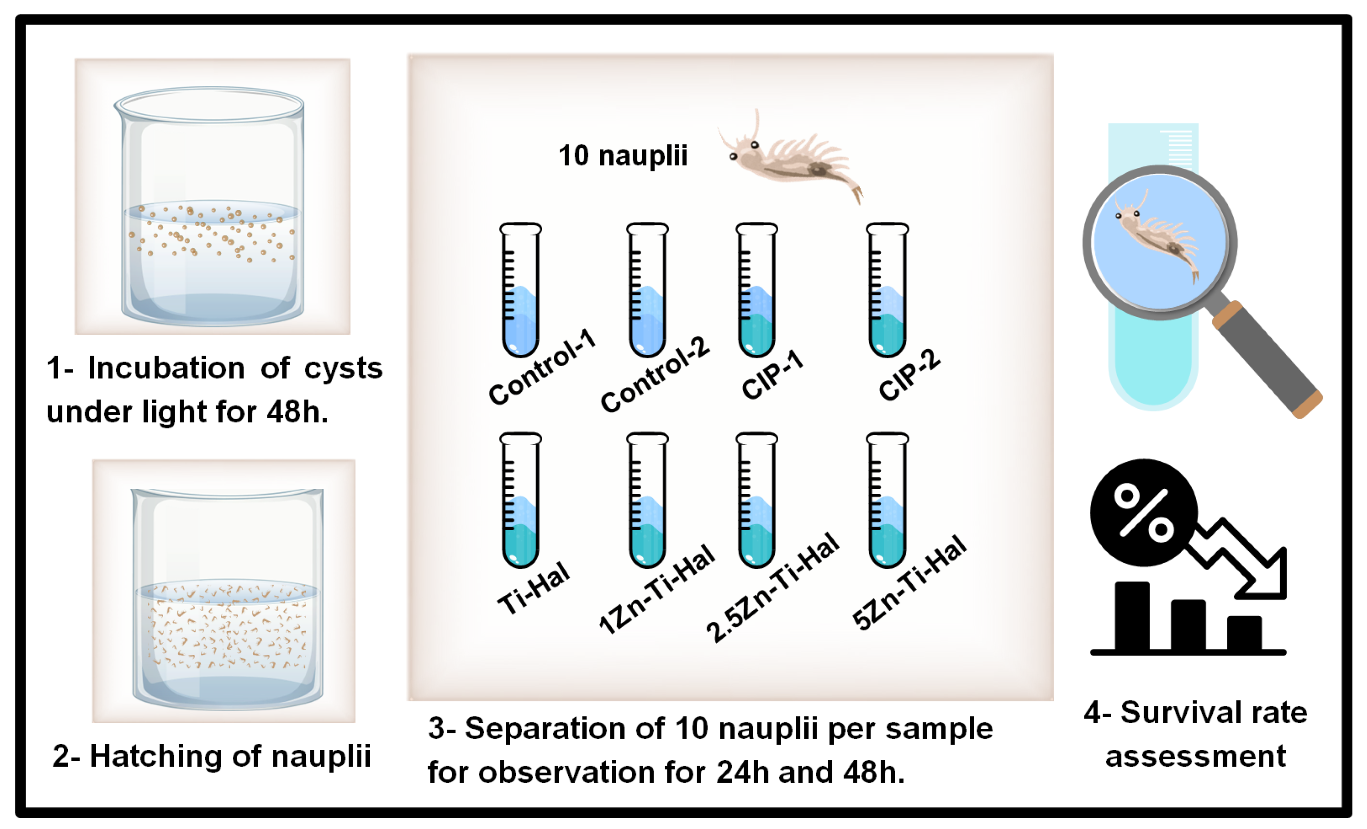
The acute ecotoxicity of CIP (30 mg L−1) before and after photocatalytic degradation was evaluated using Artemia salina nauplii, following a previously adapted methodology [29] with the selection of nauplii for direct contact with active and inactive CIP to evaluate possible toxic byproducts based on the survival rate. As shown in Table S1, the following samples were included: two controls—Control-1 (100% saline water) and Control-2 (50% saline water + 50% distilled water)—were used to evaluate basal survival and the effect of salinity; two samples containing CIP—CIP-1 (94% saline + 6% CIP) and CIP-2 (50% saline + 50% CIP)—were prepared to evaluate the effect of the antibiotic on survival; finally, for each catalyst sample (Ti-Hal, 1Zn-Ti-Hal, 2.5Zn-Ti-Hal, and 5Zn-Ti-Hal), a solution was prepared with 50% residual supernatant from CIP degradation and 50% saline water.
For the test, Artemia salina cysts were incubated in 200 mL of laboratory-prepared saline solution for 48 h to hatch nauplii. Ten nauplii were added to each sample (test tube) with the corresponding volumes: Control-1 (5 mL saline), Control-2 (2.5 mL saline + 2.5 mL distilled water), CIP-1 (0.3 mL CIP + 4.7 mL saline), CIP-2 (2.5 mL CIP + 2.5 mL saline), and catalyst samples (2.5 mL residual supernatant + 2.5 mL saline). Survival was monitored after 24 and 48 h, and all tests were performed in triplicate (Figure 2).

2.6. Antibiotic Inactivation Test

The antibiotic inactivation test was adapted from the methodology described previously [14,29] to evaluate the bactericidal effect of residual CIP on Staphylococcus aureus (ATCC 25923) [19]. As detailed in Figure S1, the test was performed in two stages. First, the supernatant from the photocatalytic removal of CIP was separated from the catalyst by centrifugation (5000 rpm, 10 min), frozen, and lyophilized to obtain the residual powder, which was then redispersed in sterilized water. Second, 100 µL of this solution was mixed with 100 µL of standardized bacterial inoculum (1.2 × 10⁴ CFU mL−1) on Mueller–Hinton agar using the spread plate method and incubated at 37 °C for 24 h [19]. All tests were performed in triplicate.
The bacterial inoculum was prepared by transferring a portion of S. aureus culture from nutrient agar into Brain Heart Infusion (BHI) medium, followed by 24 h incubation at 37 °C, and diluted (1 mL culture in 9 mL BHI) prior to the test [29].
The inhibition effect (η) was calculated using Equation (2):
ƞ =   N 1 N 2 N 1 × 100 ,
where N1 is the mean colony-forming units of the control plates, and N2 is the mean colony-forming units for each tested solution [29].

3. Results and Discussion

3.1. Elemental and Structural Properties

3.1.1. XRF Analysis

Elemental analysis by XRF confirmed the presence of Zn, Ti, Al, and Si in the samples, consistent with the expected composition derived from ZnO, TiO2, and halloysite precursors. The oxides identified and their respective quantities are listed in Table 1. Alumina (Al2O3) and silica (SiO2) were predominant in all samples, corresponding to the halloysite framework and representing approximately 66–72 wt%. TiO2 accounted for 22.6–26.6 wt%, while ZnO increased progressively with the nominal doping level, from 2.14 wt% in 1Zn-Ti-Hal to 9.19 wt% in 5Zn-Ti-Hal.
Compared to the base sample (Ti-Hal), the gradual rise in ZnO content was accompanied by a slight decrease in TiO2, Al2O3, and SiO2, confirming the successful incorporation of Zn into the TiO2–halloysite matrix. This compositional evolution supports the formation of ternary ZnO–TiO2–halloysite nanocomposites with tunable ZnO loading, which may influence charge transfer and photocatalytic performance.

3.1.2. XRD Analysis

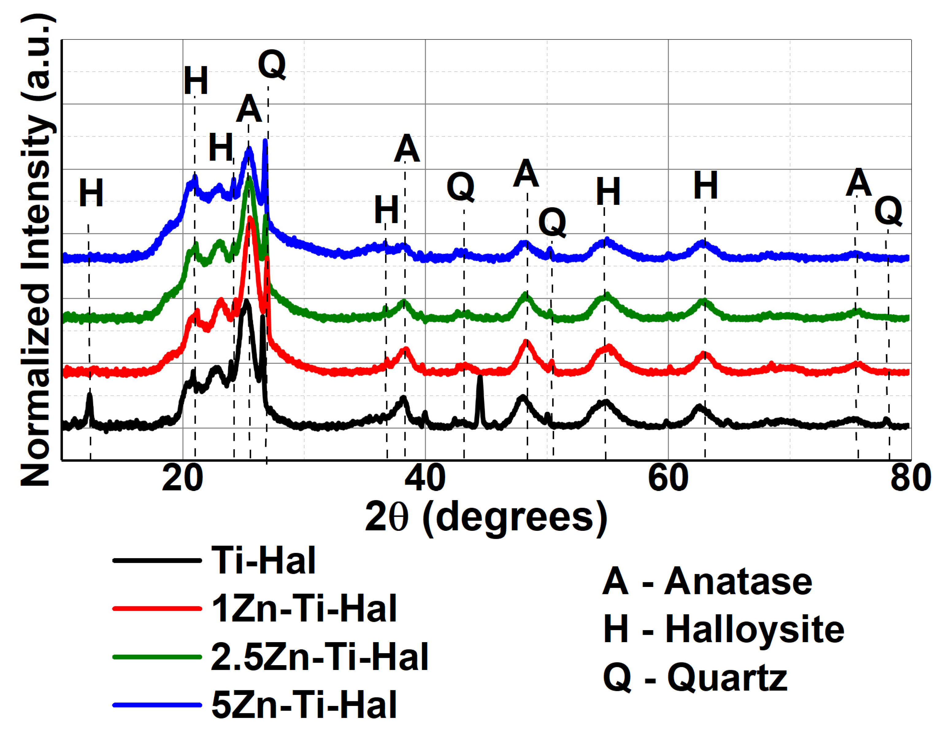
The X-ray diffractogram in Figure 3 presents the qualitative analysis of the respective samples with the characteristic peaks of the identified phases. Halloysite (7 Å) PDF 00-029-1487 was identified with prominent peaks at 12.2°, 20.9°, 24.3°, 37.0° and 54.6° corresponding to the planes (001), (100), (002), (003), (212), (002), where the appearance of the peak (300) at 63.0° referring to halloysite (10 Å) PDF 00-029-1489 may be related to the calcination applied in the synthesis of the material (Figure 1), since halloysite is subject to changes in the interplanar spacing in heat treatments in the range between 100 °C and 350 °C [1]. Additionally, according to identification from PDF 00-033-1161, quartz was found at peaks of 26.6°, 43.0°, 50.3°, and 77.9°, which correspond to the (101), (200), (112), and (220) planes. It has already been reported by Yu et al. [16] who synthesized ZnO/halloysite nanotube composites by a facile wet chemical technique in anhydrous ethanol under a mild reaction condition with different mass ratios of ZnO to halloysite nanotubes, where diffraction peaks attributed to quartz were observed, indicating the presence of quartz impurities in the raw halloysite nanotubes.
Anatase (PDF 01-070-6826) was identified at peaks 25.4°, 38.3°, 48.3°, and 75.5°, indexed to the (101) (004), (220), and (215) planes, respectively, and is the main phase of TiO2 in applications below 700 °C, exhibiting greater photocatalytic activity [3]. The (101) plane obtained relevant intensity among all phases, pointing to the successful inclusion of TiO2 in the form of anatase under the halloysite structure, where this is also related to the heat treatment applied during the synthesis (Figure 3), since at a temperature of 400 °C an increase in the grain size and a better crystalline form of their layers are obtained, which allows a decrease in optical transparency and, consequently, greater light absorption [30].
The absence of peaks related to ZnO was observed in the doped samples. Thus, as the presence of this phase has already been proven through the XRF results (Table 1), it is possible that despite the incorporation of Zn2+ ions in the base sample (Ti-Hal), the amount was insufficient for detection in this analysis. Zhan et al. [30] reported in their study that when developing a ZnO/TiO2 nanocomposite supported on montmorillonite for an agricultural antibacterial agent, observed in their samples doped with ZnO that there were no characteristic peaks of this in the XRD results, in which they related this occurrence due to the tiny size of the crystallite or to a small amount of ZnO in the composite.

3.1.3. XPS Analysis

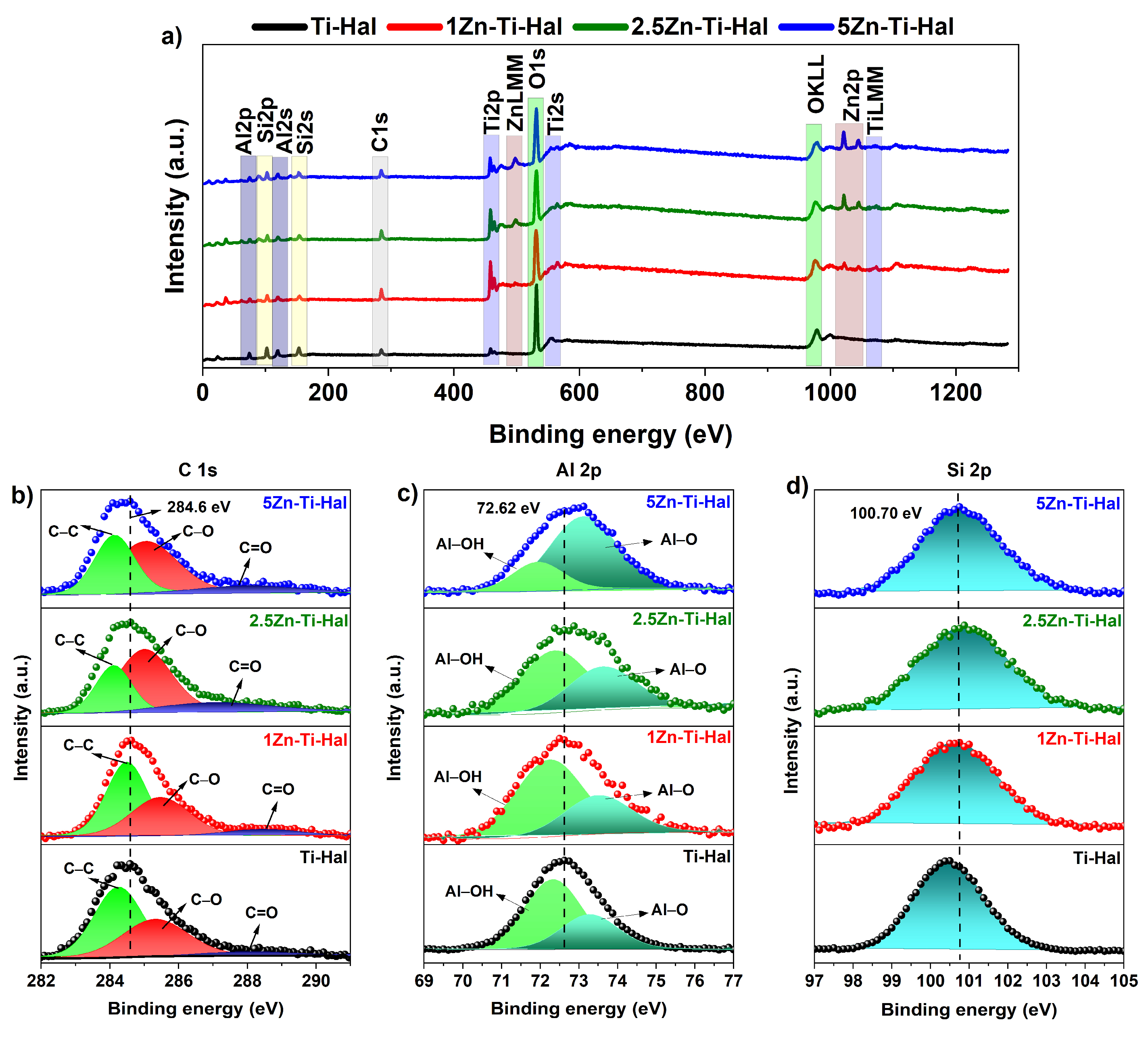
The XPS technique was used to investigate the elemental composition and chemical states of the samples. Spectral calibration was performed using the C 1s peak (C–C component) with a binding energy of 284.6 eV as a reference. Spectral analysis was performed using CasaXPS software (version 2.3.26PR1.0), with Shirley background subtraction and Gaussian–Lorentzian peak functions for deconvolution. The general spectra of the samples are illustrated in Figure 4a.
The high-resolution C 1s spectra show that the reference calibration (284.6 eV) was successfully applied since it was detected in the spectra of all samples (Figure 4b), in which the bands related to the C–C and C–O bonds may result from residual carbon and accidental compounds from the XPS instrument itself and the C=O band may be related to CO2 adsorbed on the surface [31].
The high-resolution spectra of Al 2p and Si 2p reinforce the halloysite composition. In Al 2p, the central peak at 72.62 eV (Figure 4c) is observed, associated with Al–OH and Al–O bonds present in the lumen of the halloysite tubes of the respective samples [10,11,16,22]. The Si 2p spectrum exhibits peaks at 100.7 eV (Figure 4d) characteristic of Si–O bonds on the external surface of the halloysite [10,11,16,22]. The simultaneous presence of these peaks confirms that halloysite clay is present in the samples and contributes to the XPS signal. These results ensure that the aluminosilicate matrix (halloysite) remains chemically and structurally intact in the analyzed samples.
The analysis of the high-resolution O 1s spectra (Figure 5a) reveals differences between the samples without and with Zn doping. For the Ti-Hal sample, only a single peak at 529.71 eV is observed, attributed to the lattice oxygen in TiO2 (Ti–O–Ti) [32]. In the Zn-doped samples, two distinct peaks are observed, located at 527.32 eV and 529.71 eV. This new peak, at 527.32 eV, is present only in the samples containing Zn and can be attributed to lattice oxygen, suggesting a possible interaction between ZnO and the TiO2 structure [16,22].
In the high-resolution spectrum of Ti 2p (Figure 5b), the bands corresponding to Ti 2p3/2 and Ti 2p1/2 [3,10,11,30] were identified, with binding energies of 455.91 eV and 461.80 eV, respectively, in the doped samples, and 456.46 eV and 462.35 eV in the Ti-Hal sample. Therefore, a slight shift in the bands towards lower binding energies is observed in the doped samples, which can be attributed to the effect of Zn doping [14]. Despite this shift, the separation between the Ti 2p3/2 and Ti 2p1/2 peaks in all samples was 5.89 eV, a characteristic value for TiO2 [33,34,35,36], which indicates that titanium remains in the oxidation state (Ti4+) [3,10,11,31,35,36].
Thus, confirmation of the Ti4+ ion may indicate effective gains in photocatalysis as reported in the study by Wu et al. [35] with mixed porous TiO2-SiO2 materials, in which all titania–silica samples generated hydrogen through photocatalytic water splitting under UV irradiation, it was found that the photocatalytic hydrogen generation rate increases in the same proportion as the amount of Ti4+ ions (coordinated in a tetrahedral and octahedral form), where the presence of these ions has a significant impact on the photocatalytic activity. Thus, it is considered that the XPS results correlated with those of XRD (Figure 3) point to Ti4+ in the anatase form in this structural environment [11], which is characteristic of anatase TiO2, widely recognized for its efficiency in photocatalytic applications [35,36], supporting the potential applicability of the synthesized samples for CIP removal.
The binding energies of Zn 2p (Figure 5c) indicate the characteristic bands of Zn 2p3/2 and Zn 2p1/2 [3,16,31,37] in the doped samples with the respective binding energies: 1Zn-Ti-Hal with 1022.18 eV and 1044.63 eV; 2.5Zn-Ti-Hal with 1021.47 eV and 1045.47 eV; and 5Zn-Ti-Hal with 1020.98 eV and 1044.87 eV. The energy differences between Zn 2p3/2 and Zn 2p1/2 in each sample (1Zn-Ti-Hal with 22.45 eV, 2.5Zn-Ti-Hal with 24.0 eV and 5Zn-Ti-Hal with 23.89 eV) are consistent with the reported value of ZnO and confirm the 2+ oxidation state of Zn [3,16,31,37].
These variations may be related to ZnO–halloysite interactions [16,38], demonstrating successful ZnO doping on the halloysite surface. Therefore, the XPS results indicate two important findings: first, that the signals of O, Al, Si, Ti and Zn point to these as the main elements present on the surface of the samples, which coincides with the XRF data (Table 1); second, the coexistence of Zn, Ti, and Al signals in the XPS spectra suggests potential electronic interactions between ZnO, TiO2, and halloysite, consistent with previous reports [10].

3.2. Morphological Analysis

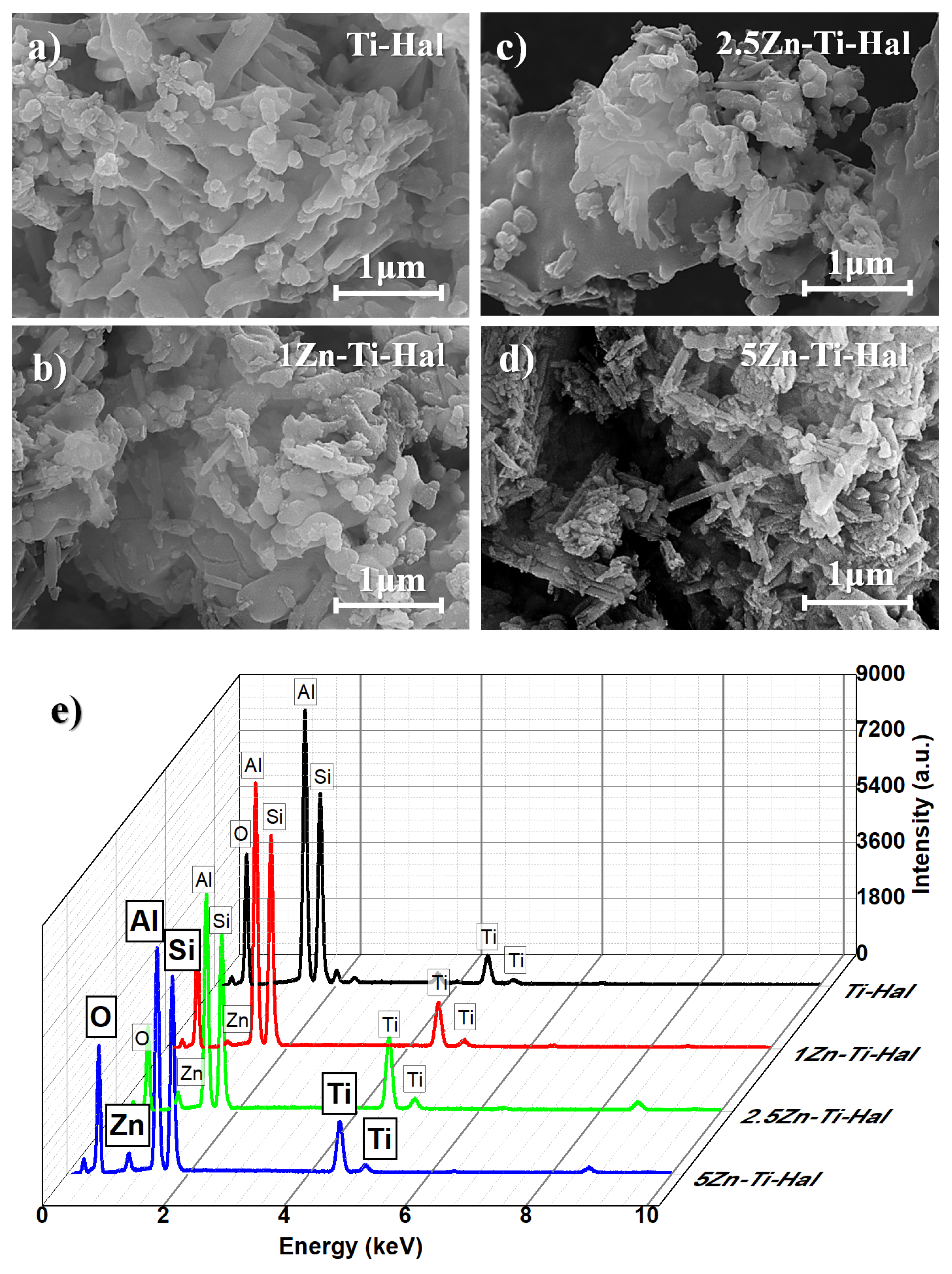
SEM analysis of Ti-Hal (Figure 6a), 1Zn-Ti-Hal (Figure 6b), 2,5Zn-Ti-Hal (Figure 6c), and 5Zn-Ti-Hal (Figure 6d) confirmed the characteristic morphology of halloysite: aluminosilicate clay with tubular nanostructure, elongated shape, empty lumen, and polished surface [10,19]. However, when comparing the sample images regarding the visual perception of halloysite nanotubes, it is evident that 5Zn-Ti-Hal and Ti-Hal exhibit greater visibility of halloysite tubes, while 2.5Zn-Ti-Hal and 1Zn-Ti-Hal show lower visibility, which should be highlighted as it can influence their performance in CIP removal.
Among the samples, the presence of small spherical particles on the halloysite nanotube structure is noticeable, which may be TiO2 particles (Figure 6a–d), especially in the sample not doped with Zn (Figure 6a). The EDS analysis of the samples under study (Figure 6e) indicated in all samples the arrangement of four elements in greater intensity in decreasing order: Al, Si, O, and Ti, where the 5Zn-Ti-Hal sample obtained a greater amount of oxygen, which can result in positive oxygen defect density indices, an important factor for good photocatalytic performance [9,39]. Additionally, the main elements identified here (Al, Si, O, Ti, and Zn) align with the XRF (Table 1) and XPS (Figure 4 and Figure 5) results, which complement each other regarding the chemical composition of the samples.

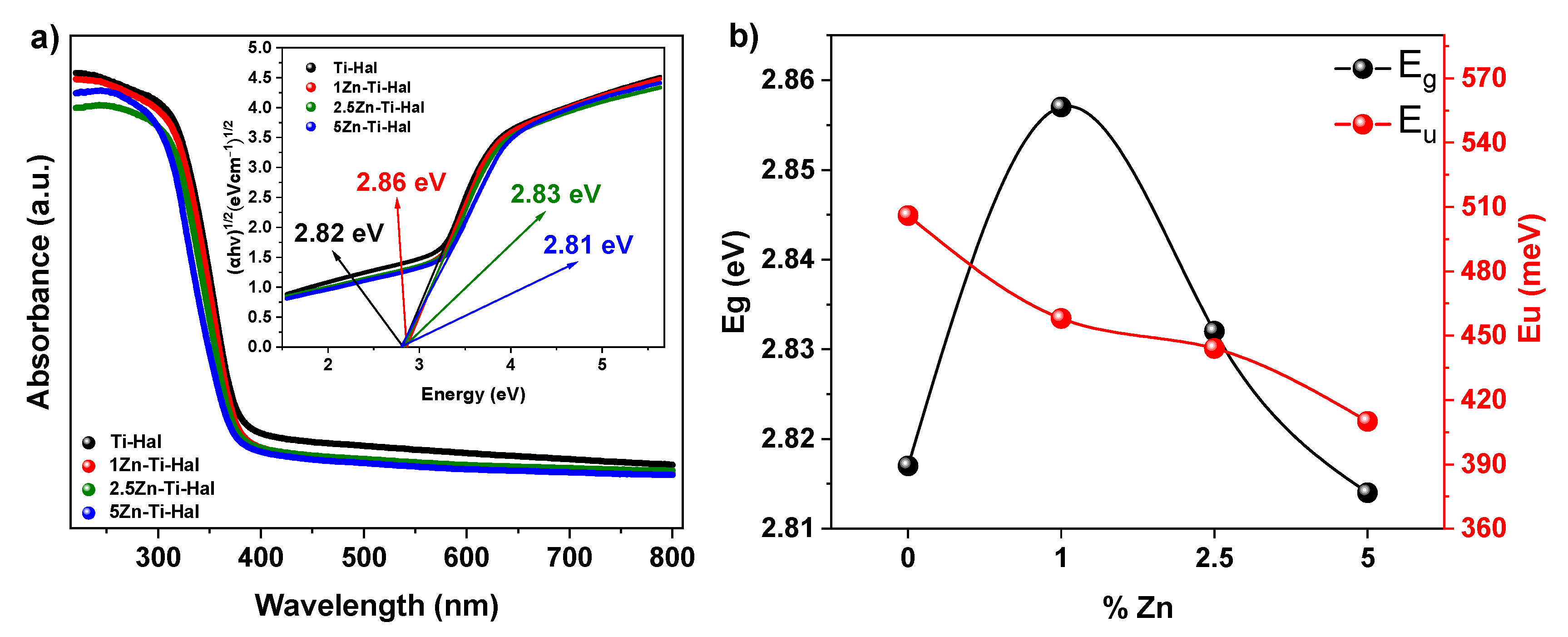
3.3. Analysis of Optical Properties

The band gap energy (Eg) of the samples was calculated using the Tauc method [2,3,17] from Equation (3):
α h ν 1 2 = A h ν E g ,
where the Tauc graph relates Eg to α (absorption coefficient), where hν is the photon energy and A is the proportionality constant.
The results in Figure 7a indicate a reduction in the band gap energy for all samples in relation to pure ZnO (3.37 eV) and TiO2 (3.2 eV) [3,10,11,15,17], where the values were: 2.82 eV for Ti-Hal, 2.86 eV for 1Zn-Ti-Hal, 2.83 eV for 2.5Zn-Ti-Hal, and 2.81 eV for 5Zn-Ti-Hal. These data indicate that the combination of ZnO, TiO2, and halloysite can improve absorption in the visible region (Table 2).
Among the doped samples, the sample with the highest percentage of ZnO (5Zn-Ti-Hal) exhibits the smallest band gap, confirming the effectiveness of the doping process. Consistent with these findings, Lee et al. [2] reported that the incorporation of lanthanum (La) into halloysite-supported TiO2 (TiO2@HNT) also led to a reduction in band gap, attributed to the absorption characteristics of halloysite within the TiO2 absorption band. It demonstrates the importance of the TiO2–halloysite system in supporting the dopant while simultaneously achieving this optical and structural improvement.
The Urbach Energy (EU) or absorption tail is a parameter associated with lattice disorder and reflects the concentration of defects in the samples [17], which can be calculated by Equation (4):
α υ   =   D   exp h υ E U
where EU is determined as a function of the absorption coefficient (α), where D is the proportionality constant, h is Planck’s constant, and υ is the frequency of the incident light.
The calculation of EU using the inverse of the slope of the linear function, below the band gap threshold, is shown in Figure S2, indicating high values for all samples [40]. As shown in Table 2, the results obtained are as follows: 506 meV for Ti-Hal, 458 meV for 1Zn-Ti-Hal, 444 meV for 2.5Zn-Ti-Hal, and 410 meV for 5Zn-Ti-Hal. Despite the gradual decrease in EU with ZnO doping, the matrix bonding between TiO2 and halloysite provided significant EU values for all samples.
This high EU value can favor photocatalytic processes, as it is directly proportional to the presence of oxygen vacancies, which are mainly responsible for reducing the band gap and separating the carriers [9,13,14,39,41]. Thus, from Figure 7b, it is noticeable that the increase in EU is reflected in a decrease in the band gap value, indicating a possible increase in the density of oxygen defects [9,39].

4. Removal Test

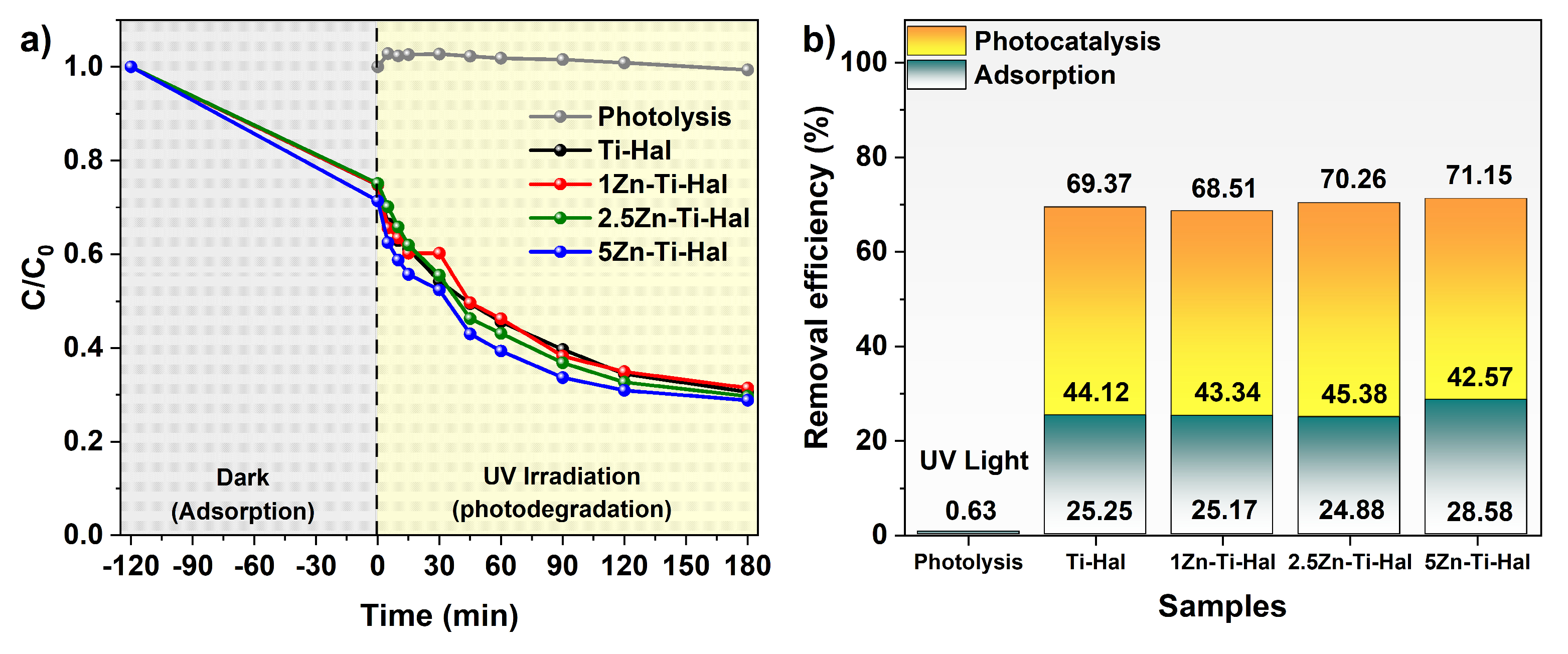
The drug removal test is presented in Figure 8, which assessed the degradation capacity of CIP (30 mg L−1) by the respective catalysts (Ti-Hal, 1Zn-Ti-Hal, 2.5Zn-Ti-Hal, and 5Zn-Ti-Hal) using 0.5 g L−1 of each material. Firstly, dark adsorption was used to achieve the equilibrium. After 120 min in the dark to obtain adsorption–desorption equilibrium, the catalyst and the CIP solution were subjected to UV irradiation to evaluate photodegradation.
Figure 8a shows the C/C0 results of CIP during the 180 min irradiation period, and Figure 8b shows the CIP removal efficiency in photolysis and using the photocatalysts synthesized in this work. The results of photolysis (degradation of the drug using only light in the absence of the photocatalyst) revealed its high stability with only 0.63% degradation, thus making it necessary to search for materials that increase this pollutant removal rate. During the 120 min in the dark stage, the nanocomposites had a removal efficiency between 24.88% and 28.58% of the drug. After 180 min of irradiation of the solution containing the drug and photocatalyst, an additional total removal of CIP through oxidative degradation was observed, ranging from 42.57% to 45.38%.
Overall, the CIP removal efficiency among the samples was Ti-Hal—69.37% (adsorption 25.25% and photocatalysis 44.12%); 1Zn-Ti-Hal—68.51% (adsorption 25.17% and photocatalysis 43.34%); 2.5Zn-Ti-Hal—70.26% (adsorption 24.88% and photocatalysis 45.38%), and 5Zn-Ti-Hal—71.15% (adsorption 28.58% and photocatalysis 42.57%). The average photocatalytic manipulation (43.85%) was 1.69 times higher than the average adsorption removal (25.97%). Thus, the results indicate that adsorption and photodegradation acted in a complementary manner, with photocatalysis contributing more significantly to the overall removal efficiency.
These results are in agreement with those found in the literature, which indicate that clay minerals enhance the interaction between the pollutant and the nanocomposite [10,14,16,26,42]. Furthermore, the synergism between adsorption and photodegradation is more efficient for removing organic pollutants compared to the use of semiconductors alone [14,16].
The 5Zn-Ti-Hal sample obtained the best CIP degradation result, which can be correlated with the data from the following characterizations: SEM, 5Zn-Ti-Hal had excellent visualization of the halloysite (Figure 6d), which can directly influence the increase (or decrease) of the nanoparticle clusters, where the halloysite are indicated for better dispersion of the incorporated semiconductors, enhancing the adsorption efficiency on the substrate and the photocatalytic capacity of the nanoparticles [2,3,6,11,16]. Additionally, 5Zn-Ti-Hal achieved a significant Urbach Energy (EU) index (Figure 7b), where better EU values indicate the possibility of a longer time in the electron-hole pair recombination process, an important factor for good results in photocatalysis [9,13,14]; Finally, 5Zn-Ti-Hal achieved the smallest band gap and 1Zn-Ti-Hal had the most significant band gap (Figure 7a), with a smaller band gap being an indicator of greater light absorption potential [1,6,11,13].
The mechanistic interpretation that emerges is as follows: halloysite provides adsorption sites and a nanotubular matrix that disperses and stabilizes TiO2 and ZnO nanoparticles, increasing contact between adsorbed CIP and photosensitive centers [16,26]. The presence of Zn, at the optimum concentration observed (5%), slightly reduces the band gap and introduces defects/tail states that prolong the average lifetime of carriers; and the Ti and Zn electronic interaction (evidenced by O 1s and Ti 2p displacement) favors charge separation at heterogeneous interfaces, decreasing recombination and increasing the probability of forming reactive species capable of mineralizing or fragmenting the CIP [26].
To assess the stability of the materials under photocatalytic conditions, SEM images taken before and after CIP degradation are compared in Figure S3. The micrographs show no significant morphological changes for any sample; the halloysite nanotubular framework and the dispersion of ZnO/TiO2 nanoparticles are preserved, with no evidence of particle sintering, structural collapse, or extensive agglomeration. These observations, particularly pronounced for 5Zn-Ti-Hal, support the morphological stability of the nanocomposites during the photocatalytic tests and reinforce their potential for reuse.
This combination of effects explains the advantage observed for 5Zn-Ti-Hal over samples with lower Zn content or without Zn, even though the differences in performance are moderate, which is consistent with the slight variation in Eg and the predominant role of the support in initial removal by adsorption. Aghababaei et al. [3] developed a work with a new heterostructure photocatalyst with O-g-C3N4 (oxygen-doped graphitic carbon nitride), ZnO, TiO2 and halloysite for photodegradation of diclofenac sodium (DCF) under UV light, studied in dark conditions for 2 h, where after removal test the sample that obtained the best DCF degradation performance was the one that had in its constitution all the aforementioned materials. It was reported that the use of halloysite enables the dispersion of semiconductors, which enhances both their photocatalytic activity and stability. Therefore, the application of halloysite incorporated with other semiconductors yields better results in photocatalysis and contaminant adsorption.

4.1. Bioassays

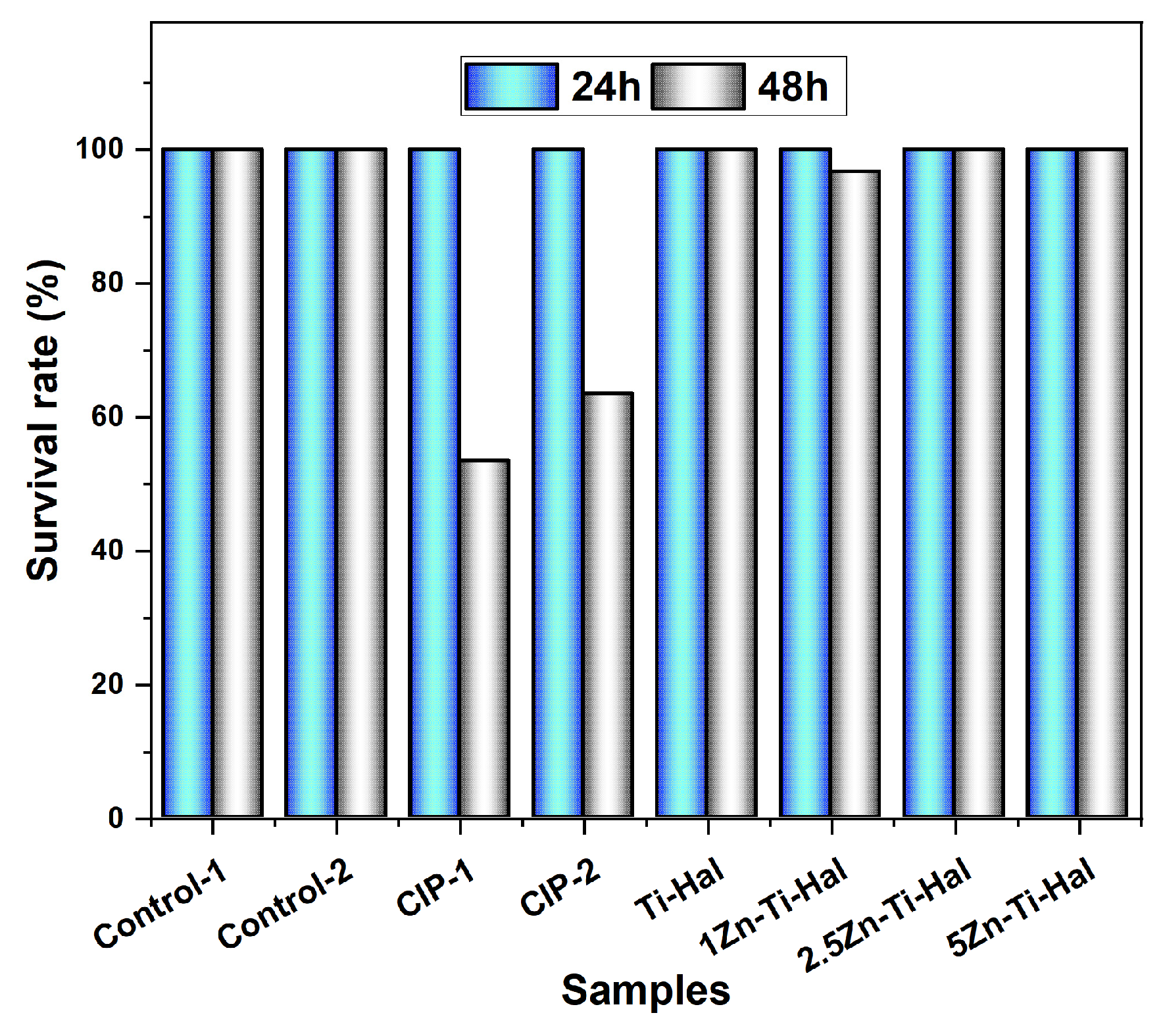
Figure 9 shows the data from the ecotoxicity test against Artemia salina after 24 h and 48 h, along with the respective survival rates for each sample analyzed. The control samples (Control-1 and Control-2) showed the maximum survival rate (100%), indicating that the dilution caused by the composition of the saline solution plus the post-degraded residual CIP does not affect the survival of the nauplii in the direct contact test.
The rate of surviving nauplii in CIP-1 (53.33%) and CIP-2 (63.33%) indicates a notable acute toxicity of CIP at 30 mg L−1 in the 48 h. According to a study conducted by Kergaravat et al. [42], who performed a complete ecotoxicological test including acute, chronic, and post-chronic recovery tests in two zooplanktonic microcrustaceans (Daphnia magna and Ceriodaphnia dubia) for six quinolones of different generations (including ciprofloxacin among these), where in the acute ecotoxicity test they classified CIP (36 mg L−1 in 48 h and 14 mg L−1 in 72 h) as harmful to the respective aquatic organisms. Thus, a CIP concentration of 30 mg L−1 made it possible to test the potential for CIP inactivation by the synthesized samples in this study at a concentration that has been proven to be harmful to aquatic microcrustaceans.
The Artemia salina subjected to contact with the samples after the CIP removal process reached a maximum survival rate (100%), equal to that of the control samples, except sample 1Zn-Ti-Hal, which had a similar measurement (96.66%). Therefore, these results indicate that after the removal treatment, the byproducts resulting from CIP remediation are non-toxic to Artemia salina, suggesting that the degradation pathway does not generate acutely hazardous intermediates under the tested conditions.

4.2. Pharmacological Inactivation Test

The antibacterial inactivation potential of CIP at 30 mg L−1 by the 5Zn-Ti-Hal sample was analyzed after the removal test, from the direct contact of the post-degraded residual CIP with the bacterium Staphylococcus aureus (S. aureus), where the growth (or not) of the respective cultured colonies will be the positive (or negative) indicator of the loss of bactericidal activity [14]. The choice of S. aureus was because its infections, in addition to being common, are associated with high mortality rates [39] and, also, because the antibacterial potential of CIP against this strain [39,41,42,43] and even antibacterial resistance [5,41,42,43] has been reported in the literature.
Three samples were used for the test: the control (Ctrl), to evaluate the number of colonies without contaminant; the CIP sample, enumerating the colonies affected by the active drug; and, finally, the residual CIP sample (5Zn-Ti-Hal), to investigate the number of colonies grown (or not) in contact with the potentially inactivated residue.
The results of the pharmacological inactivation test are presented in Figure 10. The Control sample showed significant growth of bacterial colonies, indicating that the standardized suspension of S. aureus strains prepared was successful and will be used as a reference for counting against the other samples. The CIP sample showed no colony growth, which reaffirms the bactericidal capacity of this antibiotic.
The CIP residue obtained after 180 min of photocatalytic degradation with the 5Zn-Ti-Hal sample exhibited an inhibitory efficiency of 50.35%, compared to 100% for the untreated CIP solution. It should be noted that, although 71.15% of the initial CIP was removed from the original 30 mg L−1 solution, a residual concentration remains after treatment (approximately 8.66 mg L−1). This residual CIP concentration is sufficient to produce a measurable antibacterial effect, which explains why partial inhibition (≈50.35%) was still observed in the treated sample. Therefore, when comparing the treated supernatant to the untreated drug solution, the antibacterial activity is clearly reduced (100% to 50.35% inhibition); however, the remaining fraction of CIP still retains partial bactericidal capacity at the concentration present after photocatalysis.
This behavior is consistent with previous reports. Feitosa et al. [14] for example, observed that photocatalytic removal of tetracycline (≈70.45% removal using a Ce–TiO2–Sepiolite system) led to a decreased but still measurable inhibitory effect in bacterial assays (inhibition reduced to 83.12% after 180 min and to 45.63% after 600 min), illustrating the non-linear relationship between percent removal and residual antimicrobial activity. Such literature evidence supports our observation that partial antibiotic removal can still leave sufficient active (or bioactive) residues to produce measurable inhibition.
Therefore, this result is attributed to the combined adsorption–photocatalysis effect achieved during the removal process, which reduced the antibacterial capacity of CIP and allowed the growth of bacterial colonies in direct contact with the post-degradation residue. It indicates the 5Zn-Ti-Hal sample’s ability to treat and inactivate bactericidal products, while also reducing the potential for antibacterial resistance of pharmacological residues in aquatic environments.
Comparing the data in Table S2, this study with the ZnO–TiO2–halloysite system obtained good drug degradation results at a higher concentration (30 mg L−1) compared to all the cited studies. Furthermore, another distinguishing feature of this work is the applied bioassays, as none of these studies subjected the photodegraded residues to biological tests, such as ecotoxicological evaluation and/or pharmacological inactivation tests, to investigate the impacts of the generated residual byproducts. Therefore, the ZnO–TiO2–halloysite system presents, in addition to photodegradation results, a prospective vision of combining pollutant removal efficiency with positive environmental solutions for aquatic ecosystems.

5. Conclusions

A ternary nanocomposite comprising ZnO, TiO2, and halloysite was studied, relying on the combined contribution of adsorption and photocatalysis as a green approach for the removal of ciprofloxacin hydrochloride (CIP) from aquatic media. XRD, XRF, XPS, UV-Vis, and SEM-EDS results confirmed the synthesis of the expected nanocomposite, indicating a strong structural and morphological interaction between ZnO, TiO2, and halloysite. XRF, XPS, and SEM-EDS analyses revealed Al, Si, O, Ti, and Zn as the primary elements in the samples, confirming the presence of ZnO in the doped samples. The correlation of XRD, XRF, and XPS data indicates the incorporation of Zn2+ ions without inducing significant structural distortions. XRD and XPS revealed the presence of Ti4+ in the anatase form, indicating a high photocatalytic potential of the synthesized samples. The manipulation of CIP (30 mg L−1) was achieved at a reasonable level across all samples, with an average prevalence of photocatalysis over adsorption of 1.69 times. Notably, 5Zn-Ti-Hal was the sample with the highest removal efficiency, at 71.14% (photocatalysis: 42.57% and adsorption: 28.58%). Ecotoxicological analysis against Artemia salina indicates that the degradation byproducts generated after treatment are non-toxic, suggesting that no harmful intermediates persisted in solution. The antibacterial inactivation test further demonstrated a marked reduction in the bactericidal potential of CIP, consistent with extensive structural degradation of the parent antibiotic. Therefore, the ternary ZnO–TiO2–halloysite nanocomposite shows promising potential as a green technology for aquatic remediation, since it enables CIP degradation while mitigating residual toxicity.

Supplementary Materials

The following supporting information can be downloaded at: https://www.mdpi.com/article/10.3390/min15121253/s1, Figure S1: Drug inactivation test: initial stage, the supernatant powder is obtained from the residual CIP and in the final stage, the antibacterial inhibition against bacterial growth is evaluated through direct contact of the supernatant solution with the standardized bacterial inoculum suspension; Figure S2: Results of Urbach Energy from the lnα vs. Energy graph for samples (a) Ti-Hal, (b) 1Zn-Ti-Hal, (c) 2.5Zn-Ti-Hal and (d) 5Zn-Ti-Hal; Figure S3: Comparison of SEM images of samples (a) Ti-Hal, (b) 1Zn-Ti-Hal, (c) 2.5Zn-Ti-Hal and (d) 5Zn-Ti-Hal before degradation (white caption) and post-degradation (yellow caption); Table S1: Description of samples for evaluation of their toxicological potential; Table S2: Comparison with related results in the literature with semiconductors incorporated into halloysite. References [2,3,6,16,23,44] are cited in the Supplementary Materials.

Author Contributions

J.R.M.: Research, Methodology, Conceptualization, Data Curation, Writing—Original Draft. R.P.F.: Research, Software, Writing—Review and Editing. I.S.d.L.: Methodology. L.H.O.: Research, Formal Analysis, Writing—Review and Editing. F.F.: Visualization. E.C.S.-F.: Visualization. J.A.C.: Research, Formal Analysis, Visualization, Validation, Writing—Review and Editing. J.A.O.: Research, Formal Analysis, Validation, Writing—Review and Editing, Project administration. All authors have read and agreed to the published version of the manuscript.

Funding

This research received no external funding.

Data Availability Statement

The data that support the findings of this study are available from the corresponding author upon reasonable request.

Acknowledgments

The authors thank the Coordination Support in Higher Education (CAPES) and the National Council for Scientific and Technological Development (CNPq) for support.

Conflicts of Interest

The authors declare that they have no known competing financial interests or personal relationships that could have appeared to influence the work reported in this paper.

References

  1. Papoulis, D.; Panagiotaras, D.; Tsigrou, P.; Christoforidis, K.C.; Petit, C.; Apostolopoulou, A.; Stathatos, E.; Komarneni, S.; Koukouvelas, I. Halloysite and Sepiolite–TiO2 Nanocomposites: Synthesis Characterization and Photocatalytic Activity in Three Aquatic Wastes. Mater. Sci. Semicond. Process. 2018, 85, 1–8. [Google Scholar] [CrossRef]
  2. Lee, J.; Seong, S.; Jin, S.; Jeong, Y.; Noh, J. Synergetic Photocatalytic-Activity Enhancement of Lanthanum Doped TiO2 on Halloysite Nanocomposites for Degradation of Organic Dye. J. Ind. Eng. Chem. 2021, 100, 126–133. [Google Scholar] [CrossRef]
  3. Aghababaei, N.; Abdouss, M.; Hosseini-Monfared, H.; Ghanbari, F. Photocatalytic Degradation of Diclofenac Using a Novel Double Z-Scheme Catalyst (O-g-C3N4/ZnO/TiO2@halloysite Nanotubes): Degradation Mechanism, Identification of by-Products and Environmental Implementation. J. Water Process Eng. 2023, 53, 103702. [Google Scholar] [CrossRef]
  4. Pereira, G.; Espínola, D.L.; Pinto, G.F.; Silva, V.; Lima, D.L.D.; Calisto, V. Photodegradation of Ciprofloxacin in Water Using Silver Nanostructures. Case Stud. Chem. Environ. Eng. 2024, 9, 100699. [Google Scholar] [CrossRef]
  5. Hu, X.; Hu, X.; Peng, Q.; Zhou, L.; Tan, X.; Jiang, L.; Tang, C.; Wang, H.; Liu, S.; Wang, Y.; et al. Mechanisms Underlying the Photocatalytic Degradation Pathway of Ciprofloxacin with Heterogeneous TiO2. Chem. Eng. J. 2020, 380, 122366. [Google Scholar] [CrossRef]
  6. Albuquerque, W.; Trigueiro, P.; Silva, B.V.; Neves, L.; Almeida, L.C.; Peña-Garcia, R.R. A Novel RuO2@ZnO-Alginate-Halloysite Composite for the Effective Degradation of Eosin Yellow Dye and Ciprofloxacin Drug. Mater. Res. Bull. 2025, 182, 113178. [Google Scholar] [CrossRef]
  7. Zarzzeka, C.; Goldoni, J.; de Paula de Oliveira, J.d.R.; Lenzi, G.G.; Bagatini, M.D.; Colpini, L.M.S. Photocatalytic Action of Ag/TiO2 Nanoparticles to Emerging Pollutants Degradation: A Comprehensive Review. Sustain. Chem. Environ. 2024, 8, 100177. [Google Scholar] [CrossRef]
  8. Xu, K.; Shen, J.; Zhang, S.; Xu, D.; Chen, X. Efficient Interfacial Charge Transfer of BiOCl-In2O3 Step-Scheme Heterojunction for Boosted Photocatalytic Degradation of Ciprofloxacin. J. Mater. Sci. Technol. 2022, 121, 236–244. [Google Scholar] [CrossRef]
  9. Soares, A.S.; Araujo, F.P.; Osajima, J.A.; Guerra, Y.; Viana, B.C.; Peña-Garcia, R. Nanotubes/Nanorods-like Structures of La-Doped ZnO for Degradation of Methylene Blue and Ciprofloxacin. J. Photochem. Photobiol. A Chem. 2024, 447, 115235. [Google Scholar] [CrossRef]
  10. Pouthika, K.; Madhumitha, G. Tailoring Interfacial Charge Separation in Z-Scheme CuO@TiO2@halloysite Heterostructure for Efficient Photocatalytic Removal of Congo Red. J. Taiwan. Inst. Chem. Eng. 2025, 166, 105752. [Google Scholar] [CrossRef]
  11. Luo, Y.; Zhang, Z.; Chen, S.; Jiang, A.; Chen, X.; Zhu, X.; Di, Y. Activation of Peroxymonosulfate by Halloysite Nanotube-Supported TiO2 Composite for Efficient Photocatalytic Degradation of Rhodamine B. Opt. Mater. 2024, 147, 114669. [Google Scholar] [CrossRef]
  12. Tetteh, E.K.; Rathilal, S.; Asante-Sackey, D.; Chollom, M.N. Prospects of Synthesized Magnetic TiO2-Based Membranes for Wastewater Treatment: A Review. Materials 2021, 14, 3524. [Google Scholar] [CrossRef] [PubMed]
  13. Janani Archana, K.; Christy Preetha, A.; Balasubramanian, K. Influence of Urbach Energy in Enhanced Photocatalytic Activity of Cu Doped ZnO Nanoparticles. Opt. Mater. 2022, 127, 112245. [Google Scholar] [CrossRef]
  14. Feitosa, R.P.; de Lima, I.S.; Guerra, Y.; da Silva-Filho, E.C.; Furtini, M.B.; Almeida, L.; Peña-Garcia, R.R.; Martín, I.B.; Cecília, J.A.; Osajima, J.A. Cerium-Doped TiO2 and Sepiolite Nanocomposites for Tetracycline Inactivation in Water Treatment. ACS Appl. Nano Mater. 2025, 8, 4324–4338. [Google Scholar] [CrossRef]
  15. Marana, N.L.; Sambrano, J.R.; de Souza, A.R. Propriedades Eletrônicas, Estruturais e Constantes Elásticas Do ZnO. Quim. Nova 2010, 33, 810–815. [Google Scholar] [CrossRef]
  16. Yu, H.; Xu, H.; Hao, T.; Yuan, Y.; Zhang, B.; Wang, H.; Shao, G.; Fan, B.; Lu, H. Facile Synthesis of ZnO/Halloysite Nanotube Composite with Greatly Enhanced Photocatalytic Performance. Colloids Surf. A Physicochem. Eng. Asp. 2024, 688, 133633. [Google Scholar] [CrossRef]
  17. Yuan, Z.; Zhu, J.; Lu, J.; Li, Y.; Ding, J. Preparation of Biodiesel by Transesterification of Castor Oil Catalyzed by Flaky Halloysite Supported ZnO/SnO2 Heterojunction Photocatalyst. Renew. Energy 2024, 227, 120516. [Google Scholar] [CrossRef]
  18. Rosa, D.; Remmani, R.; Bavasso, I.; Bracciale, M.P.; Di Palma, L. Biochar Supported Fe–TiO2 Composite for Wastewater Treatment: Solid-State Synthesis and Mechanistic Insights. Chem. Eng. Sci. 2025, 317, 122076. [Google Scholar] [CrossRef]
  19. Abdullayev, E.; Lvov, Y. Halloysite for Controllable Loading and Release. In Developments in Clay Science; Elsevier: Amsterdam, The Netherlands, 2016; pp. 554–605. [Google Scholar]
  20. Ruiz-Hitzky, E.; Aranda, P.; Akkari, M.; Khaorapapong, N.; Ogawa, M. Photoactive Nanoarchitectures Based on Clays Incorporating TiO2 and ZnO Nanoparticles. Beilstein J. Nanotechnol. 2019, 10, 1140–1156. [Google Scholar] [CrossRef]
  21. Yuan, Y.; Xu, H.; Li, H.; Fang, C.; Xie, W.; Li, M.; Zhang, B.; Shao, G.; Lu, H.; Wang, H. Construction of CdS/Halloysite Nanotube Composite for High Efficient Photocatalytic Degradation toward Tetracycline Hydrochloride. J. Alloys Compd. 2025, 1036, 181646. [Google Scholar] [CrossRef]
  22. Jiang, L.; Huang, Y.; Liu, T. Enhanced Visible-Light Photocatalytic Performance of Electrospun Carbon-Doped TiO2/Halloysite Nanotube Hybrid Nanofibers. J. Colloid Interface Sci. 2015, 439, 62–68. [Google Scholar] [CrossRef] [PubMed]
  23. Albuquerque, W.A.; Filho, A.J.N.; Romaguera-Barcelay, Y.; Medina-Carrasco, S.; Orta, M.d.M.; Trigueiro, P.; Peña-Garcia, R.R. Synergistic ZnO–CuO/Halloysite Nanocomposite for Photocatalytic Degradation of Ciprofloxacin with High Stability and Reusability. Minerals 2025, 15, 977. [Google Scholar] [CrossRef]
  24. Banti, C.N.; Papachristodoulou, C.; Chrysouli, M.P.; Douvalis, A.; Hadjikakou, S.K. Controlled Dual Activity of an Organometallic Antibiotic through Micelle Formulation. J. Organomet. Chem. 2024, 1012, 123130. [Google Scholar] [CrossRef]
  25. Nannou, C.; Maroulas, K.N.; Tsamtzidou, C.; Ladomenou, K.; Kyzas, G.Z. Photocatalytic Degradation of Veterinary Antibiotics in Wastewaters: A Review. Sci. Total Environ. 2025, 966, 178765. [Google Scholar] [CrossRef]
  26. Santos, G.O.; Goulart, L.A.; Cordeiro-Junior, P.J.; Sanchez-Montes, I.; Lanza, M.R. Pharmaceutical Contaminants: Ecotoxicological Aspects and Recent Advances in Oxidation Technologies for Their Removal in Aqueous Matrices. J. Environ. Chem. Eng. 2022, 10, 108932. [Google Scholar] [CrossRef]
  27. de Lima, I.S.; Silva, A.S.; Nascimento, A.M.S.S.; de Oliveira, L.H.; Morais, A.Í.S.; Barreto, H.M.; Peña-Garcia, R.; Cuevas, M.D.M.O.; Argôlo Neto, N.M.; Osajima, J.A.; et al. Synthesis and Characterization of Cassava Gum Hydrogel Associated with Chlorhexidine and Evaluation of Release and Antimicrobial Activity. Macromol. Biosci. 2024, 24, 2300507. [Google Scholar] [CrossRef]
  28. Banti, C.; Hadjikakou, S. Evaluation of Toxicity with Brine Shrimp Assay. Bio-protocol 2021, 11, e3895. [Google Scholar] [CrossRef]
  29. Hsu, C.Y.; Mahmoud, Z.H.; Abdullaev, S.; Ali, F.K.; Ali Naeem, Y.; Mzahim Mizher, R.; Morad Karim, M.; Abdulwahid, A.S.; Ahmadi, Z.; Habibzadeh, S.; et al. Nano Titanium Oxide (Nano-TiO2): A Review of Synthesis Methods, Properties, and Applications. Case Stud. Chem. Environ. Eng. 2024, 9, 100626. [Google Scholar] [CrossRef]
  30. Zhan, J.; Chen, H.; Zhou, H.; Hao, L.; Xu, H.; Zhou, X. Mt-Supported ZnO/TiO2 Nanocomposite for Agricultural Antibacterial Agent Involving Enhanced Antibacterial Activity and Increased Wettability. Appl. Clay Sci. 2021, 214, 106296. [Google Scholar] [CrossRef]
  31. Al-Gaashani, R.; Zakaria, Y.; Gladich, I.; Kochkodan, V.; Lawler, J. XPS, Structural and Antimicrobial Studies of Novel Functionalized Halloysite Nanotubes. Sci. Rep. 2022, 12, 21633. [Google Scholar] [CrossRef]
  32. Wang, J.; Wang, G.; Yu, T.; Ding, N.; Wang, M.; Chen, Y. Photocatalytic Performance of Biochar-Modified TiO2 (C/TiO2) for Ammonia–Nitrogen Removal. RSC Adv. 2023, 13, 24237–24249. [Google Scholar] [CrossRef]
  33. Qu, K.; Huang, L.; Hu, S.; Liu, C.; Yang, Q.; Liu, L.; Li, K.; Zhao, Z.; Wang, Z. TiO2 Supported on Rice Straw Biochar as an Adsorptive and Photocatalytic Composite for the Efficient Removal of Ciprofloxacin in Aqueous Matrices. J. Environ. Chem. Eng. 2023, 11, 109430. [Google Scholar] [CrossRef]
  34. Bharti, B.; Kumar, S.; Lee, H.N.; Kumar, R. Formation of Oxygen Vacancies and Ti3+ State in TiO2 Thin Film and Enhanced Optical Properties by Air Plasma Treatment. Sci. Rep. 2016, 6, 32355. [Google Scholar] [CrossRef] [PubMed]
  35. Wu, C.M.; Peng, R.; Dimitrijevic, N.M.; Rajh, T.; Koodali, R.T. Preparation of TiO2-SiO2 Aperiodic Mesoporous Materials with Controllable Formation of Tetrahedrally Coordinated Ti4+ Ions and Their Performance for Photocatalytic Hydrogen Production. Int. J. Hydrogen Energy 2014, 39, 127–136. [Google Scholar] [CrossRef]
  36. Rajkumar, N.; Ramachandran, K. Oxygen Deficiency and Room Temperature Ferromagnetism in Undoped and Cobalt-Doped TiO2 Nanoparticles. IEEE Trans. Nanotechnol. 2011, 10, 513–519. [Google Scholar] [CrossRef]
  37. Akter, N.; Ahmed, T.; Haque, I.; Hossain, M.K.; Ray, G.; Hossain, M.M.; Islam, M.S.; Ali shaikh, M.A.; Akhtar, U.S. XPS Valence Band Observable Light-Responsive System for Photocatalytic Acid Red114 Dye Decomposition Using a ZnO–Cu2O Heterojunction. Heliyon 2024, 10, e30802. [Google Scholar] [CrossRef]
  38. Papoulis, D. Halloysite Based Nanocomposites and Photocatalysis: A Review. Appl. Clay Sci. 2019, 168, 164–174. [Google Scholar] [CrossRef]
  39. Pereira da Silva, C.A.; Santos Araújo, N.J.; Morais Oliveira-Tintino, C.D.; Barbosa Filho, J.M.; Alencar, G.G.; de Araújo-Neto, J.B.; dos Santos, J.S.; Soares, J.B.; Domiciano, C.B.; Silva, D.A.; et al. Effect of Betulinic Acid on MepA Efflux Pump Inhibition in Staphylococcus aureus: Antibacterial and Molecular Study. Steroids 2025, 215, 109572. [Google Scholar] [CrossRef]
  40. Verbel-Olarte, M.I.; Serna-Galvis, E.A.; Jimenez-Lopez, D.M.; Jojoa-Sierra, S.D.; Porras, J.; Pulgarin, C.; Torres-Palma, R.A. First Evidence That Antibiotic-Resistant Bacteria Are Inactivated by Chemical Species Produced through the Solar Photosensitization of Ciprofloxacin in Water. Sci. Total Environ. 2025, 963, 178442. [Google Scholar] [CrossRef]
  41. Park, K.-H.; Kim, D.; Jung, M.; Kim, D.Y.; Lee, Y.-M.; Lee, M.S.; Hong, K.-W.; Bae, I.-G.; Hong, S.I.; Cho, O.-H. Effects of Sub-Inhibitory Concentrations of Nafcillin, Vancomycin, Ciprofloxacin, and Rifampin on Biofilm Formation of Clinical Methicillin-Resistant Staphylococcus aureus. Microbiol. Spectr. 2024, 12, e03412-23. [Google Scholar] [CrossRef]
  42. Kergaravat, S.V.; Hernández, S.R.; Gagneten, A.M. Second-, Third- and Fourth-Generation Quinolones: Ecotoxicity Effects on Daphnia and Ceriodaphnia Species. Chemosphere 2021, 262, 127823. [Google Scholar] [CrossRef]
  43. Sreekanth, K.; Sarath Josh, M.K.; Sethulakshmi, K.H.; Joseph, J.; Joseph, B.J.; Mohammed Hashim, K.K.; Manoj, E.; Aravindakumar, C.T.; Radhakrishnan, E.K. Diclofenac-Mediated Resensitisation of Methicillin-Resistant Staphylococcus aureus (MRSA) to Ciprofloxacin via NorA Efflux Pump Inhibition. Chem. Eng. J. 2025, 523, 167983. [Google Scholar] [CrossRef]
  44. Pouthika, K.; Madhumitha, G. Synergistic synthesis of Carrisa edulis fruit extract capped heterogeneous CuO-ZnO-HNT composite for photocatalytic removal of organic pollutants. Inorganica Chim Acta 2023, 551, 121457. [Google Scholar] [CrossRef]
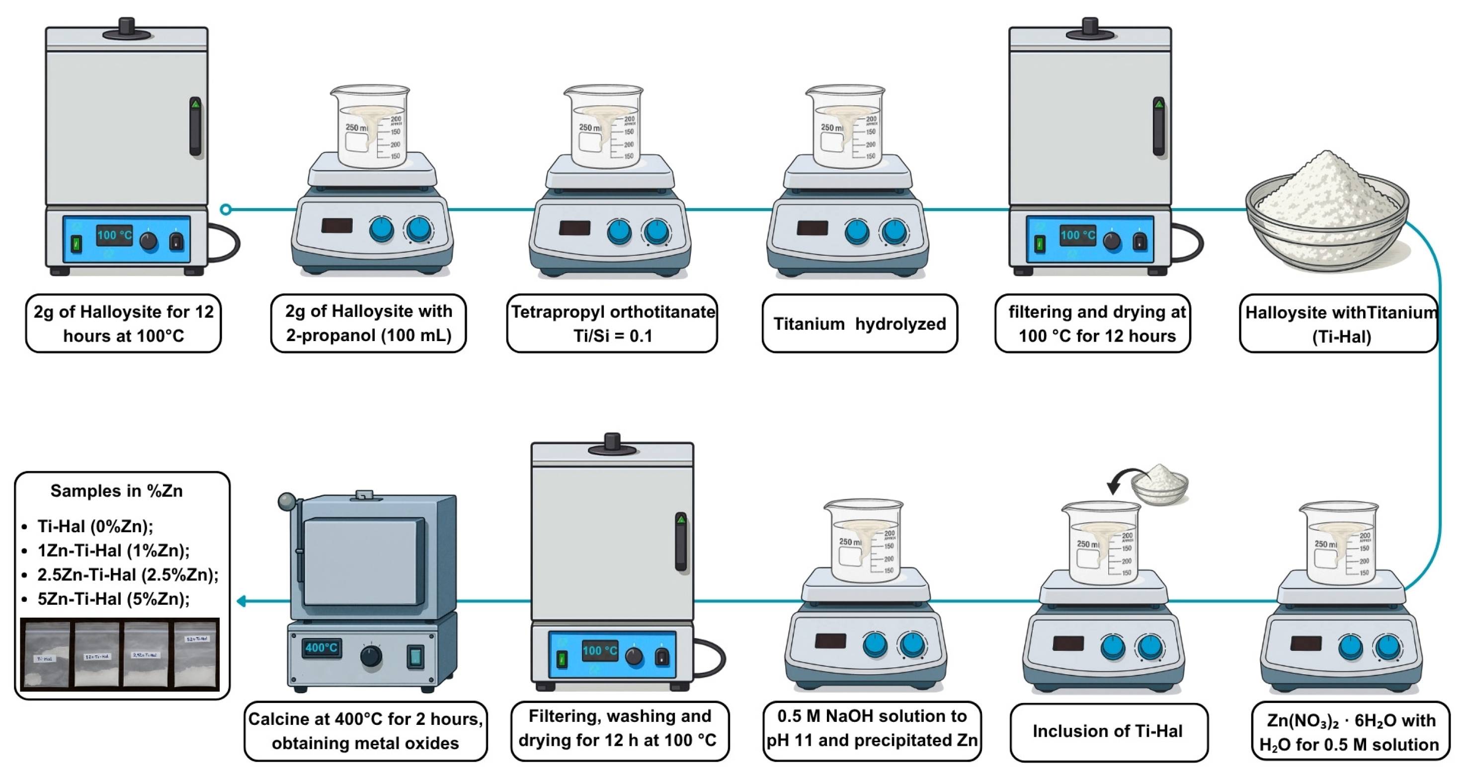
Figure 1. Synthesis methodology of nanocomposites formed by TiO2 and ZnO supported on halloysite: in the initial stage, TiO2 was incorporated into halloysite by hydrolysis, and in the final stage, ZnO was precipitated onto the TiO2–halloysite matrix. * The materials are synthesized in percentage of Zn: Ti-Hal (0% Zn), 1Zn-Ti-Hal (1% Zn), 2.5Zn-Ti-Hal (2.5% Zn), and 5Zn-Ti-Hal (5% Zn).
Minerals 15 01253 g001
Figure 2. Steps for applying the ecotoxicity test with Artemia salina: Incubation under light for 48 h (1), larval hatching (2), selection of nauplii for direct contact with active and inactive CIP for 48 h (3), and assessment of survival rate (4).
Minerals 15 01253 g002
Figure 3. XRD patterns with identification of crystalline structures in the samples Ti-Hal, 1Zn-Ti-Hal, 2.5Zn-Ti-Hal, and 5Zn-Ti-Hal.
Minerals 15 01253 g003
Figure 4. Core-level spectra of (a) general, (b) C1s, (c) Al2p, and (d) Si2p for the synthesized samples.
Minerals 15 01253 g004
Figure 5. Core-level spectra of (a) O1s, (b) Ti2p, (c) Zn2p for the synthesized samples.
Minerals 15 01253 g005
Figure 6. SEM images of Ti-Hal (a), 1Zn-Ti-Hal (b), 2.5Zn-Ti-Hal (c) and 5Zn-Ti-Hal (d). EDS with elemental mapping of the samples under study (e).
Minerals 15 01253 g006
Figure 7. DRS spectrum with band gap (via Tauc plot) (a), relationship between band gap (Eg) and Urbach Energy (EU) with the variation in Zn (b).
Minerals 15 01253 g007
Figure 8. Concentration versus time in the dark and under UV light irradiation (a); CIP removal efficiency by adsorption and photocatalysis (b).
Minerals 15 01253 g008
Figure 9. Ecotoxicity test against Artemia salina in the interval of 24 and 48 h for the control samples (Control-1 and Control-2), CIP at 30 mg L−1 (CIP-1 and CIP-2), and CIP degraded by the catalyst samples (Ti-Hal, 1Zn-Ti-Hal, 2.5Zn-Ti-Hal, and 5Zn-Ti-Hal).
Minerals 15 01253 g009
Figure 10. (a) Antibacterial inhibitory effect per sample; (b) visualization of bacterial activity (S. aureus strain) per sample.
Minerals 15 01253 g010
Table 1. Composition of Ti-Hal, 1Zn-Ti-Hal, 2.5Zn-Ti-Hal, and 5Zn-Ti-Hal by XRF.
Composition in Percent (%)
SamplesAl2O3SiO2TiO2ZnOFe2O3CaOP2O5
Ti-Hal35.8736.0926.62-0.640.290.48
1Zn-Ti-Hal35.4235.4425.762.140.600.280.44
2.5Zn-Ti-Hal35.1434.7624.094.690.580.270.46
5Zn-Ti-Hal33.7733.1422.629.190.540.270.45
Table 2. Absorption band, band gap, and Urbach Energy results for Ti-Hal, 1Zn-Ti-Hal, 2.5Zn-Ti-Hal, and 5Zn-Ti-Hal samples.
SampleAbsorption Band (nm)Band Gap (eV)Urbach Energy (meV)
Ti-Hal439.712.82506
1Zn-Ti-Hal433.562.86458
2.5Zn-Ti-Hal438.162.83444
5Zn-Ti-Hal441.282.81410
Disclaimer/Publisher’s Note: The statements, opinions and data contained in all publications are solely those of the individual author(s) and contributor(s) and not of MDPI and/or the editor(s). MDPI and/or the editor(s) disclaim responsibility for any injury to people or property resulting from any ideas, methods, instructions or products referred to in the content.