Non-Invasive Investigation of a 16th-Century Illuminated Scroll: Pigments, Fillers, and Metal-Based Decorations
Abstract
1. Introduction
2. Materials and Methods
2.1. The Illuminated Parchment
- -
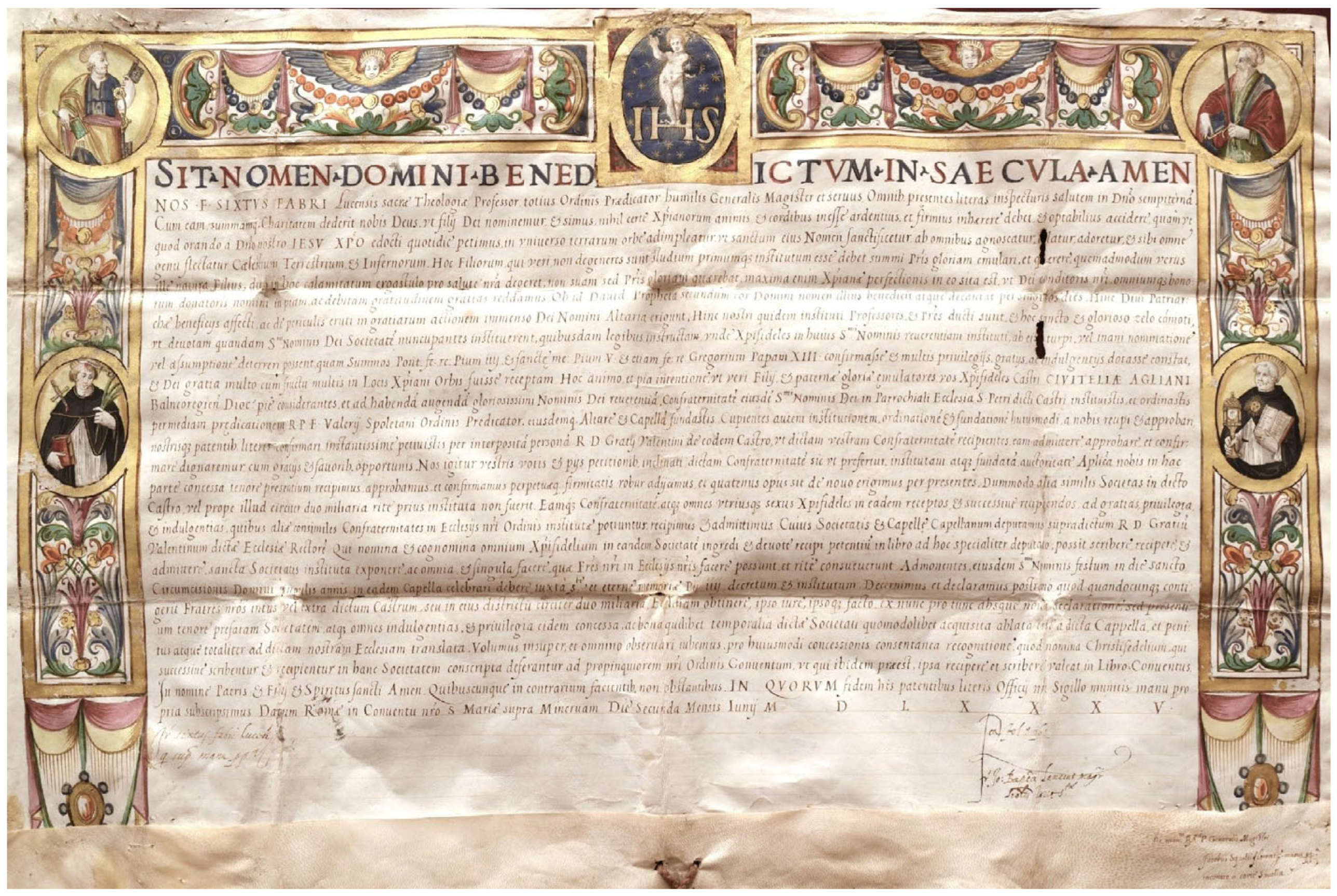
- The parchment is an important document from the Counter-Reformation period, where the strength of the orthodoxy of the Catholic Church expressed by the Dominican Order is highlighted, starting from the title (Sit Nomine Domini Benedictum in Saecula Amen; Be the Name of God Blessed Forever Amen).
- -
- The text indirectly reflects the apprehension and insistence of the members of a small and recently established Brotherhood, the Corpus Christi of Civitella di Agliano, who requested approval from the Dominicans. In fact, the members of the Brotherhood were worried about not having acted according to the expected rules, i.e., by asking permission from the Dominicans.
- -
- The document, in response to the requests of the Brotherhood, is drawn up by Frater Sixstus Fabri Lucensis, Generalis Magister of the Dominican Order and Professor of Theology. He confirmed the approval and guaranteed the authorization to constitute the Brotherhood through patentibus literis (the parchment itself) to build an altar and a chapel in the Church of San Pietro in Civitella d’Agliano in the Diocese of Bagnoregio.
2.2. Experimental Section
2.2.1. Hypercolorimetric Multispectral Imaging (HMI)
- (i)
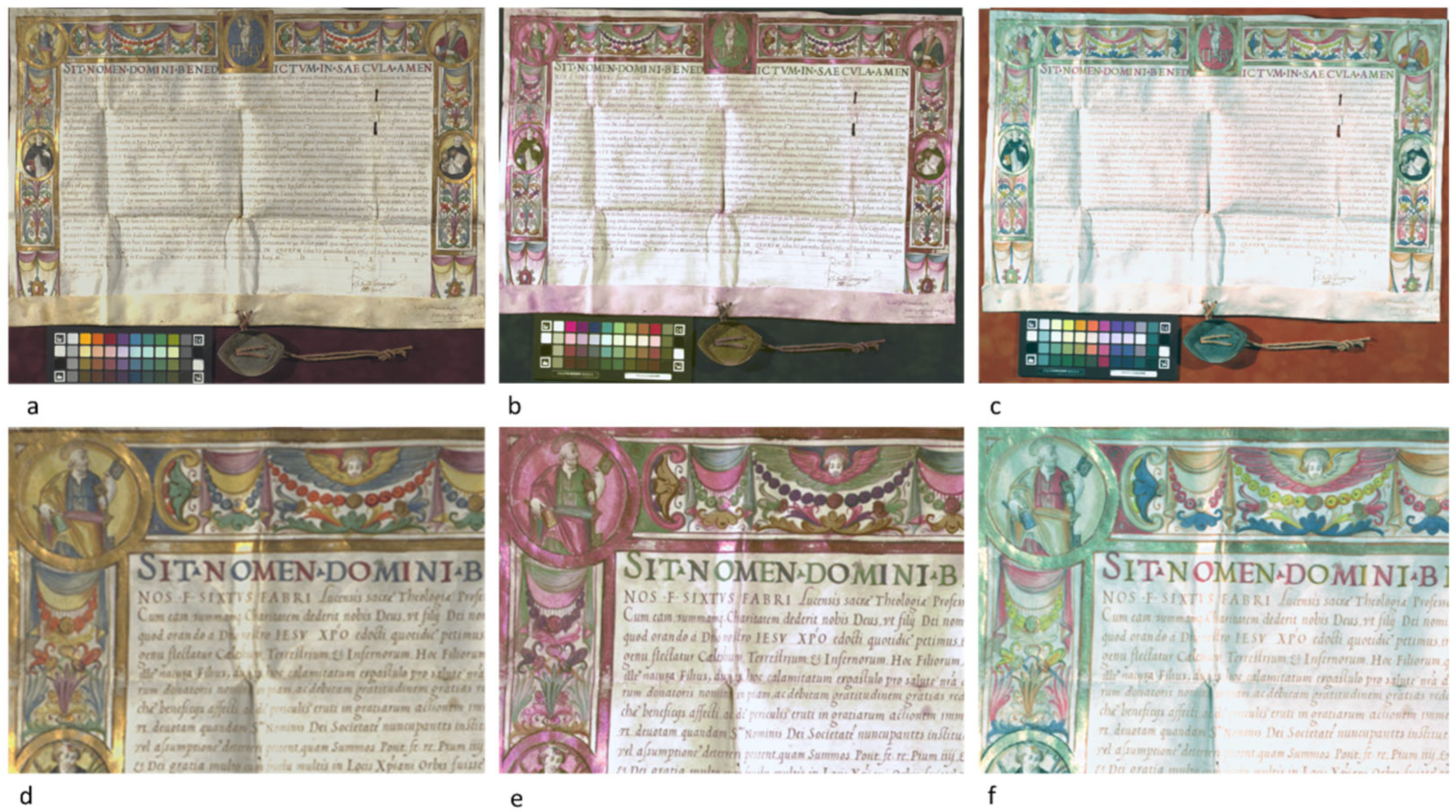
- The acquisition device is made up of a Nikon D800 camera (Nital SpA, Moncalieri Torino, Italy) modified in full range to register images in the spectral range 300–1000 nm and is equipped with a 60 mm lens; optical filters for UV-Vis and Vis-IR acquisition (named filter A and filter B, respectively), for UVF (U V-IR cut filter coupled with filter A), and light sources consisting of two modified NEEWER (Neewer, Shenzhen, China) 750II Flashes Speedlite TTL flashes with LCD display and wireless triggers. The flashes were modified by removing their front plastic lenses, thus allowing emissions in the 300–1000 nm region for multispectral imaging, and two UV CR230B-HP 10W LED projectors (peak emission at 365 nm, for ultraviolet fluorescence) (Madatec srl, Pessano con Bornago, Milano, Italy) were used; white references patches positioned around the parchment, along with a ColorChecker (version 8.0) made of 36 standard colors and 8 grayscale patches, plus two reference white and two blacks; the illuminating sources were positioned at about 45° with respect to the artwork surface;
- (ii)
- The calibration software, named SpectraPick® (Version 1.2, created by Profilocolore, Rome, Italy) allows obtaining imaging data for seven monochromatic bands (.tiff images) centered at 350, 450, 550, 650, 750, 850, and 950 nm and the visible RGB output;
- (iii)
- The processing software, named PickViewer® (Version 1.2, created by Profilocolore, Rome, Italy) enables the extraction of information to support materials characterization and to highlight surface details by applying digital image processing tools to the calibrated bands.
2.2.2. Infrared Reflectography in the 950–1700 nm Spectral Range (IRR)
2.2.3. MA-XRF
- -
- For the illuminated parts: 38 kV anode voltage, 70 uA filament current, 10 mm/s scanning velocity, and 0.5 mm step in both X and Y directions. Beam diameter on the sample surface ~1 mm, no He flow.
- -
- For the written part: 30 kV anode voltage, 95 uA filament current, 0.5 mm/s scanning velocity, and 0.25 mm step in both X and Y directions. Collimator diameter 0.4 mm, no He flow.
2.2.4. Raman Spectroscopy
2.2.5. External Reflectance FT-IR (ER-FTIR) Spectroscopy
2.2.6. Optical Microscopy
2.2.7. Fiber Optic Reflectance Spectroscopy (FORS)
3. Results and Discussion
3.1. Painting Technique
3.1.1. The Support
3.1.2. Underdrawings
3.2. Pigment Identification
3.3. Metal Layers
3.4. Written Text
3.5. Capital Letters
4. Conclusions
Author Contributions
Funding
Data Availability Statement
Acknowledgments
Conflicts of Interest
References
- Orna, M.V. Artists’ pigments in illuminated medieval manuscripts: Tracing artistic influences and connections—A review. ACS Symp. Ser. 2013, 1147, 3–18. [Google Scholar] [CrossRef]
- Panayotova, S. The Art & Science of Illuminated Manuscripts: A Handbook; Harvey Miller Publishers: London, UK, 2020; 528p, ISBN 9781912554591. [Google Scholar]
- Plesters, J. 2. Ultramarine blue, natural and artificial. Stud. Conserv. 1966, 11, 62–75. [Google Scholar] [CrossRef]
- Vetter, W.; Latini, I.; Schreiner, M. Azurite in medieval illuminated manuscripts: A reflection-FTIR study concerning the characterization of binding media. Herit. Sci. 2019, 7, 21. [Google Scholar] [CrossRef] [PubMed]
- Cucci, C.; Delaney, J.K.; Picollo, M. Reflectance Hyperspectral Imaging for Investigation of Works of Art: Old Master Paintings and Illuminated Manuscripts. Acc. Chem. Res. 2016, 49, 2070–2079. [Google Scholar] [CrossRef] [PubMed]
- Aceto, M.; Agostino, A.; Fenoglio, G.; Baraldi, P.; Zannini, P.; Hofmann, C.; Gamillscheg, E. First analytical evidences of precious colourants on Mediterranean illuminated manuscripts. Spectrochim. Acta Part. A Mol. Biomol. Spectrosc. 2012, 95, 235–245. [Google Scholar] [CrossRef]
- Nodari, L.; Ricciardi, P. Non-invasive identification of paint binders in illuminated manuscripts by ER-FTIR spectroscopy: A systematic study of the influence of different pigments on the binders’ characteristic spectral features. Herit. Sci. 2019, 7, 7. [Google Scholar] [CrossRef]
- Pessanha, S.; Manso, M.; Carvalho, M.L. Application of spectroscopic techniques to the study of illuminated manuscripts: A survey. Spectrochim. Acta Part B At. Spectrosc. 2012, 71–72, 54–61. [Google Scholar] [CrossRef]
- Ricciardi, P.; Dooley, K.A.; Maclennan, D.; Bertolotti, G.; Gabrieli, F.; Patterson, C.S.; Delaney, J.K. Use of standard analytical tools to detect small amounts of smalt in the presence of ultramarine as observed in 15th-century Venetian illuminated manuscripts. Herit. Sci. 2022, 10, 38. [Google Scholar] [CrossRef]
- Edwards, H.G.M.; Vandenabeele, P.; Colomban, P. Analytical Raman Spectroscopy of Manuscripts and Maps: The Role of Inks. In Raman Spectroscopy in Cultural Heritage Preservation; Springer: Berlin/Heidelberg, Germany, 2023; pp. 215–231. [Google Scholar] [CrossRef]
- Martinez, K.; Cupitt, J.; Saunders, D.; Pillay, R. Ten years of art imaging research. Proc. IEEE 2002, 90, 28–41. [Google Scholar] [CrossRef]
- Del Federico, E.; Shöfberger, W.; Schelvis, J.; Kapetanaki, S.; Tyne, L.; Jerschow, A. Insight into Framework Destruction in Ultramarine Pigments. Inorg. Chem. 2006, 45, 1270–1276. [Google Scholar] [CrossRef]
- Perino, M.; Pronti, L.; Moffa, C.; Rosellini, M.; Felici, A.C. New Frontiers in the Digital Restoration of Hidden Texts in Manuscripts: A Review of the Technical Approaches. Heritage 2024, 7, 683–696. [Google Scholar] [CrossRef]
- Pouyet, E.; Devine, S.; Grafakos, T.; Kieckhefer, R.; Salvant, J.; Smieska, L.; Woll, A.; Katsaggelos, A.; Cossairt, O.; Walton, M. Revealing the biography of a hidden medieval manuscript using synchrotron and conventional imaging techniques. Anal. Chim. Acta 2017, 982, 20–30. [Google Scholar] [CrossRef]
- Colantonio, C.; Clivet, L.; Laval, E.; Coquinot, Y.; Maury, C.; Melis, M.; Boust, C. Integration of multispectral imaging, XRF mapping and Raman analysis for noninvasive study of illustrated manuscripts: The case study of fifteenth century “Humay meets the Princess Humayun” Persian masterpiece from Louvre Museum. Eur. Phys. J. Plus 2021, 136, 958. [Google Scholar] [CrossRef]
- Chiriu, D.; Ricci, P.C.; Cappellini, G. Raman characterization of XIV–XVI centuries Sardinian documents: Inks, papers and parchments. Vib. Spectrosc. 2017, 92, 70–81. [Google Scholar] [CrossRef]
- Mercuri, F.; Buonora, P.; Cicero, C.; Helas, P.; Manzari, F.; Marinelli, M.; Paoloni, S.; Pasqualucci, A.; Pinzari, F.; Romani, M.; et al. Metastructure of illuminations by infrared thermography. J. Cult. Herit. 2018, 31, 53–62. [Google Scholar] [CrossRef]
- Legrand, S.; Ricciardi, P.; Nodari, L.; Janssens, K. Non-invasive analysis of a 15th century illuminated manuscript fragment: Point-based vs imaging spectroscopy. Microchem. J. 2018, 138, 162–172. [Google Scholar] [CrossRef]
- Pronti, L.; Perino, M.; Cursi, M.; Santarelli, M.L.; Felici, A.C.; Bracciale, M.P. Characterization and Digital Restauration of XIV-XV Centuries Written Parchments by Means of Nondestructive Techniques: Three Case Studies. J. Spectrosc. 2018, 2018, 1–14. [Google Scholar] [CrossRef]
- Titubante, M.; Giannini, F.; Pasqualucci, A.; Romani, M.; Verona-Rinati, G.; Mazzuca, C.; Micheli, L. Towards a non-invasive approach for the characterization of Arabic/Christian manuscripts. Microchem. J. 2020, 155, 104684. [Google Scholar] [CrossRef]
- Turner, N.K.; Patterson, C.S.; MacLennan, D.K.; Trentelman, K. Visualizing underdrawings in medieval manuscript illuminations with macro-X-ray fluorescence scanning. X-Ray Spectrom. 2019, 48, 251–261. [Google Scholar] [CrossRef]
- Deneckere, A.; De Reu, M.; Martens, M.P.J.; De Coene, K.; Vekemans, B.; Vincze, L.; De Maeyer, P.; Vandenabeele, P.; Moens, L. The use of a multi-method approach to identify the pigments in the 12th century manuscript Liber Floridus. Spectrochim. Acta Part A Mol. Biomol. Spectrosc. 2011, 80, 125–132. [Google Scholar] [CrossRef]
- Tonazzini, A.; Salerno, E.; Abdel-Salam, Z.A.; Harith, M.A.; Marras, L.; Botto, A.; Campanella, B.; Legnaioli, S.; Pagnotta, S.; Poggialini, F.; et al. Analytical and mathematical methods for revealing hidden details in ancient manuscripts and paintings: A review. J. Adv. Res. 2019, 17, 31–42. [Google Scholar] [CrossRef]
- Perino, M.; Pronti, L.; Di Forti, L.G.; Romani, M.; Taverna, C.; Massolo, L.; Manzari, F.; Cestelli-Guidi, M.; Nucara, A.; Felici, A.C. Revealing artists’ collaboration in a 14th century manuscript by non-invasive analyses. Minerals 2021, 11, 771. [Google Scholar] [CrossRef]
- Mounier, A.; Le Bourdon, G.; Aupetit, C.; Belin, C.; Servant, L.; Lazare, S.; Lefrais, Y.; Daniel, F. Hyperspectral imaging, spectrofluorimetry, FORS and XRF for the non-invasive study of medieval miniatures materials. Herit. Sci. 2014, 2, 24. [Google Scholar] [CrossRef]
- Nastova, I.; Grupče, O.; Minčeva-Šukarova, B.; Ozcatal, M.; Mojsoska, L. Spectroscopic analysis of pigments and inks in manuscripts: I. Byzantine and post-Byzantine manuscripts (10–18th century). Vib. Spectrosc. 2013, 68, 11–19. [Google Scholar] [CrossRef]
- Mazzinghi, A.; Ruberto, C.; Castelli, L.; Ricciardi, P.; Czelusniak, C.; Giuntini, L.; Mandò, P.A.; Manetti, M.; Palla, L.; Taccetti, F. The importance of being little: MA-XRF on manuscripts on a Venetian island. X-Ray Spectrom. 2021, 50, 272–278. [Google Scholar] [CrossRef]
- Marucci, G.; Beeby, A.; Parker, A.W.; Nicholson, C.E. Raman spectroscopic library of medieval pigments collected with five different wavelengths for investigation of illuminated manuscripts. Anal. Methods 2018, 10, 1219–1236. [Google Scholar] [CrossRef]
- Mash’al, N.; Razak, R.A.; Sharif, H.M. A review on methods of analysis of the pigments and inks in illuminated manuscript. J. Archit. Plan. Constr. Manag. 2023, 13, 77–89. [Google Scholar]
- Tanevska, V.; Nastova, I.; Minčeva-Šukarova, B.; Grupče, O.; Ozcatal, M.; Kavčić, M.; Jakovlevska-Spirovska, Z. Spectroscopic analysis of pigments and inks in manuscripts: II. Islamic illuminated manuscripts (16th–18th century). Vib. Spectrosc. 2014, 73, 127–137. [Google Scholar] [CrossRef]
- Faubel, W.; Staub, S.; Simon, R.; Heissler, S.; Pataki, A.; Banik, G. Non-destructive analysis for the investigation of decomposition phenomena of historical manuscripts and prints. Spectrochim. Acta Part B At. Spectrosc. 2007, 62, 669–676. [Google Scholar] [CrossRef]
- Melis, M.; Miccoli, M.; Quarta, D. Multispectral hypercolorimetry and automatic guided pigment identification: Some masterpieces case studies. Opt. Metrol. 2013, 8790, 87900W. [Google Scholar] [CrossRef]
- Melis, M.; Miccoli, M. Trasformazione evoluzionistica di una fotocamera reflex digitale in un sofisticato strumento per misure fotometriche e colorimetriche. In Colore e Colorimetria Contributi Multidisciplinari; Maggioli Editore: Santarcangelo di Romagna, Italy, 2013; pp. 28–38. [Google Scholar]
- Laureti, S.; Colantonio, C.; Burrascano, P.; Melis, M.; Calabrò, G.; Malekmohammadi, H.; Sfarra, S.; Ricci, M.; Pelosi, C. Development of integrated innovative techniques for paintings examination: The case studies of The Resurrection of Christ attributed to Andrea Mantegna and the Crucifixion of Viterbo attributed to Michelangelo’s workshop. J. Cult. Herit. 2019, 40, 1–16. [Google Scholar] [CrossRef]
- Annarilli, S.; Casoli, A.; Colantonio, C.; Lanteri, L.; Marseglia, A.; Pelosi, C.; Sottile, S. A Multi-Instrument Analysis of the Late 16th Canvas Painting, “Coronation of the Virgin with the Saints Ambrose and Jerome”, Attributed to the Tuscany-Umbria Area to Support the Possibility of Bio-Cleaning Using a Bacteria-Based System. Heritage 2022, 5, 2904–2921. [Google Scholar] [CrossRef]
- Bonizzoni, L.; Caglio, S.; Galli, A.; Lanteri, L.; Pelosi, C. Materials and Technique: The First Look at Saturnino Gatti. Appl. Sci. 2023, 13, 6842. [Google Scholar] [CrossRef]
- Taccetti, F.; Castelli, L.; Czelusniak, C.; Gelli, N.; Mazzinghi, A.; Palla, L.; Ruberto, C.; Censori, C.; Lo Giudice, A.; Re, A.; et al. A multipurpose X-ray fluorescence scanner developed for in situ analysis. Rend. Lincei 2019, 30, 307–322. [Google Scholar] [CrossRef]
- Rosi, F.; Cartechini, L.; Sali, D.; Miliani, C. Recent trends in the application of Fourier Transform Infrared (FT-IR) spectroscopy in Heritage Science: From micro- to non-invasive FT-IR. Phys. Sci. Rev. 2019, 4, 20180006. [Google Scholar] [CrossRef]
- Miliani, C.; Rosi, F.; Daveri, A.; Brunetti, B.G. Reflection infrared spectroscopy for the non-invasive in situ study of artists’ pigments. Appl. Phys. A Mater. Sci. Process. 2012, 106, 295–307. [Google Scholar] [CrossRef]
- Botticelli, M.; Maras, A.; Candeias, A. μ-Raman as a fundamental tool in the origin of natural or synthetic cinnabar: Preliminary data. J. Raman Spectrosc. 2020, 51, 1470–1479. [Google Scholar] [CrossRef]
- Nöller, R. Cinnabar reviewed: Characterization of the red pigment and its reactions. Stud. Conserv. 2015, 60, 79–87. [Google Scholar] [CrossRef]
- Arinto, A.A.F.L.G. Chapter 9: X-ray Fluorescence Spectrometry as a diagnostic tool in conservation of illuminated manuscripts. In Cultural Heritage: Protection, Developments and International Perspectives; Nova Science Publishers: New York, NY, USA, 2013; pp. 235–256. [Google Scholar]
- Mozgawa, W.; Handke, M.; Jastrzebski, W. Vibrational spectra of aluminosilicate structural clusters. J. Mol. Struct. 2004, 704, 247–257. [Google Scholar] [CrossRef]
- Bisulca, C.; Picollo, M.; Bacci, M.; Kunzelman, D. Uv-Vis-Nir Reflectance Spectroscopy of Red Lakes in Paintings. In Proceedings of the 9th International Conference on NDT of Art (Nondestructive Testing of Art), Jerusalem, Israel, 25–30 May 2008; pp. 1–7. [Google Scholar]
- Cosentino, A. Effects of Different Binders on Technical Photography and Infrared Reflectography of 54 Historical Pigments. Int. J. Conserv. Sci. 2015, 6, 287–298. [Google Scholar]
- Boust, C.; Wohlgelmuth, A. DATABASE: Pigments under UV and IR radiations. Sci. Imaging Cult. Herit. 2017. Available online: https://copa.hypotheses.org/552 (accessed on 18 November 2025).
- Melo, M.J.; Nabais, P.; Guimarães, M.; Araújo, R.; Castro, R.; Oliveira, M.C.; Whitworth, I. Organic dyes in illuminated manuscripts: A unique cultural and historic record. Philos. Trans. R. Soc. A Math. Phys. Eng. Sci. 2016, 374, 20160050. [Google Scholar] [CrossRef] [PubMed]
- Amat, A.; Rosi, F.; Miliani, C.; Sassi, P.; Paolantoni, M.; Fantacci, S. A combined theoretical and experimental investigation of the electronic and vibrational properties of red lead pigment. J. Cult. Herit. 2020, 46, 374–381. [Google Scholar] [CrossRef]
- Aze, S.; Vallet, J.-M.; Detalle, V.; Grauby, O.; Baronnet, A. Chromatic alterations of red lead pigments in artworks: A review. Phase Transit. 2008, 81, 145–154. [Google Scholar] [CrossRef]
- Dehaine, Q.; Tijsseling, L.T.; Glass, H.J.; Törmänen, T.; Butcher, A.R. Geometallurgy of cobalt ores: A review. Miner. Eng. 2021, 160, 106656. [Google Scholar] [CrossRef]
- Nimis, P.; Costa, L.D.; Guastoni, A. Cobaltite-rich mineralization in the iron skarn deposit of Traversella (Western Alps, Italy). Mineral. Mag. 2014, 78, 11–27. [Google Scholar] [CrossRef]
- Cavallo, G.; Riccardi, M.P. Glass-based pigments in painting: Smalt blue and lead–tin yellow type II. Archaeol. Anthropol. Sci. 2021, 13, 199. [Google Scholar] [CrossRef]
- Agarwal, U.P. Analysis of Cellulose and Lignocellulose Materials by Raman Spectroscopy: A Review of the Current Status. Molecules 2019, 24, 1659. [Google Scholar] [CrossRef]
- Abdel-Hamied, M.; Abdelhafez, A.A.M.; Abdel-Maksoud, G. Consolidation materials used with illuminated and non-illuminated paper manuscripts and historical leather bindings: A review. Pigment. Resin. Technol. 2025, 54, 333–342. [Google Scholar] [CrossRef]
- Beöthy-Kozocsa, I.; Sipos-Richter, T.; Szlabey, G. Parchment Codex Restoration Using Parchment and Cellulose Fibre Pulp. Restaurator 1990, 11, 95–109. [Google Scholar] [CrossRef]
- Henrist, C.; Traina, K.; Hubert, C.; Toussaint, G.; Rulmont, A.; Cloots, R. Study of the morphology of copper hydroxynitrate nanoplatelets obtained by controlled double jet precipitation and urea hydrolysis. J. Cryst. Growth 2003, 254, 176–187. [Google Scholar] [CrossRef]
- Zhan, Y.; Zhou, X.; Fu, B.; Chen, Y. Catalytic wet peroxide oxidation of azo dye (Direct Blue 15) using solvothermally synthesized copper hydroxide nitrate as catalyst. J. Hazard. Mater. 2011, 187, 348–354. [Google Scholar] [CrossRef]
- Bushong, E.J.; Yoder, C.H. The Synthesis and Characterization of Rouaite, a Copper Hydroxy Nitrate. An Integrated First-Year Laboratory Project. J. Chem. Educ. 2009, 86, 80–81. [Google Scholar] [CrossRef]
- Secco, E.A.; Wort, G.G. Infrared spectra of unannealed and of annealed Cu4(OH)6(NO3)2. Can. J. Chem. 1987, 65, 2504–2508. [Google Scholar] [CrossRef]
- Fanost, A.; Gimat, A.; de Viguerie, L.; Martinetto, P.; Giot, A.C.; Clémancey, M.; Blondin, G.; Gaslain, F.; Glanville, H.; Walter, P.; et al. Revisiting the identification of commercial and historical green earth pigments. Colloids Surf. A Physicochem. Eng. Asp. 2020, 584, 124035. [Google Scholar] [CrossRef]
- Purdy, E.H.; Critchley, S.; Holé, C.; Cotte, M.; Kirkham, A.; Casford, M. Characterisation of rouaite, an unusual copper-containing pigment in early modern English wall paintings, by synchrotron micro X-Ray diffraction and micro X-Ray absorption spectroscopy. Appl. Phys. A 2024, 130, 817. [Google Scholar] [CrossRef]
- Purdy, E.H.; Critchley, S.; Kirkham, A.; Casford, M. Illuminating the problem of blue verditer synthesis in the early modern English period: Chemical characterisation and mechanistic understanding. NPJ Herit. Sci. 2024, 12, 138. [Google Scholar] [CrossRef]
- Burgio, L.; Manca, R.; Browne, C.; Button, V.; Horsfall Turner, O.; Rutherston, J. Orange for gold? Arsenic sulfide glass on the V&A Leman Album. J. Raman Spectrosc. 2019, 50, 1169–1176. [Google Scholar] [CrossRef]
- Agresti, G.; Baraldi, P.; Pelosi, C.; Santamaria, U. Yellow pigments based on lead, tin, and antimony: Ancient recipes, synthesis, characterization, and hue choice in artworks. Color. Res. Appl. 2016, 41, 226–231. [Google Scholar] [CrossRef]
- Brøns, C.; Thavapalan, S.; Dardeniz, G.; Hodgkinson, A.K.; Moutsiou, T.; Blakolmer, F.; Pelletier-Michaud, L. The Value of Colour. Material and Economic Aspects in the Ancient World; Edition Topoi: Berlin, Germany, 2019. [Google Scholar] [CrossRef]
- Schafer, E.H. Orpiment and Realgar in Chinese Technology and Tradition. J. Am. Orient. Soc. 1955, 75, 73. [Google Scholar] [CrossRef]
- Edwards, H.G.M. Analytical Raman spectroscopic discrimination between yellow pigments of the Renaissance. Spectrochim. Acta Part A Mol. Biomol. Spectrosc. 2011, 80, 14–20. [Google Scholar] [CrossRef]
- van Loon, A.; Noble, P.; Krekeler, A.; Snickt, G.; Janssens, K.; Abe, Y.; Nakai, I.; Dik, J. Artificial orpiment, a new pigment in Rembrandt’s palette. Herit. Sci. 2017, 5, 1. [Google Scholar] [CrossRef]
- Muralha, V.S.F.; Burgio, L.; Clark, R.J.H. Raman spectroscopy analysis of pigments on 16–17th c. Persian manuscripts. Spectrochim. Acta Part A Mol. Biomol. Spectrosc. 2012, 92, 21–28. [Google Scholar] [CrossRef] [PubMed]
- Kühn, H. 4 lead-tin yellow. Stud. Conserv. 1968, 13, 7–33. [Google Scholar] [CrossRef]
- Sandalinas, C.; Ruiz-Moreno, S. Lead-tin-antimony yellow: Historical manufacture, molecular characterization and identification in seventeenth-century italian painting. Stud. Conserv. 2004, 49, 41–52. [Google Scholar] [CrossRef]
- Bevilacqua, N.; Borgioli, L.; Adrover Garcia, I.I. Pigmenti nell’Arte dalla Preistoria alla Rivoluzione Industriale; Il Prato: Villatora, Italy, 2010; ISBN 8863360901. [Google Scholar]
- Mühlethaler, B.; Thissen, J. Smalt. Stud. Conserv. 1969, 14, 47–61. [Google Scholar] [CrossRef]
- Ricciardi, P.; Pallipurath, A.; Rose, K. “It’s not easy being green”: A spectroscopic study of green pigments used in illuminated manuscripts. Anal. Methods 2013, 5, 3819–3824. [Google Scholar] [CrossRef]
- Toffolo, M.B.; Ricci, G.; Caneve, L.; Kaplan-Ashiri, I. Luminescence reveals variations in local structural order of calcium carbonate polymorphs formed by different mechanisms. Sci. Rep. 2019, 9, 16170. [Google Scholar] [CrossRef] [PubMed]
- Dolgin, B.; Bulatov, V.; Schechter, I. A complex analytical method for parchment characterization. Rev. Anal. Chem. 2009, 28, 151–307. [Google Scholar] [CrossRef]
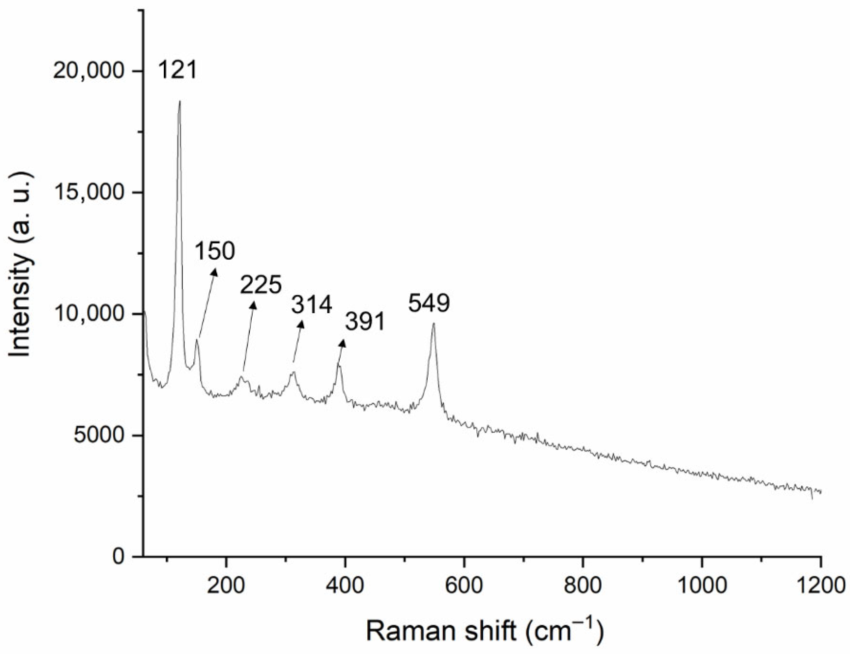
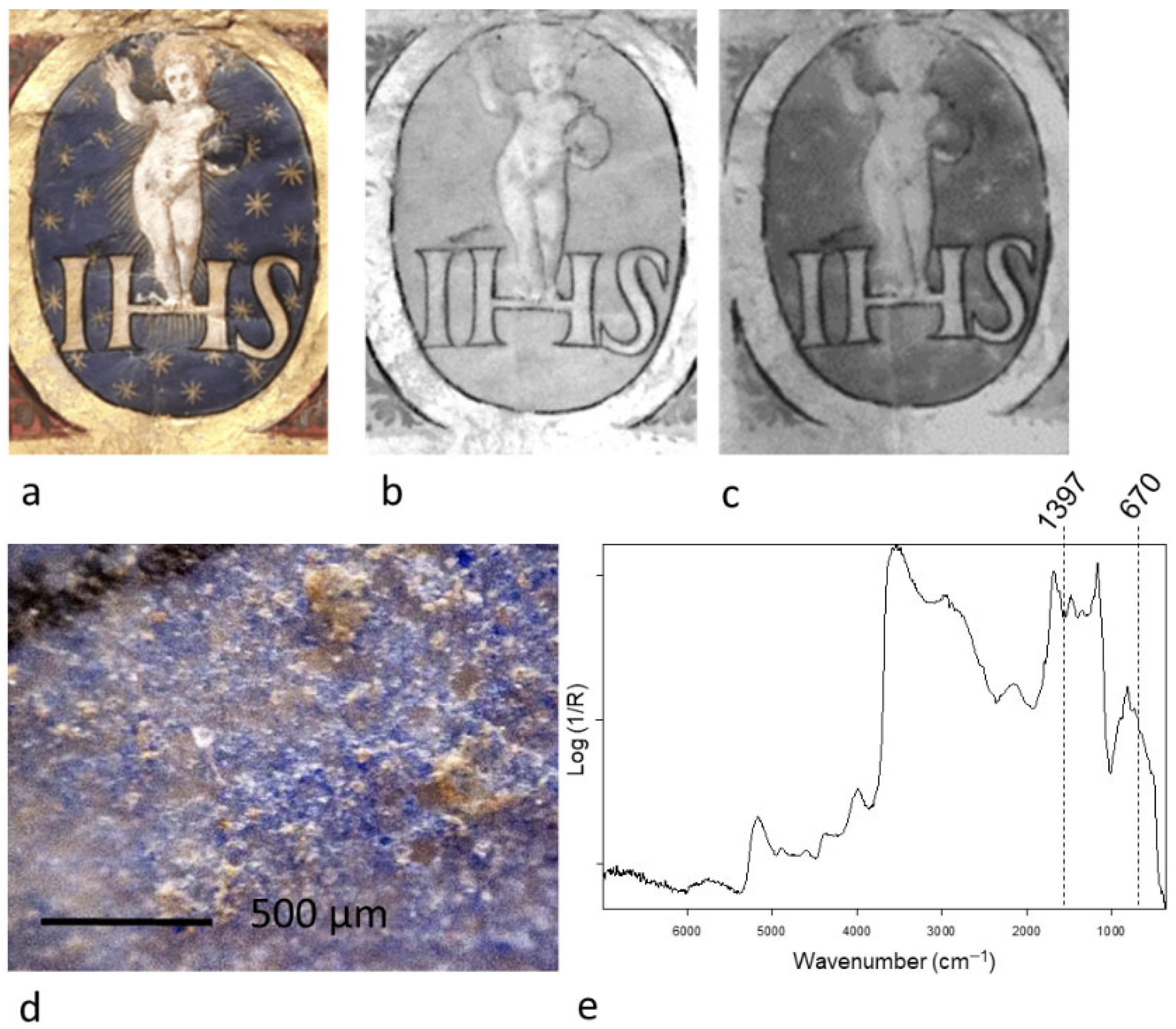
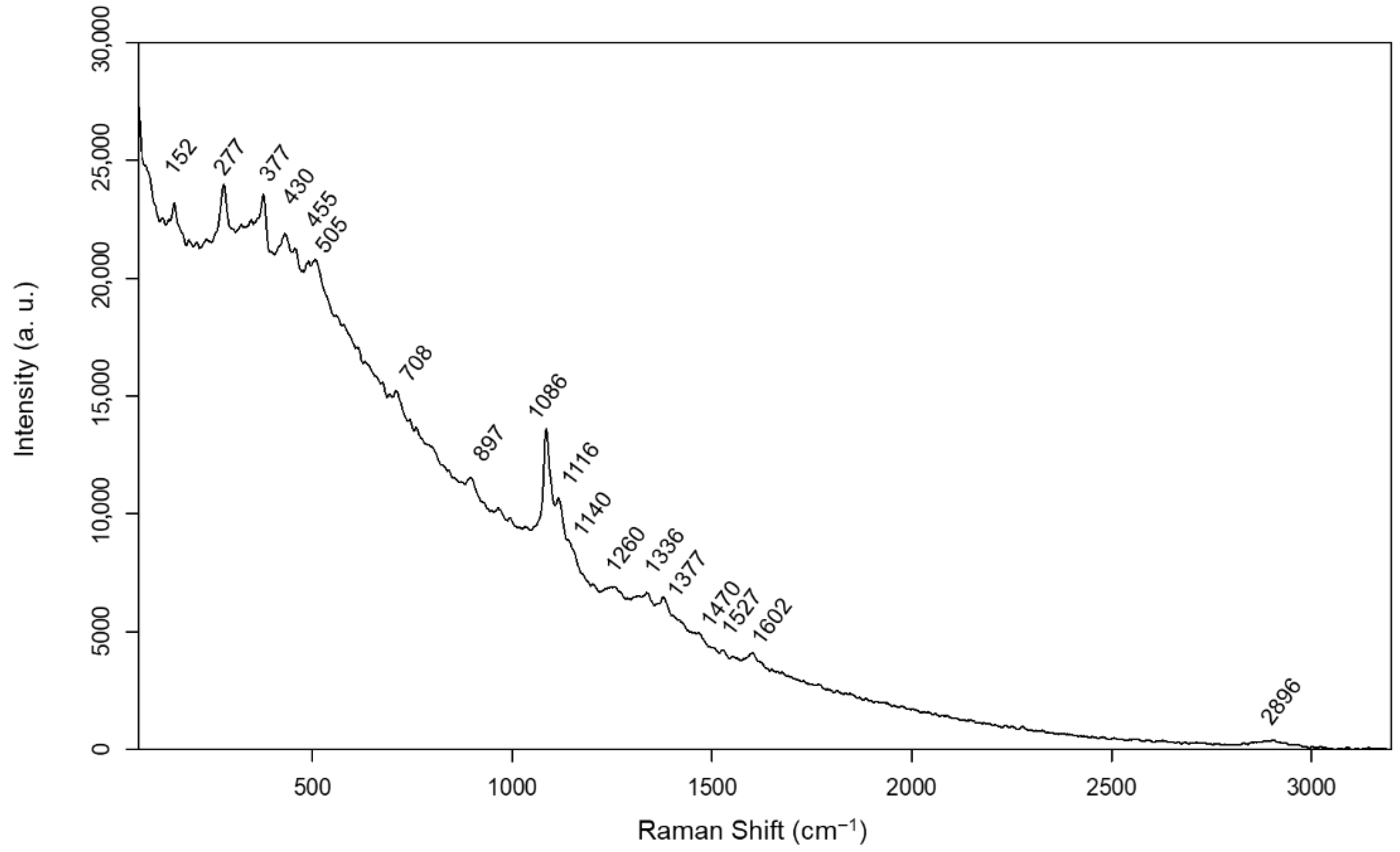
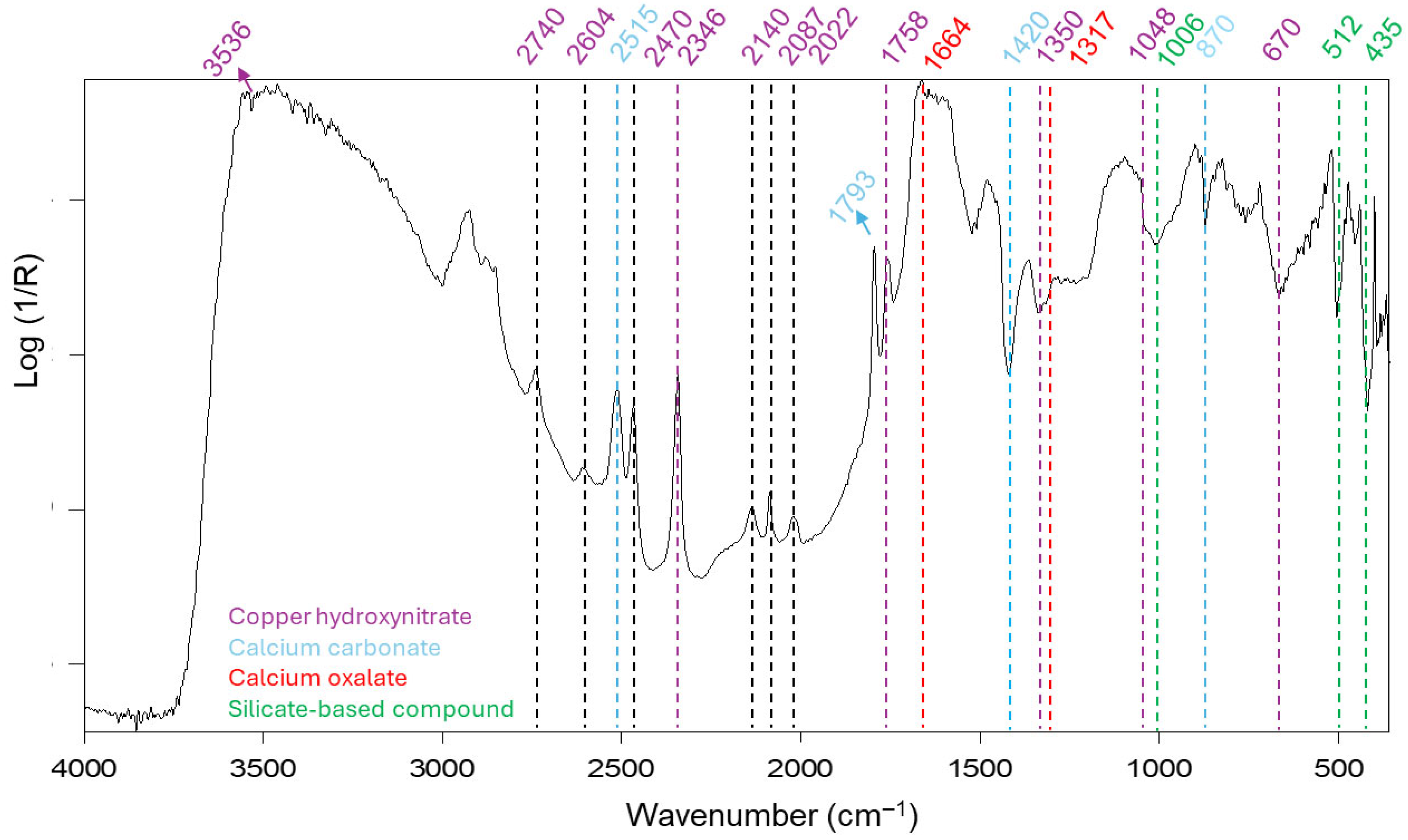

| Areas | Assignment | |
|---|---|---|
| Preparatory layer | support | Parchment |
| additives | Calcite | |
| Drawing | frame | Carbon-based pigment |
| Pictorial layer | red | Vermilion/Cinnabar |
| pink | Lead white mixed with animal derivate lake (aluminosilicates as mordant) | |
| orange | Minium/Red lead | |
| blue | Smalt mixed with lead white | |
| green | Copper hydroxynitrate (rouaite or gerhardtite) | |
| yellow | Lead, arsenic, tin-based pigments | |
| black | Carbon-based pigment mixed with lead white | |
| Metal layers | Gold and silver | |
| Written text | red | Vermilion/Cinnabar and silver tip |
| blue | Smalt and silver tip | |
| black | Iron-based ink |
Disclaimer/Publisher’s Note: The statements, opinions and data contained in all publications are solely those of the individual author(s) and contributor(s) and not of MDPI and/or the editor(s). MDPI and/or the editor(s) disclaim responsibility for any injury to people or property resulting from any ideas, methods, instructions or products referred to in the content. |
© 2025 by the authors. Licensee MDPI, Basel, Switzerland. This article is an open access article distributed under the terms and conditions of the Creative Commons Attribution (CC BY) license (https://creativecommons.org/licenses/by/4.0/).
Share and Cite
Pronti, L.; Romani, M.; Lanteri, L.; Bizzarri, F.; Colantonio, C.; Pelosi, C.; Ruberto, C.; Castelli, L.; Mazzinghi, A.; Spizzichino, V.; et al. Non-Invasive Investigation of a 16th-Century Illuminated Scroll: Pigments, Fillers, and Metal-Based Decorations. Minerals 2025, 15, 1252. https://doi.org/10.3390/min15121252
Pronti L, Romani M, Lanteri L, Bizzarri F, Colantonio C, Pelosi C, Ruberto C, Castelli L, Mazzinghi A, Spizzichino V, et al. Non-Invasive Investigation of a 16th-Century Illuminated Scroll: Pigments, Fillers, and Metal-Based Decorations. Minerals. 2025; 15(12):1252. https://doi.org/10.3390/min15121252
Chicago/Turabian StylePronti, Lucilla, Martina Romani, Luca Lanteri, Francesco Bizzarri, Claudia Colantonio, Claudia Pelosi, Chiara Ruberto, Lisa Castelli, Anna Mazzinghi, Valeria Spizzichino, and et al. 2025. "Non-Invasive Investigation of a 16th-Century Illuminated Scroll: Pigments, Fillers, and Metal-Based Decorations" Minerals 15, no. 12: 1252. https://doi.org/10.3390/min15121252
APA StylePronti, L., Romani, M., Lanteri, L., Bizzarri, F., Colantonio, C., Pelosi, C., Ruberto, C., Castelli, L., Mazzinghi, A., Spizzichino, V., & Cestelli Guidi, M. (2025). Non-Invasive Investigation of a 16th-Century Illuminated Scroll: Pigments, Fillers, and Metal-Based Decorations. Minerals, 15(12), 1252. https://doi.org/10.3390/min15121252


