Article

C14-HSL Quorum Sensing Signal Molecules: Promoting Role in Chalcopyrite Bioleaching Efficiency

1
School of Minerals Processing and Bioengineering, Central South University, Changsha 410083, China
2
Key Laboratory of Biohydrometallurgy, Ministry of Education, Changsha 410083, China
3
School of Metallurgy and Environment, Central South University, Changsha 410083, China
*
Author to whom correspondence should be addressed.
Minerals 2025, 15(12), 1248; https://doi.org/10.3390/min15121248
Submission received: 30 September 2025 / Revised: 2 November 2025 / Accepted: 11 November 2025 / Published: 26 November 2025
(This article belongs to the Special Issue Hydrometallurgical Treatments of Copper Ores, By-Products and Waste)

Abstract

N-tetradecanoyl-L-homoserine lactone (C14-HSL) is a long-chain signaling molecule belonging to acyl-homoserine lactones (AHLs), which is widely present in the quorum sensing (QS) system of Gram-negative bacteria. In this study, the effects of C14-HSL on chalcopyrite bioleaching mediated by Acidithiobacillus ferrooxidans (A. ferrooxidans) were investigated. After cultivating A. ferrooxidans with different energy substrates and exploring the potential mechanisms of signal molecule production, chalcopyrite was selected as the energy substrate for further study. Molecular docking analysis revealed that the high binding affinity between AHL and the receptor protein AfeR in A. ferrooxidans was beneficial for the activation of transcription by the AfeR-AHL complex, promoting their biological impact. The variations in the physicochemical parameters of pH, redox potential, and copper ions revealed that after adding C14-HSL, the leaching rate of chalcopyrite increased (1.15 times during the initial 12 days). Further analysis of the mechanism of extracellular polymers formation indicated that the presence of C14-HSL could promote the formation of biofilms and the adhesion of bacteria, facilitating mineral leaching rate of A. ferrooxidans. This research provides a theoretical basis for regulating the biological leaching process of chalcopyrite and metal recovery using signaling molecules, which could also be used to control environmental damage caused by acid mine/rock drainage.

1. Introduction

Chalcopyrite (CuFeS2) is a typical, widely occurring, and the most abundant primary copper sulfide mineral, and economically the most important copper mineral globally. It belongs to the category of relatively inert sulfide minerals [1,2]. The primary leaching methods for chalcopyrite can be categorized into three main approaches: pyrometallurgy-assisted leaching, direct hydrometallurgical leaching, and bioleaching. The traditional mining, beneficiation, and metallurgical processes of these mineral resources often resulted in low efficiency, severe environmental pollution, high production costs, lengthy procedures, and low resource utilization rates. Biohydrometallurgy is an established but evolving technology that utilizes microorganisms to catalyze the dissolution and extraction of valuable metals from ores and mineral waste materials. This process occurs through microbially generated acidic or oxidizing environments, and the resulting metal-rich solutions require subsequent purification steps to obtain high-purity products [3]. Compared to traditional pyrometallurgical processes, biohydrometallurgy offers both environmental and economic advantages and considered a promising mineral in processing and extractive metallurgy technology [4].
The passivation properties of chalcopyrite remain the major challenge in the bioleaching process. Low bioleaching efficiency has been frequently reported as a result of passivation of the mineral surface, such as jarosite and sulfur films formed during the leaching process. Moreover, the slow leaching rates and low extraction efficiency are the two major obstacles to the industrial application of biohydrometallurgy [1,4]. The interaction between microorganisms and metal minerals has existed on Earth for over a billion years, and microorganisms play a significant role in both the formation and dissolution of metal minerals. Theoretically, the oxidation and dissolution rates of sulfide minerals catalyzed by iron- and sulfur-oxidizing bacteria in nature are millions of times faster than under sterile conditions [5]. Therefore, enhancing the role of microorganisms is a fundamental strategy to address the challenges of chalcopyrite leaching.
Microbial adsorption on the surface of chalcopyrite is the first step in the complete oxidative dissolution process of chalcopyrite bioleaching. Bacterial attachment in bioleaching follows second-order irreversible kinetics of bacterial concentration and substrate surface area, which includes two stages: reversible adhesion and irreversible attachment [4]. Bacterial adhesion to the mineral surface enhances the bioleaching of ore sulfides and can be further divided into two steps. First, the presence of extracellular polymeric substances (EPSs) mediates the contact between the mineral surface and the cells, and then a biofilm covering the mineral surface is formed, with bacterial cells embedded in the continuous EPS layer. The formation of biofilm is the mechanism of bacterial cell adhesion and interaction with the mineral surface [6].Studies have found that the biofilm formation of attached bacteria is closely related to the production of EPS [7], and this is believed to be mediated by quorum sensing (QS) in many bacteria [8].
QS is a cell-to-cell communication mechanism that enables microorganisms to synchronize their collective behaviors by which microorganisms regulate their activities based on population density [9]. When the population density reaches a certain critical value, microorganisms exhibit “social” behaviors. They use signal molecules secreted into the environment as the basis for judging their population density. As the bacterial population density increases, signal molecules accumulate in the external environment [10]. Signal molecules are transported across the cell membrane and bind to corresponding receptors, subsequently triggering downstream gene expression and regulating physiological and biochemical processes, such as root nodulation, bioluminescence, protein secretion, flagellar movement, virulence factor production, plasmid transfer, and biofilm formation [11,12,13,14,15,16]. In the early 21st century, Farah et al. first reported the existence and characterization of a functional AHL quorum sensing system in the typical mineral-leaching bacterium A. ferrooxidans [17].This microorganism primarily produces AHLs with medium to long-chain of acyl and various substituents at the C-3 position. Genomic analysis has identified a quorum sensing-related gene locus containing two open reading frames, afeI and afeR, which are arranged in an inverted orientation and encode proteins highly similar to members of the acyl synthase (type I) and transcriptional regulatory protein (type R) families, respectively. Studies have confirmed that AfeI possesses AHL synthase activity, and both the afeI and afeR genes are transcribed normally in A. ferrooxidans [17]. In 2007, Rivas et al. further discovered that A. ferrooxidans possessed two types of quorum sensing systems, among which the AHL type serves as its principal system. The AHL-based quorum sensing system is an atypical system composed of the genes glyQ, glyS, gph, and act, with bioinformatic predictions indicating that they encode the α-subunit and β-subunit of glycine tRNA synthetase, phosphatase, and acyltransferase, respectively [18]. Notably, subsequent studies have demonstrated that the AfeI/R system exhibits substrate-dependent modulation of its regulatory function, with distinct differences in AHL production under sulfur versus ferrous iron conditions [19]. However, the specific roles of AfeI/R and AHL remain unclear and require further investigation.
Signal molecules could promote the formation of biofilms to a certain extent, thereby facilitating leaching. Alex et al. found that the addition of a mixture of C14-HSL, 3-oxo-C14-HSL, and 3-OH-C14-HSL to an A. ferrooxidans pyrite leaching system enhanced biofilm formation on the pyrite flakes [20]. In addition, Sigde et al. found that the addition of 3-OH-C14-HSL or the AHL analog tetrazole 9c promoted the formation of biofilms and the attachment of cells on sulfur flakes [21]. In this study, signal molecules were utilized to enhance the bioleaching rate of chalcopyrite. The effect of signal molecules on chalcopyrite bioleaching mediated by A. ferrooxidans was systematically examined. A. ferrooxidans was cultured with different energy substrates to analyze the potential production mechanisms of signal molecules. Furthermore, molecular docking was employed to investigate their potential regulatory mechanisms. This study aims to provide a theoretical basis and new strategies for optimizing the bioleaching process of sulfide ores by delving into the role and regulatory mechanisms of signaling molecules in the acidophilic ferrous sulfide bacteria mediated bioleaching process of chalcopyrite. In addition, we hope that this study can not only provide reference for improving the leaching efficiency of refractory sulfide ores (such as chalcopyrite) using quorum sensing mechanism, but also provide important theoretical guidance and practical basis for the future screening and development of low-cost and efficient AHLs analogs to replace expensive natural signal molecules, thereby promoting the industrial application of signal molecules in sulfide ore bioleaching.

2. Materials and Methods

2.1. Preparation of Minerals

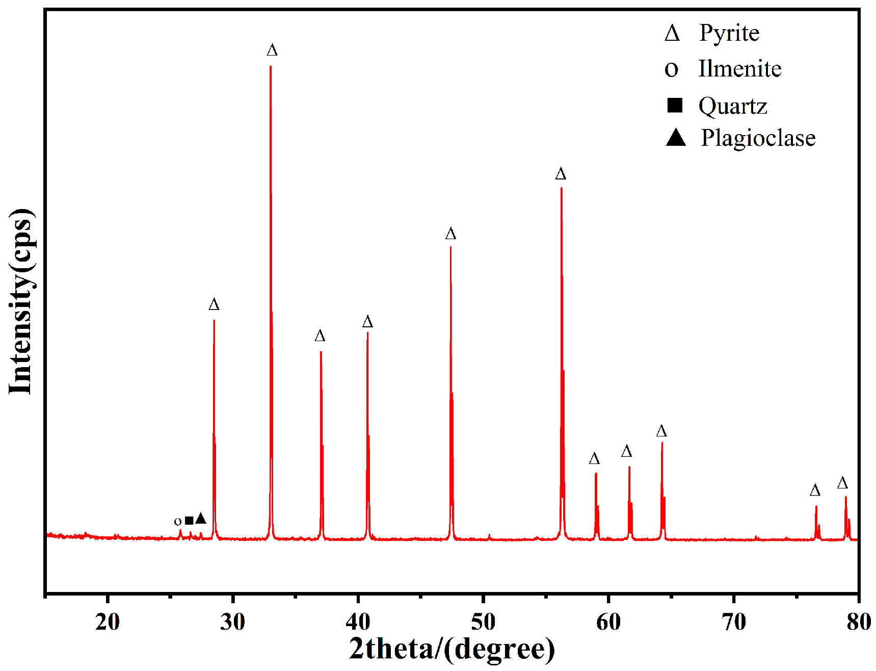
The pure chalcopyrite and pytite specimen employed in this experiment was procured from Dongchuan, Yunnan Province. The ore samples underwent a crushing process to yield particles with a size smaller than 74 μm, and the X-ray diffraction (XRD) pattern of this mineral is presented in Figure 1 and Figure 2. Mineralogical analysis via XRD indicated that the primary constituents of the chalcopyrite ore sample were chalcopyrite (80.4%), pyrite (8.0%), quartz (3.6%), and dolomite (8.0%). The main components of the pyrite sample were pyrite (96.00%), ilmenite (1.90%), quartz (0.96%), and plagioclase (1.14%).

2.2. Strain and Cultivation of Strains with Different Energy Substrates

The strain of A. ferrooxidans ATCC 23270 was obtained from the Key Lab of Biometallurgy of the Ministry of Education, Central South University. The 9K basal medium comprising (NH4)2SO4 (3.0 g/L), K2HPO4 (0.5 g/L), KCl (0.1 g/L), Ca(NO3)2 (0.01 g/L), and MgSO4·7H2O (0.5 g/L) was used in the bioleaching experiments. A total of 100 mL of the medium was added to a 250 mL Erlenmeyer flask, and the initial pH of the medium was adjusted to 2.0 using sulfuric acid, which was autoclaved at 121 °C for 20 min. Subsequently, all the energy substrates (chalcopyrite and pyrite (2% (w/v)), elemental sulfur (10 g/L), and FeSO4·7H2O (44.7 g/L)) were sterilized by ultraviolet irradiation for 30 min and individually added to the pre-sterilized medium in three replicates. The A. ferrooxidans was inoculated into the sterilized 9K medium at a cell density of 4 × 10⁷ cells/mL and incubated at 30 °C, 180 rpm/min. The bacterial cells were harvested by centrifugation at the end of the logarithmic growth phase, and the supernatant was collected to evaluate the AHL concentration.

2.3. Quantification of AHL via High-Performance Liquid Chromatography–Mass Spectrometry (HPLC-MS)

The extraction steps for AHL began with culturing the cells until the end of the log phase and then centrifuged at 10,000 rpm for 10 min to remove solid impurities. The supernatant was filtered through a 0.22 μm hydrophilic filter membrane and thoroughly mixed with chromatographic-grade ethyl acetate (HPLC, ≥99.9%, Sigma-Aldrich, St. Louis, MO, USA) in a ratio of 1:3, allowed to stand for 5 min, and the upper organic phase was collected. We repeated the above extraction step twice and evaporated the combined extracts to dryness under vacuum using a rotary evaporator. After that, 1 mL of chromatographic-grade methanol (HPLC, ≥99.9%, TEDIA, Hamilton, OH, USA) was added to redissolve the residue, obtaining a crude AHL extract. The crude extract was passed through a 0.22 μm organic-phase filter membrane and analyzed by HPLC-MS.
The N-(3-oxododecanoyl)-L-homoserine lactone (3-oxo-C12-HSL), N-(3-hydroxydodecanoyl)-DL-homoserine lactone (3-OH-C12-HSL), N-dodecanoyl-L-homoserine lactone (C12-HSL), N-(3-oxotetradecanoyl)-L-homoserine lactone (3-oxo-C14-HSL), and N-tetradecanoyl-L-homoserine lactone (C14-HSL) used in the experiment were purchased from Sigma Company with purity greater than 98%. The selection of these five signaling molecules was based on prior research, which demonstrated that long-chain AHLs exerted more significant regulatory effects on Acidithiobacillus ferrooxidans compared to short-chain variants [17,22]. The five signal molecule standard substances were separately prepared as individual standard stock solutions at 500 μg/L using methanol. Subsequently, 100 μL of each stock solution was combined and diluted with methanol to prepare a mixed standard solution. This mixed standard solution was then subjected to stepwise dilution with methanol to obtain a mixed standard working solution at a final concentration of 500 ng/L. To optimize the chromatographic conditions, 0.2% (v/v) formic acid and 5 mM ammonium acetate (NH4Ac) were added to mobile phase B (acetonitrile). A full scan was performed in electrospray ionization positive (ES+) mode, and the cone voltage and collision voltage were adjusted to optimize the mass spectrometry conditions.
The concentration of signaling molecules was detected using liquid chromatography-mass spectrometry (HPLC-MS). HPLC (LC-20A, Shimadzu, Kyoto, Japan) was operated under the following conditions: Chromatographic column: Eclipse Plus C18. Column temperature: 30 °C. Injection volume: 10 μL. Mobile phase were A:B = (aqueous phase: 0.1% formic acid—5 mM NH4AC):(acetonitrile: 0.2% formic acid—5 mM NH4AC). Flow rate: 0.3 mL/min. Elution gradient: At 0.00 min, the proportion of phase B was 40%. Within 6.00 min, the proportion of phase B linearly increases to 95% and was maintained at 95% for 5 min. From 11.00 to 13.00 min, the proportion of phase B was reduced to 40%, and was balanced at 40% until 14 min. Mass spectrometry conditions (TripleTOFTM 5600+, AB SCIEX Technologies, Redwood City, CA, USA): The ion source was an ESI source, positive ion, scan type: MRM. Curtain gas: 15 psi. Spray voltage: +4000 V. Nebulizer gas pressure: 60 psi. Auxiliary gas pressure: 70 psi. Nebulization temperature: 400 °C.

2.4. Bioleaching Experiments with Different Signal Molecules

A volume of 100 mL of 9K medium was added to a 250 mL conical flask, and the initial pH was adjusted to 2.0 using sulfuric acid. The conical flask was sterilized in a high-pressure steam autoclave at 121 °C for 20 min. Subsequently, chalcopyrite (pre-sterilized by ultraviolet irradiation for 30 min) was added to the sterilized medium to achieve a pulp concentration of 2%. The A. ferrooxidans strain was inoculated into the sterilized 9K medium at a concentration of 2% (v/v), resulting in a final cell density of 4 × 10⁷ cells/mL. Following inoculation, five AHL molecules—C12-HSL, 3-OH-C12-HSL, 3-oxo-C12-HSL, C14-HSL, and 3-OH-C14-HSL dissolved in methanol were separately added to the experimental groups to achieve a final concentration of 0.5 μM in the system. Control experiments included a biotic control, comprising an equivalent bacterial concentration supplemented with an equal volume of methanol, and an abiotic control (CK) without bacterial inoculation to account for biological and non-biological effects, respectively. Each group of experiments was cultured in triplicate using a shaker incubator maintained at 30 °C with a rotational speed of 180 rpm. The supernatant was collected every three days for physicochemical analysis.

2.5. Analysis of Main Physicochemical Properties in Solution

The supernatant was collected every three days to measure pH, oxidation-reduction potential (ORP) and Cu2+ concentration. The pH value of the leachate was determined by a pH meter, and the ORP of the leachate was determined by an ORP meter. The concentration of copper ions in the solution was determined by 2,9-dimethyl-1,10-phenanthroline spectrophotometry. We regularly supplemented an equal amount of sterile distilled water to make up for the evaporated water during the leaching process. The consumed solution for sampling and analysis was compensated with sterile 9k medium at pH 2.

2.6. Counting of Free and Adsorbed Microorganisms

The following steps were carried out: A 2 mL sample was centrifuged at 3000 rpm for 5 min using a low-speed centrifuge to separate the bacterial solution from mineral particles, and the supernatant containing free bacteria was carefully removed. The remaining mineral pellet was resuspended in sterile 9K medium to a final volume of 2 mL. Sterile 9k medium was added to the remaining mineral residue to a volume of 2 mL. After being shaken for 20 min with a vortex oscillator, the mixture was centrifuged at 3000 rpm for 6 min to separate the adsorbed microorganisms from the minerals. Both free and adsorbed microorganisms were quantified using a hemocytometer.

2.7. Extraction and Determination of Extracellular Polymeric Substances

After 24 days of microbial leaching of chalcopyrite, the culture medium was centrifuged at 9000 rpm and 4 °C for 10 min. The supernatant was collected in sterile tube I. To the remaining slag in the centrifuge tube, 10 mL of sterile water and 1 g of sterile glass beads with a diameter of 0.5 mm were added. Then, it was shaken on a micro vortex mixer for 10 min and centrifuged at 3000 r/min for 1 min. The supernatant was poured out, and the cell concentration of the supernatant was examined under an optical microscope. The above steps were repeated until no microorganisms could be observed in the supernatant under an optical microscope. All the supernatants were collected in the above steps, centrifuged at 10,000 r/min for 5 min, and the supernatant was collected into sterile tube II. Adding 30 mL of sterile water to the remaining precipitate in the centrifuge tube, the tube was incubated in a water bath at 75 °C for 30 min, then removed and allowed to cool, after which it was centrifuged at 3000 r/min for 2 min, and the supernatant was collected into sterile tube III. The supernatants in sterile tubes I, II, and III were combined and filtered through a 0.22 μm pore size filter under sterile conditions to eliminate residual bacteria, and the extracellular polymer solution was obtained. Using glucose as the standard, the anthrone–sulfuric acid method was employed to determine polysaccharides. Bovine serum albumin was used as the standard, and the Coomassie brilliant blue method was employed to determine protein.

2.8. Three-Dimensional Fluorescence Spectral Analysis of Extracellular Polymeric Substances

The three-dimensional fluorescence spectral data of EPS were determined by using the Hitachi F-7000 fluorescence spectrophotometer (Hitachi Scientific Instrument Beijing Co., Ltd., Beijing, China). The excitation spectrum was scanned from 200 to 500 nm at 5 nm intervals, while the emission spectrum was collected from 200 to 600 nm in 5 nm increments, with a scanning speed of 12,000 nm/min. In order to eliminate the influence of the Raman scattering peak of water, the fluorescence intensity of the blank water sample was subtracted from the scanning results. Matlab R2024 was used to eliminate Rayleigh scattering and Raman scattering. Finally, Origin 9.6 software was used to visualize and draw the three-dimensional fluorescence spectral data.

2.9. Molecular Docking

The protein sequences of A. ferrooxidans with AfeI (AAZ20805.1) and AfeR (AAV53702.2) were obtained from the NCBI database in FASTA format, and their homology models were generated and evaluated using SWISS-MODEL (https://swissmodel.expasy.org, accessed on 18 August 2025). The 3D structures of the donors (AHL and vanillin) were downloaded from ZINC (https://zinc.docking.org/substances/home/, accessed on 18 August 2025) and were as follows: ZINC42764482 (C12-HSL), ZINC64859360 (C14-HSL), ZINC8436851 (3-oxo-C12-HSL), ZINC38146043(3-oxo-C14-HSL), ZINC42764616 (3-OH-C12-HSL), as well as ZINC000002567933 (vanillin, an AHL analog). The receptor and the donor were prepared according to the standard protocol of the Discovery Studio 2019 software. The receptor was generated by removing the water molecules, adding hydrogen atoms, as well as defining protein receptors and binding sites for subsequent molecular docking analysis. All compounds were geometrically optimized to generate multiple conformations of the compound and minimize the molecular energy of the compound. Molecular docking was performed with LibDock, and the data were visualized using Discovery Studio software.

2.10. Statistical Analysis

All experiments were performed three times. The experimental data was presented as mean ± standard deviation (mean ± SD). Descriptive statistics were performed, and graphs were generated using Origin 9.6 software. SPSS 26 was used for one-way analysis of variance (ANOVA) and Tukey’s test, with a p-value < 0.05 representing the significant difference. Molecular docking was performed with LibDock, and the data were visualized using Discovery Studio 2019 software.

3. Results and Discussion

3.1. Generation and Identification of AHL in A. ferrooxidans

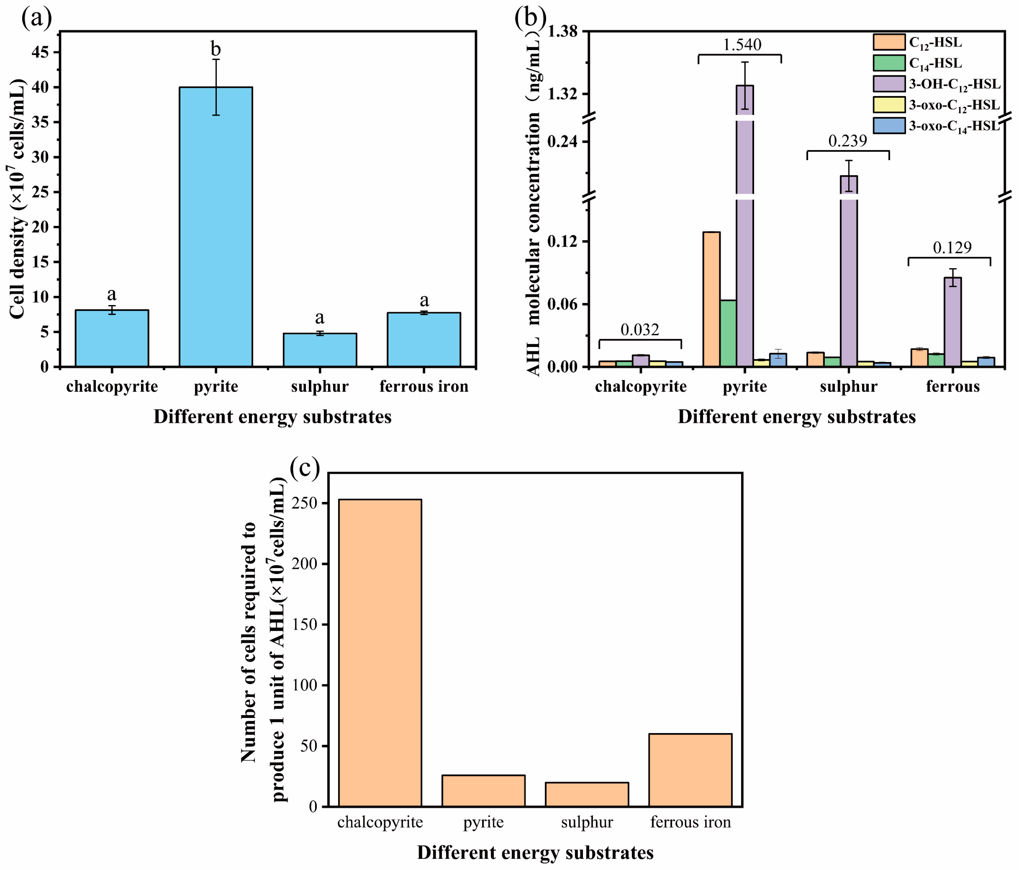
Among the Gram-negative bacteria, QS is mostly achieved through the biosynthesis and subsequent concentration-sensing of AHL signals, a small neutral lipid molecule with a homoserine lactone (HSL) moiety linked to a 4 to 18 carbon acyl side chain [23]. A. ferrooxidans mainly produces acyl-homoserine lactones (AHLs) including C12-HSL, C14-HSL, 3-OH-C8-HSL, 3-OH-C10-HSL, 3-OH-C12-HSL, 3-OH-C14-HSL, 3-OH-C16-HSL, 3-O-C12-HSL, and 3-O-C14-HSL [17]. The energy substrates could influence both the types of AHLs and the regulatory function of these AHLs [19]. The cell growth and generation of 5 commercially available AHLs (3-oxo-C12-HSL, 3-OH-C12-HSL, C12-HSL, 3-oxo-C14-HSL and C14-HSL) of A. ferrooxidans in different energy substrates including chalcopyrite, pyrite, elemental sulfur, or FeSO4·7H2O were measured. When A. ferrooxidans was cultured with different energy sources, the cell concentration at the end of the log stage was 4.0 × 10⁸ cells/mL (pyrite), 7.7 × 10⁷ cells/mL (ferrous iron), 4.8 × 10⁷ cells/mL (sulfur), or 8.1 × 10⁷ cells/mL (chalcopyrite), respectively. Under each culture condition, five kinds of AHLs were detected in the culture supernatant. Their [MS + H]+ values were 284.3, 312.2, 300.3, 298.3, and 326.3, respectively, which are close to the [MS + H]+ values of C12-HSL, C14-HSL, 3-OH-C12-HSL, 3-oxo-C12-HSL, and 3-oxo-C14-HSL (Table 1). In addition, these five kinds of AHL have the characteristics of protonation of homoserine lactone (m/z 102.1) [24], and the MS2 spectra of these five AHLs are the same as those of standard compounds. Therefore, we conclude that A. ferrooxidans can generally produce five signal molecules, namely C12-HSL, C14-HSL,3-OH-C12-HSL,3-oxo-C12-HSL, and 3-oxo-C14-HSL.
Furthermore, the relative contents of these five AHLs in the extract are detected by the peak area normalization method [25]. The results showed that the total amount of AHL produced by A. ferrooxidans cultured with pyrite was the highest (1.540 ng/mL), followed by sulfur (0.238 ng/mL), ferrous iron (0.128 ng/mL), and chalcopyrite (0.032 ng/mL). Among the AHL molecules produced by A. ferrooxidans cultured with different substrates, the content of 3-OH-C12-HSL was the highest, accounting for 86.97% (sulfur), 86.23% (pyrite), 66.41% (ferrous iron), and 34.38% (chalcopyrite), respectively. Consistent with former results, 3-OH-C12-HSL except 3-OH-C14-HSL (not commercially available) was the most abundant and may play important roles in growth and metabolism regulation [17,19]. The second was C12-HSL, accounting for 5.88% (sulfur), 54.20% (pyrite), 13.28% (ferrous iron), and 15.63% (chalcopyrite), respectively. The third was C14-HSL, accounting for 3.78% (sulfur), 4.15% (pyrite), 9.38% (ferrous iron), and 18.75% (chalcopyrite), respectively (Figure 3b). Thus, energy substances affect the production of signal molecules, among which the total AHL concentration of pyrite was the most abundant. Based on the total amount of AHL produced by A. ferrooxidans and the cell concentration at the end of the log stage, the number of cells required to produce a unit concentration of AHL (1 ng/mL) were found to occur in the following order: sulfur (20 × 107 cells/mL) < pyrite (26 × 107 cells/mL) < ferrous iron (60 × 107 cells/mL) < chalcopyrite (253 × 107 cells/mL) (Figure 3c).
The highest AHL concentration was observed when grown with pyrite as the energy source, which corresponded to the highest cell density under all tested energy conditions (ferrous iron, sulfur, pyrite, or chalcopyrite) (Figure 3a,b). However, sulfur powder-cultured specimens exhibited comparatively lower cell concentrations than iron or chalcopyrite, yet paradoxically displayed higher AHL content in their supernatant, indicating cell concentration plays a restricted regulatory role in quorum sensing molecule production. These observations were in accordance with the fact that the acyl-HSL concentrations in S0-enriched media were higher than those in Fe2+-enriched media [19].
The bacterium A. ferrooxidans obtains energy by oxidizing iron and reduced inorganic sulfur compounds (RISCs) [26,27]. The synthesis of AHL requires S-adenosyl-L-methionine (SAM), derived from methionine via amino acid/sulfur metabolism, and acyl-ACP, which originates from the fatty acid metabolism pathway [17,28,29]. The SAM, which is derived from sulfur metabolism, may be affected by S-enriched media growth conditions, thereby facilitating the acyI-HSLs synthesized by A. ferrooxidans, which was in accordance with the study that the transcription levels of the afeI gene were higher in cells grown in sulfur and thiosulfate media than in iron-grown cells [17,30]. The first step in sulfur assimilation corresponds to sulfate uptake, the pool of SAM in A. ferrooxidans should depend on sulfate availability. Therefore, AHL synthesis could be affected under the growth conditions that we employed [31]. However, the way in which the energy source (iron versus sulfur or thiosulfate) could affect the nature of the AHLs is still unknown, since no information relating to external acidic pH, energy metabolism, and cell wall metabolism is currently available for A. ferrooxidans [31]. However, based on studies of other acidophilic or chemoautotrophic microorganisms, we hypothesize that these factors may affect the characteristics of AHLs through the following pathways: Acidithiobacillus ferrooxidans is an acidophilic bacterium with an optimal growth pH range of 1.5 to 2.5, and the external acidic environment is associated with the stability of AHLs. Acidithiobacillus ferrooxidans obtains energy through redox reactions, including the oxidation of ferrous iron, sulfur, and thiosulfate. Different energy sources may affect the bacterial metabolic pathways and energy output, thereby influencing the synthesis of AHLs. The cell wall structure of Acidithiobacillus ferrooxidans is crucial for maintaining its survival in extremely acidic environments. Cell wall metabolism may affect the transport and secretion of AHLs. Changes in the composition and structure of the cell wall may influence the diffusion rate of AHLs and the concentration gradient between intracellular and extracellular spaces.

3.2. Structural Insights into the Mechanisms of AfeI/AfeR-AHL Interaction

Molecular docking is a computational method used to predict the binding interaction between small molecules and target molecules (such as proteins) [32]. In A. ferrooxidans, a typical LuxI/R-type quorum-sensing system includes functional genes afeI and afeR. Among them, the AHL synthase encoded by afeI and the AHL signal molecule receptor protein encoded by afeR [9]. Due to the common structural frameworks and biosynthetic pathways shared by AHL-based QS molecules, elucidating the mechanisms of their molecular recognition specificity has been a significant challenge.
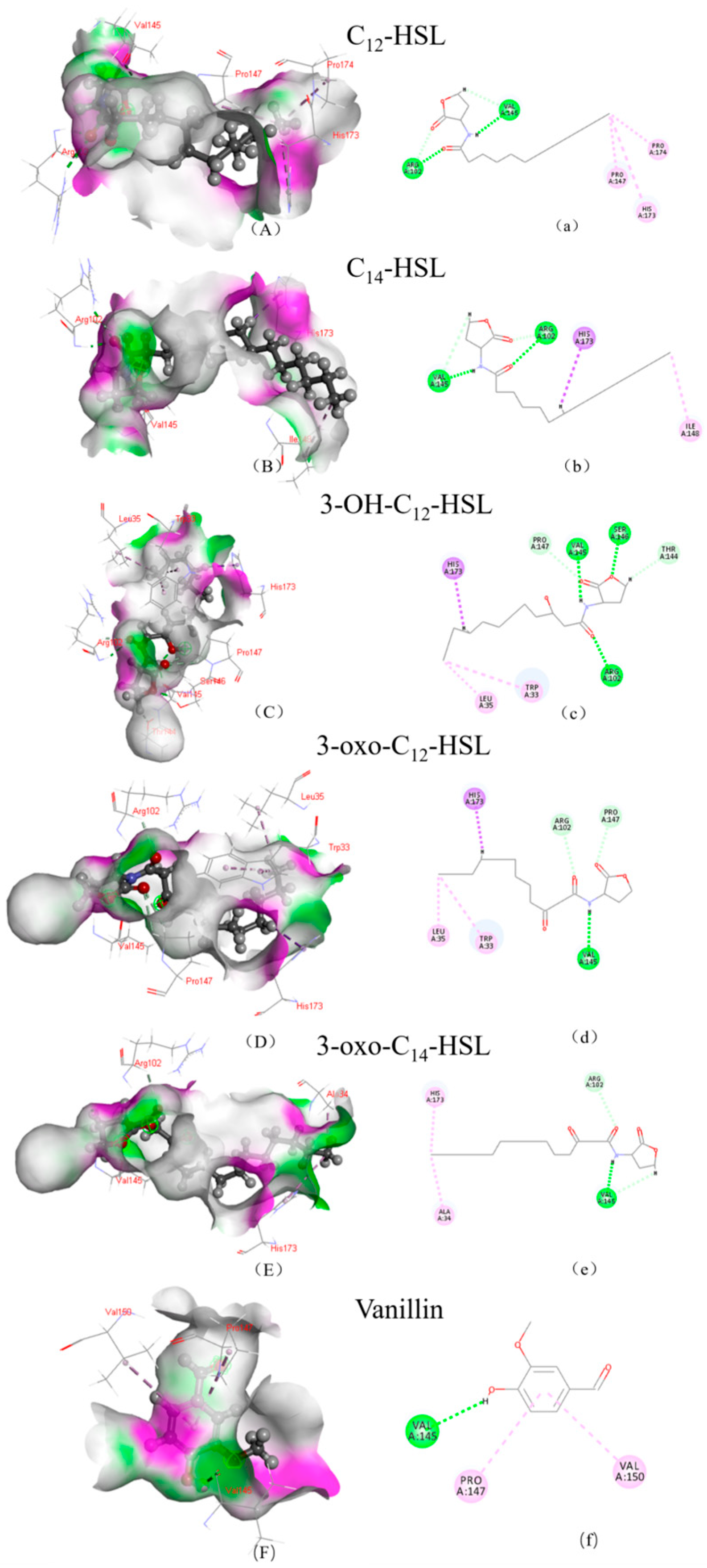
Molecular docking was used to analyze the interaction between AfeI/AfeR and HSL to explore the regulating mechanism of microbial leaching of chalcopyrite. The AHL molecules, including C12-HSL, C14-HSL, 3-OH-C12-HSL, 3-oxo-C12-HSL, and 3-oxo-C14-HSL, as well as vanillin all formed hydrogen bonds and hydrophobic interactions with the synthetic protein AfeI (Figure 4). There were two identical amino acid residue interaction sites (A/VAL:145, A/ARG:102) for hydrogen bonds and one identical amino acid residue interaction site (A/HIS:173) for hydrophobic interaction. Vanillin has one residue interaction site (A/VAL:145) for hydrogen bonds and two residue interaction sites (A/PRO:147, A/VAL:150) for hydrophobic interaction (Table 2).
The LibDock score is a parameter for comprehensive evaluation of energy, geometry, and chemical environment. The higher the LibDock score, the higher the activity of small molecules binding to the receptor. 3-oxo-C14-HSL has the highest binding affinity with AfeI protein with a LibDock score of 128.87, followed by C14-HSL (123.159), 3-OH-C12-HSL (123.039), 3-oxo-C12-HSL (122.509), C12-HSL (120.501) and vanillin (77.3768) (Table 2). The results indicated that AHL with a long side chain showed higher binding affinity with the AfeI protein [33]. The affinity between the AfeI protein and AHL with a ketone substitution at the C-3 position of the side chain is higher than that of the unsubstituted [17]. However, the relationship between the affinity and AHL concentrations in A. ferrooxidans cultures needs further study.
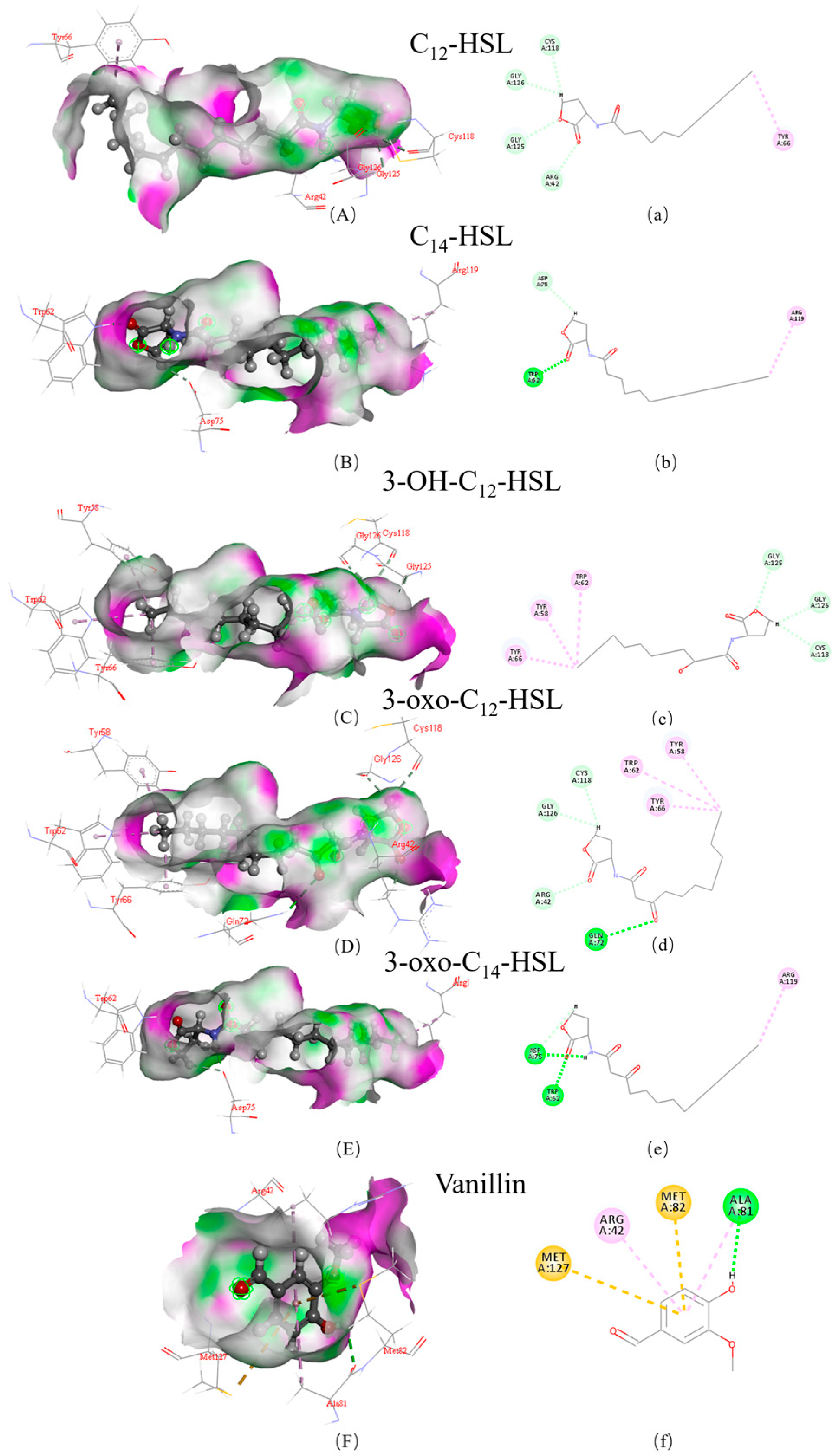
The binding situation of AHL and receptor protein AfeR is shown in Figure 5. In the interface pocket, the polar region (green) and hydrophobic region (purple) in the AfeR protein can be clearly observed. The AHL molecules, including C12-HSL, C14-HSL, 3-OH-C12-HSL, 3-oxo-C12-HSL, 3-oxo-C14-HSL, and vanillin, all formed hydrogen bonds and hydrophobic interactions with the AfeR protein. The results indicated that the affinity between the AfeR protein and AHL depends on the length of the side chain. There were two identical amino acid residue interaction sites (A/CYS:118, A/GLY:126) for hydrogen bonds and one identical amino acid residue interaction site (A/TYR:66) for hydrophobic interaction shared by C12-HSL, 3-OH-C12-HSL, and 3-oxo-C12-HSL. However, C14-HSL and 3-oxo-C14-HSL shared two identical amino acid residue interaction sites (A/TRP:62, A/ASP:75) for hydrogen bonds and one identical amino acid residue interaction site (A/ARG:119) for hydrophobic interaction (Table 2). Vanillin could form hydrogen bonds with the receptor protein AfeR, but it did not have the same amino acid residue interaction sites as AHL.
The LibDock scores of the AHL and AfeR protein complexes from high to low were 143.914 (3-oxo-C14-HSL), 139.272 (C14-HSL), 133.245 (3-OH-C12-HSL), 130.135 (3-oxo-C12-HSL), 127.537 (C12-HSL), and vanillin (78.0117) (Table 2). The LibDock score of 3-oxo-C14-HSL and C14-HSL is higher than that of the other HSLs, and they bind more tightly to the receptor protein. The high binding affinity between AHL and the AfeR receptor protein is beneficial for the activation of transcription by the AfeR-AHL complex, promoting their biological impact. In addition, key protein residues especially (A/TRP:62, A/ASP:75) and (A/ARG:119) could be promising ligands for further studies, including biochemical assays and site-directed mutagenesis, to clarify these mechanisms [34,35].
The molecular docking results indicated that there are differences in the binding affinity between different AHL molecules and AfeI and AfeR proteins. This predicted difference in binding affinity is consistent with the gene expression regulation and phenotype changes observed in previous studies. Research has shown that the binding of AHL molecules to AfeR receptor proteins is a crucial first step in activating downstream gene transcription. In particular, AHLs with high affinity for AfeR (such as C14-HSL and its derivatives) can effectively promote the formation of AfeR-AHL complex, thereby specifically recognizing and binding to the upstream promoter region of the afeI gene, significantly upregulating the expression of the afeI gene and forming a self-induced positive feedback loop [17,21,30].This transcriptional activation directly extends to the regulation of various enzyme activities. Functional genomics analysis shows that AfeR protein can regulate a complex network of over 140 genes, many of which are closely related to biofilm formation, extracellular polysaccharide (EPS) biosynthesis, and substrate metabolism. For example, after adding AHL analogs (such as tetrazole 9c), transcriptome analysis revealed that genes associated with early biofilm formation (such as those encoding proton channel proteins, phosphate transport systems, and EPS precursor synthase) were significantly induced, which directly promoted cell adhesion to sulfide ore surfaces and bioleaching efficiency. On the contrary, some genes related to biofilm maturation and carbohydrate metabolism are inhibited [21,36].

3.3. Effect of Additional Signal Molecules on Chalcopyrite Leaching by A. ferrooxidans

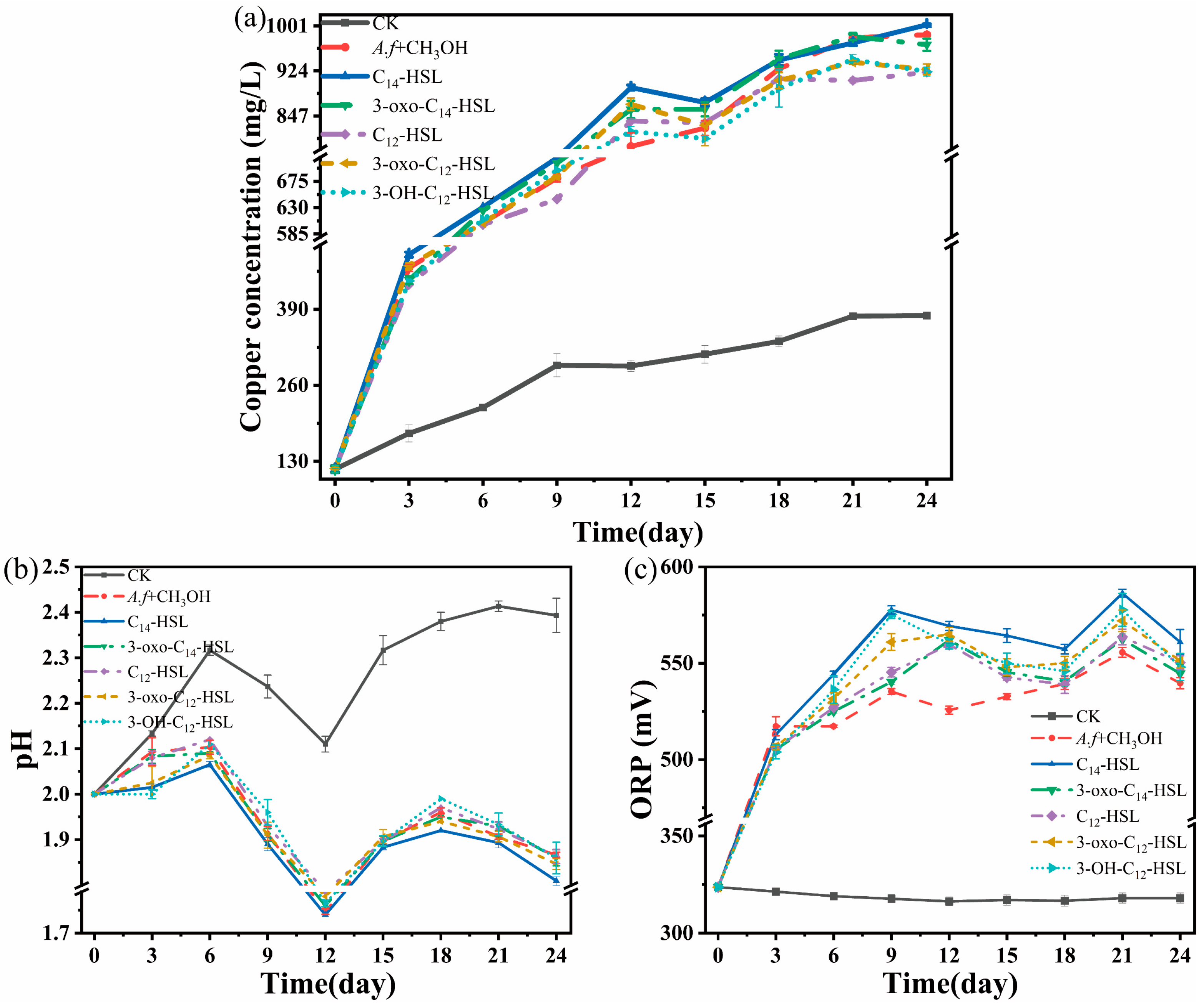
The QS receptors have a high affinity for their cognate ligands in comparison with their non-cognate ligands [37,38]. LuxR-type receptors respond to signals in a concentration range from five to a few hundred nanomolar [21,29,39]. The addition of AHL molecules or its analog promoted the formation of biofilms and the attachment of cells to sulfur or pyrite [20,21]. To verify the roles of signal molecules on chalcopyrite leaching by A. ferrooxidans, 0.5 μM of C12-HSL, C14-HSL,3-OH-C12-HSL,3-oxo-C12-HSL, and 3-oxo-C14-HSL were added to the system. The behaviors of key physicochemical factors during the bioleaching process are shown in Figure 6. The general trends of copper concentration, variations in pH and ORP were similar in the six groups.
The copper concentration of the six groups continually increased throughout the leaching process (Figure 6a). On the 12th day of leaching, the total copper ion concentration of C14-HSL was 895.65 mg/L, while that of the control group was 794.90 mg/L. The copper concentration with different acyl-HSL was found to occur in the following order: C14-HSL > 3-oxo-C12-HSL > 3-oxo-C14-HSL > C12-HSL > 3-OH-C12-HSL > methanol control on the 12th day of leaching. The copper ion concentration (1002.90 mg/L) in the leaching solution with C14-HSL was found to be the highest after 24 days of leaching.
During the bioleaching process, the pH value increased then decreased followed by another increased (Figure 6b). The pH value of all groups generally increased in the first 6 days. The pH of the leaching solution in the sterile control group slowly rose from 2.00 to 2.39, while the others increased to 2.1. From day 6 to day 12, the pH value of all groups maintained a continuous downtrend, decreasing to around 1.75. After 12 days, the pH in different groups slightly increased and then dropped to around 1.80–1.87. During the 24-day leaching process, the pH values in the group with C14 were always lower than the other groups. Two times of pH increase were related to the alkaline gangue in chalcopyrite and the formation of the passivation layer during the dissolution process [40,41].
It was generally accepted that the variation in ORP was closely related to the proportion of Fe3+/Fe2+, and played a determining role in chalcopyrite bioleaching (surface species of chalcopyrite during bioleaching by moderately thermophilic bacteria). Compared with the abiotic control, the ORP of the six groups increased continuously and then reached a plateau (about 525–575 mV) after 24 days (Figure 6c). The redox potential of the leaching solution with C14-HSL was higher than that in the other groups. With the increase in ORP higher than 550 mV, the dissolution of copper ions from chalcopyrite finally became slow and stagnant [42].
The analysis showed that AHL, especially C14-HSL, could accelerate chalcopyrite dissolution during the initial 12 days with the variation in physicochemical factors, such as redox potential and pH. Former results demonstrated that overexpression of the afeI operon could promote cell growth in So-media and inhibit cell growth in Fe2+-media. Acyl-HSLs mediated distinct regulation strategies were employed to influence bacterial metabolism of A. ferroxidase cultivated in different energy substrates [19].
This study preliminarily distinguished the primary biological effects from purely chemical actions during chalcopyrite bioleaching by establishing a sterile abiotic control (CK). The significant differences between the biological treatment and the abiotic control (CK) support the conclusion that the observed effects were predominantly driven by microbial activity. Future research will introduce a heat-killed bacterial control group to more comprehensively and precisely resolve the respective contributions of live microbial metabolic activity and the physical effects of cellular structures in the bioleaching process of sulfide ores.

3.4. The Influence of C14-HSL on EPS Production During Chalcopyrite Bioleaching

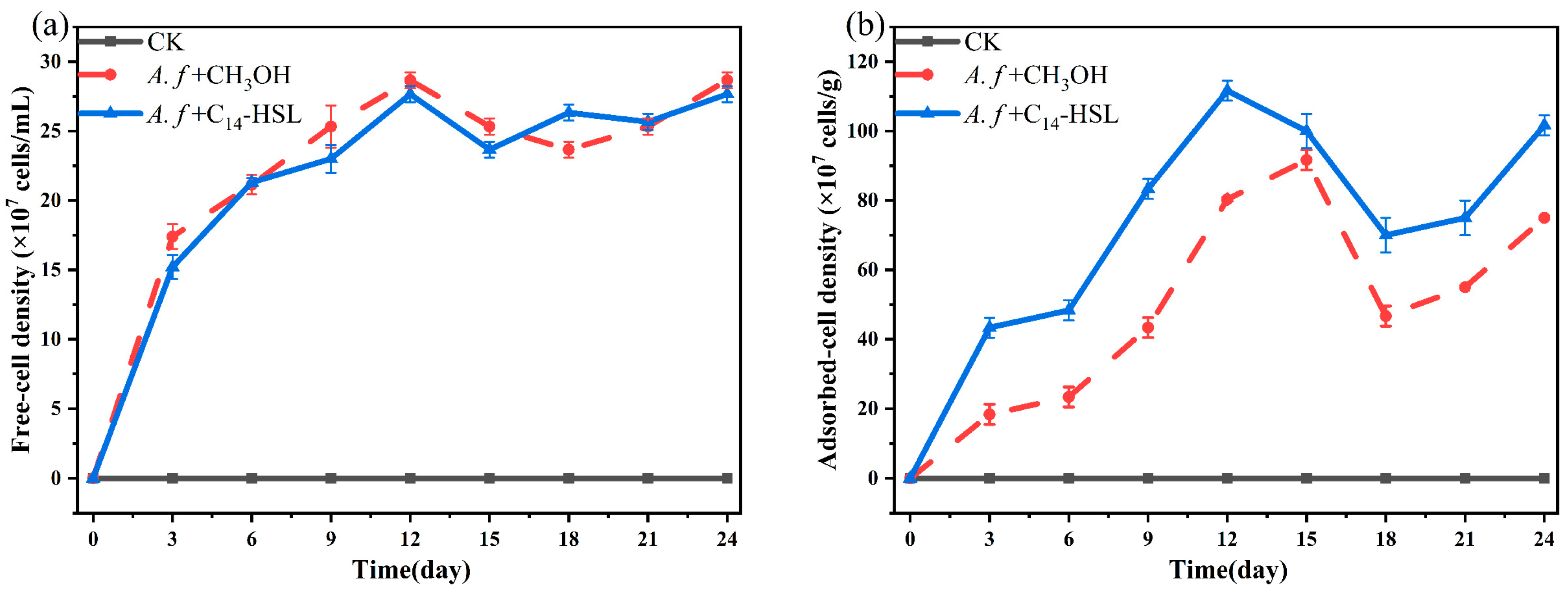
The addition of AHLs or their analogs promoted the attachment of cells and the formation of biofilms and EPS [21,43].To decipher the promotion mechanism of HSL on chalcopyrite early dissolution, the attachment behavior of A. ferrooxidans cells with the addition of C14-HSL was studied.
As shown in Figure 7, the concentration of free cells or adsorbed cells continuously increased before the 12th day. There was no significant difference in the free cells’ concentration between the C14-HSL group and the methanol control group. However, the concentration of adsorbed cells in the C14-HSL group remained higher than that in the control group. The final concentration of adsorbed cells in the C14-HSL group and control was 10.1 × 10⁸ and 8 × 10⁸ cells/g, respectively. The addition of C14-HSL promoted the adsorption and colonization of microorganisms on the mineral surface.
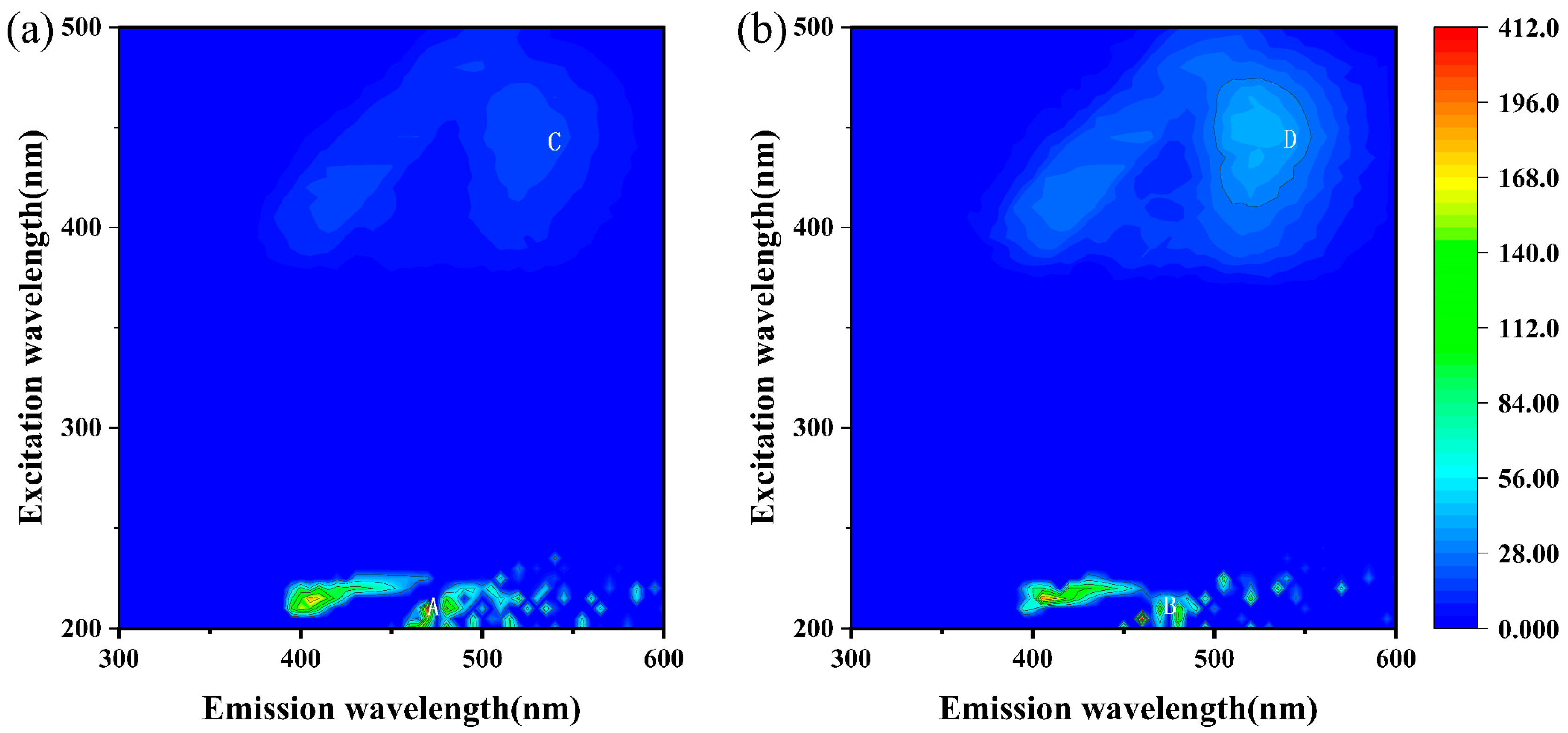
Figure 8 shows the 3D fluorescence spectra of EPS of the methanol control group and the C14-HSL experimental group at the end of bioleaching. The spectrum of EPS in the methanol control group had two peaks, Peak A (Ex/Em = 210.0 nm/470.0 nm) and Peak C (Ex/Em = 450.0 nm/515.0 nm). The spectrum of EPS cultured in the C14-HSL experimental group also had two peaks, Peak B (Ex/Em = 205.0 nm/460.0 nm) and Peak D (Ex/Em = 450.0 nm/520.0 nm). According to previous literature reports [44,45,46], Peak A and Peak B were characteristic peaks of fulvic acid-like compounds, and Peak C and D were characteristic peaks of humic acid-like compounds. Judging from the fluorescence intensity, the content of humic acid-like compounds in the EPS of the C14-HSL experimental group was significantly increased compared with the control group.
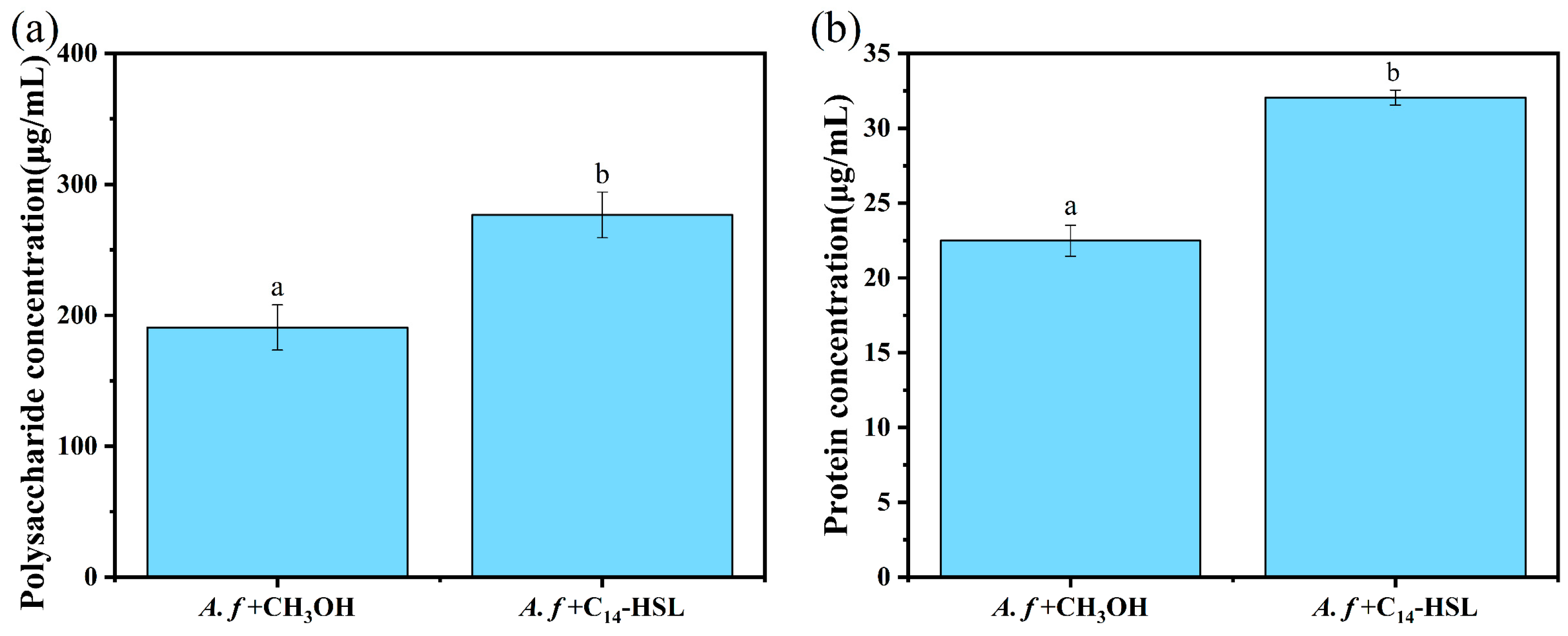
Figure 9 illustrates the polysaccharide and protein concentrations of EPS during the bioleaching process. The polysaccharide concentration of the C14-HSL group was 276.62 μg/mL, which was 45.03% higher than that of the methanol control group. The protein concentration of the C14-HSL group was 32.048 μg/mL, which was 42.55% higher than that of the methanol control group. There was a significant difference in the polysaccharide and protein concentrations of EPS between the C14-HSL group and the control (p < 0.05).
The addition of C14-HSL promoted the colonization of microorganisms on the mineral surface and EPS production including polysaccharide, protein, and humic acid-like compounds. QS signal transduction and rapid adsorption of cells on mineral surfaces help to establish the EPS matrix and the biofilm phenotype [47]. EPS are also closely related to the interaction between cells and minerals by improving the surface properties of minerals [48]. In addition, the anionic groups in polysaccharides, proteins, and humic acid-like compounds can form Fe3+ (Cu2+)-EPS complexes on the mineral surface, promoting the dissolution of minerals [49].

4. Conclusions

The addition of C14-HSL increased the early leaching rate of by approximately 1.15 times. Molecular docking revealed that the promotion mechanism involved the strong binding of C14 to the AfeI/R receptor protein. The high binding affinity between AHL and the AfeR receptor protein facilitates the activation of transcription by the AfeR-AHL complex, promoting their biological impact. Through the measurement of polysaccharides and proteins, it was found that the addition of C14-HSL promoted the production of EPS and facilitated mineral leaching rate. Three-dimensional fluorescence analysis indicated that the increase in humic acid-like compounds enhanced hydrophobic interactions, which may be one of the reasons for the greater adsorption of bacterial communities on the mineral surface in the C14-HSL experimental group, which enhances the hydrophobicity and adsorption capacity of the bacterial community onto the mineral surface. These findings highlight the pivotal role of quorum sensing as a master regulatory system that can orchestrate key microbial processes critical to bioleaching rate. Due to time and experimental limitations, future research can focus on other aspects, such as the endogenous production and coordination mechanisms of AHL, the effect of exogenous AHL addition concentration on leaching, the impact of AHL on key components and structures of biofilms, and its influence on the formation and dissolution of passive films.

Author Contributions

Conceptualization, S.C. and Y.L. (Yili Liang); methodology, S.C. and W.L.; software, S.C. and W.L.; validation, S.C. and Z.Y.; formal analysis, S.C., W.L. and Y.L. (Yili Liang); investigation, S.C.; resources, S.C.; data curation, S.C.; writing—original draft preparation, S.C.; writing—review and editing, S.C., Z.Y. and Y.L. (Yili Liang); visualization, X.W., Y.L. (Yiran Li), N.I. and S.B.; supervision, Y.L. (Yili Liang); project administration, Y.L. (Yili Liang); funding acquisition, Y.L. (Yili Liang). All authors have read and agreed to the published version of the manuscript.

Funding

This research was funded by the National Key Research and Development Program of China (Grant No.2022YFC2105303).

Data Availability Statement

The protein sequences of A. ferrooxidans with AfeI (AAZ20805.1) and AfeR (AAV53702.2) were download from the NCBI database.

Acknowledgments

Table 1 was drawn by Kingdraw v4.0. Figure 1, Figure 2, Figure 5, Figure 6 and Figure 8 were drawn by Origin. Figure 7 was drawn by Matlab and Origin. Figure 3 and Figure 4 were drawn by Discovery studio.

Conflicts of Interest

The authors declare no conflicts of interest.

Abbreviations

The following abbreviations are used in this manuscript:
C14-HSLN-tetradecanoyl-L-homoserine lactone
AHLsAcyl-homoserine lactones
EPSExtracellular polymeric substances
QSQuorum sensing
XRDX-ray diffraction
HPLC-MSHigh-performance liquid chromatography–mass spectrometry
3-oxo-C12-HSLN-3-oxododecanoyl-L-homoserine lactone
3-OH-C12-HSLN-3-hydroxydodecanoyl-DL-homoserine lactone
C12-HSLN-dodecanoyl-L-homoserine lactone
3-oxo-C14-HSLN-3-oxotetradecanoyl-L-homoserine lactone
ES+Electrospray ionization positive
ORPOxidation-reduction potential
RISCsReducing inorganic sulfur compounds
SAMS-adenosyl-L-methionine

References

  1. Kaksonen, A.H.; Boxall, N.J.; Gumulya, Y.; Khaleque, H.N.; Morris, C.; Bohu, T.; Cheng, K.Y.; Usher, K.M.; Lakaniemi, A.-M. Recent Progress in Biohydrometallurgy and Microbial Characterisation. Hydrometallurgy 2018, 180, 7–25. [Google Scholar] [CrossRef]
  2. Bakhti, A.; Moghimi, H.; Bozorg, A.; Stankovic, S.; Manafi, Z.; Schippers, A. Comparison of Bioleaching of a Sulfidic Copper Ore (Chalcopyrite) in Column Percolators and in Stirred-tank Bioreactors Including Microbial Community Analysis. Chemosphere 2024, 349, 140945. [Google Scholar] [CrossRef] [PubMed]
  3. Watling, H. Microbiological Advances in Biohydrometallurgy. Minerals 2016, 6, 49. [Google Scholar] [CrossRef]
  4. Zhao, H.; Zhang, Y.; Zhang, X.; Qian, L.; Sun, M.; Yang, Y.; Zhang, Y.; Wang, J.; Kim, H.; Qiu, G. The Dissolution and passivation mechanism of chalcopyrite in bioleaching: An overview. Miner. Eng. 2019, 136, 140–154. [Google Scholar] [CrossRef]
  5. Dunbar, W.S. Biotechnology and the Mine of Tomorrow. Trends Biotechnol. 2017, 35, 79–89. [Google Scholar] [CrossRef]
  6. Giese, E.C. Evidences of EPS-iron (III) Ions Interactions on Bioleaching Process Mini-review: The Key to Improve Performance. Orbital Electron. J. Chem. 2019, 11, 200–204. [Google Scholar] [CrossRef]
  7. Bellenberg, S.; Díaz, M.; Noël, N.; Sand, W.; Poetsch, A.; Guiliani, N.; Vera, M. Biofilm Formation, Communication and Interactions of Leaching Bacteria During Colonization of Pyrite and Sulfur Surfaces. Res. Microbiol. 2014, 165, 773–781. [Google Scholar] [CrossRef]
  8. Whiteley, M.; Diggle, S.P.; Greenberg, E.P. Progress in and Promise of Bacterial Quorum Sensing Research. Nature 2017, 551, 313–320. [Google Scholar] [CrossRef]
  9. Williams, P.; Winzer, K.; Chan, W.C.; Cámara, M. Look Who’s Talking: Communication and Quorum Sensing in the Bacterial World. Philos. Trans. R. Soc. Lond. B Biol. Sci. 2007, 362, 1119–1134. [Google Scholar] [CrossRef]
  10. Sharma, A.; Singh, P.; Sarmah, B.K.; Nandi, S.P. Quorum Sensing: Its Role in Microbial Social Networking. Res. Microbiol. 2020, 171, 159–164. [Google Scholar] [CrossRef]
  11. Hamza, F.; Zinjarde, S. Use of Marine Microorganisms in Designing Anti-infective Strategies for Sustainable Aquaculture Production. J. Appl. Microbiol. 2023, 134, lxad128. [Google Scholar] [CrossRef] [PubMed]
  12. Roca Subirà, I.; Espinal, P.; Vila-Farrés, X.; Vila Estapé, J. The Acinetobacter baumannii Oxymoron: Commensal Hospital Dweller Turned Pan-Drug-Resistant Menace. Front. Microbiol. 2012, 3, 148. [Google Scholar] [CrossRef]
  13. Saipriya, K.; Swathi, C.H.; Ratnakar, K.S.; Sritharan, V. Quorum-sensing System in Acinetobacter baumannii: A Potential Target for New Drug Development. J. Appl. Microbiol. 2020, 128, 15–27. [Google Scholar] [CrossRef] [PubMed]
  14. Fuqua, C.; Greenberg, E.P. Listening in on Bacteria: Acyl-homoserine Lactone Signalling. Nat. Rev. Mol. Cell Biol. 2002, 3, 685–695. [Google Scholar] [CrossRef]
  15. Bassler, B.L.; Losick, R. Bacterially Speaking. Cell 2006, 125, 237–246. [Google Scholar] [CrossRef]
  16. Williams, P. Quorum Sensing, Communication and Cross-kingdom Signaling in the Bacterial World. Microbiology 2008, 153, 3923–3938. [Google Scholar] [CrossRef]
  17. Farah, C.; Vera, M.; Morin, D.; Haras, D.; Jerez, C.A.; Guiliani, N. Evidence for a Functional Quorum-Sensing Type AI-1 System in the Extremophilic Bacterium Acidithiobacillus ferrooxidans. Appl. Environ. Microbiol. 2005, 71, 7033–7040. [Google Scholar] [CrossRef]
  18. Rivas, M.; Seeger, M.; Jedlicki, E.; Holmes, D.S. Second Acyl Homoserine Lactone Production System in the Extreme Acidophile Acidithiobacillus ferrooxidans. Appl. Environ. Microbiol. 2007, 73, 3225–3231. [Google Scholar] [CrossRef]
  19. Gao, X.Y.; Fu, C.A.; Hao, L.; Gu, X.F.; Wang, R.; Lin, J.Q.; Liu, X.M.; Pang, X.; Zhang, C.J.; Lin, J.Q.; et al. The Substrate-dependent Regulatory Effects of the AfeI/R System in Acidithiobacillus ferrooxidans Reveals the Novel Regulation Strategy of Quorum Sensing in Acidophiles. Environ. Microbiol. 2021, 23, 757–773. [Google Scholar] [CrossRef]
  20. González, A.; Bellenberg, S.; Mamani, S.; Ruiz, L.; Echeverría, A.; Soulère, L.; Doutheau, A.; Demergasso, C.; Sand, W.; Queneau, Y.; et al. AHL Signaling Molecules with a Large Acyl Chain Enhance Biofilm Formation on Sulfur and Metal Sulfides by the Bioleaching Bacterium Acidithiobacillus ferrooxidans. Appl. Microbiol. Biotechnol. 2013, 97, 3729–3737. [Google Scholar] [CrossRef]
  21. Mamani, S.; Moinier, D.; Denis, Y.; Soulère, L.; Queneau, Y.; Talla, E.; Bonnefoy, V.; Guiliani, N. Insights into the Quorum Sensing Regulon of the Acidophilic Acidithiobacillus ferrooxidans Revealed by Transcriptomic in the Presence of an Acyl Homoserine Lactone Superagonist Analog. Front. Microbiol. 2016, 7, 1365. [Google Scholar] [CrossRef] [PubMed]
  22. Gao, X.Y. Regulatory Function and Molecular Mechanism of the Quorum Sensing System in Acidithiobacillus ferroxidans ATCC 23270; Wanfang Data: Beijing, China, 2020. [Google Scholar]
  23. Galloway, W.R.J.D.; Hodgkinson, J.T.; Bowden, S.D.; Welch, M.; Spring, D.R. Quorum Sensing in Gram-Negative Bacteria: Small-Molecule Modulation of AHL and AI-2 Quorum Sensing Pathways. Chem. Rev. 2011, 111, 28–67. [Google Scholar] [CrossRef] [PubMed]
  24. Morin, D.; Grasland, B.; Vallée-Réhel, K.; Dufau, C.; Haras, D. On-line High-performance Liquid Chromatography–mass Spectrometric Detection and Quantification of N-acylhomoserine Lactones, Quorum Sensing Signal Molecules, in the Presence of Biological Matrices. J. Chromatogr. A 2003, 1002, 79–92. [Google Scholar] [CrossRef] [PubMed]
  25. Ni, Z.; Sousa, B.C.; Colombo, S.; Afonso, C.B.; Melo, T.; Pitt, A.R.; Spickett, C.M.; Domingues, P.; Domingues, M.R.; Fedorova, M.; et al. Evaluation of Air Oxidized PAPC: A Multi Laboratory Study by LC-MS/MS. Free Radic. Biol. Med. 2019, 144, 156–166. [Google Scholar] [CrossRef]
  26. Esparza, M.; Cárdenas, J.P.; Bowien, B.; Jedlicki, E.; Holmes, D.S. Genes and Pathways for CO2 Fixation in the Obligate, Chemolithoautotrophic Acidophile, Acidithiobacillus ferrooxidans, Carbon fixation in A. ferrooxidans. BMC Microbiol. 2010, 10, 229. [Google Scholar] [CrossRef]
  27. Zhan, Y.; Yang, M.; Zhang, S.; Zhao, D.; Duan, J.; Wang, W.; Yan, L. Iron and Sulfur Oxidation Pathways of Acidithiobacillus ferrooxidans. World J. Microbiol. Biotechnol. 2019, 35, 60. [Google Scholar] [CrossRef]
  28. Parsek, M.R.; Val, D.L.; Hanzelka, B.L.; Cronan, J.E.; Greenberg, E.P. Acyl Homoserine-lactone Quorum-Sensing Signal Generation. Proc. Natl. Acad. Sci. USA 1999, 96, 4360–4365. [Google Scholar] [CrossRef]
  29. Ahlgren, N.A.; Harwood, C.S.; Schaefer, A.L.; Giraud, E.; Greenberg, E.P. Aryl-homoserine Lactone Quorum Sensing in Stem-nodulating Photosynthetic Bradyrhizobia. Proc. Natl. Acad. Sci. USA 2011, 108, 7183–7188. [Google Scholar] [CrossRef]
  30. Rivas, M.; Seeger, M.; Holmes, D.S.; Jedlicki, E. A Lux-like Quorum Sensing System in the Extreme Acidophile Acidithiobacillus ferrooxidans. Biol. Res. 2005, 38, 283–297. [Google Scholar] [CrossRef]
  31. Valdés, J.; Veloso, F.; Jedlicki, E.; Holmes, D. Metabolic Reconstruction of Sulfur Assimilation in the Extremophile Acidithiobacillus ferrooxidans Based on Genome Analysis. BMC Genom. 2003, 4, 51. [Google Scholar] [CrossRef]
  32. Kharpan, B.; Shyam, A.; Nandi, R.; Paul, S.; Paul, P.C.; Mondal, P.; Kumar, D.; Choudhury, S.; Ray, S. Antiparasitic Activity, DNA/BSA Binding Interaction, Molecular Docking and DFT Studies of Mesogenic l-leucine Based Schiff Base and Its Derivatize Cu(II) and Zn(II) Complexes. J. Mol. Struct. 2024, 1304, 137633. [Google Scholar] [CrossRef]
  33. Soulère, L.; Guiliani, N.; Queneau, Y.; Jerez, C.A.; Doutheau, A. Molecular Insights into Quorum Sensing in Acidithiobacillus ferrooxidans bacteria Via Molecular Modelling of The Transcriptional Regulator AfeR and of the Binding Mode of Long-chain Acyl Homoserine Lactones. J. Mol. Model. 2008, 14, 599–606. [Google Scholar] [CrossRef] [PubMed]
  34. Alisaac, A. In silico analysis of quorum sensing modulators: Insights into Molecular Docking and Dynamics and Potential Therapeutic Applications. PLoS ONE 2025, 20, e0325830. [Google Scholar] [CrossRef]
  35. Luo, B.; Wu, S.; Liu, W.; Zhang, D.; Liu, R.; Liu, T.; Sun, Z.; Wei, Z.; Liu, M.; Shi, Z.; et al. Mechanistic Insights into the Orthogonal Functionality of an AHL-mediated Quorum-sensing Circuit in Yersinia pseudotuberculosis. Synth. Syst. Biotechnol. 2025, 10, 174–184. [Google Scholar] [CrossRef]
  36. Banderas, A.; Guiliani, N. Bioinformatic Prediction of Gene Functions Regulated by Quorum Sensing in the Bioleaching Bacterium Acidithiobacillus ferrooxidans. Int. J. Mol. Sci. 2013, 14, 16901–16916. [Google Scholar] [CrossRef]
  37. Malik, S.; Akram, F.; Ali, M.; Javed, M.; Mateen, R.M.; Zidan, A.; Bahadur, A.; Iqbal, S.; Mahmood, S.; Farouk, A.-E.; et al. Unveiling Quorum Sensing Mechanisms: Computational Docking and Dynamics of Bacterial Receptors and Ligands. J. Mol. Struct. 2025, 1323, 140730. [Google Scholar] [CrossRef]
  38. Zhang, Z.; Zheng, Y.; Han, P.; Dong, H.; Liang, X.; Yin, G.; Wu, D.; Yang, Y.; Liu, S.; Liu, M.; et al. N-acyl-homoserine lactones (AHLs) in Intertidal Marsh: Diversity and Potential Role in Nitrogen Cycling. Plant Soil. 2020, 454, 103–119. [Google Scholar] [CrossRef]
  39. Schaefer, A.L.; Greenberg, E.P.; Oliver, C.M.; Oda, Y.; Huang, J.J.; Bittan-Banin, G.; Peres, C.M.; Schmidt, S.; Juhaszova, K.; Sufrin, J.R.; et al. A New Class of Homoserine Lactone Quorum-sensing Signals. Nature 2008, 454, 595–599. [Google Scholar] [CrossRef]
  40. Wu, A.; Yin, S.; Qin, W.; Liu, J.; Qiu, G. The Effect of Preferential Flow on Extraction and Surface Morphology of Copper Sulphides During Heap Leaching. Hydrometallurgy 2009, 95, 76–81. [Google Scholar] [CrossRef]
  41. Córdoba, E.M.; Muñoz, J.A.; Blázquez, M.L.; González, F.; Ballester, A. Leaching of Chalcopyrite with Ferric Ion. Part I General. Aspects. Hydrometallurgy 2008, 93, 81–87. [Google Scholar] [CrossRef]
  42. Tao, J.; Liu, X.; Luo, X.; Teng, T.; Jiang, C.; Drewniak, L.; Yang, Z.; Yin, H. An Integrated Insight into bioleaching Performance of Chalcopyrite Mediated by Microbial Factors: Functional Types and Biodiversity. Bioresour. Technol. 2021, 319, 124219. [Google Scholar] [CrossRef] [PubMed]
  43. Gao, X.-Y.; Liu, X.-J.; Fu, C.-A.; Gu, X.-F.; Lin, J.-Q.; Liu, X.-M.; Pang, X.; Lin, J.-Q.; Chen, L.-X. Novel Strategy for Improvement of the Bioleaching Efficiency of Acidithiobacillus ferrooxidans Based on the AfeI/R Quorum Sensing System. Minerals 2020, 10, 222. [Google Scholar] [CrossRef]
  44. Vardanyan, A.; Vardanyan, N.; Khachatryan, A.; Zhang, R.; Sand, W. Adhesion to Mineral Surfaces by Cells of Leptospirillum, Acidithiobacillus and Sulfobacillus from Armenian Sulfide Ores. Minerals 2019, 9, 69. [Google Scholar] [CrossRef]
  45. Jacquin, C.; Lesage, G.; Traber, J.; Pronk, W.; Heran, M. Three-dimensional Excitation and Emission Matrix Fluorescence (3DEEM) for Quick and Pseudo-quantitative Determination of Protein- and Humic-like Substances in Full-scale Membrane Bioreactor (MBR). Water Res. 2017, 118, 82–92. [Google Scholar] [CrossRef]
  46. Chen, W.; Westerhoff, P.; Leenheer, J.; Booksh, K. Fluorescence Excitation−Emission Matrix Regional Integration to Quantify Spectra for Dissolved Organic Matter. Environ. Sci. Technol. 2004, 37, 5701–5710. [Google Scholar] [CrossRef]
  47. Liu, R.; Chen, J.; Zhou, W.; Cheng, H.; Zhou, H. Insight to the Early-stage Adsorption Mechanism of Moderately Thermophilic Consortia and Intensified Bioleaching of Chalcopyrite. Biochem. Eng. J. 2019, 144, 40–47. [Google Scholar] [CrossRef]
  48. Mitsunobu, S.; Zhu, M.; Takeichi, Y.; Ohigashi, T.; Suga, H.; Jinno, M.; Makita, H.; Sakata, M.; Ono, K.; Mase, K.; et al. Direct Detection of Fe(II) in Extracellular Polymeric Substances (EPS) at the Mineral-Microbe Interface in Bacterial Pyrite Leaching. Microbes Environ. 2016, 31, 63–69. [Google Scholar] [CrossRef]
  49. La Vars, S.M.; Newton, K.; Quinton, J.S.; Cheng, P.-Y.; Wei, D.-H.; Chan, Y.-L.; Harmer, S.L. Surface Chemical Characterisation of Pyrite Exposed to Acidithiobacillus ferrooxidans and Associated Extracellular Polymeric Substances. Minerals 2018, 8, 132. [Google Scholar] [CrossRef]
Figure 1. XRD pattern of the chalcopyrite sample.
Minerals 15 01248 g001
Figure 2. XRD pattern of the pyrite sample.
Minerals 15 01248 g002
Figure 3. The content of signal molecules and cell density produced under different energy substrate cultures: (a) Cell density (mean ± SD) of A. ferrooxidans cultured with different substrates at the end of the log phase (Tukey test, n = 3, p < 0.05); (b) Composition and concentration (mean ± SD) of signaling molecules in the culture supernatant of A. ferrooxidans after cultivation with different energy substrates; (c) The number of cells required to produce a unit concentration of AHL (1 ng/mL).
Minerals 15 01248 g003
Figure 4. The docking of AHL with the signal molecule synthesis protein AfeI in A. ferrooxidans: (AF): The 3D structures of C12-HSL, C14-HSL, 3-OH-C12-HSL, 3-oxo-C12-HSL, 3-oxo-C14-HSL, and vanillin docked with AfeI; (af) The 2D representations of the interactions between C12-HSL, C14-HSL, 3-OH-C12-HSL, 3-oxo-C12-HSL, 3-oxo-C14-HSL, and vanillin with AfeI. Hydrogen bonds are labeled in green and hydrophobic interactions are labeled in purple.
Minerals 15 01248 g004
Figure 5. The docking of different types of AHL with the signal molecule synthesis protein AfeR in A. ferrooxidans: (AF): The 3D structures of C12-HSL, C14-HSL, 3-OH-C12-HSL, 3- oxo-C12-HSL, 3-oxo-C14-HSL, and vanillin docked with AfeR; (af) The 2D representations of the interactions between C12-HSL, C14-HSL, 3-OH-C12-HSL, 3-oxo-C12-HSL, 3-oxo-C14-HSL, and vanillin with AfeR. Hydrogen bonds are labeled in green and hydrophobic interactions are labeled in purple.
Minerals 15 01248 g005
Figure 6. Variation (mean ± SD, n = 3) of physicochemical parameters during the leaching of chalcopyrite by the A. ferrooxidans system under the influence of five individually added signaling molecules: (a) copper concentration; (b) pH; (c) ORP.
Minerals 15 01248 g006
Figure 7. Variation (mean ± SD, n = 3) of bacterial density during chalcopyrite leaching by addition of C14-HSL to the system of A. ferrooxidans: (a) free bacteria; (b) adsorbed bacteria.
Minerals 15 01248 g007
Figure 8. Three-dimensional fluorescence spectra of EPS of A. ferrooxidans system: (a) methanol control group; (b) C14-HSL.
Minerals 15 01248 g008
Figure 9. EPS concentrations (mean ± SD) of A. ferrooxidans systems (different letters indicate significant differences): (a) polysaccharides; (b) proteins; a and b represent the significant differences among different experimental groups (Tukey test, n = 3, p < 0.05).
Minerals 15 01248 g009
Table 1. Signaling molecules in the culture supernatant of A. ferrooxidans ATCC 23270.
AHLChemical FormulaDetection of [M + H]+ IonStructured Formula
C12-HSLC16H29O3N284.3Minerals 15 01248 i001
3-OH-C12-HSLC16H29O4N300.3Minerals 15 01248 i002
3-oxo-C12-HSLC16H27O4N298.3Minerals 15 01248 i003
C14-HSLC18H33O3N312.2Minerals 15 01248 i004
3-oxo-C14-HSLC18H31O4N326.3Minerals 15 01248 i005
Table 2. Molecular docking parameters of AHL and AfeI /AfeR protein complexes.
AHLLibDock
Score
Hydrogen Bond
Binding Site
Hydrophobic Bond
Binding Site
AfeIAfeRAfeIAfeRAfeIAfeR
3-oxo-C14-HSL128.87143.914A/VAL:145
A/ARG:102
A/ASP:75
A/TRP:62
A/HIS:173
A/ALA:34
A/ARG:119
C14-HSL123.159139.272A/ARG:102
VAL:145
A/TRP:62
A/ASP:75
A/HIS:173
A/ILE:148
A/ARG:119
3-OH-C12-HSL123.039133.245A/VAL:145
A/SER:146
A/ARG:102
A/THR:144
A/PRO:147
A/GLY:125
A/GLY:126
A/CYS:118
A/HIS:173
A/TRP:33
A/LEU:35
A/TRP:62
A/TYR:58
A/TYR:66
3-oxo-C12-HSL122.509130.135A/VAL:145
A/PRO:147
A/ARG:102
A/GLN:72
A/CYS:118
A/GLY:126
A/ARG:42
A/HIS:173
A/TRP:33
A/LEU:35
A/TYR:66
A/TRP:62
A/TYR:58
C12-HSL120.501127.537A/ARG:102
A/VAL:145
A/CYS:118
A/GLY:126
A/CLY:125
A/ARG:42
A/PRO:174
A/PRO:147
A/HIS:173
A/TYR:66
Vanillin77.37778.012A/VAL:145A/ALA:81A/PRO:147
A/VAL:150
A/ARG:42
Disclaimer/Publisher’s Note: The statements, opinions and data contained in all publications are solely those of the individual author(s) and contributor(s) and not of MDPI and/or the editor(s). MDPI and/or the editor(s) disclaim responsibility for any injury to people or property resulting from any ideas, methods, instructions or products referred to in the content.