Article

Bacteria-like Ferruginous Structures in Carboniferous Limestones as Remains of Post-Variscan Hydrothermal Activity in Southern Poland

1
Faculty of Geology, Geophysics, and Environmental Protection, AGH University of Krakow, 30-059 Kraków, Poland
2
Institute of Biology and Earth Sciences, University of the National Education Commission, 30-084 Krakow, Poland
3
PROINSOL Sp. z o.o. Spółka Komandytowa, 30-126 Kraków, Poland
*
Author to whom correspondence should be addressed.
Minerals 2025, 15(11), 1158; https://doi.org/10.3390/min15111158
Submission received: 16 October 2025 / Revised: 29 October 2025 / Accepted: 30 October 2025 / Published: 1 November 2025

Abstract

Structures resembling iron-related bacteria (IRB) have been found in the Mississippian limestones that form part of the carbonate platform in the Moravo-Silesian Basin that surrounds the Upper Silesian Block, an eastern margin of the Brunovistulicum. Microfacial, petrological, and geochemical analyses were used to determine the bacteria-like structures that are present in narrow zones unrelated to bedding. We present here the morphology and chemistry of the studied microstructures showing their similarities to IRB from the present-day Sphaerotilus-Leptothrix group, the Galionella group, and the Mariprofundus ferrooxydans species. We suggest that bacterial growth occurred in the originally empty micropores of microfossil skeletons and shells, between bioclasts or in secondary voids formed during the selective dissolution of micrite or smaller sparite crystals. Hydrothermal solutions, associated probably with the post-Variscan magmatism in this area, provided iron compounds for the growth of the IRB.

Graphical Abstract

1. Introduction

Single-celled organisms are able to actively participate in changes in environmental chemistry. They mediate oxidation and reduction reactions occurring in both aquatic and terrestrial environments, e.g., [1,2]. Such processes also occurred in the geological past and could be recorded in rocks, which we can read by deciphering fossilization processes, e.g., [3,4]. The fossilization of single-celled organisms is most likely if they precipitated extracellular inorganic coatings [5,6]. Such structures are usually left as the only remnants after the decomposition of bacterial cellular matter, providing valuable information on microbial life in ancient environments, e.g., [7]. The more resistant structures to later remobilization are metallic bacterial coatings such as those created by iron related bacteria (IRB) [8,9]. Such bacterial types are able to catalyze the oxidation of Fe (II) [10,11] and accumulate (oxyhydr)oxides, either outside the cells or in the surrounding extracellular polymeric matrices, e.g., [12].
IRB are well known from modern environments as well as from the fossil record. The earliest fossilized bacterial remnants, morphologically comparable with the modern IRB have been recognized so far from the well-known Precambrian Gunflint cherts [13,14], as well as in many Precambrian sedimentary successions. They also contributed significantly to the banded iron formations (BIFs) [3,13,15].
Bacterial iron encrustations have also been described from various sedimentary environments in many Phanerozoic successions, both freshwater, e.g., [16], and marine, e.g., [17,18,19]. They were recognized in fossil record from high-energy, shallow, e.g., [20], to calm, deep-water settings [7], where they can formed Fe-oolite sands [21,22], bacterial mats [17], stromatolites [23], or incrustations binding mud mounds [24]. Especially in marine environments, bacterially mediated amorphous iron oxyhydroxides combined with manganese oxyhydroxides and silica are commonly found in areas of mid-ocean and back-arc volcanic activity, covering vast areas of the ocean floor [25], also associated with hydrothermal fluid activity.
In the present study, we report morphological and chemical evidences for the presence of bacteria-like structures belonging to the IRB group in the Mississippian limestones along the Upper Silesian Block (Figure 1a,b), which is the northern part of the larger unit known as the Brunovistulicum, e.g., [26], and we try to indicate their origin and time of appearance in these rocks. A characteristic feature of the limestones consisting of the IRB-like structures is their red colour, which does not occur in underlying carbonate successions [more than 1 km thick], which formed on the epicontinental shelf stretching from the Western Europe to the Ukraine [27]. Previous studies suggested the sedimentary origin of the red colouration of these limestones, related to the admixture of hematite, which would be redistributed from the terrestrial environment [28]. Here, we test another hypothesis, which connects the occurrence of IRB-like structures with the activity of hydrothermal waters. In the study area, they could have occurred during two different geological periods. The first one could be related to the Pennsylvanian and Cisuralian post-orogenic granitoid plutonism and bimodal volcanism, e.g., [29,30]. And the second one could be related to the Late Triassic sulphide mineralization [mainly of Zn-Pb] of unknown fluid origin [31].

2. Geological Setting

The IRB-like remnants have been found in the uppermost part of the Upper Devonian–Mississipian succession of the ancient carbonate platform in the Moravo-Silesian Basin that surrounded the Upper Silesian Block, an eastern margin of the Brunovistulicum, e.g., [34,35,36]. This carbonate body developed approximately three hundred kilometres on a passive continental margin facing a deep-water basin of the Rhenohercynian system [37,38,39]. Due to sea-level rise and tectonic extension, successive drowning of this platform occurred [39,40], partly associated with its disintegration and the development of intra-platform troughs [41,42,43]. The closure of the Rhenohercynian ocean resulted in subsequent continental collision between Lugodanubian and subducting Brunovistulicum [44], which ultimately amalgamated the Upper Silesian Block with the Bohemian Massif [28].
During Pennsylvanian–Cisuralian post-orogenic stages, granitoid plutonism and bimodal volcanism took place in both foreland and the internal parts of the Variscan orogen, including the recent southern Poland [31,45]. Their numerous occurrences, e.g., [32,46,47,48,49,50,51,52], are associated with the Kraków–Lubliniec Fault Zone, which divides the Upper Silesian Block from the Małopolska Block (Figure 1b).
The Variscan basement containing the Devonian and Carboniferous sediments is covered discordantly by the succession of Permian–Mesozoic carbonate and siliciclastic sediments of the Central European Basin, dominated by the Triassic strata (Figure 1c). All of them dip toward the northeast, reflecting the vertical crustal mobility of this region [53]. The Middle Triassic limestones and dolomites (Muschelkalk) comprise Zn-Pb deposits, locally as resources of economic value, e.g., [31,54]. However, this Zn-Pb mineralization is hosted also by older (Devonian–Permian) and younger (Jurassic) rocks [55,56].
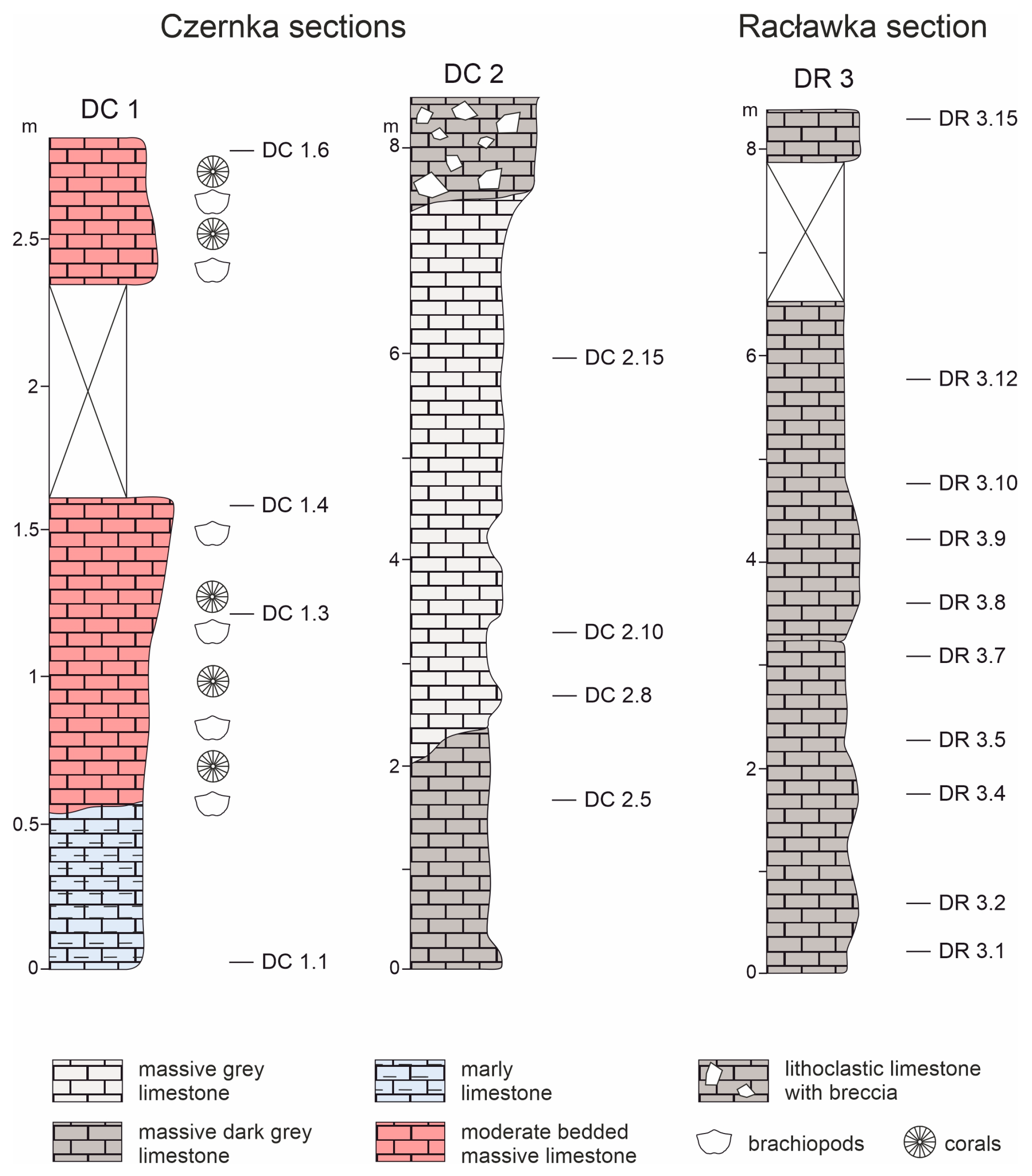
The studied Upper Devonian to Mississippian limestones of the shallow-water platform (bioclastic wackstones with brachiopods) are recently outcropped in the southernmost part of the Silesian-Kraków Monocline (southern Poland), 25 km west of Kraków (Figure 1c). In this area, the Middle Devonian–Mississippian carbonates (limestones and dolomites) are cut by several gorges containing, among others, the Racławka and Czernka valleys. Our microfacial and geochemical studies are based on samples from three sections located in these valleys. The section from the Racławka Valley (DR3; Figure 2) corresponds to the lower Tournaisian, whereas the two sections from the Czernka Valley (DC1, DC2; Figure 2) belong to the middle Viséan, based on analyses of foraminiferal assemblages [57]. Litho- and biostratigraphic studies of this carbonate succession have a history of over 160 years, as summarized in [57]. The assignment of sediments from the studied sections to lithostratigraphic units is based on the later classification [27]. Their chronostratigraphy is based on foraminiferal assemblages from the class Fusulinata studied in the same rock samples [57].

3. Material and Methods

3.1. Sampling

The criterion for selecting 18 samples for iron-related bacteria research was the reddish colour of the rocks in the fresh fracture or any ferruginous discolourations and coatings visible in subsequent microscopic observations. Samples were taken from three exposures in the selected area (Figure 1c).
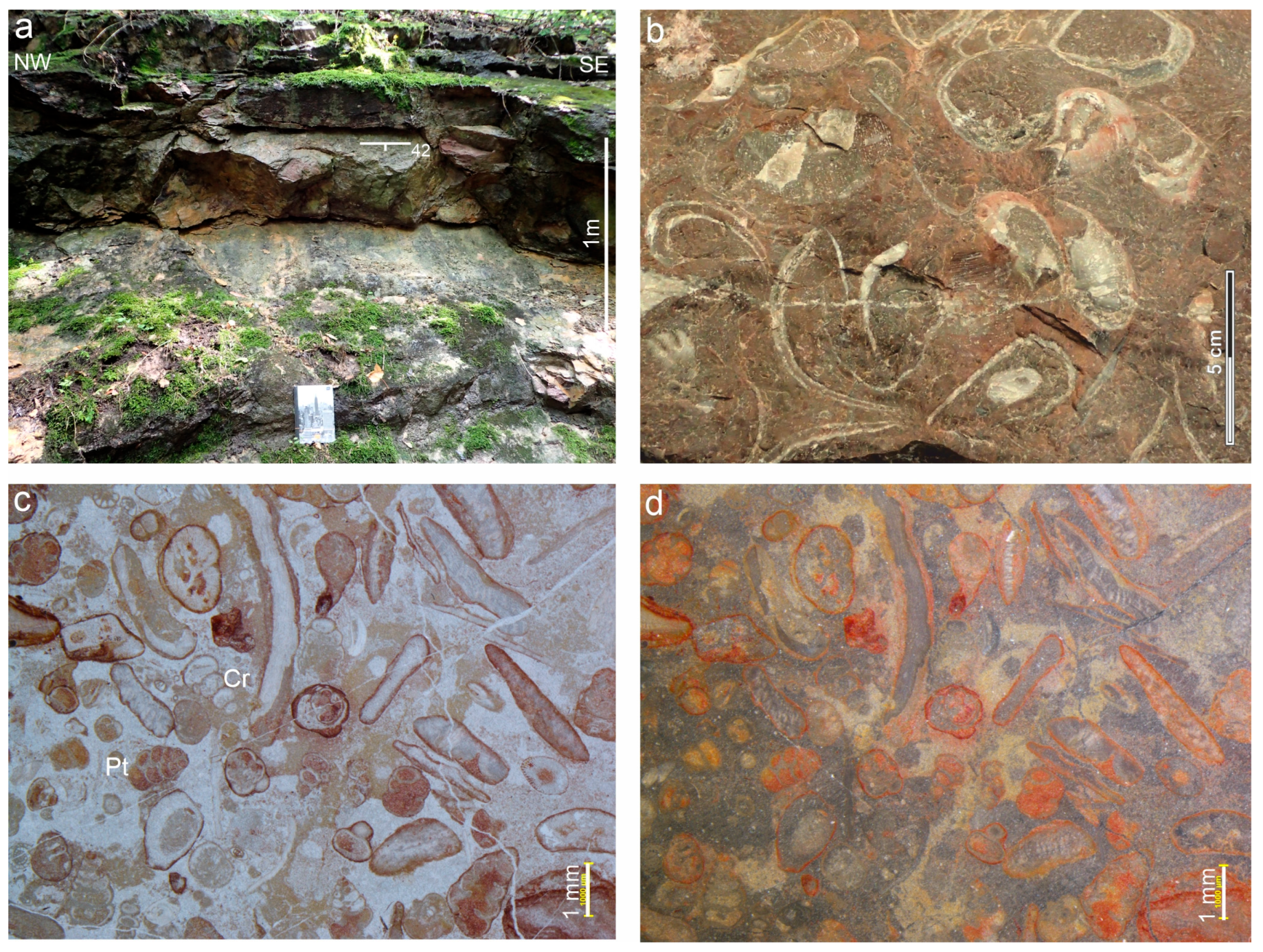
Four samples of the Viséan biogenic limestones belonging to the Czerwona Ścianka Formation [27,55] were selected from the stratigraphically youngest (DC1) section (the Red Wall in the Czernka Valley (50°10′12.9″ N, 19°37′22.1″ E; Figure 2)). The characteristic feature of this location is the red colour of rock on the exposed surface (Figure 3a,b). Another distinguishing feature of the limestones is also the red colour on the borders around the bioclasts (Figure 3c,d).
Four rock samples were selected from the stratigraphically older (DC2) section (the Commune quarry in the Czernka Valley (50°10′09.7″ N, 19°37′24.2″ E; Figure 2)) representing also the Viséan limestones of the Czerna Formation [27,55]. This outcrop is located on the orographically left bank of the Czernka stream, near a local water intake. The profile contains grey to dark grey limestones on a fresh fracture, and beige to pinkish on a weathered surface. They are characterized by a lack of lamination, but within the outcrop scale, layering can be observed, with layers ranging from 1 to 3 m thick. There are sparse cracks (0.2 to 0.5 mm) secondarily filled with white calcite. In the thin-section view, limestones are pelitic mudstone-type and, locally, bioclastic wackstone. Lithoclasts composed of carbonate rocks are visible in individual samples. In the central part of the exposure, a succession of fine-bedded limestones with a nodular structure is visible. These limestones do not show the presence of macrofossils.
Four samples of massive dark grey Tournaisian limestones belonging to the Racławka Formation [27,57] were taken from the outcrop (DR3) on the slopes of the Komarówka Hill in the Racławka Valley (50°10′28.9″ N, 19°40′26.7″ E). In the axis of the main and lateral valleys, there are two faults along which tectonic blocks containing the Famennian and Tournaisian limestones were dislocated relative to each other (Figure 1c). The profile exposes pelitic limestones without lamination, light grey on the weathered surface and dark grey on the fresh fracture. Two-plane fractures dominate, ranging in thickness from 1 to 4 mm. Cracks filled with brick red calcite are visible in the upper part of the profile. In the fillings of larger cracks, the crack rim is brick red and the interior is filled with white, creamy calcite.

3.2. Petrology and Geochemistry Studies

Twenty standard uncovered petrographic thin sections with a size of 3 × 5 cm were made from the selected samples. All thin sections were cut perpendicular to the lamination. All thin sections were observed under a conventional transmitted light microscope (TLM) using a Nikon Eclipse LV100N Pol polarizing microscope with a digital camera (Nikon DS10) (Nikon Corporation, Yokohama, Japan). The thin sections are stored as Collection IRB-2-2024 at the Faculty of Geology, Geophysics, and Environmental Protection of the AGH University of Science and Technology, Kraków.
The chemical composition of the minerals was determined using the SEM-EDS method. Carbon-coated polished thin-section samples were analyzed using a JEOL 5410 microscope (JEOL Ltd., Akishima, Japan) with a Voyager 3100 (NORAN) EDS spectrometer (NORAN Instruments, Middleton, WI, USA) as well as a HITACHI S 4700 microscope (Hitachi High-Tech Corporation, Naka, Japan) with a Vantage (NORAN) spectrometer (NORAN Instruments, Middleton, WI, USA). The time of analysis was 100 s for a point at an acceleration voltage of 20 kV. The ZAF correction algorithm was used. These studies were performed in the Laboratory of Field Emission SEM and Microanalysis at the Institute of Geological Sciences, Jagiellonian University, Kraków. Additional scanning electron microscope observations regarding mineralogical studies of iron oxides and oxyhydroxides were carried out in low-vacuum mode, using a FEI 200 Quanta FEG microscope (Fei Company, Brno, Czechia) equipped with an EDS/EDAX spectrometer (at the AGH University of Krakow). The acceleration voltage was 20 kV, and the pressure was 60 Pa. The samples were not coated with any conductive layer.
Raman microspectroscopy measurements were performed with a Thermo Scientific DXR Raman Microscope (Thermo Electronic Scientific Instruments LLC, Madison, WI, USA). An Olympus (100×) objective lens (Olympus Corporation, Nagano, Japan)was used to focus the incident laser on a sample resulting in a spot size of ca. 0.7 μm. The spectra were recorded at room temperature using a 900 grooves/mm grating and a CCD detector. The excitation wavelength was 532 nm, and the power was 10 mW. An exposure time of 3 s and 100 data accumulations were used.
All these samples were analyzed for major and minor element concentrations at the Bureau Veritas Minerals Laboratories, Vancouver, Canada. Total abundances of the major oxides, several minor elements, rare Earth and refractory elements were analyzed by inductively coupled plasma (ICP) emission spectrometry, following lithium metaborate/tetraborate fusion and dilute nitric acid digestion. Loss on ignition (LOI) was determined by the weight difference after ignition at 1000 °C for >2 h. Moreover, separate 0.5 g samples were digested in Aqua Regia and analyzed by ICP mass spectrometry to determine the precious and base metals. The detection limits ranged from 0.002 wt% to 0.01 wt% for major oxides, from 0.1 ppm to 20 ppm for trace elements, and from 0.01 ppm to 0.1 ppm for the rare Earth elements. The CANMET- and USGS-certified reference materials were used as monitors of data quality. Normalized REE patterns are shown against a logarithmically scaled concentration axis. To calculate the Ce anomaly, we used an equation (Ce/Ce*)N = CeN/(PrN × PrN/NdN) [58]. Gd anomalies were calculated using the Gd/Gd* ratio, where Gd* is the interpolated Gd concentration for a smooth Post Archean Australian Shale-normalized REE pattern, and Xn is the concentration of element X normalized to PAAS: Gd*N = SmN1/3 × TbN2/3.
Based on standards and sample replicates, the precisions for abundances and element ratios (including Gd/Gd* ratios) are in most cases much better than 5% (2 RSD). Typical errors for gadolinium excesses in shells are assumed to be about 0.25 ng/g (2σ).

4. Results

4.1. Microstructure of Limestones with Ferruginous Pigments

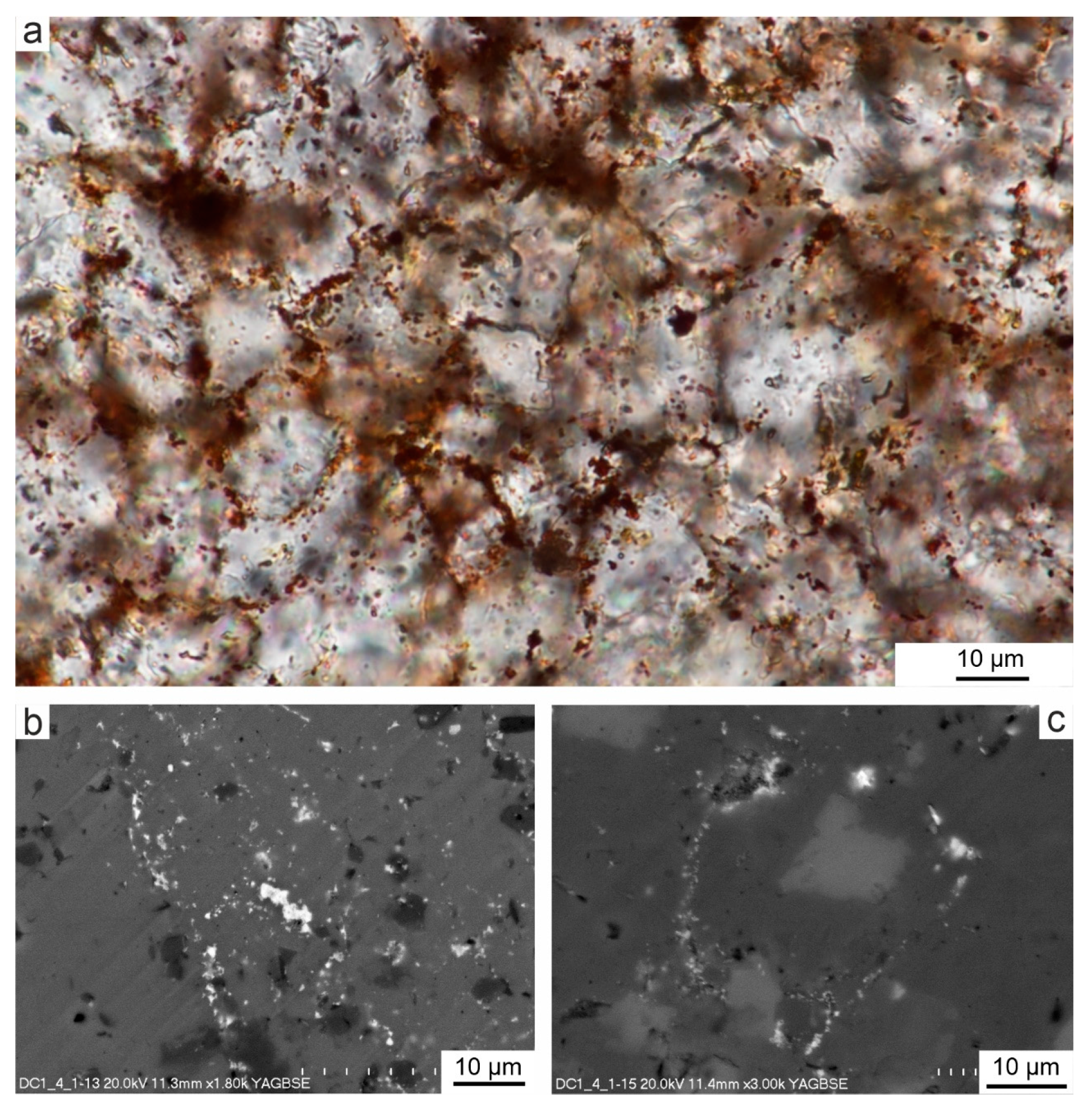
The analysis of foraminiferal assemblages and microfacies showed that all samples studied are biogenic limestones that were deposited in a shallow marine environment [55]. They contain different percentages of calcareous bioclasts, which are mainly benthic foraminifers, mostly belonging to the fusulinids (Figure 4a–d). The remaining bioclasts are fragments of skeletal plates of echinoderms (Figure 4a). Other biotic particles are very rare. A characteristic feature of these limestones is sparite binder, which comprises crystals that vary in size (Figure 4e). The smallest calcite crystals (3–5 µm) occur in the recrystallized walls of foraminifera, and the largest (5–10 µm and larger) in the infill of originally empty chambers or pores in skeletons, as well as in the space surrounding bioclasts.
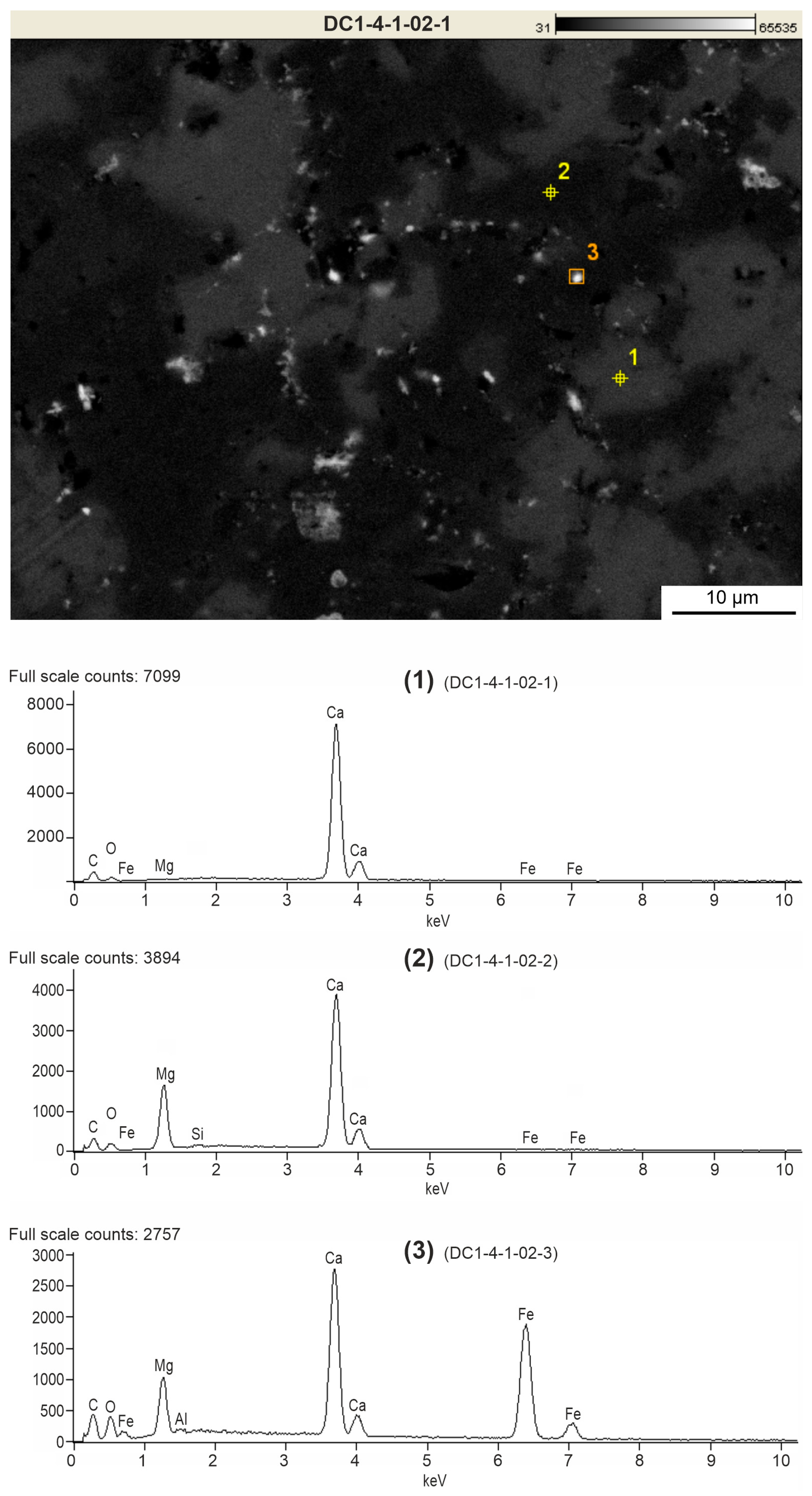
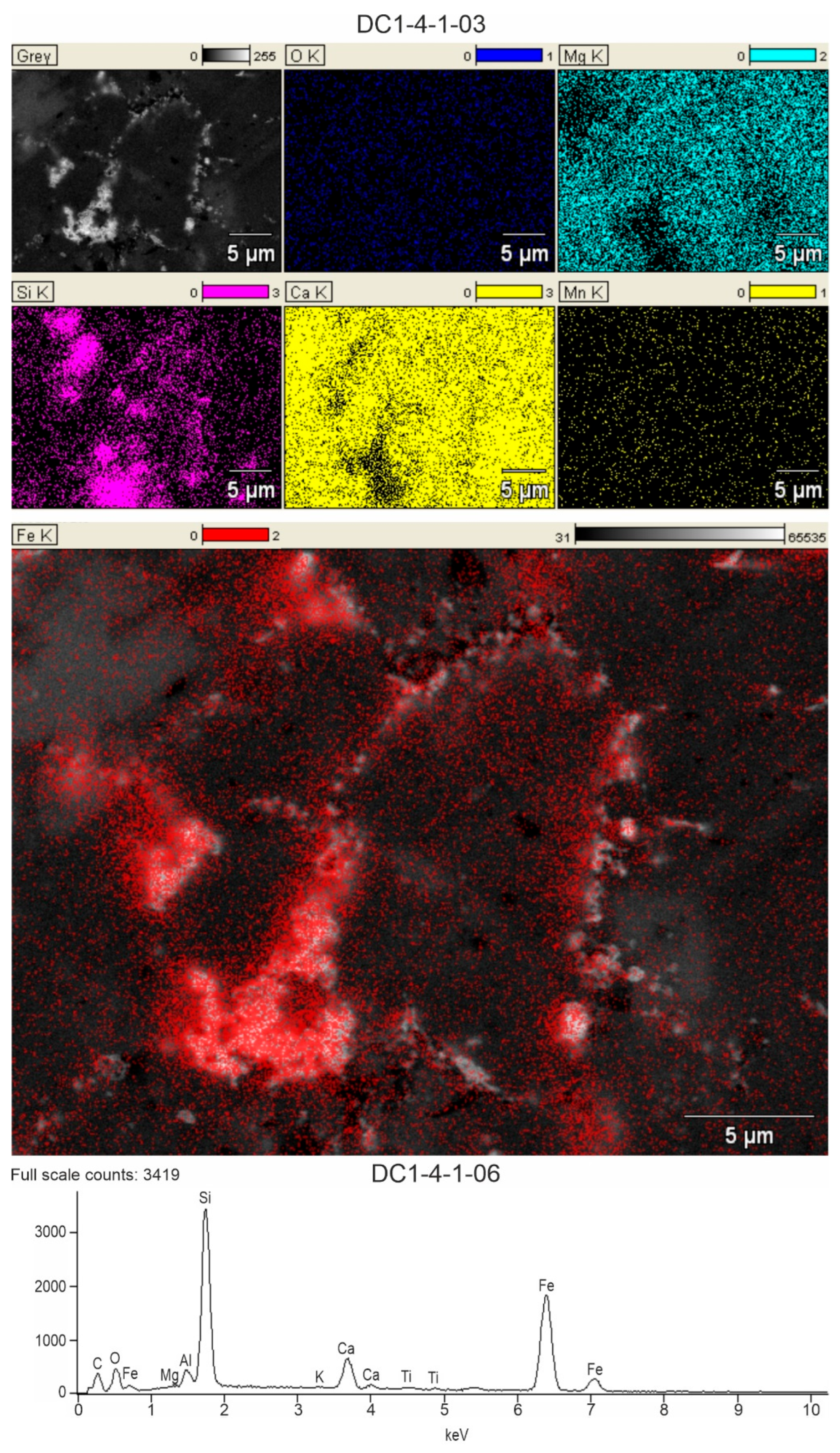
SEM-EDS images of selected surfaces of thin sections of rocks, which were characterized by the presence of redness, show their structural and chemical differences. Larger, light grey and dark grey areas seen in the BSE image (sometimes white-glowing points) correspond to the shell walls of foraminifers and other bioclasts, which is confirmed by TLM images (Figure 4b,c). Among them, white dots and streaks occur mainly around bioclasts or sparite crystals. In turn, dark grey areas indicate the filling of originally empty spaces in foraminiferal chambers and spaces between bioclasts. Chemical SEM-EDS microanalysis (Figure 5) indicates that light grey areas consist mainly of calcite, which is the main component of bioclasts. White streaks and glowing points contain an increased content of Fe oxides. In some places, these white peaks occur in microcrystalline quartz (Figure 6). On the other hand, dark grey areas contain Mg oxide and therefore may be partially composed of dolomite crystals (Figure 6).
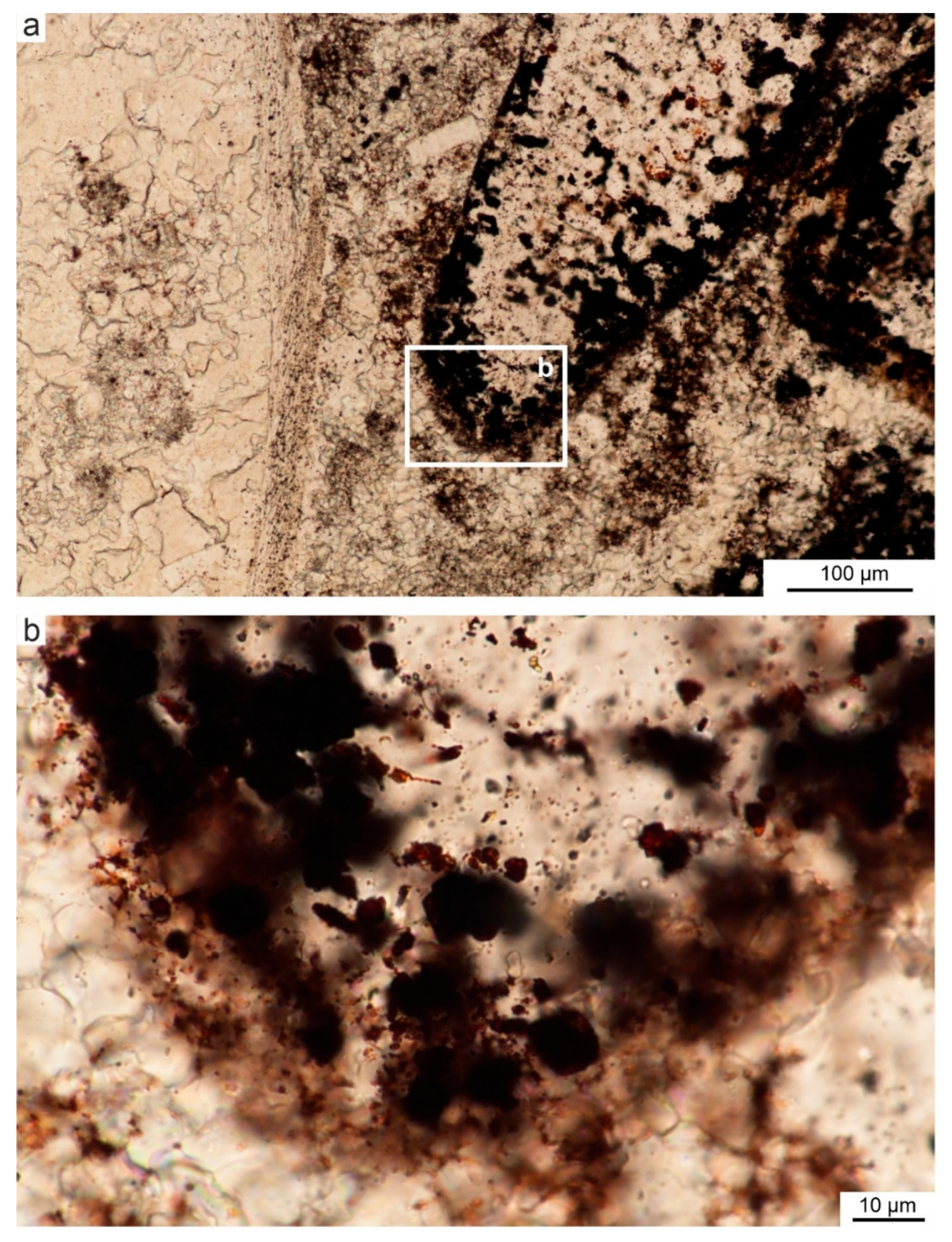
Detailed microscopic analysis of all thin sections of the rocks studied show that only red Viséan limestones (section DC1) possess irregular rust to brown areas that could have been caused by iron-related bacterial activity (Figure 4e, Figure 5 and Figure 6). Microfacies analysis (TLM) shows that these pigmented areas contain iron oxides and oxyhydroxides, accompanied by individual iron-bearing microcrystals and/or aggregates. These ferrous discolourations can occupy areas of up to 40% of the microscopic view. They occur in various positions in relation to the sparite cement crystals. They are most common between crystals, on the edges and walls of crystals, forming a characteristic network (Figure 7). This is especially the case when the calcite crystals are small. Larger crystals contain individual oval structures of 1–2 µm in size enclosed within them (Figure 8 and Figure 9). Rust-coloured and brownish structures may form thin streaks between crystals or thicker coatings with raised, oval shapes and blurred edges, whereas when these structures are thicker, they become dark brown to black inside and have oval to convex shapes that can be single or clustered in larger black areas. In most cases, these areas were the site of growth of Fe-bearing crystals.

4.2. Mineralogy of Ferruginous Discolourations in Limestones

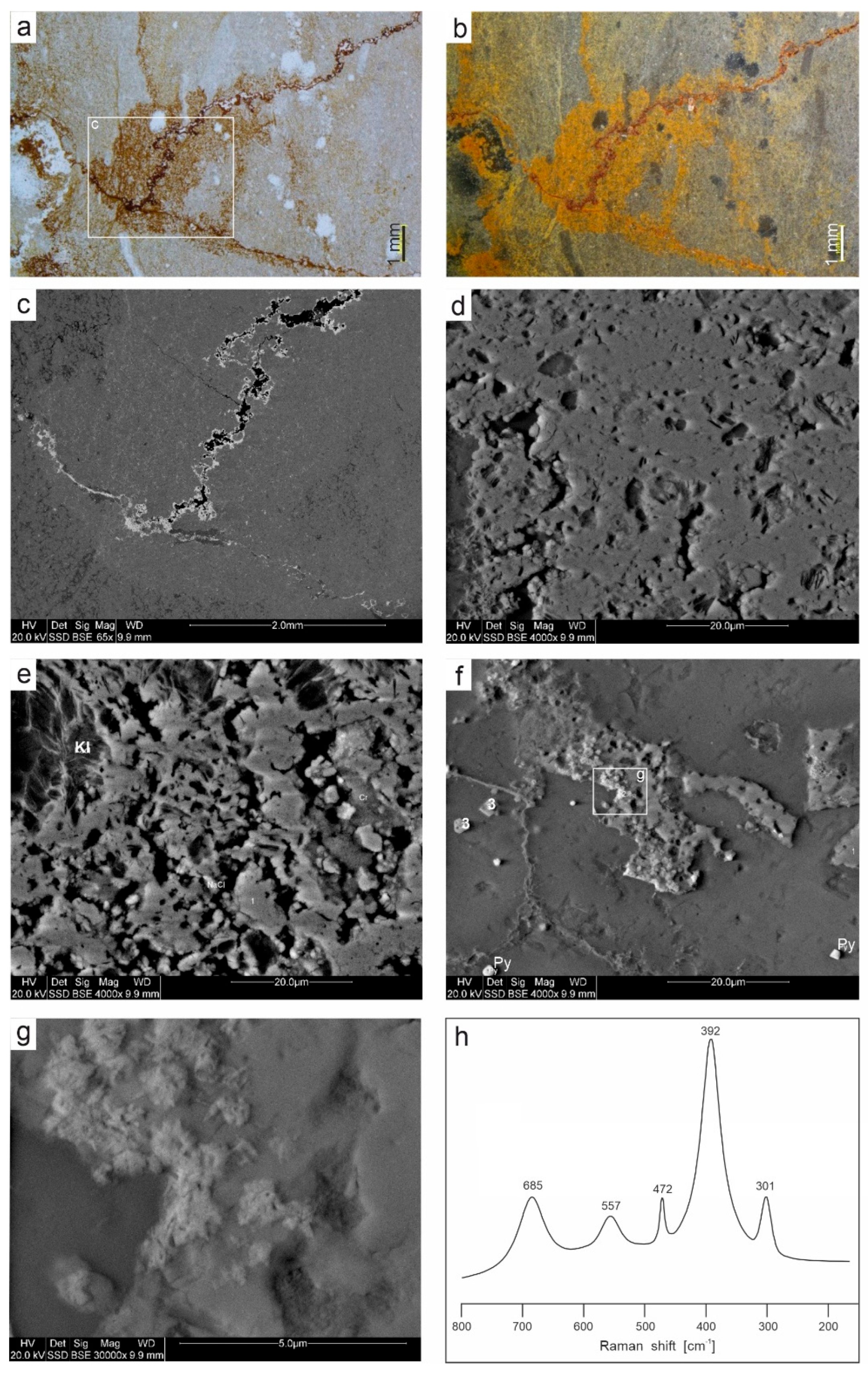
The microscopic images (TLM) show that the sources of the reddish and yellowish discolouration of the studied rocks, which gives the overall macroscopic colour and creates rusty coatings around fossils and bioclasts, are numerous veins filled with several generations of minerals (Figure 10a,b).
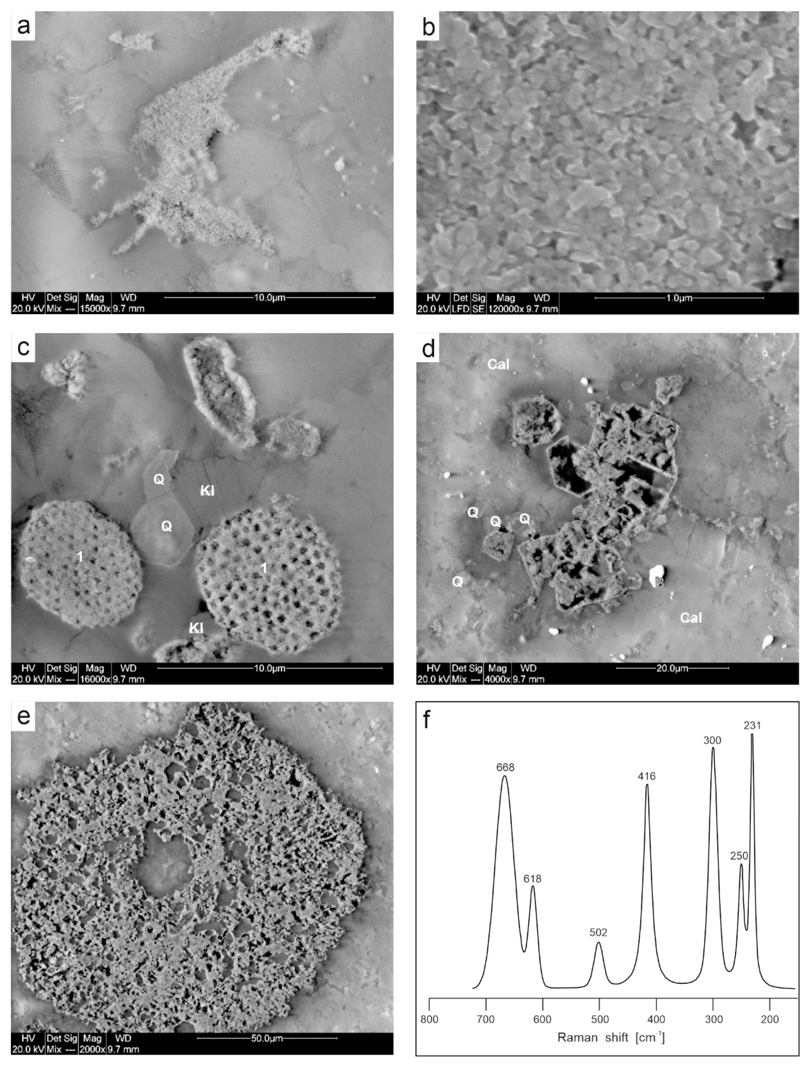
SEM observations and SEM-EDS analyses supported by Raman microspectroscopy indicate that the Fe oxyhydroxides and oxides present in the studied samples are goethite (α-FeOOH) and hematite (α-Fe2O3), both of which form very diverse morphological forms (Figure 10 and Figure 11). These are very finely dispersed crystals and indentations, several µm in size, which are responsible for the majority of the colour of the entire rock. Furthermore, pore and vein fillings are common, as are pseudomorphs after pyrite (e.g., Figure 11d) and rounded porous aggregates, likely of bacterial origin (Figure 11c). The collected Raman spectra indicate that goethite and hematite do not occur together; individual samples contain only one or the other mineral. Goethite is the dominant iron carrier in samples DC1.1 and DC1.2, which are characterized by a yellowish colour. Here, small (max 2–3 µm) crystallites (most often filling pores) can be encountered, with the acicular shape characteristic of this oxyhydroxide. In contrast, in specimens with a dominant reddish colour (samples DC1.3, DC1.4.1, DC1.4.2, DC1.5), the colouring agent is hematite, in places forming quasi-spherical or plate-like crystallites. Variation in colour within a single sample (e.g., DC1.4.2) appears to be unrelated to mineralogical diversity (in this particular case, the only Fe form detected is hematite), but rather to the degree of mineral accumulation in different locations. There is also no evidence of significant differences in chemical composition between zones of different colour within a single sample. However, subtle differences in the composition of iron compounds between samples are noticeable. Some samples do not contain EDS-detectable admixtures of other transition metals (as in sample DC1.3) but do contain admixtures of Zn (samples DC1.1, DC1.2, DC1.5), Ti (samples DC1.1, DC1.2), and Mn (in some locations in samples DC1.4.1 and DC1.5). In sample DC1.2, Mn oxides with significant Pb content are occasionally found alongside goethite. The chemistry and morphology of the fine crystallites suggest that it is coronadite Pb1-1.4(Mn4+,Mn3+)8O16 (Figure 10f,g).
Other non-carbonate components of the limestones include Fe sulphides (pyrite), quartz, and kaolinite (Figure 10e and Figure 11c). Barite (Figure 11d), Ti oxides (rutile), and fluorapatite were also encountered. Pyrite most often occurs as scattered single euhedral or subhedral crystals, with sizes of up to several µm. Framboidal clusters (up to 10 µm in diameter) composed of crystals < 1 µm in size can also be found (sample DC1.1). Pseudomorphs of Fe oxides after FeS2 are common alongside fresh, unoxidized pyrite crystals. Quartz and kaolinite typically occur in zones enriched in iron oxides and oxyhydroxides, as evidenced by elemental maps indicating correlations in Fe, Si, and Al concentrations. Both silicates are often euhedral, and euhedral quartzes with visible crystal growth zones are also found.

4.3. Geochemistry of Major Oxides and Trace Elements

The rocks studied show strong differences in the content of major elements (Table 1). In the Viséan red limestones (DC1 section), the concentration of CaO is markedly lower (44.70–51.88 wt%), in contrast to the sum of other components that are not geochemically related to limestones, i.e., SiO2, MgO, Fe2O3, Al2O3, K2O, MnO, TiO2, P2O5, and Na2O. The content of SiO2 is high (3.28–12.14 wt%), as well as the contents of Al2O3 (0.46–3.34 wt%) and Fe2O3 (0.44–1.69 wt%), while the amount of MgO is relatively small (1.26–3.28 wt.%). The contents of other elements are also clearly higher than the average contents for marine limestones [59].
The samples from the second section of the Viséan rocks (DC2) are characterized by the higher amount of CaO (49.72–55.51 wt%) and a lower sum of other components (1.19–9.83 wt%). The concentration of SiO2 is lower (0.59–9.83 wt%), similarly to the content of Al2O3 (0.02–0.88 wt%), Fe2O3 (0.07–0.33 wt%), and MgO (0.24–0.33 wt%). The abundance of other elements is lower (K2O < 0.07 wt%; TiO2 < 0.03 wt%; MnO < 0.03 wt%).
The quite different composition of main oxides characterizes the Tournaisian limestones (DR3 section). The high content of CaO (54.16–55.34 wt%) and low amounts of SiO2 (0.41–1.06 wt%), Al2O3 (0.05–0.19 wt%), and Fe2O3 (0.04–0.13 wt%) are typical of limestones. The only amount of MgO (0.40–0.66 wt%) is clearly higher in this succession. The abundances of other oxides like TiO2, MnO, P2O5, and Na2O do not exceed 0.01 wt%. Only the K2O content is slightly higher (up to 0.06 wt%).

4.4. Correlation Between Main Elements and SEM-EDS Mapping

Pearson’s coefficient values, calculated for individual pairs of oxide content in all samples from the DC1 section (Table 2), show strong linear correlation between SiO2 and Al2O3, Fe2O3, K2O, TiO2, P2O5, and MnO. Moreover, there is a strong negative correlation between CaO and Fe2O3, SiO2, Al2O3, K2O, TiO2, P2O5, and MnO. There is also significant negligible correlation between MgO and all of the above mentioned oxides, which is characteristic for all samples investigated. The same trend is distinctly visible for samples of all sections investigated (DC1, DC2, DR3) (Figure 12), although individual profiles were affected to a different extent by the impact of hydrothermal solutions, which was reflected in the different contents of individual elements.
The above tendency observed in the analytical results is also clearly depicted in the SEM-EDS mapping (Figure 6). The microanalysis of Si, Fe, Ca, Mg, Al, and K clearly shows that the micro-area of increased SiO2 and Fe oxide and oxyhydroxide contents overlap. This dependence is clearly visible in the images from rocks in profile DC1, where the weight of both oxide groups is relatively high. The same area also coincides with the fields of Al and K oxides in the same samples.
The area of co-occurrence of SiO2 and Fe oxides and oxyhydroxides and (Al, K) elements is generally separated from the area with increased CaO and MgO values, which is associated primarily with coarse crystalline cement filling the original porosity and generally does not contain Fe-bearing browning.

4.5. Trace Elements

The composition of trace elements in these limestones displays similar differences in the sections studied (Table 3). The red limestones from the Czernka Valley (DC1, DC2) are particularly distinctive in this respect. The DC1 samples are characterized by enhanced content of several elements in relation to samples from the Racławka Valley (DR3). This applies to the following elements: Zn (up to 298 ppm), Pb (up to 191 ppm), Sr (up to 758 ppm), V (up to 36 ppm), Ba (up to 30 ppm), As (up to 15.1 ppm), Th (up to 5.7 ppm), Nb (up to 4,5 ppm), U (up to 3.7 ppm), Zr (up to 35.2 ppm), Mo (up. To 1.4 ppm), Hf (up to 1 ppm), Sb (up to 1.0 ppm), Ga (up to 1.3 ppm), and Hg (0.4–0.7 ppm).
In the second section of the Viséan limestones (DC2), the increased values refer only to one sample (DC2.08), where some trace elements, i.e., Zn (86 ppm), Ba (30 ppm), Zr (35.9 ppm), Th (3.3 ppm), U (1.6 ppm), Nb (1.6 ppm), Hf (up to 0.9 ppm) have high contents, comparable to the DC1 samples. The abundances of Pb (up to 33.2 ppm), Zn (up to 86 ppm), Cu (up to 2.8 ppm), and Mo (up to 0.2 ppm) are markedly lower.
The DR3 samples have small amounts of the most trace elements compared to the rock samples described above (Table 3). Only the content of Sr (184.8–329.5 ppm) is medium, and it is higher than that of the DC2 samples.

4.6. REE Signatures

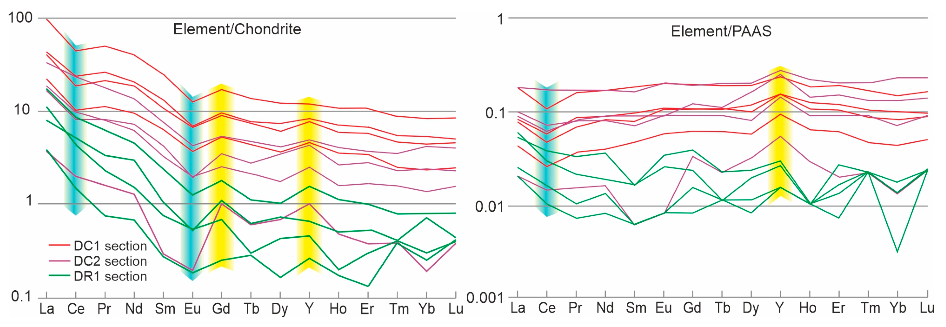
The studied sediments display a variable content of REE (Table 3). In the red limestones (section DC1; av. 60.3 ppm), it is higher than in the grey limestones containing pink spots (section DC2; av. 23.2 ppm), and much higher than in the grey limestones containing ferruginous discolourations and coatings visible in microscopic observations (section DR3; av. 5.7 ppm). The chondrite- and PAAS-normalized REE + Y patterns were similar between the sections in the Czernka Valley (DC1 and DC2), emphasized by Ce- and Eu-negative anomalies with Gd- and Y-positive anomalies (Figure 13). This pattern for limestones from the Racławka Valley (section DR2) differs among the Light-REE in the lack of cerium anomaly. The materials studied from the Czernka Valley exhibit superchondritic Y/Ho ratios (av. 35–57 in particular sections). In turn, the lowest values (32–39) are characteristic of the DC1 section. The Ce/Ce* average values in limestones vary significantly between the sections from 0.73 (DC1 section) to 1.6 (DR3 section). The values of the DyN/SmN ratio, a signature of diagenetic enrichment in the MREE [60], are above 1.2, excluding a single sample in the Czernka Valley (DC1.01). The Gd/Gd* average values in limestones vary significantly between the sections from 20 (DC1 section) to 160 (DR3 section).

5. Discussion

5.1. Bacterial Origin of Rust-Coloured and Brown Pigments

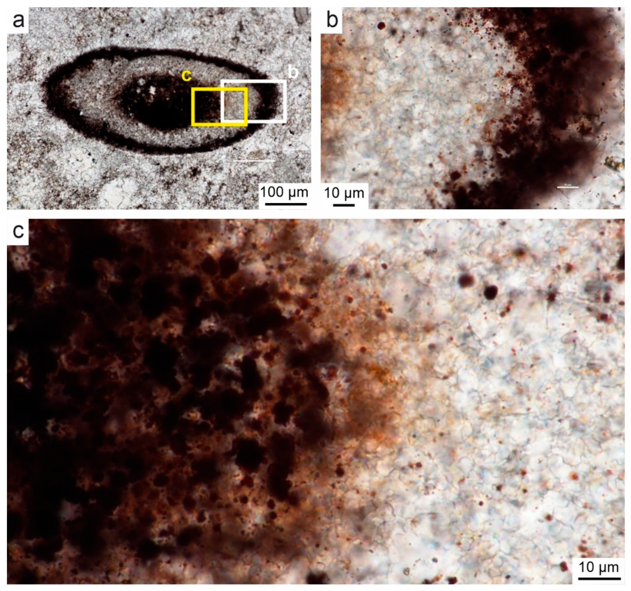
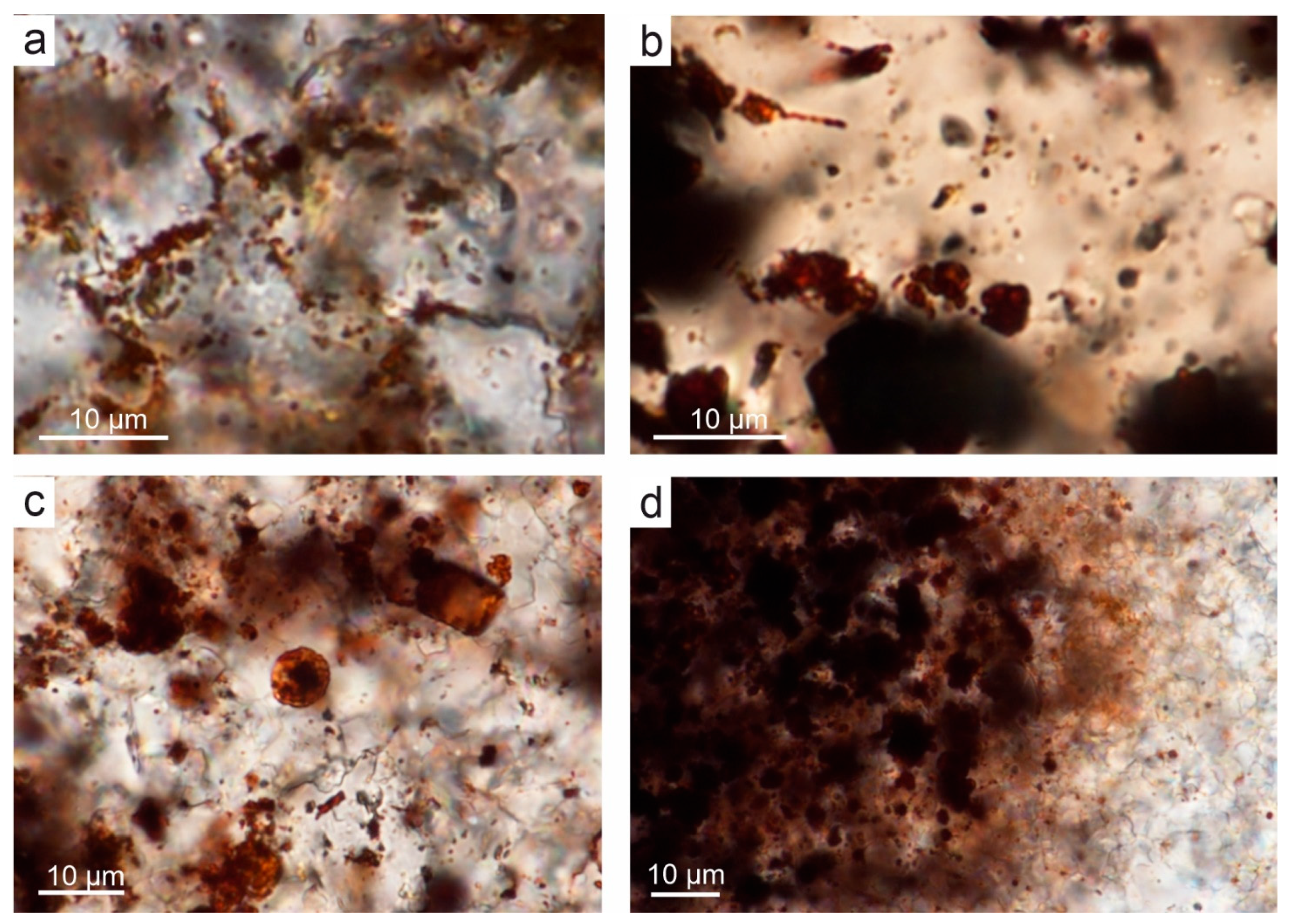
The thin-section views of samples from the DC1 profile, using TLM at magnification x 1000, show spherical to oval dark brown structures measuring 1–2 μm across (Figure 4e, Figure 7, Figure 8, Figure 9, and Figure 14). They occur as single capsules or tubes enclosed in a spary binder (e.g., Figure 14a–c), arranged linearly (Figure 14a) between crystal walls (e.g., Figure 4e), or appear in groups of several specimens (Figure 11c and Figure 14c). The single capsule is hollow inside and has a wall that is about 0.1 µm thick. SEM-EDS and Raman analysis showed that the capsule walls contain a large amount of iron oxides and hydroxides (Figure 6 and Figure 11), which may be a remnant of the original bacterial sheath from the IRB group. Oval to elongated structures of various sizes resembling bacterial cells are often arranged linearly or in rounded consorcia (Figure 11c and Figure 14a–c). This indicates a similarity to IRB from the Sphaerotilus-Leptothrix group due to the arrangement of individual cells in bacterial sheaths. In some places, empty tubular structures are visible, which may also be remnants of specimens of this group of bacteria (Figure 14c). Some of the specimens possess stalked, twisted ribbon-like structure (Figure 14b) resembling IRB from the Galionella group or another Fe oxidizer like Mariprofundus ferrooxydans, e.g., [64,65,66].
Further evidence for the association of rusty discolourations with bacterial origin are oval, dark brown pigment clusters usually surrounded by rusty, translucent haloes with an unevenly coloured, floccular structure, visible under a light microscope (Figure 14d). These structures resemble the bacterial holdfasts, whose general outline and shape might be similar to that of bacteria from the Sphaerotilus-Leptothrix group. Particular similarity to the present-day species of Leptothrix lopholea and Leptothrix cholodnii can be seen [5,67].
The structures that occur in the examined samples from the DC1 outcrop can be classified as bacteria only on the basis of the above mentioned features because in the case of fossil material from the late Paleozoic, this is the only available criterion. It is not possible to assess their belonging to any group of bacteria (or even Archaea) by applying microbiological and chemical methods to modern material. For this reason, we stick to the term “bacteria-like″ or “resembling bacteria″ to fully reflect the above impossibility of classifying the above structures as bacteria. However, in some cases, especially when bacteria precipitate inorganic compounds such as iron oxides or calcium carbonate outside the cell membrane, in the fossil record, they may preserve unique shapes and various types of bacterial sheaths and filaments, corresponding to features of specific bacterial species cf. [66].

5.2. Timing for IRB Growth

The rocks in which the bacteria-like remnants are found have a long geological history. They belong to a nearly 1200 m long sequence of the Upper Paleozoic carbonate platform [27]. Carbonate sedimentation occurred in a relatively shallow part of the epicontinental Moravo-Silesian Basin (MSB), along the shelves bordering the Upper Silesian Block—a part of a larger tectonic unit—the Brunovistulicum [34,36,68]. The older basement of this tectonic structure is interpreted as an ancient piece derived from the Gondwana continent [69].
The taphonomic analysis shows that bacteria-like remnants are not associated with the sedimentation in shallow marine environment of the original limestones. First of all, bacteria are present in part of the limestone with sparite cement, which occurs in the studied rocks as narrow zones of several millimetre to several centimetre, unrelated to bedding and cutting rocks in different directions (Figure 10a,b), also across microfossils as different types of foraminifers or echinoderm plates (Figure 7, Figure 8 and Figure 9). Rusty pigmentation occurs especially in the originally porous parts of the rock, which are microfossil chambers or porous walls of skeletal remains (Figure 3c,d, Figure 8, Figure 9 and Figure 11e). Additionally, bacteria-like structures are not associated with bacterial mats, which could have formed in the sedimentary environment of the host rocks at that time [57].
In order to draw conclusions about the origin of bacteria in the studied rocks, it is important to note that bacterial remnants are present locally, only in samples from the DC1 profile. In other profiles (DC2, DR3), bacteria were not observed despite the fact that they represent similar lithotypes of shallow-water limestones.
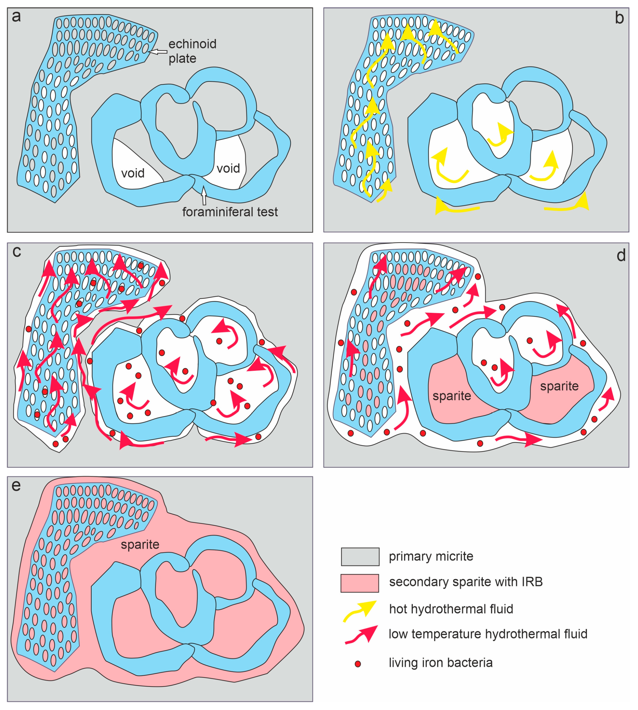
The microfacies analysis of the reddish colourations inside the microfossil walls and around them (Figure 3c,d), as well as shape of the walls and their vicinity (Figure 8, Figure 9 and Figure 15), shows that their origin is partly related to the dissolution process. The thinnest infillings were developed as seams bordering bioclasts, which usually consist of calcite spare, while they were surrounded by micrite. Reddish (iron oxide) seams started to develop in places where calcitic bioclast contacted micrite. This shows that fissures can be opened by leaching micrite from the host limestone and successively filling them with iron oxides, which are associated with calcite spar and quartz crystals (Figure 15). The more resistant parts of foraminiferal chambers or echinoid plates were left as residue (Figure 11e). Additionally, the crystals of calcitic spar are rounded (Figure 7), indicating their stepwise dissolution and crystallization during subsequent hydrothermal pulses.
This may indicate that bacterial growth occurred in the originally empty micropores of the skeletons and shells of microfossils, between bioclasts, or in secondary voids formed during the selective dissolution of micrite or smaller sparite crystals. The spatial relationship of sparite crystals to iron deposits and bacteria-like remnants indicates that iron oxide precipitation and sparite crystallization were contemporaneous processes. For the development of IRB, bacteria had to be solutions with iron content increased to such a level that individual species of bacteria could encrust cell membranes during their life. Samples from the DC1 profile contain increased contents of Fe, and in addition to this, other elements constituting a trace of the influence of hydrothermal conditions on these rocks. These solutions had to be relatively cold if IRB could live during their circulation. Another indicator of low-temperature hydrothermal activity is enrichment in elements like Ba, Sr, As, and Sb, e.g., [70]. Significantly increased contents of these elements, compared to samples from other tested profiles, occur in the DC1 profile.
Among the studied platform succession sediments, the IRB-like structures were not found in the Tournaisian limestones (DC2 profile), whose location right next to the same fault as the red limestones from the DC1 exposure suggests the possibility of their infiltration by hydrothermal waters. However, among the oxides whose elevated content may indicate hydrothermal origin, the composition and weight fraction differs from that in the DC1 profile. Additionally, the content of iron oxides and hydroxides is over 50 percent lower. Differences in chemical composition suggest different origins, and perhaps different durations, of hydrothermal solutions in these nearby areas. At the same time, the lack of visible structures resembling IRB indicates low iron concentrations in the original solutions circulating in the rock, which were insufficient for bacterial formation to produce inorganic coatings. This may also indicate the higher temperature of the solutions interacting in this part of the carbonate complex.
In turn, the limestone succession from the DR3 profile is characterized by the typical marine limestone content of oxides, accessory elements, and rare earth elements. The biotic components and the state of preservation of the clasts indicate a high-energy sedimentary environment. Therefore, the sparite present in these samples likely crystallized in the sedimentary environment and does not originate from the subsequent recrystallization of micrite during the circulation of hydrothermal solutions.
The local scope of influence of hydrothermal solutions and the possibility of the temporary development of IRB in them only in the area of neptunian dikes is evidenced by the fact that total REE content measured from the studied limestones is comparable to that of normal seawater (25–26 ppm; Table 3; e.g., [71]) or even lower in the case of the Si-enriched dyke (20 ppm). The same conclusion is suggested here on the basis of the PAAS-normalized REE patterns of the material from the dykes that were more or less flat, excluding the Y enrichment (Figure 13). This is similar to carbonate and authigenic marine phases, which mainly produced a seawater-like REE pattern [72]. The REE of the host rock was slightly higher (ca 20%; Table 3), which may reflect the contamination of phosphates in the echinoderm-foraminiferal limestone observed in thin sections of the rock (Figure 11e).

5.3. Potential Sources of Hydrothermal Fluids

Here, we consider two potential fluid sources, the first of which could be related to post-Variscan magmatism and volcanism, and the second, to a stage of Mesozoic sulphide mineralization. The first of these episodes is well documented in the study area, and the second, in its immediate vicinity.
Variscan compressional deformations in this area, which took place at the turn of the Moscovian and Kasimovian (Westphalian and Stephanian), resulted in the formation of gentle folds and faults with a latitudinal course and strike-slip character [44]. In this phase, the Dębnik anticline was formed, among others (Figure 1c). Starting from the Gzhelian (Stephanian B), this area experienced a phase of accumulation of post-orogenic deposits (arkosic sandstones and conglomerates containing silicified Dadoxylon tree trunks) and the onset of terrestrial volcanism, which lasted until the Early Permian, e.g., [73]. Volcanic, subvolcanic, and volcanoclastic rocks occur in this area on a surface of approx. 200 km2 in the form of lava flows and small intrusions in the form of domes, laccoliths, and veins, and local tuffs of an ignimbrite nature, e.g., [47,48,49,50,51,52,74]. The intrusions are local and do not form continuous covers, although they are associated with the Kraków–Lubliniec tectonic zone, which divides the Upper Silesian Block from the Małopolska Block (Figure 1b) [34,45]. This volcanism was bimodal, from andesitic to rhyolitic, and a history of more than 100 years of research indicates that both varieties of volcanites appear to be genetically related and derived from magmas originating from the lithospheric mantle and crust, e.g., [30].
Zircon U–Pb dating of various igneous rocks (granodiorites, dacites, lamprophyre, and diabase) from this igneous belt showed that magmatism spanned within a short-lived event, between 303.8 ± 2.2 and 292.7 ± 4.9 Ma [30]. Felsic calc-alkaline magmatism was a characteristic for the oldest interval (during the latest Carboniferous), followed by the slightly alkaline volcanism of mafic–intermediate composition (the oldest Permian). The last one concerns the lamprophyre and diabase dikes located near the study area, in the 20 km wide zone along the Kraków–Lubliniec tectonic zone [30]. The oldest of them is the so-called granodiorite II from the Zawiercie region located in the Lubliniec–Kraków Fault Zone, which has been dated (Sr-Rb) to 340 ± 4 Ma [75].
This type of magmatism is associated with the Cu-Mo mineralization in this area occurring both in porphyry-type deposits, and in the surrounding skarn-contact metasomatic and vein-type deposits [46,75,76,77,78,79,80,81,82,83], forming a system of quartz veins with ore minerals. This mineralization was related to felsic magmatism and could be the most likely source of the elevated trace element values found in the Czernka Valley. In turn, the younger alkaline volcanism is characterized by the REE mineralization of the metasomatic hydrothermal type [84], which was not confirmed in the examined deposits.
Another possibility of the origin of the fluid sources for IRB that we considered could be related to the Mississippi Valley-type Zn-Pb mineralization hosted mainly by the Middle Triassic limestones and dolomites (Muschelkalk), which is of economic importance in a neighbouring region, just west of the study area [31,54]. Most probably, these ore fluids have also been related to the Variscan igneous rocks, which gave rise to the polymetallic ores, and later, they became remobilized by heat flow and redeposited in various sedimentary successions [85]. The timing of the remobilization of older ores and the origin of fluids are still debated. They are most likely related to crustal extension during the Mesozoic plate–tectonic reorganization (Late Triassic according to [85] or Early Cretaceous according to [86]). This type of mineralization is also known in association with Devonian–Permian rocks, which occur close to the studied sediments, in the Czernka Valley [55]. This proximity to the studied DC1 and DC2 profiles, and the fact that Pb-Zn mineralization was associated with low-temperature hydrothermal fluids, leads us to consider this Fe source for the IRB growth. However, our geochemical data are insufficient in this respect, and geological publications contain data on trace element content without detailed information on whether they originate from mining fields or their transition zones, or from “pure″ limestones or dolomites, e.g., [87]. For this reason, we leave this issue open for further geochemical studies.

6. Conclusions

  • In the Viséan part of the thick limestone succession located along the Upper Silesian Block (northern part of the Brunovistulicum), there are local zones of red-coloured rocks, which, based on morphological and chemical evidence, have been classified here as bacterial structures. These are spherical or oval forms, 1–2 µm in diameter, that occur as individual capsules or tubes arranged linearly between the walls of calcite crystals. The single capsule is hollow inside and has a wall that is approximately 0.1 µm thick, which contains a large amount of iron oxides and oxyhydroxides; this may be a remnant of the original bacterial sheath. Some oval, dark brown pigment clusters, surrounded by translucent halos with a floccular structure, resemble the bacterial holdfasts. We present here the morphology and chemistry of the microstructures showing their similarities to IRB from the present-day Sphaerotilus-Leptothrix group, the Galionella group, and the Mariprofundus ferrooxydans species.
  • The taphonomic analysis shows that bacteria-like remnants are not associated with the sedimentation of shallow marine carbonate mud. They are present only in narrow zones that are a few millimetres to several centimetres wide, unrelated to bedding, and they also occur across various microfossils. Based on microfacies analysis, we indicate that bacterial growth occurred in the originally empty micropores of microfossil skeletons and shells, between bioclasts, or in secondary voids formed during the selective dissolution of micrite or smaller sparite crystals. The dissolution of primary micrite and the subsequent filling of spaces with iron oxides/xyhydroxideswere associated with repeated hydrothermal pulses. The solutions must have been relatively cold, which is evidenced by the increased content of Ba, Sr, As, and Sb.
  • Taking into account the types of mineralization associated with post-Variscan plutonism and volcanism, well documented in the immediate vicinity of the studied Mississippian sedimentary succession, we suggest that the hydrothermal pulses that associate with the formation of bacterial structures are probably related to felsite magmatism (Pennsylvanian), which was characterized, among other, by Cu-Mo mineralization, typical of many neighbouring porphyry deposits and skarn metasomatic deposits. However, this issue requires further geochemical studies on these sediments, in conjunction with analogous studies on carbonate rocks around the Zn-Pb mineralization zones, but outside the ore bodies and their transition zones.

Author Contributions

Conceptualization, M.B., A.W. and K.B.; Methodology, M.B.,A.W. and G.R.; Validation, K.B.; Formal analysis, M.B., A.W. and G.R.; Investigation, M.B., A.W. and G.R.; Resources, S.S. and P.S.; Data curation, M.B.; Writing—original draft, M.B., K.B., A.W., S.S., P.S. and S.B.; Visualization, K.B., P.S., S.B. and P.D.; Project administration, M.B.; Funding acquisition, K.B. and A.W. All authors have read and agreed to the published version of the manuscript.

Funding

K.B. received funding from the University of the National Education Commission, Krakow (Grant No. WPBU/2024/03/00125), and Marta Bąk, Grzegorz Rzepa, Piotr Strzeboński, and Sławomir Bębenek received funding from the Statutory Funds of the Faculty of Geology, Geophysics and Environmental Protection, AGH University of Krakow (Project 16.16.140.315).

Data Availability Statement

The original contributions presented in this study are included in the article. Further inquiries can be directed to the corresponding author.

Acknowledgments

We would like to thank two anonymous reviewers and the journal editor for constructive comments and suggestions. We are also grateful to a native English speaker for the overall grammatical and linguistic improvement of the text.

Conflicts of Interest

Author Stanisław Szczurek was employed by the company PROINSOL Sp. z o.o. Spółka Komandytowa. The remaining authors declare that the research was conducted in the absence of any commercial or financial relationships that could be construed as a potential conflict of interest.

References

  1. Volesky, B. Biosorption and Biosorbents. In Biosorption of Heavy Metals; CRC Press: Boca Raton, FL, USA, 1990; pp. 3–5. [Google Scholar]
  2. Cygnarowska, K. The use of algae to remove copper and lead from industrial wastewater. Geol. Geophys. Environ. 2023, 7, 85–93. [Google Scholar] [CrossRef]
  3. Gyollai, I.; Polgari, M.; Fintor, K.; Pal-Molnar, E.; Popp, F.; Koeberl, C. Microbial activity records in Marinoan Snowball Earth postglacial transition layers connecting diamictite WITH cap carbonate (Otavi Group, NW-Namibia). Austrian J. Earth Sci. 2017, 110, 4–20. [Google Scholar] [CrossRef]
  4. Ciurej, A.; Bąk, M.; Szczerba, M. Biostratinomy and diagenetic impact on exceptional preservation of coccospheres from Lower Oligocene coccolith limestones. Minerals 2020, 10, 616. [Google Scholar] [CrossRef]
  5. Mulder, E.G.; Deinema, M.H. The sheathed bacteria. In The Prokaryotes, A Handbook on the Biology of Bacteria: Ecophysiology, Isolation, Identification, Applications; Balows, A., Ed.; Springer-Verlag: Berlin/ Heidelberg, Germany, 1992; p. 4126. [Google Scholar]
  6. Pan, J.; Zhao, H.; Tucker, M.E.; Zhou, J.; Jiang, M.; Wang, Y.; Zhao, Y.; Sun, B.; Han, Z.; Yan, H. Biomineralization of monohydrocalcite induced by the halophile Halomonas smyrnensis WMS-3. Minerals 2019, 9, 632. [Google Scholar] [CrossRef]
  7. Bąk, M.; Bąk, K.; Górny, Z.; Stożek, B. Evidence of bacteriogenic iron and manganese oxyhydroxides in Albian–Cenomanian marine sediments of the Carpathian realm (Poland). Ann. Soc. Geol. Pol. 2015, 85, 371–385. Available online: http://www.asgp.pl/85_2_371_385 (accessed on 17 May 2025). [CrossRef]
  8. Noike, T.N.; Nakamura, K.; Matsumoto, J. Oxidation of ferrous iron by acidophilic iron-oxidizing bacteria from a stream receiving acid mine drainage. Water Res. 1983, 17, 21–27. [Google Scholar] [CrossRef]
  9. Schrenk, M.O.; Edwards, K.J.; Goodman, R.M.; Hamers, R.J.; Banfield, J.F. Distribution of Thiobacillus ferrooxidans and Leptospirillum ferrooxidans: Implications for generation of acid mine drainage. Science 1998, 279, 1519–1522. [Google Scholar] [CrossRef] [PubMed]
  10. Kirby, C.S.; Thomas, H.M.; Southam, G.; Donald, R. Relative contributions of abiotic and biological factors in Fe(II) oxidation in mine drainage. Appl. Geochem. 1999, 14, 511–530. [Google Scholar] [CrossRef]
  11. Francis, C.A.; Tebo, B.M. Marine Bacillus spores as catalysts for the oxidative precipitation and sorption of metals. J. Mol. Microbiol. Biotechnol. 1999, 1, 71–78. Available online: https://www.caister.com/backlist/jmmb/v/v1/v1n1/11.pdf (accessed on 15 October 2020).
  12. Konhauser, K.O.; Riding, R. Bacterial biomineralization. In Fundamentals in Geobiology; Knoll, A.H., Canfield, D.E., Konhauser, K.O., Eds.; Blackwell Publishing Ltd.: Oxford, UK, 2012; pp. 105–130. Available online: https://robertriding.com/pdf/konhauser_riding2012.pdf (accessed on 15 October 2020).
  13. Schopf, J.W.; Fairchild, T.R. Late Precambrian microfossils: A new stromatolitic biota from Boorthanna, south Australia. Nature 1973, 242, 537–538. Available online: https://www.nature.com/articles/242537a0 (accessed on 15 October 2020). [CrossRef]
  14. Schelble, R.T.; Westall, F.; Allen, C.C. 1.8 Ga iron-mineralized microbiota from the Gunflint Iron Formation, Ontario, Canada: Implications for Mars. Adv. Space Res. 2004, 33, 1268–1273. [Google Scholar] [CrossRef]
  15. Awramik, S.M.; Schopf, J.W.; Walter, M.R. Filamentous fossil bacteria from the Archean of Western Australia. Precamb. Res. 1983, 20, 357–374. [Google Scholar] [CrossRef]
  16. Sommaruga, R.; Psenner, R. Trophic interactions within the microbial food web in Piburger See (Austria). Archiv Hydrobiol. 1995, 132, 257–278. [Google Scholar] [CrossRef]
  17. Préat, A.; Loreau, J.P.; Durlet, C.; Mamet, B. Petrography and biosedimentology of the Rosso Ammonitico Veronese (Middle–Upper Jurassic, north-eastern Italy). Facies 2006, 52, 265–278. Available online: https://link.springer.com/article/10.1007/s10347-005-0032-2 (accessed on 8 May 2021). [CrossRef]
  18. Mamet, B.; Préat, A. Iron-bacterial mediation in Phanerozoic red limestones: State of the art. Sedim. Geol. 2006, 185, 147–157. [Google Scholar] [CrossRef]
  19. Bąk, M.; Natkaniec-Nowak, L.; Naglik, B.; Bąk, K.; Dulemba, P. Organic-walled microfossils from the early Middle Cambrian sediments of the Holy Cross Mountains, Poland: Possible implications for sedimentary environment in the SE margin of the Baltica. Acta Geol. Sin.—Engl. Ed. 2017, 91, 39–50. [Google Scholar] [CrossRef]
  20. Hanert, H.H. Bacterial and chemical iron oxide deposition in a shallow bay on Palaea Kameni, Santorini, Greece: Microscopy, electron probe microanalysis, and photometry of in situ experiments. Geomicrobiol. J. 2002, 19, 317–342. [Google Scholar] [CrossRef]
  21. Hallam, A.; Bradshaw, M.G. Bituminous shales and oolitic ironstones as indicators of transgressions and regressions. J. Geol. Soc. 1979, 136, 157–164. [Google Scholar] [CrossRef]
  22. Rudmin, R.; Reva, I.; Sokol, E.; Abdullayev, E.; Ruban, A.; Kudryavtsev, A.; Tolkachev, O.; Mazurov, A. Minerals of rare earth elements in high-phosphorus ooidal ironstones of the Western Siberia and Turgai Depression. Minerals 2020, 10, 11. [Google Scholar] [CrossRef]
  23. Krajewski, K. Pelagic stromatolites from the High-Tatric Albian limestones in the Tatra Mts. Kwart. Geol. 1981, 25, 731–759. Available online: https://gq.pgi.gov.pl/article/view/9044 (accessed on 16 November 2024).
  24. Bąk, M.; Górny, Z.; Bąk, K. Sponge growth on the Cenomanian carbonate shelves of the Carpathian Basin: A record from spicule-rich turbidites. Bull. Geosci. 2015, 90, 651–666. Available online: http://www.geology.cz/bulletin/contents/art1544# (accessed on 16 November 2024). [CrossRef]
  25. Boyd, T.D.; Scott, S.D. Microbial and hydrothermal aspects of ferric oxyhydroxides and ferrosic hydroxides: The example of Franklin Seamount, Western Woodlark Basin, Papua New Guinea. Geochem. Transact. 2001, 7, 45. [Google Scholar] [CrossRef]
  26. Mazur, S.; Aleksandrowski, P.; Gągała, Ł.; Krzywiec, P.; Żaba, J.; Gaidzik, K. Late Palaeozoic strike—Slip tectonics versus oroclinal bending at the SW outskirts of Baltica: Case of the Variscan belt’s eastern end in Poland. Int. J. Earth Sci. 2020, 109, 1133–1160. Available online: https://link.springer.com/article/10.1007/s00531-019-01814-7 (accessed on 10 December 2020). [CrossRef]
  27. Dvorák, J.; Galle, A.; Herbig, H.G.; Krejèí, Z.; Malec, J.; Paszkowski, M.; Racki, G.; Skompski, S.; Szulczewski, M.; Żakowa, H. Evolution of the Polish-Moravian carbonate platform in the Late Devonian and Early Carboniferous: Holy Cross Mts., Krakow Upland, Moravian Karst. In XIII International Congress on Carboniferous-Permian; Guide to Excursion B4; Małecka, J., Ed.; Państwowy Instytut Geologiczny: Warszawa, Poland, 1995; pp. 1–35. [Google Scholar]
  28. Alexandrowicz, S.W.; Siedlecka, A. Lithological profile of Dinantian limestones of Czerna near Krzeszowice. Ann. Soc. Geol. Pol. 1964, 34, 395–423. Available online: http://www.asgp.pl/sites/default/files/volumes/34_3_395_424.pdf (accessed on 15 December 2020).
  29. Timmerman, M.J. Timing, geodynamic setting and character of Permo-Carboniferous magmatism in the foreland of the Variscan Orogen, NW Europe. Geol. Soc. Lond. Spec. Publ. 2004, 223, 41–74. [Google Scholar] [CrossRef]
  30. Mikulski, S.Z.; Williams, I.S.; Markowski, M. Carboniferous–Permian magmatism and Mo–Cu (W) mineralization in the contact zone between the Małopolska and Upper Silesia Blocks (south Poland): An echo of the Baltica–Gondwana collision. Int. J. Earth Sci. 2019, 108, 1467–1492. Available online: https://link.springer.com/article/10.1007/s00531-019-01715-9 (accessed on 17 October 2020). [CrossRef]
  31. Sass-Gustkiewicz, M.; Dżułyński, S.; Ridge, J.D. The emplacement of zinc-lead sulfide ores in the Upper Silesian district—A contribution to the understanding of Mississippi Valley-type deposits. Econ. Geol. 1982, 77, 392–412. [Google Scholar] [CrossRef]
  32. Żelaźniewicz, A.; Aleksandrowski, P.; Buła, Z.; Karnkowski, P.H.; Konon, A.; Oszczypko, N.; Ślączka, A.; Żaba, J.; Żytko, K. Tectonic Subdivision of Poland; Komitet Nauk Geologicznych PAN: Wrocław, Poland, 2011; p. 60. Available online: https://www.kngpan.agh.edu.pl/wp-content/uploads/Regionalizacja_Tektoniczna_Polski_20111.pdf (accessed on 17 October 2024).
  33. Gradziński, R. Przewodnik Geologiczny po Okolicach Krakowa; Wydawnictwa Geologiczne: Warszawa, Poland, 1972; p. 3. [Google Scholar]
  34. Żaba, J.; Buła, A.; Jachowicz, M. Principal characteristics of the Upper Silesian Block and Małopolska Block border zone (southern Poland). Geol. Mag. 1997, 5, 669–677. [Google Scholar] [CrossRef]
  35. Kalvoda, J.; Leichmann, J.; Bábek, O.; Melichar, R. Brunovistulian terrane (Central Europe) and Istanbul Zone (NW Turkey): Late Proterozoic and Paleozoic tectonostratigraphic development and paleogeography. Geol. Carp. 2003, 54, 139–152. Available online: https://geologicacarpathica.com/browse/archive/?journal_article_no=2661 (accessed on 20 October 2020).
  36. Buła, Z.; Habryn, R.; Jachowicz-Zdanowska, M.; Żaba, J. The Precambrian and Lower Paleozoic of the Brunovistulicum (eastern part of the Upper Silesian Block, southern Poland)—The state of the art. Geol. Quart. 2015, 59, 123–134. [Google Scholar] [CrossRef]
  37. Belka, Z. The development and decline of a Dinantian carbonate platform: An example from the Moravia-Silesia Basin, Poland. In European Dinantian Environments; Miller, J., Wright, P.V., Adams, A.E., Eds.; John Wiley & Sons: Hoboken, NJ, USA, 1987; pp. 177–188. [Google Scholar]
  38. Hladil, J. Structure and microfacies of Middle and Upper Devonian Carbonate Buildups in Moravia, Czechoslovakia. Mem. Can. Soc. Petrol. Geol. 1988, 14, 607–618. Available online: https://archives.datapages.com/data/dgs/014/014002/607_cspgsp014b0607.htm (accessed on 15 January 2021).
  39. Szulczewski, M.; Belka, Z.; Skompski, S. The drowning of a carbonate platform: An example from the Devonian–Carboniferous of the south western Holy Cross Mountains, Poland. Sedim. Geol. 1996, 106, 21–49. [Google Scholar] [CrossRef]
  40. Bábek, O. Thinning and wining upward megasequence in Middle Devonian carbonate slope deposits, Moravia, Czech Republic. Neues Jahrb. Geol. Paläontologie Abh. 1996, 202, 409–432. [Google Scholar] [CrossRef]
  41. Dvořák, J. The Famennian of Moravia (CSSR); the relation between tectonics and sedimentary facies. Ann. Soc. Geol. Belg. 1986, 109, 131–136. Available online: https://popups.uliege.be/0037-9395/index.php?id=2426 (accessed on 20 October 2020).
  42. Szulczewski, M. Depositional evolution of the Holy Cross Mts. (Poland) in the Devonian and Carboniferous—A review. Geol. Quart. 1995, 39, 471–488. Available online: https://gq.pgi.gov.pl/article/view/8261 (accessed on 25 October 2020).
  43. Bábek, O.; Plikryl, T.; Hladil, J. Progressive drowning of carbonate platform in the Moravo-Silesian Basin (Czech Republic) before the Frasnian/Famennian event: Facies, compositional variations and gamma-ray spectrometry. Facies 2007, 53, 293–316. [Google Scholar] [CrossRef]
  44. Franke, W.; Cocks, R.M.; Torsvik, T.H. The Palaeozoic Variscan oceans revisited. Gondwana Res. 2017, 48, 257–284. [Google Scholar] [CrossRef]
  45. Słaby, E.; Breitkreuz, C.; Żaba, J.; Domańska-Siuda, J.; Gajdzik, K.; Falenty, K.; Falenty, A. Magma generation in an alternating transtensional–transpressional regime, the Kraków–Lubliniec Fault Zone, Poland. Lithos 2010, 119, 251–268. [Google Scholar] [CrossRef]
  46. Harańczyk, C. Mineral parageneses of Cracovides and its cover (Southern Poland). Ann. Soc. Geol. Pol. 1985, 53, 91–126. Available online: http://www.asgp.pl/sites/default/files/volumes/53_1-4_091_126.pdf (accessed on 14 October 2020).
  47. Czerny, J.; Muszyński, M. Co-magmatism of the Permian volcanites of the Krzeszowice area in the light of petrochemical data. Miner. Pol. 1997, 28, 3–25. Available online: http://www.mineralogia.pl/dokumenty/282.pdf (accessed on 14 October 2020).
  48. Lewandowska, A.; Rospondek, M.J.; Nawrocki, J. Stephanian–Early Permian intermediate volcanic rocks from the Nieporaz-Brodla and Sławków grabens near Kraków, Southern Poland. Ann. Soc. Geol. Pol. 2010, 80, 227–251. Available online: http://www.asgp.pl/sites/default/files/volumes/80_3_227_251.pdf (accessed on 14 October 2020).
  49. Lewandowska, A.; Rospondek, M.J.; Kobuszewski, Ł. Alleged Carboniferous (Viséan) volcanism at the eastern margin of the Moravo-Silesian Basin, Kraków region, southern Poland. Ann. Soc. Geol. Pol. 2018, 88, 59–69. [Google Scholar] [CrossRef]
  50. Nawrocki, J.; Lewandowska, A.; Fanning, M. Isotope and paleomagnetic ages of the Zalas rhyodacites (S Poland). Przegl. Geol. 2007, 55, 476–478. Available online: https://geojournals.pgi.gov.pl/pg/article/view/31129 (accessed on 14 October 2020).
  51. Nawrocki, J.; Fanning, M.; Lewandowska, A.; Polechońska, O.; Werner, T. Palaeomagnetism and the age of the Cracow volcanic rocks (S Poland). Geoph. J. Intern. 2008, 174, 475–488. [Google Scholar] [CrossRef]
  52. Nawrocki, J.; Krzemiński, L.; Pańczyk, M. 40Ar-39Ar ages of selected rocks and minerals from the Kraków–Lubliniec Fault Zone, and their relation to the Paleozoic structural evolution of the Malopolska and Brunovistulian terranes (S Poland). Geol. Quart. 2010, 54, 289–300. Available online: https://gq.pgi.gov.pl/article/view/7589 (accessed on 14 October 2020).
  53. Szulc, J. Anisian–Carnian evolution of the Germanic basin and its eustatic, tectonic and climatic controls. Zentralblatt Geol. Paläeontologie 1999, 1, 813–852. Available online: https://www.schweizerbart.de/publications/detail/isbn/9783510660117/Epicontinental_Triassic_Volume_1 (accessed on 14 October 2020).
  54. Leach, D.L.; Viets, J.G.; Kozłowski, A.; Kibitlewski, S. Geology, geochemistry, and genesis of the Silesia-Kraków zinc-lead district, southern Poland. Soc. Econ. Geol. Spec. Publ. 1996, 4, 171–181. [Google Scholar]
  55. Szuwarzyński, M. Ore bodies in the Silesia-Cracow Zn-Pb ore district, Poland. Prace Państw. Inst. Geol. 1996, 154, 9–24. [Google Scholar]
  56. Mikulski, S.Z.; Oszczepalski, S.; Sadłowska, K.; Chmielewski, A.; Małek, R. The occurrence of associated and critical elements in selected documented Zn-Pb, Cu-Ag, Fe-Ti-V, Mo-Cu-W, Sn, Au-As and Ni deposits in Poland. Biul. Państw. Inst. Geol. 2018, 472, 21–52. [Google Scholar] [CrossRef]
  57. Szczurek, S. Late Devonian–Early Carboniferous Foraminifera of the Upper Silesian Block (Kraków region, southern Poland). Geol. Quart. 2023, 67, 36. [Google Scholar] [CrossRef]
  58. Lawrence, M.G.; Kamber, B.S. The behaviour of the rare earth elements during estuarine mixing—Revisited. Mar. Chem. 2006, 100, 147–161. [Google Scholar] [CrossRef]
  59. Morse, J.W.; Mackenzie, F.T. Geochemistry of sedimentary carbonates. In Developments in Sedimentology; Elsevier: Amsterdam, The Netherlands, 1990; Volume 48, p. 706. [Google Scholar]
  60. Shields, G.A.; Webb, G.R. Has the REE Composition of Seawater Changed over Geological Time? Chem. Geol. 2004, 1–2, 103–107. [Google Scholar] [CrossRef]
  61. McDonough, W.F.; Sun, S.S. Composition of the Earth. Chem. Geol. 1995, 120, 223–253. [Google Scholar] [CrossRef]
  62. Taylor, S.R.; McLennan, S.M. The Continental Crust: Its Composition and Evolution: An Examination of the Geochemical Record Preserved in Sedimentary Rocks; Carlton Blackwell Scientific Publication: Oxford, UK, 1985; p. 312. Available online: https://core.ac.uk/download/621678419.pdf (accessed on 20 October 2020).
  63. McLennan, S.M. Relationships between the trace element composition of sedimentary rocks and upper continental crust. Geochem. Geophys. Geosyst. 2001, 2, 1–24. [Google Scholar] [CrossRef]
  64. Chan, C.S.; Fakra, S.C.; Emerson, D.; Fleming, E.J.; Edwards, K.J. Lithotrophic iron-oxidizing bacteria produce organic stalks to control iron mineral growth: Implications for biosignature formation. ISME J. 2011, 5, 717–727. Available online: https://www.nature.com/articles/ismej2010173 (accessed on 10 March 2020). [CrossRef]
  65. Comolli, L.R.; Luef, B.; Chan, C.S. High-resolution 2D and 3D cryo-TEM reveals structural adaptations of two stalk-forming bacteria to an Fe-oxidizing lifestyle. Environ. Microbiol. 2011, 13, 2915–2929. [Google Scholar] [CrossRef]
  66. Emerson, D.; Moyer, C.L. Microbiology of Seamounts: Common Patterns Observed in Community Structure. Oceanography 2002, 23, 148–163. [Google Scholar] [CrossRef]
  67. Van Veen, W.L.; Mulder, E.G.; Deinema, M.H. The Sphaerotilus-Leptothrix group of bacteria. Microbiol. Rev. 1978, 42, 329–356. [Google Scholar] [CrossRef] [PubMed]
  68. Kalvoda, J.; Bábek, O.; Leichmann, J.; Fatka, O.; Laichmann, J.; Melichar, R.; Nechyba, S.; Spacek, P. Brunovistulian terrane (Bohemian Massif, Central Europe) from late Proterozoic to late Paleozoic: A review. Int. J. Earth Sci. 2007, 97, 497–518. [Google Scholar] [CrossRef]
  69. Belka, Z.; Valverde-Vaquero, P.; Dörr, W.; Ahrendt, H.; Wemmer, K.; Franke, W.; Schafer, J. Accretion of first Gondwana-derived terranes at the margin of Baltica. Geol. Soc. Lond. Spec. Publ. 2002, 201, 19–36. [Google Scholar] [CrossRef]
  70. Madondo, J.; Canet, C.; Núñez-Useche, F.; González-Partida, E. Geology and geochemistry of jasperoids from the ‘Montaña de Manganeso’ district, San Luis Potosí, north-central Mexico. Rev. Mex. Cienc. Geol. 2021, 38, 193–209. [Google Scholar] [CrossRef]
  71. Piepgras, D.J.; Gacobsen, S.B. The behavior of rare earth elements in seawater: Precise determination of variations in the North Pacific water column. Geochim. Cosmochim. Acta 1992, 56, 1851–1862. [Google Scholar] [CrossRef]
  72. Piper, D.Z.; Bau, M. Normalized rare earth elements in water, sediments, and wine: Identifying sources and environmental redox conditions. Am. J. Analyt. Chem. 2013, 4, 69–83. [Google Scholar] [CrossRef]
  73. Siedlecka, A.; Krysowska, M. Studies of origin and distribution of the Karniowice sandstones north of the Krzeszowice graben. Ann. Soc. Geol. Pol. 1962, 32, 371–398. Available online: http://www.asgp.pl/sites/default/files/volumes/32_3_371_398.pdf (accessed on 12 January 2020).
  74. Wolska, A. Petrology and geochemistry of granitoids and their mafic microgranular enclaves (MME) in marginal part of the Małopolska Block (S Poland). Mineralogia 2012, 43, 3–127. [Google Scholar] [CrossRef]
  75. Truszel, M.; Karwowski, Ł.; Lasoń, K.; Markiewicz, J.; Żaba, J. Magmatism and metamorphism of the Kraków–Lubliniec tectonic zone as a clue to the occurrence of polymetallic deposits. Biul. Państw. Inst. Geol. 2006, 418, 55–103. Available online: https://geojournals.pgi.gov.pl/bp/article/view/29624 (accessed on 22 October 2020).
  76. Piekarski, D. Metallogenic and prognostic analysis of the Paleozoic sequence of the NE margin of the Upper Silesian Coal Basin. Ann. Soc. Geol. Pol. 1985, 53, 207–234. Available online: http://www.asgp.pl/sites/default/files/volumes/53_1-4_207_234.pdf (accessed on 22 October 2020).
  77. Harańczyk, C.; Lankosz, A.; Wolska, A. Jerzmanowice granodioryte, porphyres and Co-Mo ores. Rudy Met. Nieżelaz. 1995, 40, 334–341. [Google Scholar]
  78. Koszowska, E.; Wolska, A. Mineralogical and geochemical study of thermally altered country rocks of granodioritic intrusion in the Będkowska Valley near Kraków (S Poland). Ann. Soc. Geol. Pol. 2000, 70, 261–280. Available online: http://www.asgp.pl/sites/default/files/volumes/70_3-4_261_281.pdf (accessed on 22 October 2020).
  79. Truszel, M.; Karwowski, Ł. Skarns and skarn mineralization in the Cracow-Lubliniec region (Southern Poland). Mineral. Soc. Pol. Spec. Pap. 2003, 23, 175–178. [Google Scholar]
  80. Oszczepalski, S.; Markowiak, M.; Mikulski, S.Z.; Lasoń, K.; Buła, Z.; Habryn, R. Porphyry Mo–Cu–W mineralization within Precambrian-Paleozoic rocks—Prospectivity analysis of the border zone of the Upper Silesia and Małopolska Blocks. Biul. Państw. Inst. Geol. 2010, 439, 339–354. [Google Scholar]
  81. Mikulski, S.Z.; Oszczepalski, S.; Markowiak, M. The occurrence and prospective resources of molybdenum and tungsten ores in Poland. Biul. Państw. Inst. Geol. 2012, 465, 297–314. [Google Scholar]
  82. Markowiak, M. Description of ore mineralization against the background of thermal-metasomatic alterations of rocks in the Żarki-Kotowice area. Prace Państw. Inst. Geol. 2015, 203, 74. Available online: https://www.pgi.gov.pl/oferta-inst/wydawnictwa/serie-wydawnicze/prace-pig/6853-prace-pig-tom-203-2015.html (accessed on 25 October 2020).
  83. Mikulski, S.Z.; Markowiak, M.; Sadłowska, K.; Chmielewski, A.; Zieliński, G. Pilot studies of rare earths (REE) in the contact zone of the Upper Silesia Block with the Małopolska Block. Biul. Państw. Inst. Geol. 2015, 465, 77–98. [Google Scholar] [CrossRef]
  84. Małek, R. Porphyry-type Cu-Mo mineralization in the Będkowska Valley region of the Kraków-Częstochowa Upland. Prz. Geol. 2018, 66, 252–258. Available online: https://geojournals.pgi.gov.pl/pg/article/view/26933/18651 (accessed on 25 October 2020).
  85. Sass-Gustkiewicz, M.; Dżułyński, S. On the origin of strata-bound Zn-Pb ores in the Upper Silesia, Poland. Ann. Geol. Soc. Pol. 1998, 68, 267–278. Available online: http://www.asgp.pl/sites/default/files/volumes/68_4_267_278.pdf (accessed on 22 October 2020).
  86. Heijlen, W.; Muchez, P.; Banks, D.A.; Schneider, J.; Kucha, H. Carbonate-hosted Zn-Pb deposits in Upper Silesia, Poland: Origin and evolution of mineralizing fluids and constraints on genetic models. Econ. Geol. 2003, 9, 911–932. [Google Scholar] [CrossRef]
  87. Mikulski, S.Z.; Oszczepalski, S.; Sadłowska, K.; Chmielowski, A.; Małek, R. Trace Element Distributions in the Zn-Pb (Mississippi Valley-Type) and Cu-Ag (Kupferschiefer) Sediment-Hosted Deposits in Poland. Minerals 2020, 10, 75. [Google Scholar] [CrossRef]
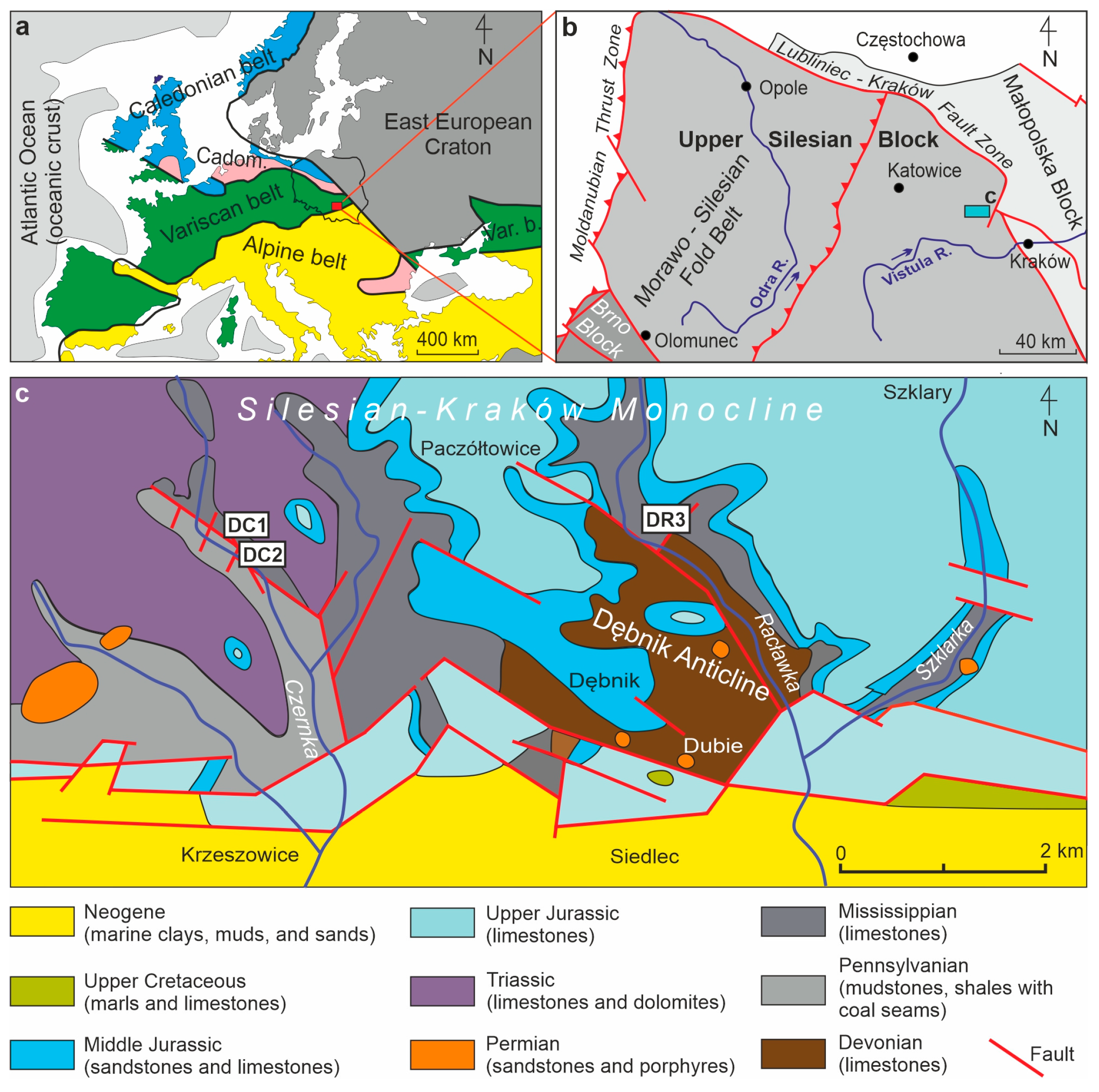
Figure 1. (a) The study area (red rectangle) against the background of the general tectonic units of Europe. (b) The study area (blue rectangle) on the map showing the tectonic units beneath the Permian–Mesozoic and Cenozoic cover [32]. (c) Location of the studied lithological profiles on the background of the geological map of the southern part of the Silesian-Kraków Monocline [33]. Geographical coordinates of the studied profiles: DC1—50°10′12.9″ N, 19°37′22.1″ E; DC2—50°10′09.7″ N, 19°37′24.2″ E; DR3—50°10′28.9″ N, 19°40′26.7″ E.
Minerals 15 01158 g001
Figure 2. Research profiles in the Czernka Valley (DC1, DC2) and in the Racławka Valley (DR3) with samples selected for microfacial, geochemical, and petrographic analyses.
Minerals 15 01158 g002
Figure 3. Details of limestones exposed in the DC1 profile (Red Wall). (a) General view of the exposure state with the middle- to thick-bedded Viséan limestones. (b) Cross-sections of brachiopod shells visible in the bed in the lower part of the exposure. (c,d) Microscopic images (TLM) of the red limestone microfacies from the DC1 profile (sample DC1.4) in plane-polarized light (c) and plane-polarized light with reflected light (EPI; (d)) showing the occurrence of bioclasts surrounded by rusty brown coatings, which appear red and/or yellow in reflected light depending on the occurrence and packing of iron minerals.
Minerals 15 01158 g003
Figure 4. Photomicrographs of bioclasts and spary binder in sample DC1.4. Foraminifers (a) and echinoids plates (b) surrounded by rusty coatings and with rusty fillings of originally porous parts of the skeletons visible in the TLM plane of polarized light. (c,d) The same types of bioclasts are observed in SEM-BSE imaging, showing different shades of grey corresponding to different structures and arrangements of calcite crystals, while white dots correspond to iron-containing particles. (e) Spary binder between bioclasts (TLM plane of polarized light) and rusty pigment, when viewed at high magnification consisting of numerous small oval or elongated elements resembling the casings of iron bacteria in size and shape.
Minerals 15 01158 g004
Figure 5. Chemical SEM-EDS microanalysis of sample DC1.4 in SEM-BSE image. (1) Light grey calcite clusters. (2) Dark grey dolomite clusters. (3) White Fe oxides/oxyhydroxides.
Minerals 15 01158 g005
Figure 6. SEM-EDS detailed chemical mapping of the selected area of sample DC1.4 showing the occurrence of Mg, Si, Ca, Mn, and Fe elements in oxide form and the overlapping of the areas of occurrence of individual elements, proving their common origin. The graph shows the total content of the analyzed elements from the entire tested surface.
Minerals 15 01158 g006
Figure 7. (a) Photomicrographs (TLM) of thin-section sample DC1.5 show rusty brown pigment between sparite crystals and rusty pigmentation within individual crystals, under plane-polarized light. (b,c) SEM-EDS imaging of the surface of sample DC1.5 showing that the rusty brown pigmentation is composed of Fe oxides/oxyhydroxides.
Minerals 15 01158 g007
Figure 8. (a) Photomicrograph of a cross-section through an echinoderm plate with the original microstructure, where the pores have been filled by rust-coloured pigment (sample DC1.5), under plane-polarized light. (b) Close-up of image (a) showing that the rust pigment is composed partly of oval and elongated forms resembling bacteria in size and shape, under plane-polarized light.
Minerals 15 01158 g008
Figure 9. (a) Photomicrograph of bioclast left from an echinoderm plate, where rusty brown pigmentation is found within the original void and along the contact with the surrounding micrite (sample DC1.5), under plane-polarized-light. (b) Close-up of the exterior showing clumps of brown pigment and accompanying oval structures resembling bacteria, under plane-polarized-light. (c) Close-up of the internal part of the bioclast, showing additional bright flocs surrounding pigment tufts resembling bacterial holdfasts, under plane-polarized-light.
Minerals 15 01158 g009
Figure 10. Iron oxides in sample DC1.2. (a) Microscopic image (TLM) of vein infillings and disseminated pigment within carbonate mass. (b) The same image in plane-polarized light with reflected light (EPI). (c) SEM-BSE image of the central part of A image. (df) Closer look at areas from image (c). (d) Compact, botryoidal cryptocrystalline mass. (e) Cryptocrystalline porous aggregates (Kl—kaolinite). (f) Tiny cryptocrystalline aggregates (Py—pyrite; 3—Fe oxide pseudomorphs after pyrite). (g) Closer view of image (f) showing accumulation of Mn oxides (probably coronadite). (h) Typical processed Raman spectrum showing diagnostic goethite features.
Minerals 15 01158 g010
Figure 11. Iron oxides in sample DC1.4.2. (a) Pore infillings, SEM-BSE+SE image. (b) A detail of A−cryptocrystalline hematite; SEM-SE image (Q−quartz; Kl−kaolinite; Cal−calcite). (c) Cryptocrystalline aggregates (1) of possible bacterial origin (Q−quartz; Cal−calcite; Kl−kaolinite). (d) Pseudomorphs after pyrite, SEM-BSE+SE (Q−quartz; Cal−calcite; B−barite). (e) Echinoderm plate with porous microstructure, SEM-BSE+SE image. (f) Typical processed Raman spectrum showing diagnostic hematite features.
Minerals 15 01158 g011
Figure 12. Correlation graphs for individual pairs of oxides in all samples investigated.
Minerals 15 01158 g012
Figure 13. REE curves of the studied samples, normalized to chondrites [61] and Post-Archean Australian Shale standards [62,63].
Minerals 15 01158 g013
Figure 14. Bacteria-like structures in rusty brown pigment in samples of the DC1 section (the Czernka Valley—the Commune quarry), under plane-polarized-light. (a) Ovals contain Fe oxides/oxyhydroxides arranged as single capsules inside crystals or chains located between crystals of spary binder. (b) Twisted chains and clusters of spheres resembling bacterial-like consortia. (c) Spherical and elongated Fe oxide/oxyhydroxide structures resembling iron bacteria capsules. (d) Flocks of rusty brown pigment with thin flocs and oval capsules resembling bacterial holdfasts.
Minerals 15 01158 g014
Figure 15. Time spatial model of diagenetic processes that occurred in the Mississippian limestones of the Upper Silesian Block in zones affected by hydrothermal activity related to post-Variscan plutonism and volcanism, leading to the formation of bacteria-like ferruginous structures. (a) Biogenic limestone containing bioclasts surrounded by primary micrite. (b) A pulse of hot hydrothermal solutions circulating primarily in porous parts of the rock, such as natural pores in fossil skeletons, which leached micrite, particularly at the boundaries with and within bioclasts. (c) Another pulse of Fe-containing hydrothermal solutions, cool enough for iron bacteria to live in them and form iron capsules. (d) The gradual crystallization of calcite sparite from cool, supersaturated solutions in which iron bacteria lived caused them to be enclosed inside the calcite crystals and, above all, between the crystals. (e) The end result of the above processes was rusty coatings of bioclasts and the filling of the voids with calcite sparite containing significant amounts of remains of the life activity of iron bacteria.
Minerals 15 01158 g015
Table 1. Content (wt%) of major elements in the rock samples studied. DC1—strong hydrothermally altered red limestones (the Czernka Valley—the Red Wall); DC2—weak/poorly hydrothermally altered pinkish grey limestones (the Czernka Valley—the Commune quarry); DR3—non-hydrothermally altered grey limestones (the Racławka Valley—slopes of the Komarówka Hill).
DC1
(n = 4)
DC2
(n = 4)
DR3
(n = 10)
CaO51.88–44.70 (av. 48.98)55.51–49.72 (av. 53.85)55.34–54.16 (av. 54.79)
Σ CaO + LOI94.38–82.00 (av. 89.40)98.82–90.22 (av. 95.57)99.03–98.02 (av. 98.59)
Σ other elements 17.81–5.56 (av. 10.49)9.83–1.19 (av. 3.44)2.02–0.97 (av. 1.40)
SiO212.14–3.28 (av. 6.80)8.04–0.59 (av. 2.58)1.06–0.41 (av. 0.66)
MgO1.26–0.30 (av. 0.68)0.33–0.24 (av. 0.27)0.66–0.40 (av. 0.58)
Fe2O31.69–0.44 (av. 0.92)0.43–0.07 (av. 0.17)0.13–0.05 (av. 0.07)
Al2O33.34–0.46 (av. 1.84)0.88–0.02 (av. 0.33)0.19–0.06 (av. 0.09)
K2O0.14–0.03 (av. 0.09)0.07–0.01 (av. 0.03)0.06–0.01 (av. 0.02)
TiO20.10–0.02 (av. 0.06)0.03–0.01 (av. 0.02)0.01
MnO 0.07–0.02 (av. 0.05)0.03–0.01 (av. 0.02)0.01
P2O50.06–0.01 (av. 0.04)0.010.01
Na2O0.03–0.01 (av. (0.02)0.010.01
Table 2. Pearson’s coefficient values, calculated for individual pairs of oxides in all samples from the DC1 section (the Czernka Valley—the Red Wall).
SiO2Al2O3Fe2O3MgOCaONa2OK2OTiO2P2O5MnO
SiO2 0.933520.93112−0.08947−0.984240.255060.88450.927250.786910.7502
Al2O30.93352 0.96874−0.10711−0.958360.349370.91380.996730.936230.76123
Fe2O30.931120.96874 −0.012−0.95980.351210.890230.969390.860910.70297
MgO−0.08947−0.10711−0.012 −0.027160.1545−0.01154−0.09549−0.090940.30166
CaO−0.98424−0.95836−0.9598−0.02716 −0.33029−0.91739−0.95411−0.83796−0.79583
Na2O0.255060.349370.351210.1545−0.33029 0.447290.395020.538250.32657
K2O0.88450.91380.89023−0.01154−0.917390.44729 0.90920.857250.6789
TiO20.927250.996730.96939−0.09549−0.954110.395020.9092 0.948170.75895
P2O50.786910.936230.86091−0.09094−0.837960.538250.857250.94817 0.75491
MnO0.75020.761230.702970.30166−0.795830.326570.67890.758950.75491
Table 3. The content of trace elements (ppm) in the rock samples studied. DC1—strong hydrothermally altered red limestones (the Czernka Valley—the Red Wall); DC2—weak/poorly hydrothermally altered pinkish grey limestones (the Czernka Valley—the Commune quarry); DR3—non-hydrothermally altered grey limestones (the Racławka Valley—slopes of the Komarówka Hill).
DC1
(n = 4)
DC2
(n = 4)
DR3
(n = 10)
Sr758.3–354.4 (av. 607.4)155.0–109.2 (av. 140.7)329.5–184.8 (av. 187.7)
Rb5.4–3.0 (av. 3.7)1.7–0.1 (av. 0.6)2.6–0.4 (av. 1.1)
Ba30–18 (av. 26)30–4 (av. 11)6–2 (av. 4)
Mo1.1–0.4 (av. 0.6)0.2–0.1 (av. 0.2)0.3–0.1 (av. 0.17)
Cu4.6–2.0 (av. 3.5)2.8–1.1 (av. 1.8)2.1.7–0.2 (av. 0.84)
Pb191.0–8.8 (av. 83.5)33.2–3.5 (av. 14.8)5.0–0.7 (av. 2.3)
Zn298–15 (av. 151)86–17 (av. 36)19–15 (av. 10.7)
Zr35.2–5.0 (av. 19.2)35.9–1.7 (av. 11.8)7.8–2.8 (av. 2.3)
U3.7–1.3 (av. 2.5)2.6–0.7 (av. 1.6)4.1–1.1 (av. 2.5)
Th5.7–0.7 (av. 2.9)3.3–1.0 (av. 1.8)0.2
Hf1.0–0.1 (av. 0.6)0.9–0.1 (av. 0.3)0.3–0.1 (av. 0.1)
ΣREE86.6–20.9 (av. 47.8) 36.1–3.5 (av. 18.3)13.1–2.5 (av. 6.5)
Disclaimer/Publisher’s Note: The statements, opinions and data contained in all publications are solely those of the individual author(s) and contributor(s) and not of MDPI and/or the editor(s). MDPI and/or the editor(s) disclaim responsibility for any injury to people or property resulting from any ideas, methods, instructions or products referred to in the content.