Review

Geochemical Characteristics and Health Risks of Coal Dust: An Integrated Review from Component-Dependent Toxicity to Emerging Oxidative Toxicity Indicators

1
State Key Laboratory of Deep Coal Safety Mining and Environmental Protection, Anhui University of Science and Technology, Huainan 232001, China
2
School of Earth and Environment, Anhui University of Science and Technology, Huainan 232001, China
3
Key Laboratory of Industrial Dust Prevention and Control & Occupational Health and Safety, Ministry of Education, Anhui University of Science and Technology, Huainan 232001, China
4
Bonanza Precision Mining and Environmental Protection Guizhou Provincial Academician Expert Workstation, Guanshanhu District, Guiyang 550081, China
*
Author to whom correspondence should be addressed.
Minerals 2025, 15(10), 1075; https://doi.org/10.3390/min15101075
Submission received: 29 August 2025 / Revised: 27 September 2025 / Accepted: 13 October 2025 / Published: 15 October 2025

Abstract

Coal mining and consumption, a persistent source of global energy, pose significant occupational health risks. Through a bibliometric analysis of 562 publications (2001–2025), this review delineates the evolution from conventional metrics (mass concentration, free silica content) toward advanced characterization of mineralogical/geochemical heterogeneity and component dependent toxicity mechanisms. Evidence confirms that multiple toxic elements are enriched in the respirable fraction, with bioaccessibility critically governed by particle size, host phase, and chemical speciation. In vitro studies using simulated lungs and gastrointestinal fluids demonstrate that acidic environments significantly accelerate toxic metal dissolution, triggering oxidative stress. While the bioaccessibility of inorganic constituents has been extensively studied, that of complex organic pollutants, particularly polycyclic aromatic hydrocarbons, remains a critical knowledge gap. Oxidative stress is now recognized as a pivotal mechanism linking coal dust exposure to inflammation and genotoxic damage. Emerging abiotic toxicity indicators, such as environmentally persistent free radicals and oxidative potential, offer promising avenues for understanding and risk prediction; however, their analytical methodologies require further standardization and refinement. This review provides a scientific foundation for developing a next-generation risk assessment framework that integrates multi-dimensional coal dust characteristics, bioaccessibility, and oxidative potential, thereby guiding future research to better protect the health of coal miners.

1. Introduction

With domestic raw coal output reaching 4.78 billion tons in 2024 and constituting >66.6% of its primary energy production, China remains the global leader in coal production and consumption [1]. Projections suggest coal will maintain its dominant role in China’s energy portfolio until at least 2050 [2]. Globally, annual coal consumption is expected to stabilize at approximately 8 billion tons by 2050, with China and India remaining the top consumers [3]. As global coal consumption patterns exhibit significant regional concentration (Figure 1), it is critical to assess the associated occupational health hazards.
The generation of coal dust during mining, transportation, and processing releases complex particulates composed of organic-inorganic mixtures (e.g., PAHs, heavy metals, silicates) [4,5,6,7]. Historically, pneumoconiosis control relied on two key metrics: respirable dust mass concentration and free silica content. Although this strategy initially reduced disease incidence, its effectiveness has been challenged by a paradoxical resurgence of severe pneumoconiosis among U.S. miners since the late 1990s, despite stable dust exposure levels [8]. This suggests that conventional mass-based metrics fail to capture critical toxicological determinants, a limitation attributed to the source-dependent heterogeneity of coal dust physicochemical properties (e.g., surface reactivity, mineralogy) [9,10,11].
To address the subjectivity of traditional literature reviews, this study employs bibliometric analysis using CiteSpace [12] to quantitatively map 562 publications (2001–2025). This data-driven approach aims to objectively identify: (i) The evolution of research hotspots; (ii) Key knowledge gaps regarding contaminant bioaccessibility; (iii) The emergence of new abiotic toxicity indicators. This profiling establishes a foundation for systematizing coal dust toxicity mechanisms and refining current risk assessment frameworks.

2. Bibliometric Analysis

2.1. Data Sources and Methods

Literature retrieval for this review was conducted in the Web of Science (WOS) Core Collection to capture the global evolution of research on coal dust toxicity. The search spanned 1 January 2001 to 31 July 2025; using the Boolean query: TI = (“coal”) AND (“particulate*” OR “PM” OR “dust” OR “particle*”) AND TS = (“risk” OR “toxicity” OR “disease” OR “occupation*” OR “health*”), where TI denotes the title field and TS represents topic fields (title; abstract; keywords). After removing duplicates and irrelevant records; 562 peer-reviewed articles and reviews were retained for bibliometric analysis. It is important to note that this bibliometric analysis relied on the Web of Science Core Collection. While this database provides a robust foundation for analyzing high-quality research, future studies could benefit from incorporating complementary databases such as Scopus or PubMed to further enhance the comprehensiveness and comparative perspectives of the analysis.

2.2. Analysis of Bibliometric Results

2.2.1. Literature Quantity Analysis

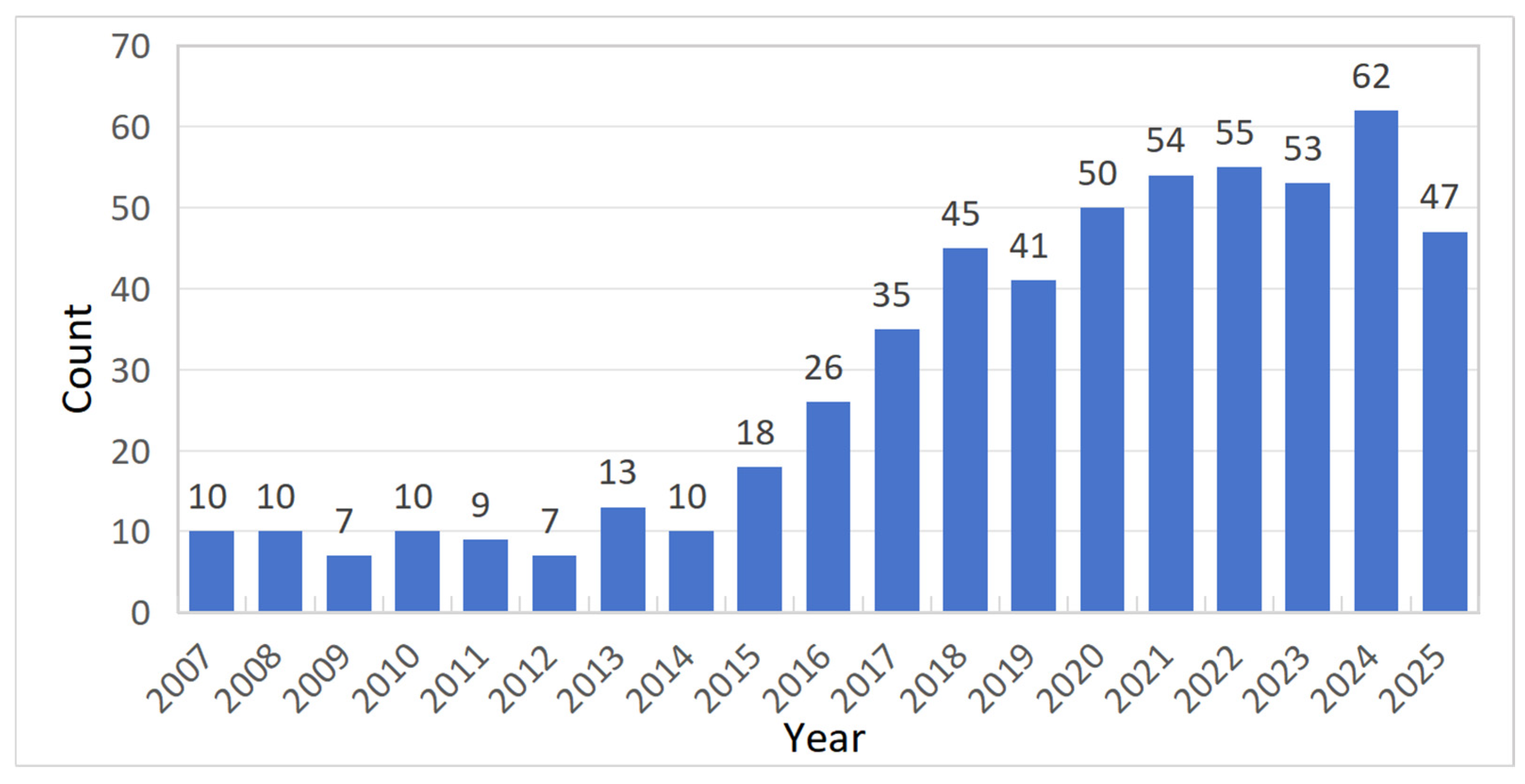
The annual number of publications provides insight into the developmental trends in coal dust exposure and health impacts. As shown in Figure 2, the research landscape can be divided into three distinct phases:
Initial Phase (2007–2014): Low output (mean: 9.5 articles/year), focusing on dust concentration exposure, free silica content, and pneumoconiosis diagnosis [9,13,14,15,16,17].
Transition Phase (2015–2019): Publications surged to 33 articles/year, largely driven by regulatory changes such as the U.S. MSHA’s 2014 mandate to reduce respirable dust limits [8]. Research shifted toward respirable dust diffusion dynamics and mineralogical toxicity impacts [18,19,20,21,22,23,24,25].
Expansion Phase (2020–2025): Further growth to 57.5 articles/year, with focus on organic components, inorganic elements, mineralogy, and toxic response potential [26,27,28,29,30].

2.2.2. Country Co-Authorship Analysis

As shown in Figure 3, a total of 55 countries published research on coal dust-related health issues between 2001 and 2025. China ranked first with 274 publications (48.75%), reflecting its dominant role in occupational health research and policy interventions. The United States followed with 150 publications (26.69%), underscoring its long-standing focus on toxicity mechanisms and epidemiological studies. Australia (45 publications, 8.01%) and India (39 publications, 6.94%) ranked third and fourth, respectively, aligning with their status as major coal-producing and consuming nations. The purple outer rings in Figure 3 indicate a country’s centrality within the global research network. The U.S. exhibited the highest centrality (0.45), reflecting its pivotal role in cross-border collaborations and foundational research, while China (0.37) demonstrated strong regional influence.

2.2.3. Keyword Burst Analysis

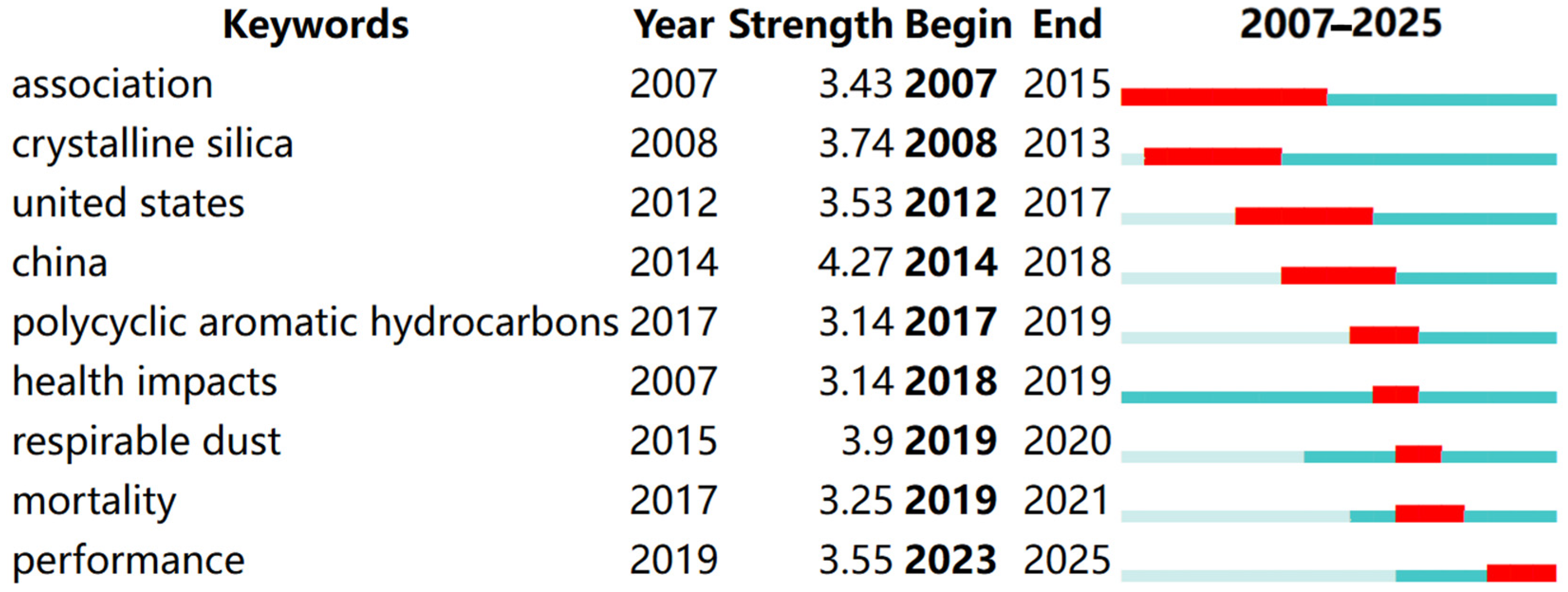
To identify research frontiers, a burst analysis of keywords was conducted using CiteSpace, and the top 9 keywords with the strongest citation bursts were chronologically sorted (Figure 4). In the figure, the light blue shading corresponds to the entire time span from 2007 to 2025. The blue bars represent the duration for which the keywords were present, while the red bars indicate their burst duration.
The analysis reveals a clear evolutionary trajectory in coal dust and health research from 2007 to 2025. Early studies (2007–2012) focused on foundational associations and regional exposure patterns (e.g., “association”, “united states”). This was followed by an intensified focus on specific toxic components, such as “crystalline silica” (2008–2013) and “polycyclic aromatic hydrocarbons” (2017–2019). A significant geographical shift was also observed, with “china” emerging as the strongest burst (2014–2018). Subsequent research transitioned toward quantifying direct health endpoints, linking “respirable dust” (2019–2020) to outcomes like “mortality” (2019–2021). The latest phase (2023–2025) is marked by the rise of “performance”, signaling a shift from hazard identification toward applied interventions, mitigation technologies, and practical risk management solutions.

2.2.4. Document Co-Citation Analysis

Co-citation analysis of highly cited papers helps to identify influential research topics and trends. As shown in Table 1, 7 of the top 10 most cited papers were authored by U.S. researchers, highlighting the nation’s leadership in this field.
The highly cited literature indicates a shift from traditional dust concentration control to exploring multi-component synergistic toxicity mechanisms. For example, Cohen et al. [37] pioneered the field by demonstrating that silicate nanoparticles, rather than bulk coal dust, drive the resurgence of progressive massive fibrosis (PMF) in Appalachia, overturning the long-held “inert dust hypothesis.” This finding was extended by Johann-Essex et al. [33], who used CCSEM-EDX mineral mapping to establish a critical quartz toxicity threshold (>8% content, OR = 5.2). Concurrently, Blackley et al. [32] leveraged 30-year epidemiological surveillance to reveal a stark disparity in PMF incidence, a finding that catalyzed the MSHA’s regulatory reform to lower the respirable dust limit. Building on this, Sarver et al. [20] developed a triaxial profiling framework integrating particle size, mineralogy, and surface reactivity, superseding conventional mass concentration metrics. More recent research elucidated size-dependent bioactivity of coal dust: Trechera et al. [34] identified 4.2-fold Cu/Sb enrichment in PM2.5 enhancing alveolar penetration and oxidative stress; Zazouli et al. [36] validated Fe2+/Cu+-mediated Fenton reactions depleting glutathione (>80%) and triggering collagen deposition, elucidating a molecular pathway for fibrosis.
These studies collectively highlight a critical shift in dust toxicity understanding from a “mass concentration core” to component-dependent toxicity. Growing concerns over bioaccessibility of heavy metals and organics demand moving beyond single-limit metrics toward a comprehensive toxicity index integrating particle size, silica content, and ROS activity. This review focuses on these two key areas.

3. Geochemical Characteristics of Coal Dust

Coal dust is a complex mineral–organic–metal assemblage, whose chemical fingerprint is shaped by stratigraphic provenance, host-rock lithology, and mine-specific processes such as cutting, equipment wear, and ventilation-driven mixing [38,39,40,41]. Multiple studies have demonstrated that coal dust comprises major inorganic constituents (Al, Si, Ca, Fe, K, Na, Mg) alongside a suite of trace metals and metalloids (notably Mn, V, Cr, Cu, Zn, As, Pb, Sb), exhibiting significant spatial variability and close associations with specific mineral hosts (e.g., As and Fe in pyrite) or organic matrices [4].
Mineralogical investigations employing X-ray diffraction (XRD) and scanning electron microscopy with energy dispersive X-ray spectroscopy (SEM-EDX) reveal that clay minerals (kaolinite, illite), quartz, and carbonates (e.g., calcite) serve as the predominant mineral carriers in coal dust, accompanied by accessory sulfides such as pyrite and, occasionally, galena [39,41]. These minerals display heterogeneous size distributions, with finer particles (<2.5 μm) frequently enriched in bioactive phases such as nanoscale anatase. The fibrous morphology of such nanoparticles enables evasion of immune clearance mechanisms, thereby promoting persistent pulmonary inflammation [42]. Relative to typical crustal minerals, coal dust exhibits substantially elevated proportions of sulfides and oxides—minerals that are trace constituents in the crust but are amplified multiple-fold in dust—reflecting the sulfur and iron enrichment processes occurring during coalification [36].
Table 2 summarizes region-specific elemental abundances in coal dust compared with global coal averages [18,36,42,43]. Notably, several trace elements, including vanadium and zinc in Iranian samples as well as manganese and iron in Slovenian samples, reach concentrations an order of magnitude or more above the global baseline [18,36,43]. These enrichments are attributed to a combination of sources: detrital inputs from host rock fragments (clay minerals, carbonates), oxidative mobilization of sulfide-hosted trace elements through pyrite weathering (As, Fe, and associated metals), and anthropogenic contributions such as machine wear releasing Fe, Cr, and Ni [39].
From an exposure and toxicological standpoint, two aspects are particularly critical: (1) the enrichment of recognized toxic elements (e.g., As, Cd, Pb, Cr) in the respirable fraction, and (2) the host mineral phase and chemical speciation governing bioavailability. Total elemental concentrations alone provide insufficient predictive power for toxicity; for example, arsenic bound within sulfide minerals typically exhibits lower bioavailability than arsenic adsorbed on oxide surfaces or present in soluble forms. Similarly, hexavalent chromium (Cr(VI)) is markedly more toxic than its trivalent form (Cr(III)). Accordingly, comprehensive risk assessments must incorporate particle size-resolved sampling, sequential chemical extractions or speciation analyses, and biological reactivity metrics such as oxidative potential, which directly link chemical composition to toxicological outcomes.

4. Bioaccessibility of Substances in Coal Dust

The health risks posed by substances in coal dust are governed not only by their intrinsic toxicity and concentration but critically by their bioavailability [44]. Assessing bioavailability typically involves two main approaches: in vitro chemical analyses and in vivo biological methods such as biomarker monitoring and bioaccumulation studies. While in vivo methods offer the most direct evaluation of human health risks, they are often costly, logistically challenging, and subject to strict ethical oversight [45,46]. Consequently, in vitro bioaccessibility assays are widely employed due to their rapidity, cost-effectiveness, and fewer ethical constraints.
Bioaccessibility from coal dust occurs primarily via two exposure pathways: pulmonary and gastrointestinal. Particles smaller than 4 μm can evade nasal filtration and penetrate deeply into alveolar lung tissues. In contrast, particles sized 4–10 μm are typically retained in the upper respiratory tract, swallowed, and subsequently enter the gastrointestinal tract, where they release toxic substances such as heavy metals and organic pollutants, posing potential health hazards [6,47,48].
Recent investigations utilizing simulated lung fluids (SLFs) and gastric simulations have advanced understanding of dissolution kinetics for coal dust constituents. These studies reveal that bioaccessible fractions—often a minor portion of total elemental concentrations—can nonetheless provoke oxidative stress, inflammation, and cellular damage, contributing to coal workers’ pneumoconiosis (CWP) and carcinogenesis [6].

4.1. Metals

Natural human lung fluid exhibits complex composition, variable pH, and ionic strength [49], leading to heterogeneous release patterns of metals and metalloids from inhaled particles. To mimic these physiological conditions, various SLFs have been developed. The predominant simulants in coal dust studies are Gamble’s solution (GS), replicating the neutral pH (7.4–7.6) interstitial fluid of deep lung alveoli, and artificial lysosomal fluid (ALF), simulating the acidic (pH 4.5–5.0) lysosomal environment of macrophages post-immune activation. Details of their formulations and extraction conditions are provided in Table 3.
SEM-EDX analyses of Appalachian and Rocky Mountain mine dust demonstrate elevated total concentrations of Si, Al, and Fe in fine particles (<2.5 μm) [40]. Despite ALF bioaccessibility being only 0.03%–0.04%, this fraction is sufficient to impair viability of lung epithelial (A549) cells, indicating toxicity is governed by bioaccessible, not total, metal content. In Czech Sokolov mine dust, Zádrapová et al. [50] observed that As and Be bioaccessibility in ALF reached 86%–95%, markedly surpassing gastric bioaccessibility (45%–73%). This suggests that acidic lung environments accelerate metal dissolution and reactive oxygen species (ROS) generation, disrupting immune function even when total metal concentrations poorly correlate with toxicity.
Similarly, studies on Australian mine dust found that reactive Fe(II) species such as pyrite and siderite exhibited elevated bioaccessibility under inflammatory conditions, catalyzing Fenton reactions and amplifying oxidative injury [51]. Bioaccessible iron levels inversely correlated with cell viability (R2 = 0.689), underscoring iron’s critical role in CWP risk resurgence. Complementary zebrafish model experiments confirmed that aqueous coal dust extracts disrupt connective tissue and modulate immune gene expression via bioaccessible As, Zn, and Cu fractions, upregulating oncogenic pathways despite minor changes in total metal concentrations [21].
Comparative analyses across multiple mine dust sources reveal significant variability in bioaccessibility. Within individual mines, floor and roof dust samples released the highest metal concentrations, exhibiting pronounced cytotoxicity in vitro, whereas seam and rock dusts showed comparatively lower bioaccessibility [4]. Investigations of lignite fly ash further demonstrated pulmonary As bioaccessibility reaching 95%, far exceeding rates in soil and other dust matrices (10%–50%), highlighting the unique dissolution behavior of coal dust [52].
Three primary in vitro methods assess metal bioaccessibility via the gastrointestinal tract: the Unified BARGE Method (UBM), the Physiologically Based Extraction Test (PBET), and the Simple Bioaccessibility Extraction Test (SBET). Their specific protocols and distinctions are summarized in Table 4.
While SBET is favored for its operational simplicity and suitability for rapid preliminary screening, it models only the gastric digestion phase, neglecting the intestinal stage where metals can complex anew, thereby often overestimating bioaccessibility values [58]. UBM offers comprehensive simulation across oral, gastric, and intestinal phases but entails complex, time-intensive procedures that may limit its routine use [56]. PBET strikes an optimal balance by sequentially simulating gastric (pH 1.5–2.5) and intestinal (pH 7.0) phases, incorporating key bioactive agents such as pepsin, pancreatin, and bile salts, thus more realistically reproducing human digestive dynamics. This makes PBET particularly well suited for assessing complex environmental matrices typical of coal mining regions.
Case studies underscore these methodological differences. In the Czech Sokolov coalfield, Zádrapová et al. [50] reported simulated gastric fluid extracting up to 47% of Cd, indicating substantial mobility under acidic conditions. Skála et al. [59] demonstrated that fine particles (<0.3 mm) released significantly more As and Be in simulated intestinal fluid than coarser particles, with As bioaccessibility reaching 9.7% versus 2.3% in coarse fractions. This size-dependent release, likely due to prolonged intestinal residence time of fine particles, offers a scientific basis for targeted source control.
A complementary study in China’s Huaibei mining area by Sun et al. [60] highlighted the critical influence of digestive phase transitions. During the gastric phase, Cd and Pb bioaccessibility reached 43.9% and 61.7%, respectively, but intestinal phase pH elevation triggered marked re-adsorption of Cd, reducing bioaccessibility to 7.3%. These findings emphasize pH’s central role in metal speciation and bioaccessibility.
Collectively, these insights reflect both advances and persistent gaps in bioaccessibility assessment methodologies. Challenges include the lack of standardized formulations for physiological fluid components (e.g., mucin, phospholipids) and limited validation of in vitro–in vivo correlations [61]. Future methodological refinement should integrate coal dust’s distinctive physicochemical traits—such as elevated organic matter and iron oxide content—into tailored extraction protocols, thereby enhancing ecological relevance and improving predictive accuracy in health risk assessments.

4.2. Organic Compounds

Coal is a complex macromolecular matrix encapsulating diverse low-molecular-weight organic compounds, including polycyclic aromatic hydrocarbons (PAHs), organic acids, and long-chain alkanes. While inorganic constituents of coal dust have been extensively studied, limited research on harmful organic pollutants impedes comprehensive risk assessment.
Recent advances in extraction and analytical techniques have revealed coal’s intricate organic profile. For example, Ying Zong et al. [62] optimized a CS2/THF co-solvent system (1:1 v/v) to extract organics from Ordos Basin coal, achieving 6.5% extraction efficiency. GC-MS analysis identified 57 species categorized into four classes: (1) long-chain n-alkanes (e.g., eicosane, hexacosane), present in trace amounts; (2) monoaromatic steranes (C28–C29 homologues), indicative of biogeochemical origins; (3) polycyclic aromatic compounds (PACs), dominated by retene, fluoranthene, and methylpyrene; and (4) heteroatom-containing species, including novel nitriles like cyclohexanecarbonitrile. Similarly, Coronado-Posada et al. [63] applied methanol Soxhlet extraction to Colombian coal dust, revealing a complex suite of alkanes, aromatic acids, phthalates, and PAHs. Tirado-Ballestas et al. [64] further confirmed this heterogeneity via hydroethanolic extraction, identifying 17 compounds ranging from linear alkanes to cytotoxic PAHs such as benzo[a]anthracene. This chemical diversity underscores coal’s role as a reservoir of bioactive contaminants.
Despite extensive study of inorganic constituents, bioaccessibility data for organic compounds remain scarce. Schulz [65] pioneered physiological fluid leaching of German coalmine dusts (72 h at room temperature), demonstrating ≤1% organic release efficiency compared to dichloromethane extraction. Phenolic compounds dominated the leachates, with lecithin enhancing solubility by 1.5-fold.
In vitro models simulating pulmonary bioavailability are evolving. Xie et al. [66] quantified PAH bioaccessibility in e-waste particles using modified Gamble’s solution (MGS) and artificial lysosomal fluid (ALF), revealing strong particle-size dependence: bioaccessibility increased for larger particles (1.8–5.6 μm) and was inversely affected by hydrophobicity, measured as the octanol-water partition coefficient (KOW). Gao et al. [67] corroborated these findings in Harbin’s PM2.5, where ALF extracted significantly lower PAH fractions than MGS, highlighting the role of pH-driven dissolution dynamics.

5. Coal Dust Oxidative Toxicity Assessment

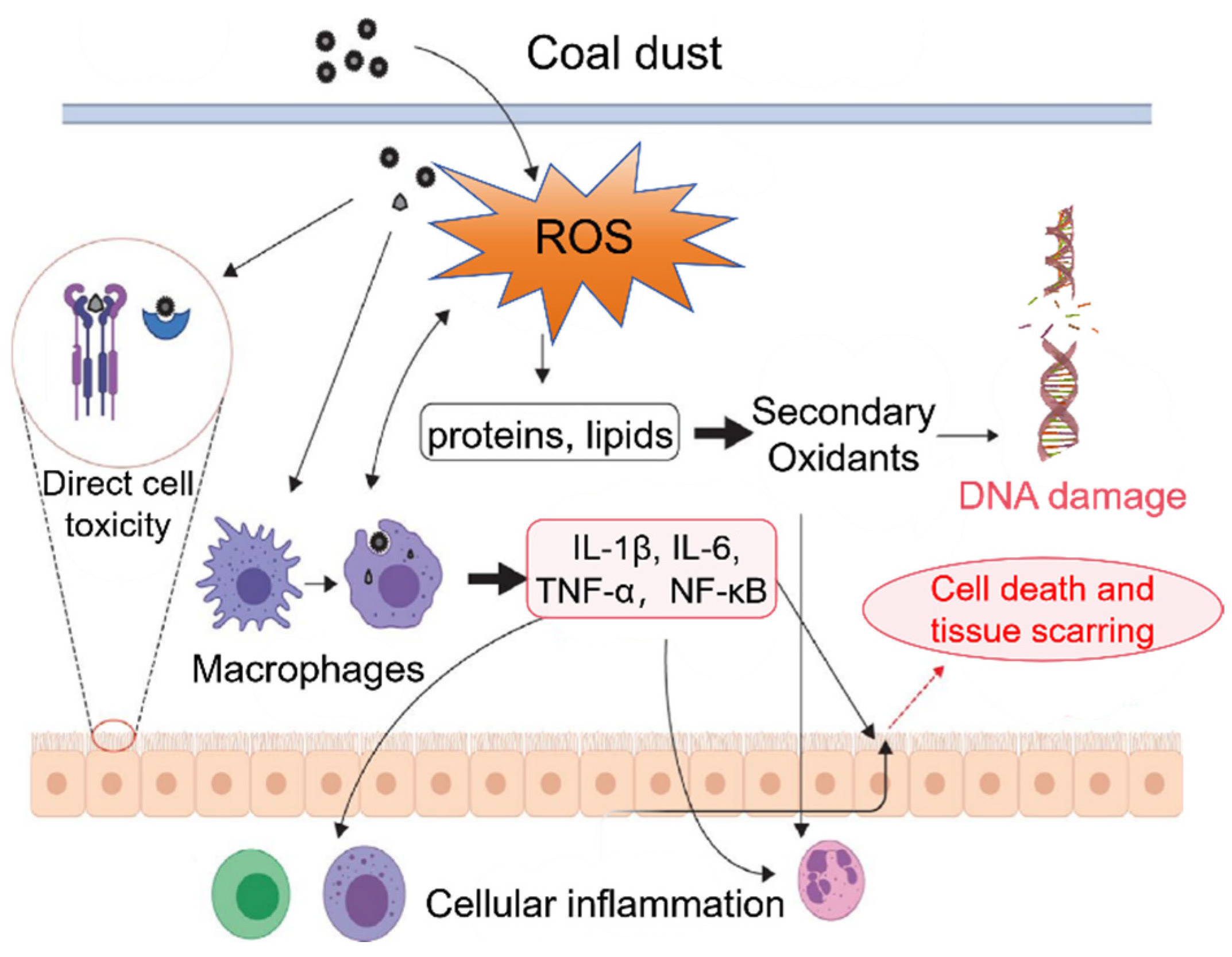
While the full toxicological mechanisms of coal dust have not been entirely elucidated, substantial evidence indicates that its effects follow a three-tier cascade: oxidative stress initiation, inflammatory amplification, and genotoxic damage (Figure 5).
1. Oxidative Stress Initiation: Coal dust particles containing transition metals (e.g., Ni, V) and polycyclic aromatic hydrocarbons (PAHs) directly generate reactive oxygen species (ROS) through Fenton-type reactions and cytochrome P450-mediated redox cycling [27,69,70,71]. This is compounded by macrophage phagocytosis, which triggers endogenous ROS bursts and disrupts cellular redox homeostasis.
2. Inflammatory Cascade Amplification: Excessive ROS production activates signaling pathways such as NF-κB, promoting the release of pro-inflammatory cytokines (e.g., IL-1β, IL-6, TNF-α) from epithelial cells and macrophages [29,72,73]. This persistent inflammatory microenvironment induces immune cell infiltration, fibroblast activation, and collagen deposition, ultimately leading to irreversible pulmonary fibrosis.
3. Terminal Genotoxic Effects: ROS and reactive nitrogen species (RNS) can damage DNA, resulting in the formation of 8-hydroxy-2′-deoxyguanosine (8-OHdG) and strand breaks, which may induce G → T carcinogenic mutations [74,75,76,77,78].
Therefore, oxidative stress is the pivotal trigger in this cascade, and its quantitative assessment is crucial for effective prevention and control. Currently, two principal cell-free chemical assays are used to quantify the oxidative potential (OP) of dust.

5.1. Environmentally Persistent Free Radicals (EPFRs)

Environmentally persistent free radicals (EPFRs) are highly reactive species with unpaired electrons that have emerged as a critical research focus in occupational health, particularly in coal mining. Due to their stability and persistence across various environmental matrices—including atmospheric particulate matter, soils, and fly ash—EPFRs are directly relevant to occupational health concerns when detected in coal dust [79,80,81,82,83]. Mechanistically, EPFRs disrupt cellular redox homeostasis by continuously generating ROS, potentially inducing oxidative stress and health impairments such as pulmonary and cardiovascular inflammation [82,84].
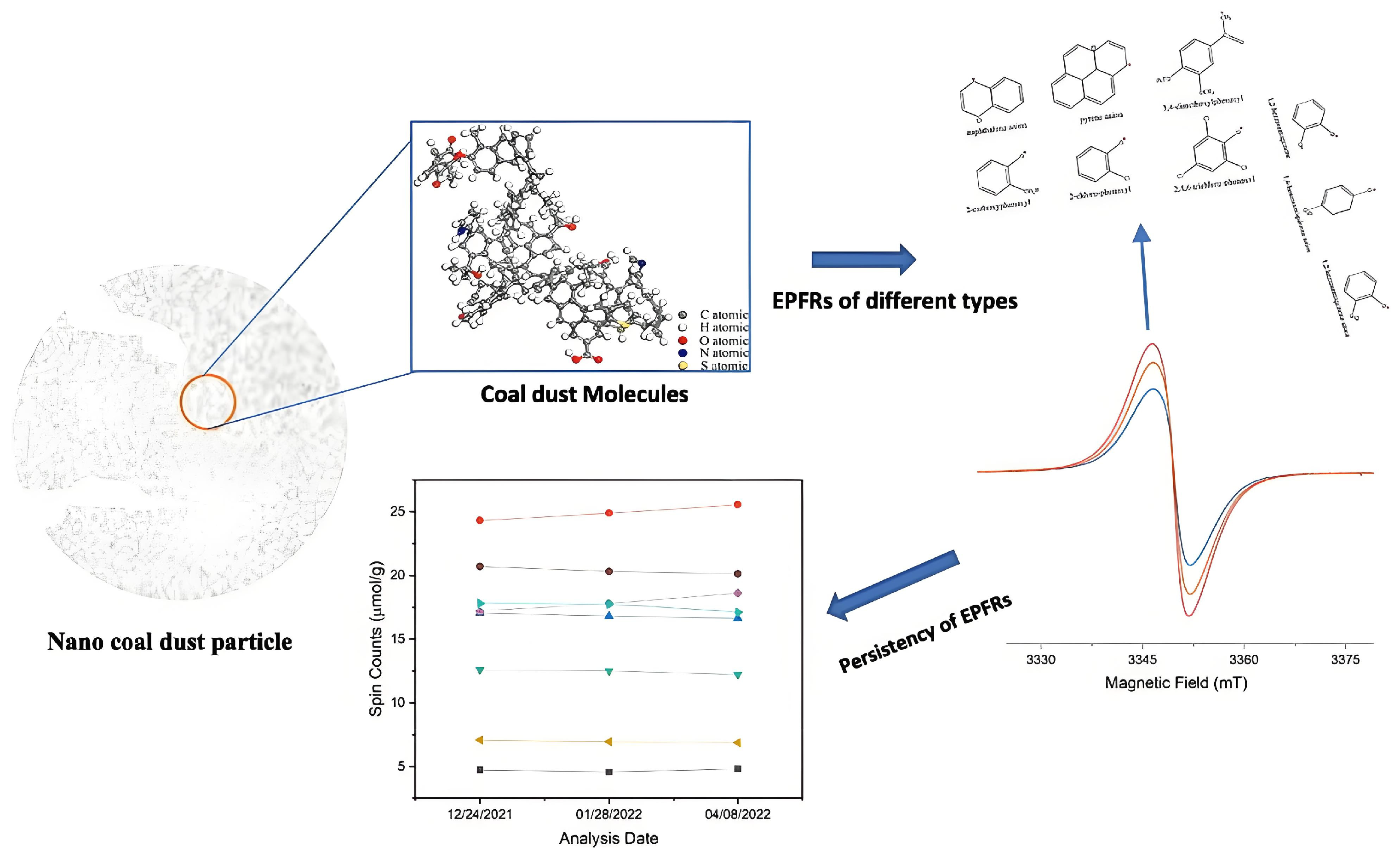
Despite their recognized significance, EPFR research remains less advanced than that of conventional pollutants, primarily due to analytical limitations. Significant technical challenges persist in the sampling, extraction, and quantification of EPFRs from complex media [85]. Currently, electron paramagnetic resonance (EPR) spectroscopy is the cornerstone technique for identifying radical species [86,87]. The analysis of spectral parameters (e.g., g-factor, peak area) allows for the inference of EPFR speciation, concentration, and physicochemical properties [88]. Based on g-factor values, EPFRs are broadly categorized into three classes: carbon-centered radicals (g < 2.003), oxygen-centered radicals (g > 2.004), and hybrid radicals (2.003 ≤ g ≤ 2.004) [89,90,91].
Emerging investigations have increasingly focused on coal dust, revealing that coal matrices are not inert but serve as reservoirs for EPFRs. The profiles of these radicals vary dramatically based on coal origin and maturity [92,93,94]. As shown in Figure 6, EPR spectroscopy confirms their persistent spectral signatures [94]. Studies employing EPR spectroscopy show that both coal rank and particle size modulate EPFR profiles. For example, bituminous coals contain higher concentrations of aromatic carbon-centered radicals than lignite, resulting in greater radical stability [95]. Particle size reduction, such as through mechanical handling, can fracture chemical bonds and elevate oxygen-centered radical concentrations, suggesting mechanochemical activation as a potent initiator of radical formation [94,96]. Notably, nano-sized coal dust exhibits EPFR stability over extended storage periods, implying that inhaled particles may retain oxidative reactivity long after deposition in the respiratory tract.
These physicochemical traits have direct implications for oxidative toxicity. EPFRs can redox-cycle with molecular oxygen to continuously generate ROS, perturbing cellular redox homeostasis, damaging biomolecules, and triggering pro-inflammatory pathways [97]. Historical pathological evidence further supports this, with stable coal-derived radicals detected in miners’ lung tissue at concentrations that correlate with the severity of coal workers’ pneumoconiosis [98]. This underscores their potential role as a mechanistic link between coal dust exposure and chronic respiratory disease.
Given these findings, future research must move beyond simple EPFR identification. A more holistic approach is needed, integrating particle-specific characterization with comprehensive in vitro and in vivo bioassays. This will be crucial for unraveling the full extent of their toxicological contributions and developing effective mitigation strategies.

5.2. Oxidative Potential (OP)

Oxidative potential (OP) is a vital health indicator integrating various physicochemical properties of particulate matter to predict toxicological effects, often providing more comprehensive insight than mass concentration or chemical composition alone [99,100]. While widely characterized in atmospheric particulate matter [79,101,102,103], OP assessment in coal dust remains limited.
Various chemical assays are used to quantify OP, each with distinct advantages and sensitivities. The dithiothreitol (DTT) assay measures the depletion rate of DTT by redox-active species, reflecting the capacity to induce oxidative stress. The ascorbic acid (AA) assay assesses AA depletion, particularly sensitive to transition metals. The glutathione depletion assay (OPGSH) evaluates the loss of reduced glutathione, relevant to antioxidant defense mechanisms.
Recent investigations reveal marked spatial heterogeneity and compositional dependence of coal dust OP. For instance, in Henan mining areas, DTT-based OP values (~2.45 nmol/min/μg) are predominantly influenced by calcite-pyrite mineral assemblages and redox-active elements like Sb and As [104]. Contrastingly, AA assays show higher sensitivity to anatase-tobelite phases and transition metals such as Ni and Cr. Notably, in Shanxi coal mines, DTT-measured OP substantially exceeds AA and DCFH assay results, underscoring DTT’s distinctive responsiveness to complex coal matrices [43]. Similar patterns emerge in Iranian mines, where OPAA values in non-coal face areas surpass OPGSH, highlighting the critical impact of sampling site and assay selection on toxicity interpretation [36]. Mechanistic studies in Southwest China’s high-sulfur coal dust demonstrate a strong correlation (r = 0.83) between iron content and OPAA, confirming that pyrite-mediated Fenton reactions significantly amplify ROS generation [34].
These findings reveal significant variability in assay sensitivity to coal dust composition. DTT excels in detecting redox reactivity linked to Fe-bearing sulfides and trace metals, whereas AA and GSH depletion assays better reflect antioxidant depletion associated with surface chemistry and transition metals.
Oxidative potential (OP) and environmentally persistent free radicals (EPFRs) represent two critical yet distinct indicators for assessing the oxidative hazard of particulate matter. OP, as a functional metric, offers the advantage of integratively reflecting the overall oxidative damage potential of particles and demonstrates stronger epidemiological associations with adverse health outcomes. However, its limitations include mechanistic ambiguity and a lack of standardized measurement protocols. In contrast, EPFRs serve as specific component-based indicators, providing a clear molecular mechanism for oxidative stress and enabling high-sensitivity source apportionment. Their primary limitations lie in their inability to represent the entirety of oxidant species and the need for further investigation into their environmental behavior and bioavailability.
Future research should focus on integrating these two metrics. This can be achieved through simultaneous measurement and multivariate analysis to delineate their respective contribution weights. Furthermore, employing simulation experiments and in vitro toxicological models is crucial to elucidate the environmental transformation processes of EPFRs and their pivotal role in OP-driven biological effects. Such an integrated approach will facilitate the development of a more precise health risk assessment framework for coal dust.

6. Conclusions and Future Perspectives

This review, underpinned by a comprehensive bibliometric analysis of 562 publications from 2001 to 2025, systematically chronicles the evolution of coal dust research from traditional exposure metrics to advanced toxicological mechanisms.
First, research emphasis has decisively shifted from simple mass concentration and free silica content toward detailed exploration of geochemical heterogeneity and component-specific toxicity. Consequently, recent studies develop a refined evidence chain integrating particle size distribution, mineralogy, and surface reactivity.
Second, bioaccessibility has become a cornerstone concept in understanding coal dust toxicity. Our analysis identifies distinct dissolution patterns for heavy metals and organic pollutants within simulated pulmonary and gastrointestinal fluids. Acidic lung environments, such as macrophage lysosomes, markedly accelerate the release of toxic metals and metalloids including As and Be, initiating oxidative stress. In contrast, pH variations in the digestive tract crucially determine metal speciation and bioaccessibility, emphasizing the necessity for context-specific assessment methods. Nonetheless, substantial knowledge gaps persist concerning the bioaccessibility of complex organic pollutants such as PAHs.
Third, oxidative stress emerges as the central mechanism underlying the inflammatory cascade and genotoxic damage. Quantitative assessment of oxidative potential is therefore imperative for developing novel risk indicators. Emerging abiotic toxicity metrics like environmentally persistent free radicals (EPFRs) and OP show promise as biomarkers; however, standardization challenges in their quantification must be addressed.
Moving forward, future research should prioritize (1) developing comprehensive risk assessment frameworks that integrate multi-component analysis, bioaccessibility, and abiotic toxicity metrics to replace outdated single-metric approaches; (2) standardizing EPFR and OP analytical methodologies to support their integration into occupational health regulations and exposure limit development; (3) intensifying research on the bioaccessibility and toxicological effects of organic pollutants, especially PAHs, in coal dust; and (4) promoting policy interventions that account for component-specific toxicity, such as revising exposure limits based on bioaccessible fractions and oxidative potential rather than total mass concentration.
These concerted efforts will significantly enhance understanding and management of coal dust-related health risks, furnishing a stronger scientific foundation to protect coal miners globally.

Author Contributions

Conceptualization, X.F.; methodology X.F. and J.Y.; writing—original draft preparation, J.Y.; writing—review and editing, X.F. All authors have read and agreed to the published version of the manuscript.

Funding

This work was supported by Anhui Jianghuai Young Talent Training Program Team Project, Anhui University of Science and Technology’s high-level talent team launch funding project (YJ20250001), Guizhou Provincial Science and Technology Support Program project (Bonanza and Precision Mining, Guizhou Provincial Academician Expert Workstation KXJZ [2024]003), the National Key R&D Program of China (2019YFC1805600, 2021YFC2902100).

Data Availability Statement

No new data were created or analyzed in this study.

Conflicts of Interest

The authors declare no conflicts of interest.

References

  1. China National Coal Association, China National Coal Association 2023 China Coal Industry Development Annual Report; China National Coal Association: Beijing, China, 2024.
  2. Wei, W.; Mushtaq, Z.; Sharif, M.; Zeng, X.; Wan-Li, Z.; Qaisrani, M.A. Evaluating the Coal Rebound Effect in Energy Intensive Industries of China. Energy 2020, 207, 118247. [Google Scholar] [CrossRef]
  3. U.S. Energy Information Administration (EIA). International Energy Outlook 2023; U.S. Energy Information Administration: Washington, DC, USA, 2023. Available online: https://www.eia.gov/outlooks/ieo/ (accessed on 1 June 2024).
  4. Sarver, E.; Keles, C.; Rezaee, M. Characteristics of Respirable Dust in Eight Appalachian Coal Mines: A Dataset Including Particle Size and Mineralogy Distributions, and Metal and Trace Element Mass Concentrations. Data Brief 2019, 25, 104032. [Google Scholar] [CrossRef]
  5. Eskanlou, A.; Arnold, B.J. An Evaluation of Pyrite as a Component of Respirable Coal Dust. J. Hazard. Mater. 2024, 477, 135340. [Google Scholar] [CrossRef]
  6. Das, M.; Salinas, V.; LeBoeuf, J.; Khan, R.; Jacquez, Q.; Camacho, A.; Hovingh, M.; Zychowski, K.; Rezaee, M.; Roghanchi, P.; et al. A Toxicological Study of the Respirable Coal Mine Dust: Assessment of Different Dust Sources within the Same Mine. Minerals 2023, 13, 433. [Google Scholar] [CrossRef]
  7. Sarver, E.; Keleş, Ç.; Afrouz, S.G. Particle Size and Mineralogy Distributions in Respirable Dust Samples from 25 US Underground Coal Mines. Int. J. Coal Geol. 2021, 247, 103851. [Google Scholar] [CrossRef]
  8. Graber, J.M.; Harris, G.; Almberg, K.S.; Rose, C.S.; Petsonk, E.L.; Cohen, R.A. Increasing Severity of Pneumoconiosis Among Younger Former US Coal Miners Working Exclusively Under Modern Dust-Control Regulations. J Occup Env. Med 2017, 59, e105–e111. [Google Scholar] [CrossRef]
  9. Fubini, B.; Fenoglio, I. Toxic Potential of Mineral Dusts. Elements 2007, 3, 407–414. [Google Scholar] [CrossRef]
  10. Sventeková, E.; Prievozník, P.; Mlčoch, J.; Vandlíčková, M. Assessment of the Dust in Underground Coal Mine. Appl. Sci. 2024, 14, 6038. [Google Scholar] [CrossRef]
  11. LaBranche, N.; Keles, C.; Sarver, E.; Johnstone, K.; Cliff, D. Characterization of Particulates from Australian Underground Coal Mines. Minerals 2021, 11, 447. [Google Scholar] [CrossRef]
  12. Chen, C.; Song, M. Visualizing a Field of Research: A Methodology of Systematic Scientometric Reviews. PLoS ONE 2019, 14, e0223994. [Google Scholar] [CrossRef]
  13. Petsonk, E.L.; Rose, C.; Cohen, R. Coal Mine Dust Lung Disease. New Lessons from an Old Exposure. Am. J. Respir. Crit. Care Med. 2013, 187, 1178–1185. [Google Scholar] [CrossRef]
  14. Laney, A.S.; Weissman, D.N. Respiratory Diseases Caused by Coal Mine Dust. J. Occup. Environ. Med. 2014, 56, S18–S22. [Google Scholar] [CrossRef]
  15. Kurth, L.M.; McCawley, M.; Hendryx, M.; Lusk, S. Atmospheric Particulate Matter Size Distribution and Concentration in West Virginia Coal Mining and Non-Mining Areas. J. Expo. Sci. Environ. Epidemiol. 2014, 24, 405–411. [Google Scholar]
  16. Onder, M.; Onder, S. Evaluation of Occupational Exposures to Respirable Dust in Underground Coal Mines. Ind. Health 2009, 47, 43–49. [Google Scholar] [PubMed]
  17. Cohen, R.; Patel, A.; Green, F. Lung Disease Caused by Exposure to Coal Mine and Silica Dust. Semin. Respir. Crit. Care Med. 2009, 29, 651–661. [Google Scholar]
  18. Moreno, T.; Trechera, P.; Querol, X.; Lah, R.; Johnson, D.; Wrana, A.; Williamson, B. Trace Element Fractionation between PM10 and PM2.5 in Coal Mine Dust: Implications for Occupational Respiratory Health. Int. J. Coal Geol. 2019, 203, 52–59. [Google Scholar] [CrossRef]
  19. Zhang, Z.; Zhang, C.; Shao, B.; Pang, D.; Han, G.; Lin, L. Effects of Abnormal Expression of Fusion and Fission Genes on the Morphology and Function of Lung Macrophage Mitochondria in SiO2-Induced Silicosis Fibrosis in Rats in Vivo. Toxicol. Lett. 2019, 312, 181–187. [Google Scholar] [CrossRef]
  20. Sarver, E.; Keles, C.; Rezaee, M. Beyond Conventional Metrics: Comprehensive Characterization of Respirable Coal Mine Dust. Int. J. Coal Geol. 2019, 207, 84–95. [Google Scholar] [CrossRef]
  21. Caballero-Gallardo, K.; Wirbisky-Hershberger, S.E.; Olivero-Verbel, J.; de la Rosa, J.; Freeman, J.L. Embryonic Exposure to an Aqueous Coal Dust Extract Results in Gene Expression Alterations Associated with the Development and Function of Connective Tissue and the Hematological System, Immunological and Inflammatory Disease, and Cancer in Zebrafish. Metallomics 2018, 10, 463–473. [Google Scholar] [CrossRef] [PubMed]
  22. Johann-Essex, V.; Keles, C.; Sarver, E. A Computer-Controlled SEM-EDX Routine for Characterizing Respirable Coal Mine Dust. Minerals 2017, 7, 15. [Google Scholar] [CrossRef]
  23. Skubacz, K.; Wojtecki, Ł.; Urban, P. Aerosol Concentration and Particle Size Distributions in Underground Excavations of a Hard Coal Mine. Int. J. Occup. Saf. Ergon. 2017, 23, 318–327. [Google Scholar] [CrossRef]
  24. Hajizadehmotlagh, M.; Paprotny, I. Miniaturized Wearable Respirable Dust Monitor (WEARDM) for Underground Coal Mines: Designs and Experimental Evaluation. In Proceedings of the 2019 IEEE SENSORS, Montreal, QC, Canada, 27–30 October; IEEE: Montreal, QC, Canada, 2019; pp. 1–4. [Google Scholar] [CrossRef]
  25. Doney, B.C.; Blackley, D.; Hale, J.M.; Halldin, C.; Kurth, L.; Syamlal, G.; Laney, A.S. Respirable Coal Mine Dust in Underground Mines, United States, 1982–2017. Am. J. Ind. Med. 2019, 62, 478–485. [Google Scholar] [CrossRef] [PubMed]
  26. Cai, F.; Xue, S.; Zhou, Z.; Zhang, X.; Kang, Y.; Zhang, J.; Zhang, M. Exposure to Coal Dust Exacerbates Cognitive Impairment by Activating the IL6/ERK1/2/SP1 Signaling Pathway. Sci. Total Environ. 2024, 946, 174202. [Google Scholar] [CrossRef]
  27. Kamanzi, C.; Becker, M.; Von Holdt, J.; Hsu, N.-J.; Konečný, P.; Broadhurst, J.; Jacobs, M. Machine Learning Demonstrates Dominance of Physical Characteristics over Particle Composition in Coal Dust Toxicity. Environ. Sci. Technol. 2024, 58, 1636–1647. [Google Scholar] [CrossRef]
  28. Buitrago-Rodríguez, M.Y.; Rangel, N.; Vega-Valderrama, J.D.; Pulido-Medellín, M.; Rondón-Lagos, M. Unraveling Chromosomal and Genotoxic Damage in Individuals Occupationally Exposed to Coal from Underground Mining. Front. Genet. 2024, 15, 1422938. [Google Scholar] [CrossRef]
  29. Wang, R.; Zhang, S.; Liu, Y.; Li, H.; Guan, S.; Zhu, L.; Jia, L.; Liu, Z.; Xu, H. The Role of Macrophage Polarization and Related Key Molecules in Pulmonary Inflammation and Fibrosis Induced by Coal Dust Dynamic Inhalation Exposure in Sprague-Dawley Rats. Cytokine 2024, 173, 156419. [Google Scholar] [CrossRef]
  30. Din, I.U.; Muhammad, S.; Faisal, S.; ur Rehman, I.; Ali, W. Heavy Metal(Loid)s Contamination and Ecotoxicological Hazards in Coal, Dust, and Soil Adjacent to Coal Mining Operations, Northwest Pakistan. J. Geochem. Explor. 2024, 256, 107332. [Google Scholar] [CrossRef]
  31. Liu, T.; Liu, S. The Impacts of Coal Dust on Miners’ Health: A Review. Env. Res 2020, 190, 109849. [Google Scholar] [CrossRef]
  32. Blackley, D.J.; Halldin, C.N.; Laney, A.S. Continued Increase in Prevalence of Coal Workers’ Pneumoconiosis in the United States, 1970–2017. Am J Public Health 2018, 108, 1220–1222. [Google Scholar] [CrossRef] [PubMed]
  33. Johann-Essex, V.; Keles, C.; Rezaee, M.; Scaggs-Witte, M.; Sarver, E. Respirable Coal Mine Dust Characteristics in Samples Collected in Central and Northern Appalachia. Int. J. Coal Geol. 2017, 182, 85–93. [Google Scholar] [CrossRef]
  34. Trechera, P.; Moreno, T.; Córdoba, P.; Moreno, N.; Zhuang, X.; Li, B.; Li, J.; Shangguan, Y.; Kandler, K.; Dominguez, A.O.; et al. Mineralogy, Geochemistry and Toxicity of Size-Segregated Respirable Deposited Dust in Underground Coal Mines. J Hazard Mater 2020, 399, 122935. [Google Scholar] [CrossRef]
  35. Hall, N.B.; Blackley, D.J.; Halldin, C.N.; Laney, A.S. Current Review of Pneumoconiosis Among US Coal Miners. Curr. Environ. Health Rep. 2019, 6, 137–147. [Google Scholar] [CrossRef]
  36. Zazouli, M.A.; Dehbandi, R.; Mohammadyan, M.; Aarabi, M.; Dominguez, A.O.; Kelly, F.J.; Khodabakhshloo, N.; Rahman, M.M.; Naidu, R. Physico-Chemical Properties and Reactive Oxygen Species Generation by Respirable Coal Dust: Implication for Human Health Risk Assessment. J Hazard Mater 2021, 405, 124185. [Google Scholar] [CrossRef]
  37. Cohen, R.A.; Petsonk, E.L.; Rose, C.; Young, B.; Regier, M.; Najmuddin, A.; Abraham, J.L.; Churg, A.; Green, F.H.Y. Lung Pathology in U.S. Coal Workers with Rapidly Progressive Pneumoconiosis Implicates Silica and Silicates. Am. J. Respir. Crit. Care Med. 2016, 193, 673–680. [Google Scholar] [CrossRef]
  38. Su, X.; Ding, R.; Zhuang, X. Characteristics of Dust in Coal Mines in Central North China and Its Research Significance. ACS Omega 2020, 5, 9233–9250. [Google Scholar] [CrossRef]
  39. Trechera, P.; Querol, X.; Lah, R.; Johnson, D.; Wrana, A.; Williamson, B.; Moreno, T. Chemistry and Particle Size Distribution of Respirable Coal Dust in Underground Mines in Central Eastern Europe. Int. J. Coal Sci. Technol. 2022, 9, 3. [Google Scholar] [CrossRef]
  40. Salinas, V.; Das, M.; Jacquez, Q.; Camacho, A.; Zychowski, K.; Hovingh, M.; Medina, A.; Rubasinghege, G.; Rezaee, M.; Baltrusaitis, J.; et al. Characterization and Toxicity Analysis of Lab-Created Respirable Coal Mine Dust from the Appalachians and Rocky Mountains Regions. Minerals 2022, 12, 898. [Google Scholar] [CrossRef]
  41. Keles, C.; Taborda, M.J.; Sarver, E. Updating “Characteristics of Respirable Dust in Eight Appalachian Coal Mines: A Dataset Including Particle Size and Mineralogy Distributions, and Metal and Trace Element Mass Concentrations” with Expanded Data to Cover a Total of 25 US Mines. Data Brief 2022, 42, 108125. [Google Scholar] [CrossRef] [PubMed]
  42. Shangguan, Y.; Zhuang, X.; Querol, X.; Li, B.; Li, J.; Moreno, N.; Trechera, P.; Sola, P.C.; Uzu, G. Mineralogical and Geochemical Variations from Coal to Deposited Dust and Toxicity of Size-Segregated Respirable Dust in a Blasting Mining Underground Coal Mine in Hunan Province, South China. Int. J. Coal Geol. 2021, 248, 103863. [Google Scholar] [CrossRef]
  43. Shangguan, Y.; Zhuang, X.; Querol, X.; Li, B.; Moreno, N.; Trechera, P.; Sola, P.C.; Uzu, G.; Li, J. Physicochemical Characteristics and Oxidative Potential of Size-Segregated Respirable Coal Mine Dust: Implications for Potentially Hazardous Agents and Health Risk Assessment. Int. J. Coal Geol. 2024, 282, 104433. [Google Scholar] [CrossRef]
  44. Huang, H.; Jiang, Y.; Xu, X.; Cao, X. In Vitro Bioaccessibility and Health Risk Assessment of Heavy Metals in Atmospheric Particulate Matters from Three Different Functional Areas of Shanghai, China. Sci. Total Environ. 2018, 610–611, 546–554. [Google Scholar] [CrossRef]
  45. Yabe, J.; Nakayama, S.M.M.; Ikenaka, Y.; Yohannes, Y.B.; Bortey-Sam, N.; Oroszlany, B.; Muzandu, K.; Choongo, K.; Kabalo, A.N.; Ntapisha, J.; et al. Lead Poisoning in Children from Townships in the Vicinity of a Lead–Zinc Mine in Kabwe, Zambia. Chemosphere 2015, 119, 941–947. [Google Scholar] [CrossRef]
  46. Bose-O’Reilly, S.; Yabe, J.; Makumba, J.; Schutzmeier, P.; Ericson, B.; Caravanos, J. Lead Intoxicated Children in Kabwe, Zambia. Environ. Res. 2018, 165, 420–424. [Google Scholar] [CrossRef]
  47. Ren, H.; Yu, Y.; An, T. Bioaccessibilities of Metal(Loid)s and Organic Contaminants in Particulates Measured in Simulated Human Lung Fluids: A Critical Review. Environ. Pollut. 2020, 265, 115070. [Google Scholar] [CrossRef] [PubMed]
  48. Ettler, V.; Cihlová, M.; Jarošíková, A.; Mihaljevič, M.; Drahota, P.; Kříbek, B.; Vaněk, A.; Penížek, V.; Sracek, O.; Klementová, M.; et al. Oral Bioaccessibility of Metal(Loid)s in Dust Materials from Mining Areas of Northern Namibia. Environ. Int. 2019, 124, 205–215. [Google Scholar] [CrossRef] [PubMed]
  49. Mukhtar, A.; Limbeck, A. Comparison of the Extraction Efficiencies of Different Leaching Agents for Reliable Assessment of Bio-Accessible Trace Metal Fractions in Airborne Particulate Matter. E3S Web Conf. 2013, 1, 05001. [Google Scholar] [CrossRef]
  50. Zádrapová, D.; Titěra, A.; Száková, J.; Čadková, Z.; Cudlín, O.; Najmanová, J.; Tlustoš, P. Mobility and Bioaccessibility of Risk Elements in the Area Affected by the Long-Term Opencast Coal Mining. J. Environ. Sci. Health Part A 2019, 54, 1159–1169. [Google Scholar] [CrossRef]
  51. Sun, Y.; Kinsela, A.S.; Cen, X.; Sun, S.; Collins, R.N.; Cliff, D.I.; Wu, Y.; Waite, T.D. Impact of Reactive Iron in Coal Mine Dust on Oxidant Generation and Epithelial Lung Cell Viability. Sci. Total Environ. 2022, 810, 152277. [Google Scholar] [CrossRef]
  52. Bourliva, A.; Kelepertzis, E.; Papadopoulou, L.; Patinha, C.; Kantiranis, N. Enhanced Gastric/Lung Arsenic Bioaccessibility from Lignite Fly Ashes: Comparing Bioaccessibility Rates with Multiple Environmental Matrices. Toxics 2023, 11, 358. [Google Scholar] [CrossRef]
  53. Tokalıoğlu, Ş.; Clough, R.; Foulkes, M.; Worsfold, P. Stability of Arsenic Species During Bioaccessibility Assessment Using the In Vitro UBM and HPLC-ICP-MS Detection. Biol. Trace Elem. Res. 2020, 198, 332–338. [Google Scholar] [CrossRef]
  54. Chen, X.; Singh, A.; Kitts, D.D. In-Vitro Bioaccessibility and Bioavailability of Heavy Metals in Mineral Clay Complex Used in Natural Health Products. Sci. Rep. 2020, 10, 8823. [Google Scholar] [CrossRef]
  55. Sun, Y.; Jones, K.; Sun, Z.; Shen, J.; Bu, F.; Ma, F.; Gu, Q. Can Arsenic Bioavailability Be Predicted in Soils Using in Vitro Gastro-Intestinal Simulation? Ecotoxicol. Environ. Saf. 2024, 275, 116235. [Google Scholar] [CrossRef] [PubMed]
  56. Zingaretti, D.; Baciocchi, R. Different Approaches for Incorporating Bioaccessibility of Inorganics in Human Health Risk Assessment of Contaminated Soils. Appl. Sci. 2021, 11, 3005. [Google Scholar] [CrossRef]
  57. Özlü, E. Investigation of Bioaccessibility and Sources of Elements in Road Dust: Implications for Ecological and Human Health Risks. Microchem. J. 2024, 205, 111374. [Google Scholar] [CrossRef]
  58. Oomen, A.G.; Hack, A.; Minekus, M.; Zeijdner, E.; Cornelis, C.; Schoeters, G.; Verstraete, W.; Van de Wiele, T.; Wragg, J.; Rompelberg, C.J.M.; et al. Comparison of Five In Vitro Digestion Models To Study the Bioaccessibility of Soil Contaminants. Environ. Sci. Technol. 2002, 36, 3326–3334. [Google Scholar] [CrossRef] [PubMed]
  59. Skála, J.; Boahen, F.; Száková, J.; Vácha, R.; Tlustoš, P. Arsenic and Lead in Soil: Impacts on Element Mobility and Bioaccessibility. Environ. Geochem. Health 2022, 44, 943–959. [Google Scholar] [CrossRef]
  60. Sun, L.; Sun, C.; Liu, F.; Bao, X. Health Risk Assessment of Oral Bioaccessibility of Heavy Metal in Soil from Coalfield in Huaibei City, China. Hum. Ecol. Risk Assess. Int. J. 2019, 25, 2045–2055. [Google Scholar] [CrossRef]
  61. Soares, M.; Oliveira, H.; Alves, C. Airborne Particulate Matter Inhalation Bioaccessibility: A Review of Methodological Aspects. Chem. Biol. Interact. 2025, 408, 111403. [Google Scholar] [CrossRef] [PubMed]
  62. Zong, Y.; Zong, Z.-M.; Ding, M.-J.; Zhou, L.; Huang, Y.-G.; Zheng, Y.-X.; Jin, X.; Ma, Y.-M.; Wei, X.-Y. Separation and Analysis of Organic Compounds in an Erdos Coal. Fuel 2009, 88, 469–474. [Google Scholar] [CrossRef]
  63. Coronado-Posada, N.; Cabarcas-Montalvo, M.; Olivero-Verbel, J. Phytotoxicity Assessment of a Methanolic Coal Dust Extract in Lemna Minor. Ecotoxicol. Environ. Saf. 2013, 95, 27–32. [Google Scholar] [CrossRef]
  64. Tirado-Ballestas, I.P.; Alvarez-Ortega, N.; Maldonado-Rojas, W.; Olivero-Verbel, J.; Caballero-Gallardo, K. Oxidative Stress and Alterations in the Expression of Genes Related to Inflammation, DNA Damage, and Metal Exposure in Lung Cells Exposed to a Hydroethanolic Coal Dust Extract. Mol. Biol. Rep. 2022, 49, 4861–4871. [Google Scholar] [CrossRef]
  65. Schulz, H.-M. Coal Mine Workers’ Pneumoconiosis (CWP): In Vitro Study of the Release of Organic Compounds from Coal Mine Dust in the Presence of Physiological Fluids. Env. Res 1997, 74, 74–83. [Google Scholar] [CrossRef]
  66. Xie, S.-Y.; Lao, J.-Y.; Wu, C.-C.; Bao, L.-J.; Zeng, E.Y. In Vitro Inhalation Bioaccessibility for Particle-Bound Hydrophobic Organic Chemicals: Method Development, Effects of Particle Size and Hydrophobicity, and Risk Assessment. Environ. Int. 2018, 120, 295–303. [Google Scholar] [CrossRef]
  67. Gao, P.; Guo, H.; Wang, S.; Guo, L.; Xing, Y.; Yao, C.; Jia, L.; Fan, Q.; Hang, J. In Vitro Investigations of High Molecular Weight Polycyclic Aromatic Hydrocarbons in Winter Airborne Particles Using Simulated Lung Fluids. Atmos. Environ. 2019, 201, 293–300. [Google Scholar] [CrossRef]
  68. Vanka, K.S.; Shukla, S.; Gomez, H.M.; James, C.; Palanisami, T.; Williams, K.; Chambers, D.C.; Britton, W.J.; Ilic, D.; Hansbro, P.M.; et al. Understanding the Pathogenesis of Occupational Coal and Silica Dust-Associated Lung Disease. Eur. Respir. Rev. 2022, 31, 210250. [Google Scholar] [CrossRef] [PubMed]
  69. Vallyathan, V.; Shi, X.; Dalal, N.S.; Irr, W.; Castranova, V. Generation of Free Radicals from Freshly Fractured Silica Dust: Potential Role in Acute Silica-Induced Lung Injury. Am. Rev. Respir. Dis. 1988, 138, 1213–1219. [Google Scholar] [CrossRef]
  70. Schoonen, M.A.A.; Harrington, A.D.; Laffers, R.; Strongin, D.R. Role of Hydrogen Peroxide and Hydroxyl Radical in Pyrite Oxidation by Molecular Oxygen. Geochim. Cosmochim. Acta 2010, 74, 4971–4987. [Google Scholar] [CrossRef]
  71. Cohn, C.A.; Mueller, S.; Wimmer, E.; Leifer, N.; Greenbaum, S.; Strongin, D.R.; Schoonen, M.A. Pyrite-Induced Hydroxyl Radical Formation and Its Effect on Nucleic Acids. Geochem. Trans. 2006, 7, 3. [Google Scholar] [CrossRef]
  72. Perret, J.L.; Plush, B.; Lachapelle, P.; Hinks, T.S.C.; Walter, C.; Clarke, P.; Irving, L.; Brady, P.; Dharmage, S.C.; Stewart, A. Coal Mine Dust Lung Disease in the Modern Era. Respirology 2017, 22, 662–670. [Google Scholar] [CrossRef]
  73. Yao, L.; Zhou, Y.; Li, J.; Wickens, L.; Conforti, F.; Rattu, A.; Ibrahim, F.M.; Alzetani, A.; Marshall, B.G.; Fletcher, S.V.; et al. Bidirectional Epithelial–Mesenchymal Crosstalk Provides Self-Sustaining Profibrotic Signals in Pulmonary Fibrosis. J. Biol. Chem. 2021, 297, 101096. [Google Scholar] [CrossRef] [PubMed]
  74. Fleming, A.M.; Burrows, C.J. Chemistry of ROS-Mediated Oxidation to the Guanine Base in DNA and Its Biological Consequences. Int. J. Radiat. Biol. 2022, 98, 452–460. [Google Scholar] [CrossRef] [PubMed]
  75. Delaney, S.; Jarem, D.A.; Volle, C.B.; Yennie, C.J. Chemical and Biological Consequences of Oxidatively Damaged Guanine in DNA. Free Radic. Res. 2012, 46, 420–441. [Google Scholar] [CrossRef]
  76. Storr, S.J.; Woolston, C.M.; Zhang, Y.; Martin, S.G. Redox Environment, Free Radical, and Oxidative DNA Damage. Antioxid. Redox Signal. 2013, 18, 2399–2408. [Google Scholar] [CrossRef]
  77. Omari Shekaftik, S.; Nasirzadeh, N. 8-Hydroxy-2′-Deoxyguanosine (8-OHdG) as a Biomarker of Oxidative DNA Damage Induced by Occupational Exposure to Nanomaterials: A Systematic Review. Nanotoxicology 2021, 15, 850–864. [Google Scholar] [CrossRef]
  78. Valavanidis, A.; Vlachogianni, T.; Fiotakis, C. 8-Hydroxy-2′-Deoxyguanosine (8-OHdG): A Critical Biomarker of Oxidative Stress and Carcinogenesis. J. Environ. Sci. Health Part C 2009, 27, 120–139. [Google Scholar] [CrossRef]
  79. Wang, Y.; Wang, M.; Li, S.; Sun, H.; Mu, Z.; Zhang, L.; Li, Y.; Chen, Q. Study on the Oxidation Potential of the Water-Soluble Components of Ambient PM2.5 over Xi’an, China: Pollution Levels, Source Apportionment and Transport Pathways. Environ. Int. 2020, 136, 105515. [Google Scholar] [CrossRef] [PubMed]
  80. Zhao, J.; Shen, G.; Shi, L.; Li, H.; Lang, D.; Zhang, L.; Pan, B.; Tao, S. Real-World Emission Characteristics of Environmentally Persistent Free Radicals in PM2.5 from Residential Solid Fuel Combustion. Environ. Sci. Technol. 2022, 56, 3997–4004. [Google Scholar] [CrossRef] [PubMed]
  81. Zhang, Y.; Zhou, Y.; Wang, H.; Xiao, K.; Luo, Z.; Li, Y.; Xing, R.; Jiang, K.; Fu, D.; Liu, W.; et al. Environmental Persistent Free Radicals in Highly Polluted Soils and the Association with Polycyclic Aromatic Compounds. Environ. Res. 2024, 262, 119853. [Google Scholar] [CrossRef]
  82. Zhao, X.; Tang, L.; Zhang, S.; Wang, J.; Czech, B.; Oleszczuk, P.; Minkina, T.; Gao, Y. Formation and Biotoxicity of Environmentally Persistent Free Radicals in Steelworks Soil under Thermal Treatment. J. Hazard. Mater. 2024, 467, 133697. [Google Scholar] [CrossRef]
  83. Xu, M.; Wu, T.; Tang, Y.-T.; Chen, T.; Khachatryan, L.; Iyer, P.R.; Guo, D.; Chen, A.; Lyu, M.; Li, J.; et al. Environmentally Persistent Free Radicals in PM2.5: A Review. Waste Dispos. Sustain. Energy 2019, 1, 177–197. [Google Scholar] [CrossRef]
  84. Xu, Y.; Lu, X.; Su, G.; Chen, X.; Meng, J.; Li, Q.; Wang, C.; Shi, B. Scientific and Regulatory Challenges of Environmentally Persistent Free Radicals: From Formation Theory to Risk Prevention Strategies. J. Hazard. Mater. 2023, 456, 131674. [Google Scholar] [CrossRef]
  85. Guo, C.; Richmond-Bryant, J. A Critical Review of Environmentally Persistent Free Radical (EPFR) Solvent Extraction Methodology and Retrieval Efficiency. Chemosphere 2021, 284, 131353. [Google Scholar] [CrossRef]
  86. Chen, Q.; Wang, M.; Wang, Y.; Zhang, L.; Xue, J.; Sun, H.; Mu, Z. Rapid Determination of Environmentally Persistent Free Radicals (EPFRs) in Atmospheric Particles with a Quartz Sheet-Based Approach Using Electron Paramagnetic Resonance (EPR) Spectroscopy. Atmos. Environ. 2018, 184, 140–145. [Google Scholar] [CrossRef]
  87. Yang, L.; Liu, G.; Zheng, M.; Jin, R.; Zhu, Q.; Zhao, Y.; Wu, X.; Xu, Y. Highly Elevated Levels and Particle-Size Distributions of Environmentally Persistent Free Radicals in Haze-Associated Atmosphere. Environ. Sci. Technol. 2017, 51, 7936–7944. [Google Scholar] [CrossRef] [PubMed]
  88. Di Valentin, C.; Neyman, K.M.; Risse, T.; Sterrer, M.; Fischbach, E.; Freund, H.-J.; Nasluzov, V.A.; Pacchioni, G.; Rösch, N. Density-Functional Model Cluster Studies of EPR g Tensors of Fs+ Centers on the Surface of MgO. J. Chem. Phys. 2006, 124, 044708. [Google Scholar] [CrossRef] [PubMed]
  89. Lomnicki, S.; Truong, H.; Vejerano, E.; Dellinger, B. Copper Oxide-Based Model of Persistent Free Radical Formation on Combustion-Derived Particulate Matter. Environ. Sci. Technol. 2008, 42, 4982–4988. [Google Scholar] [CrossRef]
  90. Zhang, Z.; Kang, Q.; Yun, T.; Yan, K. Density Functional Theory Investigation of Possible Structures of Radicals in Coal Undergoing O2 Chemisorption at Ambient Temperature. Energy Fuels 2017, 31, 953–958. [Google Scholar] [CrossRef]
  91. dela Cruz, A.L.N.; Cook, R.L.; Lomnicki, S.M.; Dellinger, B. Effect of Low Temperature Thermal Treatment on Soils Contaminated with Pentachlorophenol and Environmentally Persistent Free Radicals. Environ. Sci. Technol. 2012, 46, 5971–5978. [Google Scholar] [CrossRef]
  92. Xu, Q.; Yang, S.; Yang, W.; Tang, Z.; Hu, X.; Song, W.; Zhou, B.; Yang, K. Secondary Oxidation of Crushed Coal Based on Free Radicals and Active Groups. Fuel 2021, 290, 120051. [Google Scholar] [CrossRef]
  93. Zhang, Y.; Zhang, J.; Li, Y.; Gao, S.; Yang, C.; Shi, X. Oxidation Characteristics of Functional Groups in Relation to Coal Spontaneous Combustion. ACS Omega 2021, 6, 7669–7679. [Google Scholar] [CrossRef]
  94. Azam, S.; Kurashov, V.; Golbeck, J.H.; Bhattacharyya, S.; Zheng, S.; Liu, S. Comparative 6+ Studies of Environmentally Persistent Free Radicals on Nano-Sized Coal Dusts. Sci. Total Environ. 2023, 878, 163163. [Google Scholar] [CrossRef] [PubMed]
  95. Taub, T.; Ruthstein, S.; Cohen, H. The Involvement of Carbon-Centered Radicals in the Aging Process of Coals under Atmospheric Conditions: An EPR Study. Phys. Chem. Chem. Phys. 2018, 20, 27025–27035. [Google Scholar] [CrossRef]
  96. Liu, J.; Jiang, X.; Shen, J.; Zhang, H. Chemical Properties of Superfine Pulverized Coal Particles. Part 1. Electron Paramagnetic Resonance Analysis of Free Radical Characteristics. Adv. Powder Technol. 2014, 25, 916–925. [Google Scholar] [CrossRef]
  97. Li, X.; Zhao, H.; Qu, B.; Tian, Y. Photoformation of Environmentally Persistent Free Radicals on Particulate Organic Matter in Aqueous Solution: Role of Anthracene and Formation Mechanism. Chemosphere 2022, 291, 132815. [Google Scholar] [CrossRef]
  98. Dalal, N.S.; Jafari, B.; Petersen, M.; Green, F.H.Y.; Vallyathan, V. Presence of Stable Coal Radicals in Autopsied Coal Miners’ Lungs and Its Possible Correlation to Coal Workers’ Pneumoconiosis. Arch. Environ. Health Int. J. 1991, 46, 366–372. [Google Scholar] [CrossRef]
  99. Delfino, R.J.; Staimer, N.; Vaziri, N.D. Air Pollution and Circulating Biomarkers of Oxidative Stress. Air Qual. Atmosphere Health 2011, 4, 37–52. [Google Scholar] [CrossRef]
  100. Ayres, J.G.; Borm, P.; Cassee, F.R.; Castranova, V.; Donaldson, K.; Ghio, A.; Harrison, R.M.; Hider, R.; Kelly, F.; Kooter, I.M.; et al. Evaluating the Toxicity of Airborne Particulate Matter and Nanoparticles by Measuring Oxidative Stress Potential—A Workshop Report and Consensus Statement. Inhal. Toxicol. 2008, 20, 75–99. [Google Scholar] [CrossRef]
  101. Clemente, Á.; Gil-Moltó, J.; Yubero, E.; Juárez, N.; Nicolás, J.F.; Crespo, J.; Galindo, N. Sensitivity of PM10 Oxidative Potential to Aerosol Chemical Composition at a Mediterranean Urban Site: Ascorbic Acid versus Dithiothreitol Measurements. Air Qual. Atmosphere Health 2023, 16, 1165–1172. [Google Scholar] [CrossRef]
  102. Taghvaee, S.; Sowlat, M.H.; Diapouli, E.; Manousakas, M.I.; Vasilatou, V.; Eleftheriadis, K.; Sioutas, C. Source Apportionment of the Oxidative Potential of Fine Ambient Particulate Matter (PM2.5) in Athens, Greece. Sci. Total Environ. 2019, 653, 1407–1416. [Google Scholar] [CrossRef] [PubMed]
  103. Naraki, H.; Keshavarzi, B.; Zarei, M.; Moore, F.; Abbasi, S.; Kelly, F.J.; Dominguez, A.O.; Jaafarzadeh, N. Urban Street Dust in the Middle East Oldest Oil Refinery Zone: Oxidative Potential, Source Apportionment، and Health Risk Assessment of Potentially Toxic Elements. Chemosphere 2021, 268, 128825. [Google Scholar] [CrossRef] [PubMed]
  104. Shangguan, Y.; Zhuang, X.; Querol, X.; Li, B.; Moreno, N.; Trechera, P.; Sola, P.C.; Uzu, G.; Li, J. Characterization of Deposited Dust and Its Respirable Fractions in Underground Coal Mines: Implications for Oxidative Potential-Driving Species and Source Apportionment. Int. J. Coal Geol. 2022, 258, 104017. [Google Scholar] [CrossRef]
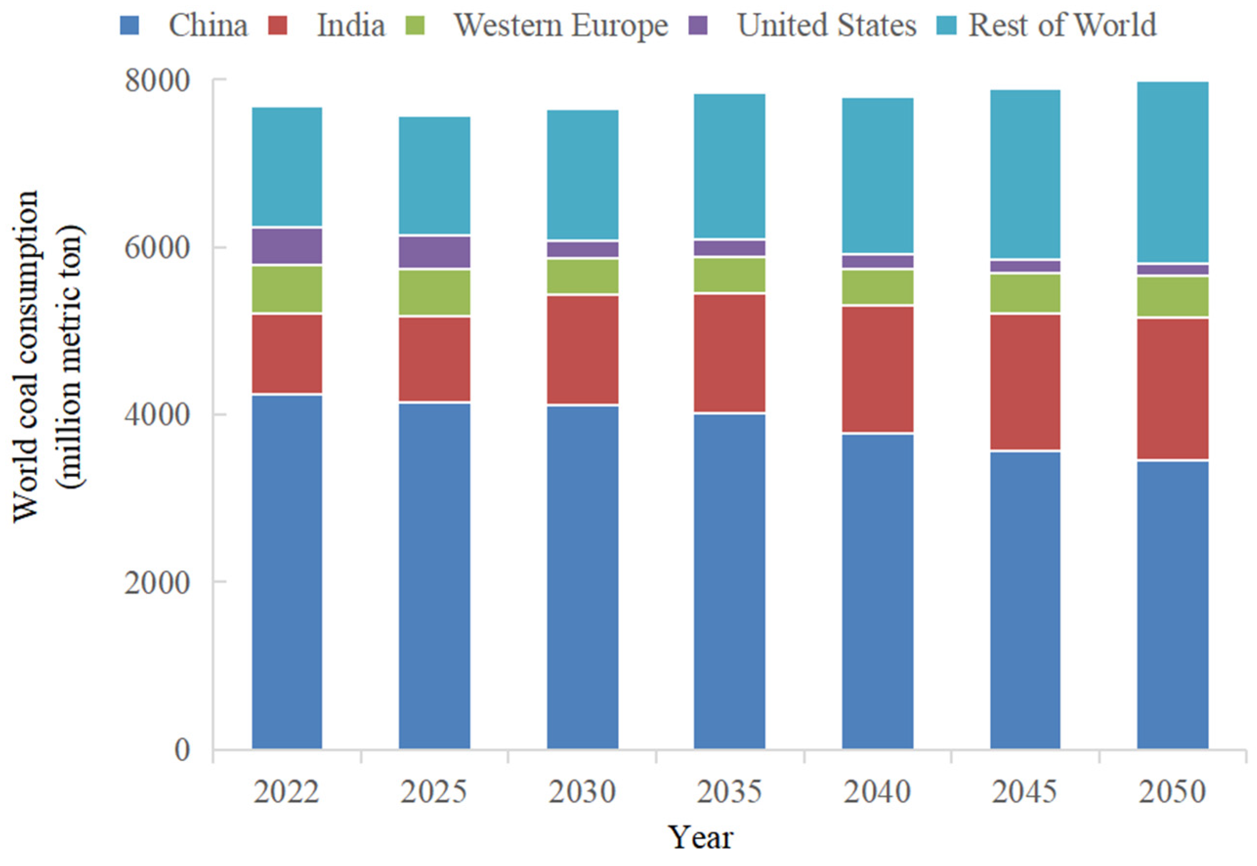
Figure 1. Global coal consumption estimates by country and groupings, 2022–2050 [3].
Minerals 15 01075 g001
Figure 2. The annual number of publications on health risks associated with coal dust exposure.
Minerals 15 01075 g002
Figure 3. Country co-authorship network.
Minerals 15 01075 g003
Figure 4. Top 9 keywords with the strongest citation bursts.
Minerals 15 01075 g004
Figure 5. Mechanisms of dust particulate matter (PM) toxicity [68].
Minerals 15 01075 g005
Figure 6. EPFRs spectroscopy of coal nanoparticles [94].
Minerals 15 01075 g006
Table 1. Top 10 cited studies from 2001 to 2025.
CountYearCountryAuthorTitle
362020USALiu TThe impacts of coal dust on miners’ health: A review [31]
342019USASarver EBeyond conventional metrics: Comprehensive characterization of respirable coal mine dust [20]
272018USABlackley DJContinued Increase in Prevalence of Coal Workers’ Pneumoconiosis in the United States, 1970–2017 [32]
212019USADoney BCRespirable coal mine dust in underground mines, United States, 1982–2017 [25]
212017USAJohann-Essex VRespirable coal mine dust characteristics in samples collected in central and northern Appalachia [33]
202020SpainTrechera PMineralogy, geochemistry and toxicity of size-segregated respirable deposited dust in underground coal mines [34]
192019USAHall NBCurrent Review of Pneumoconiosis Among US Coal Miners [35]
192021IranZazouli MAPhysico-chemical properties and reactive oxygen species generation by respirable coal dust: Implication for human health risk assessment [36]
182016USACohen RALung Pathology in U.S. Coal Workers with Rapidly Progressive Pneumoconiosis Implicates Silica and Silicates [37]
182019SpainMoreno TTrace element fractionation between PM10 and PM2.5 in coal mine dust: Implications for occupational respiratory health [18]
Table 2. Elemental abundance in coal dust from different regions (μg/g).
ElementIran Coal Dust [36]Iran
Coal [36]
Shanxi Coal
Dust [43]
Hunan Coal Dust [42]Hunan
Coal [42]
Slovenia
Coal Dust [18]
Slovenia
Coal [18]
World
Coal [36]
Be1.293.752.021.261.98<0.1-1.5
V34713.7549.4979.3340.60412825
Cr149832.5350.4421.0733-10
Mn45582115.73218.7829.3377854250
Fe31,20032,14910,700 31,95016,60010,000
Co21.4610.19.339.2420.805.852.65
Cu69.1613.523.9630.9024.33338.615
Zn425.9312.581.5777.6358.671112250
As26.213.816.8280.477.82198.226
Sr55.44354.75225.0244.3328.33121.594130
Ag0.270.07-----0.08
Cd0.860.03---0.250.20.3
Sb5.970.689.348.684.305.750.43
Ba163274.5333.5538.869.5715878120
Pb45.218.5235.9734.3345.00358.725
Al23,40043,20038,500--31,80017,20010,000
Ca8140229,00012,300--36,90025,60010,000
K21,40042835400--665027001000
Mg12,10095,2001900--525027002000
Na29,20041403200--310010002000
Ti--2284.221014.56535.671068640-
Table 3. Different simulated liquid components and extraction steps.
CategoryGamble’s Solution (GS) [40]Artificial Lysosomal Fluid (ALF) [40]
Dust dosage (g)0.0200.020
Simulated liquid volume (mL)100100
Simulated liquid composition (g/L)NaCl6.779NaCl3.210
NaHCO32.268Na2HPO40.071
Sodium citrate0.055Sodium citrate0.077
NH4Cl0.535Glycine0.059
Glycine0.357NaOH6.000
NaH2PO41.872Citric acid20.80
L-cysteine0.121CaCl2.2H2O0.128
CaC12.2H2O0.026Na2SO40.039
MgCl2.6H2O0.050
Disodium tartrate0.090
Sodium lactate0.085
Sodium pyruvate0.172
Extraction stepsStir at 1000 rpm for 24 h at 37 °C, centrifuge, and analyze the supernatantStir at 1000 rpm for 24 h at 37 °C, centrifuge, and analyze the supernatant
Table 4. Different gastrointestinal tract simulated liquid components and extraction steps.
CategoryUBM [53,54]PBET [55]SBET [56,57]
Mimic organMouth–Gastric–Intestinal phaseGastric–Intestinal phaseGastric phase
Extraction stepsOral phase: pH 6.5 ± 0.5
Add α-amylase(145 mg/L);
Mucoprotein(50 mg/L);
Uric Acid (15 mg/L)
Oscillate at 37 °C for 5 min.
Gastric Phase Simulation: pH 2.5
Add pepsin (1.25 g/L),
sodium citrate(0.5 g/L),
sodium malate (0.5 g/L),
lactic acid (420 μL/L),
acetic acid (500 μL/L);
Oscillate at 37 °C for 1 h.
Gastric Phase Simulation
Add pepsin (1.25 g/L),
Adjust pH to 1.5 ± 0.1 using HCl
Oscillate at 37 °C for 1 h.
Gastric phase: pH 1.2
Add pepsin(1.0 g/L),
mucoprotein (3.0 g/L),
bovine serum albumin(1 g/L)
shaken for 1 h.
Intestinal Phase Simulation:pH 7
Add pancreatin (0.5 g/L),
porcine bile salts (1.75 g/L), Adjust pH to 7.0 using NaHCO3.
Oscillating for 4 h
Intestinal Phase: pH 4
Duodenal fluid:pH 7.4 ± 0.2
Add CaCl2 (200 mg/L),
bovine serum albumin(1 g/L)
pancreatin (3 g/L),
lipase (500 mg/L),
Bile fluid: pH 8.0 ± 0.2
Add CaCl2 (222 mg/L),
bovine serum albumin(1.8 g/L),
bile (6 g/L),
Oscillate at 37 °C for 4 h.
solid-to-liquid ratio1:15; 1:22.5; 1:601:1001:100
Disclaimer/Publisher’s Note: The statements, opinions and data contained in all publications are solely those of the individual author(s) and contributor(s) and not of MDPI and/or the editor(s). MDPI and/or the editor(s) disclaim responsibility for any injury to people or property resulting from any ideas, methods, instructions or products referred to in the content.