Article

Biological Control Versus Environmental Influence in Serpulid Tube Calcification

Key Laboratory of Submarine Geosciences and Prospecting Techniques, Ministry of Education and College of Marine Geosciences, Ocean University of China, Qingdao 266100, China
*
Author to whom correspondence should be addressed.
Minerals 2025, 15(10), 1034; https://doi.org/10.3390/min15101034
Submission received: 9 September 2025 / Revised: 27 September 2025 / Accepted: 28 September 2025 / Published: 29 September 2025
(This article belongs to the Section Biomineralization and Biominerals)

Abstract

Serpulids are among the few annelid groups capable of building skeletal structures by secreting calcium carbonate. Compared with other biomineralizing organisms, their control over tube construction is relatively limited, making them vulnerable to environmental changes. To distinguish between intrinsic biological regulation and extrinsic environmental influence in tube formation, we examine the calcareous tube of Hydroides elegans, focusing on the tube ultrastructure, mineral composition, elemental distribution, organic-inorganic constituents, and biomineralization mechanism. The results show that the tube consists of three superimposed layers: an innermost organic sheet, an intermediate lamello-fibrillar calcite layer, and an outermost spherulitic prismatic calcite layer. The outer spherulitic prismatic layer frequently exhibits bioerosion, trapped sedimentary particles, and fan-shaped aragonite aggregates, indicating pronounced environmental influence. In contrast, the middle lamello-fibrillar calcite fabric is highly organized and closely integrated with the innermost organic sheet, indicating strictly biological controls. This study highlights the combined effect of biological controls and environmental influences in serpulid tube calcification, contributing to our understanding of their adaptive evolution in changing oceans.

1. Introduction

Annelids construct a variety of dwelling tubes, but only certain genera and species within three families—Serpulidae, Cirratulidae, and Sabellidae—are capable of producing mineralized tubes by secreting calcium carbonate minerals. In temperate marine environments, these groups represent one of the most important calcified invertebrates and are also widely recognized as significant fouling organisms [1,2,3]. Their evolutionary history can be traced back to the Permian, and their fossils have been common since the Middle Triassic [4,5]. Among them, serpulids are exceptional at building calcareous tubes with highly organized and complex skeletal ultrastructures. Their calcareous tube consists of an outer mineral layer and an inner organic layer [6]. The mineral composition is primarily calcite, aragonite, or a mix of both carbonate polymorphs [6,7,8,9]. Carbonate minerals for tube construction are secreted by specialized calcifying glands located on either side of the peristomial collar of the organism, which are then transported and assembled into diverse ultra-microstructures. Extensive investigations into serpulid tube biomineralization have identified approximately a dozen different ultrastructural types (e.g., fibrous, prismatic, and spherulitic) [1,10,11,12,13,14,15,16,17,18,19]. Of these, the lamello-fibrillar (LF) calcite structure is the most highly ordered and sophisticated ultrastructural type.
While tube construction in serpulids represents a biologically controlled mineralization process, it differs markedly from the calcification of molluscan shells, which occurs within a fully enclosed space isolated from the external environment. Instead, serpulid tube calcification somehow resembles the reef-building process of scleractinian corals [20,21], constituting a relatively weak form of biologically controlled mineralization. As a result, it is highly sensitive to changes in biotic and abiotic factors in the marine environment (e.g., temperature, pH, and seawater chemistry), which can profoundly influence the dynamics of tube calcification [22,23].
The respective influence of biological controls and environment-influenced factors in tube calcification is manifested in several aspects, such as variations in a tube’s ultrastructural types, differing degrees of organization of basic mineral building units, and the quantity and distribution of organic constituents. Recently, Grenier et al. [24] classified tube ultrastructures into two main categories: primary structures reflecting biologically controlled mineralization and secondary structures largely shaped by environmental influences. These authors further proposed an organic-mediated mineralization model for tube formation, particularly for those with an LF calcite layer. Nevertheless, the mechanism by which the organic matrix guides tube calcification remains unresolved.
Here, we examine the various ultrastructures, mineral compositions, and elemental distributions of the calcareous tube of Hydroides elegans, with focus on clarifying the primary and secondary structures and the interface between the organic and mineral layers. This work reconstructs the biomineralizing model of the LF-secreting tube of H. elegans, elucidating the relative contribution of biological control and marine environmental factors in the calcification process of serpulid tubes.

2. Materials and Methods

2.1. Specimens

The specimens of Hydroides elegans utilized in this study were from (1) calcareous tubes attached to oyster shells collected from intertidal oyster beds in Rushan, Weihai City, Shandong Province, China (36.850473° N, 121.526184° E) and (2) calcareous tubes attached to scallop shells purchased from a seafood market in Qingdao, Shandong Province, China. After collection, the samples were rinsed, with the serpulid tubes remaining intact and attached to the shells of other species, and then stored under dry conditions before being further processed according to different experimental requirements. All obtained specimens were adult, meticulously cleaned, desiccated, and stored at the College of Marine Geosciences of the Ocean University of China.

2.2. Photograph and Image Acquisition

The selected calcareous tubes were embedded in epoxy resin, ground, and polished into thin sections. These thin sections were examined using optical microscopy to characterize the overall structural distribution and ultrastructure of each tube. Some tubes were manually fractured longitudinally and transversely for further examinations. These fractured samples were mounted on T-shaped aluminum alloy stubs using conductive carbon adhesive. The mounted samples were sputter-coated with gold for 280 s (30 mA) and examined by using a Quanta 450 FEG field-emission scanning electron microscope (SEM) operated at 15 kV and 0.8 nA. Energy-dispersive X-ray spectroscopy (EDS) analysis was conducted to determine the elemental distribution within the calcareous tube.

2.3. Electron Probe Microanalysis

Quantitative determination and elemental mapping of major elements (Mg, Sr, O, P, and S) within the calcareous tubes were performed using a JEOL JXA-8530F Plus electron probe microanalyzer (EPMA). Following sectioning and polishing into thin sections, the sample surfaces were sputter-coated with platinum (Pt) for 90 s to ensure stable analysis of the insulating carbonate material. Analysis was conducted in mapping mode, with an accelerating voltage of 15 kV and a beam current of 200 nA, balancing excitation efficiency with the risk of thermal damage.

2.4. Raman Spectroscopy Analysis

Non-destructive analysis of the calcareous tube was performed using a micro-Raman spectrometer (Horiba LabRAM HR Evolution). A 532 nm laser source was employed for the experiments. Spectra were acquired across the range of 100–1730 cm−1 using a 500 nm grating. The detector temperature was set to −60.09 °C, and the laser aperture was set to 100.021. To enhance the signal-to-noise ratio, 2 accumulations per point were performed. Measurements were conducted using a 10×–50× objective lens. Spectral peak positions were calibrated using a silicon standard. Data processing, including baseline correction and Lorentzian peak fitting, was performed using LabSpec 6 software. Mineral identification was based on reference to the standard spectral library proposed by Frezzotti [25].
Experiments including high-resolution SEM imaging, EPMA mapping, and Raman spectroscopy were conducted at the State Key Laboratory of Continental Dynamics at Northwest University. Other examinations, including specimen pretreatment and optical microscopic observation, were performed at the College of Marine Geosciences of the Ocean University of China.

3. Results

3.1. Ultrastructure and Morphology

The calcareous tube of H. elegans exhibited a bi-layered structure (Figure 1A–C), comprising an outer mineral layer and an inner organic layer. The mineral layer itself consisted of an outer spherulitic prismatic (SP) and an inner lamello-fibrillar calcite (LF) structural sublayers. The organic sheet occurred on the innermost side of the calcareous tube and displayed distinct surface properties on its inner and outer surfaces (Figure 1D,E).
The outermost SP layer, with a thickness ranging from 19 to 35 µm, was composed of elongated mineral units exhibiting a radial structural arrangement. The main body of the tube was formed by multiple interlocking mineral laminae exhibiting an LF ultrastructure (Figure 2A,B). The basic mineral unit was characterized by a rhombohedral shape, indicative of calcite crystallites (Figure 2C). The individual mineral laminae were 6.5–8.5 µm thick, sometimes separated by visible thin organic membranes.
The innermost organic sheet (Figure 2D), approximately 1.5–1.8 µm thick, exhibited distinct surface characteristics; the surface facing the tube lumen (defined as the outer surface) was smooth, whereas the opposite surface was tightly connected to the adjacent mineral layer. This organic sheet comprised prominent protein fibers organized in stacked laminae, with fibers in alternating layers oriented approximately orthogonal (~90°) to each other, forming a densely interwoven network (Figure 2E). These robust fiber bundles represent composite structures of the organic matrix and carbonate minerals, probably in the form of an amorphous calcium carbonate (ACC) phase (Figure 2F). Carbonate minerals toward the outer mineral layers exhibited increased crystallinity. The basic mineral building blocks were rod-shaped, measuring 3–6 µm in length and 0.7–0.9 µm in diameter, and they were composed of aggregated nanoparticles (Figure 2G). Notably, these mineralized fiber bundles appeared to be enveloped by a thin organic membrane layer (Figure 2H).
At the mineral–organic interface, the LF structure exhibited an ordered organization, congruent with the fabrication of adjacent organic sheets (Figure 2F). These basic mineral units were aligned along their long axes and further assembled into robust mineral fiber bundles, concomitant with a progressive reduction in organic content (Figure 2F,H,I). Adjacent to the inner surface, a well-defined LF structure was visible, in which regularly ordered and mineralized fiber bundles were interlinked by organic filaments and porous organic membranes (Figure 3).

3.2. Bioerosion and Secondary Infillings

The outer surface of the calcareous tubes exhibited dense bioerosion traces and porosity, with variable boreholes (3.8–15.4 µm long and 1.3–1.8 µm wide) (Figure 4A). Numerous borings occurred between adjacent or within specific mineral layers, often filled with dumbbell-shaped calcite clusters. These clusters measured 6.7–23.8 µm in total length and were composed of densely aggregated nanoparticles (Figure 4B). Some of these borings were infilled with acicular aragonite aggregates, exhibiting hexagonal cross-sections with diameters of about 2.4–4.4 µm and lengths of 19.8–32.4 µm (Figure 4C,D). Additionally, fan-shaped fibrous aragonite aggregates were occasionally present within the mineral layers (Figure 4E), while granular prismatic calcite precipitates occurred between spaces of the robust fiber bundles of the innermost organic sheet (Figure 4F).

3.3. Mineral Composition and Elemental Distribution

Raman spectra analysis revealed a prominent broad peak at 1089.55 cm−1, slightly higher than the standard v1 vibration band of pure calcite (1085 cm−1), while closely matching the characteristic peak of Mg-calcite (1087 cm−1) (Figure 5A–C). This positive shift in the main stretching vibration band was due to the incorporation of magnesium (Mg2+) in the calcite lattice [26], indicating the presence of Mg-calcite minerals. The resulting peak at 717.42 cm−1 corresponded well with the v4 vibrational mode of calcite (712 cm−1) and clearly differed from the characteristic peaks of aragonite (704 cm−1) and vaterite (740–750 cm−1). The SEM-EDS results showed enrichment of Ca, C, O, and Mg in proximity to the well-crystallized LF structures, demonstrating an Mg-calcite mineralogy (Figure 5D,E).
Electron probe microanalysis (EPMA) revealed that Sr was mainly distributed along the inner and outer surfaces of the tubes. This distribution was consistent with the diagenetic precipitation coating the tubes. Both the SP and LF layers were enriched with Mg, indicating high-Mg calcite layers, whereas the elements P, S, Ca, C, and O were uniformly distributed throughout the mineral tubes (Figure 6).

4. Discussion

4.1. Environmental Influence and Biological Control

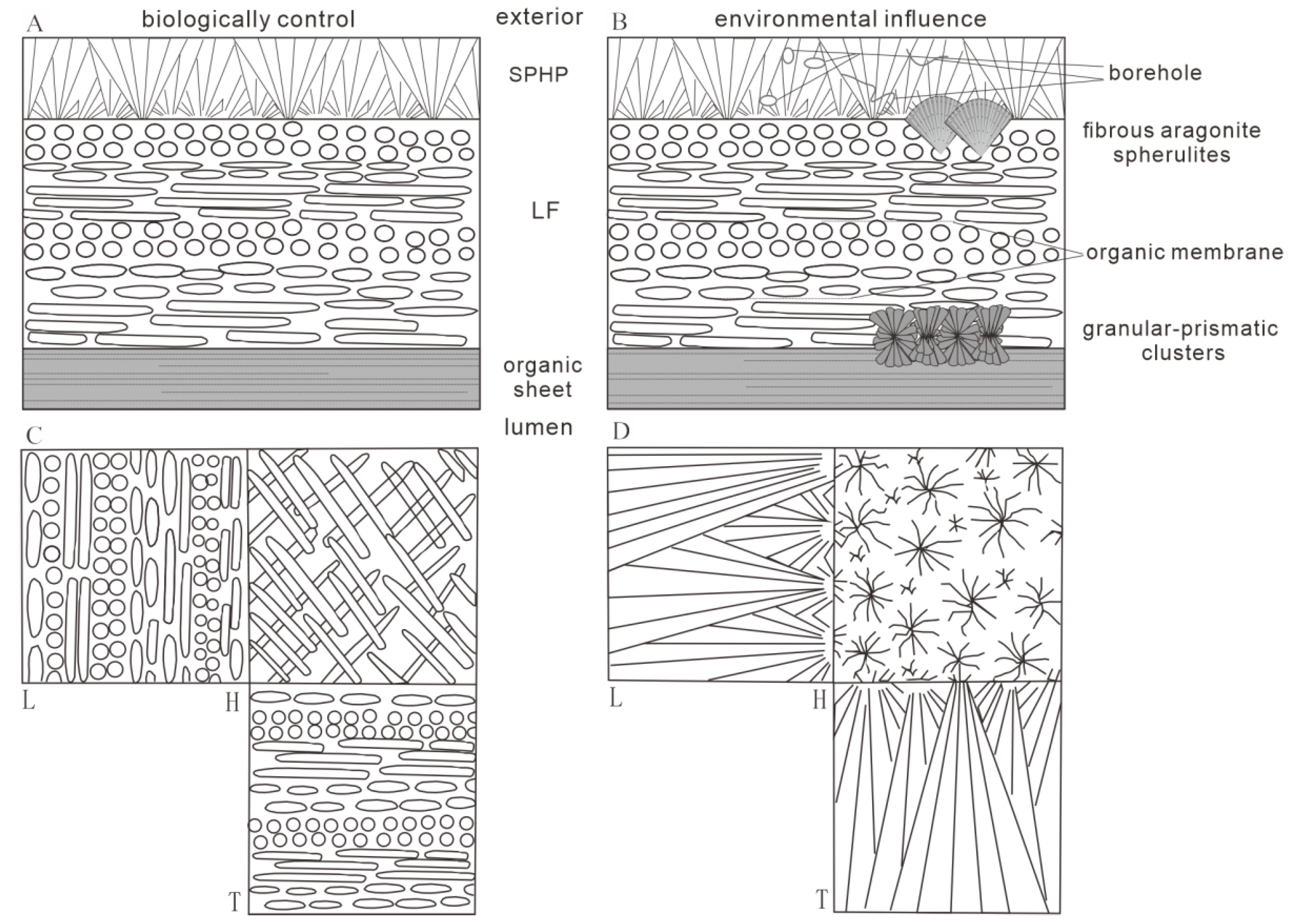
The calcareous tube of Hydroides elegans exhibited an ultrastructure essentially comparable with that of Crucigera websteri in terms of the presence of external SP calcite and inner LF calcite, forming most of the tube’s thickness [24]. Both primary and secondary structures were recognized. The primary structures included the SP and LF calcite layers that corresponded to the biologically regulated tube ultrastructure, which is widespread in modern serpulids [4,6,27,28]. The secondary structures are characterized by dense bioerosion traces and boreholes formed by undetermined predatory attacks [11,23]. Numerous void spaces and gaps are typically filled by secondary depositions, including dumbbell-shaped calcite crystallites, acicular aragonite aggregates, fan-shaped fibrous aragonite, and granular prismatic calcite precipitated between the spaces of the fiber bundles of the innermost organic sheet. One notable difference is that the Crucigera websteri tube has an LF structure comprising stacked mineral laminae clearly separated by organic membranes [10,24]. In contrast, no obvious organic membrane separation was observed in the LF structure of H. elegans. Instead, there was a distinct transition at the mineral–organic interface, where the crystallization and growth of basic mineral units is strictly biologically controlled [6,10]. It should be noted that both the primary and secondary structures together constitute the mineralized tube. Primary structures are formed under exquisite biological regulation [29,30], while the precipitation of secondary structures is more environmentally influenced [31] (Figure 7).
The mineral phase composition and elemental distribution within calcareous tubes are influenced by multiple environmental factors. The concentrations of key chemical ions in seawater, particularly the magnesium-to-calcium molar ratio (molar Mg2+/Ca2+), seem to play a central role in the mineralogy of the tube. In modern marine environments with a high Mg/Ca ratio (~5.2), serpulids tend to produce high-Mg calcite and aragonitic tubes. Fossil records indicate that the preference of a specific carbonate polymorph (calcite/aragonite) is highly consistent with secular changes in seawater chemistry over geological timescales [4,31]. The seawater carbonate saturation state is another major factor driving mineralization rates and structural integrity. Specifically, in shallow marine settings with high saturation, calcium carbonate precipitation is enhanced, promoting tube thickening and increasing crystallinity. In contrast, deep sea and acidified marine environments reduced mineralization rates, resulting in structural fragility [32]. Moreover, temperature also exerts significant effects, as increased temperatures generally promote tube growth by accelerating metabolism, and warmer environments favor aragonite precipitation [7,33]. Together, these environmental factors strongly affect tube formation, particularly the precipitation of various secondary structures. The variability in these structures in turn greatly enhances the ecological adaptability of the organism to dynamic environments.
The mineralization process of serpulid tubes exhibits pronounced characteristics for its biological controls. They have long been known to secrete calcium carbonate minerals via the calcifying glands in the collar region [27,34]. They synthesize specific carbonate mineral precursors and maintain a relatively stable mineralogical composition, a physiological process that is highly resistant to external environmental variations. The crystal arrangement and magnesium content, particularly in the highly ordered LF structure, are strictly maintained [29]. The regular formation and distribution of primary structures, namely the SP and LF calcite structure layers, are typical biologically controlled products [4,6]. The mineral layers are composed of rod-shaped calcitic units formed by an aggregation of nanoparticles within an organic cortex [6]. The organic sheet shows obvious layered structures parallel to the inner surface of the tube [10]. Each lamina comprises thick fiber bundles with a certain orientation, where the general direction of individual fibers is basically parallel to each other [24]. Within given laminae, robust organic-mineral bundles are interlinked through protein filaments and porous organic membranes [10]. Crystal nucleation, growth, and orientation and their final stacking into a crisscrossed three-dimensional architecture are all precisely controlled by the organic sheet, highlighting an organic-mediated mineralization nature [10,29].

4.2. Biomineralization Mode in Serpulid Tubes

Mineralized tubes of serpulids contain approximately 7.5% organic contents, composed mainly of chitin and proteins, which act as an organic scaffold occluded within the mineral tube [24,35]. In mineralized tubes of Spirobranchus triqueter and Crucigera websteri, the LF calcite structure exhibits distinct organic membranes separating adjacent laminar layers [36]. The periodic occurrence of these organic membranes corresponds to the continuous development of growth lines in the SP layer, reflecting rhythmic construction of the tube by a worm [10]. Based on the regular occurrence of organic membranes within the LF-secreting tube, two possible mechanisms for tube formation have been proposed: (1) the tube wall develops incrementally, with each LF calcite lamina deposited as a thin sublayer, followed by the secretion of an organic membrane, or (2) the tube forms through the isolation of an enclosed space, and LF secretion is subsequently completed in a single phase. Both biomineralization mechanisms in Spirobranchus and Crucigera highlight the critical role of organic membranes in the process of tube calcification [24].
In the tube of Hydroides, the adjacent laminar layers of the LF structure appear to be tightly stacked, lacking continuous interlaminar organic membranes. Instead, tube formation is primarily regulated by the innermost organic sheet. This suggests that serpulids have adopted species-specific biomineralization strategies in tube construction. In Hydroides elegans, the inner organic sheet is first assembled by a worm to form an enclosed compartment. However, the LF structure is not mineralized as a single unit; rather, it is formed incrementally, lamina by lamina, under the mediation of an associated organic sheet. The enclosed spaces formed by the organic sheet provide effective control over the crystal morphology and growth kinetics [22]. Similar to other metazoan biomineralizers such as molluscs, intracellular vesicles probably release amorphous carbonate precursors from the calcifying glands directly onto the pre-formed organic sheet [36,37,38]. Amorphous carbonate precursors are gradually transformed into stable high-Mg calcite crystals facilitated by magnesium and organic molecules. Necessary proteins and polysaccharides stabilize the deposition of amorphous calcium carbonate precursors, guide the directional growth of crystals, and determine the spatial arrangement and morphology of the final mineral phase [6,29]. Ultimately, through precise step-by-step regulation of the multi-layer structure, a mechanically robust, high-strength mineralized tube is formed.

5. Conclusions

The formation of calcareous tubes for Hydroides elegans occurs as a weak mineralization process in a semi-enclosed environment. The primary structures of the tube include SP calcite and LF calcite, which are formed through biological control. The secondary structures of the tube include fibrous aragonite, granular-prismatic clusters, and incorporation of external objects, whose precipitation is significantly environmentally influenced. The biomineralization of the LF calcite tube is regulated by a typical organic matrix-mediated process, during which the innermost organic sheet forms an enclosed space, and the layer-by-layer stacking of calcitic lamina occurs under the control of various organic molecules.

Author Contributions

L.L. (corresponding author): Conceptualized the overall experimental design, led the feasibility study of the research scheme, and supervised the entire research process to ensure the scientific rigor of the work. C.X.: Performed all experimental operations independently, including sample preparation, data collection, and experimental validation. She was also responsible for the systematic collation, preliminary analysis, and visualization of experimental data, laying a solid foundation for the subsequent data interpretation and manuscript writing. All authors have read and agreed to the published version of the manuscript.

Funding

This study was supported by the National Natural Science Foundation of China (Grant No. 42202001), which also covered the article-processing charges.

Data Availability Statement

Data are contained within the article.

Acknowledgments

We thank Linhao Cui, Jingjing Dai, and Baige Xia for the technical support. Thanks are given to Jingyi He, who contributed to specimen collection in the field.

Conflicts of Interest

The funders had no role in the design of the study; in the collection, analyses, or interpretation of data; in the writing of the manuscript, or in the decision to publish the results.

References

  1. Vinn, O.; Ten Hove, H.A.; Mutvei, H.; Kirsimäe, K. Ultrastructure and mineral composition of serpulid tubes (Polychaeta, Annelida). Zool. J. Linn. Soc. 2008, 154, 633–650. [Google Scholar] [CrossRef]
  2. Vinn, O.; Ten Hove, H.A.; Mutvei, H. On the tube ultrastructure and origin of calcification in sabellids (Annelida, Polychaeta). Palaeontology 2008, 51, 295–301. [Google Scholar] [CrossRef]
  3. Vinn, O. The ultrastructure of calcareous cirratulid (Polychaeta, Annelida) tubes. Est. J. Earth Sci. 2009, 58, 153–156. [Google Scholar]
  4. Vinn, O.; Mutvei, H. Calcareous tubeworms of the Phanerozoic. Est. J. Earth Sci. 2009, 58, 286–296. [Google Scholar] [CrossRef]
  5. Sanfilippo, R.; Rosso, A.; Reitano, A.; Insacco, G. First record of sabellid and serpulid polychaetes from the Permian of Sicily. Acta Palaeontol. Pol. 2017, 62, 25–38. [Google Scholar] [CrossRef]
  6. Vinn, O. The role of an internal organic tube lining in the biomineralization of serpulid tubes. Carnets Géologie 2011, 1, 13–16. [Google Scholar] [CrossRef]
  7. Smith, A.M.; Riedi, M.A.; Winter, D.J. Temperate reefs in a changing ocean: Skeletal carbonate mineralogy of serpulids. Mar. Biol. 2013, 160, 2281–2294. [Google Scholar] [CrossRef]
  8. Vinn, O.; Kupriyanova, E.K. Evolution of a dense outer protective tube layer in serpulids (Polychaeta, Annelida). Carnets Géol. 2011, 2011, 137–147. [Google Scholar] [CrossRef]
  9. Vinn, O. The role of aragonite in producing the microstructural diversity of serpulid skeletons. Minerals 2021, 11, 1435. [Google Scholar] [CrossRef]
  10. Vinn, O. Occurrence, formation and function of organic sheets in the mineral tube structures of Serpulidae (Polychaeta, Annelida). PLoS ONE 2013, 8, e75330. [Google Scholar] [CrossRef]
  11. Weedon, M.J. Tube microstructure of Recent and Jurassic serpulid polychaetes and the question of the Palaeozoic ‘spirorbids’. Acta Palaeontol. Pol. 1994, 39, 1–15. [Google Scholar]
  12. Vinn, O.; Alkahtane, A.A.; Al Farraj, S.; El Hedeny, M. New type of SIOP structure in Serpulidae: Formation and evolutionary implications. Minerals 2024, 14, 291. [Google Scholar] [CrossRef]
  13. Vinn, O.; Mutvei, H.; ten Hove, H.A.; Kirsimäe, K. Unique Mg-calcite skeletal ultrastructure in the tube of the serpulid polychaete Ditrupa. Neues Jahrb. Geol. Paläontol. Abh. 2008, 248, 79–89. [Google Scholar] [CrossRef]
  14. Vinn, O.; Kirsimäe, K.; ten Hove, H.A. Tube ultrastructure of Pomatoceros americanus (Polychaeta, Serpulidae): Implications for the tube formation of serpulids. Est. J. Earth Sci. 2009, 58, 148–152. [Google Scholar] [CrossRef]
  15. Vinn, O. On the unique isotropic aragonitic tube microstructure of some serpulids (Polychaeta, Annelida). J. Morphol. 2013, 274, 478–482. [Google Scholar] [CrossRef] [PubMed]
  16. Vinn, O. SEM study of semi-oriented tube microstructures of Serpulidae (Polychaeta, Annelida): Implications for the evolution of complex oriented microstructures. Microsc. Res. Tech. 2013, 76, 453–456. [Google Scholar] [CrossRef] [PubMed]
  17. Vinn, O.; Alkahtane, A.A.; El Hedeny, M.; Al Farraj, S. Discovery of rod-type crossed lamellar structure in Spiraserpula (Serpulidae, Polychaeta) from the Middle Miocene of Austria. Neues Jahrb. Geol. Paläontol. Abh. 2023, 310, 91–97. [Google Scholar] [CrossRef] [PubMed]
  18. Vinn, O. Distribution of growth lines in the tube wall of serpulids (Polychaeta, Annelida). J. Mar. Biol. Assoc. UK 2024, 104, e24. [Google Scholar] [CrossRef]
  19. Słowiński, J.; Vinn, O.; Zatoń, M. Ultrastructure of the Jurassic serpulid tubes—Phylogenetic and paleoecological implications. PeerJ 2024, 12, e17389. [Google Scholar] [CrossRef]
  20. Tambutté, E.; Allemand, D.; Zoccola, D. Coral biomineralization: A focus on intra-skeletal organic matrix and calcification. Minerals 2021, 11, 523. [Google Scholar]
  21. Cuif, J.P.; Dauphin, Y. Organic matrix and biomineralization of scleractinian corals. Minerals 2020, 10, 189. [Google Scholar]
  22. Sommerdijk, N.A.J.M.; de With, G. Biomimetic CaCO3 mineralization using designer molecules and interfaces. Chem. Rev. 2008, 108, 4499–4550. [Google Scholar] [CrossRef]
  23. Buckman, J.O. An overview of the tube fabric of Pomatoceros (Polychaeta, Serpulidae), illustrated by examples from the British Isles. Zool. Anz. 2015, 259, 54–60. [Google Scholar] [CrossRef]
  24. Grenier, C.; Berent, K.; Rodríguez-Navarro, A.; Vinn, O.; Checa, A.G. Microstructures, crystallography and growth patterns of serpulid tubeworms (Class Polychaeta). Mar. Biol. 2024, 171, 231. [Google Scholar] [CrossRef]
  25. Frezzotti, M.L.; Tecce, F.; Casagli, A. Raman spectroscopy for fluid inclusion analysis. J. Geochem. Explor. 2012, 112, 1–20. [Google Scholar] [CrossRef]
  26. Burke, E.A.J. Raman microspectrometry of fluid inclusions. Lithos 2001, 55, 139–158. [Google Scholar] [CrossRef]
  27. Hedley, R.H. Tube formation by Pomatoceros triqueter (Polychaeta). J. Mar. Biol. Assoc. UK 1958, 37, 315–322. [Google Scholar] [CrossRef]
  28. Hedley, R.H. Studies on serpulid tube formation. I. The secretion of the calcareous and organic components of the tube by Pomatoceros triqueter. Q. J. Microsc. Sci. 1956, 97, 411–419. [Google Scholar] [CrossRef]
  29. Weiner, S.; Addadi, L. Crystallization pathways in biomineralization. Annu. Rev. Mater. Res. 2011, 41, 21–40. [Google Scholar] [CrossRef]
  30. Gilbert, P.U.P.A.; Bergmann, K.D.; Boekelheide, N.; Tambutté, S.; Mass, T.; Marin, F.; Adkins, J.F.; Erez, J.; Gilbert, B.; Knutson, V.; et al. Biomineralization: Integrating mechanism and evolutionary history. Sci. Adv. 2022, 8, eabl9653. [Google Scholar] [CrossRef] [PubMed]
  31. Ries, J.B. Effect of ambient Mg/Ca ratio on Mg fractionation in calcareous marine invertebrates: A record of the oceanic Mg/Ca ratio over the Phanerozoic. Geology 2004, 32, 981–984. [Google Scholar] [CrossRef]
  32. Kupriyanova, E.K.; Vinn, O.; Taylor, P.D.; Schopf, J.W.; Kudryavtsev, A.; Bailey-Brock, J. Serpulids living deep: Calcareous tubeworms beyond the abyss. Deep Sea Res. I 2014, 90, 91–104. [Google Scholar] [CrossRef]
  33. Simkiss, K.; Wilbur, K.M. Biomineralization: Cell Biology and Mineral Deposition; Academic Press: New York, NY, USA, 1989. [Google Scholar]
  34. Neff, J.M. Mineral regeneration by serpulid polychaete worms. Biol. Bull. 1969, 136, 1–15. [Google Scholar] [CrossRef]
  35. Vinn, O.; Jäger, M.; Kirsimäe, K. Microscopic evidence of serpulid affinities of the problematic fossil tube ‘Serpula’ etalensis from the Lower Jurassic of Germany. Lethaia 2008, 41, 417–421. [Google Scholar] [CrossRef]
  36. Bubel, A. Electron microscope studies on the operculum of Spirorbis spirorbis (L.). In Proceedings of the 3rd International Biodegradation Symposium; Sharpley, J.M., Kaplan, A.M., Eds.; Applied Science Publishers: London, UK, 1976; pp. 495–514. [Google Scholar]
  37. Bubel, A.; Thorp, C.H.; Fitzsimmons, C.A.L. A histological and electron microscopical study of opercular regeneration in the serpulid Pileolaria (P.) granulata, with particular reference to the formation of the calcareous opercular plate. In Proceedings of the 4th International Congress on Marine Corrosion and Fouling, Juan-les-Pins, Antibes, France, 14–18 June 1977; pp. 85–96. [Google Scholar]
  38. Bubel, A.; Thorp, C.H.; Moore, M.N. A histological, histochemical and ultrastructural study of the operculum of the serpulid Pomatoceros triqueter L., with particular reference to the formation of the calcareous opercular plate during opercular regeneration. In Proceedings of the 4th International Biodegradation Symposium; Sharpley, J.M., Kaplan, A.M., Eds.; Applied Science Publishers: London, UK, 1980; pp. 275–290. [Google Scholar]
Figure 1. Ultrastructure and morphology of the Hydroides elegans tube. (A) Fractural surface showing various tube wall ultrastructures: outer spherulitic prismatic (SP), intermediate lamello-fibrillar (LF) calcite, and inner organic sheet. (B) Optical micrograph revealing distinct wall layering in the transverse section. (C) Longitudinal section of the layered structure. (D) Outer surface of the organic sheet, showing a relatively smooth texture with residual debris (E) Mineral fibers beneath the inner surface of the organic layer.
Minerals 15 01034 g001
Figure 2. Tube ultrastructures of Hydroides elegans. (A,B) Fractural surface showing gross morphology of the LF structure, with arrows indicating alternating orientations of carbonate crystallites. (C) Close-up view of the LF structure. (D) Arrangement of the innermost organic sheet with regularly stacked lamellae. (E) Close-up image showing the fabrication of the organic sheath. (F) Organic–mineral interface. (G) Assembly of rod-shaped crystallites forming robust bundles. (H) Mineral aggregates enveloped within a thin organic membrane layer. (I) Mineral fiber bundles interconnected with organic filaments and porous organic membranes.
Minerals 15 01034 g002
Figure 3. Well-defined LF structure adjacent to the inner surface of an organic sheet. Regularly ordered mineral fibers are interconnected by protein filaments and organic membranes.
Minerals 15 01034 g003
Figure 4. Secondary structures and bioerosion in Hydroides elegans tubes. (A) Bioerosion traces and vertically oriented boreholes. (B) Environmentally induced calcite precipitates with dumbbell morphology. (C) Porosity developed between structural layers (D) Acicular aragonite crystals with a hexagonal cross-section. (E) Fan-shaped aragonite clusters exhibiting a radial fibrous texture. (F) Randomly oriented granular prismatic calcite crystals.
Minerals 15 01034 g004
Figure 5. Mineralogical characterization of Hydroides elegans tubes. (A) Optical photograph, with Raman analysis point marked with a white star. (B) Close-up image of the LF structure. (C) Raman spectrum with characteristic peak of Mg-calcite. (D,E) SEM-EDS mapping.
Minerals 15 01034 g005
Figure 6. Element distribution within Hydroides elegans tubes. Distribution of Sr along inner and outer surfaces of the tubes due to secondary precipitation. Enrichment of Mg in mineral tubes indicative of Mg-calcite.
Minerals 15 01034 g006
Figure 7. Schematic diagrams of primary and secondary structures. (A) Primary structure generated under biological control. (B) Secondary structure generated under the influence of the external environment. (C) Lamello-fibrillar structure. (D) Spherulitic prismatic ((C,D) derived from [1]). Abbreviations: LF = lamello-fibrillar structure; SP = spherulitic prismatic structure; H = horizontal section; L = longitudinal section; T = transverse section.
Minerals 15 01034 g007
Disclaimer/Publisher’s Note: The statements, opinions and data contained in all publications are solely those of the individual author(s) and contributor(s) and not of MDPI and/or the editor(s). MDPI and/or the editor(s) disclaim responsibility for any injury to people or property resulting from any ideas, methods, instructions or products referred to in the content.