1. Introduction
Inactive uranium mines have proven prolific sources of new low-temperature uranyl minerals. These phases, typically found in efflorescent coatings on tunnel walls, form from aqueous solutions that permeate and alter the uranium-rich wallrocks. As these solutions, now laden with uranyl complexes and a variety of other cations and anions, seep from the tunnel walls and evaporate, a diverse array of minerals crystallizes. In the past decade, no area in the world has yielded more new low-temperature uranyl mineral species than Red Canyon in southeastern Utah (USA). Until the recent closures of the mines in Red Canyon to collecting, a team led by one of the authors (JM) methodically sampled the secondary mineralization in the Blue Lizard, Green Lizard, Markey and Giveaway-Simplot mines, resulting in the discovery of 38 new uranyl minerals, with several others yet to be described [e.g., [
1,
2]]. In addition, four new non-uranyl minerals, all sulfates, have also been described. Herein, we describe chenowethite, one of the most recent new uranyl sulfate minerals to be discovered in the Red Canyon mineral assemblages.
Chenowethite is named for American uranium geologist William L. Chenoweth (1928–2018). Dr. Chenoweth received his Ph.D. in geology from the University of New Mexico in 1953. He spent his entire career studying the uranium deposits of the western U.S. He was employed by the U.S. Atomic Energy Commission (AEC), working out of their office in Grants, Arizona until 1964 and then out of their Grand Junction, Colorado office. He was Chief of the Geology Branch in Grand Junction from 1970 until his retirement in 1983, during which time he was responsible for the activities of AEC geologists in the 14 western U.S. states. He then became a uranium mining consultant and a research associate at the New Mexico Bureau of Geology and Mineral Resources. He served as the chairman of the Nuclear Minerals Committee for the Energy Minerals Division of the American Association of Petroleum Geologists from 1983–1998. Dr. Chenoweth authored or co-authored more than 150 reports on the uranium mining history, geology and resources in New Mexico, Arizona, Colorado and Utah. The bulk of the content of these reports stem from his more than 30 years researching uranium for the AEC and the U.S. Department of Energy (DOE). Among these reports is the definitive report on the geology and production history of the uranium deposits of the White Canyon district, which includes the mines in which chenowethite was discovered. At the time of his death, Dr. Chenoweth was still serving as the secretary/treasurer of the Grand Junction Geological Society.
The new mineral and its name were approved by the International Mineralogical Association (IMA) Commission on New Minerals, Nomenclature and Classification (CNMNC) with the number 2022–063. The description is based on four cotype specimens deposited in the collections of the Natural History Museum of Los Angeles County, 900 Exposition Boulevard, Los Angeles, CA 90007, USA, under catalogue numbers 76259 (Blue Lizard mine), 76260 (Blue Lizard mine), 76261 (Green Lizard mine) and 76262 (Green Lizard mine).
2. Occurrence, Geological Setting and Mineral Association
Chenowethite was discovered on specimens collected underground in the Blue Lizard mine (37°33′26″ N 110°17′44″ W), the Green Lizard mine (37°34′37.10″ N 110°17′52.80″ W) and the Markey mine (37°32′57″ N 110°18′08″ W) in the White Canyon mining district, San Juan County, Utah, USA. The foregoing description is based only on specimens from the Blue Lizard and Green Lizard mines and only these should be considered type localities for the species. The Blue Lizard mine is on the west-facing side of Red Canyon and the Markey mine is on the east-facing side about 1 km southwest of the Blue Lizard mine. The Green Lizard mine is near the head of Low Canyon on the east side of Red Canyon, 2.1 km north of the Blue Lizard mine. The geology of all three mines is very similar [
3,
4], although the secondary mineralogy of the Green Lizard mine is richer in ammonium phases and that of the Markey mine is richer in carbonate phases. The following information on the mines and their geology is taken largely from [
3].
Mineralized channels are in the Shinarump member of the Chinle Formation. The Shinarump member consists of medium- to coarse-grained sandstone, conglomeratic sandstone beds and thick siltstone lenses. Ore minerals were deposited as replacements of wood and other organic material and as disseminations in the enclosing sandstone. Since the mine closed, oxidation of primary ores in the humid underground environment has produced a variety of secondary minerals, mainly sulfates, as efflorescent crusts on the surfaces of mine walls.
Chenowethite is a relatively rare mineral found in association with ammoniozippeite, dickite, gypsum, hexahydrite, johannite and plášilite at the Blue Lizard mine. At the Green Lizard mine, it is found in association with dickite, gypsum, natrojarosite, natrozippeite and johannite.
Uranyl sulfate minerals typically form by hydration–oxidation weathering of primary uranium minerals, mainly uraninite, by acidic solutions derived from the decomposition of associated sulfides [
5,
6,
7]. Chenowethite and other secondary minerals occurring in the efflorescent crusts of the mines of Red Canyon were formed by such a process.
3. General Appearance, Physical, Chemical and Optical Properties
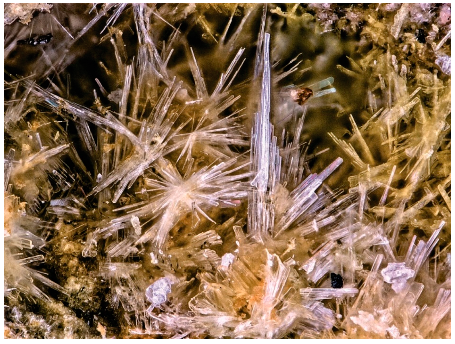
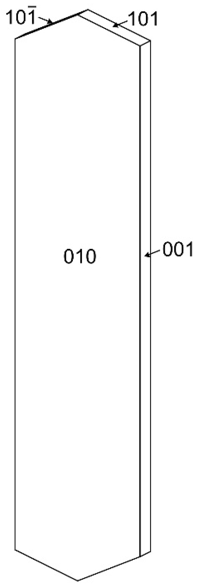
Chenowethite occurs as long, thin blades up to about 0.5 mm long, forming irregular sprays and subparallel groups (
Figure 1). The blades are flattened on {010} and elongated parallel to [100]. The observed crystal forms are {010}, {001} and {101} (
Figure 2). No twinning was observed. The color of the mineral is pale green yellow. It has white streak, vitreous to silky luster, brittle tenacity, splintery and stepped fracture and two cleavages: {010} perfect and {001} good. The Mohs hardness based on scratch tests is about 2. The mineral is nonfluorescent in both long- and short-wave ultraviolet illumination. The density measured via flotation in a mixture of methylene iodide and toluene is 3.05(2) g/cm
3. The calculated density based on the empirical formula and single-crystal cell is 3.045 g/cm
3. The mineral is easily soluble in room-temperature H
2O.
Optically, crystals are biaxial (−) with α = 1.530(2), β = 1.553(2) and γ = 1.565(2), measured in white light. The 2V measured directly by conoscopic observation of the interference figure on a spindle stage is 72(2)°, The 2V calculated from the indices of refraction is 70.7°. The dispersion was observed to be r > v, slight. The optical orientation is X = b, Y = a, Z = c. The mineral exhibits weak pleochroism in shades of pale green yellow: X < Y < Z.
The Gladstone–Dale compatibility [
8] 1 − (K
p/K
c) is 0.004 (superior) based on the empirical formula and −0.002 (superior) based on the ideal formula, in both cases using
k(UO
3) = 0.118 [
9].
4. Raman Spectroscopy
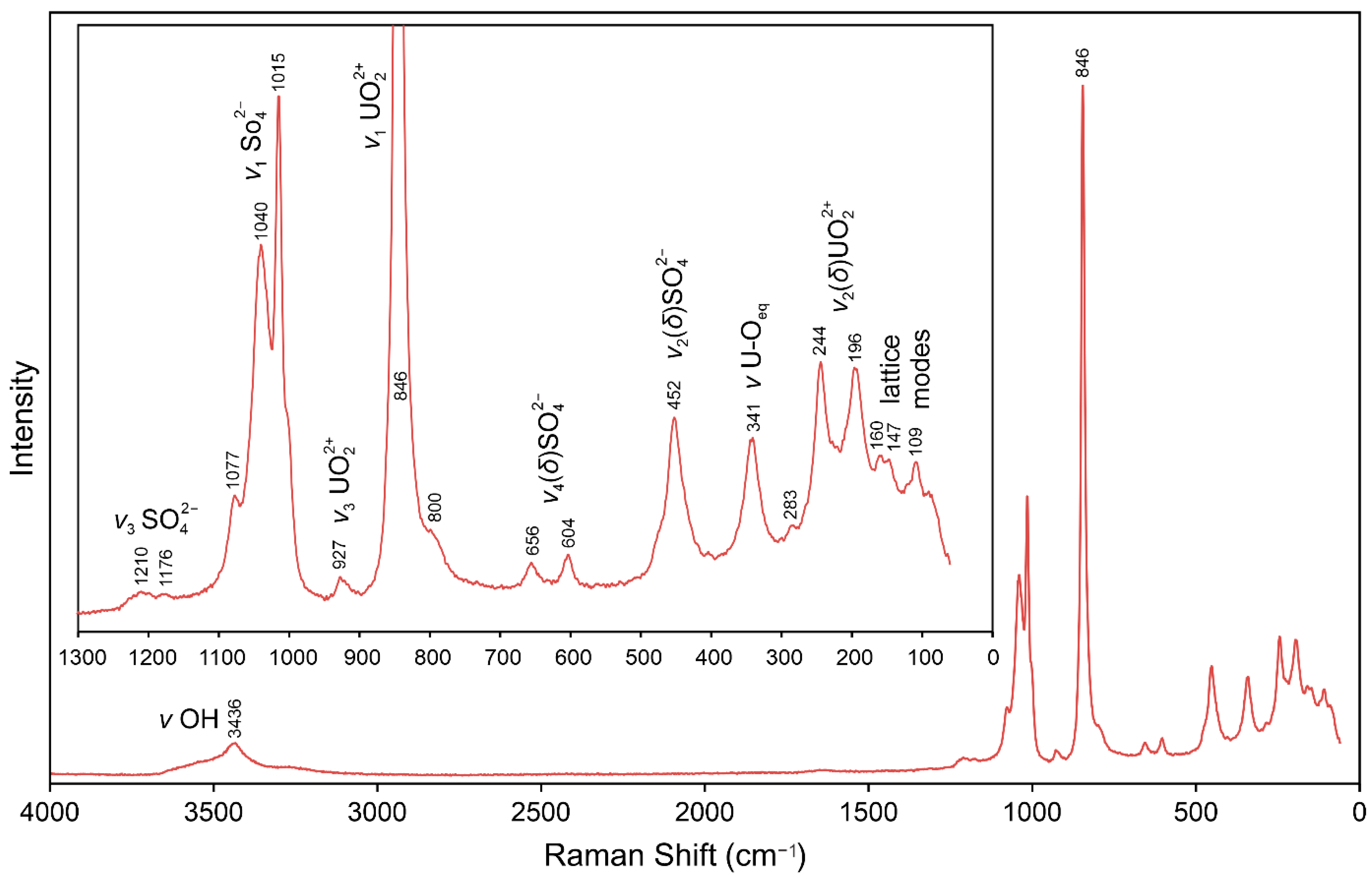
Raman spectroscopy was performed via a Horiba XploRa Plus micro-Raman spectrometer (Kyoto, Japan) using an incident wavelength of 532 nm, laser slit of 100 μm, 1800 gr/mm diffraction grating and a 100× (0.9 NA) objective. The spectrum, recorded from 4000 to 100 cm
−1, is shown in
Figure 3.
The band at 3436 cm
−1 with broad shoulders extending from about 3700 to 3200 cm
−1 is due to ν OH stretching vibrations. Using the empirically derived equation of [
10], the calculated O⋅⋅⋅O distance for the hydrogen bond corresponding to the 3436 cm
−1 band is 2.82 Å. The complex nature of the interlayer portion of the structure, which includes several different H
2O groups, some of which are disordered, clearly involves numerous hydrogen bonds covering a wide range of O⋅⋅⋅O distances. This accounts for the broad shoulders in the ν OH region of the spectrum and it makes the unambiguous assignment of the specific hydrogen bond corresponding to the 3436 cm
−1 band impossible. A barely perceptible band at about 1630–1650 cm
−1 is presumably related to the ν
2(δ) bending vibrations of H
2O.
The numerous bands in the 1300 to 190 cm
−1 portion of the spectrum are assignable to a variety of vibrational modes of SO
42− and UO
22+ as labeled in
Figure 3. According to the empirical relationship of [
11], the ν
1 UO
22+ and ν
3 UO
22+ vibrations at 846 and 927 cm
−1, respectively, both correspond to an approximate U–O
Ur bond length of 1.77 Å, in excellent agreement with U–O
Ur bond lengths from the X-ray data: 1.756(12) and 1.766(12) Å.
5. Chemical Composition
Analyses (eight points) were performed at Caltech on a JEOL 8200 electron microprobe in WDS mode. Analytical conditions were 15 kV accelerating voltage, 10 nA beam current and a beam diameter of 5 μm. The blades of chenowethite are very fragile and impossible to polish; consequently, analyses were performed on unpolished crystal faces. Because insufficient material is available for a direct determination of H
2O, it was calculated based upon the structure determination (S = 2 and O + N = 25
apfu). The high analytical total is attributed to water loss under vacuum. The analytical results are given in
Table 1.
The empirical formula (based on 2 S apfu) is (NH4)0.22Mg0.71Fe2+0.09Co0.05Ni0.04U2.00S2O24.78H23.58 or, arranged structurally, (Mg0.71Fe2+0.09Co0.05Ni0.04)∑0.89(H2O)6[(UO2)2(SO4)2(OH)2]·[(H2O)4.78(NH4)0.22]∑5.00. The simplified formula is (Mg,Fe,Co,Ni,□)(H2O)6[(UO2)2(SO4)2(OH)2]·5(H2O,NH4); or [(Mg,Fe,Co,Ni)2+1−x□x](H2O)6[(UO2)2(SO4)2(OH)2]·5[(H2O)1−2x(NH4)+2x]. The ideal formula is Mg(H2O)6[(UO2)2(SO4)2(OH)2]·5H2O, which requires MgO 4.08, UO3 57.86, SO3 16.20, H2O 21.87, total 100 wt.%. As explained below, the H2O following the dot in the formula corresponds to isolated H2O groups and the small amount of N is presumed to be present as NH4+ substituting for one or more of the isolated H2O groups. The NH4+ serves to compensate for the charge deficiency resulting from the partial vacancy in the Mg site.
6. X-ray Diffraction
X-ray powder diffraction data were recorded using a Rigaku R-Axis Rapid II curved imaging plate microdiffractometer (Tokyo, Japan) with monochromatized Mo
Kα radiation. A Gandolfi-like motion on the φ and ω axes was used to randomize the sample. Observed
d values and intensities were derived by profile fitting using JADE Pro software (Materials Data Inc., Livermore, CA, USA). Data are given in
Table 2. The orthorhombic (space group
Cmcm) unit-cell parameters refined from the powder data using JADE Pro with whole pattern fitting are
a = 6.951(2),
b = 19.053(6),
c = 16.372(5) Å and
V = 2168.19(7) Å
3.
Single-crystal X-ray studies were performed using the same diffractometer and radiation noted above. The Rigaku CrystalClear software package (Tokyo, Japan) was used for processing the structure data, including the application of an empirical absorption correction using the multi-scan method with ABSCOR [
12]. The structure was solved using the intrinsic phasing algorithm of SHELXT [
13]. SHELXL-2016 [
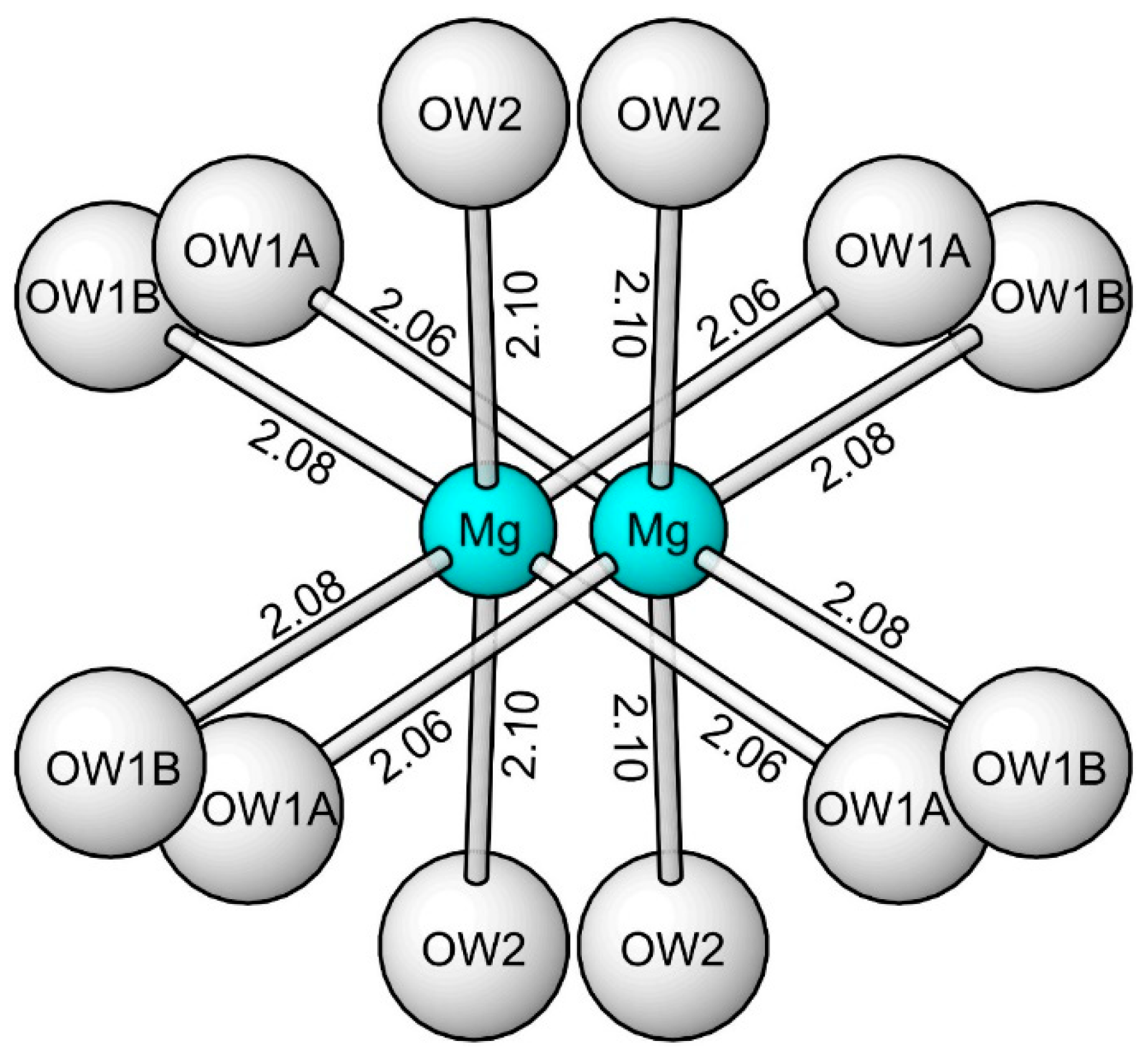
14] was used for the refinement of the structure. The octahedrally coordinated Mg site was initially located on the special position 0,0,0; however, anisotropic refinement of the site indicated it to be split along with its coordinated O sites (OW1 and OW2) (
Figure 4). The EMPA-determined cation content Mg
0.71Fe
2+0.09Co
0.05Ni
0.04□
0.11 was assigned to the split Mg site. Although the site-scattering value calculated for this occupancy (53.3) was significantly lower than the refined site-scattering value for the site (61.9), the resultant
R1 was almost identical for the two refinements (0.0396 vs. 0.0395). Difference Fourier syntheses failed to locate possible H atom positions. Data collection and refinement details are given in
Table 3, atom coordinates and displacement parameters in
Table 4, selected bond distances in
Table 5 and a bond valence analysis [
15,
16] in
Table 6.
7. Description of Crystal Structure
The U sites in the structure of chenowethite are surrounded by seven O atoms forming a squat UO
7 pentagonal bipyramid. This is the most typical coordination for U
6+, particularly in uranyl sulfates, where the two short apical bonds of the bipyramid constitute the UO
2 uranyl group. In the structure of chenowethite, pairs of pentagonal bipyramids share a common edge, forming dimers. The dimers are linked by sharing corners with SO
4 groups, yielding a [(UO
2)
2(SO
4)
2(OH)
2]
2− sheet parallel to {010} (
Figure 5). This sheet is based on the phosphuranylite anion topology [
17], with ring symbol 6
15
24
23
2 [
18]. The sheets in deliensite, Fe(H
2O)
5[(UO
2)
2(SO
4)
2(OH)
2]·2H
2O [
19]; feynmanite, Na
2(H
2O)
7[(UO
2)
2(SO
4)
2(OH)
2] [
20]; greenlizardite, (NH
4)Na(H
2O)
3[(UO
2)
2(SO
4)
2(OH)
2]·H
2O [
21]; johannite Cu(H
2O)
4[(UO
2)
2(OH)
2(SO
4)
2]·4H
2O [
22]; meitnerite, (NH
4)
2(H
2O)
4[(UO
2)
2(SO
4)
2(OH)
2] [
23]; and plášilite, Na
2(H
2O)
4[(UO
2)
2(OH)
2(SO
4)
2] [
4], are topologically identical to those in chenowethite. However, these sheets are geometrical isomers, differing in the orientation of the SO
4 groups. The sheets in chenowethite and deliensite are the same geometrical isomer (ddudd/uuduu), differing in relatively minor canting of the tetrahedra.
The interlayer region in the chenowethite structure contains a disordered Mg(H2O)6 octahedron and three isolated H2O groups (OW3, OW4 and OW5). The OW5 site is another split site. The small amount of N noted in the EPMA is presumed to represent NH4+, which is accommodated in one or more of the isolated H2O sites. This NH4+ serves to compensate the charge deficiency resulting from the partial vacancy in the Mg site noted above. Note that NH4+ is a relatively common constituent in the secondary phases of the uranium mines in Red Canyon and is an essential constituent in ammoniozippeite, which is intimately associated with chenowethite at the Blue Lizard mine.
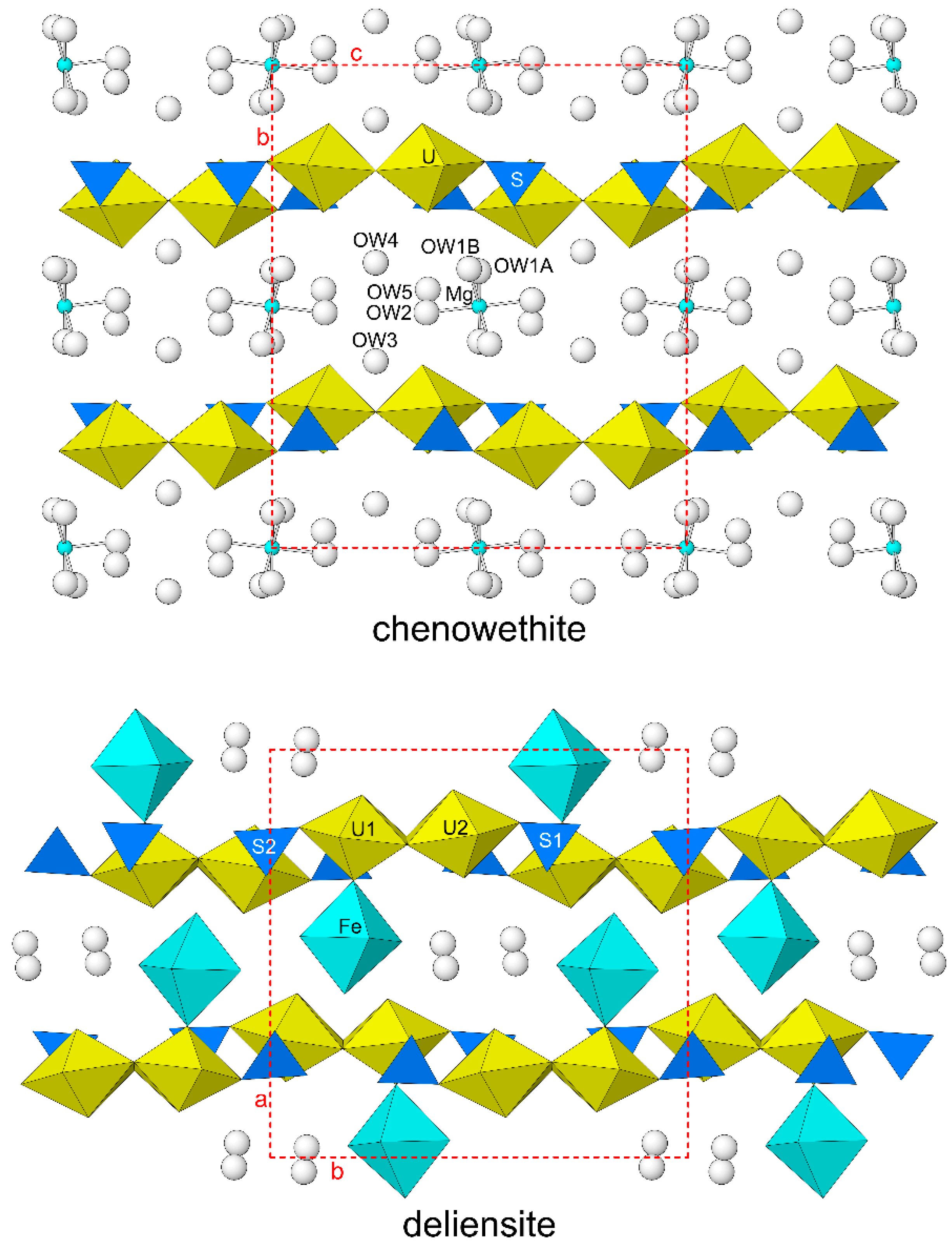
All of the structures with [(UO
2)
2(SO
4)
2(OH)
2]
2– sheets noted contain cations in their interlayer regions. In the structures of feynmanite, greenlizardite, johannite, meitnerite and plášilite, the interlayer cations serve to link [(UO
2)
2(SO
4)
2(OH)
2]
2− sheets to each other via cation–oxygen bonds. In the structure of deliensite, the interlayer Fe-centered octahedron shares one O corner with the apical vertex of a UO
7 pentagonal bipyramid in only one sheet, such that adjacent sheets are only linked via hydrogen bonds. In the structure of chenowethite, the interlayer Mg(H
2O)
6 octahedron only links to the [(UO
2)
2(SO
4)
2(OH)
2]
2− sheets via hydrogen bonds. The structures of chenowethite and deliensite are compared in
Figure 6.
8. Discussion
Our observations of uranyl sulfates in both the field and laboratory have shown them to occur as complex admixtures of species, often so visually, chemically, or spectroscopically similar to one another that identification is difficult except through a combination of methods. The high structural and chemical diversity of uranyl sulfates, which is attributed to minute variabilities in formation conditions, has greatly advanced our understanding of uranyl-sulfate mineralogy. In general, solution conditions such as ionic strength, pH, and evaporation rate, which depends greatly on local relative humidity, are dominant contributors to the diversity of species in Red Canyon.
The same conditions that drive uranyl-sulfate mineral formation also dictate anthropogenic processes, where, for example, uranyl-sulfate phases crystallized from leaking U-bearing wastes in a geological repository will be important indicators of local solution chemistry and can aid in failure analyses or diagnoses. Thus, continued research on uranyl-sulfate mineral formation in abandoned uranium mines can translate to greater control of uranium in the lab, and may eventually be used to improve industrial techniques for U-waste disposal or remediation of U contaminated sites. Most uranyl-sulfate species described from Red Canyon have yet to be reproduced synthetically; however, the high solubilities of chenowethite and others lend themselves to relatively simple evaporative synthesis techniques. Such studies could produce years of interesting results that supplement our understanding of natural and industrial processes involving uranyl sulfates.
Author Contributions
Conceptualization, A.R.K., J.P. and T.A.O.; methodology, A.R.K., J.P., T.A.O., C.M. and J.M.; investigation, A.R.K., J.P., T.A.O., C.M. and J.M.; original manuscript—draft preparation, A.R.K., J.P. and T.A.O.; manuscript—review and editing, A.R.K., J.P., T.A.O., C.M. and J.M.; figures, A.R.K. All authors have read and agreed to the published version of the manuscript.
Funding
This study was funded, in part, by the John Jago Trelawney Endowment to the Mineral Sciences Department of the Natural History Museum of Los Angeles County.
Data Availability Statement
The crystallographic information file (CIF) for this paper (deposition number 2226329) are provided free of charge by the joint Cambridge Crystallographic Data Centre and Fachinformationszentrum Karlsruhe Access Structures service
www.ccdc.cam.ac.uk/structures (accessed on 5 December 2022).
Conflicts of Interest
The authors declare no conflict of interest.
References
- Mindat.org. Available online: https://www.mindat.org/loc-37459.html (accessed on 11 November 2022).
- Kampf, A.R.; Olds, T.A.; Plášil, J.; Nash, B.P.; Marty, J. Nitscheite, (NH4)2[(UO2)2(SO4)3(H2O)2]·3H2O, a new mineral with an unusual uranyl-sulfate sheet. Am. Mineral. 2022, 107, 1174–1180. [Google Scholar] [CrossRef]
- Chenoweth, W.L. The Geology and Production History of the Uranium Deposits in the White Canyon Mining District, San Juan County, Utah; Utah Geological Survey Miscellaneous: SaltLake City, UT, USA, 1993; Volume 93-3. [Google Scholar]
- Kampf, A.R.; Kasatkin, A.V.; Čejka, J.; Marty, J. Plášilite, Na(UO2)(SO4)(OH)·2H2O, a new uranyl sulfate mineral from the Blue Lizard mine, San Juan County, Utah, USA. J. Geosci. 2015, 60, 1–10. [Google Scholar] [CrossRef]
- Finch, R.J.; Murakami, T. Systematics and paragenesis of uranium minerals. In Uranium: Mineralogy, Geochemistry and the Environment; Burns, P.C., Ewing, R.C., Eds.; Mineralogical Society of America and Geochemical Society: Chantilly, VA, USA, 1999; Volume 38, pp. 91–179. [Google Scholar]
- Krivovichev, S.V.; Plášil, J. Mineralogy and Crystallography of Uranium. In Uranium: From Cradle to Grave; Burns, P.C., Sigmon, G.E., Eds.; Mineralogical Association of Canada Short Courses: Québec City, QC, Canada, 2013; Volume 43, pp. 15–119. [Google Scholar]
- Plášil, J. Oxidation–hydration weathering of uraninite: The current state-of-knowledge. J. Geosci. 2014, 59, 99–114. [Google Scholar] [CrossRef]
- Mandarino, J.A. The Gladstone–Dale compatibility of minerals and its use in selecting mineral species for further study. Can. Miner. 2007, 45, 1307–1324. [Google Scholar] [CrossRef]
- Mandarino, J.A. The Gladstone-Dale relationship—Part 1: Derivation of new constants. Can. Miner. 1976, 14, 498–502. [Google Scholar]
- Libowitzky, E. Correlation of O–H stretching frequencies and O-H⋅⋅⋅O hydrogen bond lengths in minerals. Monat. Chem. 1999, 130, 1047–1059. [Google Scholar] [CrossRef]
- Bartlett, J.R.; Cooney, R.P. On the determination of uranium-oxygen bond lengths in dioxouranium(VI) compounds by Raman spectroscopy. J. Mol. Struct. 1989, 193, 295–300. [Google Scholar] [CrossRef]
- Higashi, T. ABSCOR; Rigaku Corporation: Tokyo, Japan, 2001. [Google Scholar]
- Sheldrick, G.M. SHELXT—Integrated space-group and crystal-structure determination. Acta Cryst. 2015, A71, 3–8. [Google Scholar] [CrossRef] [PubMed]
- Sheldrick, G.M. Crystal Structure refinement with SHELX. Acta Cryst. 2015, C71, 3–8. [Google Scholar]
- Gagné, O.C.; Hawthorne, F. C Comprehensive derivation of bond-valence parameters for ion pairs involving oxygen. Acta Crystallogr. 2015, B71, 562–578. [Google Scholar] [CrossRef] [PubMed]
- Ferraris, G.; Ivaldi, G. Bond valence vs. bond length in O…O hydrogen bonds. Acta Cryst. 1988, B44, 341–344. [Google Scholar] [CrossRef]
- Burns, P.C. U6+ minerals and inorganic compounds: Insights into an expanded structural hierarchy of crystal structures. Can. Mineral. 2005, 43, 1839–1894. [Google Scholar] [CrossRef]
- Krivovichev, S.V.; Burns, P.C. Actinide compounds containing hexavalent cations of the VI group elements (S, Se, Mo, Cr, W). In Structural Chemistry of Inorganic Actinide Compounds; Krivovichev, S.V., Burns, P.C., Tananaev, I.G., Eds.; Elsevier: Amsterdam, The Netherlands, 2007; pp. 95–182. [Google Scholar]
- Plášil, J.; Hauser, J.; Petříček, V.; Meisser, N.; Mills, S.J.; Škoda, R.; Fejfarová, K.; Čejka, J.; Sejkora, J.; Hloušek, J.; et al. Crystal structure and formula revision of deliensite, Fe[(UO2)2(SO4)2(OH)2](H2O)7. Mineral. Mag. 2012, 76, 2837–2860. [Google Scholar] [CrossRef]
- Kampf, A.R.; Olds, T.A.; Plášil, J.; Marty, J.; Perry, S.N. Feynmanite, a new sodium-uranyl-sulfate mineral from Red Canyon, San Juan County, Utah, USA. Mineral. Mag. 2019, 83, 153–160. [Google Scholar] [CrossRef]
- Kampf, A.R.; Plášil, J.; Nash, B.P.; Marty, J. Greenlizardite, (NH4)Na(UO2)2(SO4)2(OH)2·4H2O, a new mineral with phosphuranylite-type uranyl sulfate sheets from Red Canyon, San Juan County, Utah, USA. Mineral. Mag. 2018, 82, 401–411. [Google Scholar] [CrossRef]
- Mereiter, K. Die kristallstruktur des johannits, Cu(UO2)2(OH)2(SO4)2·8H2O. Tschermaks Mineral. Petrogr. Mitt. 1982, 30, 47–57. [Google Scholar] [CrossRef]
- Kampf, A.R.; Plášil, J.; Nash, B.P.; Marty, J. Meitnerite, (NH4)(UO2)(SO4)(OH)·2H2O, a new uranyl-sulfate mineral with a sheet structure. Eur. J. Mineral. 2018, 30, 999–1006. [Google Scholar] [CrossRef]
| Publisher’s Note: MDPI stays neutral with regard to jurisdictional claims in published maps and institutional affiliations. |
© 2022 by the authors. Licensee MDPI, Basel, Switzerland. This article is an open access article distributed under the terms and conditions of the Creative Commons Attribution (CC BY) license (https://creativecommons.org/licenses/by/4.0/).


