1. Introduction
The structural and textural feature fabrics of vein quartz are formed as a result of dynamometamorphism (a cataclastic, dislocation metamorphism), and they are characterized by significant diversity. They reflect the conditions for the development of the geological history of the lithosphere block to which it belongs [
1,
2,
3]. The formation of the microstructure of mineral aggregates is influenced by the rate of strain, temperature, and the presence of fluid [
4,
5,
6,
7]. In this regard, the analysis of defective quartz structures (boundaries, sub-boundaries and twins), along with the analysis of gas–liquid inclusions, indicator impurities, and other geochemical data, provides important information regarding the mechanisms, stages, and conditions of the dynamometamorphic processes.
The rate and temperature of deformation significantly affect the mechanism of destruction. For example, in conditions of low-grade metamorphism (T below 300 °C) at high strain rates (faster than 10
−6 sec
−1) the preferred structures are the brittle destruction (crushing) of grains [
8,
9]. The result is the formation of a cataclastic structure expressed with different cracks: intragrain, transgrain (intersecting several grains), and interstitial (following along the grain boundaries and twins).
The primary mechanisms of plastic deformations of vein quartz include dislocation glide, deformation twin, dissolution under pressure, creep, diffusion, recrystallization, and intergranular slippage [
10]. These determine the mechanisms of active plastic deformation or creep deformation. Each of these processes is characterized by certain characteristics in the defective subsystem. This can be a change in the dislocation structure, the formation of deformed twin or sub-boundaries of varying degrees of perfection. If the dislocation structure is available for study by transmission electron microscopy, the latter types of defects successfully are analyzed by electron back scatter diffraction (EBSD) on scanning electron microscope (SEM). This makes it possible to determine the angle of misorientation of the boundaries and other crystallographic characteristics. Thus, by analyzing the structural elements with misdirections, it is possible to obtain information regarding the degree and temperature of the plastic deformation processes.
For example, at low strain rates (slower than 10–11 s
−1) and low temperatures, the structure of dissolution under pressure and redeposition structure are the most characteristic [
11,
12]. The dissolution under pressure for quartz was noted in wide interval temperatures before 500 °C. However, this mechanism plays a leading role in the temperature range of 200–300 °C [
13]. Subsequently, as the temperature increases, new grains may be formed that are oriented orthogonally with respect to the maximum compression axis [
14].
At high temperatures close to the melting point (more than 500 °C) and low strain rates, the main mechanism is diffusion creep. This occurs when there is a directed diffusion transfer of atoms through the crystal lattice. The flow of diffusion to and from the boundary depends on whether the tension or compression prevails in the given area [
15]. If the transfer of a substance occurs by volume diffusion, this process is called Nabarro–Herring creep. If it occurs due to diffusion along the grain boundaries, this process is called Coble creep [
16]. This determines the creep mechanism.
Diffusion creep structures are difficult to diagnose. However, the implementation of one mechanism or the other leads to different effects of changing the texture and dislocation structure; thus, this can be diagnosed when conducting textural and structural analyses. In contrast to dislocation creep, which we will describe below, the crystallographic axes and planes of crystals become less ordered during diffusion [
17,
18]. In addition, fine-grained aggregates (recrystallization nuclei) can be an important indicator of diffusion creep [
17].
In low-temperature conditions at strain rates (10−11–10−6 s−1), deformation in quartz grain develops by the mechanism of dislocation creep. In this case, dislocation sliding occurs, and there is a “wavy” extinction of the grains. There is the formation of deformation bands (lamellae) and the mass development of subgrains, when the temperature rises to 400 °C.
With the leading role of dislocation glide, the processes of accumulation of dislocations and their annihilation (return) occur simultaneously. The resulting dislocation structures can be characterized by a uniform distribution of dislocations, and the tendency to form dislocation clusters (tangles and cell walls) or the formation of low-angle boundaries that determine the blockiness of the grain, or even the formation of a fragmentary structure with high-angle grain boundaries. In the latter case, the grain can be divided into fragments, and those fragments into blocks in order to form a block structure. The formation of these types of substructures is typical for all crystalline materials in which the deformation is performed by sliding dislocations [
16,
19].
Under the conditions of constant applied stresses, dislocation creep leads to dynamic recrystallization, which is characterized by the process of enlarging the original grains and the appearance of new undeformed grains [
16,
20,
21]. The size of recrystallized grains depends directly on the temperature and applied stresses [
22,
23]. Based on this, three mechanisms of dynamic recrystallization have been identified, successively replacing each other [
21,
22,
24,
25]. For each mechanism, researchers observed different types of grain boundary morphology, which clearly differ in temperature conditions and accordingly, in the mechanism of grain boundary migration. In addition, they may depend on the internal (impurity) structure within the grain.
Urai [
26] described recrystallization, which is accompanied by the migration of the curved grain boundary (BLG—bulging recrystallization) as a “slow” migration of grain boundaries at low temperatures. According to Stipe’s research in natural conditions, this process is characterized by temperatures of 280–400 °C [
27]. With this recrystallization mechanism, Blenkinsop [
28] defined the fine-toothed boundaries of the original grains as “sutures”, noting the formation of small recrystallization grains from individual teeth. When the recrystallization mechanism is associated with the rotation of subgrains (SGR—subgrain rotation recrystallization), the material incompatibility compensation along their boundaries occurs by dislocation sliding. At the same time, the misorientation angle between neighboring subgrains increases, and when the misorientation angle reaches 10°, they are classified as large angle [
29,
30], noting the recrystallization process. “Fast” [
26] grain boundary migration (GBM—grain boundary migration recrystallization) contributes to the formation of large grains that may have a sinuous shape or a more complex boundary configuration. This creates complex “stitched” boundaries [
27]. During the described mechanisms of dynamic recrystallization, mosaic structures are formed [
22].
The temperature regimes of the described mechanisms of dynamic recrystallization mainly depend on the extrapolation of strain rates [
7], and as a result, the grain size is an important indicator for determining the recrystallization temperature [
22,
31]. The described mechanisms correlate with the modes of experimental dislocation creep, which successively change each other when the temperature increases and the strain rate and the differential stresses decrease [
21,
24]. An important parameter that affects the temperature and strain rate conditions is the presence of water [
24].
In addition to all of the above, mechanical Dauphiné twinning occurs in quartz under applied stresses characteristic of tectonic deformations. This effect is associated to the host by a 180° rotation (or 60° considering the trigonal symmetry) around the
C axis [0001]. This reduces the strain energy, reduces the crystal’s stiffness, and increases its flexibility to deformations, but does not change the orientation of the
C axis [
32,
33]. Researchers interpret the double Dauphiné boundaries as boundaries between the grains of two right and two left quartz crystals. This is characterized by the presence of the same
C axis, which is the boundary of the slope. This is expressed in the same Euler angles φ1 and Φ and a difference in the Euler angles φ2 of 60° [
34]. According to Manegon [
35], the process of boundary twinning can develop both synkinematically to the main deformation and after the recrystallization of quartz grains. In the first case, within the initially homogeneous crystal, there is a separation and localization of deformation between the twins. In this case, the low-angle boundaries that occur in different doubles never intersect. In the second case, the twinning after recrystallization is explained by the absence of recrystallized grains at the boundaries of the twins or exclusively inside individual twin crystals [
36].
Thus, the considered quartz substructures characterize various deformation mechanisms that are realized under different conditions. They are thermodynamic indicators. In this regard, the aim of this work is to systematize the morphological and crystallographic microstructures of vein quartz in the Bambukoy river basin for their subsequent connection with the mechanisms and conditions of formation in the Earth’s crust.
2. Geological Setting and Samples
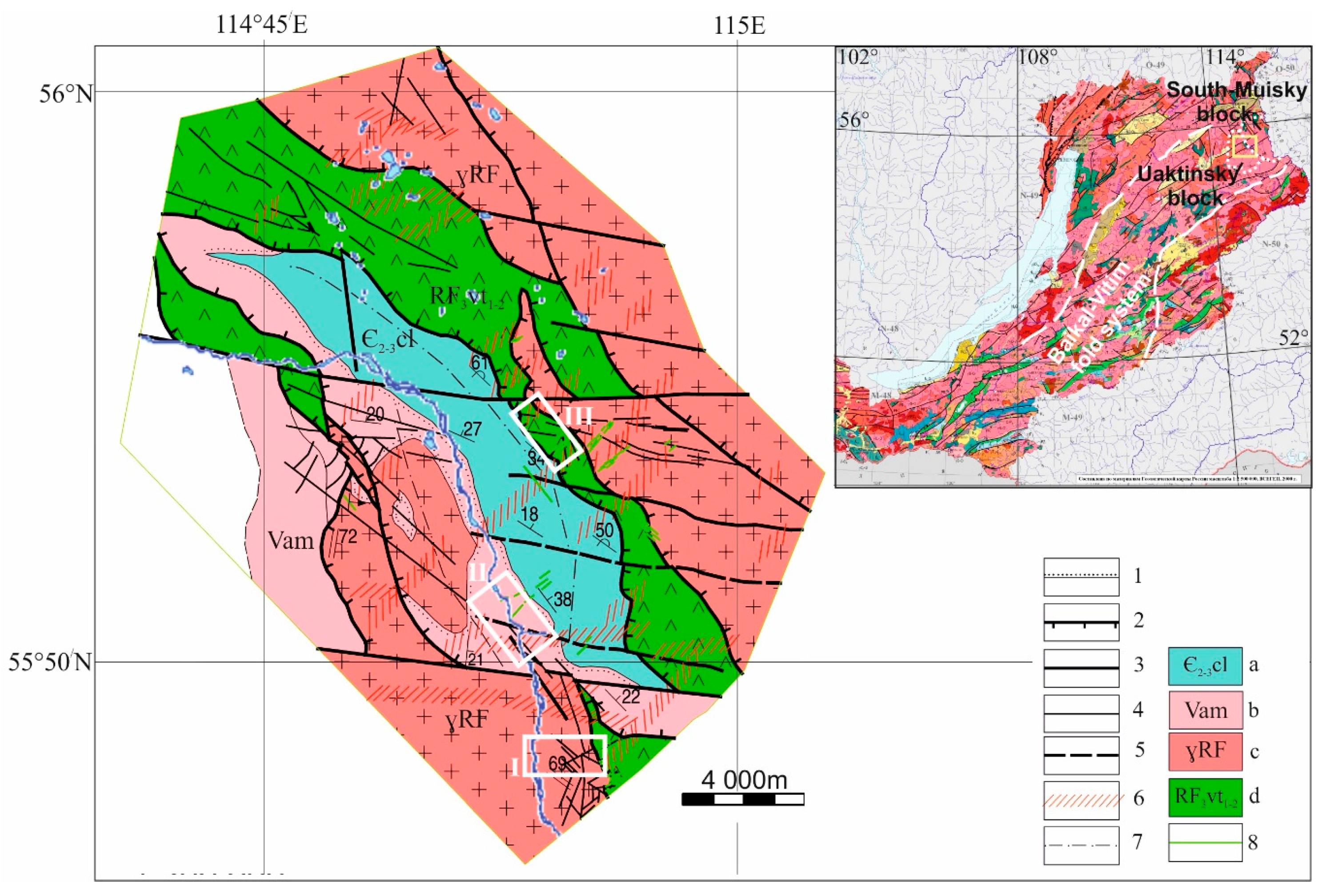
The work area (
Figure 1) is located in the Bambukoy river basin within the Muya administrative region of the Northern Buryatia at a distance of 11 km from the village of Taksimo (Western Transbaikalia, Russia).
The folded structures of the Western Transbaikalia, which are united into the Baikal folded region, occupy the territory of the southern frame of the Siberian platform. They are considered as part of the Central Asian belt, which includes structural and formation zones (terrains) of the Baikal, Caledonian, and Varis–Hercynian age. The object under study is located within the Baikal–Vitim folded system and is confined to the border of the Baikal–Muiskoy (South Muisky block) and Barguzino–Vitimskoy (Uaktinsky block) structural formation zones. According to Gordienko [
37], this place also marks the border of two terranes of the southeastern folded frame of the Siberian craton: the middle-upper-fainian Bambuisky and upper-fainian Kelyansky. The late-Riphean island-arc Baikal–Muiskaya zone and the Vend-early Paleozoic Barguzino–Vitimskaya zone of marginal basins represent geodynamic settings.
Using the results of structural tectonic mapping performed by the authors, in the construction of the study area, we marked terrigenous structural–material complexes of Amatkanskoy (Vam) and Chulemginsky (Є2-3cl) Formation, and also Verkhnetuluinsky subvolcanic (RF3vt1-2) and granitoid-undifferentiated (ɣRF) complexes. Terrigenous deposits of the Amatkanskoy suite form a section of cyclically alternating and interleaved aleurolites and sandstones of various grain sizes (from fine-grained to coarse-grained), intensively dislocated into oblique, recumbent, diving and ridge folds. At the base of the horizon section, rocks are located that are defined as the lithified products of the ancient weathering crusts of the kaolinite profile. The section of the chulegminsky suite represented by a rather monotonous stratum of greenish gray calcareous aleurolites, silt sands, and aleuro sandstone, often in alternation with fine-grained brownish carbonate sandstone. Not rarely, under the influence of tectonics in the zones of schismatization, fillites formed.
The group of metamorphosed subvolcanic and dyke rocks of a rather variegated composition, closely related to each other and spatially confined to a single structural zone, is assigned to the Verkhnetuluinsky complex. The complex includes volcanic rocks, mainly of acidic and medium composition, with a small amount of volcanic rocks of the basites composition. Along the South to North stretch, the volcanogenic section is replaced by a volcanic and terrigenous section where tuffs of basic and acidic composition predominate. The composition of the undifferentiated granitoid complex includes three igneous formations, according to age characteristics, occupying a middle position between the Chulegminsky and Verkhnetuluinsky complexes. Rocks of these complexes are intensively subject to metasomatic changes, expressed mainly by sericitization, carbonatization, and albitization.
The tectonic development of the region determined by the formation of the thrust system of the northwestern direction, which involves all the selected structural–material complexes. The thrust system is complicated by the left-lateral strike slip of the West–North–West direction. The left-lateral strike slip subdivides the thrust system into separate segments. The final stage of deformations is marked by steeply dipping disjunctives of the northeastern direction.
In the course of field work within the study area, the authors investigated three complex systems of quartz veins and veinlets. Their locations are marked by white rectangles on the structural and tectonic diagram of the work area (
Figure 1). The internal structure of the vein systems was studied in bedrock outcrops and ditch webs. As a result, the kinematic analysis of the tectonic nature of the cracks filled with vein material was performed. Based on the nature of the cracks that filled the fluid among the veins, we identified the main and echelon quartz veins. Among the echelon veins, we considered the syndeformational and submain veins. Syndeformational cores perform the gashes formed during stretching. Submain veins were considered by the authors as the former main ones that performed cracks in the stretching systems formed during the formation or rotation of the displacers. The structure and analysis of each system will be presented below.
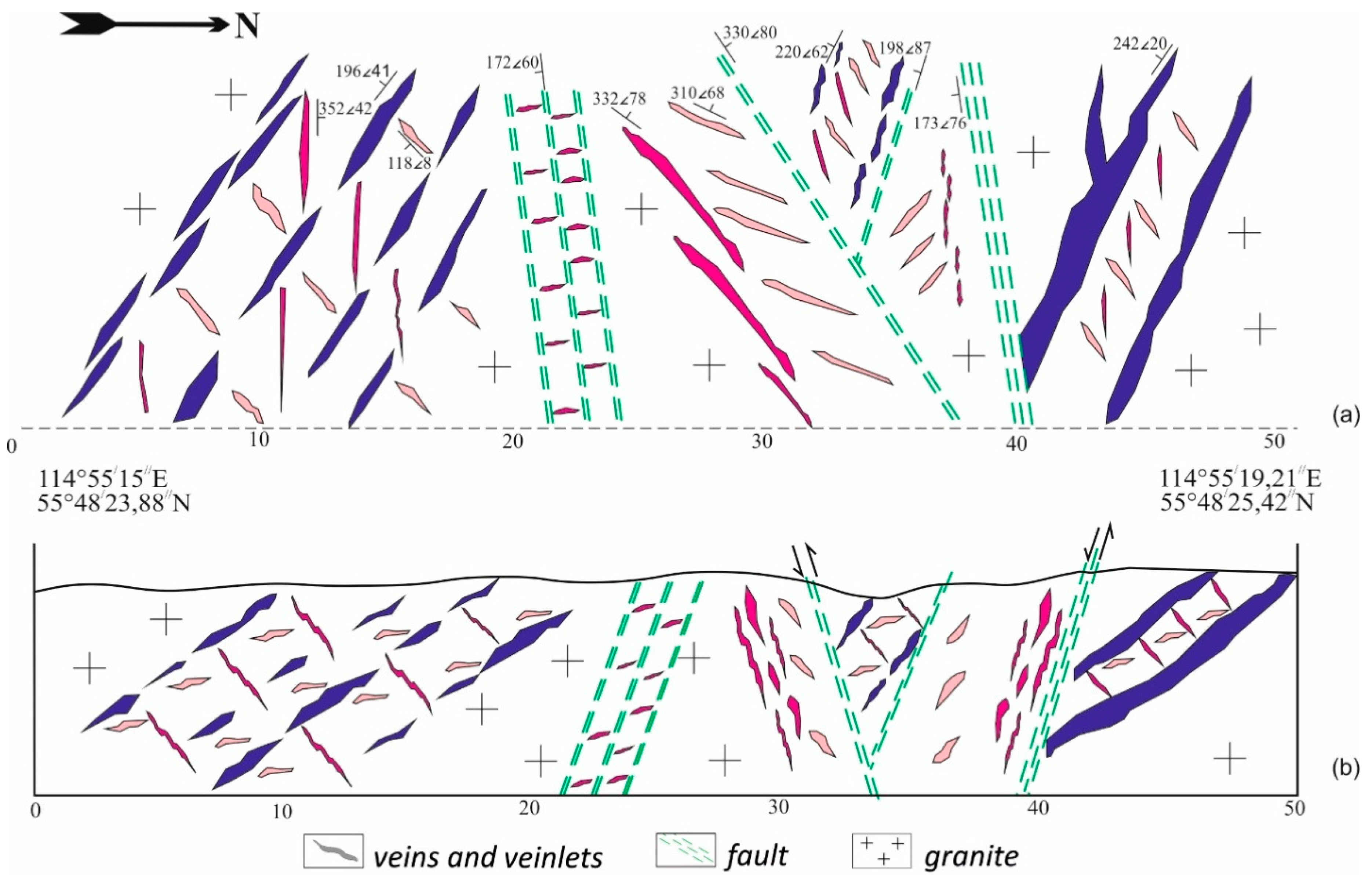
The Kryvoy vein system is located within the granite rocks. Subsequently, the structure of this zone became more complicated by sinistral shear deformations of latitudinal strike. In the structure of this system (
Figure 2) the main veins of southwestern dipping take the basis positions (shown in blue in
Figure 2). With these main veins are associated small flat dipping veins in the northeastern direction, limited in strike (shown in peach in the
Figure 2). The third type of vein bodies are represented by steeply and moderately dipping to the southeastern and northwestern veins up to 10 cm thick (shown in pink in the
Figure 2).
The structure of the vein system of Kryvoy is complicated by the manifestations of brittle deformations recorded by local zones of northeastern, sub-latitudinal and northwestern strike (
Figure 2). At the same time, a disjunctive of the northeastern direction can be associated with zones of stretching and/or with left-lateral shear deformations. For the fault of the northwestern strike, it is assumed that the discharge kinematics of normal fault deformations of the rear extension of the thrust system. Disjunctive sub-latitudinal directions are considered as left-lateral shear deformations complicating the southern flanks of the thrust faults.
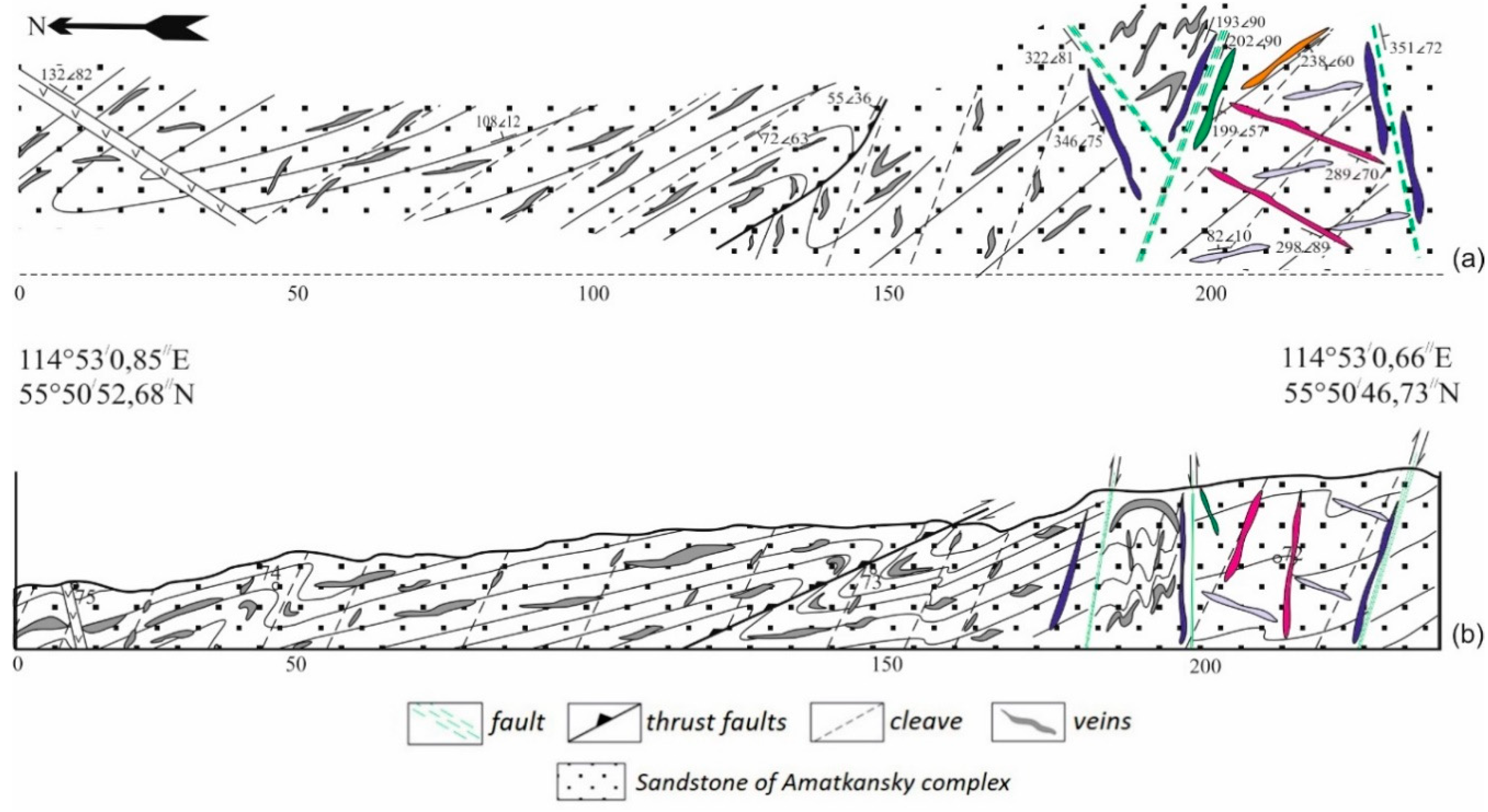
The Beregovoy vein system (
Figure 3) is located among Amatkanskoy suite sandstones (Vam). There is the sub-concordant lamination and cleavage of metamorphized and cross veins. The veins of the first type, shown in gray in
Figure 3, have a ribbon morphology with an irregularly varying thickness from a few centimeters to 0.5 m in the veins and a rather poor sulfide mineralization. The structural pattern of cross veins is determined by the development of the Riedel shear paragenesis, which connects the main steeply falling veins that correspond to the shear surfaces (shown in blue in the
Figure 3) and the veins that perform echelon cracks. Among the feathering, steeply falling veins oriented at an angle of 60° to the shear surface (shown in pink in the
Figure 3) perform syn-shear stretching systems. The sub-latitudinal veinlets (shown in orange and lilac) are controlled by synthetic and antithetic shears, respectively. The submain vein is the one highlighted in green in the drawing.
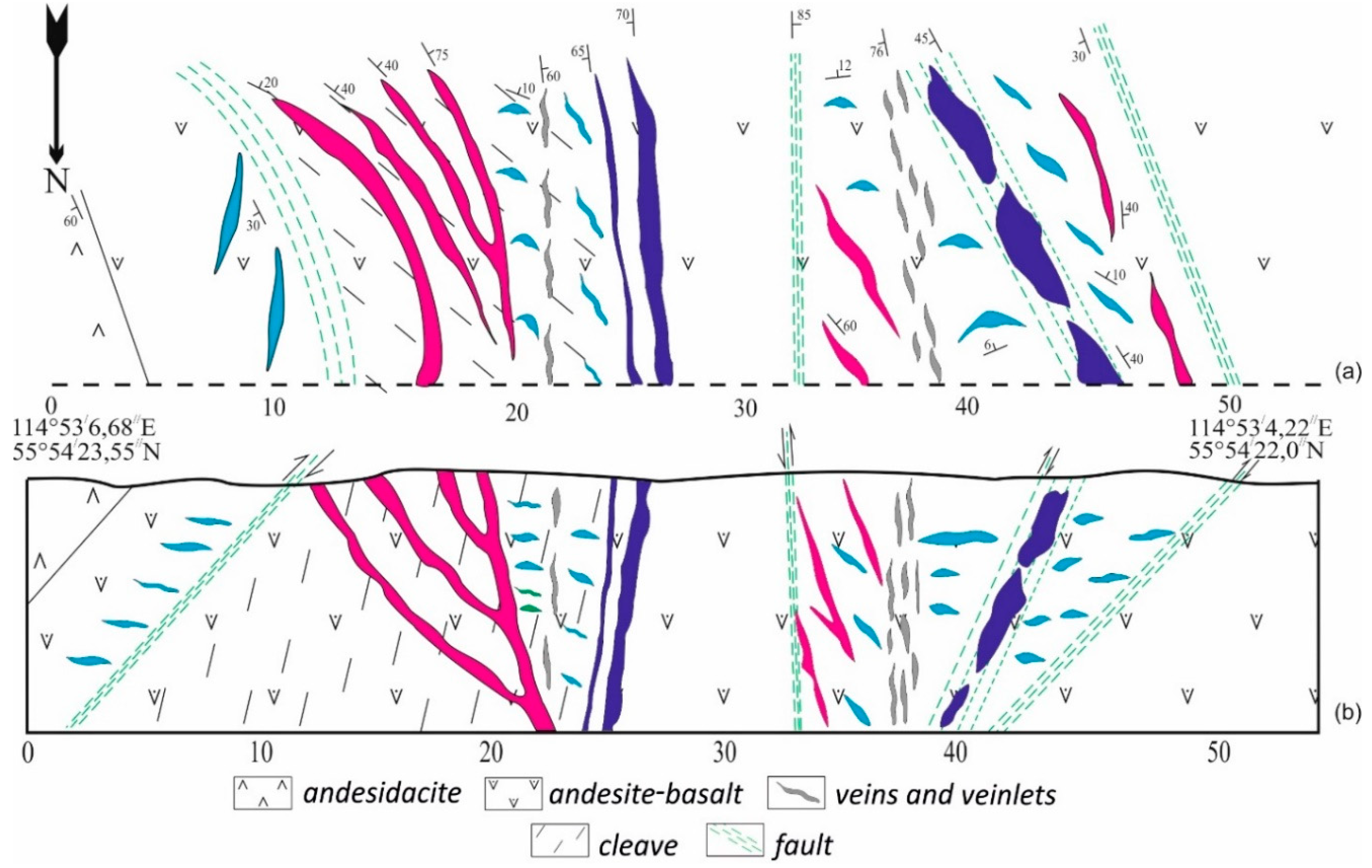
The Otvesny vein system is observed in the subvolcanic rocks of the Verkhnetuluinsk complex (RF
3vt
1-2). Quartz veins are controlled by the structure of the flank part of the trust system including the reverse and normal faults. In the structure (
Figure 4), there are several series with a characteristic orientation of veins and veinlets. Each series is determined by the development of three zones.
The first zone or footwall zone is characterized by the development of moderately and steeply dipping large main veins of the northwestern strike. The thickness of these veins varies from 0.5 to 2 m, and they are often laminated and represented by a series of lenses (shown in blue in
Figure 4). The hanging zone of vein formations is characterized by bodies with a western fall (shown in pink in
Figure 4). At the same time, a decrease in the angle of their incidence is set from West to East. The thickness of such veins varies from a few centimeters to 0.5–0.7 m. A subordinate value is the gently falling veins (shown in light blue in the drawing) that feathered the main veins. The axial zone of vein formations is represented by a thin-veined series with a thickness of up to 5 m, a complex system of converging veins with a thickness of up to 5 cm (shown in gray in the
Figure 4). As a rule, these formations are characterized by a meridional or North–North–West orientation and are associated with low lying northwest veinlets.
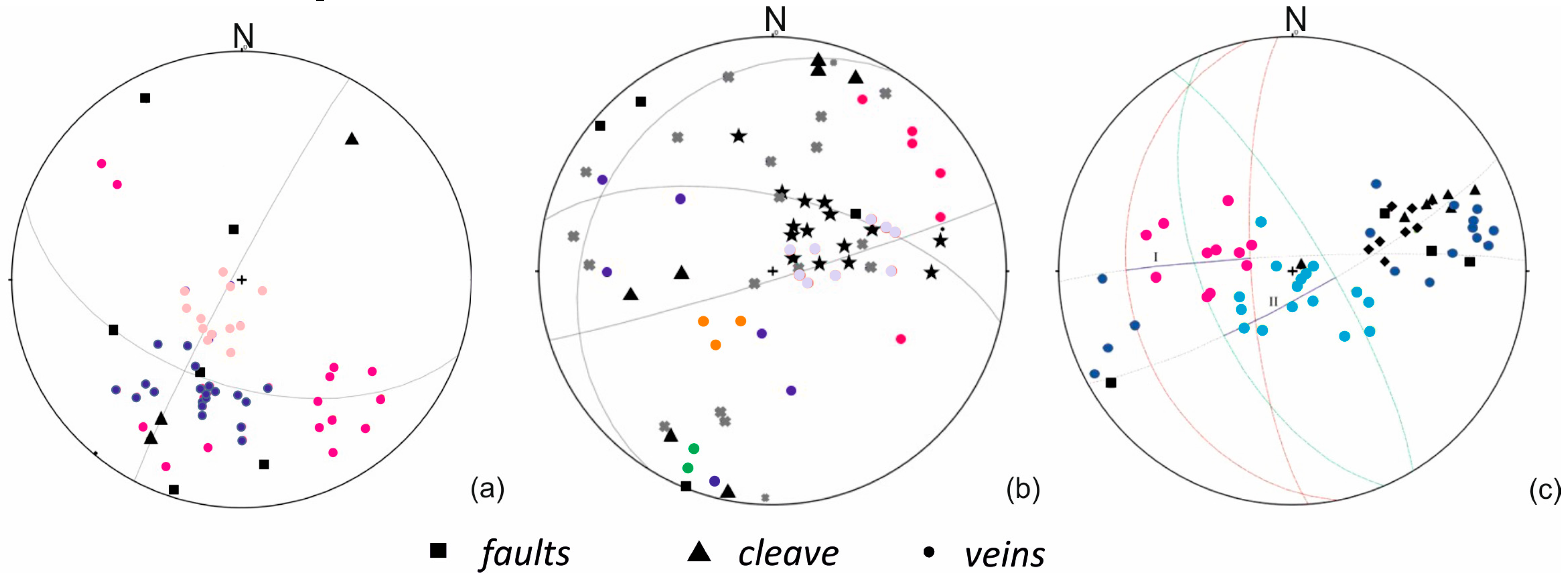
The analysis of the spatial orientation of the elements of the deformation structure (fault zones, shale and cleavage) veins, and veinlets allowed us to conduct a kinematic reconstruction of the formation of vein systems (
Figure 5). The development of quartz veins in the Krivoy system (
Figure 5a) is controlled by the rear zones of thrust deformations and is complicated by sinistral shear deformations. The main veins of the southwestern fall (blue color) are associated with the development of discharge deformations during the formation of the thrust system. The gently sloping veins (peach color) and the veins that fall steeply to the southeast (pink color) are associated with the process of stretching during the development of two generations of separation cracks during the formation of flank upwash shear deformations.
The formation of sub-consistent veins of the Beregovoy system (
Figure 5b) is associated with the initial stages of the formation of the thrust system and is controlled by the stretching vector during the formation of upwash deformations. On a stereographic projection showing the structural position of elements, veins, and veinlets, one can observe two trends of folded variations in layering, according to which sub-consistent veins develop. Cross veins are derived from left-shear deformations (Central shear) and deformations of the northwestern direction, which complicate the thrust system.
The control of quartz veins in the Otvesny zone (
Figure 5c) is carried out by disjunctives of the flank zone of the thrust system: upwash-thrusts of the northwestern strike and meridionally steeply normal faults. In this case, the veins of the hanging wall (pink) perform tensile cracks formed during the development of normal faults (stretch sector II).
The formation of large budinated veins of the recumbent wing is associated with the implementation of intensely dislocated nodes and cracks in the vein material in the conditions of stress inversion as a result of periodic activation of upwash and downwash type movements. The stretching vector determines the development of gently falling veins (blue color) during the formation of upwash faults (stretch sector I).
Samples of vein quartz performing the crack paragenesis of all three systems of deformations typical for the studied region were selected. After we examined all the samples using optical microphotography, five extreme types of quartz vein microstructures were identified and studied in more detail.
3. Materials and Methods
The research material was the vein quartz of the suture zone of the Baikal–Muyskoy and Barguzino–Vitimskoy structural–formation zones of the Baikal–Vitim folded system of Western Transbaikalia (Russia). Samples of vein quartz were selected by the authors during fieldwork in the period 2017–2018. Sampling was performed in three areas marked with white rectangles in
Figure 1. We selected at least three samples of main, submain, and syndeformational quartz from each section, and 30 of the samples were studied. Vein quartz was selected both from the core of wells and from veins that came to the surface. After studying all samples using the structural and petrographic description of each sample, five extreme types of veins that outcrop quartz microstructures were identified, and were studied in more detail using the EBSD method.
The kinematic nature of quartz veins is determined by the structural position of localization cracks and the identification of characteristic fracture parageneses. Among the tectonic structures available to direct observation, special attention was paid to the analysis of splitting and fracturing zones. The splitting zones are understood as sustained in the thickness and extent of the schistosity and intensely fractured rocks with a characteristic fracture paragenesis. Among isolated cracks, for which, under field conditions it difficult to determine paragenic structures, three genetic types distinguished interlayer cracks, the systematic tear of the “joints” type and cleavage cracks. To visualize the spatial data, the authors used stereographic projections constructed in the lower hemisphere.
The laboratory studies were conducted at the Center for the Collective Use of Geochemistry of Natural Systems of Tomsk State University (Tomsk). We studied the microstructural and crystallographic features of vein quartz using an optical microscope (Leica DM750P, Leica Microsystems, Wetzlar, Germany) by reflected light and transmitted light using a scanning electron microscope (SEM) Tescan Mira 3 LMU (Tescan, Brno, Czech). The scanning electron microscope had a Nordlys F device (Oxford Instruments, Oxford, UK) for the electron backscatter diffraction method (EBSD). This allowed us to study the structure, orientation, and misorientation. The surfaces of the quartz samples were prepared by mechanical polishing and ion milling on a SEMPrep2 multifunction device (Technoorg Linda Co. Ltd., Budapest, Hungary). The carbon coating of the samples was carried out on the installed EMITECH K450X (Vitesco Technologies Emitec GmbH, Lohmar, Germany). The carbon layer was 30 nm. The survey was conducted using an accelerating voltage of 20 kV, a beam current of ~0.8 nA, a working distance of approximately 15 mm, and the tilt of the sample at 70°. EBSD images were obtained on rectangular grids by moving an electron beam with a regular step of 2 or 4 μm (at least 10 pixels per grain). The scanning area was 1 mm2. Post processing was performed with the licensed software Oxford Instruments AZtec. The EBSD analysis of the obtained data was performed with the HKL Channel 5 software (Oxford Instruments, Oxford, UK). The overall indexing for trigonal quartz was 85–95%.
For identifying Dauphiné twinning in quartz when processing EBSD data, we used boundary trace analysis. In this study, we determined the presence of Dauphiné twins by the following group of features: first, the presence of a peak of about 60° in the misorientation angle distribution, associated with rotation around the C axis; secondly, the same Euler angles φ1 and Φ and a difference in the Euler angles φ2 of 60°; thirdly, the occurrence of one C axis orientation, but six rather than three pairs of positive {r} and negative {z} rhombohedral planes, and the preferential alignment of the superposed {r} and {z} poles nearly parallel to the maximum compressive axis [
33,
34,
35].
4. Results
There were five types of quartz microstructures in the studied veins. Each type corresponded to separate morphological parameters and the degree of deformation manifested corresponded to the different conditions for the formation of mineral aggregates and preferred modes of deformation. The selected types and their characteristics are listed below:
Quartz with continuous Dauphiné boundaries;
Quartz of block structure;
Quartz with deformation bands;
Quartz of mosaic-like structure;
Quartz of medium-grained structure.
4.1. Type A. Quartz with Continuous Dauphiné Boundaries
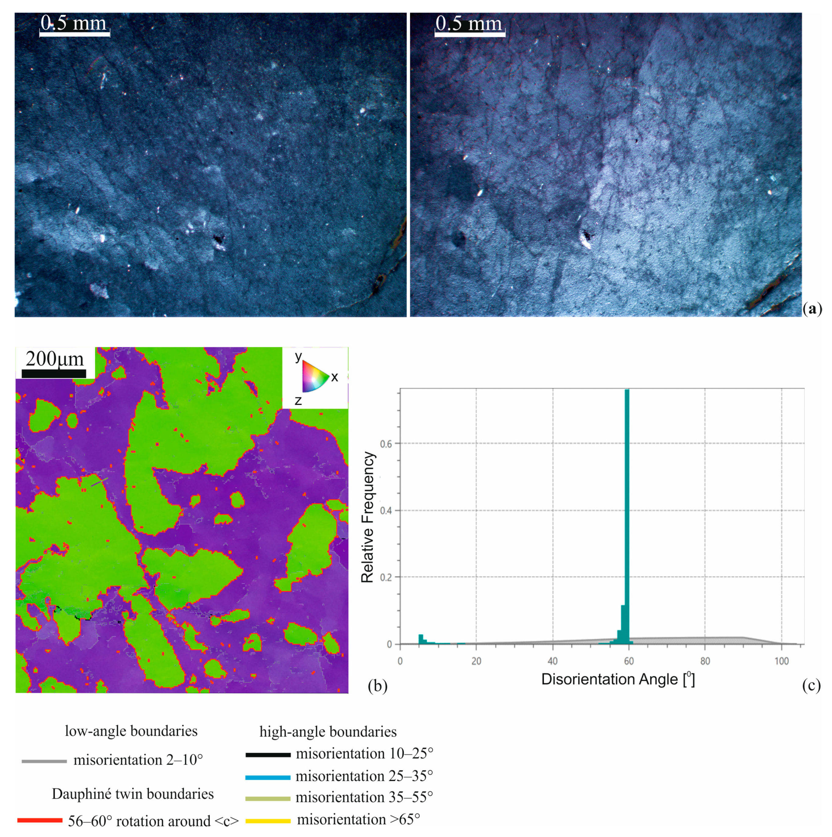
Large grains with an internal block structure characterized the microstructure of type A vein quartz. In the optical image, inside the large quartz grains, we observed small changes in contrast to when the microscope table rotated under cross-polarized light (
Figure 6a). On the EBSD map (
Figure 6b), two main orientations were highlighted, corresponding to orientations close to the <1010> corners of a standard stereographical triangle. Grains with a closed sinuous boundary (green) were located in the matrix of the parent grain (purple). They were separated from each other by a boundary with a misorientation angle of 60°. The analysis showed that the axis of rotation lay parallel to the boundary line, forming an inclined boundary of a special type. This special type of boundary in quartz is called the Dauphiné twin boundary [
35]. A type A structure is characterized by a high proportion of high-angle boundaries (90%) and a small proportion of low-angle boundaries (10%) in the grain body. The area of peaks on the histogram disorientation angle distribution is proportional to the percentage of highlighted boundaries (
Figure 6c).
4.2. Type B. Quartz of Block Structure
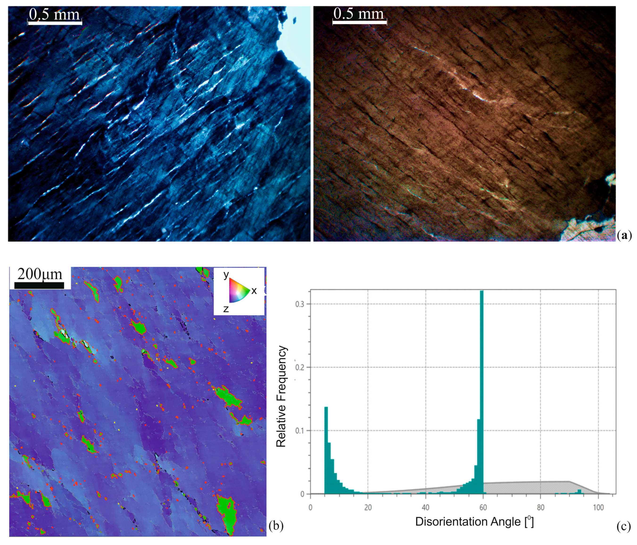
In optical micrography (
Figure 7a), the microstructure of quartz type B is characterized by a large number of low-angle boundaries in the grain body (74% of the total fraction of boundaries) that divide it into blocks. With the microscope table rotating, the subgrain extinguished any visible order. Quartz separations of less than 0.05 mm were confined to the low-angle boundaries of the blocks. The texture drawing demonstrated a trend developing in the direction of the microstructure, located at an angle of 60°. This correlates with the image of the disorientation map obtained in the EBSD analysis (
Figure 7b). On the processed EBSD map, individual blocks of green color up to 0.06 mm wide were fixed, and outlined with special sinuous, high-angle 60° boundaries of the twin type (
Figure 7c). On the histogram of misorientation angles, only two peaks presented as typical for this structure: in the low-angle region up to 15°, and in the high-angle region from 50° to 60°. The last interval ended with a high 60° peak. From this analysis, it follows that this structure is characterized by the rearrangement of low-angle boundaries into high-angle and then into the most favorable 60° special boundaries.
4.3. Type C. Quartz with Deformation Bands
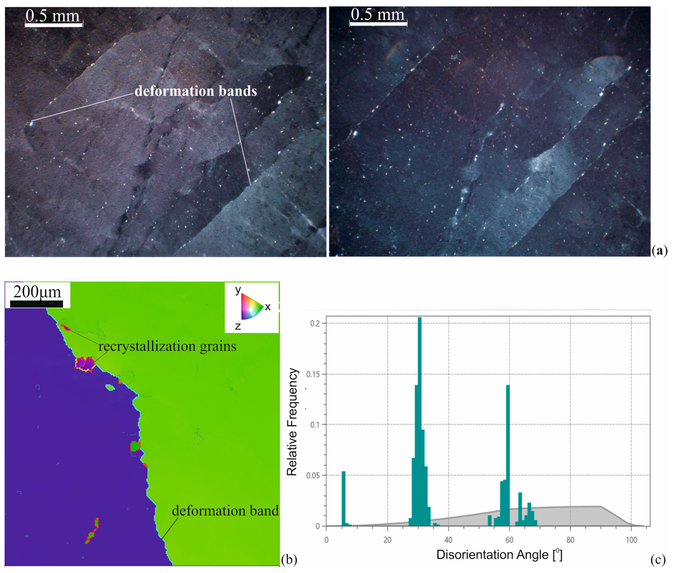
The microstructure of type C is characterized by the formation of deformation bands that divide the grain into separate elongated fragments of different widths from 0.5 to 1.5 mm, which are alternately extinguished when turning the table of the microscope (
Figure 8a). In some cases, the width of such fragments may be less than 0.5 mm. The space between the deformation bands has a small proportion of low-angle boundaries, most of which are localized near high-angle boundaries, which are close to straight-line boundaries. A structure that has mostly parallel boundaries is called a band structure. In some cases, these boundaries are closed, fixing the transition to a fragmented structure. In the presence of isometric formations, when bounded by closed deformation boundaries, we speak of a fragmented substructure.
For type C structures, we observed the appearance of 0.05 mm recrystallization nuclei (
Figure 8b) near the deformation bands. As a rule, embryos have sections of the boundary with a 60° to 10° misorientation, indicating the Dauphiné twin boundaries. The analysis of the EBSD map showed that the angle of misorientation of fragments separated by a deformation band was approximately 30°. The peak on the histogram of disorientation corresponding to an angle of 30° (
Figure 8c) was slightly larger than the peak of the Dauphiné boundary. In addition, there was a small peak in the area of low angles and a peak between 62° and 70°, which linked with the formation of high-angle boundaries of a general type for grains of recrystallization. The percentage of highlighted boundaries were low-angle—35%, 30°—41%, 60°—21%, and boundaries that lie in the range from 62° to 70°—4%.
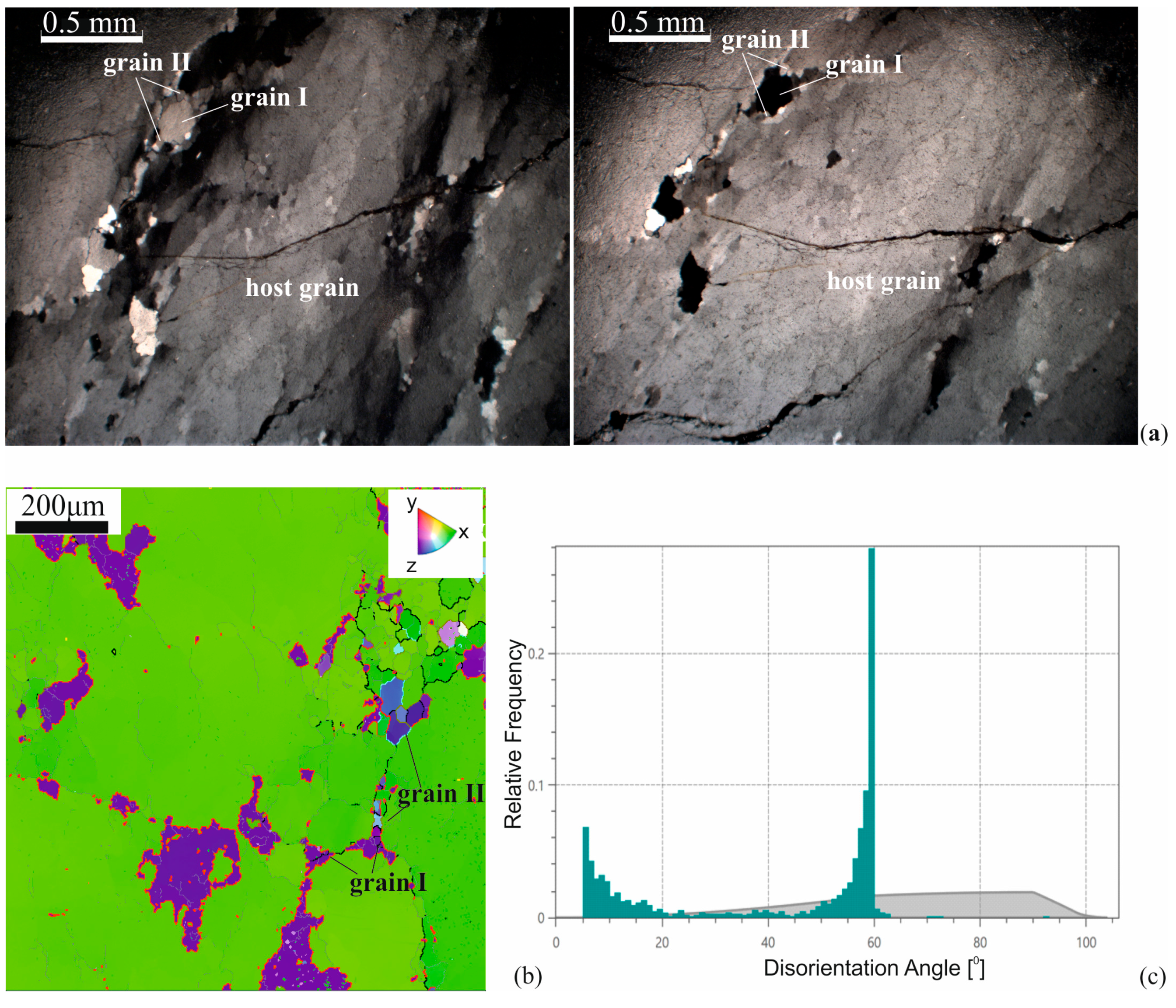
4.4. Type D. Quartz of Mosaic-Like Structure
Optical studies of quartz with a D-type mosaic microstructure demonstrated the presence of development sites for groups of recrystallized grains (
Figure 9a). There were two types of grains among them. Large (up to 0.3 mm) grains of type I had the same color of interference with the host grain at the microscope table turned by 60°. In other cases, they had different colors. The recrystallization grains of type II were small in size (up to 0.1 mm) and were adjacent to type I grains. In addition, this structure was characterized by the presence of cracks.
The inverse pole figure (IPF) map in quartz highlighted the orientation of a large parent grain (green domain) and individual islands of grains with special boundaries of Dauphiné twinning (purple grain) (
Figure 9b). Within the green domain, there was an intensive development of low-angle boundaries, which were gradually closed into subgrains. They connected the grains with the boundaries of the Dauphiné. Within these grains (grain I), adjacent recrystallization grains (grain II) were observed with a misorientation boundary of approximately (in this image) 25° (
Figure 9c). There were also a small number of freestanding recrystallization grains of type II.
The histogram of grain misorientation of this type of structure differed from the histogram of grain misorientation of type B structures by the presence of a small proportion of boundaries between low-angle and 60° boundaries.
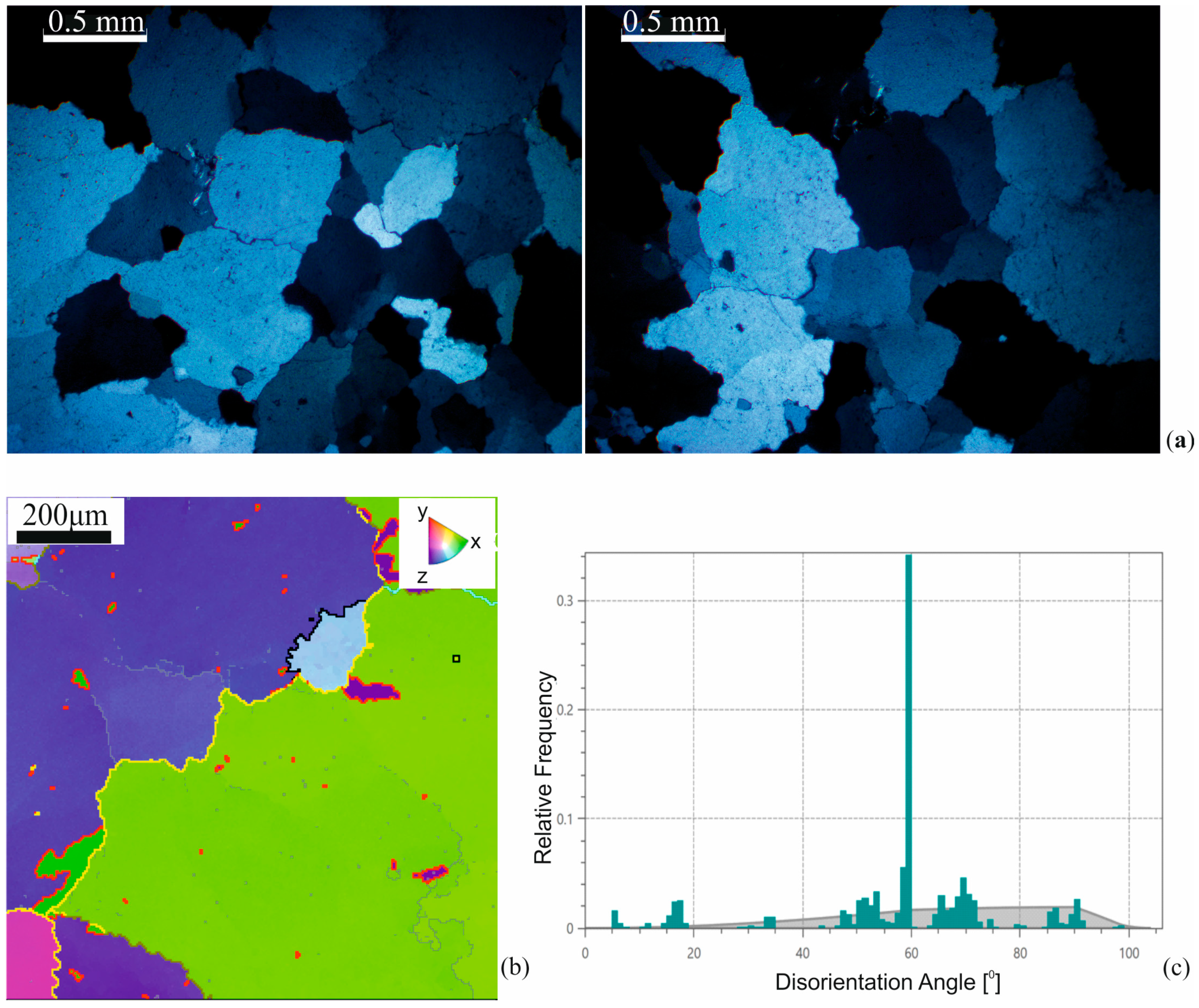
4.5. Type E. Quartz of Medium-Grained Structure
Isometric grains (up to 1 mm) with undulatory extinction characterized the microstructure of E type quartz. The grain was demonstrated to be mostly smooth with wavy boundaries (
Figure 10a). In the processed EBSD map (
Figure 10b), the quartz grains had a low percentage (34%) of low-angle boundaries (gray boundaries). The grains were separated from each other by low angles. These boundaries on the processed EBSD are marked in yellow. At the grain contacts, the regions separated by boundaries identified as Dauphiné twins (red boundaries) were fixed as well as the recrystallization grains with a 25° misorientation boundary (black boundaries). According to the histogram (
Figure 10c), the widest range of misorientation angles were observed for this structure. The histogram shows several peaks in the area of low-angle boundaries, a 17° peak, from 45° to 60°, a pronounced 60° peak, from 65° to 75°, and close to 90°. This indicates the active processes of dynamic restructuring of boundaries and sub-boundaries in this type of structure.
5. Discussion
The microstructures of the quartz from veins in the Western Transbaikalia demonstrated the manifestation of dynamometamorphic processes, which confirms the tectonic origin of cracks filled with fluid. The selected morphological types of microstructures meet special conditions for the transformation of mineral aggregates, expressed in the preferred influence of stress, temperature, or fluid present in the system. The interpretation of vein quartz microstructures according to the data presented above allows us to make conclusions regarding the mechanisms of their formation and the possibility of transition from one type to another. The correlation of the microstructure features of quartz veins and the structural confinement of quartz veins under the conditions of regional deformations gives a more complete picture of the development of a complex system of fluid–strain interactions under the conditions of a specific hydrothermal–magmatic system.
Variations in the distribution of microstructure types among the veins of different systems are indicated in
Table 1. Quartz with continuous Dauphiné boundaries (type A) is ubiquitous and is often observed in combination with elements of types of microstructures D and E. The types of microstructures of vein quartz B and C that are characterized by the formation of subgrains and deformation bands are most typical for veins associated with the development of the thrust system. Quartz with a mosaic microstructure (Type D) is observed in veins that perform main and echelon cracks of strike slip deformations. Type E is fixed in the veins associated with the development of the thrust system. However, quartz with an E-type microstructure is more typical for the main veins of Krivoy and Otvesny systems.
Based on the results of the analysis of the EBSD data of the vein quartz, typical structural elements were identified, which were characterized by the parameter of the grain boundary misorientation angle (
Table 2). The presence of selected structural elements in the microstructure picture, as well as their geometric parameters, determine the prevailing modes of deformation mechanisms.
Under conditions of high stress and moderate temperatures (greenschist-facies), the gliding of dislocations leads to the formation of subgrains outlined by low-angle boundaries (<10°). When the temperature increases or the deformation rate decreases, dislocations are organized into ordered, lower energy structures—deformation bands. We fixed the misorientation angle for the deformation bands of 30°. Active deformation contributes to the appearance of continuous twin Dauphiné boundaries with a misorientation angle of 60°. Increasing the temperature to a level close to the α–β transition reduces the size of the twins and the accumulation of recrystallization nuclei at the boundaries of a special type. In the conditions of dynamic recrystallization with the participation of a fluid, the recrystallization grains of 0.005–0.1 mm with a misorientation angle >10° formed. The formation of quartz grains larger than 1 mm occurs by static recrystallization, during which the prevailing modes were temperature and the presence of a fluid phase.
Based on the data on the conditions of the formation of structural elements, it is possible to interpret the conditions of the transformations of vein quartz during which microstructures of various types were formed.
The presence of special boundaries of the Dauphiné twinning characterized by a disorientation angle of 60° is an important indicator of the influence of stress. In Tullis’s opinion [
32,
33], such boundaries in quartz are formed at conditions of constant stress and relaxation. In our research, we noted the presence of continuous (Type A) and discontinuous twin-type boundaries (Type D). In addition, individual recrystallization grains with special boundaries were found (Types B, D, and E). The continuous boundaries of the twin type (type A) developed synkinematically with respect to the main deformation. According to Manegon [
35], in such cases, one of the twins retains the original orientation of the grain when subjected to mechanical twinning at the initial stages of deformation, and the other shows a higher degree of deformation.
Analyzing the obtained data, we concluded that when the temperature of the medium increases after the formation of Dauphiné twins, the twins boundaries undergo rearrangement. At the initial stages, this was expressed in the appearance of subgrains and recrystallization grains in the rotated twin. Likely, superimposed heating of the type A microstructure can cause the special 60° boundaries to crumble. Further increases in temperature initiates the annihilation of block walls and the active rearrangement of the boundaries and sub-boundaries. This process becomes possible at α–β transitions (T 573°C) [
39,
40]. Researchers noted a size reduction in Dauphiné twins and that twin boundaries are highly mobile near the phase transition [
41]. Experiments of Xu and Heaney [
42], related to the learning memory effects of domain structures before phase transitions demonstrated a change in the morphology of twin boundaries when the temperature approached 573 °C. When the temperature decreased, there was a tendency to accumulate twin boundaries near defects. The degree retention of twin boundaries in defect-free areas was lower than in highly defective areas.
The process of decay of 60° boundaries is well interpreted with the example of type D. In this case, the twins boundaries alternate with inclined boundaries close to 50° (φ1 176.3–172.4; Φ 112.4–110.4; φ2 38.9–85.7). In this case, an E type microstructure is an example of the resulting boundary realignment process. We assumed that the phenomenon decay of twin boundaries and the further annihilation of block walls occurs at the conditions of not only high temperature but also the presence of a fluid phase. This is confirmed by the presence of mineralized veinlets in the quartz microstructure of type D. However, the need for the simultaneous role of both (high temperature and the presence of fluid) factors has not been proven. Mutual consideration of the Dauphiné twinning boundary and general type boundaries makes it possible to evaluate the sequence of the transformation of vein quartz. The types of microstructures B, C, and D demonstrate the development of the dislocation glide mechanism, which results in the formation of subgrains and deformation bands [
16,
19].
For the type B microstructure, the recrystallization grains with special and general type boundaries repeat the shape of the subgrain. In this case, the block microstructure is formed before the appearance of mechanical Dauphiné twinning at a temperature of approximately 300°. In the case of quartz microstructure type C, the emergence of recrystallization grains is observed at the boundaries of the deformation bands. The deformation bands were formed under active deformation conditions at average deformation rates (10−11–10−6 s−1) at a temperature of approximately 400° resulting in dislocation glide. In the EBSD image in a D-type structure, recrystallization grains with special boundaries are marked on the contacts of the main grains or are confined to the remaining small-angle boundaries.
Further temperature increases involve the mechanism of dislocation creep and subsequent dynamic recrystallization under constant applied stresses. This entails the rotation of the subgrains, the rearrangement of twin boundaries, and the formation of recrystallization grains. The proximity of newly formed recrystallization grains to the boundaries of the Dauphiné twins also indicates a gradual rearrangement of the boundaries. This process is correlated with subgrain rotation recrystallization (SGR) as the mode of dynamic recrystallization [
22,
26].
Our analysis of the conditions for the formation of quartz microstructures of vein systems indicated their post-deformational origin. Taking the conditions that are typical for transformations of quartz, we suggest some features of the region’s development.
This distribution of microstructural types of quartz demonstrates the multi-stage development of the territory of the Bambukoy river basin of Western Transbaikalia. Given the mobility of twin boundaries with increasing temperature and their tendency to accumulate near defects, we concluded that quartz with a block structure and deformation bands is the earliest in the system under study. This suggests that the initial stages of territory formation are associated with the formation of a thrust system under tangential compression. Later, during the development of left-lateral strike slip deformations, this quartz underwent plastic deformation at a medium rate and temperatures of 300–400 °C. The subsequent tension led to the formation of double Dauphiné boundaries.
The peculiarities of the quartz microstructure suggest that the further development of the territory most likely took place in the conditions of a transpression mode for which there is a simultaneous compression and stick slip. At this time, along with the shear thrust faults of the northwestern direction and sub-latitude stick slip, faults of the northeastern strike were activated. This marks a change in the stress tensor, an increase in the temperature, and the injection of a new portion of fluid. These processes likely initiated the scattering of twin boundaries and the subsequent dynamic recrystallization in quartz. Another orientation of recrystallized grains is naturally obtained due to the mechanism of the emergence and subsequent growth. The static recrystallization of quartz in the main veins of the thrust system is either parallel to the dynamic recrystallization in the quartz of the shear zone, or to a later process associated with magmatic activations. The formation of the quartz microstructure of the main veins of thrust faults meets the conditions of static recrystallization.
Based on the study of the structural features of quartz and its association with regional deformation systems, we can speak about the allocation of three generations of vein quartz. The first generation is associated with the earliest processes and shows the greatest degree of deformation. This generation can be characterized by the development of such structural elements, such as subgrains with low-angle boundaries, deformation bands, and recrystallization nuclei with boundaries of special and general types. The second generation of quartz is correlated with shear deformations. The selection of the second generation of quartz is based on the presence of structural elements, such as continuous Dauphiné twins, as well as recrystallization grains with boundaries of the special and general types. The third generation of quartz was found in the veins that performed the main cracks of the thrust faults. Signs of static recrystallization characterized this generation by the migration of boundaries. This was expressed in the presence of isometric grains greater than 1 mm. In third generation quartz grains, the regions with twin boundaries are fixed near the contacts. Further study of the thermobarogeochemical parameters of fluid inclusions of isolated quartz generations will provide additional data on the evolution of tectonic activity in the region.
6. Conclusions
The features of the microstructure of quartz aggregates of veins of different orientations in the suture zone of the Baikal–Muiskoy and Barguzino–Vitimskoy structural and formation zones of Western Transbaikalia (Russia) are studied. Differently oriented quartz veins perform tectonic cracks associated with the development of regional thrust and left-thrust deformations, as a result of which they bear signs of dynamometamorphic processes. Based on the structural position of quartz veins, the main veins corresponding to the position of the main fault and the echelon ones performing cracks closely associated with the main fault are distinguished.
Structural and crystallographic analysis of quartz samples allowed us to distinguish five structural types. Each type is characterized by certain morphological features of structural elements, the spectrum of grain misorientation and intra-grain blocks. Typical elements of structural types are subgrains, deformation bands, Dauphiné mechanical twins, and recrystallization grains. Along with special Dauphiné twinning boundaries, 30° and 90° boundaries defined by the trigonal symmetry of quartz were formed in the quartz structure. We proceeded from the fact that these types of low-energy boundaries are what defines the profitability of their formation. The presence of low-angle and other high-angle boundaries in the spectrum determines various stages of deformation transformations and recrystallization processes. The process of the rearrangement of the twinning boundaries with an increase in temperature close to the α–β transition, described by researchers as a result of experiments, was noted.
The regularities of the distribution of the selected types of microstructure in the veins suggest the existence of three generations of vein quartz. Quartz of the first generation is associated with the earliest tectonic processes of the thrust system development. Features of the microstructure of first-generation grains indicate their transformation at average strain rates and temperatures in the range of 300–400 °C, followed by heating and dynamic recrystallization. Quartz veins of shear deformations show the influence of dynamic recrystallization, which indicates its later isolation as a second generation. Quartz grains of the third generation were formed as a result of static recrystallization at high temperatures and the influence of a fluid. The structural affinity of the latter is not clearly defined, but they are more pronounced in the veins associated with thrust deformations. Microstructures of quartz of the first and second generation were formed as a result of active deformation processes and are syntectonic. The fluid dominated the formation of the microstructure of quartz of the third generation, and as a result, it can be defined as post-deformation.
Thus, the microstructural features of vein quartz reflect the course of development of the hydrothermal–magmatic system in the conditions of th etectonic evolution of the region. The results obtained, along with further study of the thermobarogeochemical parameters of fluid inclusions in the selected generations and the determination of the fluid source, will add to the understanding of the development of a complex system of fluid–strain interactions in the middle and lower crust.

 ,
, 


