Efflorescent Sulfate Crystallization on Fractured and Polished Colloform Pyrite Surfaces: A Migration Pathway of Trace Elements
Abstract
1. Introduction
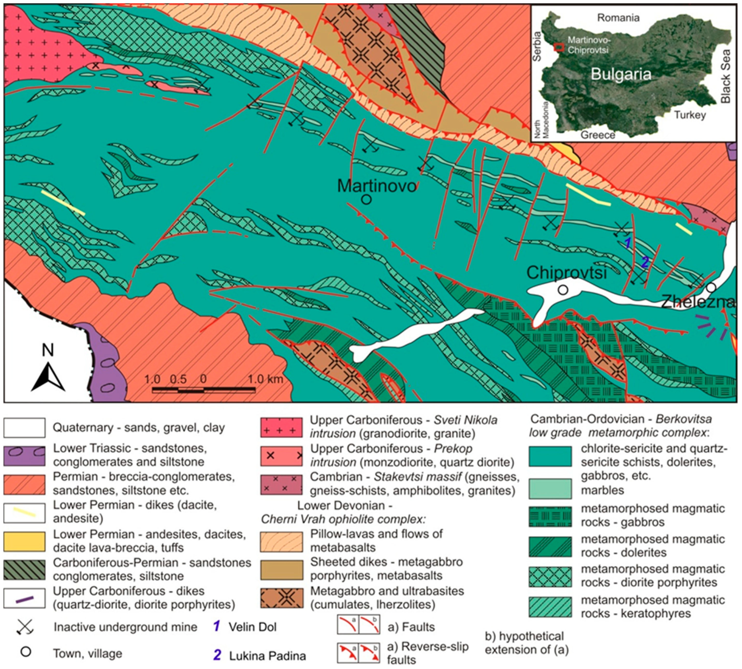
2. Geological Background
3. Materials and Methods
3.1. Samples
3.2. Analytical Procedures
3.2.1. X-Ray Diffractometry (XRD), Scanning Electron Microscopy (SEM), and Electron Microprobe Analysis (EMPA)
3.2.2. Laser Ablation Inductively Coupled Plasma Mass Spectrometry (LA-ICP-MS)
4. Results
4.1. Colloform Pyrite
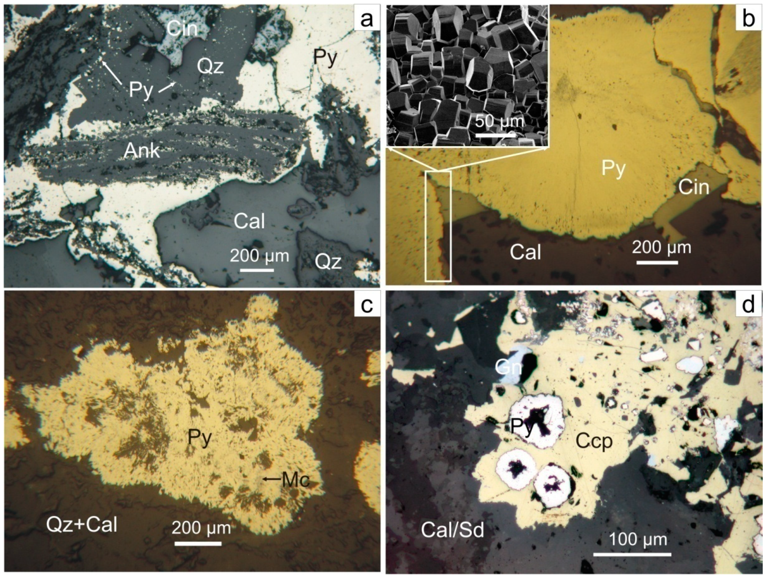
4.1.1. Colloform Pyrite Morphology and Mineral Assemblage
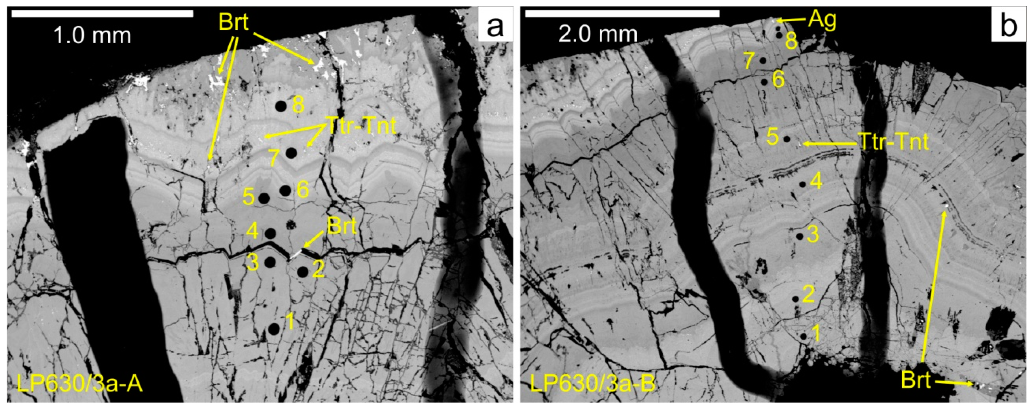
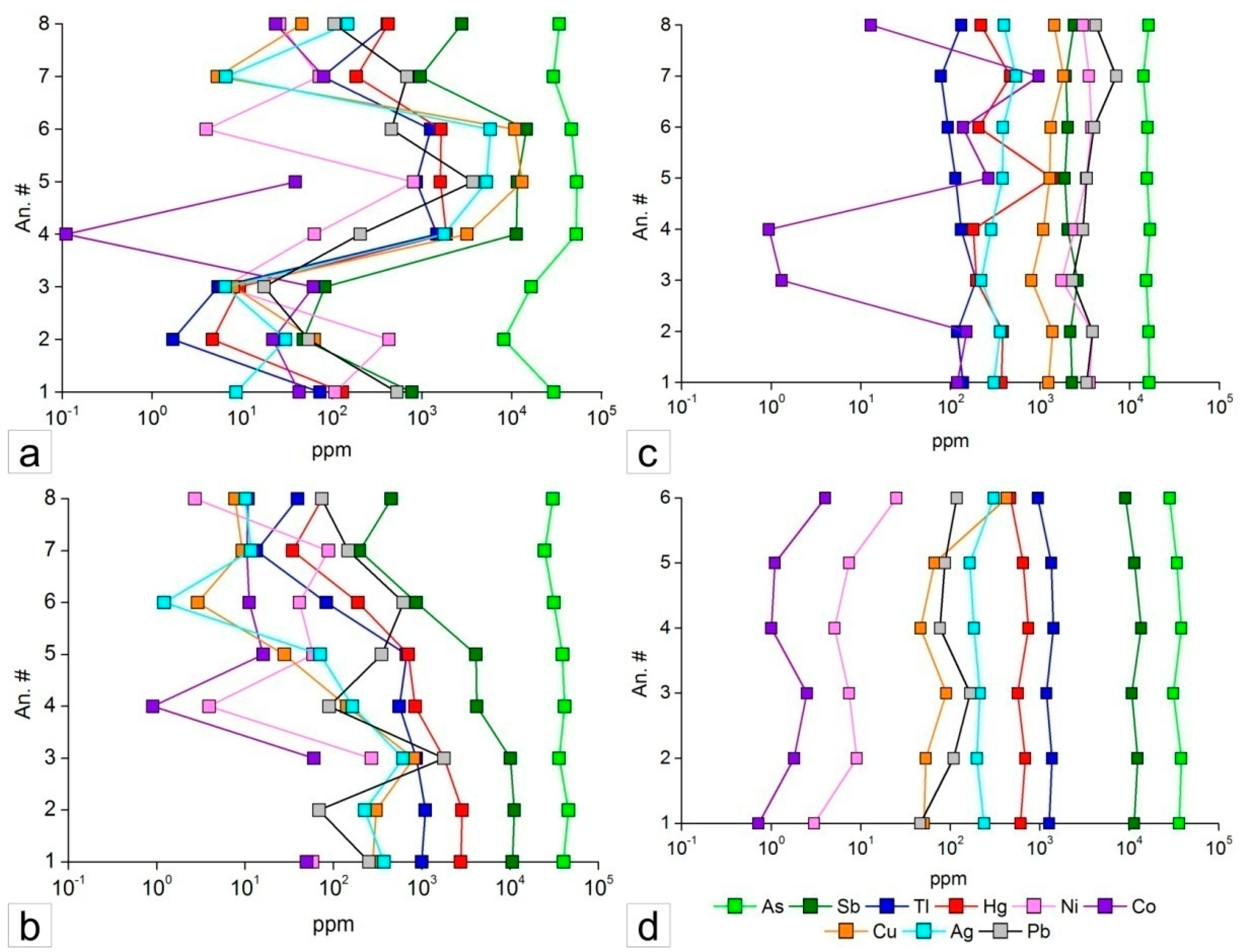
4.1.2. Chemical Composition of Colloform Pyrite
4.2. Efflorescent Mineral Assemblage
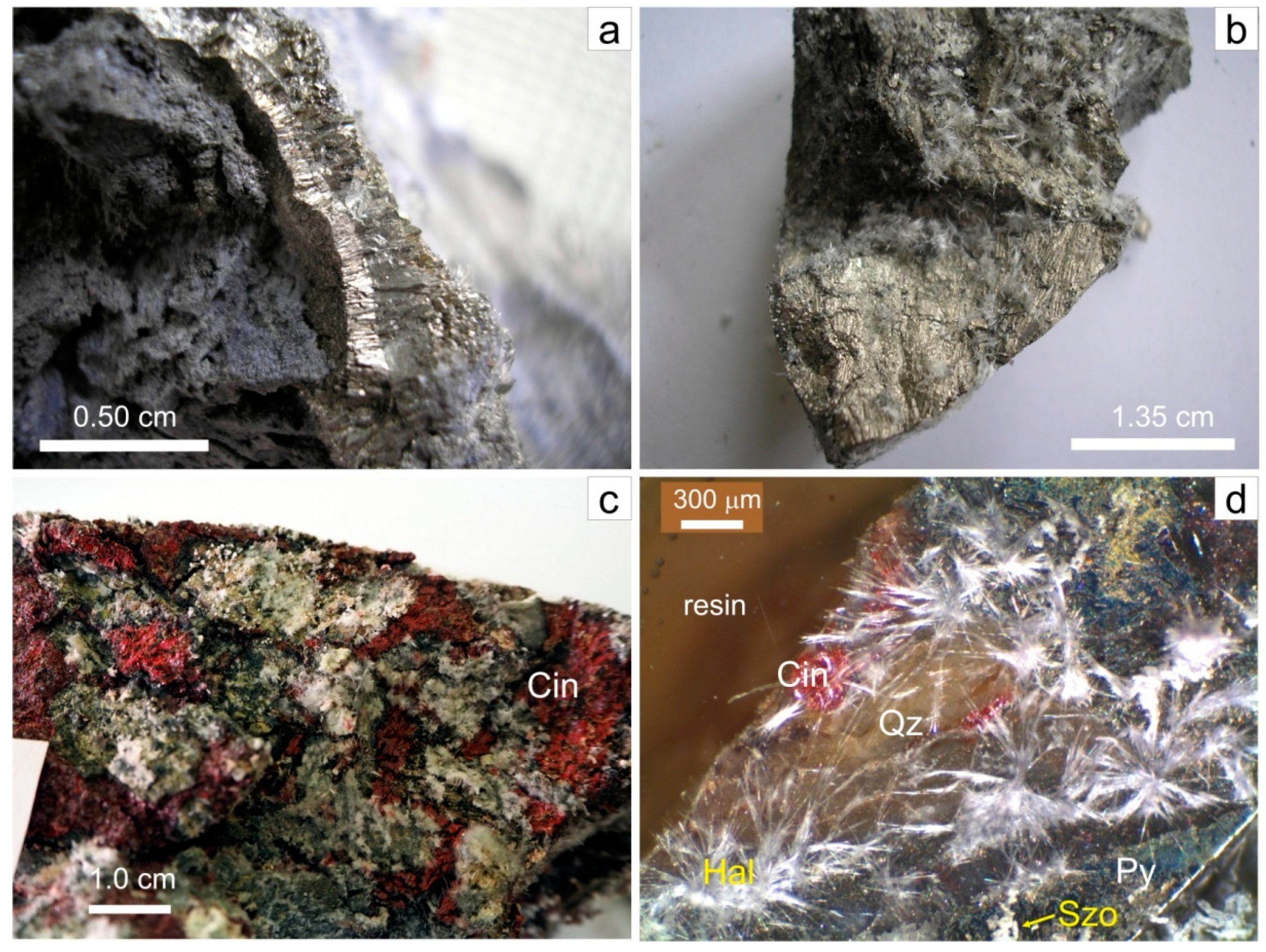
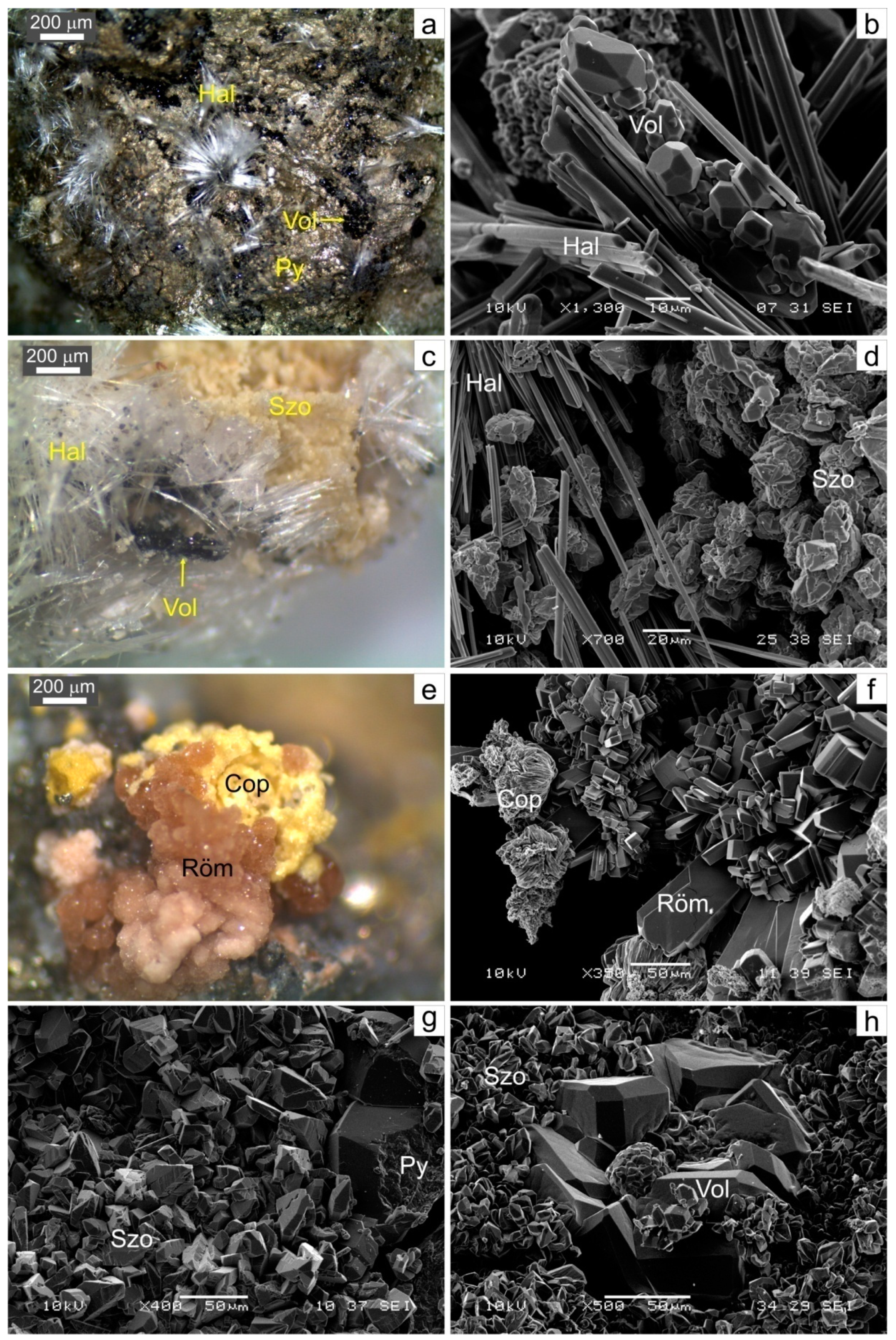
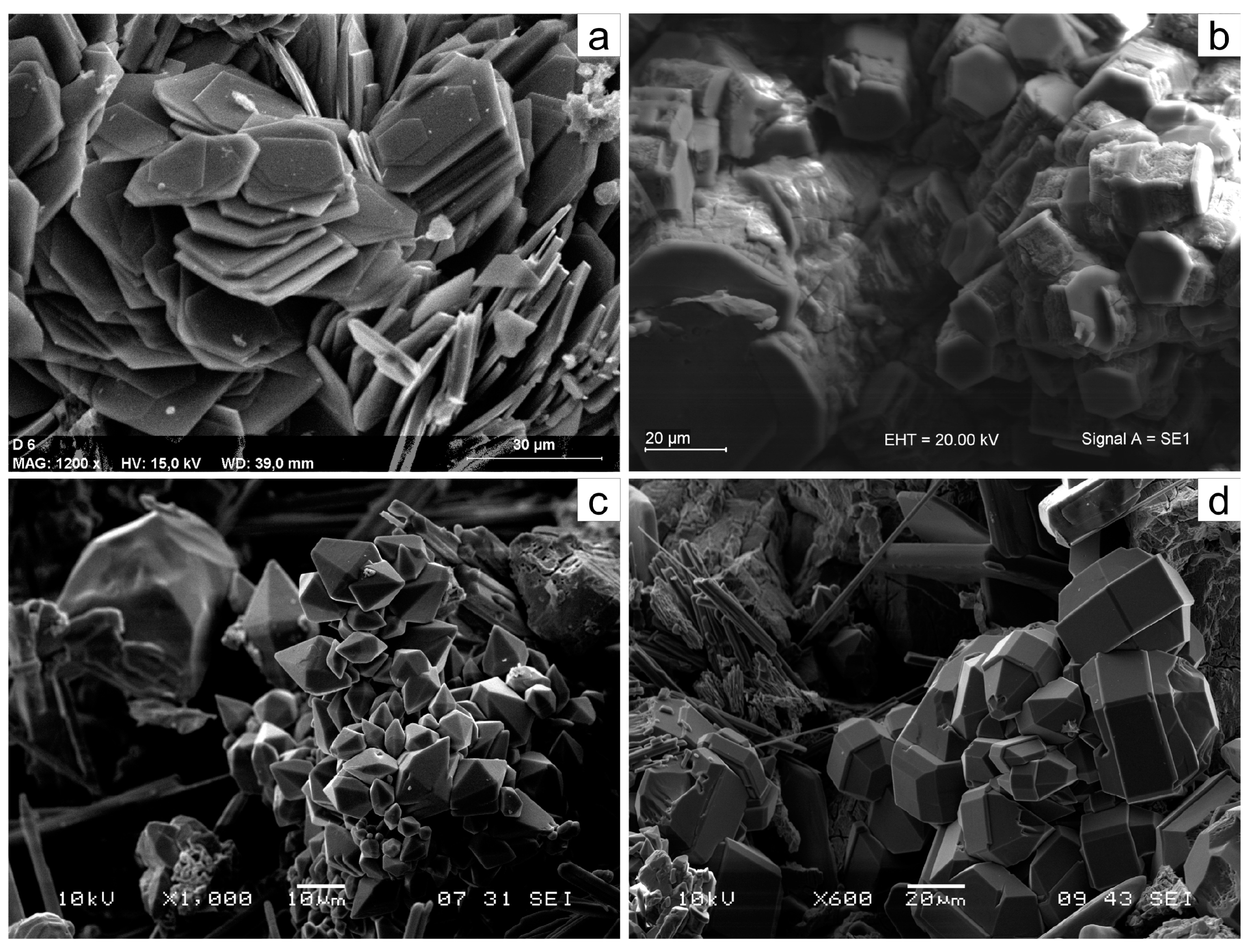
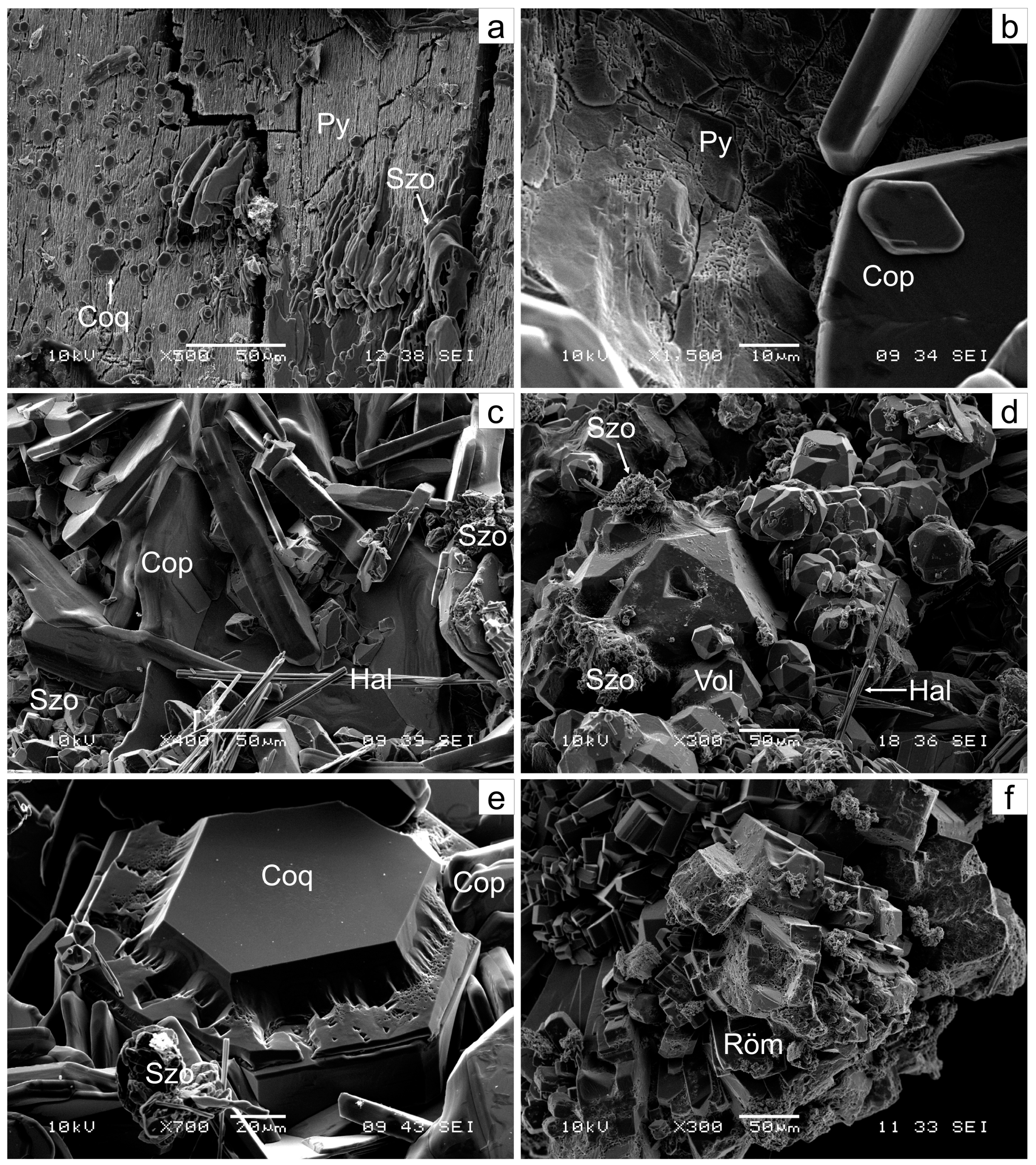
4.2.1. Mineralogy
4.2.2. Chemical Composition of the Identified Efflorescent Minerals
5. Discussion
5.1. Colloform Pyrite Chemistry: Mode of Occurrence of Trace Elements and Genetic Implications
5.2. Transformation Mechanisms
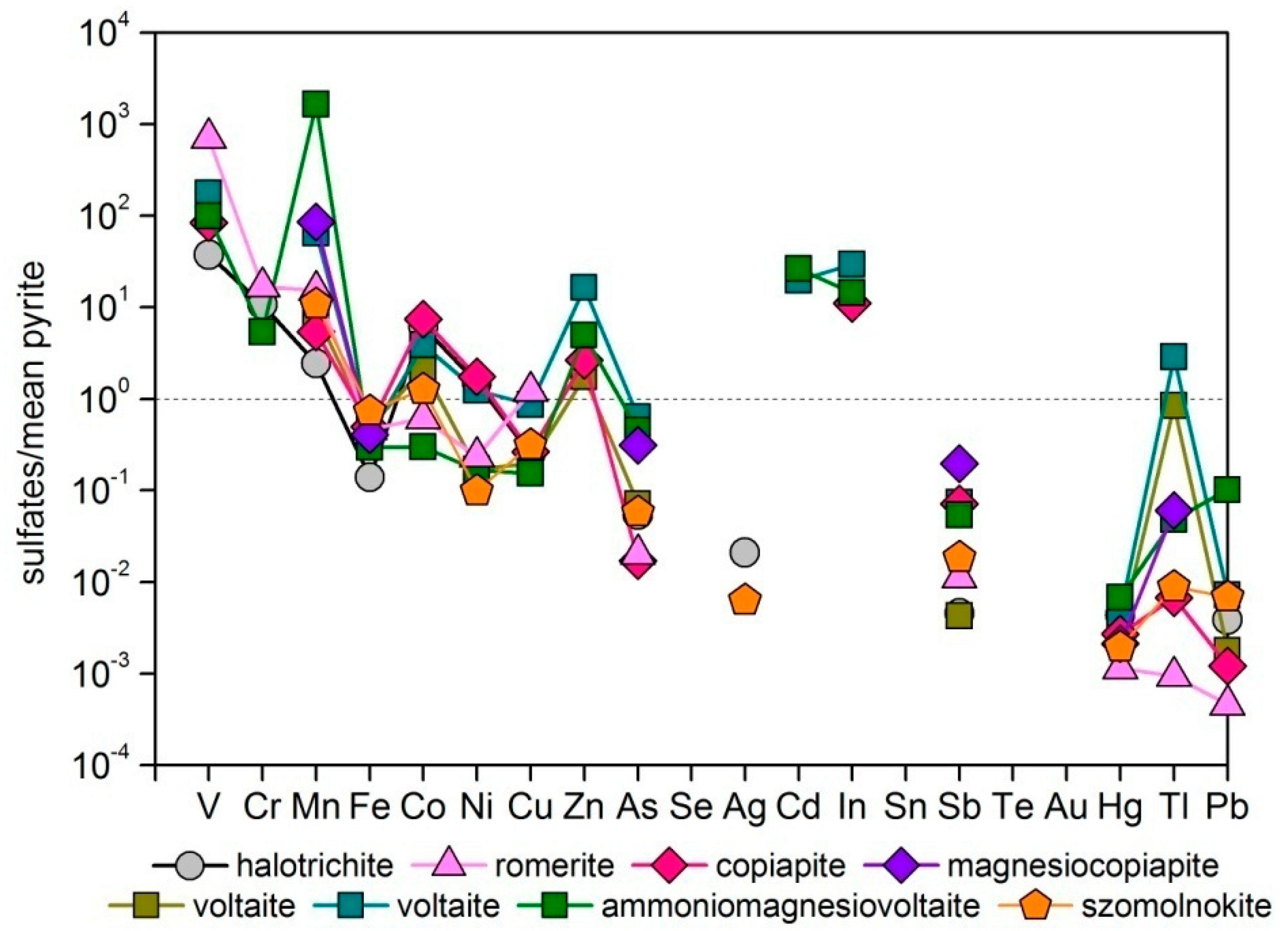
5.3. Partitioning of Inherited Trace Elements in Sulfates
5.4. Sources of Introduced Elements in Sulfates and Incorporation
5.5. Environmental Significance
6. Conclusions
Author Contributions
Funding
Acknowledgments
Conflicts of Interest
References
- Abraitis, P.K.; Pattrick, R.A.D.; Vaughan, D.J. Variations in the compositional, textural and electrical properties of natural pyrite: A review. Int. J. Miner. Process. 2004, 74, 41–59. [Google Scholar] [CrossRef]
- Barker, S.L.L.; Hickey, K.A.; Cline, J.S.; Dipple, G.M.; Kilburn, M.R.; Vaughan, J.R.; Longo, A. Uncloaking invisible gold: Use of NanoSIMS to evaluate gold, trace elements, and sulfur isotopes in pyrite from Carlin-type gold deposits. Econ. Geol. 2009, 104, 897–904. [Google Scholar] [CrossRef]
- Cook, N.J.; Ciobanu, C.L.; Mao, J.W. Textural control on gold distribution in As-free pyrite from the Dongping, Huangtuliang and Hougou gold deposits, North China Craton (Hebei Province, China). Chem. Geol. 2009, 264, 101–121. [Google Scholar] [CrossRef]
- Large, R.R.; Danyushevsky, L.; Hollit, C.; Maslennikov, V.; Meffre, S.; Gilbert, S.; Bull, S.; Scott, R.; Thomas, H.; Singh, B.; et al. Gold and trace element zonation in pyrite using a laser imaging technique: Implications for the timing of gold in orogenic and Carlin-style sediment hosted deposits. Econ. Geol. 2009, 104, 635–668. [Google Scholar] [CrossRef]
- Reich, M.; Deditius, A.; Chryssoulis, S.; Li, J.-W.; Ma, C.-Q.; Parada, M.A.; Barra, F.; Mittermayr, F. Pyrite as a record of hydrothermal fluid evolution in a porphyry copper system: A SIMS/EMPA trace element study. Geochim. Cosmochim. Acta 2013, 104, 42–62. [Google Scholar] [CrossRef]
- Wohlgemuth-Ueberwasser, C.C.; Viljoen, F.; Petersen, S.; Vorster, C. Distribution and solubility limits of trace elements in hydrothermal black smoker sulfides: An in-situ LA-ICP-MS study. Geochim. Cosmochim. Acta 2015, 159, 16–41. [Google Scholar] [CrossRef]
- Keith, M.; Häckel, F.; Haase, K.M.; Schwarz-Schampera, U.; Klemd, R. Trace element systematics of pyrite from submarine hydrothermal vents. Ore Geol. Rev. 2016, 72, 728–745. [Google Scholar] [CrossRef]
- Sykora, S.; Cooke, D.R.; Meffre, S.; Stephanov, A.S.; Gardner, K.; Scott, R.; Selley, D.; Harris, A.C. Evolution of pyrite trace element compositions from porphyry-style and epithermal conditions at the Lihir gold deposit: Implications for ore genesis and mineral processing. Econ. Geol. 2018, 113, 193–203. [Google Scholar] [CrossRef]
- Cook, N.J.; Chryssoulis, S.L. Concentration of “invisible gold” in the common sulfides. Can. Miner. 1990, 28, 1–16. [Google Scholar]
- Fleet, M.E.; Chryssoulis, S.L.; MacLean, P.J.; Davidson, R.; Weisener, C.G. Arsenian pyrite from gold deposits: Au and As distribution investigated by SIMS and EMP, and color staining and surface oxidation by XPS and LIMS. Can. Miner. 1993, 31, 1–17. [Google Scholar]
- Fleet, M.E.; Mumin, H. Gold-bearing arsenian pyrite and marcasite and arsenopyrite from Carlin Trend gold deposits and laboratory synthesis. Am. Miner. 1997, 82, 182–193. [Google Scholar] [CrossRef]
- Maddox, L.M.; Bancroft, G.M.; Scaini, M.J.; Lorimer, J.M. Invisible gold: Comparison of Au deposition on pyrite and arsenopyrite. Am. Miner. 1998, 83, 1240–1245. [Google Scholar] [CrossRef]
- Simon, G.; Huang, H.; Penner-Hahn, J.E.; Kesler, S.E.; Kao, L.S. Oxidation state of gold and arsenic in gold-bearing arsenian pyrite. Am. Miner. 1999, 84, 1071–1079. [Google Scholar] [CrossRef]
- Palenik, C.S.; Utsunomiya, S.; Reich, M.; Kesler, S.E.; Ewing, R.C. Invisible gold revealed: Direct imaging of gold nanoparticles in a Carlin-type deposit. Am. Miner. 2004, 89, 1359–1366. [Google Scholar] [CrossRef]
- Reich, M.; Kesler, S.E.; Utsunomiya, S.; Palenik, C.S.; Chryssoulis, S.L.; Ewing, R.C. Solubility of gold in arsenian pyrite. Geochim. Cosmochim. Acta 2005, 69, 2781–2796. [Google Scholar] [CrossRef]
- Deditius, A.P.; Reich, M.; Kesler, S.E.; Utsunomiya, S.; Chryssoulis, C.L.; Walshe, J.; Ewing, R.C. The coupled geochemistry of Au and As in pyrite from hydrothermal ore deposits. Geochim. Cosmochim. Acta 2014, 140, 644–670. [Google Scholar] [CrossRef]
- Vikentyev, I.V. Invisible and microscopic gold in pyrite: Methods and new data for massive sulfide ores of the Urals. Geol. Ore Depos. 2015, 57, 237–265. [Google Scholar] [CrossRef]
- Trigub, A.L.; Tagirov, B.R.; Kvashnina, K.O.; Chareev, D.A.; Nickolsky, M.S.; Shiryaev, A.A.; Baranova, N.N.; Kovalchuk, E.V.; Mokhov, A.V. X-ray spectroscopy study of the chemical state of “invisible” Au in synthetic minerals in the Fe-As-S system. Am. Miner. 2017, 102, 1057–1065. [Google Scholar]
- Moller, P.; Kersten, G. Electrochemical accumulation of visible gold on pyrite and arsenopyrite surfaces. Miner. Depos. 1994, 29, 404–413. [Google Scholar] [CrossRef]
- Nicholson, R.V.; Gillham, R.W.; Reardon, E.J. Pyrite oxidation in carbonate-buffered solution: 1. Experimental kinetics. Geochim. Cosmochim. Acta 1988, 52, 1077–1085. [Google Scholar] [CrossRef]
- Nicholson, R.V.; Gillham, R.W.; Reardon, E.J. Pyrite oxidation in carbonate-buffered solution: 2. Rate control by oxide coating. Geochim. Cosmochim. Acta 1990, 54, 395–402. [Google Scholar] [CrossRef]
- Moses, C.O.; Herman, J.S. Pyrite oxidation at circumneutral pH. Geochim. Cosmochim. Acta 1991, 55, 471–482. [Google Scholar] [CrossRef]
- Rimstidt, J.D.; Vaughan, D.J. Pyrite oxidation: A state-of-the-art assessment of the reaction mechanism. Geochim. Cosmochim. Acta 2003, 67, 873–880. [Google Scholar] [CrossRef]
- Todd, E.C.; Sherman, D.M.; Purton, J.A. Surface oxidation of pyrite under ambient atmospheric and aqueous (pH = 2 to 10) conditions: Electronic structure and mineralogy from X-ray absorption spectroscopy. Geochim. Cosmochim. Acta 2003, 67, 881–893. [Google Scholar] [CrossRef]
- Buurman, P. In vitro weathering products of pyrite. Geol. Mijnvouw 1975, 54, 101–105. [Google Scholar]
- Nordstrom, D.K.; Alpers, C.N. Negative pH, efflorescent mineralogy, and consequences for environmental restoration at the Iron Mountain Superfund site, California. Proc. Natl. Acad. Sci. USA 1999, 96, 3455–3462. [Google Scholar] [CrossRef]
- Jambor, J.J.; Nordstrom, D.K.; Alpers, C.N. Metal-sulfate salts from sulfide mineral oxidation. Rev. Miner. Geochem. 2000, 40, 303–350. [Google Scholar] [CrossRef]
- Buckby, T.; Black, S.; Coleman, M.L.; Hodson, M.E. Fe-sulphate-rich evaporative mineral precipitates from the Rio Tinto, southwest Spain. Miner. Mag. 2003, 67, 263–278. [Google Scholar] [CrossRef]
- Jerz, J.K.; Rimstidt, J.D. Efflorescent iron sulfate minerals: Paragenesis, relative stability, and environmental impact. Am. Miner. 2003, 88, 1919–1932. [Google Scholar] [CrossRef]
- Buzatu, A.; Dill, H.G.; Buzgar, N.; Damian, G.; Maftei, A.E.; Apopei, A.I. Efflorescent sulfates from Baia Sprie mining area (Romania)—Acid mine drainage and climatological approach. Sci. Total Environ. 2016, 542, 629–641. [Google Scholar] [CrossRef]
- Bonev, I.K.; Reiche, M.; Marinov, M. Morphology, perfection and growth of natural pyrite whiskers and thin platelets. Phys. Chem. Miner. 1985, 12, 223–232. [Google Scholar] [CrossRef]
- Chen, T.T. Colloform and framboidal pyrite from the Caribou deposit, New Brunswick. Can. Miner. 1978, 16, 9–15. [Google Scholar]
- Ohfuji, H.; Rickard, D. Experimental syntheses of framboids—A review. Earth-Sci. Rev. 2005, 71, 147–170. [Google Scholar] [CrossRef]
- Bonev, I.K.; Atanassova, R. Crystallography and origin of colloform pyrite. In Proceedings of the IMA 2006, Kobe, Japan, 23–28 July 2006; p. 174. [Google Scholar]
- Barrie, C.D.; Boyce, A.J.; Boyle, A.P.; Williams, P.J.; Blake, K.; Ogawara, T.; Akai, J.; Prior, D.J. Growth controls in colloform pyrite. Am. Miner. 2009, 94, 415–429. [Google Scholar] [CrossRef]
- Atanassova, R. Environmental significance of pyrite with colloform textures. C. R. Acad. Bulg. Sci. 2010, 63, 1335–1340. [Google Scholar]
- Gao, S.; Huang, F.; Wang, Y.; Gao, W. A review of research progress in the genesis of colloform pyrite and its environmental indications. Acta Geol. Sin. 2016, 90, 1353–1369. [Google Scholar]
- Atanassov, V.; Pavlov, I. Notes about mineralogy and paragenetic zoning of minerals deposits in Chiprovtsi ore region. Ann. Ecol. Sup. Miner. Geol. 1983, 28, 159–178. (In Bulgarian) [Google Scholar]
- Tarasova, E.; Tarassov, M. Horizontal zonality and mineral composition of skarns in the Martinovo deposit. Geochem. Miner. Pet. 1988, 24, 68–76. (In Bulgarian) [Google Scholar]
- Carrigan, C.W.; Mukasa, S.B.; Haydoutov, I.; Kolcheva, K. Age of Variscan magmatism from the Balkan sector of the orogen, Central Bulgaria. Lithos 2005, 82, 125–147. [Google Scholar] [CrossRef]
- Amov, B.; Arnaudov, V.; Pavlova, M.; Dragov, P.; Baldjieva, T.; Evstatieva, S. Lead isotope data on the Paleozoic granitoids and ore mineralizations from the Western Balkan Mountains and the Tran district (West Bulgaria). I. Isotopic ratios and geochronology. Geol. Balc. 1981, 11, 3–26. [Google Scholar]
- Dragov, P.; Galiy, S.A.; Zhukov, F.I. Carbon, oxygen and sulfur isotopes in minerals from the Chiprovtsi ore zone. Rev. Bulg. Geol. Soc. 1991, 52, 50–61. (In Bulgarian) [Google Scholar]
- Haydoutov, I. Precambrian ophiolites, Cambrian island arc and Variscan suture in South Carpathian-Balkan region. Geology 1989, 17, 905–908. [Google Scholar] [CrossRef]
- Haydoutov, I. Origin and Evolution of the Precambrian Balkan-Carpathian Ophiolite Segment; BAS Press: Sofia, Bulgaria, 1991; p. 179. (In Bulgarian) [Google Scholar]
- Haydoutov, I.; Yanev, S. The Protomoesian microcontinent of the Balkan Peninsula—A peri-Gondvanian piece. Tectonophysics 1997, 272, 303–313. [Google Scholar] [CrossRef]
- Plissart, G.; Monnier, C.; Diot, H.; Maruntiu, M.; Berger, J.; Triantafyllou, A. Petrology, geochemistry and Sm-Nd analyses on the Balkan-Carpathian Ophiolite (BCO—Romania, Serbia, Bulgaria): Remnants of a Devonian back-arc basin in the easternmost part of the Variscan domain. J. Geodyn. 2017, 105, 27–50. [Google Scholar] [CrossRef]
- Dragov, P.; Obretenov, N. Morphology and genesis of the siderite bodies in the Chiprovtsi deposit. Bull. Geol. Inst. Ser. Met. Non-Met. Miner. Depos. 1972, 21, 37–53. [Google Scholar]
- Obretenov, N. The Chiprovtsi ore field—Chiprovtsi deposit. In Lead-Zinc Deposits in Bulgaria; Technika: Sofia, Bulgaria, 1988; pp. 193–206. [Google Scholar]
- Milev, V.; Stanev, V.; Ivanov, V. The Extracted Ores in Bulgaria during 1878–1995—Statistical Reference Book. Sofia Zemya 1996, 93, 196. (In Bulgarian) [Google Scholar]
- Angelov, V.; Antonov, M.; Gerdzhikov, S.; Aydanlyiski, G.; Petrov, P.; Kisselinov, H. Geological Map of Bulgaria M 1:50000, Map Sheet “Chiprovtsi”; Ministry of Environment and Water, Bulgarian Geological Survey: Sofia, Bulgaria, 2008. [Google Scholar]
- Wilson, S.A.; Ridley, W.I.; Koenig, A.E. Development of sulfide calibration standards for the laser ablation inductively-coupled plasma mass spectrometry technique. J. Anal. At. Spectrom. 2002, 17, 406–409. [Google Scholar] [CrossRef]
- Guillong, M.; Meier, D.L.; Allan, M.M.; Heinrich, C.A.; Yardley, B.W.D. Appendix A6: SILLS: A MATLAB-based program for the reduction of laser ablation ICP-MS data of homogeneous materials and inclusions. In Laser Ablation ICP–MS in the Earth Sciences: Current Practices and Outstanding Issues; Sylvester, P., Ed.; Mineralogical Association of Canada Short Course 40: Vancouver, BC, Canada, 2008; pp. 328–333. [Google Scholar]
- Szakáll, S.; Sajó, I.; Fehér, B.; Bigi, S. Ammoniomagnesiovoltaite, a voltaite-related mineral species from Pécs-Vasas, Hungary. Can. Miner. 2012, 50, 65–72. [Google Scholar] [CrossRef]
- Deditius, A.P.; Utsunomiya, S.; Renock, D.; Ewing, R.C.; Ramana, C.V.; Becker, U.; Kesler, S.E. A proposed new type of arsenian pyrite: Composition, nanostructure and geological significance. Geochim. Cosmochim. Acta 2008, 72, 2919–2933. [Google Scholar] [CrossRef]
- Deditius, A.P.; Reich, M. Constraints on the solid solubility of Hg, Tl, and Cd in arsenian pyrite. Am. Miner. 2016, 101, 1451–1459. [Google Scholar] [CrossRef]
- George, L.L.; Biagioni, C.; D’Orazio, M.; Cook, N.J. Textural and trace element evolution of pyrite during greenschist facies metamorphic recrystallization in the southern Apuan Alps (Tuscany, Italy): Initiation of Tl-rich sulphosalt melt formation. Ore Geol. Rev. 2018, 102, 59–105. [Google Scholar] [CrossRef]
- George, L.L.; Biagioni, C.; Lepore, G.O.; Lacalamita, M.; Agrosi, G.; Capitani, G.C.; Bonaccorsi, E.; d’Acapito, F. The speciation of thallium in (Tl, Sb, As)-rich pyrite. Ore Geol. Rev. 2019, 107, 364–380. [Google Scholar] [CrossRef]
- Kolker, A. Minor element distribution in iron disulfides in coal: A geochemical review. Int. J. Coal Geol. 2012, 94, 32–43. [Google Scholar] [CrossRef]
- Large, R.R.; Halpin, J.A.; Danyushevsky, L.V.; Maslennikov, V.V.; Bull, S.W.; Long, J.A.; Gregory, D.D.; Lounejeva, E.; Lyons, T.W.; Sack, P.J.; et al. Trace element content of sedimentary pyrite as a new proxy for deep-time ocean-atmosphere evolution. Earth Planet. Sci. Lett. 2014, 389, 209–220. [Google Scholar] [CrossRef]
- Wells, J.D.; Mullens, T.E. Gold-bearing arsenian pyrite determined by microprobe analysis, Cortez and Carlin gold mines, Nevada. Econ. Geol. 1973, 28, 187–201. [Google Scholar] [CrossRef]
- Deditius, A.P.; Utsunomiya, S.; Kesler, S.E.; Reich, M.; Ewing, R.C. Trace elements nanoparticles in pyrite. Ore Geol. Rev. 2011, 42, 32–46. [Google Scholar] [CrossRef]
- Pearce, C.I.; Pattrick, R.A.D.; Vaughan, D.J. Electrical and magnetic properties of sulphides. Rev. Miner. Geochem. 2006, 61, 127–180. [Google Scholar] [CrossRef]
- Klemm, D.D. Synthesen und analysen in den dreieeks diagrammen FeAsS-CoAsS-NiAsS und FeS2-CoS2-NiS2. Neues Jahrbueh Fur Miner. Abh. 1965, 103, 205–255. (In German) [Google Scholar]
- Huston, D.L.; Sie, S.H.; Suter, G.F.; Cooke, D.R.; Both, R.A. Trace elements in sulfide minerals from Eastern Australian volcanic-hosted massive sulfide deposits: Part 1. Proton microprobe analyses of pyrite, chalcopyrite and sphalerite, and Part II. Selenium levels in pyrite: Comparison with δ34S values and implications for the source of sulfur in volcanogenic hydrothermal systems. Econ. Geol. 1995, 90, 1167–1196. [Google Scholar]
- Zidarova, B.; Zidarov, N. Main elements of the local geogenetic model for fluorite formation: III. Chiprovci-East deposit. C. R. Acad. Bulg. Sci. 2000, 53, 63–66. [Google Scholar]
- Dimitrova, D.; Kerestedjian, T.; Petrova, M. Cinnabar-pyrite mineralization associated with fluorite replacement bodies in the Chiprovtsi silver-lead deposit, Northwestern Bulgaria. In Proceedings of the 9th Biennial SGA Meeting Mineral Exploration and Research: Digging Deeper, Dublin, Ireland, 20–23 August 2007; pp. 873–876. [Google Scholar]
- Nesbitt, H.W.; Scaini, M.; Hochst, H.; Bancroft, G.M.; Schaufuss, A.G.; Szargan, R. Synchrotron XPS evidence for Fe2+-S and Fe3+-S surface species on pyrite fracture surfaces, and their 3D electronic states. Am. Miner. 2000, 85, 850–857. [Google Scholar] [CrossRef]
- Xiong, Y. Hydrothermal thallium mineralization up to 300 °C: A thermodynamic approach. Ore Geol. Rev. 2007, 32, 291–313. [Google Scholar] [CrossRef]
- Hawthorne, F.C.; Krivovichev, S.V.; Burns, P.C. The crystal chemistry of sulfate minerals. Rev. Miner. Geochem. 2000, 40, 1–112. [Google Scholar] [CrossRef]
- Bigham, J.M.; Nordstrom, D.K. Iron and aluminum hydroxysulfates from acid sulfate waters. Rev. Miner. Geochem. 2000, 40, 351–403. [Google Scholar] [CrossRef]
- Kelly, D.P.; Wood, A.P. Reclassification of some species of Thiobacillus to the newly designated genera Acidithiobacillus gen. nov.; Halothiobacillus gen. nov. and Thermithiobacillus gen. nov. Int. J. Syst. Evol. Microbiol. 2000, 50, 511–516. [Google Scholar] [CrossRef]
- Chukanov, N.V.; Aksenov, S.M.; Rastvetaeva, R.K.; Möhn, G.; Rusakov, V.S.; Pekov, I.V.; Scholz, R.; Eremina, T.A.; Belakovskiy, D.I.; Lorenz, J.A. Magnesiovoltaite, K2Mg5Fe3+3Al(SO4)12∙18H2O, a new mineral from the Alcaparossa mine, Antofagasta region, Chile. Eur. J. Miner. 2016, 28, 1005–1017. [Google Scholar] [CrossRef]
- Majzlan, J.; Schlicht, H.; Wierzbicka-Wieczorek, M.; Giester, G.; Pöllmann, H.; Brömme, B.; Doyle, S.; Buth, G.; Koch, C.B. A contribution to the crystal chemistry of the voltaite group: Solid solutions, Mössbauer and infrared spectra, and anomalous anisotropy. Miner. Petrol. 2013, 107, 221–233. [Google Scholar] [CrossRef]
- Jamieson, H.E.; Robinson, C.; Alpers, C.N.; Blaine McCleskey, R.; Nordstrom, D.K.; Peterson, R.C. Major and trace element composition of copiapite-group minerals and coexisting water from the Richmond mine, Iron Mountain, California. Chem. Geol. 2005, 215, 387–405. [Google Scholar] [CrossRef]
- Majzlan, J.; Michalik, R. The crystal structures, solid solutions and infrared spectra of copiapite-group minerals. Miner. Mag. 2007, 71, 553–569. [Google Scholar] [CrossRef]
- Shannon, R.D. Revised effective ionic radii and systematic studies of interatomic distances in halides and chalcogenides. Acta Crystallogr. 1976, A32, 751–767. [Google Scholar] [CrossRef]
- Majzlan, J.; Alpers, C.N.; Koch, C.B.; McCleskey, R.B. Vibrational, X-ray absorption, and Mössbauer spectra of sulfate minerals from the weathered massive sulfide deposit at Iron Mountain, California. Chem. Geol. 2011, 284, 296–305. [Google Scholar] [CrossRef]
- Moncur, M.C.; Ptacek, C.J.; Blowes, D.W.; Peterson, R.C. The occurrence and implications of efflorescent sulfate minerals at the former Sherrit-Gordon Zn-Cu mine, Sherridon, Manitoba, Canada. Can. Miner. 2015, 53, 961–977. [Google Scholar] [CrossRef]
- Romero, A.; González, I.; Galán, E. The role of efflorescent sulfates in the storage of trace elements in stream waters polluted by acid mine-drainage: The case of Peña del Hierro, Southwestern Spain. Can. Miner. 2006, 44, 1431–1446. [Google Scholar] [CrossRef]
- Hammarstrom, J.M.; Seal, R.R., II; Meierb, A.L.; Kornfeld, J.M. Secondary sulfate minerals associated with acid drainage in the eastern US: Recycling of metals and acidity in surficial environments. Chem. Geol. 2005, 215, 407–431. [Google Scholar] [CrossRef]
- Kruszewski, Ł. Supergene sulfate minerals from the burning coal mining dumps in the Upper Silesian Coal Basin, South Poland. Int. J. Coal Geol. 2013, 105, 91–109. [Google Scholar] [CrossRef]
- Stamatakis, M.G.; Baltatzis, E.G.; Skounakis, S.B. Sulfate minerals from a mud volcano in the Katacolo area, western Peloponnesus, Greece. Am. Miner. 1987, 72, 839–841. [Google Scholar]
- Dimitrova, D.; Velitchkova, N.; Mladenova, V.; Kotsev, T.; Antonov, D. Heavy metal and metalloid mobilization and rates of contamination of water, soil and bottom sediments in the Chiprovtsi mining district, Northwestern Bulgaria. Geol. Balc. 2016, 45, 47–63. [Google Scholar]
| Sample An.# | Fe | Ni | Cu | Ag | As | Sb | S | Sum | Chemical Formulae |
|---|---|---|---|---|---|---|---|---|---|
| VD1-A p.1 | 44.16 | 0.02 | 0.05 | bdl | 3.20 | 1.44 | 51.42 | 100.29 | Fe0.97As0.05Sb0.01S1.96 |
| VD1-A p.3 | 45.73 | 0.01 | bdl | 0.02 | 1.81 | 0.44 | 52.50 | 100.51 | Fe0.99As0.03S1.98 |
| VD1-A p.5 | 44.17 | bdl | 0.06 | bdl | 3.14 | 1.51 | 51.27 | 100.15 | Fe0.97As0.05Sb0.02S1.96 |
| VD1-A p.7 | 44.30 | 0.01 | 0.01 | bdl | 3.36 | 1.44 | 51.30 | 100.42 | Fe0.97As0.05Sb0.01S1.96 |
| VD1-A p.10 | 44.59 | bdl | bdl | bdl | 3.05 | 1.38 | 51.40 | 100.48 | Fe0.98As0.05Sb0.01S1.96 |
| VD1-A p.12 | 44.59 | bdl | 0.01 | bdl | 3.77 | 1.28 | 50.44 | 100.09 | Fe0.98As0.06Sb0.01S1.94 |
| VD1-B p.1 | 45.03 | 0.03 | 0.03 | 0.14 | 3.42 | 0.96 | 49.90 | 99.51 | Fe1.00As0.06Sb0.01S1.93 |
| VD1-B p.2 | 43.45 | bdl | 0.07 | bdl | 3.63 | 2.43 | 50.94 | 100.52 | Fe0.96As0.06Sb0.02S1.96 |
| VD1-B p.3 | 46.03 | 0.01 | bdl | bdl | 1.55 | 0.23 | 52.92 | 100.74 | Fe0.99As0.02S1.98 |
| VD1-B p.4 | 44.77 | bdl | 0.04 | 0.02 | 2.75 | 1.18 | 51.88 | 100.64 | Fe0.97As0.04Sb0.01S1.97 |
| VD1-B p.5 | 45.54 | bdl | 0.01 | bdl | 1.70 | 0.65 | 52.37 | 100.27 | Fe0.99As0.03Sb0.01S1.98 |
| VD1-B p.6 | 45.82 | bdl | 0.05 | bdl | 1.56 | 0.33 | 52.27 | 100.03 | Fe0.99As0.03S1.98 |
| VD2-A p.10 | 46.35 | bdl | bdl | bdl | 0.90 | 0.09 | 52.74 | 100.08 | Fe1.00As0.01S1.99 |
| VD2-A p.12 | 46.35 | bdl | 0.05 | bdl | 1.07 | bdl | 52.19 | 99.66 | Fe1.01As0.02S1.97 |
| VD2-A p.4 | 46.31 | bdl | 0.04 | 0.08 | 1.07 | 0.12 | 52.27 | 99.89 | Fe1.00As0.02S1.98 |
| VD2-A p.1 | 46.40 | 0.01 | 0.02 | bdl | 1.21 | 0.05 | 52.26 | 99.95 | Fe1.01As0.02S1.97 |
| VD2-A p.2 | 46.39 | bdl | 0.03 | 0.01 | 1.22 | 0.16 | 52.94 | 100.75 | Fe1.00As0.02S1.98 |
| VD2-B p.1 | 46.14 | 0.02 | 0.07 | 0.1 | 1.21 | 0.43 | 52.38 | 100.35 | F1.00As0.02S1.98 |
| VD2-B p.2 | 46.34 | bdl | 0.07 | 0.12 | 1.03 | 0.12 | 52.86 | 100.54 | Fe1.00As0.02S1.98 |
| VD2-B p.4 | 46.45 | bdl | bdl | bdl | 1.29 | 0.29 | 52.84 | 100.87 | Fe1.00As0.02S1.98 |
| VD2-B p.6 | 46.80 | bdl | 0.07 | 0.05 | 1.00 | 0.15 | 52.70 | 100.77 | Fe1.01As0.02S1.97 |
| ch603 p.1 | 45.24 | 0.19 | 0.15 | bdl | 1.40 | 0.23 | 51.81 | 99.02 | Fe0.99As0.02S1.97 |
| ch603 p.7 | 45.51 | 0.34 | 0.14 | bdl | 2.16 | 0.14 | 51.11 | 99.40 | Fe0.99Ni0.01As0.04S1.96 |
| ch603 p.8 | 45.47 | 0.35 | 0.30 | bdl | 2.10 | 0.17 | 51.27 | 99.66 | Fe0.99Ni0.01Cu0.01As0.03S1.96 |
| LP630/3a-A p.1 | 45.42 | 0.01 | bdl | bdl | 3.30 | 0.08 | 50.87 | 99.68 | Fe1.00As0.05S1.95 |
| LP630/3a-A p.2 | 46.12 | bdl | 0.02 | bdl | 1.88 | 0.05 | 51.65 | 99.72 | Fe1.01As0.03S1.96 |
| LP630/3a-A p.3 | 46.09 | bdl | 0.02 | bdl | 1.95 | 0.06 | 51.32 | 99.44 | Fe1.01As0.03S1.96 |
| LP630/3a-A p.4 | 45.78 | bdl | 0.07 | bdl | 1.82 | 0.07 | 51.52 | 99.26 | Fe1.00As0.03S1.97 |
| LP630/3a-A p.5 | 46.00 | bdl | 0.06 | bdl | 1.84 | 0.01 | 51.63 | 99.54 | Fe1.00As0.03S1.97 |
| LP630/3a-A p.6 | 42.82 | bdl | 0.61 | 0.30 | 4.64 | 1.48 | 49.40 | 99.25 | Fe0.96Cu0.01As0.08Sb0.02S1.93 |
| LP630/3a-A p.8 | 43.11 | bdl | 0.75 | 0.53 | 4.61 | 1.25 | 48.92 | 99.17 | Fe0.97Cu0.01As0.08Sb0.01S1.92 |
| LP630/3a-B p.2 | 42.26 | 0.05 | 0.06 | 0.11 | 4.24 | 1.15 | 51.53 | 99.52 | Fe0.93As0.07Sb0.01S1.99 |
| LP630/3a-B p.5 | 43.45 | bdl | 0.07 | bdl | 3.78 | 0.48 | 51.26 | 99.10 | Fe0.96As0.06S1.98 |
| LP630/3a-B p.6 | 46.24 | 0.01 | bdl | bdl | 0.74 | 0.04 | 52.48 | 99.42 | Fe1.00As0.01S1.99 |
| LP630/3a-B p.7 | 45.63 | bdl | bdl | 0.03 | 3.13 | 0.1 | 50.83 | 99.70 | Fe1.00As0.05S1.95 |
| LP630/3a-B p.8 | 45.14 | nd | 0.02 | nd | 3.02 | 0.03 | 50.91 | 99.12 | Fe1.00As0.05S1.95 |
| Sample An.# | Cr | Mn | Co | Ni | Cu | Zn | As | Se | Ag | Cd | In | Sb | Te | Au | Hg | Tl | Pb | Bi |
|---|---|---|---|---|---|---|---|---|---|---|---|---|---|---|---|---|---|---|
| VD1-A p.1 | 4.6 | 8.4 | 4.0 | 25.0 | 425 | 8.6 | 28,225 | 11.3 | 305 | bdl | bdl | 9032 | bdl | 0.90 | 467 | 953 | 118 | bdl |
| VD1-A p.3 | bdl | 8.7 | 1.1 | 7.4 | 66.5 | 12.3 | 33,863 | 12.3 | 165 | bdl | bdl | 11,371 | bdl | 1.82 | 649 | 1342 | 87.0 | bdl |
| VD1-A p.5 | 4.0 | 8.5 | 1.0 | 5.1 | 46.5 | 11.1 | 38,131 | 21.7 | 184 | bdl | 0.02 | 13,550 | bdl | 2.45 | 746 | 1422 | 76.7 | bdl |
| VD1-A p.7 | 5.6 | 7.9 | 2.5 | 7.4 | 89.8 | 9.1 | 30,933 | 16.0 | 214 | bdl | bdl | 10,636 | bdl | 2.59 | 567 | 1186 | 167 | bdl |
| VD1-A p.10 | 4.8 | 8.7 | 1.8 | 9.0 | 53.2 | 12.2 | 37,960 | 13.6 | 198 | 0.27 | bdl | 12,318 | bdl | 2.77 | 690 | 1363 | 109 | bdl |
| VD1-A p.12 | bdl | 9.1 | 0.72 | 3.0 | 50.9 | 12.4 | 35,970 | 14.8 | 239 | bdl | 0.04 | 11,302 | bdl | 1.43 | 610 | 1272 | 46.1 | bdl |
| VD1-B p.1 | bdl | 7.7 | 0.10 | bdl | 117.1 | 7.8 | 34,697 | 7.5 | 492 | bdl | bdl | 9442 | bdl | 0.61 | 495 | 1182 | 13.6 | bdl |
| VD1-B p.2 | 6.0 | 7.5 | 1.0 | 3.8 | 229 | 12.4 | 35,056 | 9.4 | 506 | bdl | bdl | 16,680 | bdl | 0.94 | 1242 | 2042 | 47.5 | bdl |
| VD1-B p.3 | bdl | 6.6 | 0.10 | bdl | 1248 | 7.3 | 20,033 | 4.6 | 1016 | bdl | 0.03 | 8673 | bdl | 0.58 | 489 | 782 | 60.0 | bdl |
| VD1-B p.4 | 3.9 | 7.2 | 0.12 | 2.0 | 864 | 10.7 | 27,475 | bdl | 780 | bdl | bdl | 13,015 | bdl | 0.53 | 867 | 1421 | 38.8 | bdl |
| VD1-B p.5 | bdl | 7.9 | 0.26 | bdl | 24 | 14.9 | 36,788 | 5.9 | 85.9 | bdl | bdl | 17,792 | bdl | 0.39 | 1361 | 2336 | 13.1 | bdl |
| VD1-B p.6 | 6.6 | 7.1 | 0.90 | bdl | 225 | 8.7 | 30,291 | 10.2 | 358 | bdl | bdl | 11,939 | bdl | 0.74 | 760 | 1304 | 177 | bdl |
| VD2-A p.10 | bdl | 7.9 | 1.4 | 1.8 | 127 | bdl | 10,557 | 10.3 | 357 | bdl | bdl | 1321 | bdl | 0.53 | 12.6 | 45.8 | 229 | bdl |
| VD2-A p.12 | 3.9 | 8.1 | 1.3 | 13.0 | 162 | 6.4 | 11,591 | bdl | 297 | 1.33 | bdl | 1103 | bdl | 0.31 | 15.9 | 37.9 | 418 | bdl |
| VD2-A p.4 | 2.9 | 7.1 | 1.2 | bdl | 99.3 | 5.4 | 10,551 | 7.4 | 374 | 0.26 | bdl | 1038 | bdl | 0.42 | 6.8 | 34.4 | 201 | bdl |
| VD2-A p.6 | 3.3 | 8.8 | 0.88 | bdl | 42.4 | 6.9 | 11,341 | bdl | 159 | bdl | bdl | 561 | bdl | 0.36 | 4.0 | 25.2 | 43.0 | bdl |
| VD2-A p.3 | 3.8 | 7.4 | 2.0 | 5.5 | 81.7 | 6.5 | 10,680 | bdl | 238 | bdl | bdl | 809 | bdl | 0.66 | 10.8 | 38.1 | 118 | bdl |
| VD2-A p.7 | 6.0 | 7.7 | 0.56 | bdl | 64.4 | 5.2 | 10,524 | 5.2 | 256 | bdl | bdl | 726 | bdl | 0.32 | 10.6 | 28.8 | 69.7 | bdl |
| VD2-A p.1 | 4.0 | 7.8 | 1.17 | bdl | 64.1 | 5.5 | 10,872 | 5.0 | 217 | bdl | bdl | 659 | 0.86 | 0.49 | 5.0 | 19.1 | 128 | bdl |
| VD2-A p.2 | bdl | 7.7 | 1.0 | bdl | 77.6 | 6.0 | 10,863 | 5.8 | 295 | 0.31 | bdl | 852 | 0.98 | 0.40 | 7.1 | 22.7 | 159 | bdl |
| VD2-B p.1 | 2.7 | 7.7 | 0.92 | 5.0 | 156 | 5.5 | 11,893 | 11.0 | 634 | 0.55 | bdl | 1649 | 0.62 | 1.28 | 14.9 | 84.9 | 101 | bdl |
| VD2-B p.2 | 3.9 | 8.5 | 0.90 | 2.1 | 137 | 4.8 | 11,947 | 13.0 | 507 | 0.56 | 0.02 | 1436 | 0.72 | 1.37 | 15.2 | 73.5 | 93.4 | bdl |
| VD2-B p.3 | 3.0 | 7.2 | 0.85 | bdl | 139 | 5.1 | 11,777 | 9.1 | 533 | bdl | bdl | 1376 | 0.94 | 1.18 | 13.3 | 73.5 | 90.2 | bdl |
| VD2-B p.4 | bdl | 7.1 | 1.2 | 3.3 | 142 | 6.0 | 11,818 | 14.1 | 570 | bdl | 0.03 | 1410 | 1.16 | 1.01 | 14.5 | 62.6 | 87.7 | bdl |
| VD2-B p.5 | bdl | 6.4 | 0.45 | bdl | 73.5 | 3.6 | 10,360 | 10.8 | 296 | bdl | bdl | 853 | bdl | 0.76 | 15.7 | 57.9 | 62.5 | bdl |
| VD2-B p.6 | 3.5 | 7.7 | 1.7 | 4.7 | 175 | 7.7 | 11,242 | 9.8 | 690 | 0.35 | bdl | 1644 | bdl | 1.14 | 12.5 | 83.5 | 95.1 | bdl |
| ch603 p.1 | 20.9 | 5.9 | 1.3 | 1741 | 799 | 5.8 | 15,259 | 2.7 | 220 | bdl | bdl | 2617 | bdl | 1.19 | 195 | 193 | 2292 | 0.09 |
| ch603 p.2 | 4.2 | 5.2 | 0.94 | 2415 | 1082 | 4.8 | 16,809 | bdl | 283 | 0.57 | 0.03 | 2033 | bdl | 1.00 | 180 | 131 | 3014 | 0.13 |
| ch603 p.3 | 53.6 | 4.6 | 265 | 3280 | 1281 | 5.3 | 15,621 | 4.7 | 381 | bdl | bdl | 1885 | bdl | 1.43 | 1359 | 114 | 3317 | 0.09 |
| ch603 p.4 | 4.6 | 4.1 | 138 | 3724 | 1314 | 5.1 | 15,775 | 4.1 | 385 | 0.54 | bdl | 2035 | bdl | 1.71 | 206 | 93.7 | 3986 | bdl |
| ch603 p.5 | 11.1 | 5.2 | 964 | 3518 | 1812 | 4.3 | 14,183 | 6.5 | 536 | bdl | 0.05 | 1932 | bdl | 2.15 | 467 | 78.4 | 7072 | 0.09 |
| ch603 p.6 | 4.9 | 4.7 | 12.9 | 2990 | 1442 | 4.0 | 16,252 | bdl | 394 | 0.46 | bdl | 2386 | bdl | 2.19 | 218 | 132 | 4183 | bdl |
| ch603 p.7 | 4.5 | 5.0 | 119 | 3549 | 1244 | 3.1 | 16,486 | 3.8 | 305 | bdl | bdl | 2262 | bdl | 1.90 | 370 | 138 | 3305 | 0.38 |
| ch603 p.8 | 4.4 | 4.7 | 151 | 3858 | 1381 | 3.9 | 16,234 | 5.0 | 359 | 0.41 | bdl | 2172 | bdl | 1.56 | 383 | 119 | 3840 | 0.59 |
| LP630/3a-A p.1 | 47.7 | 9.5 | 43.1 | 108 | <8.8 | bdl | 29,409 | 5.7 | 8.6 | bdl | bdl | 777 | bdl | bdl | 131 | 73.8 | 529 | bdl |
| LP630/3a-A p.2 | bdl | 5.0 | 21.9 | 431 | 63.7 | bdl | 8179 | bdl | 30.6 | bdl | bdl | 48.2 | bdl | bdl | 4.7 | 1.7 | 55.1 | bdl |
| LP630/3a-A p.3 | 6.8 | 5.5 | 62.3 | 6.3 | 8.0 | bdl | 16,432 | bdl | 6.5 | bdl | bdl | 83.3 | bdl | bdl | 9.4 | 5.4 | 17.7 | bdl |
| LP630/3a-A p.4 | bdl | 5.3 | 0.11 | 63.9 | 3188 | 48.3 | 52,322 | bdl | 1771 | 8.2 | bdl | 11,215 | 0.85 | bdl | 1874 | 1467 | 206 | bdl |
| LP630/3a-A p.5 | 4.8 | 7.2 | 39.1 | 809 | 12,971 | 1152 | 52,787 | bdl | 5230 | 32.3 | bdl | 11,541 | 0.72 | 0.38 | 1602 | 871 | 3702 | bdl |
| LP630/3a-A p.6 | bdl | 4.9 | bdl | 4.0 | 10,798 | 77.2 | 46,126 | bdl | 5746 | 37.2 | bdl | 14,527 | 0.81 | 0.51 | 1629 | 1250 | 457 | bdl |
| LP630/3a-A p.7 | 4.6 | 5.8 | 80.9 | 72.5 | 5.3 | 4.9 | 29,101 | 5.9 | 6.6 | 0.32 | 0.04 | 957 | 0.94 | bdl | 186 | 78.5 | 682 | bdl |
| LP630/3a-A p.8 | 2.3 | 5.2 | 23.7 | 26.2 | 46.4 | 4.3 | 33,788 | 6.0 | 150 | bdl | 0.03 | 2792 | bdl | bdl | 425 | 413 | 106 | bdl |
| LP630/3a-B p.1 | 13.3 | 70.1 | 49.6 | 58.0 | 278 | 25.3 | 40,192 | 3.6 | 372 | 0.58 | 0.02 | 10,567 | bdl | 0.07 | 2752 | 998 | 253 | bdl |
| LP630/3a-B p.2 | bdl | 4.7 | bdl | bdl | 303 | 16.3 | 45,571 | 3.4 | 226 | 0.70 | 0.02 | 11,092 | 1.45 | 0.15 | 2854 | 1095 | 68.5 | bdl |
| LP630/3a-B p.3 | 3.0 | 4.8 | 60.2 | 269 | 808 | 11.5 | 35,901 | bdl | 613 | bdl | bdl | 10,072 | bdl | 0.40 | 1785 | 864 | 1783 | 0.06 |
| LP630/3a-B p.4 | 2.8 | 5.0 | 0.90 | 3.9 | 142 | 14.6 | 41,365 | bdl | 164 | 0.84 | 0.02 | 4190 | 0.63 | bdl | 840 | 556 | 89.3 | bdl |
| LP630/3a-B p.5 | 3.0 | 4.7 | 16.0 | 58.9 | 27.8 | 5.3 | 39,163 | 8.1 | 70.7 | 1.1 | 0.02 | 4096 | bdl | 0.07 | 705 | 672 | 350 | bdl |
| LP630/3a-B p.6 | 3.1 | 5.1 | 11.1 | 41.2 | 2.9 | 4.1 | 31,246 | 5.8 | 1.21 | bdl | 0.02 | 864 | 1.40 | bdl | 189 | 83.0 | 618 | bdl |
| LP630/3a-B p.7 | 6.2 | 6.2 | 10.4 | 87.6 | 9.3 | 3.7 | 24,563 | bdl | 11.6 | bdl | bdl | 197 | 0.97 | bdl | 34.4 | 13.4 | 147 | bdl |
| LP630/3a-B p.8 | 5.1 | 5.1 | 10.8 | 2.7 | 7.7 | bdl | 30,402 | bdl | 10.1 | 0.22 | bdl | 451 | bdl | bdl | 74.3 | 39.1 | 73.7 | bdl |
| Mineral | Class and Space Group | Content (%) | Method |
|---|---|---|---|
| Gypsum supergroup | |||
| Gypsum CaSO4∙2H2O | Monoclinic–Prismatic I2/a | M (14) | XRD, SEM-EDS |
| Anhydrite CaSO4 | Orthorombic–Dipyramidal Amma | M (27) | XRD |
| Römerite Fe2+Fe3+2(SO4)4∙14H2O | Triclinic–Pinacoidal | M (21) | XRD, LA-ICP-MS |
| Kieserite group | |||
| Szomolnokite Fe2+SO4∙H2O | Monoclinic–Prismatic B2/b | M (33) | XRD, SEM-EDS LA-ICP-MS |
| Copiapite group | |||
| Copiapite Fe2+Fe3+4(SO4)6(OH)2∙20H2O | Triclinic–Pinacoidal | m | SEM-EDS LA-ICP-MS |
| Aluminocopiapite Al2/3Fe3+4(SO4)6(OH)2∙20H2O | Triclinic–Pinacoidal | m | SEM-EDS |
| Magnesiocopiapite MgFe3+4(SO4)6(OH)2∙20H2O | Triclinic–Pinacoidal | m (2.6) | XRD LA-ICP-MS |
| Coquimbite group | |||
| Coquimbite Fe3+2(SO4)3∙9H2O | Trigonal–Hexagonal Scalenohedral | a | SEM-EDS |
| Aluminocoquimbite AlFe3+(SO4)3∙9H2O | Trigonal–Hexagonal Scalenohedral | a (0.9) | XRD |
| Voltaite group | |||
| Voltaite K2Fe2+5Fe3+3Al(SO4)12∙18H2O | Isometric–Hexoctahedral | m | SEM-EDS LA-ICP-MS |
| Ammoniomagnesiovoltaite (NH4)2Mg2+5Fe3+3Al(SO4)12∙18H2O | Isometric–Hexoctahedral | a | LA-ICP-MS |
| Halotrichite group | |||
| Halotrichite Fe2+Al2(SO4)4∙22H2O | Monoclinic–Sphenoidal P21c | M | SEM-EDS LA-ICP-MS |
| Mineral | Na2O | K2O | MgO | Al2O3 | SiO2 | CaO | FeOt/Fe2O3t * | As2O3 | P2O5 | SO3 | H2O | Sum |
|---|---|---|---|---|---|---|---|---|---|---|---|---|
| szomolnokite | bdl | bdl | bdl | bdl | bdl | bdl | 43.73 | bdl | bdl | 45.77 | 10.50 | 100 |
| szomolnokite | bdl | bdl | 0.17 | bdl | bdl | bdl | 44.13 | 2.31 | bdl | 42.99 | 10.40 | 100 |
| szomolnokite | bdl | bdl | bdl | bdl | bdl | bdl | 38.95 | bdl | bdl | 50.30 | 10.75 | 100 |
| szomolnokite | bdl | bdl | bdl | bdl | bdl | bdl | 43.09 | bdl | bdl | 51.74 | 10.70 | 100 |
| szomolnokite | bdl | bdl | bdl | bdl | 0.42 | bdl | 41.23 | bdl | bdl | 48.05 | 10.30 | 100 |
| aluminocopiapite | bdl | bdl | 0.02 | 2.86 | bdl | bdl | 25.87 | 1.83 | bdl | 38.22 | 31.20 | 100 |
| aluminocopiapite | bdl | bdl | 0.02 | 3.62 | bdl | bdl | 25.35 | 2.13 | bdl | 37.69 | 31.20 | 100 |
| aluminocopiapite | bdl | bdl | bdl | 6.35 | bdl | bdl | 23.30 | bdl | bdl | 39.25 | 31.10 | 100 |
| copiapite | bdl | bdl | 0.96 | bdl | bdl | 1.11 | 28.88 | 3.68 | bdl | 34.32 | 31.15 | 100 |
| magnesiocopiapite | bdl | bdl | 3.15 | bdl | bdl | bdl | 23.13 | bdl | bdl | 42.72 | 31.00 | 100 |
| coquimbite | 1.40 | 0.21 | 0.42 | 0.25 | 2.11 | 0.32 | 27.48 | 0.36 | bdl | 38.60 | 29.85 | 100 |
| coquimbite | 2.70 | bdl | 0.26 | bdl | bdl | bdl | 27.95 | 0.85 | bdl | 39.35 | 29.90 | 100 |
| coquimbite | bdl | bdl | bdl | 5.41 | bdl | bdl | 18.98 | 0.57 | bdl | 46.18 | 29.85 | 100 |
| coquimbite | bdl | bdl | bdl | 3.65 | bdl | bdl | 22.00 | bdl | bdl | 45.34 | 29.00 | 100 |
| voltaite | bdl | 1.01 | 0.96 | 2.05 | 0.46 | 0.25 | 32.86 | 1.18 | 3.35 | 41.88 | 16.00 | 100 |
| voltaite | bdl | 1.68 | bdl | 4.36 | bdl | bdl | 37.00 | bdl | bdl | 41.06 | 15.90 | 100 |
| voltaite | bdl | 2.53 | 0.63 | 2.66 | 0.40 | bdl | 26.99 | 1.46 | 0.54 | 48.69 | 16.10 | 100 |
| voltaite | bdl | 1.32 | 0.58 | 2.76 | 0.35 | bdl | 29.39 | bdl | bdl | 49.65 | 15.96 | 100 |
| voltaite | bdl | 1.40 | 0.67 | 3.28 | bdl | 0.14 | 28.78 | bdl | bdl | 49.80 | 15.93 | 100 |
| voltaite | bdl | 2.02 | 0.47 | 3.20 | bdl | bdl | 28.28 | bdl | bdl | 50.02 | 16.00 | 100 |
| halotrichite | bdl | bdl | bdl | 7.13 | bdl | bdl | 9.73 | bdl | bdl | 40.14 | 43.00 | 100 |
| Element | Hal | Hal | Hal | Vol | Vol | Vol | Vol | NH4–Mg–Vol | NH4–Mg–Vol | NH4–Mg–Vol | Szo | Szo |
| Na | 151 | 142 | 118 | 22.9 | 61.4 | 748 | 484 | 505 | 323 | 354 | 263 | 257 |
| Mg | 880 | 1139 | 985 | 3338 | 2000 | 25,903 | 28,544 | 33,821 | 39,981 | 39,730 | 420 | 239 |
| Al | 78,559 | 81,867 | 61,641 | 20,917 | 20,243 | 20,898 | 20,896 | 16,456 | 16,535 | 16,743 | bdl | bdl |
| Si | bdl | bdl | bdl | bdl | 4118.61 | bdl | bdl | bdl | bdl | bdl | bdl | bdl |
| P | bdl | bdl | bdl | 538 | 812 | 7011 | 6849 | 378 | 468 | 480 | bdl | bdl |
| K | 184 | bdl | 209 | 31,284 | 29,306 | 16,754 | 11,496 | 1018 | bdl | 255 | bdl | bdl |
| Ca | bdl | bdl | bdl | bdl | bdl | bdl | bdl | 4758 | bdl | bdl | bdl | bdl |
| Sc | bdl | bdl | bdl | bdl | bdl | 140.66 | 89.47 | 14.9 | bdl | 15.24 | bdl | bdl |
| Ti | 265 | bdl | bdl | 68.4 | bdl | bdl | bdl | bdl | bdl | bdl | bdl | bdl |
| V | bdl | bdl | 3.1 | 9.0 | 8.5 | 14.5 | 14.7 | 10.2 | 7.3 | 7.0 | bdl | bdl |
| Cr | bdl | bdl | 73.5 | bdl | bdl | bdl | bdl | bdl | bdl | 36.7 | bdl | bdl |
| Mn | 19.6 | 21.6 | 20.6 | 75.3 | 65.8 | 519 | 575 | 14,377 | 11,920 | 15,306 | 101 | 82.2 |
| Fe | 62,700 | 62,700 | 62,700 | 220,200 | 220,200 | 187,700 | 176,900 | 131,300 | 133,400 | 131,800 | 328,700 | 328,700 |
| Co | 337 | 378 | 353 | 149 | 100 | 217 | 230 | 15.2 | 19.6 | 17.1 | 77.2 | 69.6 |
| Ni | 1151 | 1174 | 1274 | 146 | 127 | 987 | 1008 | 73.3 | 173 | 154 | 72.5 | 86.9 |
| Cu | 96.4 | 95.2 | 156 | 108 | 78.5 | 361 | 458 | 77.2 | 65.8 | 74.8 | 139 | 157 |
| Zn | bdl | bdl | bdl | 14.9 | 13.3 | 128 | 137 | 40.2 | 44.0 | 36.3 | bdl | bdl |
| Ga | 12.0 | bdl | 13.3 | bdl | bdl | bdl | bdl | bdl | bdl | 3.35 | bdl | bdl |
| As | 1462 | 1207 | 1213 | 1120 | 2287 | 16,634 | 13,754 | 11,082 | 9353 | 11,953 | 1464 | 1308 |
| Rb | bdl | bdl | bdl | 182 | 183 | 63.6 | 53.4 | 5.1 | bdl | bdl | bdl | bdl |
| Sr | 1.19 | bdl | bdl | bdl | bdl | bdl | bdl | 3.7 | 0.59 | 1.0 | bdl | bdl |
| Y | bdl | bdl | bdl | 2.3 | 1.9 | 73.8 | 30.7 | 1.5 | 1.7 | 1.3 | bdl | bdl |
| Zr | 1.9 | bdl | 0.65 | 14.5 | 12.3 | 8.8 | 6.3 | 4.7 | bdl | bdl | bdl | bdl |
| Ag | 1.1 | bdl | 12.5 | bdl | bdl | bdl | bdl | bdl | bdl | bdl | 2.1 | bdl |
| Cd | bdl | bdl | bdl | bdl | bdl | bdl | 9.90 | 13.3 | bdl | bdl | bdl | bdl |
| In | bdl | bdl | bdl | bdl | bdl | 0.96 | 0.70 | bdl | 0.28 | 0.54 | bdl | bdl |
| Sn | bdl | bdl | bdl | bdl | bdl | bdl | bdl | bdl | bdl | bdl | bdl | bdl |
| Sb | 28.6 | 11.7 | 30.1 | 22.4 | 21.2 | 450 | 304 | 413 | 172 | 246 | 110 | 81.5 |
| Cs | 1.1 | 1.1 | 0.56 | 3.9 | 5.6 | 3.7 | 6.4 | 8.4 | 10.4 | 10.9 | 0.93 | 0.61 |
| Ba | bdl | bdl | bdl | bdl | 4.32 | bdl | bdl | 16.2 | bdl | bdl | bdl | bdl |
| La | bdl | bdl | bdl | bdl | bdl | bdl | bdl | 0.52 | 0.66 | bdl | bdl | bdl |
| Ce | bdl | bdl | bdl | 1.0 | 1.0 | bdl | 0.47 | 2.6 | 2.0 | 2.0 | bdl | bdl |
| Nd | bdl | bdl | bdl | bdl | bdl | 7.0 | bdl | bdl | bdl | bdl | bdl | bdl |
| Sm | bdl | bdl | bdl | bdl | bdl | 6.7 | 3.4 | bdl | bdl | bdl | bdl | bdl |
| Eu | bdl | bdl | bdl | bdl | bdl | 4.4 | 1.6 | bdl | bdl | bdl | bdl | bdl |
| Hg | 1.7 | 4.6 | 1.3 | 2.1 | 1.2 | 2.2 | 3.1 | 6.8 | bdl | 1.1 | 1.7 | 0.60 |
| Tl | bdl | bdl | bdl | 377 | 508 | 1378 | 1646 | 34.2 | 17.6 | 24.2 | 4.9 | 4.4 |
| Pb | 4.4 | bdl | bdl | 1.5 | 2.5 | 8.3 | 8.1 | 214 | 80.5 | 48.2 | 9.6 | 5.8 |
| Th | bdl | bdl | bdl | 1.6 | 2.4 | bdl | bdl | bdl | bdl | bdl | bdl | bdl |
| U | bdl | bdl | bdl | 7.6 | 3.5 | bdl | bdl | bdl | 0.25 | bdl | bdl | bdl |
| Element | Röm | Röm | Röm | Cop | Cop | Cop | Cop | Mg-Cop | Mg-Cop | Mg-Cop | Mg-Cop | |
| Na | 20.4 | bdl | 16.0 | 23.5 | 17.1 | bdl | bdl | bdl | bdl | 88.2 | bdl | |
| Mg | 173 | 153 | 221 | 5676 | 5596 | 5686 | 5989 | 16,312 | 20,904 | 20,453 | 21,213 | |
| Al | 132 | 104 | 163 | 862 | 1129 | 775 | 792 | 1337 | bdl | bdl | 171 | |
| Si | bdl | bdl | bdl | bdl | bdl | bdl | bdl | bdl | bdl | bdl | bdl | |
| P | bdl | bdl | bdl | bdl | bdl | bdl | bdl | bdl | bdl | bdl | bdl | |
| K | bdl | bdl | bdl | bdl | bdl | bdl | bdl | bdl | bdl | bdl | bdl | |
| Ca | bdl | bdl | bdl | bdl | bdl | bdl | bdl | bdl | bdl | bdl | bdl | |
| Sc | bdl | bdl | bdl | bdl | bdl | bdl | bdl | bdl | bdl | bdl | bdl | |
| Ti | bdl | bdl | bdl | bdl | bdl | bdl | bdl | bdl | bdl | bdl | bdl | |
| V | 50.0 | 71.4 | 54.7 | 8.3 | 5.6 | bdl | bdl | 9.9 | bdl | bdl | bdl | |
| Cr | 96.2 | 92.0 | 157 | bdl | bdl | bdl | bdl | 28.2 | bdl | bdl | bdl | |
| Mn | 94.8 | 165 | 125 | 45.1 | 42.6 | 45.7 | 48.3 | 497 | 714 | 778 | 664 | |
| Fe | 209,200 | 209,200 | 209,200 | 223,400 | 223,400 | 223,400 | 223,400 | 183,300 | 183,300 | 183,300 | 183,300 | |
| Co | 37.4 | 32.0 | 36.8 | 399 | 440 | 439 | 443 | 69.0 | bdl | bdl | bdl | |
| Ni | 177 | 136 | 252 | 1254 | 1361 | 1492 | 1551 | 957 | bdl | bdl | bdl | |
| Cu | 659 | 524 | 530 | 114 | 115 | 130 | 140 | 30.0 | bdl | bdl | bdl | |
| Zn | bdl | bdl | bdl | 17.2 | 19.6 | 28.0 | 20.4 | 19.3 | bdl | bdl | bdl | |
| Ga | bdl | bdl | bdl | bdl | bdl | bdl | bdl | 1.9 | bdl | bdl | bdl | |
| As | 474 | 417 | 557 | 266 | 361 | 209 | 797 | 30.1 | 9021 | 8155 | 6712 | |
| Rb | bdl | bdl | bdl | bdl | bdl | bdl | bdl | bdl | bdl | bdl | bdl | |
| Sr | bdl | bdl | bdl | bdl | bdl | bdl | bdl | bdl | bdl | bdl | bdl | |
| Y | bdl | bdl | bdl | bdl | bdl | bdl | bdl | bdl | bdl | bdl | bdl | |
| Zr | bdl | bdl | 0.72 | bdl | bdl | bdl | bdl | bdl | bdl | bdl | bdl | |
| Ag | bdl | bdl | bdl | bdl | bdl | bdl | bdl | bdl | bdl | bdl | bdl | |
| Cd | bdl | bdl | bdl | bdl | bdl | bdl | bdl | bdl | bdl | bdl | bdl | |
| In | bdl | bdl | bdl | 0.35 | 0.29 | 0.30 | 0.30 | 0.29 | bdl | bdl | bdl | |
| Sn | 14.2 | 16.3 | 25.1 | bdl | bdl | bdl | bdl | bdl | bdl | bdl | bdl | |
| Sb | 52.1 | 49.5 | 71.8 | 162 | 216 | 164 | 927 | 5.2 | 1156 | 1208 | 809 | |
| Cs | 1.1 | 0.70 | 1.2 | bdl | bdl | bdl | 0.68 | bdl | 1.6 | 2.2 | bdl | |
| Ba | bdl | bdl | bdl | bdl | bdl | bdl | bdl | bdl | bdl | bdl | bdl | |
| La | bdl | bdl | bdl | bdl | bdl | bdl | bdl | bdl | bdl | bdl | bdl | |
| Ce | bdl | bdl | bdl | bdl | bdl | bdl | bdl | bdl | bdl | bdl | bdl | |
| Nd | bdl | bdl | bdl | bdl | bdl | bdl | bdl | bdl | bdl | bdl | bdl | |
| Sm | bdl | bdl | bdl | bdl | bdl | bdl | bdl | bdl | bdl | bdl | bdl | |
| Eu | bdl | bdl | bdl | bdl | bdl | bdl | bdl | bdl | bdl | bdl | bdl | |
| Hg | bdl | bdl | 0.66 | 1.9 | 1.6 | 0.72 | 2.0 | 0.35 | bdl | bdl | 1.2 | |
| Tl | 0.44 | 0.34 | 0.69 | 0.60 | 0.86 | 2.06 | 10.6 | bdl | 37.6 | 39.2 | 24.2 | |
| Pb | bdl | bdl | 0.52 | bdl | bdl | bdl | 1.4 | bdl | bdl | bdl | bdl | |
| Th | bdl | bdl | bdl | bdl | bdl | bdl | bdl | bdl | bdl | bdl | bdl | |
| U | bdl | bdl | bdl | bdl | bdl | bdl | bdl | bdl | bdl | bdl | bdl |
© 2019 by the authors. Licensee MDPI, Basel, Switzerland. This article is an open access article distributed under the terms and conditions of the Creative Commons Attribution (CC BY) license (http://creativecommons.org/licenses/by/4.0/).
Share and Cite
Dimitrova, D.; Mladenova, V.; Hecht, L. Efflorescent Sulfate Crystallization on Fractured and Polished Colloform Pyrite Surfaces: A Migration Pathway of Trace Elements. Minerals 2020, 10, 12. https://doi.org/10.3390/min10010012
Dimitrova D, Mladenova V, Hecht L. Efflorescent Sulfate Crystallization on Fractured and Polished Colloform Pyrite Surfaces: A Migration Pathway of Trace Elements. Minerals. 2020; 10(1):12. https://doi.org/10.3390/min10010012
Chicago/Turabian StyleDimitrova, Dimitrina, Vassilka Mladenova, and Lutz Hecht. 2020. "Efflorescent Sulfate Crystallization on Fractured and Polished Colloform Pyrite Surfaces: A Migration Pathway of Trace Elements" Minerals 10, no. 1: 12. https://doi.org/10.3390/min10010012
APA StyleDimitrova, D., Mladenova, V., & Hecht, L. (2020). Efflorescent Sulfate Crystallization on Fractured and Polished Colloform Pyrite Surfaces: A Migration Pathway of Trace Elements. Minerals, 10(1), 12. https://doi.org/10.3390/min10010012


