Article

Unveiling the Influence of the Menstrual Cycle on Mental Rotation Abilities: A Comparative Analysis of Three-Dimensional vs. Two-Dimensional Tasks

Faculty of Humanities and Social Sciences, University of Zagreb, 10000 Zagreb, Croatia
*
Author to whom correspondence should be addressed.
Symmetry 2024, 16(2), 172; https://doi.org/10.3390/sym16020172
Submission received: 10 December 2023 / Revised: 24 January 2024 / Accepted: 30 January 2024 / Published: 1 February 2024
(This article belongs to the Special Issue Individual Differences in Behavioral and Neural Lateralization)

Abstract

:
The activational effects of sex hormones on spatial ability have been well documented. It has been suggested that these effects might be related to hormonally induced changes in interhemispheric communication. In this EEG study, we opted to explore menstrual cycle-related changes in the efficacy of solving mental rotation tasks and laterality indices while participants were engaged with the tasks. We compared 2D and 3D mental rotation tasks, as they differ both psychometrically and in terms of lateralization. A group (n = 39) of healthy young women was tested twice, once during the mid-luteal (high estrogen and progesterone) and once during the early follicular (low levels of sex hormones) phase of menstrual cycle. The differences in power within the same frequency band on homologous sites of the left and right hemispheres were then calculated. Participants were faster, more accurate, and showed higher laterality scores when solving 3D mental rotation tasks in the early follicular phase compared to the mid-luteal phase. This indicates a higher lateralization of this specific spatial function when the levels of sex hormones are low. However, for 2D mental rotation tasks, participants showed neither shifts in efficacy nor in hemispheric laterality as a function of the menstrual cycle. This pattern of results provides further support for the notion that fluctuations in sex hormones affect laterality, and consequently, the expression of lateralized cognitive abilities.

1. Introduction

The role of sex steroids in the prenatal organization of the brain has been well documented. Effects of both testosterone and estrogen during the critical period of development on neuronal size, volume, number and shape, dendrite density, and synaptic connections have been extensively described (for a review, see [1,2,3]). It has also been noted early on that these organizational effects do not stop at the structural level but exert their influence on the behavioral and cognitive domains as well (e.g., spatial cognition in rats and primates [4,5,6,7]).
Typically, the organizational effects of sex hormones in humans have been studied in clinical populations, including those prenatally exposed to dyethylstilbestrol [8,9,10,11], or with conditions such as congenital adrenal hyperplasia and hypogonadotropic hypogonadism [12,13,14]. Studies in healthy populations include fraternal twins, with girls who have a twin brother showing higher spatial abilities than a comparable group of girls who have twin sisters [13,15], and longitudinal studies in which prenatal testosterone levels were directly measured and showed covariations with some forms of sexually dimorphic behaviors, such as children’s play or interests for other children [16,17].
This study, however, is oriented toward activational effects of sex hormones, i.e., the ones which occur later in development, parallel with fluctuations in circulatory levels of sex hormones, and are reversible in their nature. Most of the research in the area of activational effects deals with the menstrual cycle-related fluctuations of sex hormones or with daily and seasonal fluctuations in the level of testosterone in men. General conclusions of the majority of these studies were that fluctuating sex hormones only influenced performance on sex-biased cognitive tasks (i.e., those which favor either men, such as spatial tasks, or women, such as verbal fluency and perceptual speed), and that estrogen facilitated the functions primarily lateralized in the left cerebral hemisphere, while at the same time hindering the functions primarily lateralized in the right hemisphere [13,14,18,19,20,21,22]. There is also an abundance of studies showing the generally beneficial effects of estrogen on memory and working memory [23,24,25,26,27,28]. Several studies in the domain of visuo-spatial abilities [19,21,29,30] showed that subjects perform better on tests of mental rotation in the early follicular phase (low levels of circulating estrogen) than in any other phase of the cycle. Phillips and Silverman [31] showed that the performance on different spatial tests depends on gender, the phase of the menstrual cycle, and whether the tests contained two- or three-dimensional tasks: although a higher performance of male subjects was observed on all tests, their advantage was greater in tests involving tasks with 3D stimuli, and accordingly, the menstrual cycle-related shifts appeared only in the performance of 3D tasks.
Patterns of cortical lateralization vary on two relatively independent dimensions: the magnitude of asymmetries and their direction [32,33]. Contemporary brain imaging techniques have only partially confirmed older assumptions that hemispheres are not equally activated when solving different tasks, i.e., that there is greater activation of the right hemisphere during the solving of most spatial tasks, and vice versa, there is a greater activation of the left hemisphere during verbal problem solving. In fact, the picture is much more complex than that and often shows the bilateral activation of different regions during cognitive processes that have traditionally been considered the function of one or the other hemisphere [34,35,36,37,38,39,40,41]. However, there are many findings that support the relation between asymmetries and the level of performance: for example, the EEG measures of parietal and central asymmetries positively correlate with cognitive performance, but frontal ones do not [42]. A functional importance of resting EEG asymmetries has also been shown, as changes in spontaneous cortical activity influence perception and cognition [43]. Again, resting-state prefrontal asymmetries predict performance in evaluating affective stimuli only [44], whereas variation in cognitive performance is associated with posterior cortical asymmetries during activation only [45]. Nettle [46] showed that the direction of lateralization is not as important as its degree.
Previous findings suggest that in women, the higher the levels of circulating estrogens, the lower the level of hemispheric asymmetries. Hausmann and Güntürkün [47] used the split-field technique to show that the degree of lateralization does indeed vary throughout the menstrual cycle. The dominance of the left hemisphere during the processing of verbal content was lower mid-cycle than at the beginning. Likewise, they found less asymmetry during the solving of a spatial task in the mid-luteal phase, while in a comparable group of postmenopausal women, measures of laterality did not differ significantly from those in men and younger women in the menstrual phase. They suggested that progesterone levels are the key to the observed pattern: higher levels of the hormone appeared to stimulate the left hemisphere (nondominant for the spatial task), thus reducing the asymmetry (interhemispheric decoupling—if there is no communication, there are no asymmetries either). The same group of authors [48] followed a group of healthy young women with natural menstrual cycles for a period of 6 weeks and measured their sex hormone levels and performance in a lexical decision task. Of all the hormones measured, only progesterone correlated with hemispheric asymmetry. Estradiol was related to the accuracy and speed of both hemispheres but in the same direction, so it did not affect the size of the asymmetries. The interhemispheric decoupling hypothesis is based on the idea that progesterone reduces the influence of glutamate on non-NMDA receptors, which in turn reduces cortico-cortical transmission (mostly dependent on glutamate-evoked initial EPSPs in pyramidal neurons that receive information via the corpus callosum), and a reduction in callosal transmission reduces functional asymmetries. However, this hypothesis was not confirmed in later studies [49]. Interhemispheric inhibition as a function of the menstrual cycle was further corroborated in an fMRI study by Weis et al. [50]: when solving the word matching task, the inhibitory influence of the language areas of the left hemisphere on the homotopic areas of the right hemisphere was strongest during menstruation, leading to increased lateralization and thus showing a strong neuromodulatory activational influence of estradiol on the dynamics of functional brain organization.
In this study, we opted to further elaborate on the notion that menstrual cycle-related fluctuations in estrogen and progesterone affect both cognitive performance and the degree of hemispheric asymmetries and that these two effects are related. We chose two types of mental rotation tasks, 2D vs. 3D, as it has previously been shown that mental strategies involved in their processing differ and that the ability to mentally rotate 3D objects is more right-hemisphere-dependent. More specifically, we aimed to gain further insights into hormonally induced shifts in laterality as a possible underlying mechanism of the activational effects of sex hormones on spatial cognition.

2. Materials and Methods

2.1. Participants and Procedure

The procedure was in compliance with the Declaration of Helsinki guidelines. The study was approved by the Ethics Committee of the Department of Psychology (Faculty of Humanities and Social Sciences, University of Zagreb; approval code EPOP_2021_22_13).
The inclusion criteria were the following: regular menstrual cycles (not shorter than 26 days or longer than 32 days), right-handedness, no medical condition which might impact the EEG recording (e.g., epilepsy, recent concussions, some other neurological disorders, etc.) and no use of oral contraceptives during the six months prior to the study. If any of these criteria were not met, the participants were not eligible to participate in the study. The health status and regularity of the menstrual cycles of the participants were assessed by a questionnaire completed at the time of application for participation in the study. A total of 39 healthy (age range: 21–33 years; mean age = 23.8, SD = 2.6), normally cycling women were recruited and tested twice, once in the early follicular phase and once in the mid-luteal phase of the menstrual cycle. The order of testing was counterbalanced across participants. The analyzed dataset consists of the data of 32 participants, because for 4 of them, the follow up (regarding the onset of the next menstrual cycle) showed that at least one of the measurements was performed at the wrong time-point in the menstrual cycle (a detailed description of how the phases of the cycle were determined can be found below), 1 participant became pregnant between the two measurements, and for 2 of them, the EEG recordings contained too much noise to be analyzed meaningfully.
Two phases of the menstrual cycle were of interest: the early follicular (low levels of estradiol and progesterone) and the mid-luteal phase (high levels of estradiol and progesterone). Dates of testing were determined individually for each participant, based on the date of the last menstrual bleeding and the expected date of the next menstruation, using the backward-counting method, which estimates ovulation by subtracting 14 days from the predicted next menses onset. This method is considered to be more reliable than the forward-counting method when using self-reported data [51], with a satisfactory level of validity (0.77, as compared to 0.4–0.55 for forward-counting methods) [52]. For each participant, the early follicular phase session took place between days 3 and 5 of the cycle (the first two days were avoided, due to the possible effect of menstrual pain or discomfort on mood) and the mid-luteal phase session was scheduled 5–8 days before the expected date of the next menstruation. Participants were subsequently contacted and asked to report the actual onset of the next menstrual cycle when it occurred.

2.2. Measures

Both paper–pencil and computerized measures were used. The paper–pencil versions had more items and were administered first, enabling participants to familiarize themselves with the task and adopt the optimal strategy for solving them (so that later, during the EEG recording, we could be more certain that we were measuring an adopted strategy and not the process of trying out and practicing different strategies). Total scores on these tests were used as dependent variables for accuracy, providing psychometrically more valid estimates of the cognitive ability than a smaller number of items adapted for a computerized version to be solved during the EEG recording.
Paper–pencil measure of mental rotations:
Three-dimensional mental rotations: Peters et al. [53] redrew the original version of the classic Vandenberg and Kuse mental rotation test. In this study, we split the test into two parallel forms, so that the participants solved one form during the first visit and the other form during the second visit (the forms did not differ in difficulty). Each task consists of a target 3D figure, followed by four more figures, two of which are obtained by rotating the target figure, while two are different figures. The participant must mark those two obtained by rotating the given figure. The test was limited to 3 min. The maximum number of points that can be achieved is 24.
Two-dimensional mental rotations: This included the card rotation test [54]. This test also measures the ability to mentally rotate, but with two-dimensional stimuli. The test consists of two parts that we applied as parallel forms in repeated measurements. Each part consists of 28 items, and each item consists of one given character and eight more offered, four of which are obtained by rotating the given character (in the same plane), while four are mirror images. The participants’ task was to mark the two that were obtained by rotating the given character. The test was limited to three minutes. The total score is computed by subtracting the number of incorrect answers from the total number of correct answers. The maximal score is 112.
Computerized versions of mental rotation tasks:
Computerized versions of the spatial tasks were programmed using E-Prime [55]. The content of the spatial tasks was identical to those described above, with the difference that each item consisted of two figures only and the subject’s task was to press the ″I″ key on the keyboard if the objects in the pictures were identical or ″D″ if the objects were different. Since repeated measurements were carried out, A and B forms, equalized in difficulty level, were made for all sets of items. The E-Prime v2 (Psychology Software Tools) software recorded the time it took participants to answer.

2.3. EEG Processing

The EEG was recorded using a Nihon Kohden electroencephalograph with electrodes placed according to the international 10–20 system, using the Cz and linked earlobes as reference electrodes. Impedances were kept below 5 kΩ. Since central and linked earlobe referencing has been criticized [56] and is not recommended when indices of asymmetry are calculated, all recordings were re-referenced offline using the grand average reference algorithm. The EEG data were filtered using a 0.3–30 Hz band-pass filter. Noisy trials and epochs containing eye movements and/or muscle contractions were excluded to remove artifacts from the neural data. Clear EEG epochs were then extracted using a time window of min 3 s (512 data points) during the items’ presentation. These epochs were then fast Fourier transformed in order to extract spectral power bands.
All further analyses were carried out using SPSS v25. The asymmetry indices lnR—lnL were calculated within both the low (8–10 Hz) and high (11–13 Hz) alpha frequency bands on analogue pairs of electrodes of interest (T3/T4, T5/T6, P3/P4, and O1/O2). Only these posterior pairs of electrodes were relevant, as they reflect activity in parieto-occipital-temporal regions, known to be heavily involved in spatial cognition (see Figure 1).

3. Results

Firstly, we tested whether asymmetries on homologous electrodes vary as a function of the menstrual cycle in resting states (without cognitive load), with eyes open or closed. For each frequency band, analyses of variance for repeated measures were conducted, with the cycle phase as the source of variance and asymmetries on homologous pairs of electrodes as the dependent variables. The results are presented in the Supplementary Materials. As can be seen from Tables S1 and S2, in the resting state, there were no shifts in the asymmetries throughout the menstrual cycle in either the lower (α1: 8–10 Hz) or higher (α2: 11–13 Hz) frequency band, which is in accordance with some earlier findings that both alpha band power and coherence between individual regions in a state of wakeful rest remained stable during the cycle [57]. This also implies that all other changes within the alpha power spectrum that we show here are not the result of changes in the baseline EEG activity throughout the menstrual cycle: rather, they reflect a specific interaction of the cycle phase and the task in question.
Next, ANOVAs for repeated measures and paired sample t-tests with the phase of the menstrual cycle (early follicular vs. mid-luteal) as a source of variance, and both behavioral (accuracy and speed of solving the 2D and 3D mental rotation tasks) and neural (laterality indices for homologous pairs of electrodes in the alpha frequency band) measures as dependent variables, were conducted.

3.1. Accuracy and Speed of Solving the Mental Rotation Tasks as a Function of the Menstrual Cycle

The effects of menstrual cycle phase on the accuracy and speed of solving the mental rotation tasks as a function of the menstrual cycle can be seen in Table 1. As predicted based on previous findings, only 3D mental rotations were affected by the menstrual cycle phase (presumably, by the levels of circulating sex hormones). The participants were both faster and more accurate in the early follicular phase, when the levels of both estrogen and progesterone are lower than in other phases of the cycle.

3.2. Laterality Indices While Solving the Mental Rotation Tasks as a Function of the Menstrual Cycle

The repeated measures ANOVAs with the menstrual cycle phase as a source of variance and laterality scores as dependent variables were conducted for each pair of homologous sites. In order to take baseline differences into account, we first tested the shifts in laterality scores during relaxed, eyes closed and eyes open conditions. None of the homologous pairs of sites showed significant differences in laterality as a function of the menstrual cycle during those resting conditions (see Supplementary Materials). Thus, the changes in laterality scores while solving a specific task can be explained by the interaction of the cycle phase and mental activity only and not by changes in baseline laterality indices.
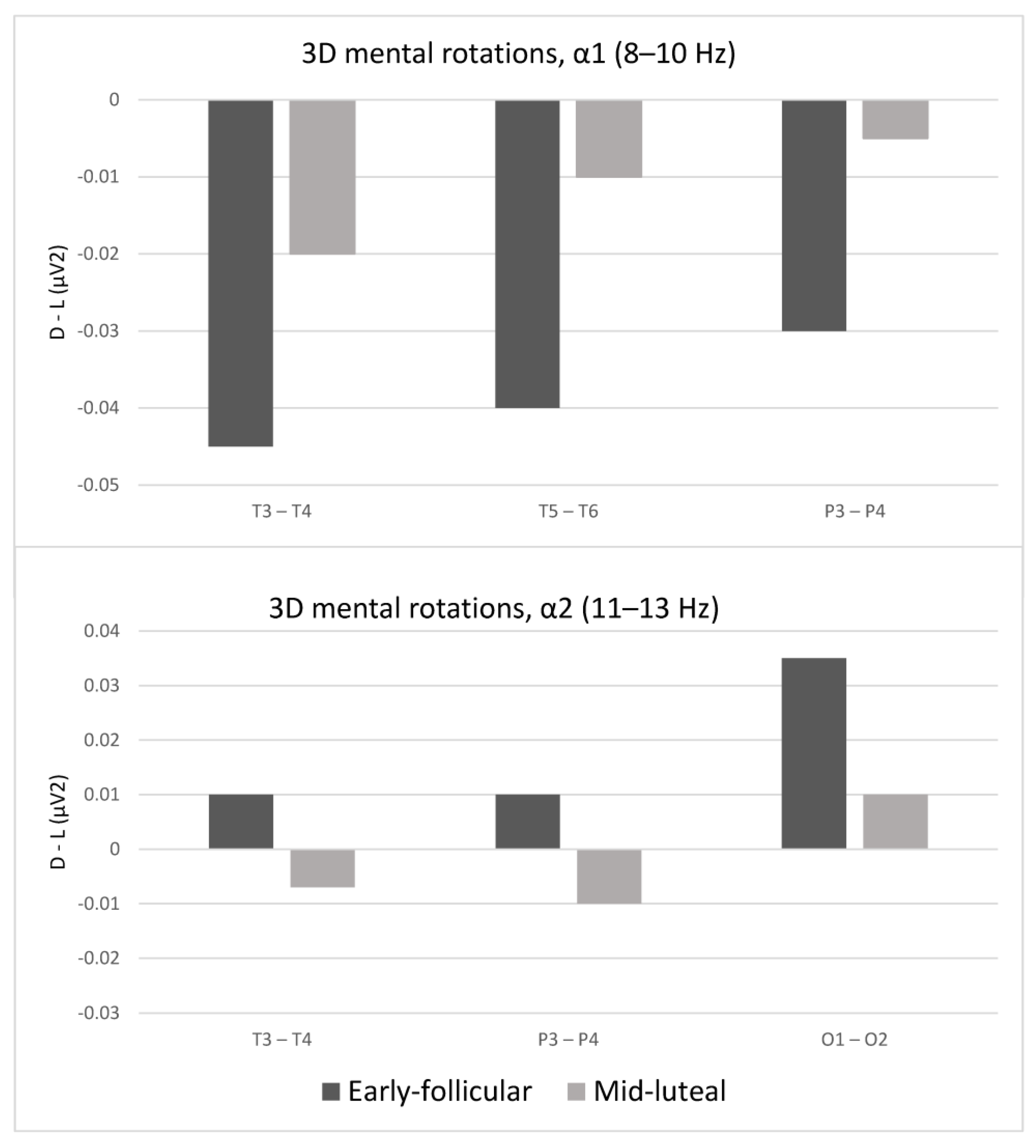
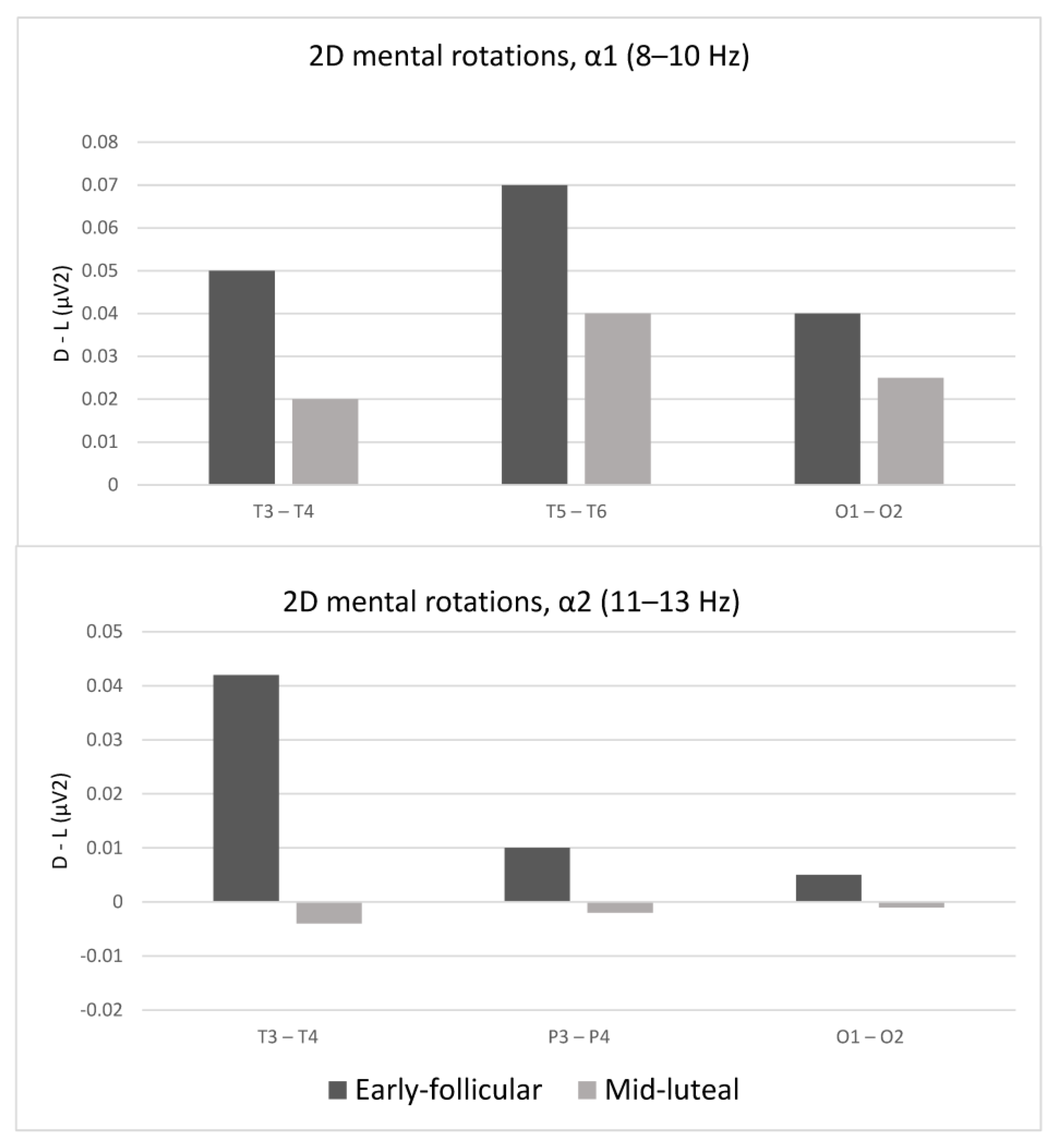
The results of ANOVAs for laterality shifts during the solving of mental rotations are shown in Table 2. As can be seen from the table, several posterior locations showed significant shifts in laterality scores, in both lower (8–10 Hz) and higher (11–13 Hz) α frequency bands. The direction of these changes is shown in Figure 2 and Figure 3 (for easier interpretation, the figures depict only significant shifts in laterality scores).

4. Discussion

Regarding the speed and accuracy of solving the mental rotation tasks, our results follow the pattern observed in previous studies [31,58]: only the efficiency of solving the 3D tasks changes as a function of the menstrual cycle; women are faster and more accurate in the early follicular phase, i.e., when estrogen and progesterone levels are low.
As for neural asymmetries, we observed several regularities in the lower alpha (α1) frequency band while participants were solving the 3D tasks: first, in both phases of the menstrual cycle, the asymmetry indices were negative, indicating a greater relative activation of the right hemisphere compared to the left. This was an expected result, and it is in line with the notion that the effect of the menstrual cycle phase on the efficiency of solving 3D mental rotation tasks is related to the hormonally induced shifts in laterality [13,18,19,47]. In the lower alpha (α2) frequency band, the pattern of laterality shifts was somewhat different. Asymmetries in the occipital channels were greater in the early follicular than in the mid-luteal phase of the cycle, which was expected, but the asymmetries in the parietal and temporal locations shifted not only in magnitude but also in their direction, as a function of the menstrual cycle: in the early follicular phase, a greater activation was observed in left hemisphere locations. However, it should also be noted that these asymmetries (regardless of their direction) were relatively small: following the notion that the α2 part of the spectrum is particularly sensitive to processes related to the processing of spatial information [57,59], we can interpret this finding as a consequence of the complexity of the task, the successful solution of which requires equal activation of both hemispheres. Based on this, it could be hypothesized that as a function of the menstrual cycle, there are changes in the domain of attention and working memory, as well as in the domain of specific mental operations required for processing the spatial components of the task.
While participants were solving the 2D mental rotation tasks, in the α1 frequency band, we observed positive asymmetry indices on the posterior locations, which indicates a relatively greater activation of the left rather than the right hemisphere. This is consistent with previous reports about possible alternative strategies for solving 2D spatial tasks [13,34,57,58,59,60,61].
Furthermore, the asymmetries on these locations were greater in the early follicular phase of the cycle, which was expected. However, it can be seen from the figures that these asymmetries were comparatively smaller during the 2D mental rotations as compared to the 3D mental rotations, which is also in line with the idea that less hemispheric specialization is required to successfully mentally rotate a 2D figure. The pattern of changes in the α2 frequency band was very similar to that observed in the 3D rotation task: differences in spectral powers measured on the left and right homologous electrodes on the temporal, parietal and occipital electrodes show greater asymmetries in the early follicular versus the mid-luteal phase of the cycle. Moreover, in the luteal phase, asymmetries were almost non-existent, which is in accordance with the interhemispheric decoupling hypothesis [58]. Another peculiarity regarding this task was the fact that even though shifts in neural asymmetries as a function of the menstrual cycle appeared, there were no differences in the efficiency of solving the tasks. This is similar to the finding of an fMRI study [62] in which the participants were presented with a perceptual speed task and a semantic task: although the performance in those two tasks did not differ across the menstrual cycle, there were differences in the patterns of brain activation during the cognitive load, as well as a significant correlation between steroid hormones’ levels and the activation of specific brain regions. The semantic task caused a dominant activation of the left hemisphere (various parts of the frontal lobe and the temporo-parietal area), while the perceptual speed task caused a bilateral activation of the posterior parts of the brain (occipital lobe, inferior temporal gyrus and dorsal parietal lobe). A positive correlation was observed both between the level of progesterone and the activation of the upper gyrus of the temporal lobe and between the level of both progesterone and estradiol and the activation of the medial part of the upper gyrus of the frontal lobe in both hemispheres. The activation of the middle and lower gyrus of the frontal lobe was not related to the amount of steroid hormones.
In summary, the 3D mental rotation tasks showed an unambiguous pattern of results: the participants solved these tasks more accurately and faster in the early follicular phase of the cycle, when estrogen and progesterone levels were low. Accordingly, in both lower and higher alpha frequency bands, larger asymmetries were observed in posterior (temporal, parietal and occipital) locations in the early follicular than in the mid-luteal phase. In the lower alpha band, which reflects attention and working memory, asymmetry indices showed a relatively higher activation of the right hemisphere in both phases of the cycle. In the higher alpha band, which reflects the processing of specific spatial components of the task, the asymmetry indices changed in both size and direction, but these indices were relatively small. This would indicate a likely bilateral activation in this part of the spectrum. Drawing from the notion that different parts of the alpha frequency spectrum are correlates of different psychological processes, we could infer that the effects of sex hormones on the efficacy of solving the mental rotation tasks can be attributed mostly to changes in the processes of attention and working memory and somewhat less to the changes in the specific processing of the spatial components of the task. The shifts in the task-solving efficacy as a function of the menstrual cycle can be attributed to the shifts in hemispheric asymmetries during engagement in the task.

Limitations of the Study

The presented study has several limitations and drawbacks. While it is not unusual for EEG studies, and especially the repeated-measures studies, to have rather small samples, a larger sample might enhance the robustness and reliability of our findings. Furthermore, it might account for interindividual variability in both hormonal profiles throughout the menstrual cycle and cognitive strategies used for solving specific tasks. Due to the time-sensitive nature of menstrual cycle phases and the lack of objective hormonal measures, we tested the participants at only two time-points in the menstrual cycle (early follicular and mid-luteal): the late follicular phase would also be of interest, as it is typically characterized by high estrogen levels and low progesterone levels (which would potentially enable us to differentiate between the specific effects of estrogen vs. progesterone on laterality and/or performance). Moreover, mental rotations are just one facet of spatial ability: in order to fully explore the activational effects of sex hormones on spatial abilities, it would be necessary to include tasks designed to measure other facets such as spatial visualization and orientation. Finally, the low spatial resolution of the EEG makes it impossible to elucidate possible hormonal effects on more specific brain regions, which is why we relied on a rather basic measure of laterality on homologue locations on the left vs. right cerebral hemispheres: studies of a similar design but using more sophisticated methods of functional neuroimaging would surely provide more nuanced conclusions.

5. Conclusions

When solving 3D mental rotation tasks, participants were faster, more accurate and showed higher laterality scores (on posterior locations) in the early follicular compared to mid-luteal phase of the menstrual cycle. For this specific cognitive function, higher laterality scores were observed in both lower and higher alpha bands, indicating changes in processes of attention, working memory and the specific processing of the spatial components of the task. However, when solving 2D mental rotation tasks, there were no shifts in either efficacy or hemispheric laterality as a function of the menstrual cycle. This pattern of results provides further support for the notion that fluctuations in sex hormones affect laterality and thus exert their effect on the lateralized cognitive abilities.

Supplementary Materials

The following supporting information can be downloaded at: https://www.mdpi.com/article/10.3390/sym16020172/s1, Table S1: The results of repeated measures ANOVAs, with menstrual cycle phase as a source of variance and laterality scores during eyes open baseline activity (no cognitive load), as dependent variables; Table S2: The results of repeated measures ANOVAs, with menstrual cycle phase as a source of variance and laterality scores during eyes closed baseline activity (no cognitive load), as dependent variables.

Author Contributions

Conceptualization, I.H. and M.T.; methodology, I.H. and M.T.; software, I.H.; validation, I.H. and M.T.; formal analysis I.H.; data curation, I.H.; writing—original draft preparation, I.H. and M.T.; writing—review and editing, I.H. and M.T.; visualization, I.H.; supervision, M.T. All authors have read and agreed to the published version of the manuscript.

Funding

This research received no external funding.

Institutional Review Board Statement

The study was conducted in accordance with the Declaration of Helsinki and approved by the Institutional Ethics Committee of Department of Psychology, Faculty of Humanities and Social Sciences (approval code EPOP_2021_22_13, 13 April 2021).

Informed Consent Statement

Informed consent was obtained from all subjects involved in the study.

Data Availability Statement

The data presented in this study are available on request from the corresponding author.

Conflicts of Interest

The authors declare no conflicts of interest.

References

  1. Hyer, M.M.; Phillips, L.L.; Neigh, G.N. Sex Differences in Synaptic Plasticity: Hormones and Beyond. Front. Mol. Neurosci. 2018, 11, 266. [Google Scholar] [CrossRef] [PubMed]
  2. Marrocco, J.; McEwen, B.S. Sex in the brain: Hormones and sex differences. Dialog-Clin. Neurosci. 2016, 18, 373–383. [Google Scholar] [CrossRef] [PubMed]
  3. Schwarz, J.M.; McCarthy, M.M. Steroid-induced sexual differentiation of the developing brain: Multiple pathways, one goal. J. Neurochem. 2008, 105, 1561–1572. [Google Scholar] [CrossRef]
  4. Juraska, J.M. Sex differences in “cognitive” regions of the rat brain. Psychoneuroendocrinology 1991, 16, 105–119. [Google Scholar] [CrossRef] [PubMed]
  5. Williams, C.L.; Meck, W.H. The organizational effects of gonadal steroids on sexually dimorphic spatial ability. Psychoneuroendocrinology 1991, 16, 155–176. [Google Scholar] [CrossRef] [PubMed]
  6. Isgor, C.; Sengelaub, D.R. Prenatal Gonadal Steroids Affect Adult Spatial Behavior, CA1 and CA3 Pyramidal Cell Morphology in Rats. Horm. Behav. 1998, 34, 183–198. [Google Scholar] [CrossRef]
  7. Bachevalier, J.; Hagger, C. Sex differences in the development of learning abilities in primates. Psychoneuroendocrinology 1991, 16, 177–188. [Google Scholar] [CrossRef]
  8. Reinisch, J.M.; Sanders, S.A. Effects of prenatal exposure to diethylstilbestrol (DES) on hemispheric laterality and spatial ability in human males. Horm. Behav. 1992, 26, 62–75. [Google Scholar] [CrossRef]
  9. Reinisch, J.M.; Ziemba-Davis, M.; Sanders, S.A. Hormonal contributions to sexually dimorphic behavioral development in humans. Psychoneuroendocrinology 1991, 16, 213–278. [Google Scholar] [CrossRef]
  10. Hines, M.; Sandberg, E.C. Sexual Differentiation of Cognitive Abilities in Women Exposed to Diethylstilbestrol (DES) Prenatally. Horm. Behav. 1996, 30, 354–363. [Google Scholar] [CrossRef]
  11. Wilcox, A.J.; Maxey, J.; Herbst, A.L. Prenatal diethylstilbestrol exposure and performance on college entrance examinations. Horm. Behav. 1992, 26, 433–439. [Google Scholar] [CrossRef]
  12. Hampson, E. Spatial cognition in humans: Possible modulation by androgens and estrogens. J. Psychiatry Neurosci. 1995, 20, 397–404. [Google Scholar]
  13. Kimura, D. Sex and Cognition; The MIT Press: Cambridge, MA, USA, 2001. [Google Scholar]
  14. Hampson, E. Sexual Differentiation of Spatial Functions in Humans. In Sexual Differentiation of the Brain; CRC Press: Boca Raton, FL, USA, 1999. [Google Scholar]
  15. Hines, M.; Golombok, S.; Rust, J.; Johnston, K.J.; Golding, J. Avon Longitudinal Study of Parents and Children Study Team Testosterone during Pregnancy and Gender Role Behavior of Preschool Children: A Longitudinal, Population Study. Child Dev. 2002, 73, 1678–1687. [Google Scholar] [CrossRef] [PubMed]
  16. Hines, M. Prenatal testosterone and gender-related behaviour. Eur. J. Endocrinol. 2006, 155, S115–S121. [Google Scholar] [CrossRef] [PubMed]
  17. Knickmeyer, R.C.; Wheelwright, S.; Taylor, K.; Raggatt, P.; Hackett, G.; Baron-Cohen, S. Gender-Typed Play and Amniotic Testosterone. Dev. Psychol. 2005, 41, 517–528. [Google Scholar] [CrossRef]
  18. Hampson, E. Variations in sex-related cognitive abilities across the menstrual cycle. Brain Cogn. 1990, 14, 26–43. [Google Scholar] [CrossRef] [PubMed]
  19. Hampson, E. Estrogen-related variations in human spatial and articulatory-motor skills. Psychoneuroendocrinology 1990, 15, 97–111. [Google Scholar] [CrossRef]
  20. Hampson, E.; Kimura, D. Reciprocal effects of hormonal fluctuations on human motor and perceptual-spatial skills. Behav. Neurosci. 1988, 102, 456–459. [Google Scholar] [CrossRef]
  21. Rosenberg, L.; Park, S. Verbal and spatial functions across the menstrual cycle in healthy young women. Psychoneuroendocrinology 2002, 27, 835–841. [Google Scholar] [CrossRef]
  22. Saucier, D.M.; Kimura, D. Intrapersonal motor but not extrapersonal targeting skill is enhanced during the midluteal phase of the menstrual cycle. Dev. Neuropsychol. 1998, 14, 385–398. [Google Scholar] [CrossRef]
  23. Smith, C.A.; McCleary, C.A.; Murdock, G.A.; Wilshire, T.W.; Buckwalter, D.K.; Bretsky, P.; Marmol, L.; Gorsuch, R.L.; Buckwalter, J. Lifelong Estrogen Exposure and Cognitive Performance in Elderly Women. Brain Cogn. 1999, 39, 203–218. [Google Scholar] [CrossRef]
  24. Carlson, L.E.; Sherwin, B.B. Steroid Hormones, Memory and Mood in a Healthy Elderly Population. Psychoneuroendocrinology 1998, 23, 583–603. [Google Scholar] [CrossRef]
  25. Resnick, S.M.; Metter, E.J.; Zonderman, A.B. Estrogen replacement therapy and longitudinal decline in visual memory: A Possible Protective Effect? Neurology 1997, 49, 1491–1497. [Google Scholar] [CrossRef] [PubMed]
  26. Maki, P.M.; Rich, J.B.; Rosenbaum, R.S. Implicit memory varies across the menstrual cycle: Estrogen effects in young women. Neuropsychologia 2001, 40, 518–529. [Google Scholar] [CrossRef] [PubMed]
  27. Sherwin, B.B. Estrogen and cognitive aging in women. Trends Pharmacol. Sci. 2002, 23, 527–534. [Google Scholar] [CrossRef] [PubMed]
  28. Duff, S.J.; Hampson, E. A Beneficial Effect of Estrogen on Working Memory in Postmenopausal Women Taking Hormone Replacement Therapy. Horm. Behav. 2000, 38, 262–276. [Google Scholar] [CrossRef] [PubMed]
  29. Ho, H.-Z.; Gilger, J.W.; Brink, T.M. Effects of Menstrual Cycle on Spatial Information-Processes. Percept. Mot. Ski. 1986, 63, 743–751. [Google Scholar] [CrossRef] [PubMed]
  30. Silverman, I.; Phillips, K. Effects of estrogen changes during the menstrual cycle on spatial performance. Ethol. Sociobiol. 1993, 14, 257–269. [Google Scholar] [CrossRef]
  31. Phillips, K.; Silverman, I. Differences in the Relationship of Menstrual Cycle Phase to Spatial Performance on Two- and Three-Dimensional Tasks. Horm. Behav. 1997, 32, 167–175. [Google Scholar] [CrossRef]
  32. Galaburda, A.M. Anatomic Basis of Cerebral Dominance. In Brain Asymmetry; Davidson, R.J., Hugdahl, K., Eds.; The MIT Press: Cambridge, MA, USA, 1995; pp. 51–73. [Google Scholar]
  33. Zaidel, E.; Aboitiz, F.; Clarke, J.M.; Kaiser, D.; Matteson, R. Hemispheric Communication: Mechanisms & Models. In Sex Differences in Interhemispheric Relations for Language; Kitterle, U.F., Ed.; Lawrence Erlbaum Associates: Hillsdale, NJ, USA, 1995; pp. 85–175. [Google Scholar]
  34. Serrati, C.; Finocchi, C.; Calautti, C.; Bruzzone, G.L.; Colucci, M.; Gandolfo, C.; Del Sette, M.; Lantieri, P.B.; Favale, E. Absence of Hemispheric Dominance for Mental Rotation Ability: A Transcranial Doppler Study. Cortex 2000, 36, 415–425. [Google Scholar] [CrossRef]
  35. Richter, W.; Somorjai, R.; Summers, R.; Jarmasz, M.; Menon, R.S.; Gati, J.S.; Georgopoulos, A.P.; Tegeler, C.; Ugurbil, K.; Kim, S.-G. Motor Area Activity During Mental Rotation Studied by Time-Resolved Single-Trial fMRI. J. Cogn. Neurosci. 2000, 12, 310–320. [Google Scholar] [CrossRef]
  36. Cabeza, R.; Nyberg, L. Imaging Cognition II: An Empirical Review of 275 PET and fMRI Studies. J. Cogn. Neurosci. 2000, 12, 1–47. [Google Scholar] [CrossRef]
  37. Jordan, K.; Heinze, H.-J.; Lutz, K.; Kanowski, M.; Jäncke, L. Cortical Activations during the Mental Rotation of Different Visual Objects. NeuroImage 2001, 13, 143–152. [Google Scholar] [CrossRef]
  38. Michel, C.M.; Thut, G.; Morand, S.; Khateb, A.; Pegna, A.J.; de Peralta, R.G.; Gonzalez, S.; Seeck, M.; Landis, T. Electric source imaging of human brain functions. Brain Res. Rev. 2001, 36, 108–118. [Google Scholar] [CrossRef]
  39. Levin, S.L.; Mohamed, F.B.; Platek, S.M. Common Ground for Spatial Cognition? A Behavioral and fMRI Study of Sex Differences in Mental Rotation and Spatial Working Memory. Evol. Psychol. 2005, 3, 147470490500300. [Google Scholar] [CrossRef]
  40. Luo, H.; Ni, J.-T.; Li, Z.-H.; Li, X.-O.; Zhang, D.-R.; Zeng, F.-G.; Chen, L. Opposite patterns of hemisphere dominance for early auditory processing of lexical tones and consonants. Proc. Natl. Acad. Sci. USA 2006, 103, 19558–19563. [Google Scholar] [CrossRef] [PubMed]
  41. Harris, I.M.; Egan, G.F.; Sonkkila, C.; Tochon-Danguy, H.J.; Paxinos, G.; Watson, J.D.G. Selective right parietal lobe activation during mental rotation. Brain 2000, 123, 65–73. [Google Scholar] [CrossRef] [PubMed]
  42. Davidson, R.J.; Chapman, J.P.; Chapman, L.J.; Henriques, J.B. Asymmetrical Brain Electrical Activity Discriminates between Psychometrically-Matched Verbal and Spatial Cognitive Tasks. Psychophysiology 1990, 27, 528–543. [Google Scholar] [CrossRef] [PubMed]
  43. Hoptman, M.J.; Davidson, R.J. Baseline eeg asymmetries and performance on neuropsychological tasks. Neuropsychologia 1998, 36, 1343–1353. [Google Scholar] [CrossRef] [PubMed]
  44. Sutton, S.K.; Davidson, R.J. Prefrontal brain electrical asymmetry predicts the evaluation of affective stimuli. Neuropsychologia 2000, 38, 1723–1733. [Google Scholar] [CrossRef] [PubMed]
  45. Davidson, R.J.; Hugdahl, K. Brain Asymmetry; MIT Press: Cambridge, MA, USA, 1995; Available online: https://mitpress.mit.edu/9780262540797/brain-asymmetry/ (accessed on 9 December 2023).
  46. Nettle, D. Hand laterality and cognitive ability: A multiple regression approach. Brain Cogn. 2003, 52, 390–398. [Google Scholar] [CrossRef] [PubMed]
  47. Hausmann, M.; Güntürkün, O. Steroid fluctuations modify functional cerebral asymmetries: The hypothesis of progesterone-mediated interhemispheric decoupling. Neuropsychologia 2000, 38, 1362–1374. [Google Scholar] [CrossRef] [PubMed]
  48. Hausmann, M. Functional cerebral asymmetries during the menstrual cycle: A cross-sectional and longitudinal analysis. Neuropsychologia 2002, 40, 808–816. [Google Scholar] [CrossRef] [PubMed]
  49. Compton, R.J.; Costello, C.; Diepold, J. Interhemispheric integration during the menstrual cycle: Failure to confirm progesterone-mediated interhemispheric decoupling. Neuropsychologia 2004, 42, 1496–1503. [Google Scholar] [CrossRef] [PubMed]
  50. Weis, S.; Hausmann, M.; Stoffers, B.; Vohn, R.; Kellermann, T.; Sturm, W. Estradiol Modulates Functional Brain Organization during the Menstrual Cycle: An Analysis of Interhemispheric Inhibition. J. Neurosci. 2008, 28, 13401–13410. [Google Scholar] [CrossRef] [PubMed]
  51. Blake, K.R.; Dixson, B.J.; O’Dean, S.M.; Denson, T.F. Standardized protocols for characterizing women’s fertility: A data-driven approach. Horm. Behav. 2016, 81, 74–83. [Google Scholar] [CrossRef] [PubMed]
  52. Gangestad, S.W.; Haselton, M.G.; Welling, L.L.; Gildersleeve, K.; Pillsworth, E.G.; Burriss, R.P.; Larson, C.M.; Puts, D.A. How valid are assessments of conception probability in ovulatory cycle research? Evaluations, recommendations, and theoretical implications. Evol. Hum. Behav. 2016, 37, 85–96. [Google Scholar] [CrossRef]
  53. Peters, M.; Laeng, B.; Latham, K.; Jackson, M.; Zaiyouna, R.; Richardson, C. A Redrawn Vandenberg and Kuse Mental Rotations Test—Different Versions and Factors That Affect Performance. Brain Cogn. 1995, 28, 39–58. [Google Scholar] [CrossRef]
  54. French, J.W.; Ekstrom, R.B.; Price, L.A. Manual for Kit of Reference Tests for Cognitive Factors (Revised 1963); Defense Technical Information Center: Fort Belvoir, VA, USA, 1963. [Google Scholar]
  55. Schneider, W.; Eschman, A.; Zuccolotto, A. E-Prime User’s Guide; Psychology Software Tools, Inc.: Pittsburgh, PA, USA, 2002. [Google Scholar]
  56. Corsi-Cabrera, M.; Solís-Ortiz, S.; Guevara, M. Stability of EEG inter-and intrahemispheric correlation in women. Electroencephalogr. Clin. Neurophysiol. 1997, 102, 248–255. [Google Scholar] [CrossRef]
  57. Gevins, A.; Smith, M.E.; Mcevoy, L.; Yu, D. High Resolution EEG Mapping of Cortical Activation Related to Working Memory: Effects of Task Difficulty, Type of Processing, and Practice. Cereb. Cortex 1997, 7, 374–385. [Google Scholar] [CrossRef]
  58. Geary, D.C. Male, Female: The Evolution of Human Sex Differences, 3rd ed.; American Psychological Association: Washington, DC, USA, 2020. [Google Scholar]
  59. Roberts, J.E.; Bell, M.A. 2- and 3-dimensional mental rotation tasks lead to different parietal laterality for men and women. Int. J. Psychophysiol. 2003, 50, 235–246. [Google Scholar] [CrossRef] [PubMed]
  60. Jordan, K.; Wüstenberg, T.; Heinze, H.-J.; Peters, M.; Jäncke, L. Women and men exhibit different cortical activation patterns during mental rotation tasks. Neuropsychologia 2002, 40, 2397–2408. [Google Scholar] [CrossRef] [PubMed]
  61. Voyer, D.; Voyer, S.; Bryden, M.P. Magnitude of sex differences in spatial abilities: A meta-analysis and consideration of some critical variables. Psychol. Bull. 1995, 117, 250–270. [Google Scholar] [CrossRef] [PubMed]
  62. Fernández, G.; Weis, S.; Stoffel-Wagner, B.; Tendolkar, I.; Reuber, M.; Beyenburg, S.; Klaver, P.; Fell, J.; de Greiff, A.; Ruhlmann, J.; et al. Menstrual Cycle-Dependent Neural Plasticity in the Adult Human Brain Is Hormone, Task, and Region Specific. J. Neurosci. 2003, 23, 3790–3795. [Google Scholar] [CrossRef]
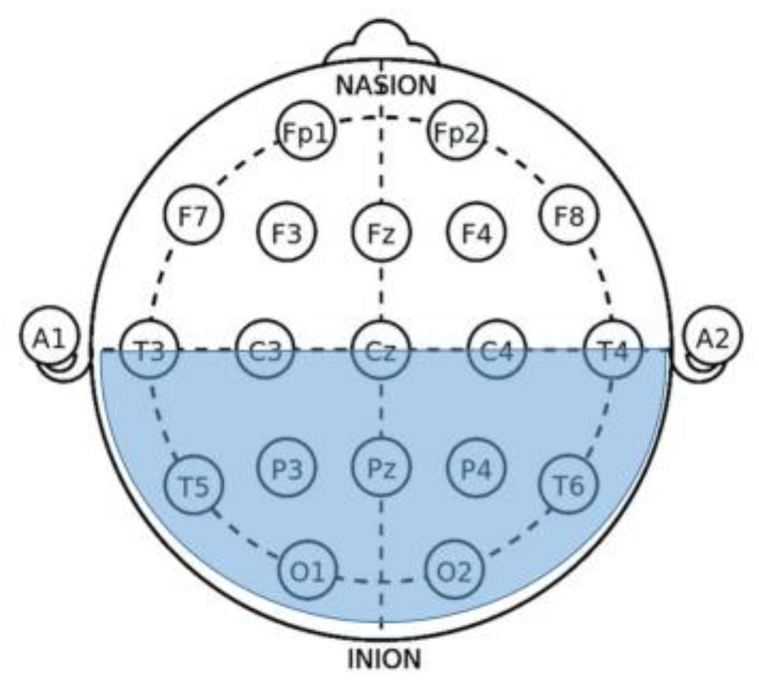
Figure 1. The locations of homologue pairs of electrodes relevant for this study (shaded).
Symmetry 16 00172 g001
Figure 2. Laterality indices while solving 3D mental rotation tasks in the luteal vs. early follicular phase of the menstrual cycle. (Upper panel) Laterality scores within lower α (8–10 Hz) frequency band; (Lower panel) laterality scores within higher α (11–13 Hz) frequency band. Negative scores indicate higher right hemisphere activation; positive scores indicate higher left hemisphere activation.
Symmetry 16 00172 g002
Figure 3. Laterality indices while solving 2D mental rotation tasks in the luteal vs. early follicular phase of the menstrual cycle. (Upper panel) Laterality scores within lower α (8–10 Hz) frequency band; (Lower panel) laterality scores within higher α (11–13 Hz) frequency band. Negative scores indicate higher right hemisphere activation; positive scores indicate higher left hemisphere activation.
Symmetry 16 00172 g003
Table 1. The results of paired samples t-test, with menstrual cycle phase as an independent variable and accuracy and speed of solving the 2D and 3D mental rotation tasks as dependent variables.
Early Follicular PhaseMid-Luteal Phase
MSDMSDt
3D mental rotationsAccuracy11.201.18.31.210.08 ***
Speed (s)8.011.0211.312.018.28 ***
2D mental rotationsAccuracy66.0920.4564.2717.70.38
Speed (s)3.541.353.691.400.44
Note. *** p < 0.001.
Table 2. The results of repeated measures ANOVAs, with menstrual cycle phase as a source of variance and laterality scores (within lower and higher α frequency bands) on each pair of homologous sites as dependent variables, during the solving of 3D and 2D mental rotation tasks.
3D Mental Rotations2D Mental Rotations
Frequency BandFrequency Band
α1 (8–10 Hz)α2 (11–13 Hz)α1 (8–10 Hz)α2 (11–13 Hz)
FFFF
Fp1–Fp20.120.270.760.15
F3–F46.97 **0.561.99 *0.01
F7–F81.380.460.110.98
C3–C42.38 *2.32 *0.190.01
T3–T43.04 *4.38 *2.91 *2.12 *
T5–T62.56 *0.142.28 *0.80
P3–P42.69 *2.86 *0.072.34 *
O1–O20.142.44 *2.03 *1.88 *
Note. ** p < 0.01, * p < 0.05.
Disclaimer/Publisher’s Note: The statements, opinions and data contained in all publications are solely those of the individual author(s) and contributor(s) and not of MDPI and/or the editor(s). MDPI and/or the editor(s) disclaim responsibility for any injury to people or property resulting from any ideas, methods, instructions or products referred to in the content.