Article

Diversity in Primate External Eye Morphology: Previously Undescribed Traits and Their Potential Adaptive Value

by
Juan Olvido Perea-García
1,*,
Dariusz P. Danel
2 and
Antónia Monteiro
1,3
1
Department of Biological Sciences, National University of Singapore, Singapore 117543, Singapore
2
Department of Anthropology, Hirszfeld Institute of Immunology and Experimental Therapy Polish Academy of Sciences, ul. Rudolfa Weigla 12, 53-114 Wroclaw, Poland
3
Science Division, Yale-NUS College, Singapore 138609, Singapore
*
Author to whom correspondence should be addressed.
Symmetry 2021, 13(7), 1270; https://doi.org/10.3390/sym13071270
Submission received: 28 June 2021 / Revised: 13 July 2021 / Accepted: 13 July 2021 / Published: 15 July 2021
(This article belongs to the Special Issue Biometric and Symmetry Issues in Animal and Human Morphology)

Abstract

:
Comparative examinations of external eye morphology in primates initially focused on communicative functions of the eye. Subsequent work has failed to find consistent associations between specific eye morphologies and communicative functions. In this article, we review the field of primate external eye morphology and inspect publicly available and unpublished photographs. We identify and describe five commonly occurring traits that have not received attention so far. We cross-examined the clinical and psychological literature to propose potential adaptive functions. These potential adaptive functions include communicative functions, but also photoregulatory functions and photoprotective functions.

1. Introduction

The field of primate external eye morphology has been shaped by Kobayashi and Kohshima’s seminal paper [1] and its follow-up [2]. In their studies, Kobayashi and Kohshima examined and compared eye shapes in 88 primate species by measuring two morphological indices. The first one, width-to-height ratio (WHR), reflected the distance between the eye corners divided by the longest perpendicular dimension of the eye fissure. The second, developed by the authors, was the index of the amount of exposed sclera (SSI) and was calculated as the width of the exposed eyeball divided by the iris diameter. Interpreting results, the authors concluded that humans had exceptionally horizontally elongated eyes that showed a higher proportion of sclera than other examined species [2]. They also attributed horizontal elongation to habitat (plains) and standing height (related to bipedal locomotion), proposing that it serves as an exaptation to enhance visual access to horizontal movement in the eyes of conspecifics.
In addition to eye shape, Kobayashi and Kohshima [2] classified primate eyes in relation to their patterns of coloration. They compared photographs of the primate species and classified them into four categories, based on contrast patterns between the iris, sclera, and surrounding skin or fur. The visibility of eye contours and iridial and scleral color were the main descriptors used in the original classification. Humans, with highly visible eye outline, light sclera, and contrasting and darker iridial color, were the only species classified in the category considered conspicuous. The other species, having low contrast between these traits, and darker sclerae, were all categorized as having cryptic eye coloration [2]. This observation gave rise to the “gaze camouflage hypothesis”—the proposal that scleral pigmentation evolved for crypticity, while scleral depigmentation evolved for conspicuousness.
The results and conclusions drawn from Kobayashi and Kohshima’s study were attractive in their elegance and attracted follow-up studies that build on them in important ways. Kaplan and Rogers [3] measured the WHR and SSI on a larger sample of photographs of orangutans, obtaining similar values as Kobayashi and Kohshima [2], though they recommend the measurement of the total area of tissue of interest rather than ratios, or indexes, composed from linear measurements (cf. [4] and their SSR index used to measure human eye morphology). Kaplan and Rogers’ study [3] also included, for the first time, the Sumatran orangutan subspecies (Pongo abelii). Using detailed video analyses of gazing patterns, they found, consistent with the literature on sign language in great apes, that orangutans preferred to gaze sideways rather than face forward. Since sideways gazing in orangutans may expose a similar proportion of sclera as direct gazing in humans, the authors suggested that measuring gaze direction patterns may be important when comparing communicative behaviors in different species [3]. Kaplan and Rogers’ article [3] also pointed out that some nonhuman primates feature conspicuous eye outlines owing to the contrast between eyelids and surrounding skin or hair. For example, infant orangutans have light eyelids that remain depigmented in Sumatran orangutans [3]. They also suggested eyelashes as a trait that may be involved in social signaling in Sumatran orangutans.
Mayhew and Gómez [5] continued focusing on great apes. They sampled photographs of 85 gorillas of both extant species (G. gorilla gorilla, n = 60; G. beringei beringei, n = 25), measured their width-to-height ratio and amount of exposed sclera, and categorized the coloration of the eyes relying on the pigmentation in the sclera, with no regard for the color of the surrounding fur or skin, nor iris color. In judging scleral pigmentation, Mayhew and Gómez [5] used an ordinal scale from 0, for completely pigmented, to 10, for human-like depigmentation, as well as a qualitative classification which distinguished six different patterns of scleral pigmentation found in the gorillas. Figure 1 illustrates this classification system. By including both recognized species of gorilla and increasing the sample number from 4 in Kobayashi and Kohshima’s study [2] to 85, Mayhew and Gómez [5] found that scleral pigmentation patterns are variable across species, and that different populations within a species show differences in the frequency of each of the patterns, i.e., species are also polymorphic. With their study, Mayhew and Gómez [5] challenged the “gaze camouflage” hypothesis [2] in two ways. Firstly, they showed that scleral depigmentation occurs within a continuum in gorillas rather than being a discrete, fixed trait. Secondly, they established that the trait is polymorphic, so that more than a few photographs are required to accurately represent natural variation. Calling attention to the observation that not all humans have completely depigmented sclerae, Mayhew and Gómez [5] finished their study concluding that, at least among great apes, it is the horizontal elongation of the eye rather than scleral depigmentation that distinguishes the human eye.
Perea-García (Perea-García 2016) later showed that different subspecies of orangutan (P. pygmaeus and P. abelii) also presented distinct patterns of scleral coloration at the species level. To circumvent the issues derived from subjective judgements of photographs, Perea-García [6] applied quantitative methods to measure the contrast between iris and sclera. A proxy for eye conspicuity was proposed, which the author termed highest contrast (HC). Highest Contrast is the difference in grayscale values between the iris and sclera in the same eye. In grayscale, values can go from 0 (completely dark) to 255 (completely bright). For example, a sclera with a grayscale value of 180 and an iris with a grayscale value of 80 would result in a HC of 100. Perea-García’s study [6] showed that P. abelii can present a HC between the sclera and iris that is comparable to that of humans.
In another study, Perea-García et al. [7] compared the eye coloration of chimpanzees (Pan troglodytes, n = 50) and bonobos (Pan paniscus, n = 51) with that of humans (n = 52). Building on Perea-García et al.’s [8] methods, Perea-García et al. [7] used relative measurements of contrast (RIL). Unlike HC, which is an absolute measure of the difference in brightness between two adjacent parts of the eye, RIL measures how bright the darker part of the eye is when compared to the lighter part. Using these methods, Perea-García et al. [7] proposed two patterns of contrasting coloration: Type 1, or human-like, where the sclera is lighter than the iris (Figure 2A); Type 2, or chimp-like, where the sclera is darker than the iris (Figure 2B). The authors also showed that, despite these differences, the three examined species show similar levels of relative contrast between the iris and the sclera [7]. These results contributed to our understanding of great ape external eye morphology with two important observations: firstly, they strongly suggested that external eye morphology is under evolutionary pressures leading to bonobos and humans sharing more similarities (light sclera) than the more closely related bonobos and chimpanzees (light and dark sclera). Secondly, it provided further morphological evidence against the “gaze camouflage” hypothesis as put forward by Kobayashi and Kohshima, since all three species showed a comparable relative contrast between their iris and sclera, even when the sclera was dark. This may be due to Kobayashi and Kohshima’s assumption that irises are uniformly dark in nonhuman primates.
Recently, Caspar et al. [9] combined the methods from Perea-García [6] and Perea-García et al. ([7,8] to compare the adequacy of absolute (HC) and relative (RIL) measurements in comparing Hominoid external eye morphology. They concluded that interspecific comparisons with RIL may overestimate similarities, especially in comparisons between species in which the sclerae are very different in brightness. Caspar et al. suggested the use of HC instead. The authors found no consistent patterns of coloration that could point at communicative functions of eye coloration across all these species and called for a reassessment of our understanding of the adaptive pressures that drive patterns of eye color.
Lastly, Mearing and Koops (2021) [10] compared the greyscale values of irises and sclerae of images of chimpanzees (P. troglodytes, n = 58), bonobos (P. paniscus, n = 49) and humans (n = 65). Humans had the most depigmented sclerae, followed by bonobos and chimpanzees. No significant differences were reported in grayscale values of the iris. They also compared the RIL of blue- and brown-eyed individuals in their human sample, finding significant differences in the RIL between them. Interspecies comparisons in RIL were, however, not significant. Pointing at the fact that RIL measurements consist of a ratio, Mearing and Koops also bring attention to the inadequacy of the measurement to appropriately characterize cryptic colorations. This led Mearing and Koops to propose absolute grayscale values of the sclera to be the better estimator of gaze conspicuity in interspecies comparisons.

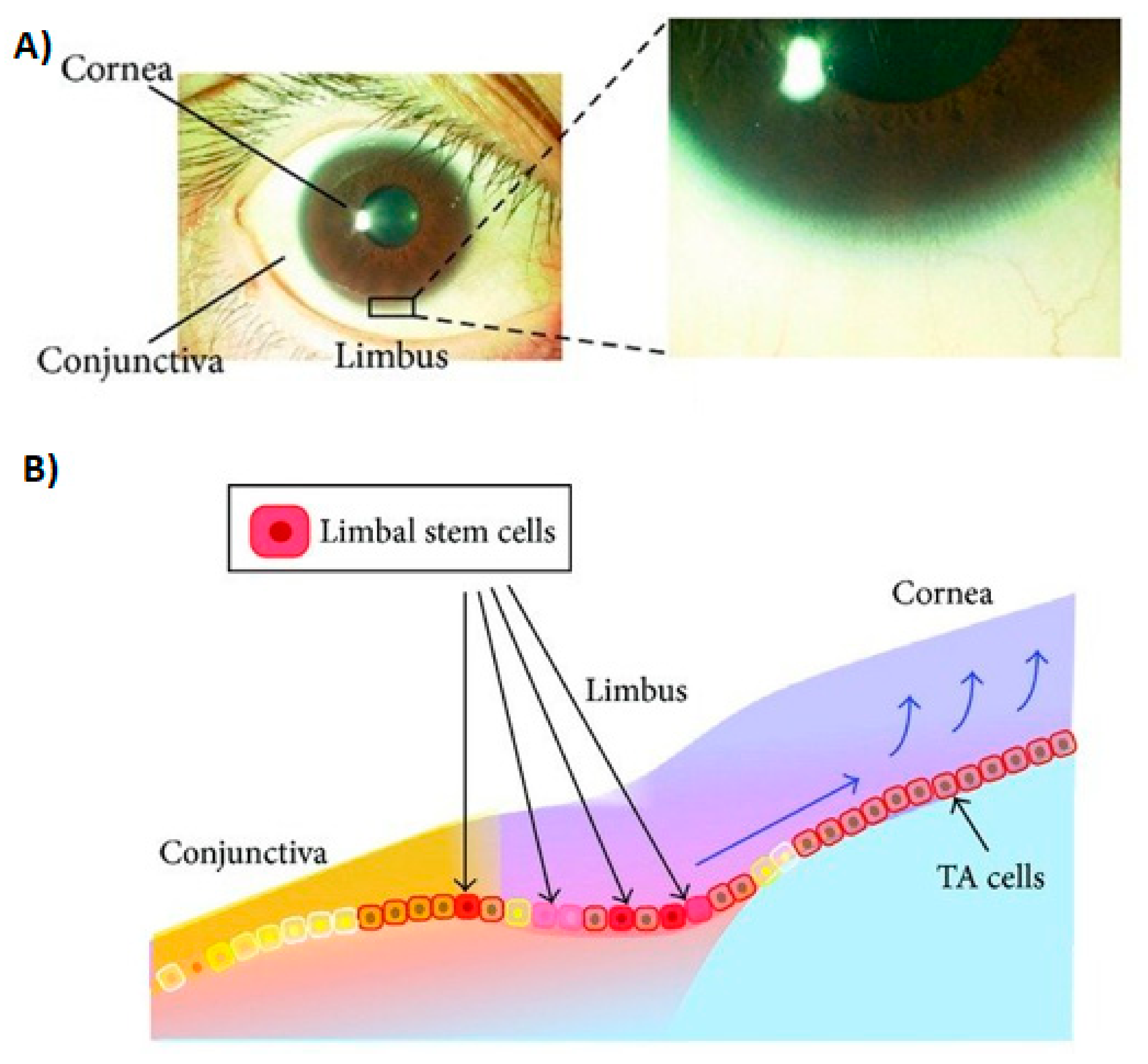
2. Unreported Traits in External Primate Eye Morphology and Their Potential Adaptive Value

Primate eyes are complex organs that have a series of unexplored traits that might additionally play a variety of communication or protective functions. We conducted preliminary observations of photographic samples in simian primates (Old and New World monkeys and apes). We found that there are other traits that may contribute to communication by informing observers about aspects such as gaze direction, emotional or health status (Figure 3). Cross-examination of human psychological and ophthalmological literature suggests that these traits may also be involved in photoprotection. Below, we present the five most notable features that have been poorly explored in the primate literature.

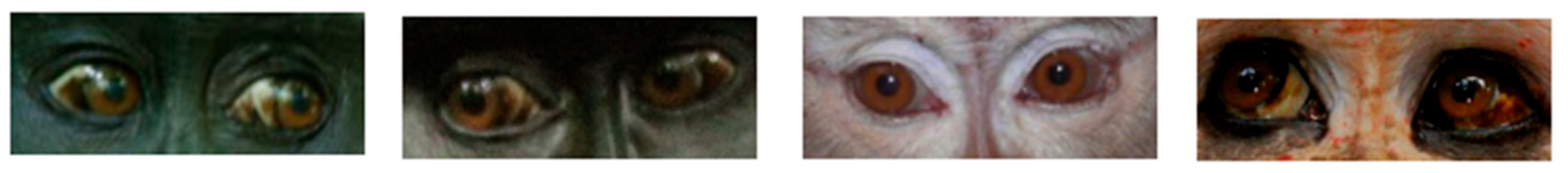
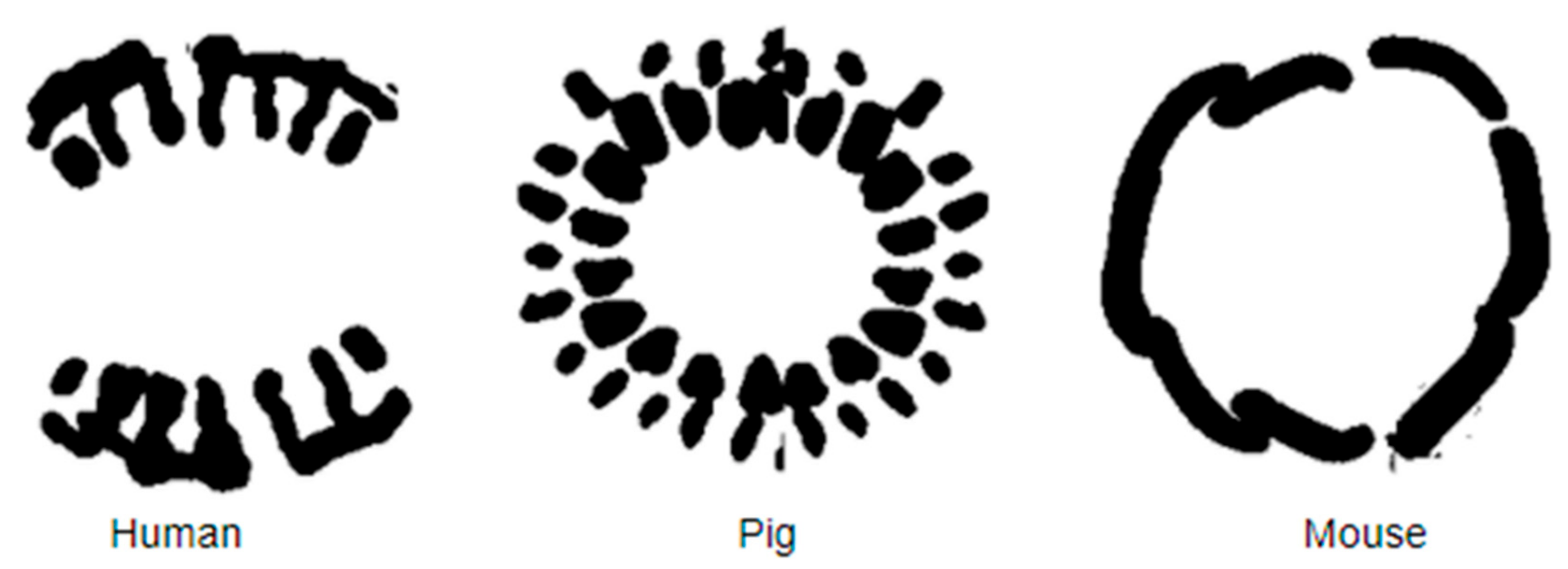
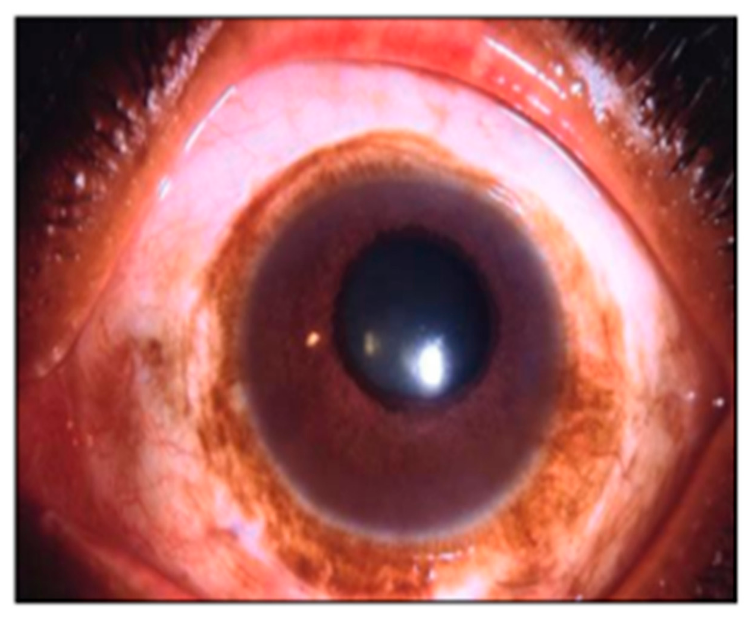
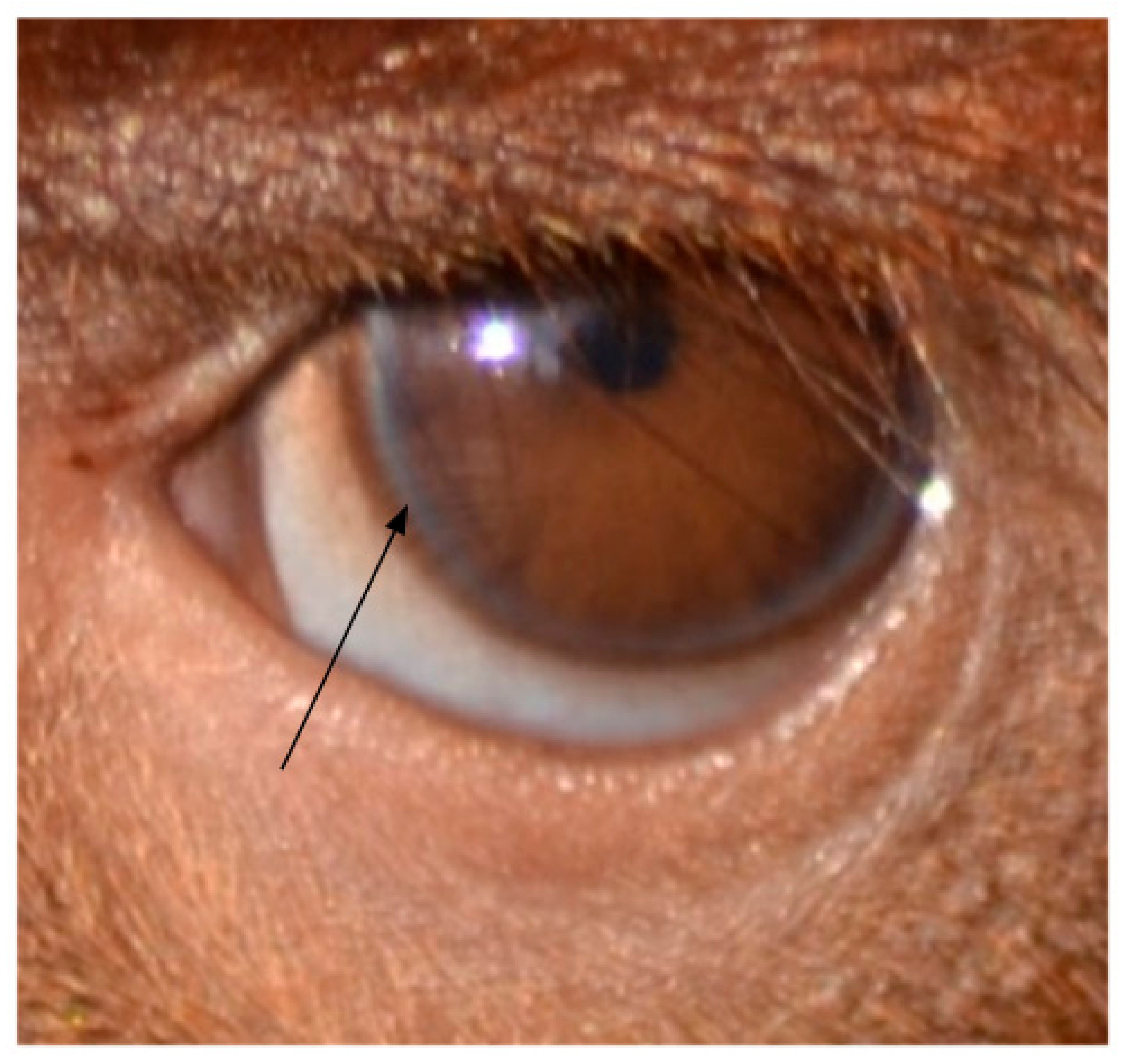
2.1. Dark Symmetric Rings Around the Iris
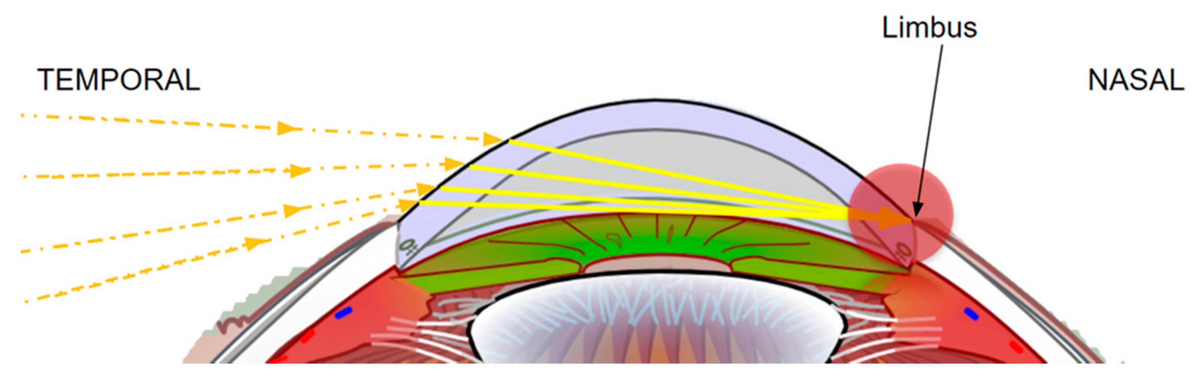
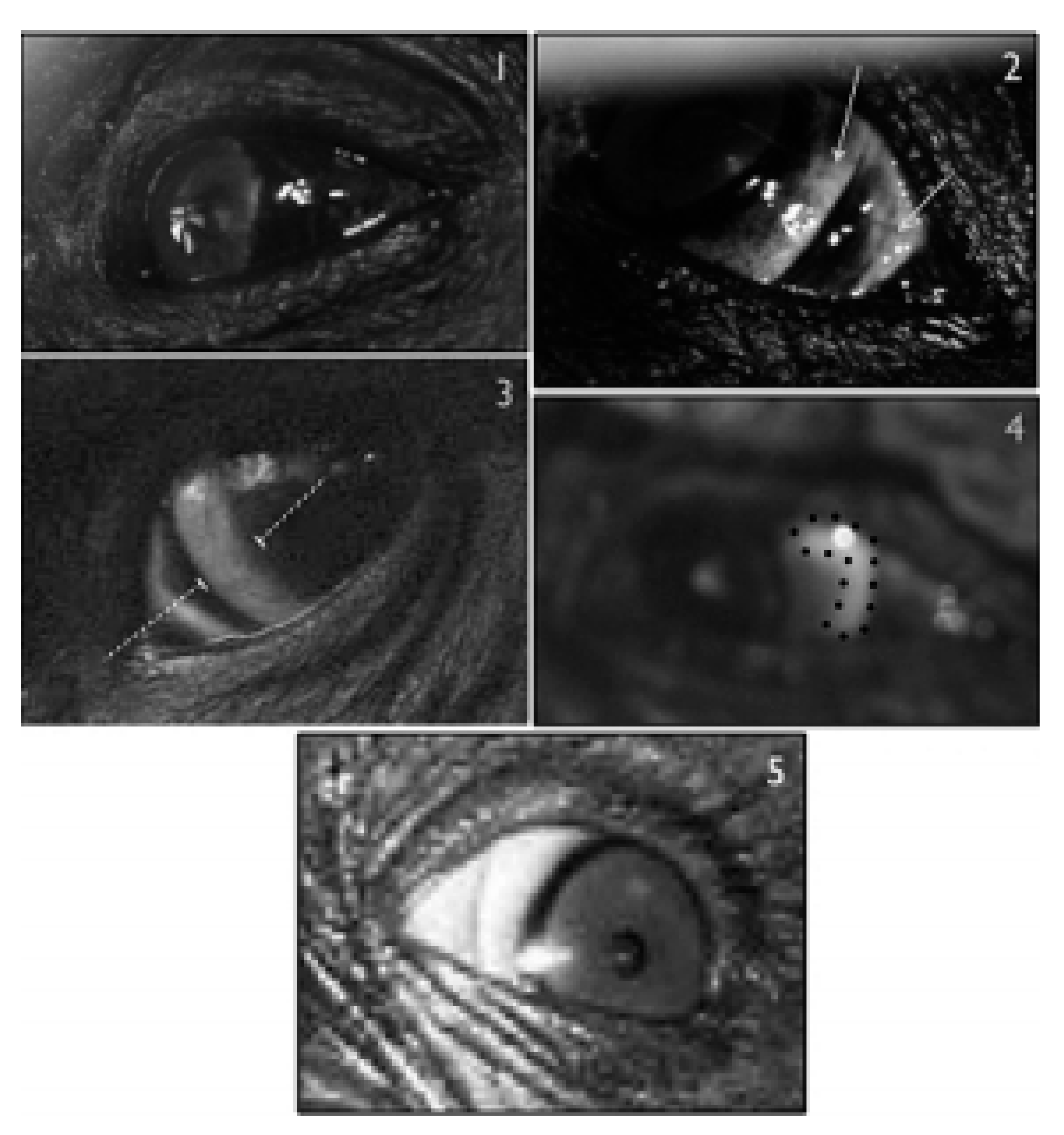
We frequently observed dark rings that were bilaterally symmetric. Sometimes, these rings seemed to be caused by pigment, as in Figure 2. In humans with lighter irises, this area where the cornea and conjunctiva meet is called the limbus, and it may appear darker than the surrounding iris or conjunctiva (Figure 4 below). While some of the darkness can be attributed to structural coloration of the ocular tissues in humans [11], pigments may also contribute to its appearance [12]. The clinical literature has looked at the importance of the limbus as a repository of epithelial stem cells that continuously renew the cornea to keep its transparency, presumably by migrating from the palisades of Vogt, where they are protected from external pathogens and lesions. Keeping the transparency of the cornea is vital for normal vision, and the greater proliferative potential of stem cells can result in an accumulation of transcription errors during meiosis [13]. Protecting the stem cells in the limbus from ultraviolet light [14] or infections [15] seems thus likely functions of limbal rings in humans. It should be noted, however, that protective functions of the limbal ring have not been proposed in the clinical literature.
The psychological literature has explored the role of limbal rings in the perception of age and attractiveness, as well as health. The thickness of limbal rings diminishes with age, and images of eyes with limbal rings were scored as more attractive than the same images without limbal rings [16]. The association between limbal rings and attractiveness has been explained in terms of an honest signal for youth and health (though see [17]). This association seems stronger when rated by women in short-term mating domains [18,19,20,21]). In addition to these functions, we propose that the limbal ring, or similar dark symmetric rings around the iris, may also facilitate the perception of eye-gaze direction because of the contrast between the limbal ring and adjacent lighter parts of the eye (iris and sclera), and should thus be considered when assessing the relationship between external eye morphology and sociocognitive functions across all primates. It should be noted that the resemblance between bilaterally symmetric rings around the iris in humans and nonhumans may be superficial. Further studies examining these structures in nonhumans will be required to ascertain their structure and potential functions.

2.2. Temporal Wedge

The temporal wedge has not been previously named, to our knowledge. It consists of a curved area of bluish tissue surrounding the iris, wider on the temporal than on the nasal side of the eye (visible in Figure 3A,B), sometimes disappearing at the anterior (top) and posterior (bottom) areas of the eye. The temporal wedge may be related to the arcus senilis (see below), which would explain why we observed species displaying either one or the other trait but not both. Two characteristics that distinguish the temporal wedge from the arcus senilis are its color and asymmetrical width. The temporal wedge is a relatively common trait among primates, yet we could only find two mentions of this tissue in Kobayashi and Kohshima [2] and Caspar et al. [9], respectively. Kobayashi and Kohshima [2] place eyes with this trait in a category containing partly white sclera. This category included four callitrichid species: Saguinus midas, S. labiatus, Callithrix argentata and Callimico goeldii that had brown sclera “with a white part in the corner of the eye”. Caspar et al. [9] noted its occurrence and described it as a thin gray line resembling the arcus senilis. There are no putative functions assigned to the temporal wedge since the trait has not been previously individuated. Like dark symmetric rings around the iris resembling the limbal ring, the temporal wedge may aid in inferring gaze direction due to the contrast between it and adjacent tissue like the iris or sclera. Its asymmetrical thickness could aid in recognizing whether the nasal or temporal sides are being observed. For instance, the temporal wedge could be useful to detect gaze aversion—especially in individuals with more pigmented sclera (against which the temporal wedge becomes more conspicuous).

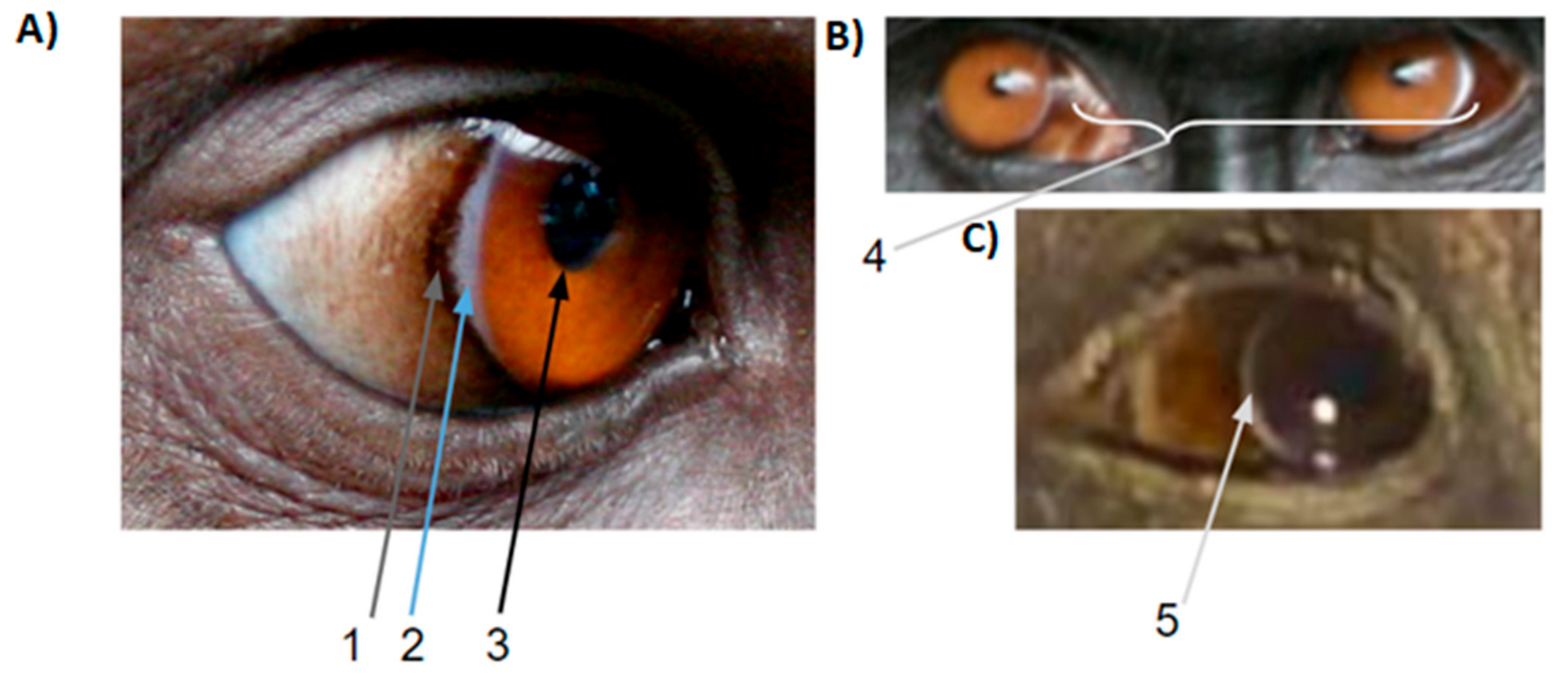
2.3. White Symmetric Rings around the Iris

We frequently observed white symmetric rings around the iris, at least superficially resembling human arcus senilis. Arcus senilis (“elder bow” in Latin) is a whitish ring that becomes apparent with age in humans. It consists of deposits of lipids around the cornea. While it is believed to be of little clinical significance, there may be a weak association with cardiovascular disease, as the deposits are believed to result from mild deficiencies in lipid metabolism [23]. Preliminary inspection of photographs indicates that the arcus senilis is present both in humans and in other primates, where it has also been observed in juveniles (Figure 5). While the occurrence of an arcus senilis in humans older than 50 is not considered pathological, its occurrence in those below 50 may be a sign of abnormally high cholesterol (in which case it is termed arcus juvenilis). The whitish ring observed in juveniles may be present before the development of melanogenic processes is completed [24]. In which case, the arcus would become the limbal ring. In addition to its potential functions in communicating access to fat-rich foods, youth, or aging, the arcus senilis may also aid in inferring gaze direction if the surrounding tissue is dark, because observers could see the orientation of the eyeball.

2.4. Iris and Pupil

The pupil is the orifice in the iris through which light enters the retina. The iris, a sphincter muscle, can dilate or constrict to adjust the quantity of light entering the retina, protecting the eye from excessive light and radiation. Therefore, the size of the pupil is inversely correlated to that of the iris. The iris may constrict or dilate as a result of emotional arousal and psychological states [25]. The perception of sadness and fear can be inferred by large pupils, while constriction contributes to the perception of anger [26]. Furthermore, evidence suggests that coordination of pupil size is linked to interpersonal trust [27], especially between members of in-groups [28]. Pupil size synchrony appears early in ontogeny in humans [29] and has been observed in nonhuman primates (chimpanzees: [30]).
In humans, the contrast between pupils and light irises (compared to dark irises) has also been shown to affect judgements of gaze direction [31]. We use the contrast between these two traits to infer direction when the contrast between sclera and iris is experimentally eliminated [32]. Previous studies on primate eye morphology have ignored the contrast between iris and pupil. This could be because of the assumption that iris color is always dark in nonhuman primates [1,2]. However, several nonhuman primate species have light irises [7,9,33] and the contrast between irises and pupils may also be used in gaze detection in these species.
Other features of the iris may also have signaling functions including iris crypts, Wölfflin nodules, contraction furrows, nevi, Brushfield and pigment spots. These elements of human iris architecture are potential markers of our genetic makeup. Although their presence was tested in relation to some personality traits [34], other aspects of their signaling potential appear to be scarcely investigated. Visual access to pupils and variations in iris color should therefore be considered in future investigations of the relationship between external eye morphology and sociocognitive functions.
Lastly, we are unaware of any studies that attempt to explain variation in iris color across primates as an order (although, see [35] for a qualitative examination of iris color in Macaca fuscata). In humans, iris color has been explained predominantly as a result of sexual selection [36]. Blue eye color in lineages with predominantly brown eyes could act as a reproductive barrier preventing hybridization in sympatric species [37]. Correlational evidence suggests that, in humans living in northern latitudes, blue eye color could be an adaptation to maximize blue light reaching the retina, because blue irises have few light-absorbing pigments [38,39]. Blue light is an important stimulus to regulate circadian patterns [40,41] but is scarcer in northern latitudes [42]. Iris color and features of the iris should be investigated to better understand the social or ecological drivers underlying its diversity in primates.

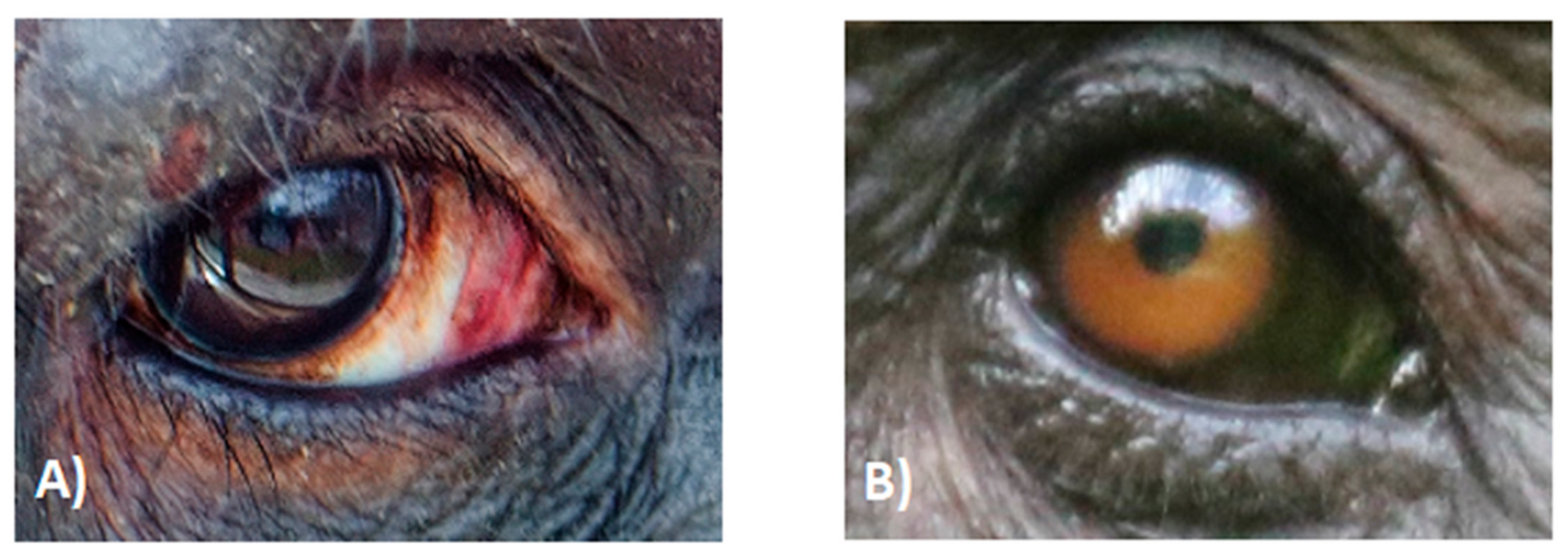
2.5. Local Variation in Scleral Pigmentation

Histological examinations have shown that the pigmented part of the eyeball is the conjunctiva, rather than the sclera [2], but it has become customary in comparative examinations of eye coloration to refer to the conjunctiva as the sclera. Unevenness in scleral pigmentation seems to be common in great apes [5,7] and may allow gaze direction to be inferred in this group, as it would be easy to discriminate which side is being shown. This is especially because the temporal side tends to be more heavily pigmented than the nasal side of the eyeball (Figure 6 below, or “Crescent” pattern in [5]). This uneven pigmentation may also have an important function in protecting the eyeball from UV-B radiation. Due to the shape of the cornea, incident light reaching the temporal side of the eyeball is concentrated and directed to the nasal side, where UV-B radiation can be up to twenty times higher than in the temporal side [43]. In humans, the nasal side of the limbus is associated with greater occurrence of tumours of the conjunctiva linked to UV radiation [44]. Thus, when pigmentation absorbs incident light in the temporal side, it will not be refracted or concentrated in the nasal side of the eyeball (see schematic in Figure 7). Temporal pigmentation may also protect the temporal side itself from UV radiation, since this side of the eye is more exposed to it.
Another aspect of human eye anatomy that reinforces the idea that scleral pigmentation protects the eyeball from harmful UV-B radiation is the near absence of stem cells in the lateral eyeball. By contrast, the vertical eye axis concentrates the great majority of stem cells [45]. Research comparing stem cell distribution in pigs, mice, and humans shows that the orientation of the eyeball relative to the face, the presence or absence of brow ridge, and patterns of activity all influence stem cell distribution [45]. For example, pig eyes are more laterally oriented and are not shielded by brow ridges, resulting in less variation in light and UV-B incidence over the circumference of the eye. As such, pig eyes display stem cells all around the cornea. Mice, which are active at night, have a more even distribution of stem cells [45]. In humans, stem cells are concentrated on the anterior and posterior limbus, protected by the upper and lower eyelids, respectively. Humans, thus, can do without scleral pigmentation to protect these cells. A comparison of the distribution of epithelial stem cells in the limbus of humans, pigs, and mice is represented in Figure 8. We are not aware of any account of the distribution of epithelial stem cells in the limbus of nonhuman primates. Examining the co-distribution of epithelial stem cells in primates and areas of pigmented sclerae could provide additional support for the function of pigmented sclerae in stem cell protection.
In humans, the most notable nonpathogenic source of color variation in sclera is known as complexion-associated melanosis (CAM) [47] (Figure 9). This pattern of pigmentation is described as usually bilateral and symmetrical, more pigmented at the limbus and less pigmented out closer to the posterior eyeball [47] (Figure 9). The distribution of pigmentation in CAM strongly resembles what can be found in many extant nonhuman primates (i.e., pigmented conjunctiva), suggesting that CAM could be an atavism.
Transient changes in scleral coloration in humans have also been suggested to indicate health status, age, and even emotional states [48,49,50]. All these data suggest that the variation in scleral pigmentation should be included while investigating sociocognitive functions of external eye morphology across all primates [47].

3. Discussion

Our understanding of external primate eye morphology and coloration has increased greatly since Kobayashi and Kohshima’s first study. Refinements in qualitative assessment [5] enabled us to capture intraspecies variation and disrupted the notion that humans were qualitatively unique. Quantitative methods provided an estimation of the gradual changes that distinguish species from one another [6,7]. Recent studies have suggested that RIL, one of the currently used proxies of ocular conspicuity, should be used with caution [9,10], especially when establishing interspecies comparisons, or in very dark morphologies. Caspar et al. [9] proposed the use of absolute differences in grayscale values of iris and sclera (following [6]), while Mearing and Koops [10] proposes the use of absolute values of scleral brightness as long as the grayscale values of irises do not differ significantly across the examined species. Because there is substantial variation in iridial brightness in primates (see Section 2.4), we agree with Caspar et al.’s [9] view that HC may be the most adequate measurement of ocular conspicuity currently at use. However, Mearing and Koops [10] pointed out that brighter morphologies may be more noticeable than darker morphologies, even if their HC is similar, simply because brighter objects reflect more light and are, as such, easier to perceive [32]. Until more appropriate measurements are proposed, we think the use of HC in a sufficiently high number of samples (as in [9]) remains the best method for an approximation that is accurate enough to move the field forward. We also recommend that basic measurements of grayscale value, and of HC, be reported so that findings can be critically assessed and built upon. Whenever possible, the photographs from which those measurements were taken should also be made available.
We wanted to bring attention to primate ocular traits that have been overlooked. These traits could be used to facilitate the inference of gaze direction by observers (due to the contrast between them and other adjacent ocular traits). However, beyond aiding with gaze following, these traits could be important for other functions. Eyes are first and foremost organs that mediate the relationship of an organism with its environment through light. The assessment of eyes as organs used in communication should build on a deep understanding of their more basic functions, such as perception and protection. These functions have received surprisingly little attention in the literature, perhaps because Kobayashi and Kohshima’s seminal study [1] focused on communicative functions.
Lastly, the studies reviewed assumed that the detectability of one or several parts of the eye is a reliable proxy of their communicative functions, due to their potential to inform observers. However, functional testing is essential before we can conclude that these traits are used for communicative (or other) functions in any specific species. For example, the less horizontally elongated eyes of orangutans (compared to humans) could suggest that they are not used as cues of attention. However, given that orangutans have a propensity to gazing sideways, and that orangutan sideways gazing exposes as much of the eyeball as in humans, it is possible that they are important for species-specific communicative behaviors [3].

4. Conclusions

In the present study, we show that external eye morphology has not been sufficiently inspected with regards to photoregulatory or photoprotective functions. We reviewed the comparative literature examining external eye morphology in primates. We also inspected publicly available and unpublished photographic samples, finding five commonly occurring traits that have not been identified or discussed in the field of comparative eye morphology. Cross-inspection with the clinical and psychological literature in humans suggests adaptive functions for these traits across a wide range of primate taxa. Since Kobayashi and Kohshima [1,2] rejected the anti-glare hypothesis, the focus in the field has been on the potential for communicative functions of different eye morphologies [5,6,7,9]. Our study calls for a more detailed examination of the ecological drivers, including abiotic drivers, of the diversity underlying the diversity of primate external eye morphology.

Author Contributions

Conceptualization, J.O.P.-G., A.M. and D.P.D.; methodology, J.O.P.-G. and D.P.D.; investigation, J.O.P.-G.; resources, A.M. and J.O.P.-G.; data curation, J.O.P.-G.; writing—original draft preparation, J.O.P.-G.; writing—review and editing, J.O.P.-G., D.P.D. and A.M.; supervision, A.M.; project administration, A.M.; funding acquisition, A.M. All authors have read and agreed to the published version of the manuscript.

Funding

J.O.P.-G.’s Ph.D. was supported by a SINGA scholarship. Partial funding for this project was provided by the National Research Foundation (NRF) Singapore, under its CRP-NRF program (NRF-CRP 20-2017-0001 Award).

Institutional Review Board Statement

Not applicable.

Informed Consent Statement

Not applicable.

Data Availability Statement

The photographs we inspected for this study are available upon reasonable request.

Acknowledgments

We thank Catherine Hobaiter, Jérôme Micheletta (https://www.macaca-nigra.org/en/) (accessed on 2 June 2021). Aurélie Albert-Daviaud, Mariska Kret, Julia Ostner, and Oliver Schülke for letting us use their photographs. The Singapore Zoo and their staff for helping us photograph their orangutans.

Conflicts of Interest

The authors declare no conflict of interest.

References

  1. Kobayashi, H.; Kohshima, S. Unique morphology of the human eye. Nat. Cell Biol. 1997, 387, 767–768. [Google Scholar] [CrossRef] [PubMed]
  2. Kobayashi, H.; Kohshima, S. Unique morphology of the human eye and its adaptive meaning: Comparative studies on external morphology of the primate eye. J. Hum. Evol. 2001, 40, 419–435. [Google Scholar] [CrossRef] [Green Version]
  3. Kaplan, G.; Rogers, L.J. Patterns of Gazing in Orangutans (Pongo pygmaeus). Int. J. Primatol. 2002, 23, 501–526. [Google Scholar] [CrossRef]
  4. Danel, D.P.; Wacewicz, S.; Kleisner, K.; Lewandowski, Z.; Kret, M.E.; Żywiczyński, P.; Perea-Garcia, J.O. Sex differences in ocular morphology in Caucasian people: A dubious role of sexual selection in the evolution of sexual dimorphism of the human eye. Behav. Ecol. Sociobiol. 2020, 74, 1–10. [Google Scholar] [CrossRef]
  5. Mayhew, J.A.; Gomez, J.-C. Gorillas with white sclera: A naturally occurring variation in a morphological trait linked to social cognitive functions. Am. J. Primatol. 2015, 77, 869–877. [Google Scholar] [CrossRef] [Green Version]
  6. García, J.O.P. Quantifying ocular morphologies in extant primates for reliable interspecific comparisons. J. Lang. Evol. 2016, 1, 151–158. [Google Scholar] [CrossRef]
  7. Perea-García, J.O.; Kret, M.E.; Monteiro, A.; Hobaiter, C. Scleral pigmentation leads to conspicuous, not cryptic, eye morphology in chimpanzees. Proc. Natl. Acad. Sci. USA 2019, 116, 19248–19250. [Google Scholar] [CrossRef] [Green Version]
  8. Perea-García, J.O.; Grenzner, T.; Hešková, G.; Mitkidis, P. Not everything is blue or brown: Quantification of ocular coloration in psychological research beyond dichotomous categorizations. Commun. Integr. Biol. 2017, 10, 1. [Google Scholar] [CrossRef] [Green Version]
  9. Caspar, K.R.; Biggemann, M.; Geissmann, T.; Begall, S. Ocular pigmentation in humans, great apes, and gibbons is not suggestive of communicative functions. Sci. Rep. 2021, 11, 1–14. [Google Scholar] [CrossRef]
  10. Mearing, A.S.; Koops, K. Quantifying gaze conspicuousness: Are humans distinct from chimpanzees and bonobos? J. Hum. Evol. 2021, 157, 103043. [Google Scholar] [CrossRef] [PubMed]
  11. Shyu, B.P.; Wyatt, H.J. Appearance of the human eye: Optical contributions to the ‘limbal ring’. Optom. Vis. Sci. 2009, 86, E1069–E1077. [Google Scholar] [CrossRef] [PubMed]
  12. Miri, A.; Al-Aqaba, M.; Otri, A.M.; Fares, U.; Said, D.G.; Faraj, L.; Dua, H.S. In vivo confocal microscopic features of normal limbus. Br. J. Ophthalmol. 2012, 96, 530–536. [Google Scholar] [CrossRef] [PubMed]
  13. Levine, M.S.; Holland, A.J. The impact of mitotic errors on cell proliferation and tumorigenesis. Genes Dev. 2018, 32, 620–638. [Google Scholar] [CrossRef] [Green Version]
  14. Coroneo, M. Ultraviolet Radiation and the Anterior Eye. Eye Contact Lens Sci. Clin. Pract 2011, 37, 214–224. [Google Scholar] [CrossRef]
  15. Das, P.; Pereira, J.A.; Chaklader, M.; Law, A.; Bagchi, K.; Bhaduri, G.; Chaudhuri, S.; Law, S. Phenotypic alteration of limbal niche–associated limbal epithelial stem cell deficiency by ultraviolet-B exposure–induced phototoxicity in mice. Biochem. Cell Biol. 2013, 91, 165–175. [Google Scholar] [CrossRef] [PubMed]
  16. Peshek, D.; Semmaknejad, N.; Hoffman, D.; Foley, P. Preliminary Evidence that the Limbal Ring Influences Facial Attractiveness. Evol. Psychol. 2011, 9, 137–146. [Google Scholar] [CrossRef]
  17. Ilicic, J.; Baxter, S.M.; Kulczynski, A. White eyes are the window to the pure soul: Metaphorical association and overgeneralization effects for spokespeople with limbal rings. Int. J. Res. Mark. 2016, 33, 840–855. [Google Scholar] [CrossRef]
  18. Brown, M.; Sacco, D.F. Put a (Limbal) Ring on It: Women Perceive Men’s Limbal Rings as a Health Cue in Short-Term Mating Domains. Pers. Soc. Psychol. Bull. 2017, 44, 80–91. [Google Scholar] [CrossRef]
  19. Brown, M.; Sacco, D.F.; Medlin, M.M. Women’s short-term mating goals elicit avoidance of faces whose eyes lack limbal rings. Evol. Behav. Sci. 2019, 13, 278–285. [Google Scholar] [CrossRef] [Green Version]
  20. Sacco, D.F.; Brown, M.; Medlin, M. Perfectionism and Relationship Status Influence Health Evaluations of Faces with Limbal Rings. Evol. Psychol. Sci. 2019, 5, 447–453. [Google Scholar] [CrossRef]
  21. Brown, M.; Keefer, L.A.; Sacco, D.F. Relational insecurity heightens sensitivity to limbal rings in partnered women. Pers. Relatsh. 2020, 27, 61–75. [Google Scholar] [CrossRef]
  22. Oie, Y.; Nishida, K. Regenerative Medicine for the Cornea. BioMed Res. Int. 2013, 2013, 428247. [Google Scholar] [CrossRef] [PubMed]
  23. Christoffersen, M.; Frikke-Schmidt, R.; Schnohr, P.; Jensen, G.B.; Nordestgaard, B.G.; Tybjærg-Hansen, A. Xanthelasmata, arcus corneae, and ischaemic vascular disease and death in general population: Prospective cohort study. BMJ 2011, 343. [Google Scholar] [CrossRef] [Green Version]
  24. Brenes, J.A. White Rings: Arcus Lipoides. Am. J. Med. 2013, 126, 112–113. [Google Scholar] [CrossRef]
  25. Harrison, N.A.; Wilson, C.E.; Critchley, H. Processing of observed pupil size modulates perception of sadness and predicts empathy. Emotion 2007, 7, 724–729. [Google Scholar] [CrossRef]
  26. Kret, M.E. The role of pupil size in communication. Is there room for learning? Cogn. Emot. 2017, 32, 1139–1145. [Google Scholar] [CrossRef] [PubMed] [Green Version]
  27. Behrens, F.; Moulder, R.G.; Boker, S.M.; Kret, M. Quantifying physiological synchrony through windowed cross-correlation analysis: Statistical and theoretical considerations. BioRxiv 2020. [Google Scholar] [CrossRef]
  28. Kret, M.E.; de Dreu, C.K. Pupil-mimicry conditions trust in partners: Moderation by oxytocin and group membership. Proc. R. Soc. B Biol. Sci. 2017, 284, 20162554. [Google Scholar] [CrossRef] [Green Version]
  29. Fawcett, C.; Arslan, M.; Falck-Ytter, T.; Roeyers, H.; Gredebäck, G. Human eyes with dilated pupils induce pupillary contagion in infants. Sci. Rep. 2017, 7, 9601. [Google Scholar] [CrossRef] [Green Version]
  30. Kret, M.E.; Tomonaga, M.; Matsuzawa, T. Chimpanzees and humans mimic pupil-size of conspecifics. PLoS ONE 2014, 9, 8. [Google Scholar] [CrossRef]
  31. West, R.W. Perceived Direction of Gaze from Eyes with Dark vs. Light Irises. Optom. Vis. Sci. 2011, 88, 303–311. [Google Scholar] [CrossRef]
  32. Yorzinski, J.L.; Thorstenson, C.A.; Nguyen, T.P. Sclera and Iris Color Interact to Influence Gaze Perception. Front. Psychol. 2021, 12, 676. [Google Scholar] [CrossRef]
  33. Meyer, W.K.; Zhang, S.; Hayakawa, S.; Imai, H.; Przeworski, M. The convergent evolution of blue iris pigmentation in primates took distinct molecular paths. Am. J. Phys. Anthr. 2013, 151, 398–407. [Google Scholar] [CrossRef] [Green Version]
  34. Larsson, M.; Pedersen, N.L.; Stattin, H. Associations between iris characteristics and personality in adulthood. Biol. Psychol. 2007, 75, 165–175. [Google Scholar] [CrossRef] [PubMed]
  35. Zhang, P.; Watanabe, K. Preliminary study on eye colour in Japanese macaques (Macaca fuscata) in their natural habitat. Primates 2006, 48, 122–129. [Google Scholar] [CrossRef]
  36. Frost, P. European hair and eye color: A case of frequency-dependent sexual selection? Evol. Hum. Behav. 2006, 27, 85–103. [Google Scholar] [CrossRef] [Green Version]
  37. Bradley, B.J.; Pedersen, A.; Mundy, N.I. Brief communication: Blue eyes in lemurs and humans: Same phenotype, different genetic mechanism. Am. J. Phys. Anthr. 2009, 139, 269–273. [Google Scholar] [CrossRef] [PubMed]
  38. Goel, N.; Terman, M.; Terman, J.S. Depressive symptomatology differentiates subgroups of patients with seasonal affective disorder. Depress. Anxiety 2002, 15, 34–41. [Google Scholar] [CrossRef]
  39. Workman, L. Blue Eyes Keep away the Winter Blues: Is Blue Eye Pigmentation an Evolved Feature to Provide Resilience to Seasonal Affective Disorder. OA J. Behav. Sci. Psychol. 2018, 1, 180002. [Google Scholar]
  40. Münch, M.; Nowozin, C.; Regente, J.; Bes, F.; De Zeeuw, J.; Hädel, S.; Wahnschaffe, A.; Kunz, D. Blue-Enriched Morning Light as a Countermeasure to Light at the Wrong Time: Effects on Cognition, Sleepiness, Sleep, and Circadian Phase. Neuropsychobiology 2016, 74, 207–218. [Google Scholar] [CrossRef] [PubMed]
  41. Wahl, S.; Engelhardt, M.; Schaupp, P.; Lappe, C.; Ivanov, I.V. The inner clock—Blue light sets the human rhythm. J. Biophotonics 2019, 12, e201900102. [Google Scholar] [CrossRef]
  42. Goldberg, B.; Klein, W. Variations in the spectral distribution of daylight at various geographical locations on the earth’s surface. Sol. Energy 1977, 19, 3–13. [Google Scholar] [CrossRef]
  43. Notara, M.; Lentzsch, A.; Coroneo, M.; Cursiefen, C. The Role of Limbal Epithelial Stem Cells in Regulating Corneal (Lymph)angiogenic Privilege and the Micromilieu of the Limbal Niche following UV Exposure. Stem Cells Int. 2018, 2018, 1–15. [Google Scholar] [CrossRef] [PubMed] [Green Version]
  44. Coroneo, M.T.; Müller-Stolzenburg, N.W.; Ho, A. Peripheral light focusing by the anterior eye and the ophthalmohelioses. Ophthalmic Surg. 1991, 22, 705–711. [Google Scholar] [PubMed]
  45. Grieve, K.; Ghoubay, D.; Georgeon, C.; Thouvenin, O.; Bouheraoua, N.; Paques, M.; Borderie, V. Three-dimensional structure of the mammalian limbal stem cell niche. Exp. Eye Res. 2015, 140, 75–84. [Google Scholar] [CrossRef]
  46. Alqahtani, J.M. Conjunctival Melanosis: Review of the Literature. Bahrain Med Bull. 2013, 35, 206–210. [Google Scholar] [CrossRef]
  47. Shields, J.A.; Shields, C.L.; Mashayekhi, A.; Marr, B.P.; Benavides, R.; Thangappan, A.; Phan, L.; Eagle, R.C., Jr. Primary acquired melanosis of the conjunctiva: Experience with 311 eyes. Trans. Am. Ophthalmol. Soc. 2007, 105, 61. [Google Scholar] [PubMed]
  48. Leung, T.S.; Outlaw, F.; Macdonald, L.W.; Meek, J. Jaundice Eye Color Index (JECI): Quantifying the yellowness of the sclera in jaundiced neonates with digital photography. Biomed. Opt. Express 2019, 10, 1250–1256. [Google Scholar] [CrossRef] [PubMed]
  49. Provine, R.R.; Nave-Blodgett, J.; Cabrera, M.O. The emotional eye: Red sclera as a uniquely human cue of emotion. Ethology 2013, 119, 993–998. [Google Scholar] [CrossRef]
  50. Provine, R.R.; Cabrera, M.O.; Nave-Blodgett, J. Red, Yellow, and Super-White Sclera. Hum. Nat. 2013, 24, 126–136. [Google Scholar] [CrossRef]
Figure 1. Illustration of qualitative patterns and quantitative degree of depigmentation by Mayhew and Gómez (2015) [5]. Image 1 would be ranked as 0 in degree of depigmentation, while image 5 would be ranked as 10, or human-like. The five images illustrate the qualitative patterns. 1: All dark sclera; 2: Patchy pattern; 3: Banded patterns; 4: Crescent pattern; 5: All white pattern. Reprint with permission © 2015 John Wiley & Sons, Inc. (Hoboken, NJ, USA).
Symmetry 13 01270 g001
Figure 2. Examples of Type 1 (A) and Type 2 (B) colorations, belonging to a bonobo and chimpanzee, respectively. Bonobo image taken from Tambako the Jaguar’s flickr under CC license: https://creativecommons.org/licenses/by-nd/2.0/ (accessed on 2 June 2021). Chimpanzee image by Catherine Hobaiter.
Symmetry 13 01270 g002
Figure 3. Unexplored eye traits that might play a variety of communication or protective functions. (A) 1: Dark symmetric rings around the iris; 2: Temporal wedge: a wedge of bluish tissue surrounding the iris, thicker on the temporal side and thinner on the nasal side of the eye; 3: Pupil: the opening that lets the light into the retina, which may be visible when the iris is light; (B) 4: Local variation in scleral pigmentation: in a large number of photographs, the nasal side is consistently less pigmented than the temporal side of the eye; (C) 5: Arcus senilis: a white ring of constant width all around the iris. Left photo of Cercopithecus lhoesti at Colchester Zoo by Keven Law, used under CC2. Top right photo of wild Macaca nigra provided by Jerome Micheletta (Macaca Nigra Project). Bottom right photo of Pongo pygmaeus at the Singapore Zoo, taken by JOPG.
Symmetry 13 01270 g003
Figure 4. (A) The limbus is the point where the conjunctiva and cornea meet. (B) Epithelial stem cells from the limbus differentiate and migrate to the cornea constantly so that it remains transparent. Both figures taken from [22] under CC BY 3.0 license.
Symmetry 13 01270 g004
Figure 5. A whitish ring all around the iris, of roughly equal width, resembles an arcus senilis but is found here in a juvenile Alouatta seniculus. Photograph by Mariska Kret.
Symmetry 13 01270 g005
Figure 6. The temporal side of the eyeball tends to be more heavily pigmented than the nasal side. This is more noticeable in more pigmented individuals. The two photos on the left represent Macaca nigra (by Jerome Micheletta, Macaca Nigra Project). The third photo shows a M. leonina (by Aurélie Albert-Daviaud) and the fourth a M. assamensis (by Oliver Schülke and Julia Ostner).
Symmetry 13 01270 g006
Figure 7. Incident light from the temporal side enters the cornea unobstructed and is focused by the cornea to the nasal side, where small spots of the cornea can reach up to twenty times the amount of UV-B radiation compared to equivalent spots on the temporal side. Image modified for clarity, added light details and labels. Based on an original illustration by Jmarchn, used under CC BY-SA 3.0 license.
Symmetry 13 01270 g007
Figure 8. Stem cell locations in the eyes of humans, pigs, and mice. In humans, the presence of a brow ridge results in a greater incidence of UV-B and visible light on the lateral side of the eyeball, where stem cells are absent. In pigs, whose eyes are more evenly exposed to light, stem cells are equally distributed all around the eye. Mice, who evolved nocturnal activity patterns, display evenly distributed stem cells even on the cornea itself. Adapted from [46] under CC 4.0 license.
Symmetry 13 01270 g008
Figure 9. Complexion-associated melanosis (CAM) is a case of intraspecific variation in humans that is not associated with any condition. It is present in more heavily pigmented individuals. Taken from [47] under CC license.
Symmetry 13 01270 g009
Publisher’s Note: MDPI stays neutral with regard to jurisdictional claims in published maps and institutional affiliations.

