Abstract
Insight is described as the sudden solution of a problem and is contrasted with an analytical, step-by-step approach. Traditionally, insight is thought to be associated with activity of the right hemisphere, whereas analytical solutions are thought to be associated with activity of the left hemisphere. However, empirical evidence as to the localization of insight-related brain activity is mixed and inconclusive. Some studies seem to confirm the traditional view, whereas others do not. Moreover, results of EEG and fMRI studies frequently contradict each other. In this study, EEG and fMRI data were recorded while subjects performed the remote association test and for each solved problem were asked to report whether the solution was reached analytically or insightfully. The data were analyzed in a 16-second fragment preceding the subject’s response. Source localization techniques were used in the analysis of EEG data. Based on EEG data, insightful as compared to analytical problem solving was accompanied by high-frequency synchronization in semantic cortical areas of the left hemisphere 10–12 s before the subject’s response. Based on fMRI data, however, insightful solutions were accompanied by increased activity in frontal and temporal regions of the right hemisphere. The results are interpreted in terms of different cognitive processes involved in insightful problem solving, which could be differently reflected in EEG and fMRI data.
1. Introduction
Insight is described as a feeling of sudden comprehension that can point to the solution to a problem [1,2,3]. This feeling is usually accompanied by confidence in the correctness of the solution and often really indicates the correct answer, although occasionally the insight solution might be incorrect [4]. This phenomenon is repeatedly described in the memoirs of prominent representatives of creative professions [5] and quite early became a component of theoretical models of creativity. Thus, according to Wallace’s theory, the process of the emergence of a creative idea goes through four stages–preparation (accumulation of necessary information), incubation (a period when conscious attention is not directed to solving a problem), insight (a creative idea flashes in the mind), and verification (a creative idea undergoes verification) [6]. However, insight as a form of cognition is not limited to creative thinking and may occur in a number of domains, including the understanding of a joke or metaphor, the identification of an object in an ambiguous or blurry picture, or a realization about oneself [7].
The study of insight in the laboratory is obviously fraught with difficulties. This is particularly true for the study of the brain’s underpinning of insight, which requires accurate timing of the psychological event being studied. It is unlikely that subjects will be able to find a creative solution and experience insight at a strictly defined time. As Dietrich and Kanso [8] put it, “the prospect of looking for creativity in the brain must seem like trying to nail jelly to the wall”. This is probably why the existing data on fMRI and EEG correlates of insight are quite contradictory. A significant part of these studies was aimed at testing one of several popular hypotheses in the field of creativity and insight. According to one hypothesis, the right temporal-parietal cortex and, in particular, the right superior temporal gyrus (STG), plays a special role in insight [9,10]. Although several studies do indicate the special role of the rSTG in the occurrence of insight [10,11,12,13], even more studies do not confirm this hypothesis [14,15,16,17,18,19,20]. Some studies, on the contrary, indicate the special role of the left hemispheric brain regions, in particular the left STG, both in the occurrence of insight and in other stages of creative thinking [19].
In some EEG studies of insight, an increase of alpha power in the frontal, parietal, and temporal regions was noted [11,13], which is interpreted as evidence of decreased activity of consciousness. However, this contradicts fMRI and PET data indicating activation of the frontal and temporoparietal regions during insight [8], since an increase of alpha power usually correlates with a decrease in blood flow in the corresponding areas of the cortex [21,22]. In other studies, an insight-related decrease in alpha power and an increase in delta, theta, beta and gamma power are described [10,14,15]. One of the few well-reproduced findings in the study of insight is the involvement of the STG [10,11,12,13,19,20], which is not surprising, given the role of this region in verbal processes. This finding, however, refers to only one experimental paradigm—the remote associations test (RAT) [23].
In general, existing data give a rather controversial picture of brain activity accompanying the occurrence of insight. One might think that insight is only the culmination of a series of brain states and processes that operate on different time scales [7]. Accordingly, creativity and insight are not uniquely associated with any one mental process or brain area [8]. Because the timing of insight is uncertain, it is important to investigate the dynamics of brain activity in the period preceding and accompanying the occurrence of insight. This was our main motivation in this study. Besides, we aimed to compare results obtained using the analysis of source-level EEG and fMRI data. In contrast with most published studies of insight, in which the resting state was used as a reference state [10,14,17], in this study we aimed to compare insight and analytical solutions not only with each other, but also with trials in which no solution was found. Due to the uncertainty of the existing evidence, we did not aim to test any specific hypothesis and considered our study as a pilot one. We used RAT as an experimental paradigm because it is considered a classic task for studying insight and gives the most reproducible results. To distinguish between insight and non-insight solutions, we used the subjects’ self-reports, which, despite all the well-known shortcomings, are perhaps the only reliable source of information about the internal feelings that accompany insight [9,24,25].
2. Materials and Methods
2.1. Participants
EEG and fMRI data were collected in different cities (Novosibirsk and Moscow, respectively). Sixty-three undergraduate and postgraduate students and staff members participated in the EEG study (31 females, mean age 26.3, SD 10.3, all right-handed) and 10 subjects participated in the fMRI study (4 females, mean age 29, SD 2, all right-handed). All participants received a monetary reward for their participation. Exclusion criteria were major medical illness and history of seizures or substance abuse. The study conforms with World Medical Association Declaration of Helsinki. The EEG study was approved by the Institute of Physiology and Basic Medicine ethical committee. The fMRI study was approved by the Ethics Committee of the National Research Center “Kurchatov Institute”. All participants gave written informed consent.
2.2. Stimuli and Task
EEG and fMRI experimental tasks were the same. We used a Russian version of the RAT [23,26]. The task is a trio of words, for which one needs to find a fourth word that forms a stable phrase with each of the three words. For example, the word “fire” connects the three cue words “smoke”, “sword”, and “set”, by forming the phrases “no smoke without fire”, “consign to fire and sword”, and “set to fire”. Problem solving was divided into three stages: preparatory (first attempt), incubation, and post-incubation (second attempt). As an incubation task, a five-minute audio recording of an excerpt from the Russian translation of S. Lem’s book The Magellanic Cloud was used. At the first stage after the instructions, there was a training series with three tasks, after which eight tasks were presented in a random order (30 s per task, or until the answer). The subjects were asked to press the space bar as soon as they thought they found the right solution. In addition, in order to identify differences between solutions that come in the form of insight, or are achieved using an analytical approach, the subjects were given the following instruction: “In relation to each solved problem, we would like to know whether this solution appeared in the form of insight, or was achieved through consistent analytical steps. The feeling of insight (‘Aha!’) is characterized by the suddenness and ‘obviousness’ of the decision. You may not know how it appeared, but you are relatively confident in its correctness without the need for verification. The right word appeared suddenly, and you immediately realized that this is the correct answer. In contrast, the analytical way to the solution comes step by step, and at each step there is a need to compare the result with the previous steps and the original task. As an example, imagine a lamp that either flashes instantly, immediately illuminating everything around it, or it warms up very slowly. In the case of each decision, we ask you to express your subjective opinion—whether it was ‘insightful’ or ‘analytical’. There can be no right or wrong answer—just focus on your intuition.” An important question is the choice of a reference state, which is used as a kind of “baseline” against which the changes in brain activity related to problem solving are analyzed. Indeed, it is interesting to compare insight and analytical solutions not only with each other, but also with a reference state. Obviously, the choice of the reference state has a great influence on the effects revealed by comparison. In most published studies of insight, the resting state was used as a reference state [9,13,16]. In this study, we chose as the reference state the trials in which the subjects searched but did not find a solution.
2.3. EEG Data Acquisition and Analysis
EEG was recorded using 127 electrodes mounted in the Quik-Cap128 NSL according to the extended International 10-5 system and Brain Products (Gilching, Germany) amplifiers with a 0.1–100 Hz analog band-pass filter. The electrooculogram was recorded simultaneously. The sampling rate was set at 1000 Hz. A fronto-central electrode was used as the ground and Cz as the reference. Electrode impedances were kept at or below 5 kilo-ohms. EEG data were recomputed to the average reference offline and artifact corrected using independent component analysis via the EEGlab toolbox (http://www.sccn.ucsd.edu/eeglab/(accessed on 26 December 2020)). To identify differences in brain activity that accompany decisions that come in the form of insights and are achieved using the analytical approach, all available trials were used, including training as well as pre- and post-incubation periods. The analysis was carried out using the within-subject approach, which required the identification of a group of subjects who had at least two insightful and two analytical solutions during the experiment. We used a 16-second EEG fragment preceding the space-bar pressing, which was divided into eight 2-second epochs and submitted to source localization using eLORETA in five standard frequency bands (delta 1–4 Hz, theta 4–8 Hz, alpha 8–12 Hz, beta 12–30 Hz, and gamma 30–45 Hz). The insight–analytic comparison was made for each of the eight epochs using a nonparametric analog of the paired samples t-tests as implemented in the sLORETA package at a confidence level of p < 0.05. Given the pilot nature of the study, no correction for multiple tests was made. In addition, both variants of problem solving were compared with trials in which the problem was not solved, using a 16-second EEG recording preceding the appearance of the message “time is up”. In addition to the current source density (CSD), connectivity estimates in the five frequency bands were also analyzed. Lagged phase synchronization (LPS [27]) was chosen for connectivity estimation. It is defined as the phase synchronization between two signals after the instantaneous zero phase contribution has been partialled out. Such a correction is necessary because zero phase synchronization is often due to non-physiological effects, such as volume conduction and low spatial resolution [28]. Connectivity measures were averaged across the five frequency bands. To identify connectivity at the source level, one needs to select regions of interest (ROI) between which connectivity measures are calculated. They can be determined on the basis of empirical data in the centers of the identified activation clusters, or on the basis of literature, in theoretically relevant areas of the brain. Here we used a mixed approach. First, the default mode network (DMN) centers, which, according to existing data [29,30], take part in cognitive processes related to the search for creative solutions, were chosen: medial prefrontal cortex (MPFC: −1, 49, −2), posterior cingulate cortex (PCC: −5, −53, 41), and left (LPC: −45, −71, 35) and right (RPC: 45, −71, 35) parietal cortex [31]. In addition, the centers of seven clusters were used, in which the data analysis revealed significant differences in the current source density when comparing insight and analytical solutions (see the Results section). For each ROI, the data were averaged within a sphere centered at the corresponding point with a diameter of 10 mm. To analyze the EEG correlates of insight and analytical solutions, a group of subjects was identified who had at least two insightful and two analytical solutions during the experiment. There were 27 such subjects.
2.4. FMRI Data Acquisition and Analysis
MRI data were obtained from 10 healthy subjects (6 men and 4 women, mean age 29, SD 2, all right-handed without neurological symptoms). Informed consent was obtained from each participant. Permission to undertake this experiment was granted by the local Ethics Committee of the National Research Center “Kurchatov Institute” (Protocol No.10 from 1 August 2018).
The MRI data were acquired using a 3 Tesla SIEMENS Magnetom Verio MR tomograph. The T1- weighted sagittal 3D magnetization-prepared rapid gradient echo (MPRAGE) sequence was acquired for anatomical scans with the following imaging parameters: 176 slices, TR = 1900 ms, TE = 2.21 ms, slice thickness = 1 mm, voxel size 1 × 1 × 1 mm3, flip angle = 9°, inversion time = 900 ms, and FOV = 250 × 218 mm2. Functional MRI data were acquired as ultrafast T2*-weighted echo-planar images (EPIs) by using a 32-channel MRI head coil with the following parameters: 51 slices, TR = 1100 ms, TE = 24 ms, MB = 3, iPAT = 2, slice thickness = 2 mm, flip angle = 90°, and FOV = 192 × 192 mm2. The ultrafast fMRI protocol was obtained from the Center for Magnetic Resonance Research, Department of Radiology, University of Minnesota. Additionally, the GFM (gre_field_map) MRI scan mode was used to take into account the inhomogeneity of the magnetic field of the tomograph and correct artifacts associated with this. Field map imaging was performed with a double-echo spoiled gradient echo sequence (gre_field_map; TR = 580.0 ms, TE = 4.92/7.38 ms, voxel size: 2 × 2 × 2 (0.6-mm gaps), flip angle 90°) that generated a magnitude image and 2 phase images.
The fMRI and anatomical MRI data were pre-processed using Statistical Parametric Mapping (SPM12; Wellcome Trust Centre for Neuroimaging, London, UK; available free at http://www.fil.ion.ucl.ac.uk/spm/software/spm12/(accessed on 26 December 2020)) based on Matlab 2016a. After importing the Siemens DICOM files into the SPM NIFTI format, the center of anatomical and functional data was manually set to the anterior commissure for each subject. Broccoli’s algorithm has been used to correct motor artifacts [32]. The head movement parameter was less than 0.5 mm after the correction. Motion parameters were recalculated in the realignment step and were then included as additional regressors in the general linear model (GLM) analysis [33]. Spatial distortions of the EPIs resulting from motion-by-field inhomogeneity interactions were reduced using the FieldMap toolbox implemented in SPM12. Next, slice-timing correction for fMRI data was performed (the correction of the hemodynamic response in space and then in time to avoid pronounced motion artifacts). Anatomical MPRAGE images were segmented using the segmentation algorithm implemented in SPM, and both anatomical and functional images were normalized into the ICBM stereotactic reference frame (MNI (Montreal Neurological Institute) coordinates, Montreal, QC, Canada) using the warping parameters obtained from segmentation. Anatomical data were segmented into 3 possible tissues (grey matter, white matter, cerebrospinal fluid). The “fslmaths” function in the FSL module was used to normalize the intensity to 10,000 [34]. The MELODIC ICA module of the FSL program was used for hand classification of fMRI ICA noise components and removal of physiological and motor artifacts [35]. Functional data were smoothed using a Gauss function with an isotropic kernel of 6 mm FWHM.
Next, for each subject, data were modeled using fixed effects analysis. Regressors were formed using onsets and durations of psychological events of interest and convolving them with the canonical hemodynamic response function. Specifically, the following events were modelled using box-car functions: (1) n seconds before the subject’s response in insight trials with duration 1 s (regressor 1); (2) the time period starting from the beginning of the trial (i.e., the onset of task presentation) and ending n seconds prior to button press in insight trials (regressor 2); and (3) the 45 s during which the subjects attempted but failed to solve the problem in unsuccessful trials (regressor 3). After model estimation, the following contrasts were evaluated in each subject: (1) regressor 1 > regressor 2; and (2) regressor 1 > regressor 3. The second level analysis was performed using the one sample t-test and setting the height threshold at p < 0.001 and cluster size > 10 voxels. In the beginning, the study of the insight dynamics was carried out. The contrast was the following: (1) n seconds before the subject’s response in insight trials with duration 1 s (regressor 4); and (2) the time period starting from the beginning of the trial (i.e., the onset of task presentation) and ending n seconds prior to button press in insight trials (regressor 5). The first level analysis was performed using the t-test and setting the height threshold at p < 0.001.
3. Results
3.1. Behavioral Results
Before incubation, of the eight presented tasks, an average of 2.8 were solved (range from 0 to 7). After incubation, the average proportion of solved problems, from the number of those repeatedly presented, was 0.86 (range from 0 to 3). On average, 143 correct solutions were marked as insight and 104 as analytical. Among the wrong decisions, 80 were marked as insight and 171 as analytical. The paired samples t-test showed that the first difference was marginally significant (t = 1.71, p = 0.093), and the second one was significant (t = −3.12, p = 0.003). The difference between the number of correct and incorrect decisions separately for insight (m = 0.98, SD = 2.4) and analytical (m = −1.05, SD = 2.7) solutions was also calculated. This indicator was significantly higher when the solution was described as insight (t = 4.58, p < 0.001). The average time to solve the problem with insight was 10.2 (SD = 6.2) seconds, and analytically 11.9 (SD = 6.1) seconds. The difference was significant (t = −2.26, p = 0.026).
3.2. EEG Analysis
Comparison of the analytical solution with the lack of a solution revealed a single significant effect in 2–4 s before the end of the trial. This effect was found in the delta frequency range in the occipital cortex (MNI coordinates of the cluster center: x = 40, y = −90, z = 5, Brodmann area (BA) 19, corrected p = 0.026). CSD was higher in the solution than in the absence of a solution. Comparison of the insight solution with the lack of a solution revealed a number of effects in different frequency ranges and at different time intervals. For 12–14 s before the end of the trial, an increase in CSD during the insight solution, compared with the absence of a solution, was detected in the delta range in the right postcentral gyrus (65, −30, 20, BA 40, p = 0.022). For 10–12 s before the end of the trial, in the beta range, an increase in CSD in the left occipital cortex was detected (−30, −90, 20, BA 19, p = 0.013). For 8–10 s before the end of the trial, an increase in CSD in the beta range (−40, −30, 35, BA 2, p = 0.015) was observed in the left sensorimotor cortex. For 6–8 s before the end of the trial, a similar effect was observed in the alpha (−30, −25, 45, BA 3, p = 0.035) and gamma (−55, −25, 50, BA 2, p = 0.035) bands. The latest marginal effect was detected in 2–4 s before the end of the trial in the alpha (40, −45, 35, BA 40, p = 0.08) and beta (40, −20, 65, BA 4, p = 0.08) ranges. In this case, the CSD was higher in the absence of a solution than in the insight solution (Figure 1).
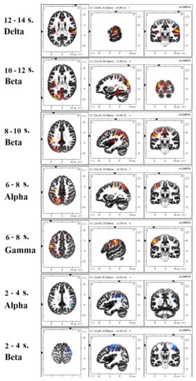
Figure 1.
EEG data. Significant effects revealed when contrasting insight with no solution trials in each of the eight 2-second epochs preceding the button press, or the appearance of the “time out” sign (the time in seconds before the end of the trail is indicated). The images are presented in neurological convention. Hot tints show regions in which current source density was higher in insight than in no solution trials, while cool tints show the opposite effect.
The effects identified by comparing the insight solution with the analytical one are shown in Figure 2. Already in 14–16 s before pressing the key, the insight solutions, in comparison with the analytical ones, were accompanied by an increase in delta activity in the PCC (10, −65, 10, BA 30, p = 0.017). Then (12–14 s), increased delta activity was observed in the right inferior frontal gyrus (45, 35, 15, BA 46, p = 0.038). For 10–12 s before pressing the key, increased beta and gamma activity was observed in the left insula (−35, −15, 20, BA 13, p = 0.021), and in the left superior (−50, −15, 5, BA 22, Wernicke zone, p = 0.021) and inferior (−55, −35, 20, BA 20, p = 0.021) temporal gyri. For 6–8 s, delta activity was increased in the left middle frontal gyrus (−25, 30, 45, BA 8, p = 0.005). Finally, in 4–6 s, reduced delta activity was observed in the PCC (−5, −55, 5, BA 30, p = 0.034) for the insight, as compared to analytical, solution. In the last 4 s before pressing the key, there were no differences.
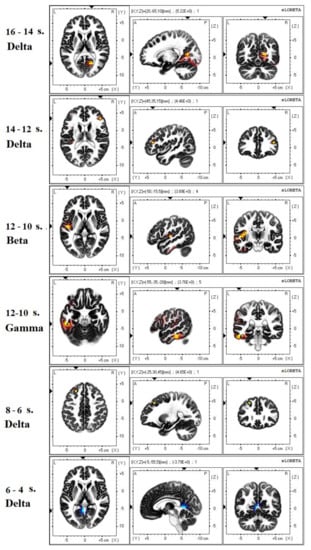
Figure 2.
EEG data. Significant effects revealed when contrasting insight with analytical solution in each of the eight 2-second epochs preceding the button press. The images are presented in neurological convention. Hot tints show regions in which current source density was higher in insight than in analytical solution trials, while cool tints show the opposite effect.
We additionally analyzed connectivity between the identified activation centers and the centers of the DMN using the LPS measures. Comparison of insight with the absence of a solution revealed a greater connectivity in the former case between the PCC and the right inferior frontal gyrus in the delta range in 8–10 s before the end of the trial (p = 0.040). Comparison of insight with analytical solution in the same period revealed a greater connectivity in the former case between the right inferior frontal gyrus and the MPFC in the delta range (p = 0.038). Comparison of insight with analytical solution in the period of 14–16 s revealed a lower connectivity during insight between the left insula and the left STG in theta range (p = 0.025) (Figure 3).
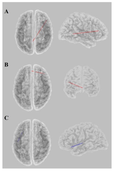
Figure 3.
EEG data. Significant effects revealed when contrasting insight with no solution or analytical solution trials using connectivity measures. (A) Insight > no solution at 8–10 s before the end of the trial (delta band); (B) insight > analytical solution at 8–10 s before the end of the trial (delta band); (C) insight > analytical solution at 14–16 s before the end of the trial (theta band). The images are presented in neurological convention. Red lines show connections that are stronger in insight trials, while blue lines show the opposite effect.
3.3. FMRI Analysis
Figure 4 shows examples of the activation dynamics of brain neural networks for two subjects (S1 and S2) in insight processes according to the fMRI data. Dynamics had an individual character. Similar behavior was observed in the activity of the prefrontal and temporoparietal areas 3 or more seconds before the subject’s reaction.
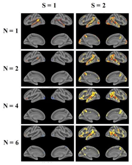
Figure 4.
fMRI data. Individual analysis (two subjects: S1 and S2) of the brain activity when contrasting insight with previous moment at various N sec (p = 0.001). The color indicates the values of the T-statistics: warm tones, positive; cold, negative.
Individual statistical maps were used to visually compare the homogeneity of brain activity under two conditions during insight (n = 1 s before the subject’s response) in contrast to: condition 1, the previous moment from the start of the task presentation; and condition 2, the moments of unsuccessful solution of tasks (45 s per task). Individual statistical maps were similar and all subjects were selected for group analysis. The results of group analysis are presented for condition 1 in Figure 5 (p = 0.001, cluster size > 10 voxel) and Table 1 (p = 0.001, cluster size > 3 voxel), and for condition 2 in Figure 6 (p = 0.001, cluster size > 10 voxel) and Table 2 (p = 0.001, cluster size > 10 voxel). Both contrasts showed the activation and deactivation of neural networks in the right and left hemispheres (Table 1 and Table 2). The activation of brain neural networks was observed in the precuneus, the anterior cingulate cortex (ACC), and the brainstem. The obtained data show that insight processes are activated mainly in the right hemisphere in frontal and temporal regions, as well as in associative precuneus regions, in the ACC and paracingulate cortex, in internal hippocampus structures and in insular cortex of the left hemisphere. It is interesting that the cerebellar structures of the left hemisphere are also involved in the insight process.
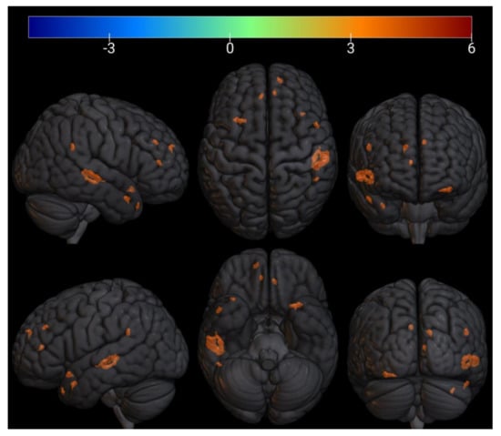
Figure 5.
fMRI data. Group analysis of the brain activity when contrasting insight with previous moment at n = 1 s (p = 0.001, cluster size > 10 voxel). The activation and deactivation of brain neural networks are presented in Table 1. The color indicates the values of the T-statistics: warm tones–positive, cold–negative.

Table 1.
fMRI data. The activation and deactivation of brain neural networks when contrasting insight with previous moment at n = 1 s (p = 0.001, cluster size > 3 voxel) for the right (r) and left (l) hemisphere (a, anterior division; p, posterior division; to, temporooccipital part).
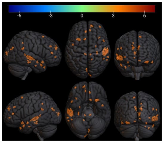
Figure 6.
fMRI data. Group analysis of the brain activity when contrasting insight with the moments of unsuccessful solution of tasks at n = 1 s (p = 0.001, cluster size > 10 voxel). The activation and deactivation of brain neural networks are presented in Table 2. The color indicates the values of the T-statistics: warm tones, positive; cold, negative.

Table 2.
fMRI data. The activation and deactivation of brain neural networks when contrasting insight with the moments of unsuccessful solution of tasks at n = 1 s (p = 0.001, cluster size > 3 voxel) for the right (r) and left (l) hemisphere (a, anterior division; p, posterior division; to, temporooccipital part).
4. Discussion
Our task in this study was to identify the dynamics of brain activity in the period preceding and accompanying the occurrence of insight. Due to the uncertainty of the existing data, we did not set ourselves the task of testing any hypothesis and considered our study as a pilot one. Identified effects have three components of information: time of occurrence, frequency range, and localization in the brain. In this study, contrary to most published studies, we used as a reference state the trials in which the subjects searched but did not find a solution. An interesting result of this choice is that the detected brain activity during analytical decisions is minimally different from the brain activity in unsuccessful trials. This suggests that in most unsuccessful trials, subjects used an analytical strategy to find a solution as well.
It should also be noted that most of the incorrectly solved problems were also solved by the analytical method. Since analytical solutions on average require more time, one might think that in many cases it was either not found (unsuccessful trials) or announced that the solution was ready without a sufficient check of its correctness (incorrectly solved problems). Insight solutions, on average, were faster, and the subjects had more time to check their correctness. The only difference between brain activity in analytical decisions and brain activity in unsuccessful trials was detected in the visual cortex in 2 s before the spacebar pressing. It can be assumed that this effect reflects a state of “return” to reality (including the switching on of visual perception) and preparation for pressing the key after the solution is found.
Brain activity accompanying insight decisions was significantly different from that in unsuccessful trials, and in trials where an analytical strategy was used. Interestingly, a significant number of effects when comparing insights with both the analytical solution and unsuccessful trials were detected in the delta range, which is associated with motivational processes and with attention to motivationally significant stimuli. In addition, delta activity is traditionally associated with a decrease in conscious perception and correlates with processes occurring at the unconscious level [36,37]. Effects in the delta frequency range have also been described in other studies of insight [15]. Unfortunately, the delta range is often not included in the analysis by researchers who aim to identify effects in the alpha, beta, and gamma ranges.
The earliest effect when comparing insight and non-insight decisions is an increase in delta activity in the PCC, which is one of the DMN hubs and according to existing data is involved in cognitive processes related to the search for creative solutions [29,30,38]. In the same period, a reduced (compared to analytical decisions) connectivity between the left insula and the left STG in theta frequency range was revealed. This combination can be interpreted as a reduced (compared to analytical decisions) search in the memory of the desired word at the conscious level and the initiation of the search process at the subconscious level. At the next stage, increased delta activity is observed in the lateral prefrontal cortex associated with working memory and executive control. One might think that all of these effects reflect activity prior to insight. The insight itself is probably associated with high-frequency activity in the cortical areas of the left hemisphere participating in semantic processes (left STG (Wernicke zone) and inferior temporal gyrus) and emotional support of insight (insula) in the 10–12 s period. Immediately after this period (8–10 s), there is an increase in connectivity in the delta range between the DMN centers (MPFC and PCC) and the right inferior frontal gyrus, which is involved in the detection of important signals in the information flow. Then (6–8 s), increased delta activity is observed in the left middle and inferior frontal gyri, in a region that partially overlaps with the Broca’s area and participates in semantic analysis of verbal information [39,40]. This “post-insight” activity in the delta range probably reflects the verification of the correctness of the solution, possibly even silent “pronunciation” of the found word. Then (4–6 s), reduced delta activity in the PCC probably reflects the cessation of the search for a solution. This interpretation is certainly speculative, but it is in good agreement with existing data.
The fMRI study of the insight state dynamics also showed that the change in the brain neural network state begins 3 or more seconds before the subject’s response. Neural network activity has different individual dynamic profiles; in most cases, there is a decrease in the activity of the dorsal-medial prefrontal cortex (DMPFC) with an increase or persistence of activity in the temporal and parieto-occipital regions (Figure 4). DMPFC are associated with goal-direction and selective memory retrieval, while the bilateral IFG and insular cortex, inferior parietal lobules (IPL), and precuneus are elements of the semantic memory network [41].
According to fMRI data, insight is associated with activation of the precuneus and ACC (DMN regions), the insular cortex in the left hemisphere, and a number of frontal and temporal regions. In contrast to EEG data, the activation of the frontal and temporal regions was more pronounced in the right than in the left hemisphere (see Table 1 and Table 2). Also, the fMRI data showed an important role of the hippocampus and parahippocampal cortex in insight. These observations are consistent with data from earlier works [9,38,41]. A new fact was the discovery of insight-related activation in the left cerebellum, which requires further verification.
Thus, in ours, as in many other studies [10,11,12,13,19,20], the role of the STG and high-frequency beta and gamma activity in insight-related processes in solving RAT problems is revealed. On the whole, these effects show distinct differences in the dynamics of brain activity during insight and analytical solutions and confirm the effectiveness of the within-subject approach to experimental design and data analysis. Also in line with previous publications, we observed a kind of “dissociation” between EEG and fMRI data with the former highlighting the involvement of the left temporal regions and the latter pointing at the role of the right frontal and temporal areas. The reason for this apparent inconsistency needs further investigation. However, it should be noted that fMRI and EEG evidence do not have to coincide, because they reflect very different processes in the brain. fMRI BOLD signal correlates with energy-consuming processes, whereas large-scale synchronization of EEG oscillations may occur without much energy consumption [42]. Large-scale synchronization, particularly of gamma oscillations, has been linked with subjective awareness and conscious perception (e.g., [43,44]). One may speculate that insight-related right-hemispheric processes captured by fMRI data are mostly associated with unconscious stages, whereas the burst of gamma activity in the left STG captured by EEG data reflects the moment of conscious awareness of the found solution.
This study has a number of limitations. First of all, it has to be acknowledged that most of our interpretations of our findings are highly speculative. This is inevitable because so far the phenomenon of insight eludes all attempts to pin it down in terms of both “objective” measuring and exact timing. We think, however, that if we want to study such fuzzy constructs as insight at all, even speculative interpretations may help to canalize the future research in some direction or another. As a limitation of the experimental design, it should be noted that the classification of insight and non-insight trials was based on self-reports, which, of course, are subject to error. However, other methods described in the literature are associated with no fewer potential sources of errors, and self-reports in this case are considered the most reliable source of information [9,24,25]. The second potential limitation may be that the instruction given to the subjects did not emphasize that the space bar should be pressed as soon as possible after the solution was found. This may explain why the effects were found relatively far from the moment of fixing the readiness of the solution. The third limitation is the small sample size of our fMRI study, which, therefore, should be considered preliminary and in need of further verification. The fourth limitation is that EEG and fMRI data were collected in different subjects and in different laboratories. The observed differences between EEG and fMRI experiments could have partially resulted from this as well as from differences inherent in EEG and fMRI data acquisition procedures. Ideally, simultaneous EEG and fMRI recordings in the same subjects are necessary in order to reveal method-specific effects. Finally, it has to be acknowledged that although the reported findings do show meaningful differences in brain underpinning of insight versus analytical problem solving, they ultimately do not shed much light on the concrete neuronal mechanisms of either insightful or analytic reasoning, which reflects critical shortcomings in the current level of understanding of cognitive mechanisms of problem solving.
Summing up, comparison of the two ways of solving RAT problems, which were classified by the subjects as “insight” or “analytical”, revealed a number of differences both at the behavioral level and at the level of brain activity. Insight solutions were faster on average and correct in more cases. Brain activity during analytical solving was minimally different from that in the absence of a solution, which suggests the presence of an analytical method in the latter case. Brain activity during insight was significantly different from that in unsuccessful trials, and in trials where an analytical strategy was used. In the early stages, differences are mainly detected in the delta frequency range in the cortical areas related to the DMN and executive control network. Brain activity, presumably associated with the occurrence of an insight, was detected in beta and gamma ranges in the cortical areas of the left hemisphere, which are involved in semantic and emotional processing. On the whole, the revealed effects show distinct differences in the dynamics of brain activity during insight and analytical problem solving.
Author Contributions
Conceptualization, G.G.K., V.L.U., and B.M.V.; methodology, G.G.K., V.L.U., and B.M.V.; software, D.G.M.; validation, V.A.O.; formal analysis, G.G.K., V.L.U., and V.A.O.; investigation, G.G.K., V.L.U., S.I.K., A.N.S., and A.V.B.; resources, S.I.K. and D.G.M.; data curation, G.G.K., V.L.U., and V.A.O.; writing—original draft preparation, G.G.K. and V.L.U.; writing—review and editing, G.G.K. and V.L.U.; visualization, G.G.K., V.L.U., and V.A.O.; supervision, G.G.K., V.L.U., and B.M.V.; project administration, G.G.K., V.L.U., and B.M.V.; funding acquisition, G.G.K. and B.M.V. All authors have read and agreed to the published version of the manuscript.
Funding
The study was partially supported by the Russian Foundation for Basic Research (RFBR) under grant # 20-013-00404, by the Ministry of Science and Higher Education of the Russian Federation (Grant № 075-15-2020-801), and the Institute of Neuroscience and Medicine budgetary theme # AAAA-A21-121011990039-2.
Institutional Review Board Statement
The study conforms with World Medical Association Declaration of Helsinki and was approved by the Institute of Neuroscience and Medicine ethical committee. Permission to undertake this experiment was granted by the local Ethics Committee of the National Research Center “Kurchatov Institute” (Protocol No.10 from 1 August 2018).
Informed Consent Statement
Informed consent was obtained from all subjects involved in the study.
Data Availability Statement
The data presented in this study are available upon request from the corresponding author.
Conflicts of Interest
The authors declare no conflict of interest.
References
- Davidson, J.E.; Sternberg, R.J. (Eds.) The Psychology of Problem Solving; Cambridge University Press: Cambridge, UK, 2003. [Google Scholar]
- Cushen, P.J.; Wiley, J. Cues to solution, restructuring patterns, and reports of insight in creative problem solving. Conscious. Cogn. 2012, 21, 1166–1175. [Google Scholar] [CrossRef]
- Weisberg, R.W. Toward an integrated theory of insight in problem solving. Think. Reason. 2014, 21, 5–39. [Google Scholar] [CrossRef]
- Irvine, W.B. Aha! The Moments of Insight That Shape Our World; Oxford University Press: New York, NY, USA, 2015. [Google Scholar]
- Poincaré, H. The Foundations of Science; Science Press: Lancaster, PA, USA, 1913. [Google Scholar]
- Wallas, G. The Art of Thought; Harcourt Brace: NewYork, NY, USA, 1926. [Google Scholar]
- Kounios, J.; Jung-Beeman, M. The Aha! Moment the cognitive neuroscience of insight. Curr. Dir. Psychol. Sci. 2009, 18, 210–216. [Google Scholar] [CrossRef]
- Dietrich, A.; Kanso, R. A Review of EEG, ERP, and Neuroimaging Studies of Creativity and Insight. Psychol. Bull. 2010, 136, 822–848. [Google Scholar] [CrossRef]
- Bowden, E.M.; Jung-Beeman, M. Aha! Insight experience correlates with solution activation in the right hemisphere. Psychon. Bull. Rev. 2003, 10, 730–737. [Google Scholar] [CrossRef]
- Jung-Beeman, M.; Bowden, E.; Haberman, J.; Frymiare, J.; Aramber-Liu, S.; Greenblatt, R.; Kounios, J. Neural activity when people solve problems with insight. PloS Biol. 2004, 2, 500–510. [Google Scholar] [CrossRef]
- Kounios, J.; Fleck, J.; Green, D.; Payne, L.; Stevenson, J.; Bowden, E.; Jung-Beeman, M. The origins of insight in resting-state brain activity. Neuropsychologia 2008, 46, 281–291. [Google Scholar] [CrossRef] [PubMed]
- Qiu, J.; Luo, Y.; Wu, Z.; Zhang, Q. A further study of ERP effects of “insight” in a riddle guessing task. Acta Psychol. Sin. 2006, 38, 507–514. [Google Scholar]
- Sandkuhler, S.; Bhattacharya, J. Deconstructing insight: EEG correlates of insightful problem solving. PLoS ONE 2008, 3, e1459. [Google Scholar] [CrossRef] [PubMed]
- Danko, S.; Starchenko, M.; Bechtereva, N. EEG local and spatial synchronization during a test on the insight strategy of solving creative verbal tasks. Hum. Physiol. 2003, 29, 129–132. [Google Scholar]
- Kounios, J.; Frymiare, J.; Bowden, E.; Fleck, J.; Subramaniam, K.; Parrish, T.; Jung-Beeman, M. The prepared mind: Neural activity prior to problem presentation predicts subsequent solution by sudden insight. Psychol. Sci. 2006, 17, 882–891. [Google Scholar] [CrossRef] [PubMed]
- Lang, S.; Kanngieser, N.; Jaskowski, P.; Haider, H.; Rose, M.; Verleger, R. Precursors of insight in event-related brain potentials. J. Cogn. Neurosci. 2006, 18, 2152–2166. [Google Scholar] [CrossRef] [PubMed]
- Lavric, A.; Forstmeier, S.; Rippon, G. Differences in working memory involvement in analytical and creative tasks: An ERP study. NeuroReport 2000, 11, 1613–1618. [Google Scholar] [CrossRef][Green Version]
- Mai, X.; Luo, J.; Wu, J.; Luo, Y. “Aha!” effects in a guessing riddle task: An event related potential study. Hum. Brain Mapp. 2004, 22, 261–270. [Google Scholar] [CrossRef]
- Qiu, J.; Li, H.; Yang, D.; Luo, Y.; Li, Y.; Wu, Z.; Zhang, Q. The neural basis of insight problem solving: An event-related potential study. Brain Cogn. 2008, 68, 100–106. [Google Scholar] [CrossRef]
- Qiu, J.; Li, H.; Yang, D.; Luo, Y.; Li, Y.; Wu, Z.; Zhang, Q. Spatiotemporal cortical activation underlies mental preparation for successful riddle solving: An event-related potential study. Exp. Brain Res. 2008, 186, 629–634. [Google Scholar] [CrossRef]
- Goldman, R.I.; Stern, J.M.; Engel, J., Jr.; Cohen, M.S. Simultaneous EEG and fMRI of the alpha rhythm. NeuroReport 2002, 13, 2487–2492. [Google Scholar] [CrossRef]
- Sadato, N.; Nakamura, S.; Oohashi, T.; Nishina, E.; Fuwamoto, Y.; Waki, A.; Yonekura, Y. Neural networks for generation and suppression of alpha rhythm: A PET study. NeuroReport 1998, 9, 893–897. [Google Scholar] [CrossRef]
- Mednick, S.A. The associative basis of the creative process. Psychol. Rev. 1962, 69, 220–232. [Google Scholar] [CrossRef] [PubMed]
- Bowden, E.M. The effect of reportable and unreportable hints on anagram solution and the aha! experience. Conscious. Cogn. 1997, 6, 545–573. [Google Scholar] [CrossRef] [PubMed]
- MacGregor, J.N.; Cunningham, J.B. Rebus puzzles as insight problems. Behav. Res. Methods 2008, 40, 263–268. [Google Scholar] [CrossRef]
- Valueva, E.A.; Belova, S.S. Creativity diagnostics: Methods, problems, perspectives. In Creativity: From Biological Prerequisites to Cultural Phenomena; Ushakov, D.V., Ed.; Institute of Psychology RAS: Moscow, Russia, 2011; pp. 625–647. (In Russian) [Google Scholar]
- Pascual-Marqui, R.D. Instantaneous and Lagged Measurements of Linear and Nonlinear Dependence between Groups of Multivariate Time Series: Frequency Decomposition. arXiv 2007, arXiv:0711.1455. Available online: http://arxiv.org/abs/0711.1455 (accessed on 9 November 2007).
- Nolte, G.; Bai, O.; Wheaton, L.; Mari, Z.; Vorbach, S.; Hallett, M. Identifying true brain interaction from EEG data using the imaginary part of coherency. Clin. Neurophysiol. 2004, 115, 2292–2307. [Google Scholar] [CrossRef]
- Jung, R.E.; Segall, J.M.; Bockholt, H.J.; Chavez, R.S.; Flores, R.; Haier, R.J. Neuroanatomy of creativity. Hum. Brain Mapp. 2010, 31, 398–409. [Google Scholar] [CrossRef] [PubMed]
- Kühn, S.; Ritter, S.M.; Müller, B.C.N.; van Baaren, R.B.; Brass, M.; Dijksterhuis, A. The Importance of the Default Mode Network in Creativity—A Structural MRI Study. J. Creat. Behav. 2014, 48, 152–163. [Google Scholar] [CrossRef]
- Fox, M.D.; Snyder, A.Z.; Vincent, J.L.; Corbetta, M.; Van Essen, D.C.; Raichle, M.E. The human brain is intrinsically organized into dynamic, anticorrelated functional networks. Proc. Natl. Acad. Sci. USA 2005, 102, 9673–9678. [Google Scholar] [CrossRef] [PubMed]
- Eklund, A.; Dufort, P.; Villani, M.; LaConte, S. BROCCOLI: Software for fast FMRI analysis on many-core CPUs and GPUs. Front. Neuroinform. 2014, 8, 24. [Google Scholar] [CrossRef]
- Friston, K.J.; Williams, S.; Howard, R.; Frackowiak, R.S.J.; Turner, R. Movement-Related effects in fMRI time-series. Magn. Reson. Med. 1996, 35, 346–355. [Google Scholar] [CrossRef]
- Jenkinson, M.; Beckmann, C.F.; Behrens, T.E.J.; Woolrich, M.W.; Smith, S.M. Fsl. Neuroimage 2012, 62, 782–790. [Google Scholar] [CrossRef]
- Griffanti, L.; Douaud, G.; Bijsterbosch, J.; Evangelisti, S.; Alfaro-Almagro, F.; Glasser, M.F.; Duff, E.P.; Fitzgibbon, S.; Westphal, R.; Carone, D.; et al. Hand classification of fMRI ICA noise components. NeuroImage 2017, 154, 188–205. [Google Scholar] [CrossRef]
- Knyazev, G.G. Motivation, emotion, and their inhibitory control mirrored in brain oscillations. Neurosci. Biobehav. Rev. 2007, 31, 377–395. [Google Scholar] [CrossRef]
- Knyazev, G.G. EEG delta oscillations as a correlate of basic homeostatic and motivational processes. Neurosci. Biobehav. Rev. 2012, 36, 677–695. [Google Scholar] [CrossRef] [PubMed]
- Subramaniam, K.; Kounios, J.; Parrish, T.; Beeman, M. A Brain Mechanism for Facilitation of Insight by Positive Affect. J. Cogn. Neurosci. 2008, 21, 415–432. [Google Scholar] [CrossRef]
- Brown, S.; Martinez, M.J.; Parsons, L.M. Music and language side by side in the brain: A PET study of the generation of melodies and sentences. Eur. J. Neurosci. 2006, 23, 2791–2803. [Google Scholar] [CrossRef] [PubMed]
- Martins, R.; Simard, F.; Monchi, O. Differences between patterns of brain activity associated with semantics and those linked with phonological processing diminish with age. PLoS ONE 2014, 9, e99710. [Google Scholar] [CrossRef] [PubMed]
- Tik, M.; Sladky, R.; Luft, C.D.B.; Willinger, D.; Hoffmann, A.; Banissy, M.J.; Bhattacharya, J.; Windischberger, C. Ultra-high-field fMRI insights on insight: Neural correlates of the Aha!-moment. Hum. Brain Mapp. 2018, 39, 3241–3252. [Google Scholar] [CrossRef] [PubMed]
- Buzsaki, G.; Draguhn, A. Neuronal oscillations in cortical networks. Science 2004, 304, 1926–1929. [Google Scholar] [CrossRef]
- Lou, H.C.; Gross, J.; Biermann-Ruben, K.; Kjaer, T.W.; Alfons, S. Coherence in consciousness: Paralimbic gamma synchrony of self-reference links conscious experiences. Hum. Brain Mapp. 2009, 31, 185–192. [Google Scholar] [CrossRef] [PubMed]
- Meador, K.J.; Ray, P.G.; Echauz, J.R.; Loring, D.W.; Vachtsevanos, G.J. Gamma coherence and conscious perception. Neurology 2002, 59, 847–854. [Google Scholar] [CrossRef]
Publisher’s Note: MDPI stays neutral with regard to jurisdictional claims in published maps and institutional affiliations. |
© 2021 by the authors. Licensee MDPI, Basel, Switzerland. This article is an open access article distributed under the terms and conditions of the Creative Commons Attribution (CC BY) license (http://creativecommons.org/licenses/by/4.0/).