Article

Histological Evaluation of a New Beta-Tricalcium Phosphate/Hydroxyapatite/Poly (1-Lactide-Co-Caprolactone) Composite Biomaterial in the Inflammatory Process and Repair of Critical Bone Defects

by
Elizabeth Ferreira Martinez
1,*,
Ana Elisa Amaro Rodrigues
2,
Lucas Novaes Teixeira
1,
Andrea Rodrigues Esposito
2,
Walter Israel Rojas Cabrera
2,
Ana Paula Dias Demasi
1 and
Fabricio Passador-Santos
1
1
Division of Oral Pathology and Cell Biology, Faculdade São Leopoldo Mandic, Campinas 13045-755, Brazil
2
Division of Periodontology, Faculdade São Leopoldo Mandic, Campinas 13045-755, Brazil
*
Author to whom correspondence should be addressed.
Symmetry 2019, 11(11), 1356; https://doi.org/10.3390/sym11111356
Submission received: 18 September 2019 / Revised: 24 October 2019 / Accepted: 30 October 2019 / Published: 2 November 2019

Abstract

:
Background: The use of biomaterials is commonplace in dentistry for bone regeneration. The aim of this study was to evaluate the performance of a new alloplastic material for bone repair in critical defects and to evaluate the extent of the inflammatory process. Methods: Forty-five New Zealand rabbits were divided into five groups according to evaluation time (7, 14, 30, 60, 120 days), totaling 180 sites with six-millimeter diameter defects in their tibiae. The defects were filled with alloplastic material consisting of poly (lactide-co-caprolactone), beta-tricalcium phosphate, hydroxyapatite and nano-hydroxyapatite (BTPHP) in three different presentations: paste, block, and membrane. Comparisons were established with reference materials, such as Bio-ossTM, Bio-oss CollagenTM, and Bio-gideTM, respectively. The samples were HE-stained and evaluated for inflammatory infiltrate (scored for intensity from 0 to 3) and the presence of newly formed bone at the periphery of the defects. Results: Greater bone formation was observed for the alloplastic material and equivalent inflammatory intensity for both materials, regardless of evaluation time. At 30 days, part of the synthetic biomaterial, regardless of the presentation, was resorbed. Conclusions: We concluded that this novel alloplastic material showed osteoconductive potential, biocompatibility, low inflammatory response, and gradual resorption, thus an alternative strategy for guided bone regeneration.

1. Introduction

The use of biomaterials is common practice in dentistry for bone regeneration, especially in procedures that require bone neoformation [1,2]. Among the materials used, autologous grafting is regarded the “gold standard” due to its osteogenic, osteoconductive, and osteoinductive properties [3]. Due to donor site morbidity, the loss of temporary function and limitations in the quantity and the quality of gained bone, other allogeneic, xenogeneic, and alloplastic materials have been widely used for such procedures [4,5,6].
The most commonly used xenograft is deproteinized bovine mineral bone [7]. This material has demonstrated excellent results in terms of space maintenance and induction of bone formation. However, inorganic bovine bone particles undergo very slow resorption [8], and the amount of new mineralized tissue may be lower when compared to other bone substitutes [9,10,11]. In addition, an association between xenogenic material and polymers has been proposed in order to modulate not only mechanical properties but also biological features, including material resorption, tridimensional morphology, and osteocondutivity [12].
As an alternative to xenografts, some studies have proposed alloplastic materials. In addition to biocompatibility, immunologic inertness and easy access, no risk of cross-infection and higher rates of resorption and bone repair have been reported [13]. Although many synthetic materials have recently been developed with various chemical compositions and structural characteristics to suit different types of clinical applications, no synthetic material has been able to achieve the biological and mechanical properties of human bone [14].
In fact, synthetic grafts continue to have limitations, mainly relating to immune response and foreign body reaction, with an intense inflammatory process [15]. Moreover, most of the alloplastic materials available in the market are granulated, which limits their use in regions where tissue scaffolding is required, such as those promoted by block grafts [16]. In this context, synthetic polymers can represent a promising alternative biomaterial, since this class of materials allows the construction of scaffolds easily tailored in different shapes that mimic bone tissue in terms of porosity and mechanic resistance [17,18]. Despite this advantage, the polymers exhibit inferior cell adhesion properties [19]. To overcome this limitation, modifications in the electric charges of polymers’ surfaces favoring the electronegative potential may catalysis cell signaling, attracting proteins and undifferentiated cells to the bone regeneration process [20,21,22].
Thus, the aim of this study was to evaluate the performance of an alloplastic material composed of poly (l-lactide-co-ε-caprolactone) arranged with electronegative charges, polyethylene glycol, beta-tricalcium phosphate, hydroxyapatite, and nanohydroxyapatite, available in three different presentations (paste, block, and membrane), in the process of bone repair in critical defects in rabbit tibia, as well as to evaluate the extent of the inflammatory process formed in this type of grafting procedure.

2. Materials and Methods

Forty-five rabbits of the New Zealand lineage (Oryctolagus cunniculus) were obtained from the CPQBA-Unicamp. The present study was conducted with prior approval of the Ethics Committee for Animal Use of the State University of Campinas—CEUA/Unicamp (protocol #4058-1). All experiments were performed in accordance with the guidelines and regulations of the Committee. The animals were kept under controlled conditions of temperature and illumination with a light-dark cycle of 12 h, with balanced feed and water ad libitum.
The experiment was performed on animals from both sexes weighing between 2.5 and 3.0 kg, and minimum three months of age. Animals received pre-anesthetic medication with acepromazine 1 mg/kg intramuscularly. The surgical procedure was performed in animals under general anesthesia, by application of 3% sodium pentobarbital (30 mg/kg) intravenously. The surgical procedures were performed respecting the principles of biosafety to prevent infectious processes in the surgical wounds.

2.1. Preparation of Bony Defects

The animals were divided into five groups according to the evaluation time (7, 14, 30, 60, and 120 days), totaling 180 evaluation sites. The animals were submitted to trichotomy of both pelvic limbs and posterior antisepsis of the region with PVPI alcohol with 1% active iodine. To access the bone region, a 4 cm longitudinal incision was made in the skin of the medial aspect of the tibia, with muscular and periosteal flap raising.
In each tibia, two surgical cortical defects were made with a 6-mm trephine drill and constant irrigation with sterile saline for removal of any bone fragments from the artificial defect. The defects were made approximately 15 mm from the femoro-tibial-patellar joint, approximately 10 mm apart, and filled with the alloplastic material composed of poly (l-lactide-co-caprolactone) arranged with electronegative charges and treated with beta-tricalcium phosphate, hydroxyapatite and nano-hydroxyapatite (named as BTPHP and provided by Bioactive Biomateriais SA, Indaiatuba, São Paulo, Brazil), in three different presentations: block, paste, and membrane. Each presentation was compared with a reference material (control, CTRL) by the company Geistlich Pharma AG (Switzerland), such as Bio-Oss collagen, Bio-Oss and Bio-Gide, respectively. The animals were divided into five evaluation times and each animal was evaluated in duplicate (two defects for each material and time of analysis, in each tibia), totalizing n = 6 defects per group in each period of analysis. After surgery, the periosteum was carefully repositioned and sutured, followed by the remaining tissues (muscle layer, subcutaneous tissue and skin) using 5-0 needle-threaded polygalactin. A cutaneous dressing containing 0.2% nitrofurazone ointment and elastic bandage was placed. The animals were treated with 10% enrofloxacin for 3 days and analgesia with morphine three times a day for 3 days. The dressing was removed after 24 hours. Rabbits were monitored daily to ensure behavioral activity level, integrity of the surgical sites, water and food consumption, as well as stools and urine elimination.
Euthanasia was performed on specific postoperative days, namely the seventh postoperative day (n = 9), the 14th day (n = 9), the 30th day (n = 9), the 60th day (n = 9) and the 120th postoperative day (n = 9). The animals were euthanized by deepening the general anesthetic dose, according to the protocol 90–150 mg/kg of sodium thiopental intraperitoneally. Subsequently, the samples were removed via cross-sectional cutaneous and muscular incisions that surrounded the femoro-tibial-patellar joint, facilitating disarticulation of the tibia. Samples were stored in flasks containing 10% buffered formalin solution for microscopic analysis.

2.2. Preparation for Histological Analysis

The tibiae removed from the five groups were prepared for conventional light microscopy. They were immersed in 10% buffered formalin solution and then demineralized in 5% formic acid. The pieces were included in histological paraffin and 4-μm cross-sections were made along the long axis in the central region of the defects so that two cuts were made per sample.
The samples were stained with Hematoxylin-Eosin and subsequently mounted with resin and coverslips for photomicrographs, which were taken under light microscopy for the analyses described below.

2.3. Histological Analysis
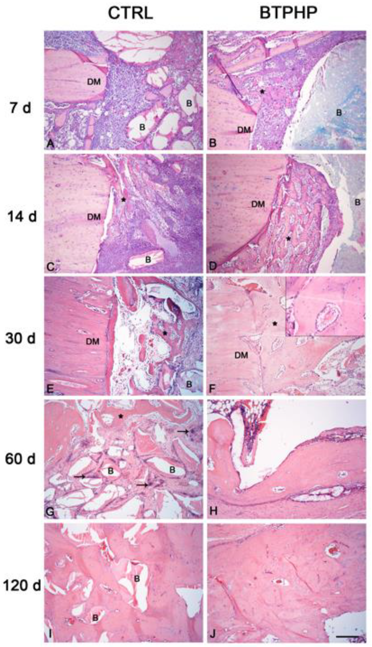
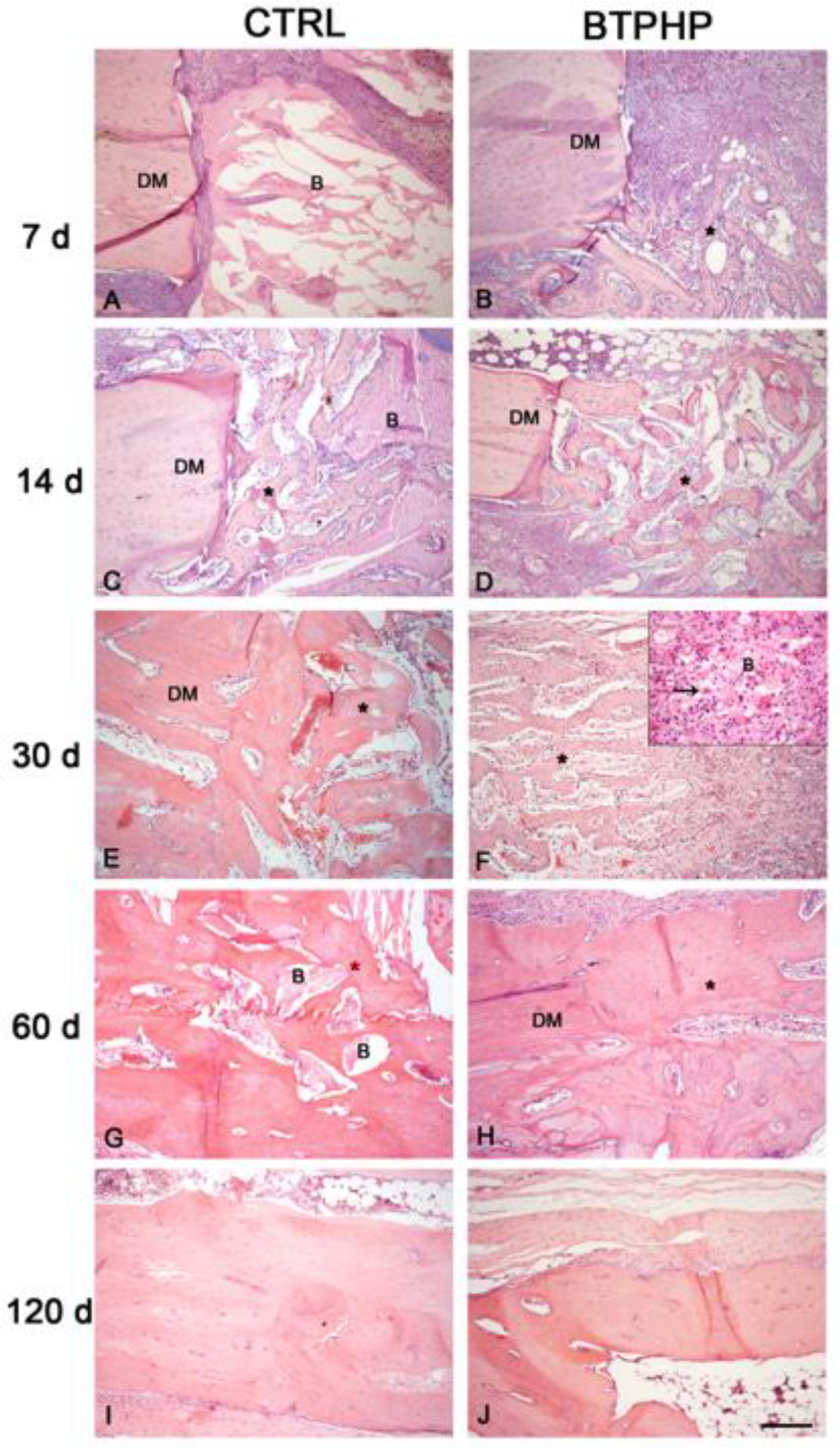
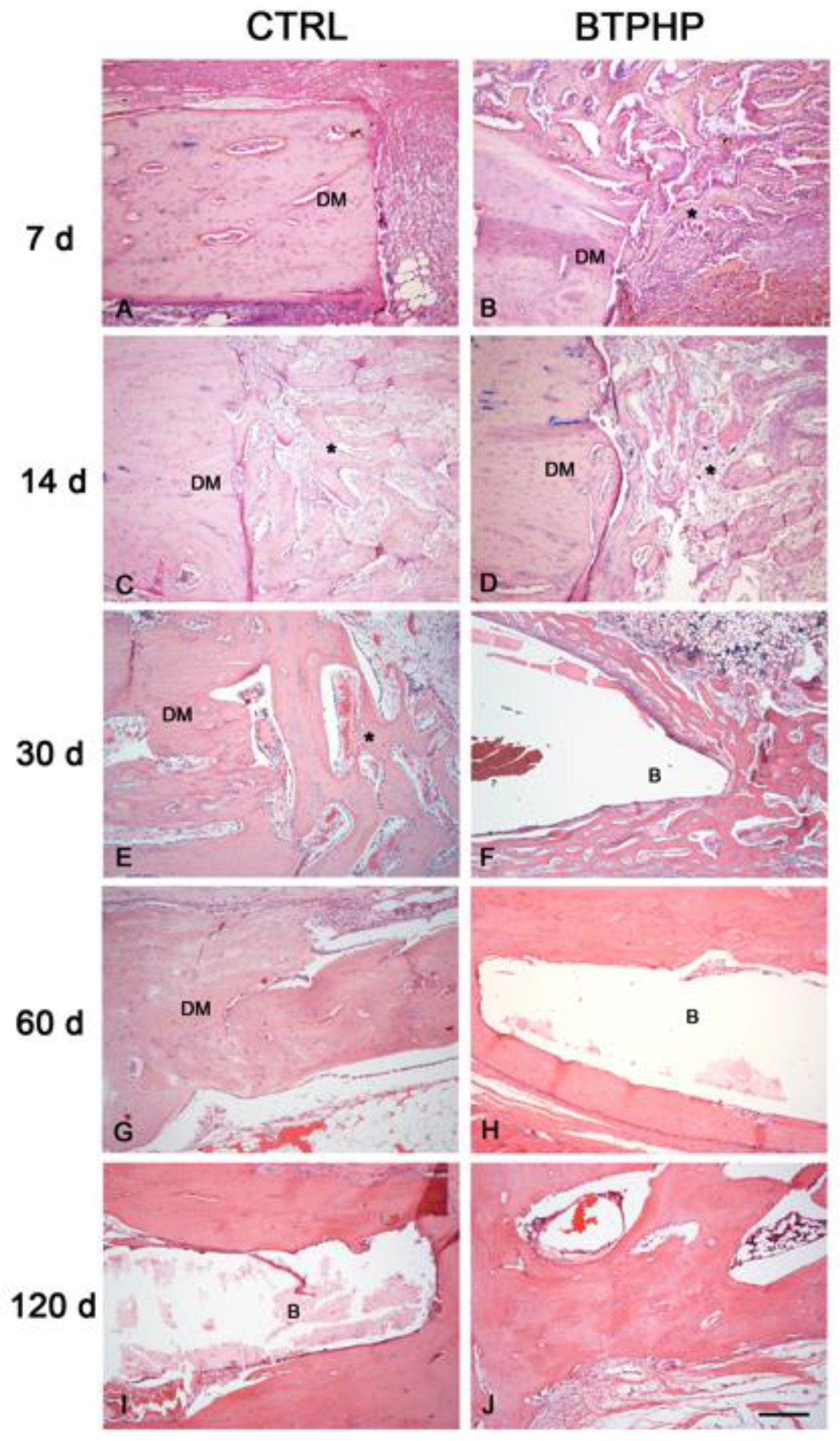
For the histological analyses, the osteogenic potential of the materials was described based on the presence of neoformed bone at the edges of the defects, as well as the presence of inflammation, including the presence of blood vessels, giant cells, leukocyte infiltrate and phagocytosis of the evaluated tissues at different times (7, 14, 30, 60, and 120 days). A classification score was adopted, considering the extent of the inflammatory process within the defect area, ranging from 0 to 3, where 0 meant up to 15%, 1 (15%–50%), 2 (50%–75%) and 3 (>75%).
Images from the slides were captured in a computerized imaging system, AxioVision rel 4.8, (Carl Zeiss, Oberkochen, Germany) under a light microscope Axioskop 2 plus (Carl Zeiss, Oberkochen, Germany).

3. Results

Representative images of bone neoformation and description of inflammatory process scores are shown, respectively, in Figure 1, Figure 2 and Figure 3. At 7 days of evaluation of the CTRL (Bio-Oss Collagen), a discrete leukocytic infiltrate (score 0) was observed along with absence of bone neoformation (Figure 1). For the BTPHP block, a discrete presence of inflammatory cells was observed, including some giant cells. However, neoformed bone was detected near the defect margins, which were lined with osteoblastic cells. At 14 days, a discrete inflammatory process (score 0) and bone neoformation was observed on the edges of the defects as well as the periphery of the biomaterial in both groups. At 30, 60, and 120 days of evaluation, no inflammatory process was observed in any of the evaluated groups (score 0), and new bone formation from the edges of the bony defect was detected along with evident hematopoietic medullary tissue with vascularization. In the BTPHP group, the defect was completely closed at 30 days of analysis, showing bone with mature histological appearance and Haversian organization.
For BTPHP Paste and Bio-Oss (CTRL), at 7 days of evaluation, the presence of both biomaterials in the bony defects was observed along with an inflammatory infiltrate occupying more than 50% of the area of the bony defect (score 2), where the BTPHP Paste material was placed, the presence of macrophages and refringent biomaterial could be visualized. For both groups, in some specimens, the presence of immature neoformed bone was observed, highly cellular, close to the edges of the defect. At 14 and 30 days, a discrete inflammatory process (score 0) and bone neoformation, essentially immature bone, very close to the edges of the bony defect was observed. At 30 days, the defect was completely closed only in the BTPHP Paste specimens, which also showed infiltrates of macrophages and multinucleated giant cells. At 60 and 120 days, no inflammatory process (score 0) was observed, but bone neoformation (*) was evident at the edges of the defects for all evaluated specimens, with complete closure of the bony defect, with histological features of mature bone and Haversian organization. In addition, remnants of biomaterial were present in the reference group (CTRL, Bio-Oss) but not in the group filled with BTPHP Paste.
Regarding the membrane material at 7 days of evaluation, there was a discrete presence of leukocytes (score 0) and bone neoformation from the edges of the defect only in the BTPHP Membrane group.
At 14 and 30 days, a discrete inflammatory process (0 score) and bone neoformation at the edges of the defects were observed for all the specimens evaluated, being more evident in the BTPHP Membrane group, which showed bone neoformation on both sides of the membrane (upper and lower) and complete closure of the defect at 30 days. At 60 days and 120 days, no inflammatory infiltrate was observed in any of the groups evaluated (score 0), while bone neoformation was evident from the edges of the bony defect, featuring hematopoietic medullary tissue. In the BTPHP Membrane group, there was complete closure of the defect with mature bone and Haversian organization histologically on both sides of the membrane.

4. Discussion

In clinical practice, successful bone regeneration is achieved when wound repair occurs based on appropriate cellular occupation of the defect space, which is necessary for closure of the bony defect. Especially for periodontal regeneration, a period of six to eight weeks is suggested for repair to occur so that grafting materials, when used, should allow bone formation in short periods of time [23].
The xenogeneic materials used as controls in this study were the most widely used and demonstrated excellent results in terms of space maintenance and induction of bone neoformation [24]. Inorganic bovine bone particles, however, undergo very slow resorption [8], and the amount of new mineralized tissue may be lower when compared to other bone substitutes [9,10,11]. In addition, some xenogeneic and allogeneic materials may induce an immunogenic response or a foreign body reaction [25,26,27,28]. Thus, some alloplastic materials have emerged as an attractive alternative to guided tissue regeneration (GTR), although their efficacy is debatable in the literature, especially with regards to the intense inflammatory reaction and large amount of remaining material associated with them [29]. Furthermore, its use is limited since most available materials are found as membranes or granules, which makes it difficult to use them in procedures that require a great deal of bone gain. Materials of synthetic origin containing hydroxyapatite (HA) have been an option because of the osteogenic potential attributed to this compound, which is ideal to promote mineralization within collagen fibrils acting as a mediator of the binding between the inorganic phase of bone with the fibrils of the organic phase [30,31]. HA is normally used in polymeric materials since they can be manufactured according to the desired size and shape, increasing its clinical applicability [32].
In addition to HA advantages, electric charges of polymers’ surfaces may contribute to migration and cell adhesion. Indeed, this treatment has been used in some surfaces application in order to improve mesenchymal stem cell recruitment and consequently, bone formation [33]. Considering that this combination has an osteoconductive potential, in the present study, a new material of synthetic origin composed of copolymer of lactic acid and caprolactone negatively charged, polyethylene glycol, beta-tricalcium phosphate, hydroxyapatite and nanohydroxyapatite (BTPHP) was evaluated in three different presentations (membrane, block, and paste). The membrane is recommended as a biological barrier in bone reconstruction and repair procedures, the block is indicated for extensive vertical and horizontal defects when high bone gain is required, whereas the paste is indicated for filling small bony defects with preserved walls [34].
The results of the present study showed a greater amount of bone formation from the margin of the defect to the center of the repair area when using the alloplastic material BTPHP, when compared to the respective reference groups and similar scores of inflammatory infiltrate throughout, independently of the evaluation time. These results evidenced an osteoconductive, biocompatible, low inflammatory potential, as well as a gradual capacity of resorption of the evaluated material, thus suggesting an alternative tool for guided bone regeneration. It is important to highlight that studies using xenogeneic biomaterial for long periods of time evidenced bone formation with no evidence of remodeling [35,36]. This study was conducted up to 120 days, when it was still possible to observe some remainder of the studied biomaterial. In this context, it is not possible to assure that complete remodeling of the material will occur.
The favorable results are due in part to the polymer-ceramic composition, since the inorganic fraction of the bone matrix constituted by HA favored cellular recognition and tissue growth, being integrated in the process of natural bone remodeling. Over the weeks, it was possible to observe new bone tissue in the defect region, with an incipient inflammatory process sometimes absent, when compared to the xenogeneic materials used in this study. Together with the osteoconductive potential of HA [37], the association with the polymeric fraction and tricalcium phosphate (TCP), allows faster resorption of the biomaterial while allowing bone formation at early stages [38,39,40], whereas the HA particles stabilize the healing area [41], maintaining the mechanical and dimensional stability of the grafted volume and contributing to maintaining the scaffold.
The analysis showed that at 30 days, part of the grafted synthetic biomaterial, regardless of presentation (paste, membrane or block), had already been resorbed when compared to the xenograft, eliminating the need for a second surgical procedure which is normally necessary to remove non-resorbable alloplastic materials.
The evidence of the low immunogenic potential of the alloplastic material BTPHP found in the present study comes from the low numbers of inflammatory cells and the scarce multinucleated giant cells. The presentation with the highest inflammatory potential was the paste, especially at seven days (score 2), however, after 14 days, discrete amounts of inflammatory infiltrate (score 0) were observed alongside rapid bone neoformation and numerous blood vessels.
For bone repair, adequate angiogenesis is required to provide for metabolic needs, including not only local nutrition but also recruitment of progenitor cells and various growth factors. One of the factors that contributes to proliferation and migration of endothelial cells is nanohydroxyapatite [42], present in the BTPHP biomaterial, which may also be facilitated by adequate pH in the microenvironment [43,44] due to biomaterial composition [45]. This may suggest an increase of blood vessels observed at the grafting site when using the alloplastic BTPH material, especially as a paste. This effect may be in part attributed to the presence of poly (l-lactic acid-co-ε-caprolactone), tricalcium beta phosphate, and associated with hydroxyapatite and nano-hydroxyapatite in the formulation, which have adequate physical and mechanical properties that influence not only the rate of degradation, but also contribute to the creation of a porous scaffold conducive of neovascularization to the site [46], as well as a low inflammatory potential [45].
Such results bring safety and predictability in bone formation at the required site. It is clear that mechanical properties, biological behavior and biodegradation mechanisms vary across different graft materials, and the physico-chemical properties are among the most important factors influencing the performance of the material in vivo, causing significantly different biological responses [17,19,44,47,48]. The results of the present study evidenced the osteoconductive, biocompatible, and bioactive potentials of the BTPHP material used in three different presentations, as an alloplastic alternative to manage bony defects, either for tissue gain or closure of small bony defects in comparison with materials currently available in the market for tissue and bone regeneration procedures. Such findings denote the effectiveness and versatility of this material depending on the area of the defect.

5. Conclusions

We concluded that this novel alloplastic material, irrespective of its presentation, showed osteoconductive potential due to the new bone formation, low inflammatory response, as demonstrated qualitatively, as well as the gradual resorption evaluated up to 120 days. Altogether, these findings support the biocompatibility feature of this new alloplastic biomaterial and emphasize its use as an alternative strategy for guided bone regeneration.

Author Contributions

Conceptualization, E.F.M., W.I.R.C., A.P.D.D. and F.P.-S.; formal analysis, L.N.T.; investigation, E.F.M. and A.R.E.; methodology, E.F.M., A.E.A.R., L.N.T., and F.P.-S.; supervision, E.F.M., A.P.D.D., and F.P.-S.; visualization, A.E.A.R.; writing—original draft, E.F.M.; writing—review & editing, L.N.T., A.R.E., W.I.R.C., A.P.D.D., and F.P.-S. All authors critically revised the article.

Funding

This research received no external funding.

Conflicts of Interest

The authors declare no conflict of interest.

References

  1. Figueiredo, A.; Coimbra, P.; Cabrita, A.; Guerra, F.; Figueiredo, M. Comparison of a xenogeneic and an alloplastic material used indental implants in terms of physico-chemical characteristics and in vivo inflammatory response. Mater. Sci. Eng. C-Mater. Biol. Appl. 2013, 33, 3506–3513. [Google Scholar] [CrossRef] [PubMed]
  2. Galindo-Moreno, P.; de Buitrago, J.G.; Padial-Molina, M.; Fernández-Barbero, J.E.; Ata-Ali, J.; O Valle, F. Histopathological comparison of healing after maxillary sinus augmentation using xenograft mixed with autogenous bone versus allograft mixed with autogenous bone. Clin. Oral Implants Res. 2018, 29, 192–201. [Google Scholar] [CrossRef] [PubMed]
  3. Kolk, A.; Handschel, J.; Drescher, W.; Rothamel, D.; Kloss, F.; Blessmann, M.; Heiland, M.; Wolff, K.D.; Smeets, R. Current trends and future perspectives of bone substitute materials e from space holders to innovative biomaterials. J. Cranio-Maxillofac. Surg. 2012, 40, 706–718. [Google Scholar] [CrossRef] [PubMed]
  4. Le, B.T.; Borzabadi-Farahani, A. Simultaneous implant placement and bone grafting with particulate mineralized allograft in sites with buccal wall defects, a three-year follow-up and review of literature. J. Cranio-Maxillofac. Surg. 2014, 42, 552–559. [Google Scholar] [CrossRef] [PubMed]
  5. Shue, L.; Yufeng, Z.; Mony, U. Biomaterials for periodontal regeneration: A review of ceramics and polymers. Biomatter 2012, 2, 271–277. [Google Scholar] [CrossRef] [PubMed]
  6. Leonetti, J.A.; Koup, R. Localized maxillary ridge augmentation with a block allograft for dental implant placement: Case reports. Implant Dent. 2003, 12, 217–226. [Google Scholar] [CrossRef]
  7. Liu, X.; Li, Q.; Wang, F.; Wang, Z. Maxillary sinus floor augmentation and dental implant placement using dentin matrix protein-1 gene-modified boné marrow stromal cells mixed with deproteinized boving bone: A comparative study in beagles. Arch. Oral Biol. 2016, 64, 102–108. [Google Scholar] [CrossRef]
  8. Galindo-Moreno, P.; Hernández-Cortés, P.; Mesa, F.; Carranza, N.; Juodzbalys, G.; Aguilar, M.; O’Valle, F. Slow resorption of anorganic bovine bone by osteoclasts in maxillary sinus augmentation. Clin. Implant Dent. Relat. Res. 2013, 15, 858–866. [Google Scholar] [CrossRef]
  9. Froum, S.J.; Wallace, S.S.; Elian, N.; Cho, S.C.; Tarnow, D.P. Comparison of mineralized cancellous bone allograft (Puros) and anorganic bovine bone matrix (Bio-Oss) for sinus augmentation: Histomorphometry at 26 to 32 weeks after grafting. Int. J. Periodontics Restor. Dent. 2006, 26, 543–551. [Google Scholar]
  10. Jensen, T.; Schou, S.; Stavropoulos, A.; Terheyden, H.; Holmstrup, P. Maxillary sinus floor augmentation with Bio-Oss or Bio-Oss mixed with autogenous bone as graft: A systematic review. Clin. Oral Implants Res. 2012, 23, 263–273. [Google Scholar] [CrossRef]
  11. Orsini, G.; Traini, T.; Scarano, A.; Degidi, M.; Perrotti, V.; Piccirilli, M.; Piattelli, A. Maxillary sinus augmentation with Bio-Oss® particles: A light, scanning, and transmission electron microscopy study in man. J. Biomed. Mater. Res. Part B Appl. Biomater. 2005, 74, 448–457. [Google Scholar] [CrossRef] [PubMed]
  12. Stacchi, C.; Lombardi, T.; Ottonelli, R.; Berton, F.; Perinetti, G.; Traini, T. New bone formation after transcrestal sinus floor elevation was influenced by sinus cavity dimensions: A prospective histologic and histomorphometric study. Clin. Oral Implants Res. 2018, 29, 465–479. [Google Scholar] [CrossRef] [PubMed]
  13. Lee, M.J.; Kim, B.O.; Yu, S.J. Clinical evaluation of a biphasic calcium phosphate grafting material in the treatment of human periodontal intrabony defects. J. Periodontal Implant Sci. 2012, 42, 127–135. [Google Scholar] [CrossRef] [PubMed] [Green Version]
  14. Catros, S.; Guillemot, F.; Lebraud, E.; Chanseau, C.; Perez, S.; Bareille, R.; Amédeé, J.; Fricain, J.C. Physico-chemical and biological properties of a nano-hydroxyapatite powder synthesized at room temperature. IRBM 2010, 31, 226–233. [Google Scholar] [CrossRef]
  15. Anderson, J.M. Biological Responses to Materials. Annu. Rev. Mater. Res. 2001, 31, 81–110. [Google Scholar] [CrossRef]
  16. Da Cruz, A.C.; Pochapski, M.T.; Daher, J.B.; da Silva, J.C.; Pilatti, G.L.; Santos, F.A. Physico-chemical characterization and biocompatibility evaluation of hydroxyapatites. J. Oral Sci. 2006, 48, 219–226. [Google Scholar] [CrossRef]
  17. Rossi, F.; Santoro, M.; Perale, G. Polymeric scaffolds as stem cell carriers in bone repair. J. Tissue Eng. Regen. Med. 2015, 9, 1093–1119. [Google Scholar] [CrossRef]
  18. Pilipchuk, S.P.; Plonka, A.B.; Monje, A.; Taut, A.D.; Lanis, A.; Kang, B.; Giannobile, W.V. Tissue engineering for bone regeneration and osseointegration in the oral cavity. Dent. Mater. 2015, 31, 317–338. [Google Scholar] [CrossRef] [Green Version]
  19. Haugen, H.J.; Lyngstadaas, S.P.; Rossi, F.; Perale, G. Bone grafts: Which is the ideal biomaterial? J. Clin. Periodontol. 2019, 46, 92–102. [Google Scholar] [CrossRef]
  20. Dekhtya, Y.; Dvornichenko, M.V.; Karlov, A.V.; Khlusov, I.A.; Polyaka, N.; Sammons, R.; Zaytsev, K.V. Electrically Functionalized Hydroxyapatite and Calcium Phosphate Surfaces to Enhance Immobilization and Proliferation of Osteoblasts In Vitro and Modulate Osteogenesis In Vivo. IFMBE Proc. 2009, 25, 245–248. [Google Scholar]
  21. Kabaso, D.; Gongadze, E.; Perutková, S.; Matschegewski, C.; Kralj-Iglic, V.; Beck, U.; van Rienen, U.; Iglic, A. Mechanics and electrostatics of the interactions between osteoblasts and titanium surface. Comput. Methods Biomech. Biomed. Eng. 2011, 14, 469–482. [Google Scholar] [CrossRef] [PubMed]
  22. Do Nascimento, R.M.; de Carvalho, V.R.; Govone, J.S.; Hernandes, A.C.; da Cruz, N.C. Effects of negatively and positively charged Ti metal surfaces on ceramic coating adhesion and cell response. J. Mater. Sci. Mater. Med. 2017, 28, 33. [Google Scholar] [CrossRef] [PubMed] [Green Version]
  23. Calciolari, E.; Ravanetti, F.; Strange, A.; Mardas, N.; Bozec, L.; Cacchioli, A.; Kostomitsopoulos, N.; Donos, N. Degradation pattern of a porcine collagen membrane in an in vivo model of guided bone regeneration. J. Periodontal Res. 2018, 53, 430–439. [Google Scholar] [CrossRef] [PubMed]
  24. Tadjoedin, E.S.; de Lange, G.L.; Bronckers, A.L.; Lyaruu, D.M.; Burger, E.H. Deproteinized cancellous bovine bone (Bio-Oss) as bone substitute for sinus floor elevation. A retrospective, histomorphometrical study of five cases. J. Clin. Periodontol. 2003, 30, 261–270. [Google Scholar] [CrossRef] [PubMed]
  25. Coutinho, L.F.; Amaral, J.B.D.; Santos, É.B.D.; Martinez, E.F.; Montalli, V.A.M.; Junqueira, J.L.C.; Araújo, V.C.; Napimoga, M.H. Presence of Cells in Fresh-Frozen Allogeneic Bone Grafts from Different Tissue Banks. Braz. Dent. J. 2017, 28, 152–157. [Google Scholar] [CrossRef] [PubMed] [Green Version]
  26. Klopfleisch, R.; Jung, F.J. The pathology of the foreign body reaction against biomaterials. J. Biomed. Mater. Res. A 2017, 105, 927–940. [Google Scholar] [CrossRef]
  27. De Lacerda, P.E.; Pelegrine, A.A.; Teixeira, M.L.; Montalli, V.A.; Rodrigues, H.; Napimoga, M.H. Homologous transplantation with fresh frozen bone for dental implant placement can induce HLA sensitization: A preliminary study. Cell Tissue Bank 2016, 17, 465–472. [Google Scholar] [CrossRef]
  28. Asikainen, A.J.; Pelto, M.; Noponen, J.; Kellomäki, M.; Pihlajamäki, H.; Lindqvist, C.; Suuronen, R. In vivo degradation of poly (DTE carbonate) membranes. Analysis of the tissue reactions and mechanical properties. J. Mater. Sci. Mater. Med. 2008, 19, 53–58. [Google Scholar] [CrossRef]
  29. Huang, Y.; Wu, C.; Zhang, X.; Chang, J.; Dai, K. Regulation of immune response by bioactive ions released from silicate bioceramics for bone regeneration. Acta Biomater 2018, 66, 81–92. [Google Scholar] [CrossRef]
  30. Li, S.H.; De Wijn, J.R.; Layrolle, P.; de Groot, K. Synthesis of macroporous hydroxyapatite scaffolds for bone tissue engineering. J. Biomed. Mater. Res. 2002, 61, 109–120. [Google Scholar] [CrossRef]
  31. Chen, Y.; Huang, Z.; Li, X.; Li, S.; Zhou, Z.; Zhang, Y.; Feng, Q.L.; Yu, B. In vitro biocompatibility and osteoblast differentiation of an injectable chitosan/nano-hydroxyapatite/collagen scaffold. J. Nanomater 2012, 2012, 1–6. [Google Scholar] [CrossRef]
  32. Pielichowska, K.; Blazewicz, S. Bioactive polymer/hydroxyapatite (nano) composites for bone tissue regeneration. In Advances in Polymer Science; Abe, A., Dušek, K., Kobayashi, S., Eds.; Springer: Berlin, Germany, 2010; Volume 232, pp. 97–207. [Google Scholar]
  33. Dey, R.E.; Wimpenny, I.; Gough, J.E.; Watts, D.C.; Budd, P.M. Poly (vinylphosphonic acid-co-acrylic acid) hydrogels: The effect of copolymer composition on osteoblast adhesion and proliferation. J. Biomed. Mater. Res. A 2018, 106, 255–264. [Google Scholar] [CrossRef] [PubMed]
  34. Reynolds, M.A.; Kao, R.T.; Nares, S.; Camargo, P.M.; Caton, J.G.; Clem, D.S.; Fiorellini, J.P.; Geisinger, M.L.; Mills, M.P.; Rosen, P.S. Periodontal regeneration—Intrabony defects: Practical applications from the AAP regeneration workshop. J. Periodontol. 2015, 86, S105–S107. [Google Scholar] [CrossRef] [PubMed]
  35. Piattelli, M.; Favero, G.A.; Scarano, A.; Orsini, G.; Piattelli, A. Bone reactions to anorganic bovine bone (Bio-Oss) used in sinus augmentation procedures: A histologic long-term report of 20 cases in humans. Int. J. Oral Maxillofac. Implants 1999, 14, 835–840. [Google Scholar]
  36. Sartori, S.; Silvestri, M.; Forni, F.; Icaro Cornaglia, A.; Tesei, P.; Cattaneo, V. Ten-year follow-up in a maxillary sinus augmentation using anorganic bovine bone (Bio-Oss). A case report with histomorphometric evaluation. Clin. Oral Implants Res. 2003, 14, 369–372. [Google Scholar] [CrossRef] [Green Version]
  37. Sanz, M.; Vignoletti, F. Key aspects on the use of bone substitutes for bone regeneration of edentulous ridges. Dent. Mater. 2015, 31, 640–647. [Google Scholar] [CrossRef]
  38. Trisi, P.; Rao, W.; Rebaudi, A.; Fiore, P. Histologic effect of pure-phase beta-tricalcium phosphate on bone regeneration in human artificial jawbone defects. Int. J. Periodontics Restor. Dent. 2003, 23, 69–77. [Google Scholar]
  39. Jensen, S.S.; Broggini, N.; Hjorting-Hansen, E.; Schenk, R.; Buser, D. Bone healing and graft resorption of autograft, anorganic bovine bone and beta-tricalcium phosphate. A histologic and histomorphometric study in the mandibles of minipigs. Clin. Oral Implants Res. 2006, 17, 237–243. [Google Scholar] [CrossRef]
  40. Artzi, Z.; Weinreb, M.; Givol, N.; Rohrer, M.D.; Nemcovsky, C.E.; Prasad, H.S.; Tal, H. Biomaterial resorption rate and healing site morphology of inorganic bovine bone and beta-tricalcium phosphate in the canine: A 24-month longitudinal histologic study and morphometric analysis. Int. J. Oral Maxillofac. Implants 2004, 19, 357–368. [Google Scholar]
  41. Schwarz, F.; Herten, M.; Ferrari, D.; Wieland, M.; Schmitz, L.; Engelhardt, E.; Becker, J. Guided bone regeneration at dehiscence-type defects using biphasic hydroxyapatite + beta tricalcium phosphate (Bone Ceramic®) or a collagen-coated natural bone mineral (BioOss Collagen®): An immunohistochemical study in dogs. Int. J. Oral Maxillofac. Surg. 2007, 36, 1198–1206. [Google Scholar] [CrossRef]
  42. Pezzatini, S.; Solito, R.; Morbidelli, L.; Lamponi, S.; Boanini, E.; Bigi, A.; Ziche, M. The effect of hydroxyapatite nanocrystals on microvascular endothelial cell viability and functions. J. Biomed. Mater. Res. A 2006, 76, 656–663. [Google Scholar] [CrossRef] [PubMed]
  43. Chakkalakal, D.A.; Mashoof, A.A.; Novak, J.; Strates, B.S.; McGuire, M.H. Mineralization and pH relationships in healing skeletal defects grafted with demineralized bone matrix. J. Biomed. Mater. Res. 1994, 28, 1439–1443. [Google Scholar] [CrossRef] [PubMed]
  44. Hammerle, C.H.; Jung, R.E. Bone augmentation by means ofbarrier membranes. Periodontol. 2000 2003, 33, 36–53. [Google Scholar] [CrossRef] [PubMed]
  45. Kinoshita, Y.; Matsuo, M.; Todoki, K.; Ozono, S.; Fukuoka, S.; Tsuzuki, H.; Nakamura, M.; Tomihata, K.; Shimamoto, T.; Ikada, Y. Alveolar bone regeneration using absorbable poly (L-lactide-co-epsilon-caprolactone)/beta-tricalcium phosphate membrane and gelatin sponge incorporating basic fibroblast growth factor. Int. J. Oral Maxillofac. Surg. 2008, 37, 275–281. [Google Scholar] [CrossRef] [PubMed]
  46. Yassin, M.A.; Leknes, K.N.; Pedersen, T.O.; Xing, Z.; Sun, Y.; Lie, S.A.; Finne-Wistrand, A.; Mustafa, K. Cell seeding density is a critical determinant for copolymer scaffolds-induced bone regeneration. J. Biomed. Mater. Res. A 2015, 103, 3649–3658. [Google Scholar] [CrossRef] [PubMed]
  47. Jung, R.E.; Zwahlen, R.; Weber, F.E.; Molenberg, A.; van Lenthe, G.H.; Hammerle, C.H. Evaluation of an in situ formed synthetic hydrogel as a biodegradable membrane for guided bone regeneration. Clin. Oral Implants Res. 2006, 17, 426–433. [Google Scholar] [CrossRef] [PubMed]
  48. Ferracini, R.; Martínez Herreros, I.; Russo, A.; Casalini, T.; Rossi, F.; Perale, G. Scaffolds as Structural Tools for Bone-Targeted Drug Delivery. Pharmaceutics 2018, 10, 122. [Google Scholar] [CrossRef]
Figure 1. HE-stained photomicrograph of bony defects filled with reference materials (CTRL, Bio-Oss Collagen) and BTPHP block at 7 (A,B), 14 (C,D), 30 (E,F), 60 (G,H), and 120 days (I,J) of evaluation. Legend: B = biomaterial; arrow = giant cell; * = neoformed bone, DM = defect margins, CTRL = control, BTPHP = beta-tricalcium phosphate, hydroxyapatite, nano-hydroxyapatite and poly (l-lactide-co-caprolactone). Inset = detail of a Haversian organization. Bar = 100 μm.
Symmetry 11 01356 g001
Figure 2. HE-stained photomicrograph of bony defects filled with reference materials (CTRL, Bio-Oss) and BTPHP Paste at 7 (A,B), 14 (C,D), 30 (E,F), 60 (G,H), and 120 days (I,J) of evaluation. Legend: B = biomaterial, * = neoformed bone, DM = defect margins. Inset = detail of refringent biomaterial (B) and macrophages (arrow). Bar = 100 μm.
Symmetry 11 01356 g002
Figure 3. HE-stained photomicrograph of bony defects filled with reference materials (CTRL, Bio-Gide) and BTPHP Membrane at 7 (A,B), 14 (C,D), 30 (E,F), 60 (G,H), and 120 days (I,J) of evaluation. Legend: B = biomaterial, * = neoformed bone, DM = defect margins. Bar = 100 μm.
Symmetry 11 01356 g003

