Article

Development of a Novel Anti-EpCAM Monoclonal Antibody for Various Applications

1
Department of Molecular Pharmacology, Tohoku University Graduate School of Medicine, 2-1 Seiryo-machi, Aoba-ku, Sendai 980-8575, Japan
2
Department of Antibody Drug Development, Tohoku University Graduate School of Medicine, 2-1 Seiryo-machi, Aoba-ku, Sendai 980-8575, Japan
3
Department of Pathology and Laboratory Medicine, Sendai Medical Center, 2-11-12, Miyagino, Miyagino-ku, Sendai 983-0045, Japan
*
Authors to whom correspondence should be addressed.
Antibodies 2022, 11(2), 41; https://doi.org/10.3390/antib11020041
Submission received: 28 March 2022 / Revised: 23 May 2022 / Accepted: 6 June 2022 / Published: 8 June 2022
(This article belongs to the Special Issue Monoclonal Antibody-Directed Therapy Series II)

Abstract

:
The epithelial cell adhesion molecule (EpCAM) is a cell surface glycoprotein, which is widely expressed on normal and cancer cells. EpCAM is involved in cell adhesion, proliferation, survival, stemness, and tumorigenesis. Therefore, EpCAM is thought to be a promising target for cancer diagnosis and therapy. In this study, we established anti-EpCAM monoclonal antibodies (mAbs) using the Cell-Based Immunization and Screening (CBIS) method. We characterized them using flow cytometry, Western blotting, and immunohistochemistry. One of the established recombinant anti-EpCAM mAbs, recEpMab-37 (mouse IgG1, kappa), reacted with EpCAM-overexpressed Chinese hamster ovary-K1 cells (CHO/EpCAM) or a colorectal carcinoma cell line (Caco-2). In contrast, recEpMab-37 did not react with EpCAM-knocked out Caco-2 cells. The KD of recEpMab-37 for CHO/EpCAM and Caco-2 was 2.0 × 10⁻⁸ M and 3.2 × 10⁻⁸ M, respectively. We observed that EpCAM amino acids between 144 to 164 are involved in recEpMab-37 binding. In Western blot analysis, recEpMab-37 detected the EpCAM of CHO/EpCAM and Caco-2 cells. Furthermore, recEpMab-37 could stain formalin-fixed paraffin-embedded colorectal carcinoma tissues by immunohistochemistry. Taken together, recEpMab-37, established by the CBIS method, is useful for detecting EpCAM in various applications.

1. Introduction

The epithelial cell adhesion molecule (EpCAM) is an approximately 40 kilodalton (kDa), type I transmembrane glycoprotein, which is expressed on the basolateral membrane of most epithelial cells [1]. EpCAM is a unique cell adhesion molecule that does not resemble other classical adhesion molecules (e.g., cadherins, selectins, and integrins) in either structure or function. EpCAM forms homophilic and intercellular adhesion, which plays critical roles in the maintenance of the epithelial integrity [2]. EpCAM also serves as a signaling molecule. Upon cleavage of EpCAM at the cell membrane, the intracellular domain of EpCAM serves as a transcriptional cofactor with β-catenin and regulates the transcription of target genes involved with cell proliferation, survival, and the maintenance of stem cell properties [3].
In addition to its role in normal epithelial tissues, EpCAM plays an important role in tumor development, and was the first identified human tumor-associated antigen [4]. Studies have shown that EpCAM is overexpressed in many epithelial cancers including pancreas, colorectal, and prostate cancers [5]. Due to high expression and antigenicity, EpCAM has been considered as a target of monoclonal antibody (mAb) therapies in colorectal cancer [6,7]. More recently, a bispecific EpCAM/CD3-antibody, Catumaxomab, serves as the trifunctional antibody. Catumaxomab binds to EpCAM-overexpressed tumors and recruits T cells to tumor cells, which promote cytotoxic synapse formation and tumor cell lysis. Furthermore, through the Fc domain, Catumaxomab also recruits natural killer cells and macrophages and enhances antibody-dependent cellular cytotoxicity (ADCC) [8,9]. Several clinical trials have demonstrated promising outcomes [10,11,12], and Catumaxomab has been approved by the European Union for the treatment of patients with malignant ascites in EpCAM-positive carcinomas [13].
Moreover, anti-EpCAM mAbs are most widely used to collect circulating tumor cells (CTCs), which are useful indicators of micro-metastasis and provide important prognostic information to determine the response to therapeutic interventions [14]. The clinical importance of CTCs was confirmed by the U.S. Food and Drug Administration (FDA) approval of CellSearch, a platform that could extract EpCAM-positive, CD45-negative cells from whole blood samples [15]. Therefore, the establishment of anti-EpCAM mAbs is important to improve the efficacy of the abovementioned applications and to develop novel modalities for tumor therapy. In this study, we developed anti-EpCAM mAbs using the Cell-Based Immunization and Screening (CBIS) method [16,17,18,19,20,21,22,23,24,25,26,27,28,29,30,31], and evaluated it for various applications, including flow cytometry, Western blotting, and immunohistochemical analyses.

2. Materials and Methods

2.1. Plasmids

The Genome Network Project clone IRAK021G03 (EpCAM) was provided by the RIKEN BioResource Research Center through the National BioResource Project of the MEXT and AMED agencies of Japan. EpCAM cDNA plus a C-terminal PA tag that is recognized by the anti-PA tag mAb (NZ-1), was subcloned into a pCAG-Ble vector (FUJIFILM Wako Pure Chemical Corporation, Osaka, Japan). N-terminal PA-tagged EpCAM deletion mutants (dN44, dN64, dN84, dN104, dN124, dN144, dN164, dN184, dN204, dN224, and dN244) were produced using a HotStar HiFidelity Polymerase Kit (Qiagen Inc., Hilden, Germany), and subcloned into the pCAG-Ble vector.

2.2. Cell Lines

P3X63Ag8U.1 (P3U1), CHO-K1, and Caco-2 (human colorectal carcinoma) cells were obtained from the American Type Culture Collection (ATCC, Manassas, VA, USA). CHO/EpCAM and CHO/EpCAM deletion mutants were established by transfecting pCAG/EpCAM vectors into CHO-K1 cells using the Neon Transfection System (Thermo Fisher Scientific, Inc., Waltham, MA, USA) as described previously [32]. EpCAM-knocked out Caco-2 (BINDS-16) was established as described previously [33], and the related cell lines are available from Tohoku University Cell Bank (Kato’s Lab; http://www.med-tohoku-antibody.com/topics/001_paper_cell.htm (accessed on 5 June 2022)). P3U1, CHO-K1, and CHO/EpCAM were cultured in RPMI 1640 media (Nacalai Tesque, Inc., Kyoto, Japan). Caco-2 was cultured in Dulbecco’s modified Eagle’s medium (DMEM; Nacalai Tesque, Inc., Kyoto, Japan). The media were supplemented with 10% heat-inactivated fetal bovine serum (FBS; Thermo Fisher Scientific Inc., Waltham, MA, USA), 100 U/mL of penicillin, 100 μg/mL streptomycin, and 0.25 μg/mL amphotericin B (Nacalai Tesque, Inc.). Cells were incubated at 37 °C in a humidified atmosphere containing 5% CO2.

2.3. Animals

A BALB/c mouse was purchased from CLEA Japan (Tokyo, Japan). The animal was housed under specific pathogen-free conditions. All animal experiments were conducted following the relevant guidelines and regulations to minimize animal suffering and distress in the laboratory. Animal experiments were approved by the Animal Care and Use Committee of Tohoku University (Permit number: 2019NiA-001). The mouse was monitored daily for health during the full four weeks duration of the experiment. A reduction of more than 25% of the total body weight was defined as a humane endpoint. The mouse was euthanized by cervical dislocation, and death was verified by respiratory and cardiac arrest.

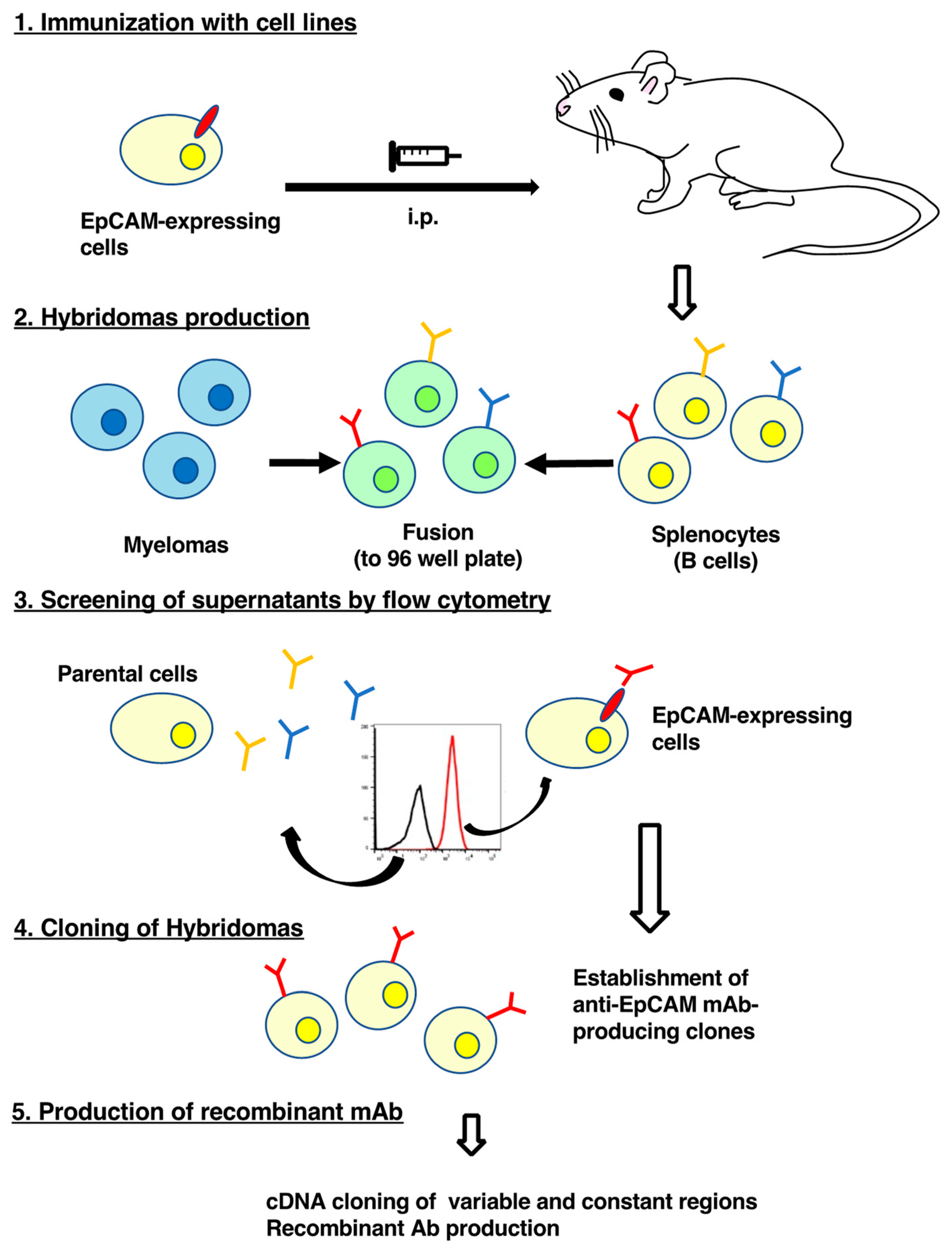
2.4. Hybridoma Production

We used the CBIS method to develop mAbs against EpCAM. Briefly, one BALB/c mouse was intraperitoneally (i.p.) immunized with CHO/EpCAM cells (1 × 10⁸) together with Imject Alum adjuvant (Thermo Fisher Scientific Inc.). The procedure included three additional immunizations, followed by a final booster injection-administered i.p. two days before spleen cell harvesting. Spleen cells were then fused with P3U1 cells using PEG1500 (Roche Diagnostics, Indianapolis, IN, USA). Hybridomas were grown in RPMI 1640, supplemented with 10% FBS, 100 U/mL of penicillin, 100 μg/mL streptomycin, and 0.25 μg/mL amphotericin B. Hypoxanthine, aminopterin, and thymidine (HAT; Thermo Fisher Scientific Inc.) were also added for hybridoma selection. Culture supernatants were screened using flow cytometry.

2.5. Production of the Recombinant Antibody

Variable (VH) and constant (CH) regions of heavy chain cDNAs of EpMab-37 were subcloned into the pCAG-Neo vector (FUJIFILM Wako Pure Chemical Corporation, Osaka, Japan) along with variable (VL) and constant (CL) regions of light chain cDNAs of EpMab-37 into the pCAG-Ble vector (FUJIFILM Wako Pure Chemical Corporation) to produce recombinant EpMab-37 (recEpMab-37). EpMab-37 vectors were transfected into ExpiCHO-S cells using the ExpiCHO Expression System (Thermo Fisher Scientific Inc.). recEpMab-37 was purified using Ab-Capcher (ProteNova, Kagawa, Japan).

2.6. Flow Cytometry

CHO/EpCAM, CHO-K1, Caco-2, and BINDS-16 cells were harvested after brief exposure to 0.25% trypsin in 1 mM ethylenediaminetetraacetic acid (EDTA; Nacalai Tesque, Inc.). After washing with 0.1% bovine serum albumin (BSA) in phosphate-buffered saline (PBS), cells were treated with recEpMab-37 for 30 min at 4 °C, and then with Alexa Fluor 488-conjugated anti-mouse IgG (1:1000; Cell Signaling Technology, Inc., Danvers, MA, USA). Fluorescence data were collected using SA3800 Cell Analyzer (Sony Corp., Tokyo, Japan).

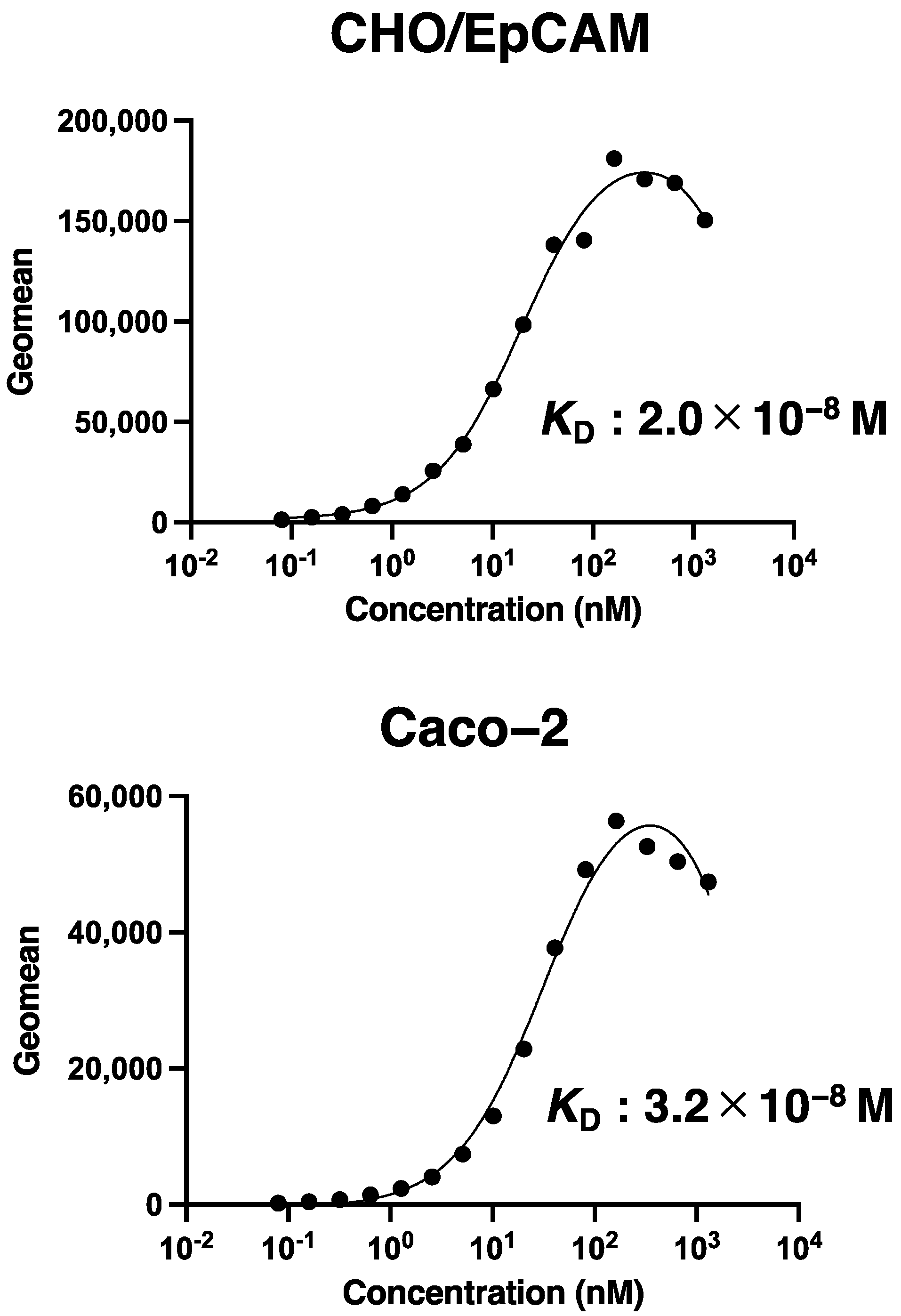
2.7. Determination of the Binding Affinity

CHO/EpCAM and Caco-2 cells were suspended in 100 μL of serially diluted recEpMab-37 (6 ng/mL–100 μg/mL), followed by the addition of Alexa Fluor 488-conjugated anti-mouse IgG (1:200). Fluorescence data were collected using BD FACSLyric (BD Biosciences, Franklin Lakes, NJ). The dissociation constant (KD) was calculated by fitting binding isotherms to built-in, one-site binding models in GraphPad Prism 8 (GraphPad Software, Inc., La Jolla, CA, USA).

2.8. Western Blot Analysis

Cell lysates of CHO/EpCAM, CHO-K1, Caco-2, and BINDS-16 cells were boiled in sodium dodecyl sulfate sample buffer (Nacalai Tesque, Inc.). The samples were then electrophoresed on 5–20% polyacrylamide gels (Nacalai Tesque, Inc.) and transferred onto polyvinylidene difluoride (PVDF) membranes (Merck KGaA, Darmstadt, Germany). After blocking with 4% skim milk (Nacalai Tesque, Inc.) for 1 h, the membrane was incubated with recEpMab-37 (1 μg/mL) or anti-β-actin (1 μg/mL) for 1 h, followed by incubation with HRP-conjugated anti-mouse immunoglobulins (Agilent Technologies, Inc., Santa Clara, CA, USA) at a 1:2000 dilution for 1 h. The membrane was developed using the ImmunoStar LD Chemiluminescence Reagent (FUJIFILM Wako Pure Chemical Corporation) and a Sayaca-Imager (DRC Co., Ltd., Tokyo, Japan). All Western blot procedures were performed at room temperature.

2.9. Immunohistochemical Analysis

Histologic sections that were 4 μm thick of colorectal carcinoma tissue array (Catalog number: CO243b; US Biomax Inc., Rockville, MD, USA) were autoclaved in citrate buffer (pH 6.0; Agilent Technologies Inc.) for 20 min. Sections were then incubated with 1 μg/mL of recEpMab-37 for 1 h at room temperature and treated using an Envision+ kit (Agilent Technologies, Inc.) for 30 min. The color was developed using 3,3′-diaminobenzidine tetrahydrochloride (DAB; Agilent Technologies, Inc.) for 2 min, and sections were then counterstained with hematoxylin (FUJIFILM Wako Pure Chemical Corporation).

3. Results

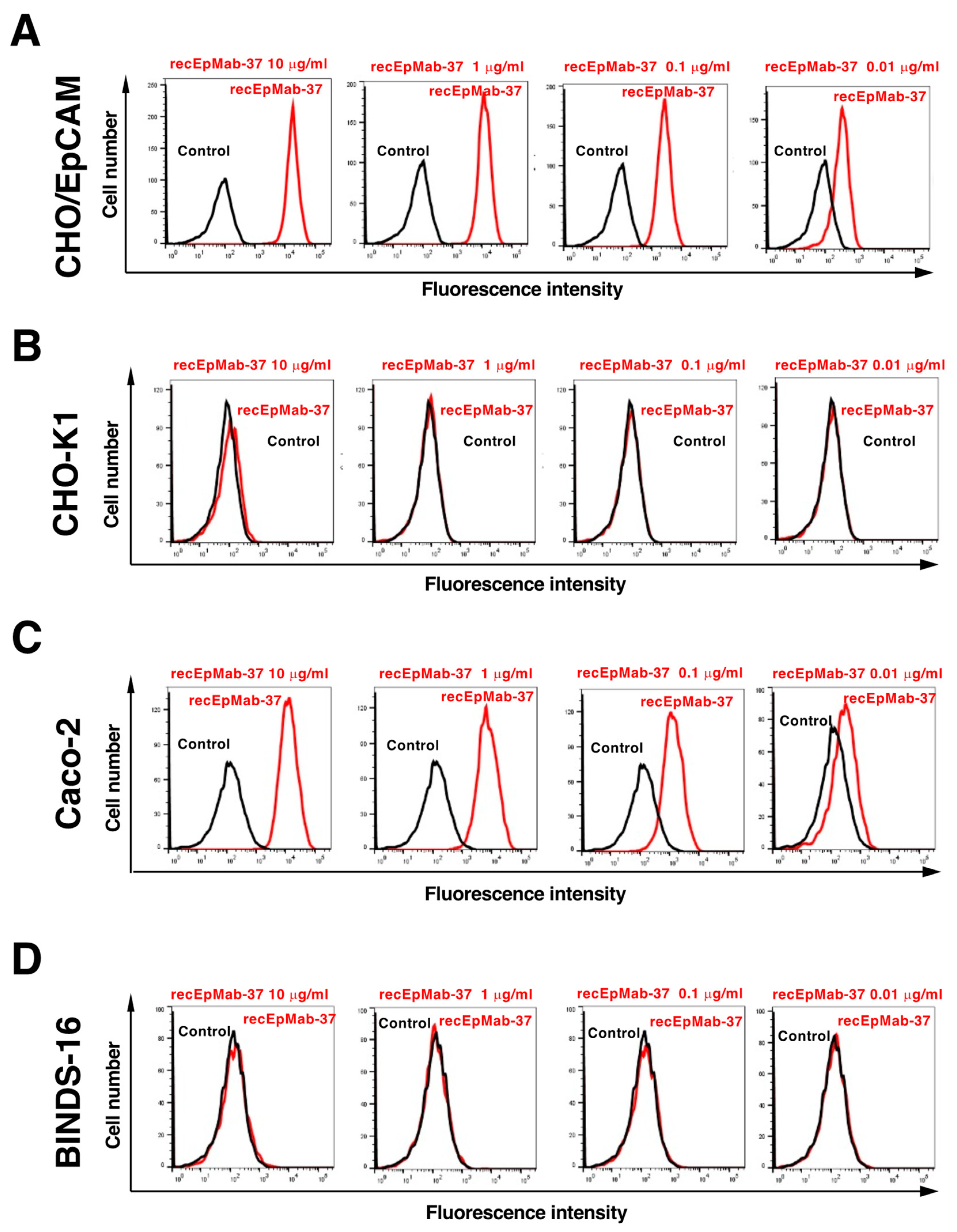
3.1. Flow Cytometric Analysis of recEpMab-37 to EpCAM-Expressing Cells

In this study, EpMab-37 was established by immunizing one mouse with CHO/EpCAM cells (Figure 1). We confirmed the reactivity of EpMab-37 to CHO/EpCAM cells, cloned the cDNAs of EpMab-37, and produced the recombinant mAb, recEpMab-37.
We first confirmed the reactivity of recEpMab-37 against CHO/EpCAM by flow cytometry. As shown in Figure 2A, recEpMab-37 recognized CHO/EpCAM cells in a dose-dependent manner, but not CHO-K1 cells (Figure 2B). We next examined whether recEpMab-37 could recognize endogenous EpCAM in colorectal carcinoma Caco-2 cells. recEpMab-37 reacted with Caco-2 cells in a dose-dependent manner (Figure 2C). However, recEpMab-37 showed no reaction with EpCAM-knocked out Caco-2 (BINDS-16) cells (Figure 2D).
Then, we determined the apparent binding affinity of recEpMab-37 with CHO/EpCAM and Caco-2 using flow cytometry. The KD of recEpMab-37 for CHO/EpCAM and Caco-2 was 2.0 × 10⁻⁸ M and 3.2 × 10⁻⁸ M, respectively, indicating that recEpMab-37 possesses the moderate affinity for EpCAM-expressing cells (Figure 3).
We next generated eleven N-terminal PA tagged EpCAM deletion mutants transfected CHO-K1 cells, namely dN44 (corresponding to 44–314 amino acids [aa]), dN64 (corresponding to 64–314 aa), dN84 (corresponding to 84–314 aa), dN104 (corresponding to 104–314 aa), dN124 (corresponding to 124–314 aa), dN144 (corresponding to 144–314 aa), dN164, (corresponding to 164–314aa), dN184 (corresponding to 184–314 aa), dN204, (corresponding to 204–314 aa), dN224, (corresponding to 224–314 aa), and dN244, (corresponding to 244–314 aa). All deletion mutants of EpCAM containing the N-terminal PA tag were detected by NZ-1 (an anti-PA tag mAb), indicating the expression level of each construct to be similar (Figure 4A). Although recEpMab-37 detected dN44, dN64, dN84, dN104, dN124, and dN144, it did not react with dN164, dN184, dN204, dN224, and dN244 (Figure 4B), suggesting that the recEpMab-37 epitope exists between 144 and 164 aa of EpCAM.

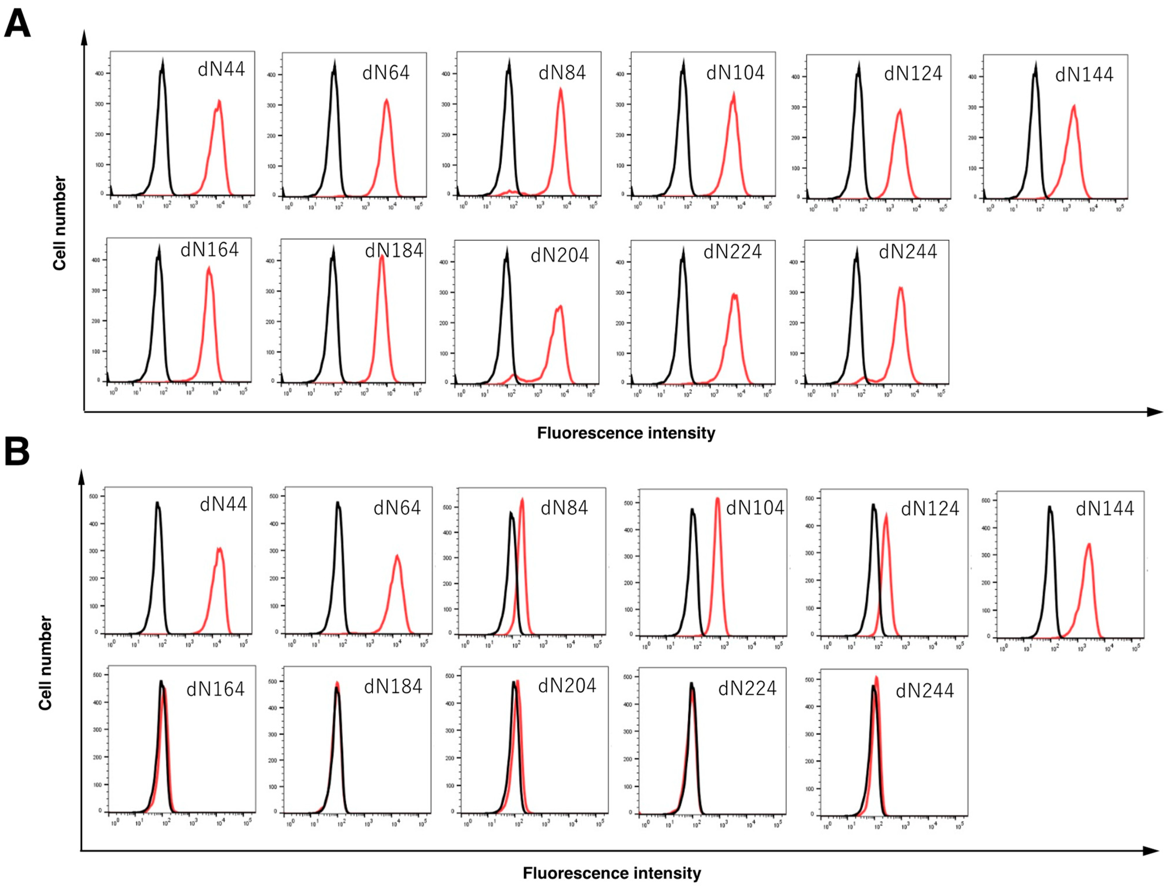
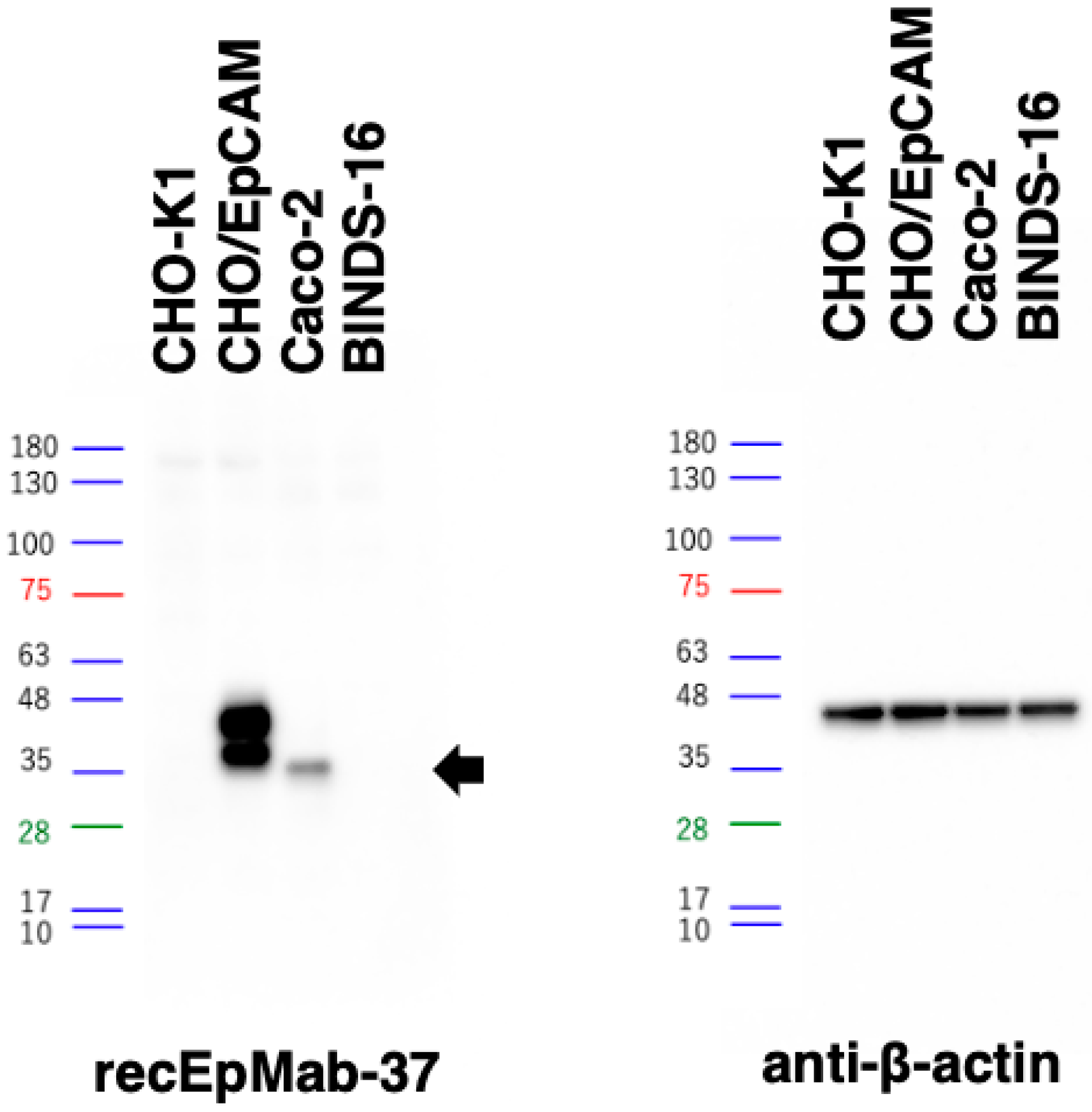
3.2. Western Blot Analysis

Western blotting was performed to further assess the sensitivity of recEpMab-37. Lysates of CHO-K1 and CHO/EpCAM cells were probed. As shown in Figure 5, recEpMab-37 detected EpCAM as 35~40 kDa double bands. However, recEpMab-37 did not detect any bands from lysates of CHO-K1 cells. These results indicate that recEpMab-37 specifically detects exogenous EpCAM. Furthermore, recEpMab-37 could detect endogenous EpCAM as a 35-kDa band from lysates of Caco-2 cells. In contrast, recEpMab-37 could not detect any bands from lysates of EpCAM-knocked out Caco-2 (BINDS-16) cells. These results suggest that recEpMab-37 specifically recognizes endogenous EpCAM in the colorectal carcinoma cell line.

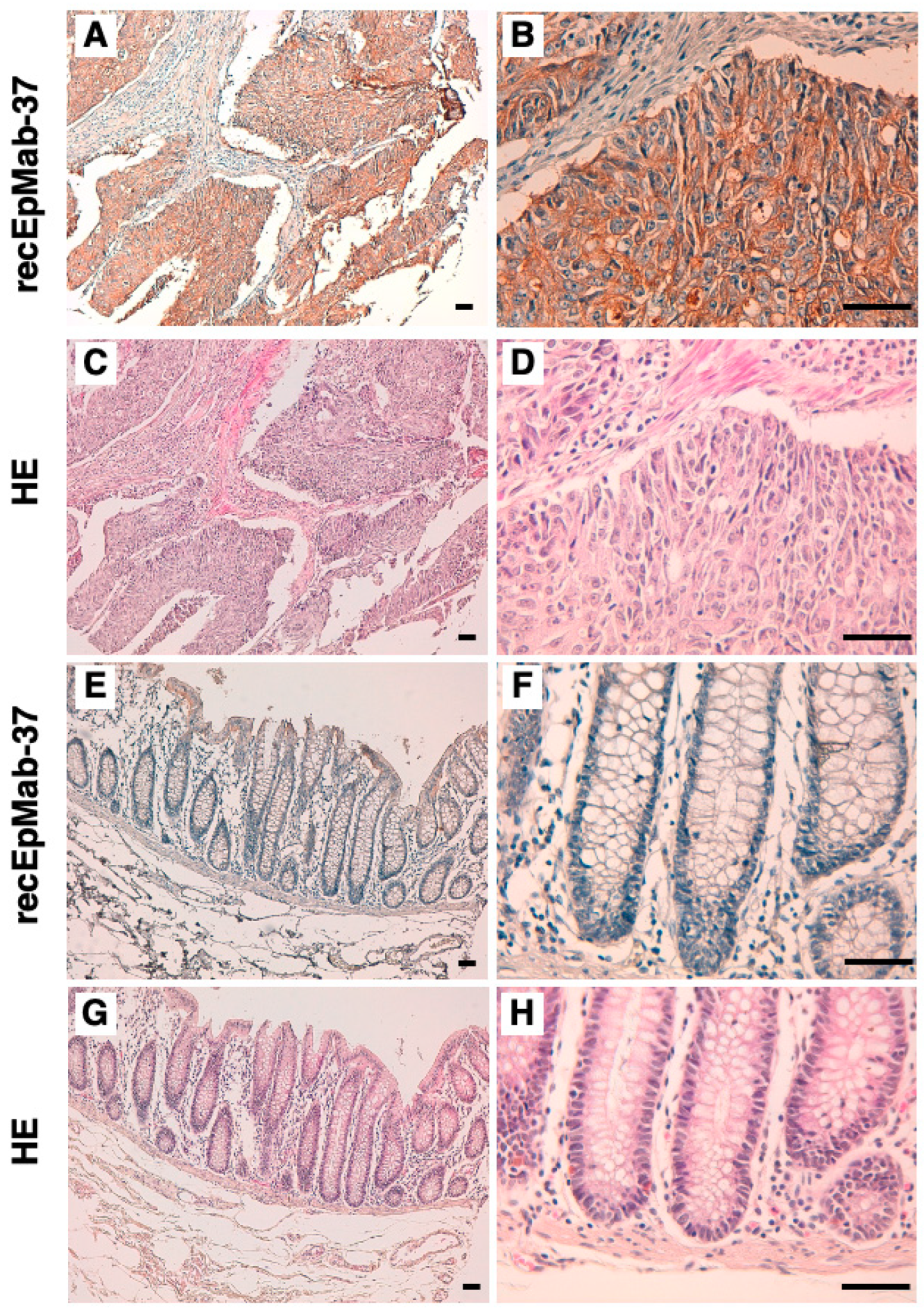
3.3. Immunohistochemical Analysis Using recEpMab-37 against Colorectal Carcinoma Tissues

We investigated whether recEpMab-37 is applicable for immunohistochemical analyses using formalin-fixed paraffin-embedded (FFPE) colorectal carcinoma sections. recEpMab-37 strongly stained colorectal carcinoma cells in a colorectal carcinoma section (Figure 6A,B), but not stromal tissues (Figure 6A). A cytoplasmic staining pattern in carcinoma cells was observed (Figure 6B). Hematoxylin and eosin (HE) staining was performed using the serial sections (Figure 6C,D). In contrast, recEpMab-37 showed faint staining in the surface of normal colorectal epithelium (Figure 6E,F). HE staining was also performed using the serial sections (Figure 6G,H). These results indicate that recEpMab-37 is useful for the immunohistochemical analysis of FFPE tumor sections.

4. Discussion

In this study, we first developed EpMab-37 using the CBIS method and next produced its recombinant mAb (recEpMab-37) (Figure 1). Then, we investigated the usefulness of recEpMab-37 for flow cytometry (Figure 2), Western blotting (Figure 5), and immunohistochemistry (Figure 6). Since recEpMab-37 does not detect any bands in EpCAM-knocked out Caco-2 cells, recEpMab-37 exhibited high specificity to EpCAM. To determine the epitope of recEpMab-37, we first performed an enzyme-linked immunosorbent assay using linear peptides in the EpCAM extracellular domain. However, EpMab-37 never recognized any peptides including 144 to 163 aa of EpCAM (Supplemental Figure S1). Next, we confirmed that the 144 to 164 aa of EpCAM are involved in recEpMab-37 binding using the deletion mutant-expressed CHO-K1 cells (Figure 4). Because EpMab-37 was established by the immunization of EpCAM-overexpressed CHO-K1 cells, EpMab-37 could recognize the native and conformational epitope. However, we cannot rule out the possibility that the deletion of EpCAM affects the native conformation and collapse the recEpMab-37 epitope. We have developed the Arg, Ile, Glu, Asp, and Leu (RIEDL)-insertion for epitope mapping (REMAP) and His-tag insertion for the epitope mapping (HisMAP) method to determine the conformational epitopes. We successfully determined the epitopes of anti-EGFR mAbs (EMab-51 and EMab-134) [34,35] and anti-CD44 mAbs (C44Mab-5 and C44Mab-46) [36,37] by the REMAP method, and anti-CD20 mAbs (C20Mab-11 and C20Mab-60) using the HisMAP method [38,39]. The determination of EpMab-37 epitope may provide useful information for future applications.
EpCAM plays critical roles in tumorigenesis to promote tumor cell proliferation, survival, and tumor-initiating potential [2]. EpCAM has been reported to be overexpressed in several tumors [40,41,42,43,44], which correlates with poor prognosis, therapeutic failure, and early tumor recurrence [43]. Therefore, EpCAM has been considered as a promising therapeutic target. A humanized scFv against EpCAM, fused to Pseudomonas exotoxin A, Oportuzumab monatox (Vicinium, VB4-845), was developed [3]. After Vicinium targets EpCAM on tumor cells, it is internalized, receives the cleavage of the linker, releases the payload exotoxin, and exhibits cytotoxicity. Vicinium was granted the FDA fast track designation for the treatment of bacillus Calmette-Guérin (BCG)-unresponsive non-muscle invasive bladder cancer (NMIBC) [45]. Vicinium was evaluated in the single-arm Phase 3 VISTA study (NCT02449239) for the treatment of patients with high-grade NMIBC in situ and high-grade papillary disease previously treated with BCG. However, FDA did not approve Vicinium for BCG-unresponsive NMIBC in August 2021 [46].
As mentioned above, clinical applications of anti-EpCAM mAbs are still limited since anti-EpCAM mAbs might generate a degree of toxicity against normal tissues. We previously established cancer-specific mAbs (CasMabs) targeting podoplanin [47,48,49,50] and podocalyxin [51], which are expressed on many tumors [52,53,54,55]. It is worthwhile to develop cancer-specific anti-EpCAM mAbs using the CasMab method. We previously converted a mouse IgG1 subclass into a mouse IgG2a, and produced a defucosylated version which exhibited high ADCC activities in vitro, and significantly reduced tumor growth [56]. Therefore, the production of a class-switched and defucosylated version of EpMab-37 is warranted to evaluate in vivo antitumor activity.

Supplementary Materials

The following supporting information can be downloaded at: https://www.mdpi.com/article/10.3390/antib11020041/s1, Figure S1: Determination of the recEpMab-37 epitope for EpCAM by ELISA using synthesized peptides of EpCAM extracellular domain.

Author Contributions

G.L., T.T. and T.A. performed the experiments; H.S. (Hiroyoshi Suzuki), M.K.K. and Y.K. designed the experiments; G.L. analyzed the data; G.L., H.S. (Hiroyuki Suzuki) and Y.K. wrote the manuscript. All authors have read and agreed to the published version of the manuscript.

Funding

This research was supported in part by Japan Agency for Medical Research and Development (AMED) under Grant Numbers: JP22ama121008 (to Y.K.), JP22am0401013 (to Y.K.), JP22bm1004001 (to Y.K.), JP22ck0106730 (to Y.K.), and JP21am0101078 (to Y.K.).

Institutional Review Board Statement

The animal study protocol was approved by the Animal Care and Use Committee of Tohoku University (Permit number: 2019NiA-001) for studies involving animals.

Data Availability Statement

The data presented in this study are available in the article and supplementary material.

Conflicts of Interest

The authors declare no conflict of interest.

References

  1. Trzpis, M.; McLaughlin, P.M.; de Leij, L.M.; Harmsen, M.C. Epithelial cell adhesion molecule: More than a carcinoma marker and adhesion molecule. Am. J. Pathol. 2007, 171, 386–395. [Google Scholar] [CrossRef] [PubMed] [Green Version]
  2. Brown, T.C.; Sankpal, N.V.; Gillanders, W.E. Functional Implications of the Dynamic Regulation of EpCAM during Epithelial-to-Mesenchymal Transition. Biomolecules 2021, 11, 956. [Google Scholar] [CrossRef]
  3. Eyvazi, S.; Farajnia, S.; Dastmalchi, S.; Kanipour, F.; Zarredar, H.; Bandehpour, M. Antibody Based EpCAM Targeted Therapy of Cancer, Review and Update. Curr. Cancer Drug Targets 2018, 18, 857–868. [Google Scholar] [CrossRef] [PubMed]
  4. Herlyn, M.; Steplewski, Z.; Herlyn, D.; Koprowski, H. Colorectal carcinoma-specific antigen: Detection by means of monoclonal antibodies. Proc. Natl. Acad. Sci. USA 1979, 76, 1438–1442. [Google Scholar] [CrossRef] [Green Version]
  5. Went, P.T.; Lugli, A.; Meier, S.; Bundi, M.; Mirlacher, M.; Sauter, G.; Dirnhofer, S. Frequent EpCam protein expression in human carcinomas. Hum. Pathol. 2004, 35, 122–128. [Google Scholar] [CrossRef] [PubMed]
  6. Riethmüller, G.; Holz, E.; Schlimok, G.; Schmiegel, W.; Raab, R.; Höffken, K.; Gruber, R.; Funke, I.; Pichlmaier, H.; Hirche, H.; et al. Monoclonal antibody therapy for resected Dukes’ C colorectal cancer: Seven-year outcome of a multicenter randomized trial. J. Clin. Oncol. 1998, 16, 1788–1794. [Google Scholar] [CrossRef]
  7. Sears, H.F.; Atkinson, B.; Mattis, J.; Ernst, C.; Herlyn, D.; Steplewski, Z.; Häyry, P.; Koprowski, H. Phase-I clinical trial of monoclonal antibody in treatment of gastrointestinal tumours. Lancet 1982, 1, 762–765. [Google Scholar] [CrossRef]
  8. Schönberger, S.; Kraft, D.; Nettersheim, D.; Schorle, H.; Casati, A.; Craveiro, R.B.; Mohseni, M.M.; Calaminus, G.; Dilloo, D. Targeting EpCAM by a Bispecific Trifunctional Antibody Exerts Profound Cytotoxic Efficacy in Germ Cell Tumor Cell Lines. Cancers 2020, 12, 1279. [Google Scholar] [CrossRef]
  9. Zhang, X.; Yang, Y.; Fan, D.; Xiong, D. The development of bispecific antibodies and their applications in tumor immune escape. Exp. Hematol. Oncol. 2017, 6, 12. [Google Scholar] [CrossRef]
  10. Knödler, M.; Körfer, J.; Kunzmann, V.; Trojan, J.; Daum, S.; Schenk, M.; Kullmann, F.; Schroll, S.; Behringer, D.; Stahl, M.; et al. Randomised phase II trial to investigate catumaxomab (anti-EpCAM × anti-CD3) for treatment of peritoneal carcinomatosis in patients with gastric cancer. Br. J. Cancer 2018, 119, 296–302. [Google Scholar] [CrossRef]
  11. Seeber, A.; Martowicz, A.; Spizzo, G.; Buratti, T.; Obrist, P.; Fong, D.; Gastl, G.; Untergasser, G. Soluble EpCAM levels in ascites correlate with positive cytology and neutralize catumaxomab activity in vitro. BMC Cancer 2015, 15, 372. [Google Scholar] [CrossRef] [PubMed] [Green Version]
  12. Ruf, P.; Kluge, M.; Jäger, M.; Burges, A.; Volovat, C.; Heiss, M.M.; Hess, J.; Wimberger, P.; Brandt, B.; Lindhofer, H. Pharmacokinetics, immunogenicity and bioactivity of the therapeutic antibody catumaxomab intraperitoneally administered to cancer patients. Br. J. Clin. Pharmacol. 2010, 69, 617–625. [Google Scholar] [CrossRef] [PubMed] [Green Version]
  13. Linke, R.; Klein, A.; Seimetz, D. Catumaxomab: Clinical development and future directions. MAbs 2010, 2, 129–136. [Google Scholar] [CrossRef] [PubMed] [Green Version]
  14. Xiao, J.; Pohlmann, P.R.; Isaacs, C.; Weinberg, B.A.; He, A.R.; Schlegel, R.; Agarwal, S. Circulating Tumor Cells: Technologies and Their Clinical Potential in Cancer Metastasis. Biomedicines 2021, 9, 1111. [Google Scholar] [CrossRef] [PubMed]
  15. de Bono, J.S.; Scher, H.I.; Montgomery, R.B.; Parker, C.; Miller, M.C.; Tissing, H.; Doyle, G.V.; Terstappen, L.W.; Pienta, K.J.; Raghavan, D. Circulating tumor cells predict survival benefit from treatment in metastatic castration-resistant prostate cancer. Clin. Cancer Res. 2008, 14, 6302–6309. [Google Scholar] [CrossRef] [PubMed] [Green Version]
  16. Tanaka, T.; Nanamiya, R.; Takei, J.; Nakamura, T.; Yanaka, M.; Hosono, H.; Sano, M.; Asano, T.; Kaneko, M.K.; Kato, Y. Development of Anti-Mouse CC Chemokine Receptor 8 Monoclonal Antibodies for Flow Cytometry. Monoclon. Antib. Immunodiagn. Immunother. 2021, 40, 65–70. [Google Scholar] [CrossRef] [PubMed]
  17. Tanaka, T.; Asano, T.; Sano, M.; Takei, J.; Hosono, H.; Nanamiya, R.; Nakamura, T.; Yanaka, M.; Harada, H.; Fukui, M.; et al. Development of Monoclonal Antibody PMab-269 Against California Sea Lion Podoplanin. Monoclon. Antib. Immunodiagn. Immunother. 2021, 40, 124–133. [Google Scholar] [CrossRef] [PubMed]
  18. Takei, J.; Asano, T.; Nanamiya, R.; Nakamura, T.; Yanaka, M.; Hosono, H.; Tanaka, T.; Sano, M.; Kaneko, M.K.; Harada, H.; et al. Development of Anti-human T Cell Immunoreceptor with Ig and ITIM Domains (TIGIT) Monoclonal Antibodies for Flow Cytometry. Monoclon. Antib. Immunodiagn. Immunother. 2021, 40, 71–75. [Google Scholar] [CrossRef]
  19. Sayama, Y.; Kaneko, M.K.; Takei, J.; Hosono, H.; Sano, M.; Asano, T.; Kato, Y. Establishment of a novel anti-TROP2 monoclonal antibody TrMab-29 for immunohistochemical analysis. Biochem. Biophys. Rep. 2021, 25, 100902. [Google Scholar] [CrossRef]
  20. Sayama, Y.; Kaneko, M.K.; Kato, Y. Development and characterization of TrMab-6, a novel anti-TROP2 monoclonal antibody for antigen detection in breast cancer. Mol. Med. Rep. 2021, 23, 1. [Google Scholar] [CrossRef]
  21. Nanamiya, R.; Takei, J.; Asano, T.; Tanaka, T.; Sano, M.; Nakamura, T.; Yanaka, M.; Hosono, H.; Kaneko, M.K.; Kato, Y. Development of Anti-Human CC Chemokine Receptor 9 Monoclonal Antibodies for Flow Cytometry. Monoclon. Antib. Immunodiagn. Immunother. 2021, 40, 101–106. [Google Scholar] [CrossRef] [PubMed]
  22. Hosono, H.; Asano, T.; Takei, J.; Sano, M.; Tanaka, T.; Kaneko, M.K.; Kato, Y. Development of an Anti-Elephant Podoplanin Monoclonal Antibody PMab-265 for Flow Cytometry. Monoclon. Antib. Immunodiagn. Immunother. 2021, 40, 141–145. [Google Scholar] [CrossRef] [PubMed]
  23. Asano, T.; Nanamiya, R.; Takei, J.; Nakamura, T.; Yanaka, M.; Hosono, H.; Tanaka, T.; Sano, M.; Kaneko, M.K.; Kato, Y. Development of Anti-Mouse CC Chemokine Receptor 3 Monoclonal Antibodies for Flow Cytometry. Monoclon. Antib. Immunodiagn. Immunother. 2021, 40, 107–112. [Google Scholar] [CrossRef] [PubMed]
  24. Kaneko, M.K.; Sano, M.; Takei, J.; Asano, T.; Sayama, Y.; Hosono, H.; Kobayashi, A.; Konnai, S.; Kato, Y. Development and Characterization of Anti-Sheep Podoplanin Monoclonal Antibodies PMab-253 and PMab-260. Monoclon. Antib. Immunodiagn. Immunother. 2020, 39, 144–155. [Google Scholar] [CrossRef] [PubMed]
  25. Furusawa, Y.; Kaneko, M.K.; Kato, Y. Establishment of C(20)Mab-11, a novel anti-CD20 monoclonal antibody, for the detection of B cells. Oncol. Lett. 2020, 20, 1961–1967. [Google Scholar] [CrossRef]
  26. Furusawa, Y.; Kaneko, M.K.; Kato, Y. Establishment of an Anti-CD20 Monoclonal Antibody (C(20)Mab-60) for Immunohistochemical Analyses. Monoclon. Antib. Immunodiagn. Immunother. 2020, 39, 112–116. [Google Scholar] [CrossRef]
  27. Kato, Y.; Furusawa, Y.; Yamada, S.; Itai, S.; Takei, J.; Sano, M.; Kaneko, M.K. Establishment of a monoclonal antibody PMab-225 against alpaca podoplanin for immunohistochemical analyses. Biochem. Biophys. Rep. 2019, 18, 100633. [Google Scholar] [CrossRef]
  28. Furusawa, Y.; Yamada, S.; Itai, S.; Nakamura, T.; Yanaka, M.; Sano, M.; Harada, H.; Fukui, M.; Kaneko, M.K.; Kato, Y. PMab-219: A monoclonal antibody for the immunohistochemical analysis of horse podoplanin. Biochem. Biophys. Rep. 2019, 18, 100616. [Google Scholar] [CrossRef]
  29. Yamada, S.; Itai, S.; Nakamura, T.; Yanaka, M.; Kaneko, M.K.; Kato, Y. Detection of high CD44 expression in oral cancers using the novel monoclonal antibody, C(44)Mab-5. Biochem. Biophys. Rep. 2018, 14, 64–68. [Google Scholar] [CrossRef]
  30. Yamada, S.; Itai, S.; Nakamura, T.; Yanaka, M.; Chang, Y.W.; Suzuki, H.; Kaneko, M.K.; Kato, Y. Monoclonal Antibody L(1)Mab-13 Detected Human PD-L1 in Lung Cancers. Monoclon. Antib. Immunodiagn. Immunother. 2018, 37, 110–115. [Google Scholar] [CrossRef] [Green Version]
  31. Itai, S.; Yamada, S.; Kaneko, M.K.; Harada, H.; Kagawa, Y.; Konnai, S.; Kato, Y. Expression of Cat Podoplanin in Feline Squamous Cell Carcinomas. Monoclon. Antib. Immunodiagn. Immunother 2017, 36, 243–250. [Google Scholar] [CrossRef] [PubMed]
  32. Kaneko, M.K.; Ohishi, T.; Takei, J.; Sano, M.; Nakamura, T.; Hosono, H.; Yanaka, M.; Asano, T.; Sayama, Y.; Harada, H.; et al. Anti-EpCAM monoclonal antibody exerts antitumor activity against oral squamous cell carcinomas. Oncol. Rep. 2020, 44, 2517–2526. [Google Scholar] [CrossRef] [PubMed]
  33. Hosono, H.; Ohishi, T.; Takei, J.; Asano, T.; Sayama, Y.; Kawada, M.; Kaneko, M.K.; Kato, Y. The anti-epithelial cell adhesion molecule (EpCAM) monoclonal antibody EpMab-16 exerts antitumor activity in a mouse model of colorectal adenocarcinoma. Oncol. Lett. 2020, 20, 383. [Google Scholar] [CrossRef] [PubMed]
  34. Sano, M.; Kaneko, M.K.; Aasano, T.; Kato, Y. Epitope Mapping of an Antihuman EGFR Monoclonal Antibody (EMab-134) Using the REMAP Method. Monoclon. Antib. Immunodiagn. Immunother. 2021, 40, 191–195. [Google Scholar] [CrossRef]
  35. Nanamiya, R.; Sano, M.; Asano, T.; Yanaka, M.; Nakamura, T.; Saito, M.; Tanaka, T.; Hosono, H.; Tateyama, N.; Kaneko, M.K.; et al. Epitope Mapping of an Anti-Human Epidermal Growth Factor Receptor Monoclonal Antibody (EMab-51) Using the RIEDL Insertion for Epitope Mapping Method. Monoclon Antib. Immunodiagn. Immunother. 2021, 40, 149–155. [Google Scholar] [CrossRef]
  36. Asano, T.; Kaneko, M.K.; Kato, Y. Development of a Novel Epitope Mapping System: RIEDL Insertion for Epitope Mapping Method. Monoclon. Antib. Immunodiagn. Immunother. 2021, 40, 162–167. [Google Scholar] [CrossRef]
  37. Asano, T.; Kaneko, M.K.; Takei, J.; Tateyama, N.; Kato, Y. Epitope Mapping of the Anti-CD44 Monoclonal Antibody (C44Mab-46) Using the REMAP Method. Monoclon. Antib. Immunodiagn. Immunother. 2021, 40, 156–161. [Google Scholar] [CrossRef]
  38. Suzuki, H.; Asano, T.; Tanaka, T.; Kaneko, M.K.; Kato, Y. Epitope Mapping of the Anti-CD20 Monoclonal Antibodies (C20Mab-11 and 2H7) Using HisMAP Method. Monoclon. Antib. Immunodiagn. Immunother. 2022, 41, 20–26. [Google Scholar] [CrossRef]
  39. Asano, T.; Suzuki, H.; Kaneko, M.K.; Kato, Y. Epitope Mapping of Rituximab Using HisMAP Method. Monoclon. Antib. Immunodiagn. Immunother. 2022, 41, 8–14. [Google Scholar] [CrossRef]
  40. Jing, Y.; Zhou, L.; Chen, J.; Xu, H.; Sun, J.; Cai, M.; Jiang, J.; Gao, J.; Wang, H. Quantitatively Mapping the Assembly Pattern of EpCAM on Cell Membranes with Peptide Probes. Anal. Chem. 2020, 92, 1865–1873. [Google Scholar] [CrossRef] [PubMed]
  41. Vorobyeva, A.; Konovalova, E.; Xu, T.; Schulga, A.; Altai, M.; Garousi, J.; Rinne, S.S.; Orlova, A.; Tolmachev, V.; Deyev, S. Feasibility of Imaging EpCAM Expression in Ovarian Cancer Using Radiolabeled DARPin Ec1. Int. J. Mol. Sci. 2020, 21, 3310. [Google Scholar] [CrossRef] [PubMed]
  42. van den Brand, D.; van Lith, S.A.M.; de Jong, J.M.; Gorris, M.A.J.; Palacio-Castaneda, V.; Couwenbergh, S.T.; Goldman, M.R.G.; Ebisch, I.; Massuger, L.F.; Leenders, W.P.J.; et al. EpCAM-Binding DARPins for Targeted Photodynamic Therapy of Ovarian Cancer. Cancers 2020, 12, 1762. [Google Scholar] [CrossRef] [PubMed]
  43. Pandit, H.; Li, Y.; Zheng, Q.; Guo, W.; Yu, Y.; Li, S.; Martin, R.C.G. Carcinogenetic initiation contributed by EpCAM+ cancer cells in orthotopic HCC models of immunocompetent and athymic mice. Oncotarget 2020, 11, 2047–2060. [Google Scholar] [CrossRef] [PubMed]
  44. Zhang, D.; Yang, L.; Liu, X.; Gao, J.; Liu, T.; Yan, Q.; Yang, X. Hypoxia modulates stem cell properties and induces EMT through N-glycosylation of EpCAM in breast cancer cells. J. Cell Physiol. 2020, 235, 3626–3633. [Google Scholar] [CrossRef]
  45. Kaplon, H.; Muralidharan, M.; Schneider, Z.; Reichert, J.M. Antibodies to watch in 2020. MAbs 2020, 12, 1703531. [Google Scholar] [CrossRef] [Green Version]
  46. Karlovitch, S. FDA Does Not Approve Vicinium for BCG-unresponsive NMIBC. 2021. Available online: https://www.targetedonc.com/view/fda-does-not-approve-vicinium-for-bcg-unresponsive-nmibc (accessed on 5 June 2022).
  47. Kaneko, M.K.; Nakamura, T.; Kunita, A.; Fukayama, M.; Abe, S.; Nishioka, Y.; Yamada, S.; Yanaka, M.; Saidoh, N.; Yoshida, K.; et al. ChLpMab-23: Cancer-Specific Human-Mouse Chimeric Anti-Podoplanin Antibody Exhibits Antitumor Activity via Antibody-Dependent Cellular Cytotoxicity. Monoclon. Antib. Immunodiagn. Immunother. 2017, 36, 104–112. [Google Scholar] [CrossRef]
  48. Kaneko, M.K.; Yamada, S.; Nakamura, T.; Abe, S.; Nishioka, Y.; Kunita, A.; Fukayama, M.; Fujii, Y.; Ogasawara, S.; Kato, Y. Antitumor activity of chLpMab-2, a human-mouse chimeric cancer-specific antihuman podoplanin antibody, via antibody-dependent cellular cytotoxicity. Cancer Med. 2017, 6, 768–777. [Google Scholar] [CrossRef]
  49. Kato, Y.; Kaneko, M.K. A cancer-specific monoclonal antibody recognizes the aberrantly glycosylated podoplanin. Sci. Rep. 2014, 4, 5924. [Google Scholar] [CrossRef]
  50. Miyazaki, A.; Nakai, H.; Sonoda, T.; Hirohashi, Y.; Kaneko, M.K.; Kato, Y.; Sawa, Y.; Hiratsuka, H. LpMab-23-recognizing cancer-type podoplanin is a novel predictor for a poor prognosis of early stage tongue cancer. Oncotarget 2018, 9, 21156–21165. [Google Scholar] [CrossRef] [Green Version]
  51. Kaneko, M.K.; Ohishi, T.; Kawada, M.; Kato, Y. A cancer-specific anti-podocalyxin monoclonal antibody (60-mG(2a)-f) exerts antitumor effects in mouse xenograft models of pancreatic carcinoma. Biochem. Biophys. Rep. 2020, 24, 100826. [Google Scholar] [CrossRef]
  52. Kaneko, M.K.; Itai, S.; Yamada, S.; Kato, Y. 47-mG(2a): A Mouse IgG(2a)-Type of PcMab-47 Useful for Detecting Podocalyxin in Esophageal Cancers by Immunohistochemistry. Monoclon. Antib. Immunodiagn. Immunother. 2018, 37, 158–161. [Google Scholar] [CrossRef] [PubMed]
  53. Tanaka, M.; Kijima, H.; Shimada, H.; Makuuchi, H.; Ozawa, S.; Inokuchi, S. Expression of podoplanin and vimentin is correlated with prognosis in esophageal squamous cell carcinoma. Mol. Med. Rep. 2015, 12, 4029–4036. [Google Scholar] [CrossRef] [PubMed] [Green Version]
  54. Nakashima, Y.; Yoshinaga, K.; Kitao, H.; Ando, K.; Kimura, Y.; Saeki, H.; Oki, E.; Morita, M.; Kakeji, Y.; Hirahashi, M.; et al. Podoplanin is expressed at the invasive front of esophageal squamous cell carcinomas and is involved in collective cell invasion. Cancer Sci. 2013, 104, 1718–1725. [Google Scholar] [CrossRef] [PubMed]
  55. Rahadiani, N.; Ikeda, J.; Makino, T.; Tian, T.; Qiu, Y.; Mamat, S.; Wang, Y.; Doki, Y.; Aozasa, K.; Morii, E. Tumorigenic role of podoplanin in esophageal squamous-cell carcinoma. Ann. Surg. Oncol. 2010, 17, 1311–1323. [Google Scholar] [CrossRef]
  56. Takei, J.; Kaneko, M.K.; Ohishi, T.; Hosono, H.; Nakamura, T.; Yanaka, M.; Sano, M.; Asano, T.; Sayama, Y.; Kawada, M.; et al. A defucosylated antiCD44 monoclonal antibody 5mG2af exerts antitumor effects in mouse xenograft models of oral squamous cell carcinoma. Oncol. Rep. 2020, 44, 1949–1960. [Google Scholar] [CrossRef]
Figure 1. A schematic procedure of anti-EpCAM mAbs production. A mouse was intraperitoneally immunized with the CHO/EpCAM cells. The screening was then conducted by flow cytometry using parental cells and EpCAM-overexpressed CHO-K1 cells. Variable and constant regions cDNAs were cloned from an established clone, and the recombinant Ab was produced.
Antibodies 11 00041 g001
Figure 2. Flow cytometry of EpCAM expressing cells using recEpMab-37. CHO/EpCAM (A), CHO-K1 (B), Caco-2 (C), and BINDS-16 (EpCAM-knocked out Caco-2) (D) cells were treated with 0.01–10 µg/mL of recEpMab-37, followed by treatment with Alexa Fluor 488-conjugated anti-mouse IgG. Black line represents the negative control.
Antibodies 11 00041 g002
Figure 3. The determination of the binding affinity of recEpMab-37. CHO/EpCAM and Caco-2 cells were suspended in 100 µL serially diluted recEpMab-37 (6 ng/mL to 100 µg/mL). Then, cells were treated with Alexa Fluor 488-conjugated anti-mouse IgG. Fluorescence data were subsequently collected using a BD FACSLyric, following the calculation of the dissociation constant (KD) by GraphPad PRISM 8.
Antibodies 11 00041 g003
Figure 4. Epitope mapping of recEpMab-37 using deletion mutants of EpCAM. CHO-K1 expressed EpCAM deletion mutants were analyzed using flow cytometry. The EpCAM mutants-transfected cells were incubated with NZ-1 (anti-PA tag; red line, (A)), recEpMab-37 (anti-EpCAM mAb; red line, (B)), or buffer control (black line, (A,B)) for 30 min at 4 °C, followed by secondary antibodies.
Antibodies 11 00041 g004
Figure 5. Western blotting by recEpMab-37. Cell lysates of CHO-K1, CHO/EpCAM, Caco-2, and BINDS-16 (10 µg) were electrophoresed and transferred onto polyvinylidene fluoride membranes. The membranes were incubated with 1 µg/mL of recEpMab-37 and 1 µg/mL of anti-β-actin and subsequently with peroxidase-conjugated anti-mouse immunoglobulins. Black arrows indicate the predicted size of EpCAM (~35 kDa).
Antibodies 11 00041 g005
Figure 6. Immunohistochemical analysis using recEpMab-37 against colorectal carcinoma tissues. (A,B,E,F) After antigen retrieval, sections were incubated with 1 µg/mL of recEpMab-37 followed by treatment with the Envision+ kit. Color was developed using DAB, and sections were counterstained with hematoxylin. Cytoplasmic brown staining indicates the EpCAM positive cells. (C,D,G,H) Hematoxylin and eosin (HE) staining. Scale bar = 100 µm.
Antibodies 11 00041 g006
Publisher’s Note: MDPI stays neutral with regard to jurisdictional claims in published maps and institutional affiliations.

