Article

Immunochemistry-Based Diagnosis of Extrapulmonary Tuberculosis: A Strategy for Large-Scale Production of MPT64-Antibodies for Use in the MPT64 Antigen Detection Test

1
Centre for International Health, Department of Global Public Health and Primary Care, University of Bergen, 5020 Bergen, Norway
2
Department of Clinical Science, University of Bergen, 5020 Bergen, Norway
3
Department of Clinical Medicine, University of Bergen, 5020 Bergen, Norway
4
Department of Histopathology, Gulab Devi Chest Hospital Lahore, Lahore 54000, Pakistan
5
Department of Pathology, Haukeland University Hospital, 5021 Bergen, Norway
6
Department of Thoracic Medicine, Haukeland University Hospital, 5021 Bergen, Norway
*
Author to whom correspondence should be addressed.
Antibodies 2021, 10(3), 34; https://doi.org/10.3390/antib10030034
Submission received: 1 July 2021 / Revised: 26 July 2021 / Accepted: 24 August 2021 / Published: 26 August 2021

Abstract

:
Tuberculosis (TB) is a global health problem. The immunohistochemistry (IHC)-based MPT64 antigen detection test has shown promising results for diagnosing extrapulmonary TB in previous studies. However, the anti-MPT64 antibody currently used in the test is in limited supply, and reproduction of a functional antibody is a prerequisite for further large-scale use. Various antigen-adjuvant combinations and immunisation protocols were tested in mice and rabbits to generate monoclonal and polyclonal antibodies. Antibodies were screened in IHC, and the final new antibody was validated on clinical human specimens. We were not able to generate monoclonal antibodies that were functional in IHC, but we obtained multiple functional polyclonal antibodies through careful selection of antigen-adjuvant and comprehensive screening in IHC of both pre-immune sera and antisera. To overcome the limitation of batch-to-batch variability with polyclonal antibodies, the best performing individual polyclonal antibodies were pooled to one final large-volume new anti-MPT64 antibody. The sensitivity of the new antibody was in the same range as the reference antibody, while the specificity was somewhat reduced. Our results suggest that it possible to reproduce a large-volume functional polyclonal antibody with stable performance, thereby securing stable supplies and reproducibility of the MPT64 test, albeit further validation remains to be done.

1. Introduction

Tuberculosis (TB) is a global health problem with an estimated 10 million new cases and 1.4 million deaths in 2019 [1]. Approximately one-third of the estimated new TB cases each year are not diagnosed or reported. Extrapulmonary TB (EPTB), which is more common in children and people with HIV [2,3,4,5,6], poses a special diagnostic challenge due to the paucibacillary nature of the disease. This leads to variable and generally low sensitivity of routine microscopy, PCR and culture [1,7], and new improved diagnostic tests are needed. Tests based on the detection of mycobacterial antigens are of special interest, as they have the potential to provide rapid and direct evidence of active TB disease [8]. Two antigen-detection tests for TB diagnosis are currently commercially available (Alere Determine TB LAM and Fujifilm SILVAMP TB LAM, both detecting lipoarabinomannan in urine) but their clinical use is restricted due to suboptimal sensitivity [9,10]. The novel MPT64 antigen-detection test (the MPT64 test) for the diagnosis of EPTB, which is based on detection of the mycobacterial antigen MPT64 in tissue samples from the site of infection, has shown promising results in previous validation studies [11,12,13,14,15,16,17,18], with higher sensitivity than routine smear and culture in high TB incidence settings [12,13,14,15,16,17]. The test is feasible to implement in the low-resource setting and can contribute towards timely and accurate diagnosis of EPTB [16]. These results warrant further research to evaluate the diagnostic test accuracy in larger cohorts and to investigate the potential for large-scale use and clinical roll-out of the test. However, the in-house polyclonal rabbit anti-MPT64 antibody used in the test is currently available in limited amounts, and reproduction of an antibody with stable performance, which is a pre-requisite for large-scale use, can be challenging due to batch-to-batch to variations in polyclonal antibodies [19]. The aim of the study was to develop an anti-MPT64 antibody to secure stable supplies and performance of the MPT64 test. Here, we describe different aspects to be considered when developing antibodies for use in immunohistochemistry (IHC), the challenges faced with the production of a monoclonal antibody and strategies to make large volumes of a functional polyclonal antibody.

2. Materials and Methods

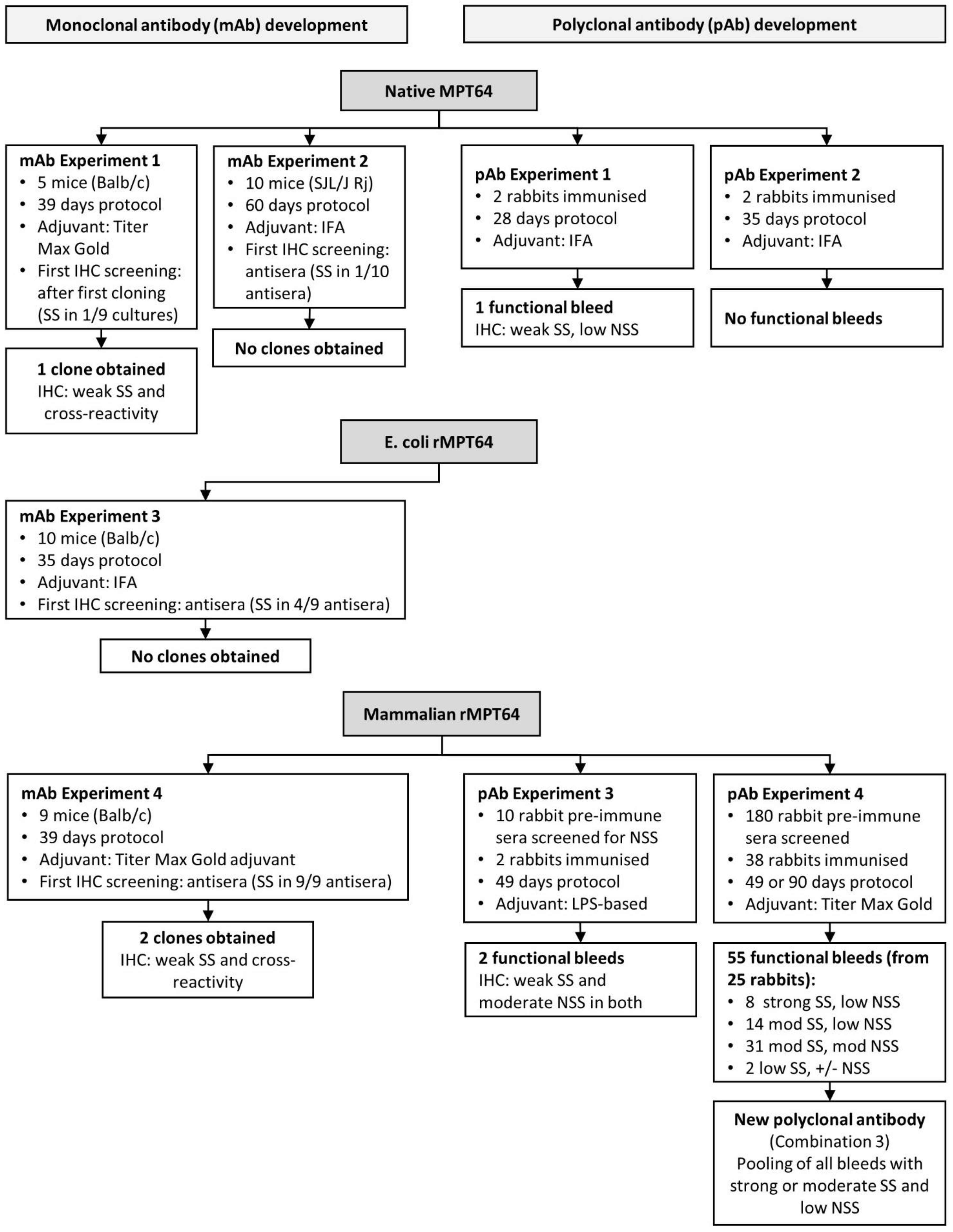
2.1. Production of MPT64 Antigen

Several strategies were used to produce MPT64 antigen for immunisation. Native MPT64 was produced because the conformational epitopes, which may be important targets in IHC, are conserved in native proteins. Native MPB64 protein was obtained from cultures of Mycobacterium bovis bacillus Calmette-Guérin Moreau (BCG Moreau), according to previously developed protocols for culturing and purification [20,21,22], with some modifications (Supplementary Materials. MPB64 is homologous to the M. tuberculosis-derived MPT64 protein used as an antigen when the reference anti-MPT64 antibody was generated [23], and the two proteins are hereafter collectively referred to as MPT64. Because production of native MPT64 is time-consuming due to the slow growth of the bacilli and the resulting low yield of MPT64 protein, we also produced recombinant MPT64 antigen in several expression systems. In our laboratory, untagged recombinant MPT64 protein was expressed in the non-pathogenic, fast-growing M. smegmatis mc2 155 [24] transformed with the mycobacterial plasmid (pUV15tetORm [25]) modified to contain the mpb64 gene with its predicted secretion signal sequence (GenBank Accession No. AM412059.2; BCGM locus 1981c), according to previously developed protocols [25,26,27] (Supplementary Materials, Figure S1). His-tagged recombinant MPT64 was produced in E. coli (by Trenzyme Life Science Services, Konstanz, Germany) and in a mammalian cell line (by InVivo Biotech Services, Berlin, Germany), based on the MPT64 amino acid sequence from M. bovis BCG Moreau without the signal sequence (Supplementary Materials). Table 1 shows the predicted amino acid sequence of the different MPT64 proteins that were used as antigens in the study.

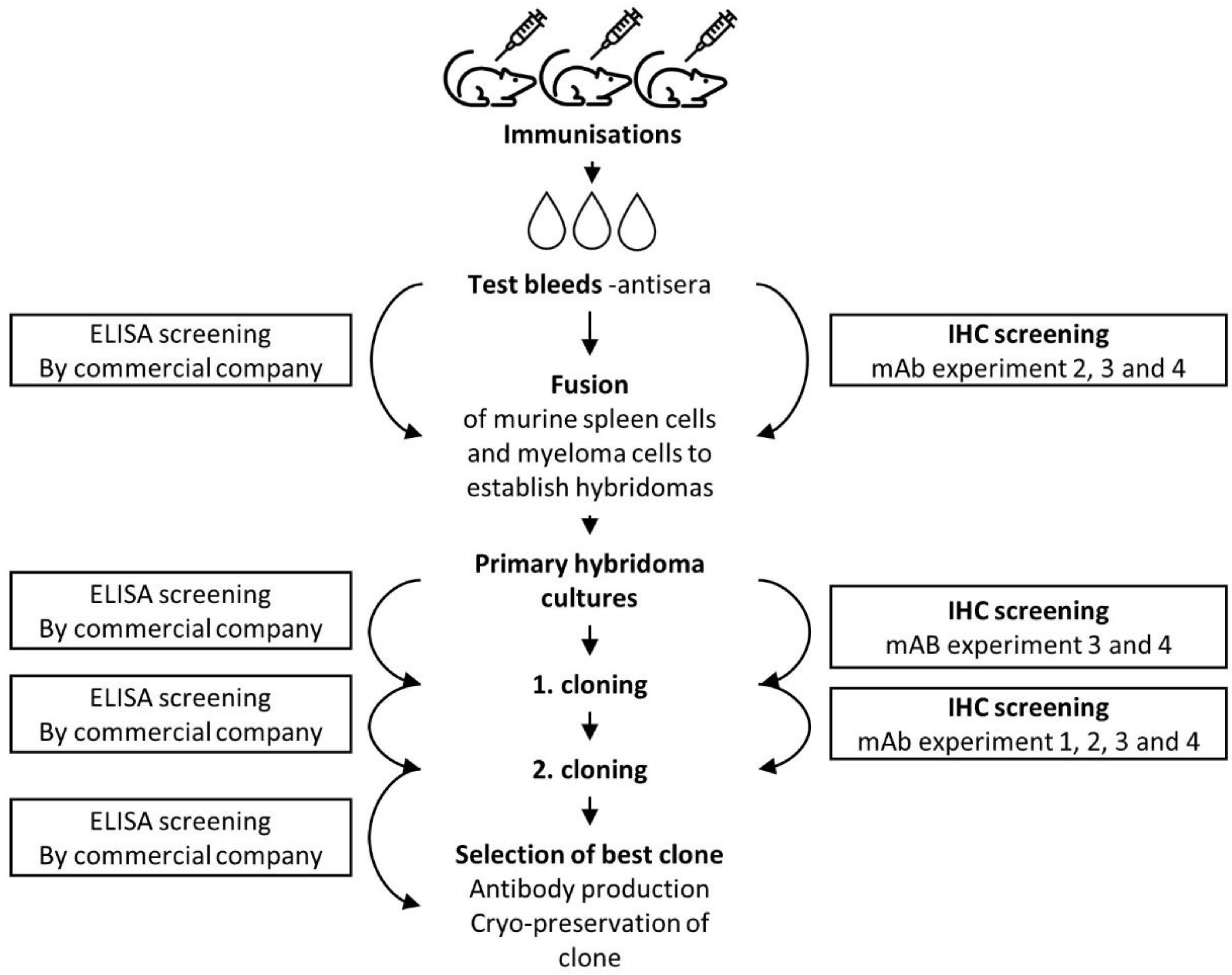
2.2. Development of a Monoclonal Anti-MPT64 Antibody

Monoclonal antibodies from mice were generated by hybridoma technology according to standard methods [28] by commercial companies (Biogenes, Berlin, Germany; PharmAbs, Leuven, Belgium and InVivo BioTech Services, Hennigsdorf, Germany). Four different strategies to develop a functional monoclonal antibody for the MPT64 test were investigated (mAb experiment 1–4, Figure 1). Different combinations of MPT64 antigen and adjuvants were tested, and an increasing number of screening steps in IHC were added in subsequent experiments. Antibodies were screened in parallel in indirect enzyme-linked immunosorbent assay (ELISA) by the commercial companies, and in IHC at our laboratory, to identify the hybridomas that produced MPT64-specific antibodies. Antibody performance in IHC was assessed in positive and negative control tissue sections using serial dilution to find the optimal working dilution, by several readers (T.M. and I.M.H.). Several antigen retrieval methods were tested to optimise the MPT64 test protocol for murine monoclonal antibodies (Supplementary Materials). Figure 2 provides an overview of the different stages of hybridoma production and target points for screening of clones for monoclonal antibodies.

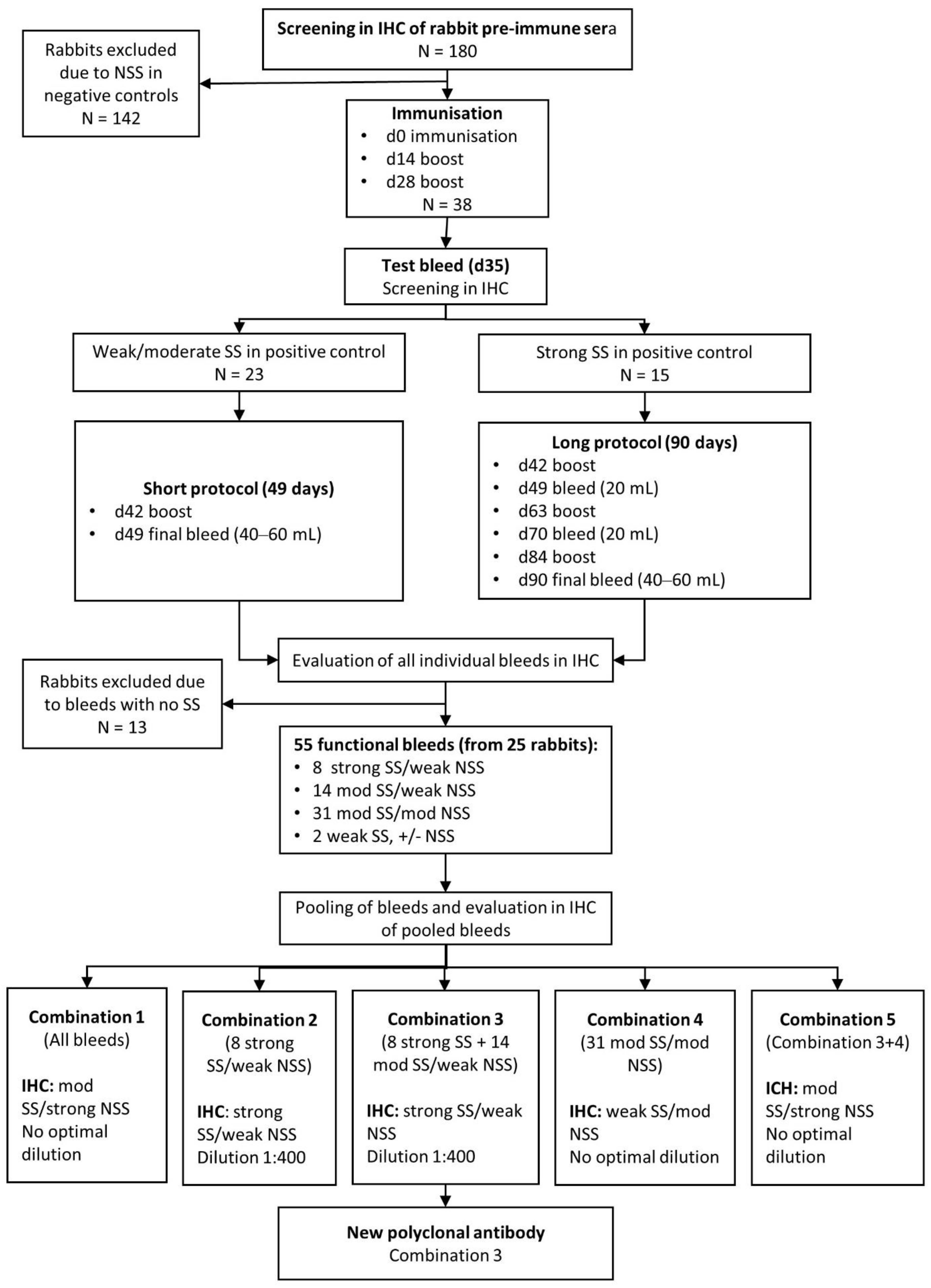
2.3. Development of Polyclonal Rabbit Anti-MPT64 Antibodies

2.3.1. Immunisations

Immunisation of rabbits to produce polyclonal anti-MPT64 antibodies was performed by commercial companies (Biogenes, PharmAbs and InVivo). Figure 1 shows the four different strategies for development of polyclonal MPT64-antibodies that were investigated (pAb experiment 1–4). In experiments 1 and 2, the original protocol for development of the reference anti-MPT64 antibody was followed [29], with the exception that the antigen was not immunoprecipitated with polyclonal rabbit anti-MPT64 antibodies before immunisation. In brief, outbred female rabbits were immunised intradermally with native MPT64 emulsified in incomplete Freund adjuvant (IFA), using a standard immunisation protocol. In later experiments, recombinant MPT64 and other adjuvants were also tested; rabbits whose pre-immune sera gave non-specific staining were excluded from immunisation, and the immunisation protocols were longer for rabbits whose antisera tests bleeds gave particularly strong specific staining (Figure 3). In all experiments, pre-immune sera were collected at baseline, antisera test bleeds were collected seven days after the second or third immunisation and the final bleed was taken seven days after the last immunisation. Pre-immune sera and all individual bleeds were tested by indirect ELISA by the companies, and in IHC in our laboratory on positive and negative control tissue sections. The staining in IHC was evaluated by several readers (T.M., I.A.M.A. and I.M.H.). The optimal working dilution was determined using serial dilution.

2.3.2. Selection and Pooling of MPT64-Specific Antibodies

The performance of the different bleeds in IHC were evaluated according to previously developed guidelines for interpretation of the MPT64 test [16]. The antibodies were categorised as (1) strong specific staining (SS) and low non-specific staining (NSS), (2) moderate SS and low NSS, (3) moderate to strong SS and moderate to strong NSS and (4) not functional, defined as no or weak SS and various degrees of NSS. In order to reduce batch-to-batch variation and to enable large-scale use of the polyclonal antibodies, the bleeds were pooled. Different combinations of pooled bleeds were tested in IHC (Figure 3), and the combination of bleeds with the best sensitivity and specificity in IHC, hereafter referred to as the new polyclonal antibody, was chosen for further experiments.

2.4. Background Blocking and Antibody Absorption Experiments

Blocking experiments were performed to reduce the non-specific binding of the new polyclonal antibody. Before application of the MPT64 antibody, the tissue sections were incubated with blocking solutions containing either (1) bovine serum albumin and serum free protein block with casein (Dako, Agilent), (2) normal goat serum or (3) recombinant Fc domain protein (Hu Fc block pure, BD, Becton Dickinson). Table 2 provides an overview of the different dilutions, incubation times and combinations of blocking solutions that were tested. Experiments with negative absorption were carried out at our laboratory by mixing the new polyclonal antibody with different proteinaceous solutions to allow non-specific antibodies or antibodies with cross-reactivity to bind proteins in the solutions and precipitate (Supplementary Materials). Positive purification by affinity column chromatography was also tested at an early stage of the study on one of the best-performing individual polyclonal antibodies, but resulted in a loss of specific staining in IHC, and the method was, therefore, not further explored.

2.5. Immunohistochemistry (the MPT64 Test)

The MPT64 test was performed using the Dako Envision + System-HRP kit (Agilent, Santa Clara, CA, USA), according to the manufacturer’s protocol, with some modifications. Briefly, 4 µm thick tissue sections on Superfrost Plus slides (Thermo Fisher Scientific, Waltham, MA, USA) were deparaffinised with xylene and rehydrated through decreasing grades of alcohol. When rabbit antibodies were used as primary antibody, heat-induced antigen retrieval (HIER) was performed by microwave boiling the sections in citrate buffer at pH 9, for 20 min. For murine antibodies, HIER was performed by pressure cooker boiling at 125 °C in TE-buffer at pH 9, for 1 min. The sections were left to cool for 20 min at RT, washed in distilled water for 10 min and incubated with peroxidase block for 20 min. For IHC with the new polyclonal antibody, an additional protein blocking step was then added to the protocol, in which a combination of 10% normal goat serum and 3% bovine serum albumin was applied to the sections overnight at 4 °C, followed by a serum-free protein block (Dako) for 12 min at RT the next day. The primary antibody was applied, and the slides were incubated for 60 min, before horseradish peroxidase conjugated secondary anti-rabbit antibody was applied for 45 min. Thereafter, the substrate (3-amino-9-ethylcarbazol) was added to the slides for 15 min, followed by counterstaining with Mayer’s haematoxylin and mounting with Immu-Mount (Thermo Fisher Scientific, Waltham, MA, USA). Slides were washed with wash buffer (0.05 mol/L Tris/HCl buffered saline with 0.05% Tween 20, pH 7.6) between all incubation steps. In each IHC run, tissue sections from known TB and non-TB cases were added as controls. In addition, the primary antibody was substituted with antibody diluent on one non-TB tissue section, to assess any non-specific binding of the secondary antibody or other reagents during IHC.

2.6. Validation of the New Polyclonal Antibodies

Human clinical samples were used for the validation of the new anti-MPT64 polyclonal antibody. Twenty extrapulmonary biopsies from TB cases with a confirmed (culture and/or Xpert MTB/RIF positive) or clinical TB diagnosis (defined as patients with presumptive extrapulmonary TB, and histology suggestive of TB, and response to treatment, as assessed by the primary investigator) were used. These materials were collected as part of another project where the clinical samples were obtained from a cohort of EPTB patients [30]. Twenty-four non-TB samples with histopathological diagnoses other than TB were used as controls. The immunostaining was screened at a total magnification of 200× and evaluated in detail at 400× by one designated reader (S.I.) according to previously developed guidelines for interpretation of the MPT64 test [16]. The reader was blinded to the TB status of the samples. Briefly, a sample was positive if a minimum of two granular red-brown coloured spots, either observed intracytoplasmic in inflammatory cells or extracellularly in necrotic material, were present in the sample. No staining, nuclear staining or extracellular granular staining in non-necrotic areas were interpreted as negative.

2.7. Statistical Methods

The sensitivity and specificity of the new polyclonal antibody were calculated using 2 × 2 cross-tabulation against a reference standard that included culture, Xpert MTB/RIF and clinical TB diagnosis.

2.8. Ethical Considerations

All animal experiments were performed by commercial companies that are certified according to ISO 9001. The animal keeping and corresponding works were performed according to German/Belgian country specific, European and US NIH/OLAW guidelines. The human biopsies used for the validation of the test were used from a study approved by the National Bioethics Committee of Pakistan (Islamabad, Pakistan) and the Regional Committee for Medical and Health Research Ethics of Western Norway (REK vest) (ethical approval code: 2014/46/REK vest). A written informed consent was obtained from all the participants in the study.

3. Results

3.1. Production of MPT64 Antigen

During a period of one year, 156 litres of BCG Moreau culture filtrates were produced and subsequently purified by chromatography (Figure S2), resulting in a yield of approximately 15 mg MPT64 protein with a purity of >90%, based on visual evaluation of Coomassie-stained SDS-PAGE gels (Figure S3). Untagged recombinant MPT64 was expressed in M. smegmatis at our laboratory, and the presence of MPT64 in the cultures was confirmed by a positive MGIT TBc identification test. No band of the expected size of MPT64 was found on Coomassie stained SDS-PAGE gel, neither from the concentrated culture filtrate nor cell lysate solution, but both solutions gave a band of the right size in western blot. This indicated that soluble MPT64 had been expressed, but in low quantities. Purified HIS-tagged recombinant MPT64 protein from E. coli (Trenzyme) and mammalian HEK-cells (In Vivo) were provided from commercial companies.

3.2. Development of Monoclonal MPT64 Antibody

Figure 1 provides the results from the four strategies that were tested to develop a monoclonal anti-MPT64 antibody. Monoclonal antibodies that were functional in ELISA were obtained with all the strategies, but most of these antibodies gave no specific staining in IHC. As ELISA was not suitable to select clones that were functional in IHC, we included earlier and more frequent screening in IHC in the latter experiments (Figure 1 and Figure 2). In experiment 2–4, only mice whose antisera showed strong SS in IHC were used for fusion, and we observed that the combination of mammalian recombinant MPT64 and Titer Max Gold adjuvant resulted in particularly good polyclonal antisera in the mice (SS in 9/9 mice), as compared to native MPT64 and IFA (SS in 1/10 mice), or E. coli recombinant MPT64 and IFA (SS in 4/9 mice). However, hybridoma cultures from mice with particularly good antisera did not result in more SS on IHC despite very good reactivity on ELISA. The few clones that gave possible SS with IHC also displayed cross-reactivity, and development of a monoclonal antibody was not further pursued after the fourth experiment.

3.3. Development of a Polyclonal Antibody

The results from the four strategies used to develop a polyclonal antibody are summarised in Figure 1. Based on the results from experiment 1–2, where non-specific staining and weak specific staining were observed in the majority of antibodies, the strategy of screening was modified in experiment 3–4 (Figure 1 and Figure 4). To minimise non-specific binding, we adopted a strategy of only selecting the rabbits whose pre-immune sera showed minimal or no staining in IHC for further immunisations. Additionally, the adjuvant was changed to Titer Max Gold together with mammalian recombinant MPT64 as antigen in experiment 4, as particularly good polyclonal antisera had been obtained with this combination in mice during development of monoclonal antibodies. Furthermore, the rabbits whose antisera gave particularly strong specific staining in IHC in experiment 4, were selected for a longer immunisation protocol (90 days) to generate larger volumes of antisera (Figure 3). Using this strategy, a total of 38 rabbits were immunised in experiment 4 (whereas 142 rabbits were excluded after screening of pre-immune sera and released for other projects within the company), resulting in 55 bleeds from 25 animals that were functional in IHC (Figure 3). These bleeds were further pooled in different combinations (combination 1–5), to increase the total antibody volume and reduce the batch-to-batch variation. Combination 2 and 3 gave the best results in IHC, both with equal performance, showing strong SS that was comparable to the reference antibody, and weak NSS (Figure 4). Combination 3, which included all individual bleeds with good SS (defined as strong or moderate SS) and low NSS, was chosen as the new polyclonal antibody because of the larger volume as compared to combination 2, making it suitable for future large-scale use. Among the various strategies employed to reduce NSS in the new polyclonal antibody, blocking with bovine serum albumin 3% and normal goat serum 10% overnight followed by serum-free block for 12 min gave the best results with clearly reduced NSS (Table 2). This blocking step was incorporated into the MPT64 test protocol for the new polyclonal antibody. Negative absorption of the new antibody with different proteinaceous solutions, including M. bovis BCG Copenhagen components, did not reduce NSS.

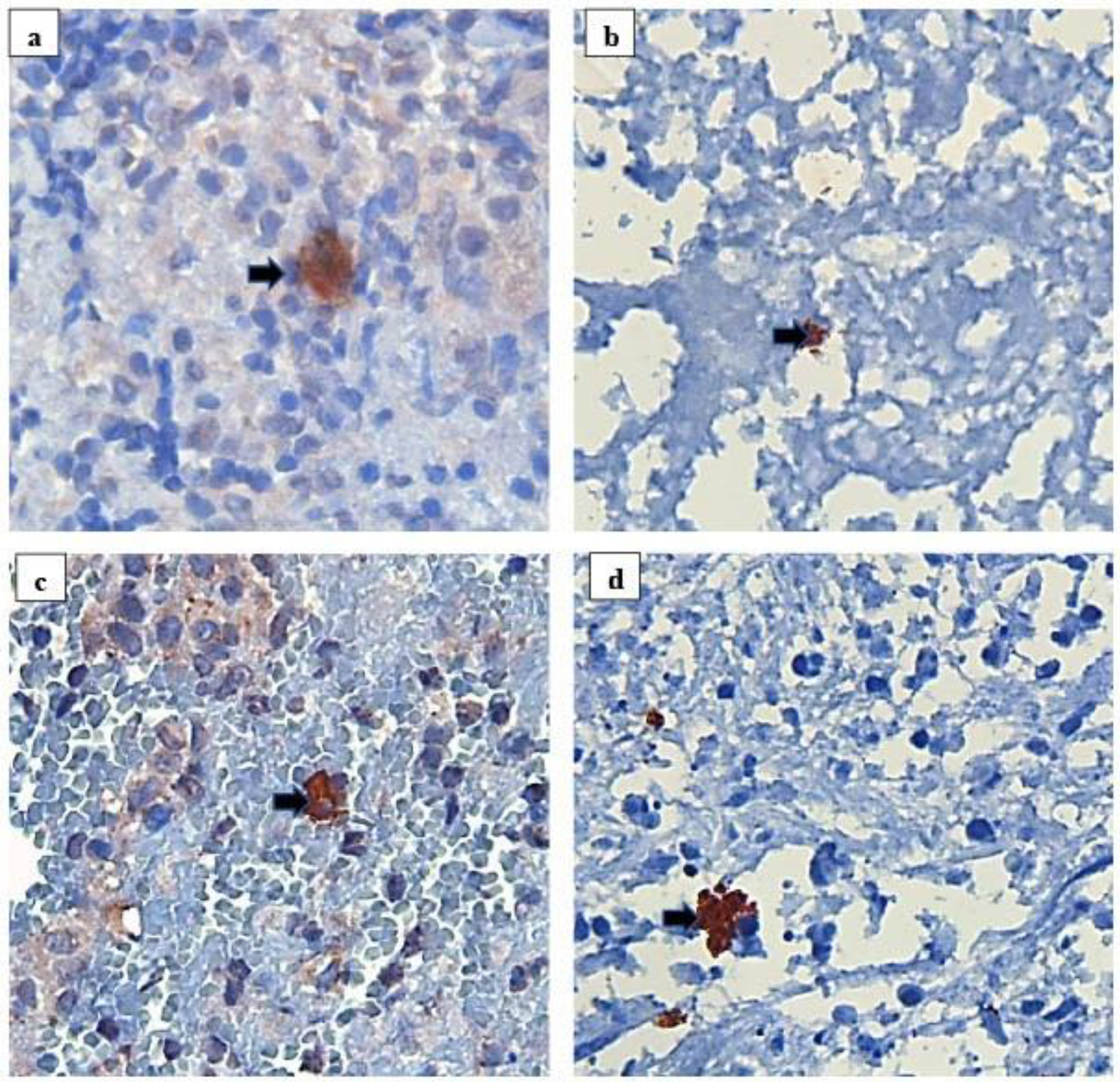
3.4. Validation of the New Polyclonal Antibody

Validation of the new polyclonal antibody was performed on human clinical extrapulmonary specimens, including 20 biopsies from confirmed or clinically diagnosed TB cases and 24 biopsies from non-TB cases. In the TB samples, the sensitivity of ZN, Xpert, Culture and the new polyclonal MPT64 antibody was 11% (1/9), 67% (6/9), 40% (6/15) and 95% (19/20), respectively (Table 3). The new polyclonal antibody was positive in all the culture and/or Xpert positive TB samples (n = 12), and positive in 7/8 samples from clinically diagnosed TB cases. Non-specific staining was observed in 4/24 non-TB samples with the new polyclonal antibody, yielding an overall specificity of 83%.

4. Discussion

In this study, we have investigated different strategies to develop a new functional antibody for use in the TB diagnostic MPT64 test, which has shown promising results for diagnosing EPTB in low-resource settings [11,12,13,14,15,16,17]. The test uses an in-house polyclonal antibody for the detection of the mycobacterial antigen MPT64, but the antibody is in limited supply and further large-scale use of the test requires reproduction of the antibody. Despite generation of several monoclonal anti-MPT64 antibodies with good reactivity in ELISA, none of the antibodies were fully functional in IHC. We, therefore, opted for the development of a polyclonal antibody. By careful selection of animals for immunisation, optimal antigen-adjuvant combination, screening of antibodies in IHC and pooling of the best performing individual antibodies, the generation of a sensitive new polyclonal antibody in a large volume was achieved. The new antibody was more sensitive than routine microscopy, Xpert and culture when validated on a small number of clinical samples, albeit a reduced specificity warrants more work with background reduction.
The choice of antigen and adjuvant can greatly affect the performance of the resulting antibody [31,32,33,34]. We used native MPT64 as antigen in the initial experiments, because the conformational epitopes, which may be important targets in IHC [35], are conserved in native proteins. However, as the resulting antibodies were neither sensitive nor specific in IHC, and the production of native MPT64 was time-consuming, we changed to recombinant MPT64 in the latter experiments. Recombinant protein expression allows for rapid production but may alter conformational epitopes depending on the choice of host system, partly because of differences in post-translational modification systems between species. Recombinant MPT64 from E. coli has been reported to elicit weaker immune responses than native MPT64 and recombinant MPT64 from M. smegmatis [36], possibly due to different post-translational modification systems [37,38,39,40,41,42,43], suggesting that important MPT64 epitopes can be affected by the host system. Despite this possible drawback, immunisation of mice with recombinant MPT64 from both E. coli and mammalian cells resulted in polyclonal antisera with as strong, or stronger, specific staining in IHC as compared to immunisation with native MPT64 (strong specific staining was observed in 9/9, 4/9 and 1/10 murine antisera with mammalian MPT64, E. coli MPT64 and native MPT64 as antigen, respectively). Poor quality of the native MPT64 antigen, possibly due to misfolding of the protein, could be one reason for the low performance of the resulting antibodies, but this was not further investigated. Several adjuvants, which can enhance and prolong the immune response [33,34], were also tested in the study (Figure 2). Based on the particularly strong specific staining obtained after immunisation of mice with mammalian recombinant MPT64 and Titer Max Gold adjuvant, we chose to change to this antigen-adjuvant combination in rabbits as well. This resulted in some of the best individual antisera in our study, with almost as strong specific staining as the reference antibody, and low non-specific staining. Furthermore, the dose of antigen injected may also impact the immune response and antibodies generated [33], but this was not explored in our study, as all rabbits received the same dose of antigen.
The main reason for choosing monoclonal antibodies in a diagnostic test is to avoid batch-to-batch variability, thereby securing reproducible test performance. Still, polyclonal antibodies offer some important advantages [19]. The development of polyclonal antibodies is relatively simple, fast and inexpensive and can result in highly sensitive antibodies because several epitopes are recognised simultaneously, leading to efficient signal amplification and improved detection. High test sensitivity is of great importance in TB diagnostics, as the sensitivity of routine TB diagnostic tests is low in paucibacillary EPTB disease [44]. Furthermore, the biological diversity of polyclonal antibodies allows for use under a wide range of chemical conditions and temperatures, which is an advantage in low-resource settings. Thus, as long as batch-to-batch consistency is managed through standardised validation of all new batches, or through creation of a large batch as in this study, polyclonal antibodies may be used, and are being used, for diagnostic purposes [45,46,47]. Still, cross-reactivity is a common issue with polyclonal antibodies, and antibody purification or background blocking is often required. Surprisingly, negative absorption with BCG Copenhagen culture filtrate proteins, which successfully reduced non-specific staining in the reference antibody, had no effect on the new polyclonal antibody. We experienced that a combined strategy of careful selection of animals for immunisation and optimised background blocking prior to IHC were the most effective measures to reduce non-specific staining. The specificity of the new antibody is still not optimal, but ongoing work indicates that increased duration of the washing steps in IHC removes the non-specific staining without reducing the specific staining of the antibody. This will be further explored in an upcoming validation study.
The development of monoclonal antibodies for use in IHC can be challenging, as demonstrated in our study. The screening of hybridoma cultures in ELISA was not an optimal method to select clones that are functional in IHC. Clones with high performance in ELISA may not detect relevant epitopes in IHC, because formalin fixation prior to IHC can mask or alter the three-dimensional conformation of the epitopes that were exposed during ELISA [48]. Antigen retrieval partly reverses these alterations, but the effect varies between epitopes, and antibody screening should, therefore, preferably be performed directly in IHC. This was done in the latter experiments, but is more time-consuming than ELISA and requires large numbers of positive control tissue sections. In the latter experiments, antisera from several immunised mice gave relatively strong specific staining in IHC, but the resulting few monoclonal antibodies that showed some specific staining also displayed cross-reactivity, indicating that their target epitopes were not unique to the MPT64 protein. We may have lost clones that recognised unique MPT64 epitopes during fusion or hybridoma selection, especially in the initial experiments where IHC was not used for screening. Another possible explanation is that our choice of antigen was not optimal to generate antibodies against unique epitopes [49]. If the less unique epitopes are immunodominant, most of the antibodies in the mice will be generated against these, whereas less immunogenic, but unique epitopes could be missed by the immune system. To avoid this, so-called subtractive immunisation techniques can be applied, in which the undesirable antibodies are used to mask their epitopes on the antigen before immunisation, so that antibodies are only generated against other epitopes [49]. This remains to be explored during further development of monoclonal MPT64-antibodies for IHC.

5. Conclusions

Reproduction of a functional polyclonal MPT64 antibody for large-scale use of the TB diagnostic MPT64 test was achieved through a combination of careful selection of antigen-adjuvant for the immunisation protocol, comprehensive screening in IHC of both pre-immune sera and antisera to find the best performing antibodies, followed by pooling the best individual antisera to obtain a large volume of polyclonal antibodies with stable performance, thereby securing stable supplies and reproducibility of the MPT64 test. Further validation of the new polyclonal antibody in clinically relevant larger populations remains to be done.

Supplementary Materials

The following are available online at https://www.mdpi.com/article/10.3390/antib10030034/s1, Supplementary Materials: Detailed protocols for production and purification of MPT64 antigen, development of antibodies and immunohistochemistry background reduction, Figure S1: The completed vector construct, pUV15tetORmMpt64, expressing the MPT64 protein, Figure S2: Chromatograms from the three-step chromatography purification strategy applied to purify native MPT64 protein, Figure S3: Overview of the purity of the MPT64-containing fractions after each step of chromatography.

Author Contributions

Conceptualisation, T.M., H.W. and L.S.; methodology, H.W. and T.M.; validation, I.M.H., I.A.M.A. and S.I.; formal analysis, I.M.H. and I.A.M.A.; investigation, I.M.H., I.A.M.A. and S.I.; data curation, I.M.H., I.A.M.A. and S.I.; writing—original draft preparation, I.M.H.; writing—review and editing, I.M.H., I.A.M.A., S.I., H.W., L.S. and T.M.; visualisation, I.M.H. and I.A.M.A.; supervision, T.M.; project administration, T.M. and I.M.H.; funding acquisition, T.M. and I.M.H. All authors have read and agreed to the published version of the manuscript.

Funding

This work was partly supported by the Research Council of Norway through the Global Health and Vaccination Programme [project number 234457], and partly supported by Blakstad og Marschalk tuberkulosefond. This project is part of the EDCTP2 programme supported by the European Union. The APC was funded by the University of Bergen.

Institutional Review Board Statement

The study was conducted according to the guidelines of the Declaration of Helsinki, and approved by the National Bioethics Committee of Pakistan (Islamabad, Pakistan) and the Regional Committee for Medical and Health Research Ethics of Western Norway (REK vest) (2014/46/REK vest). All animal experiments were performed by commercial companies that are certified according to ISO 9001. The animal keeping and corresponding works were performed according to German/Belgian country specific, European and US NIH/OLAW guidelines.

Informed Consent Statement

Written informed consent was obtained from all subjects involved in the study.

Data Availability Statement

The data presented in this study are available on request from the corresponding author.

Acknowledgments

We thank Sonja Ljostveit, University of Bergen, and Diana Turcu, University of Bergen, for contribution to antigen production and purification. We also thank Edith Marianne Fick, University of Bergen/Haukeland University Hospital, and Shaukat Siddiq, Histopathology Laboratory, Gulab Devi Chest Hospital, for technical assistance with immunohistochemistry.

Conflicts of Interest

The authors declare no conflict of interest. The funders had no role in the design of the study; in the collection, analyses or interpretation of data; in the writing of the manuscript, or in the decision to publish the results.

References

  1. World Health Organization. Global Tuberculosis Report. 2020. Available online: https://www.who.int/publications/i/item/9789240013131 (accessed on 13 November 2020).
  2. Nelson, L.; Wells, C. Global epidemiology of childhood tuberculosis. Int. J. Tuberc. Lung Dis. 2004, 8, 636–647. [Google Scholar]
  3. Smith, S.; Jacobs, R.F.; Wilson, C.B. Immunobiology of childhood tuberculosis: A window on the ontogeny of cellular immunity. J. Pediatrics 1997, 131, 16–26. [Google Scholar] [CrossRef]
  4. Jones, B.E.; Young, S.M.; Antoniskis, D.; Davidson, P.T.; Kramer, F.; Barnes, P.F. Relationship of the manifestations of tuberculosis to CD4 cell counts in patients with human immunodeficiency virus infection. Am. J. Respir. Crit. Care Med. 1993, 148, 1292–1297. [Google Scholar] [CrossRef]
  5. Harries, A. Tuberculosis and human immunodeficiency virus infection in developing countries. Lancet Infect. Dis. 1990, 335, 387–390. [Google Scholar] [CrossRef]
  6. Small, P.M.; Schecter, G.F.; Goodman, P.C.; Sande, M.A.; Chaisson, R.E.; Hopewell, P.C. Treatment of tuberculosis in patients with advanced human immunodeficiency virus infection. N. Engl. J. Med. 1991, 324, 289–294. [Google Scholar] [CrossRef] [PubMed]
  7. World Health Organization. Improving the Diagnosis and Treatment of Smear-Negative Pulmonary and Extrapulmonary Tuberculosis among Adults and Adolescents: Recommendations for HIV-Prevalent and Resource-Constrained Settings. 2007. Available online: http://www.who.int/iris/handle/10665/69463 (accessed on 26 August 2021).
  8. World Health Organization. High-Priority Target Product Profiles for New Tuberculosis Diagnostics. Report of a Consensus Meeting. 2014. Available online: https://www.who.int/tb/publications/tpp_report/en/ (accessed on 5 February 2020).
  9. Broger, T.; Sossen, B.; du Toit, E.; Kerkhoff, A.D.; Schutz, C.; Reipold, E.I.; Ward, A.; Barr, D.A.; Macé, A.; Trollip, A.; et al. Novel lipoarabinomannan point-of-care tuberculosis test for people with HIV: A diagnostic accuracy study. Lancet Infect. Dis. 2019, 19, 852–861. [Google Scholar] [CrossRef] [Green Version]
  10. World Health Organization. Lateral Flow Urine Lipoarabinomannan Assay (LF-LAM) for the Diagnosis of Active Tuberculosis in People Living with HIV-Policy Update (2019). 2019. Available online: https://www.who.int/tb/publications/2019/LAMPolicyUpdate2019/en/ (accessed on 23 June 2020).
  11. Mustafa, T.; Wiker, H.G.; Mfinanga, S.G.; Mørkve, O.; Sviland, L. Immunohistochemistry using a Mycobacterium tuberculosis complex specific antibody for improved diagnosis of tuberculous lymphadenitis. Mod. Pathol. 2006, 19, 1606–1614. [Google Scholar] [CrossRef] [PubMed]
  12. Purohit, M.R.; Mustafa, T.; Wiker, H.G.; Mørkve, O.; Sviland, L. Immunohistochemical diagnosis of abdominal and lymph node tuberculosis by detecting Mycobacterium tuberculosis complex specific antigen MPT64. Diagn. Pathol. 2007, 2, 36. [Google Scholar] [CrossRef] [PubMed] [Green Version]
  13. Purohit, M.R.; Mustafa, T.; Wiker, H.G.; Sviland, L. Rapid diagnosis of tuberculosis in aspirate, effusions, and cerebrospinal fluid by immunocytochemical detection of Mycobacterium tuberculosis complex specific antigen MPT64. Diagn. Cytopathol. 2012, 40, 782–791. [Google Scholar] [CrossRef] [PubMed]
  14. Tadele, A.; Beyene, D.; Hussein, J.; Gemechu, T.; Birhanu, A.; Mustafa, T.; Tsegaye, A.; Aseffa, A.; Sviland, L. Immunocytochemical detection of Mycobacterium Tuberculosis complex specific antigen, MPT64, improves diagnosis of tuberculous lymphadenitis and tuberculous pleuritis. BMC Infect. Dis. 2014, 14, 585. [Google Scholar] [CrossRef] [PubMed] [Green Version]
  15. Purohit, M.R.; Sviland, L.; Wiker, H.; Mustafa, T. Rapid and specific diagnosis of extrapulmonary tuberculosis by immunostaining of tissues and aspirates with anti-MPT64. Appl. Immunohistochem. Mol. Morphol. 2017, 25, 282–288. [Google Scholar] [CrossRef] [PubMed] [Green Version]
  16. Jørstad, M.D.; Marijani, M.; Dyrhol-Riise, A.M.; Sviland, L.; Mustafa, T. MPT64 antigen detection test improves routine diagnosis of extrapulmonary tuberculosis in a low-resource setting: A study from the tertiary care hospital in Zanzibar. PLoS ONE 2018, 13, e0196723. [Google Scholar] [CrossRef] [PubMed] [Green Version]
  17. Baba, K.; Dyrhol-Riise, A.M.; Sviland, L.; Langeland, N.; Hoosen, A.A.; Wiker, H.G.; Mustafa, T. Rapid and specific diagnosis of tuberculous pleuritis with immunohistochemistry by detecting Mycobacterium tuberculosis complex specific antigen MPT64 in patients from a HIV endemic area. Appl. Immunohistochem. Mol. Morphol. 2008, 16, 554–561. [Google Scholar] [CrossRef]
  18. Hoel, I.M.; Sviland, L.; Syre, H.; Dyrhol-Riise, A.M.; Skarstein, I.; Jebsen, P.; Jørstad, M.D.; Wiker, H.; Mustafa, T. Diagnosis of extrapulmonary tuberculosis using the MPT64 antigen detection test in a high-income low tuberculosis prevalence setting. BMC Infect. Dis. 2020, 20, 130. [Google Scholar] [CrossRef] [PubMed] [Green Version]
  19. Lipman, N.S.; Jackson, L.R.; Trudel, L.J.; Weis-Garcia, F. Monoclonal versus polyclonal antibodies: Distinguishing characteristics, applications, and information resources. ILAR J. 2005, 46, 258–268. [Google Scholar] [CrossRef] [Green Version]
  20. Nagai, S.; Wiker, H.G.; Harboe, M.; Kinomoto, M. Isolation and partial characterization of major protein antigens in the culture fluid of Mycobacterium tuberculosis. Infect. Immun. 1991, 59, 372–382. [Google Scholar] [CrossRef] [Green Version]
  21. Harboe, M.; Nagai, S.; Patarroyo, M.E.; Torres, M.; Ramirez, C.; Cruz, N. Properties of proteins MPB64, MPB70, and MPB80 of Mycobacterium bovis BCG. Infect. Immun. 1986, 52, 293–302. [Google Scholar] [CrossRef] [Green Version]
  22. Nagai, S.; Matsumoto, J.; Nagasuga, T. Specific skin-reactive protein from culture filtrate of Mycobacterium bovis BCG. Infect. Immun. 1981, 31, 1152–1160. [Google Scholar] [CrossRef] [Green Version]
  23. Oettinger, T.; Andersen, A.B. Cloning and B-cell-epitope mapping of MPT64 from Mycobacterium tuberculosis H37Rv. Infect. Immun. 1994, 62, 2058–2064. [Google Scholar] [CrossRef] [Green Version]
  24. Snapper, S.; Melton, R.; Mustafa, S.; Kieser, T.; Jr, W.J. Isolation and characterization of efficient plasmid transformation mutants of Mycobacterium smegmatis. Mol. Microbiol. 1990, 4, 1911–1919. [Google Scholar] [CrossRef]
  25. Ehrt, S.; Guo, X.V.; Hickey, C.M.; Ryou, M.; Monteleone, M.; Riley, L.W.; Schnappinger, D. Controlling gene expression in mycobacteria with anhydrotetracycline and Tet repressor. Nucleic Acids Res. 2005, 33, e21. [Google Scholar] [CrossRef] [PubMed]
  26. Parish, T.; Roberts, D.M. Mycobacteria Protocols, 3rd ed.; Springer Science+Business Media: New York, NY, USA, 2015. [Google Scholar]
  27. Guo, X.V.; Monteleone, M.; Klotzsche, M.; Kamionka, A.; Hillen, W.; Braunstein, M.; Ehrt, S.; Schnappinger, D. Silencing essential protein secretion in Mycobacterium smegmatis by using tetracycline repressors. J. Bacteriol. 2007, 189, 4614–4623. [Google Scholar] [CrossRef] [PubMed] [Green Version]
  28. Köhler, G.; Milstein, C. Continuous cultures of fused cells secreting antibody of predefined specificity. Nature 1975, 256, 495–497. [Google Scholar] [CrossRef] [PubMed]
  29. Harboe, M.; Closs, O.; Bjorvatn, B.; Kronvall, G.; Axelsen, N. Antibody response in rabbits to immunization with Mycobacterium leprae. Infect. Immun. 1977, 18, 792–805. [Google Scholar] [CrossRef] [PubMed] [Green Version]
  30. Tahseen, S.; Ambreen, A.; Masood, F.; Qadir, M.; Hussain, A.; Jamil, M.; Safdar, N.; Sviland, L.; Mustafa, T. Primary drug resistance in extra-pulmonary tuberculosis: A hospital-based prospective study from Pakistan. Int. J. Tuberc. Lung Dis. 2019, 23, 900–906. [Google Scholar] [CrossRef]
  31. Bergmann-Leitner, E.S.; Leitner, W.W. Adjuvants in the driver’s seat: How magnitude, type, fine specificity and longevity of immune responses are driven by distinct classes of immune potentiators. Vaccines 2014, 2, 252–296. [Google Scholar] [CrossRef] [Green Version]
  32. Schunk, M.K.; Macallum, G.E. Applications and optimization of immunization procedures. ILAR J. 2005, 46, 241–257. [Google Scholar] [CrossRef]
  33. Leenaars, M.; Hendriksen, C.F. Critical steps in the production of polyclonal and monoclonal antibodies: Evaluation and recommendations. ILAR J. 2005, 46, 269–279. [Google Scholar] [CrossRef] [Green Version]
  34. Stils, H., Jr. Adjuvants and antibody production: Dispelling the myths associated with Freund’s complete and other adjuvants. ILAR J. 2005, 46, 280–293. [Google Scholar] [CrossRef] [Green Version]
  35. Mason, J.T.; O’leary, T.J. Effects of formaldehyde fixation on protein secondary structure: A calorimetric and infrared spectroscopic investigation. J. Histochem. Cytochem. 1991, 39, 225–229. [Google Scholar] [CrossRef]
  36. Roche, P.; Winter, N.; Triccas, J.; Feng, C.; Britton, W. Expression of Mycobacterium tuberculosis MPT64 in recombinant Myco. smegmatis: Purification, immunogenicity and application to skin tests for tuberculosis. Clin. Exp. Immunol. 1996, 103, 226–232. [Google Scholar] [CrossRef]
  37. Sartain, M.J.; Belisle, J.T. N-Terminal clustering of the O-glycosylation sites in the Mycobacterium tuberculosis lipoprotein SodC. Glycobiology 2009, 19, 38–51. [Google Scholar] [CrossRef] [PubMed] [Green Version]
  38. Dobos, K.M.; Khoo, K.-H.; Swiderek, K.M.; Brennan, P.J.; Belisle, J.T. Definition of the full extent of glycosylation of the 45-kilodalton glycoprotein of Mycobacterium tuberculosis. J. Bacteriol. 1996, 178, 2498–2506. [Google Scholar] [CrossRef] [Green Version]
  39. Daugelat, S.; Kowall, J.; Mattow, J.; Bumann, D.; Winter, R.; Hurwitz, R.; Kaufmann, S.H. The RD1 proteins of Mycobacterium tuberculosis: Expression in Mycobacterium smegmatis and biochemical characterization. Microbes Infect. 2003, 5, 1082–1095. [Google Scholar] [CrossRef]
  40. Garbe, T.; Harris, D.; Vordermeier, M.; Lathigra, R.; Ivanyi, J.; Young, D. Expression of the Mycobacterium tuberculosis 19-kilodalton antigen in Mycobacterium smegmatis: Immunological analysis and evidence of glycosylation. Infect. Immun. 1993, 61, 260–267. [Google Scholar] [CrossRef] [Green Version]
  41. Herrmann, J.; O’Gaora, P.; Gallagher, A.; Thole, J.; Young, D.B. Bacterial glycoproteins: A link between glycosylation and proteolytic cleavage of a 19 kDa antigen from Mycobacterium tuberculosis. EMBO J. 1996, 15, 3547–3554. [Google Scholar] [CrossRef] [PubMed]
  42. Herrmann, J.L.; Delahay, R.; Gallagher, A.; Robertson, B.; Young, D. Analysis of post-translational modification of mycobacterial proteins using a cassette expression system. FEBS Lett. 2000, 473, 358–362. [Google Scholar] [CrossRef] [Green Version]
  43. Parra, J.; Marcoux, J.; Poncin, I.; Canaan, S.; Herrmann, J.L.; Nigou, J.; Burlet-Schiltz, O.; Rivière, M. Scrutiny of Mycobacterium tuberculosis 19 kDa antigen proteoforms provides new insights in the lipoglycoprotein biogenesis paradigm. Sci. Rep. 2017, 7, 43682. [Google Scholar] [CrossRef]
  44. World Health Organization. Global Tuberculosis Report. 2019. Available online: https://www.who.int/tb/publications/global_report/en/ (accessed on 22 November 2019).
  45. Torres-González, P.; Niembro-Ortega, M.D.; Martínez-Gamboa, A.; Ahumada-Topete, V.H.; Andrade-Villanueva, J.; Araujo-Meléndez, J.; Chaparro-Sánchez, A.; Crabtree-Ramírez, B.; Cruz-Martínez, S.; Gamboa-Domínguez, A.; et al. Diagnostic accuracy cohort study and clinical value of the Histoplasma urine antigen (ALPHA Histoplasma EIA) for disseminated histoplasmosis among HIV infected patients: A multicenter study. PLoS Negl. Trop. Dis. 2018, 12, e0006872. [Google Scholar] [CrossRef] [Green Version]
  46. Petrini, I.; Zucali, P.A.; Lee, H.S.; Pineda, M.A.; Meltzer, P.S.; Walter-Rodriguez, B.; Roncalli, M.; Santoro, A.; Wang, Y.; Giaccone, G. Expression and mutational status of c-kit in thymic epithelial tumors. J. Thorac. Oncol. 2010, 5, 1447–1453. [Google Scholar] [CrossRef]
  47. Eyzaguirre, E.; Haque, A.K. Application of immunohistochemistry to infections. Arch. Pathol. Lab. Med. 2008, 132, 424–431. [Google Scholar] [CrossRef] [PubMed]
  48. Otali, D. The Combined Effect of Formalin Fixation and Individual Steps in Tissue Processing on Immunorecognition; University of Alabama at Birmingham: Birmingham, AL, USA, 2007. [Google Scholar]
  49. de Almeida, R.; Nakamura, C.N.; de Lima Fontes, M.; Deffune, E.; Felisbino, S.L.; Kaneno, R.; Fávaro, W.J.; Billis, A.; Cerri, M.O.; Fusco-Almeida, A.M.; et al. Enhanced Immunization Techniques to Obtain Highly Specific Monoclonal Antibodies; Taylor & Francis: Boca Raton, FL, USA, 2018. [Google Scholar]
Figure 1. Overview of the different strategies used to develop monoclonal and polyclonal anti-MPT64 antibodies. Abbreviations; IFA, incomplete Freund adjuvant, IHC, immunohistochemistry; mod, moderate; NSS, non-specific staining; SS, specific staining; rMPT64, recombinant MPT64.
Antibodies 10 00034 g001
Figure 2. The steps of hybridoma production and different target points for screening of clones during the development of monoclonal antibodies. Abbreviations: ELISA, enzyme-linked immunosorbent assay, IHC, immunohistochemistry; mAb, monoclonal antibody.
Antibodies 10 00034 g002
Figure 3. Flow chart showing immunisation protocols, screening and pooling strategies used to develop the new polyclonal antibody (polyclonal antibody experiment 4). Abbreviations: d, day; IHC, immunohistochemistry; mod, moderate; NSS, non-specific staining; SS, specific staining.
Antibodies 10 00034 g003
Figure 4. Immunohistochemistry with the new (a,c) and reference anti-MPT64 polyclonal antibody (b,d) on tissue sections from patients with tuberculosis lymph adenitis. Specific staining is seen as reddish-brown granular staining (arrow) located intracellularly in the lesions. The weak diffuse staining in a and c is non-specific staining.
Antibodies 10 00034 g004
Table 1. Amino acid sequence of the various forms of MPT64 protein used as antigen in the study. The native MPT64 sequence is derived from M. bovis BCG Moreau and includes a N-terminal cleavable protein secretion signal sequence (bold), which is not present in the final, secreted form of the protein. The sequence of the recombinant protein produced in Escherichia coli (E. coli rMPT64) includes a C-terminal Thrombin-cleavable 8XHis-tag (underlined) to simplify purification. The sequence of the recombinant protein produced in human HEK cells (mammalian rMPT64) includes a N-terminal HSA signal peptide (bold) to secrete the protein, and a C-terminal 6XHis-tag (underlined) to simplify purification.
ProteinAmino Acid Sequence
Native MPT64MRIKIFMLVTAVVLLCCSGVATAAPKTYCEELKGTDTGQACQIQMSDPAYNINISLPSYYPDQKSLENYIAQTRDKFLSAATSSTPREAPYELNITSATYQSAIPPRGTQAVVLKVYQNAGGTHPTTTYKAFDWDQAYRKPITYDTLWQADTDPLPVVFPIVQGELSKQTGQQVSIAPNAGLDPVNYQNFAVTNDGVIFFFNPGELLPEAAGPTQVLVPRSAIDSMLA
E. coli rMPT64MAPKTYCEELKGTDTGQACQIQMSDPAYNINISLPSYYPDQKSLENYIAQTRDKFLSAATSSTPREAPYELNITSATYQSAIPPRGTQAVVLKVYQNAGGTHPTTTYKAFDWDQAYRKPITYDTLWQADTDPLPVVFPIVQGELSKQTGQQVSIAPNAGLDPVNYQNFAVTNDGVIFFFNPGELLPEAAGPTQVLVPRSAIDSMLAVLVPRGSAAALEHHHHHHHH
Mammalian rMPT64MKWVTFISLLFLFSSAYSAPKTYCEELKGTDTGQACQIQMSDPAYNINISLPSYYPDQKSLENYIAQTRDKFLSAATSSTPREAPYELNITSATYQSAIPPRGTQAVVLKVYQNAGGTHPTTTYKAFDWDQAYRKPITYDTLWQADTDPLPVVFPIVQGELSKQTGQQVSIAPNAGLDPVNYQNFAVTNDGVIFFFNPGELLPEAAGPTQVLVPRSAIDSMLAHHHHHH
Table 2. Strategies employed to reduce non-specific staining in immunohistochemistry with the new polyclonal antibody.
Level of Non-Specific Staining
StrategyPositive TB ControlNegative Non-TB Control
Blocking experiments
Serum free block (12 min, 30 min, 60 min, or overnight)--
BSA 3% or 10% and NGS 10% (60 min), followed by serum free block (60 min) -/↓-/↓
BSA 3% or 10% and NGS 10% (overnight), followed by serum free block (12 min)↓↓↓↓
Fc block --
Absorption experiments
M. bovis BCG Copenhagen, culture filtrates-/↓-/↓
M. bovis BCG Copenhagen, cell sonicate--
Homogenised non-TB lung and lymph node tissue sections (deparaffinised and hydrated)-
Abbreviations: TB, tuberculosis; BSA, bovine serum albumin; NGS, normal goat serum; NSS, non-specific staining; SS, specific staining; M. bovis, mycobacterium bovis; (-), no change; (↑), increased; (↓), decreased.
Table 3. Results of routine diagnostic tests and the MPT64 test performed on clinical TB and non-TB samples.
Routine Diagnostic Tests Positive/Total (%)The MPT64 Test Positive/Total (%)
NZiehl-NeelsenXpert MTB/RIFCulture LJNew Polyclonal MPT64 Antibody
TB cases total201/9 (11)6/9 (67)6/15 (40)19/20 (95)
Lymph node biopsies141/6 (17)3/4 (75)6/12 (50)14/14 (100)
Other biopsies60/3 (0)3/5 (60)0/3 (0)5/6 (83)
Confirmed TB cases120/5 (0)6/6 (100)6/11 (55)12/12 (100)
Clinically diagnosed TB cases81/4 (25)0/4 (0)0/3 (0)7/8 (88)
Non-TB cases total24N/AN/AN/A4/24 (17)
Lymph node biopsies7N/AN/AN/A2/7 (29)
Other biopsies17N/AN/AN/A2/17 (12)
Abbreviations: LJ, Lowenstein Jensen; TB, tuberculosis.
Publisher’s Note: MDPI stays neutral with regard to jurisdictional claims in published maps and institutional affiliations.

