Article

Effects of Climate Change on 2-Methylisoborneol Production in Two Cyanobacterial Species

1
Beijing Climate Change Response Research and Education Center, Beijing University of Civil Engineering and Architecture, Beijing 100044, China
2
Key Laboratory of Drinking Water Science and Technology, Research Center for Eco-Environmental Sciences, Chinese Academy of Sciences, Beijing 100085, China
3
College of Resources and Environment, University of Chinese Academy of Sciences, Beijing 100019, China
*
Authors to whom correspondence should be addressed.
Water 2017, 9(11), 859; https://doi.org/10.3390/w9110859
Submission received: 24 September 2017 / Revised: 30 October 2017 / Accepted: 2 November 2017 / Published: 22 November 2017

Abstract

:
2-Methylisoborneol (2-MIB) is a common off-flavor metabolite, and it has a very low odor threshold value. 2-MIB is produced by a variety of cyanobacteria and heterotrophic bacteria. In this study, Dolichospermum spiroides (Anabaena spiroides, FACHB 498) and Planktothrix sp. (FACHB 1371) were investigated. The influence of high temperature (27, 35, and 40 °C) and dissolved inorganic carbon (DIC, 18.4, 27.5, and 36.7 mg/L) on the two 2-MIB-producing cyanobacteria were simulated to study the effect of climate change on 2-MIB production. DIC had an effect on 2-MIB production by Planktothrix sp. The results showed that the highest DIC concentration (36.7 mg/L) led to the fastest algal growth, which increased 2-MIB production by Planktothrix sp. However, 40 °C killed all the cyanobacteria. The higher temperature (35 °C) shortened the lifecycle of the two cyanobacteria because it moved forward their logarithmic phase compared to 27 °C. The 2-MIB-producing potential of Dolichospermum spiroides may increase at 35 °C. The Dolichospermum spiroides results showed that the fluorescence intensity and the concentration of extracellular 2-MIB were highly correlated (r2 ≥ 0.90). At 35 °C, there was a stronger correlation between extracellular dissolved organic matters’ (DOMs, fulvic acid (P3 (r2 = 0.90)), humic acid (P4 (r2 = 0.92)) and the soluble microbial metabolites’ (P5 (r2 = 0.71)) fluorescence peak intensities and 2-MIB concentration. The Dolichospermum sproides 2-MIB-producing potential was affected by extracellular humus. The Planktothrix sp. 2-MIB-producing potential declined in general over the growth period. In addition, temperature had no significant effect on the Planktothrix sp. 2-MIB-producing potential and the extracellular 2-MIB component.

1. Introduction

The combustion of fossil fuels during the past two centuries has significantly increased the atmospheric carbon dioxide (CO2) concentration, a trend that has been projected to continue over the coming decades [1]. Atmospheric CO2 concentrations that had previously increased at a rate of 1% per year in the 20th century are now increasing at 3% per year and may exceed 900 ppm. Furthermore, there may be a more than 2 °C increase by 2100 [1,2]. If the concentration of CO2 in the atmosphere increased from 350 to 900 ppm, the pH and dissolved inorganic carbon (DIC) in water bodies would change. The carbonate ion concentration ( c CO 3 2 ) would decrease while the concentration of bicarbonate ions ( c CO 3 2 ) would increase [3,4,5,6,7]. Since a natural water body is an open system, the CO2 exchange between air and water is a long-term and stable process, and so are its effects on cyanobacteria and green algae [8]. Yu et al. reported that the microcystin concentration in water declined when CO2 concentrations increased [7].
Figure 1 shows the longitudinal carbon cycling in the water environment, and the increase in atmospheric CO2 concentration has an effect on the inorganic carbon in the water. Algal cells transport HCO3 into the cytosol via plasma membrane active transport and HCO3 is converted to CO2 in the cells by carbonic anhydrase [9]. An increased atmospheric CO2 concentration changes cyanobacteria and algae growth because it alters the open water carbonate equilibrium, although this effect is buffered to some degree by the carbonate equilibrium. Furthermore, substantial CO2 increases also affect carbon sequestration. The effect on filamentous bacteria is even more obvious [10]. Under high temperature conditions in summer, especially during heat waves, the inorganic carbon utilization mechanism becomes more effective [3,11].
Harmful algal blooms caused by cyanobacteria have been reported in scientific literature for more than 130 years [12]. It is one of the most important waterborne microbial hazards to human and freshwater ecosystems [13]. Periodic taste and odor events are common problems that influence the water quality in reservoirs and lakes [14,15]. 2-Methylisoborneol (2-MIB), geosmin, and other volatile organic compounds are the primary off-flavor compounds in surface and drinking water [16,17]. The odorous 2-MIB is a secondary metabolite produced by cyanobacteria, such as Dolichospermum sp., Planktothrix sp., Pseudanabaena, and Oscillatoria sp. [18,19,20]. 2-MIB is synthesized in algal cells. The natural substrate (E)-2-methylgeranyl diphosphate (3,2-MeGPP) is formed from the methyl group S-adenosyl-l-methionine (SAM) and 2-MIB is synthesized from MeGPP by 2-methylisoborneol synthase (MIBS) [21,22]. The presence of 2-MIB in source waters decreases the water quality, and the smell in water leads to frequent complaints from consumers [23]. Furthermore, Planktothrix sp. has been identified as a potential 2-MIB producer [18].
Extracellular and intracellular 2-MIB are produced from different algae organic matter (AOM) fractions [24,25,26,27]. Aqueous AOMs are derived from algae metabolites that are generally categorized into extracellular organic matter and intracellular organic matter [28]. Natural organic matters present in surface water are complex and multiphase mixtures consisting of humic acids, fulvic acids, low molecular weight organic acids, carbohydrates, proteins, and other compounds [29]. Dissolved organic matters (DOMs) include organic molecules with chromophoric (light absorbing) and fluorophoric (light emitting) moieties. The fluorescence excitation–emission matrix (EEM) analysis of water samples has become increasingly common because it can interpret the fluorescence characteristics of DOM with a high degree of sensitivity [30]. Fluorescence spectroscopy provides a high optical resolution and the fluorescence given off by different spectral regions is related to different functional groups. Therefore, DOM fractions may have their own EEM [31]. However, there has been little research on the relationship between 2-MIB and AOM.
This study selected two types of cyanobacteria that produce 2-MIB and investigated the primary environmental factors that affect 2-MIB production. The objectives of this study were to (1) explore Dolichospermum spiroides 2-MIB production potential under heat wave temperatures; (2) determine the relationships between AOM and the 2-MIB intracellular and extracellular concentrations; (3) investigate the effects of temperature and inorganic carbon on Planktothrix sp. 2-MIB production; and (4) investigate the influence of CO2 increases on algal metabolism.

2. Materials and Methods

2.1. Chemicals and Cell Culture

Standards with the highest level of purity for the target analyte (2-MIB) were purchased from Sigma–Aldrich (St. Louis, MO, USA). Stock standard solutions of 100 mg/L were prepared by diluting the different standard solutions with HPLC grade methanol, which was purchased from Fisher Scientific Co. (Shanghai, China). Grade analytical NaCl was purchased from the Sinopharm Chemical Reagent Co., Ltd. (Shanghai, China), and was heated to 450 °C for 2 h before use. Ultrapure water (18 MΩ cm) was produced using the Milli-Q Integral 5 purification system (Millipore, Molsheim, France).
Dolichospermum spiroides (FACHB 498) and Planktothrix sp. (FACHB 1371) were purchased from the Institute of Hydrobiology, Chinese Academy of Science (Wuhan, China). They were cultured in BG11 medium in an incubator at 25 °C under a constant light flux (2000 lx) with a light/dark cycle of 12 h/12 h. Each sample was replicated twice and the experiment lasted one growth cycle.

2.2. Odorant Quantification

The odorants were pretreated by the solid phase micro extraction process (SPME) with a commercial fiber (50/30 μm DVB/CAR/PDMS, 57348U, Supelco, Bellefonte, PA, USA) and an automatic fiber holder model (Gerstel, Mülheim an der Ruhr, Germany). The concentrations of the odorants in the samples were determined using a GC-MS system (GCMS-QP2010 Plus, Shimadzu, Tokyo, Japan) in the selected ion monitoring (SIM) mode. The SIM mode was m/z 95, 107, and 108. A capillary column (30 m × 0.25 μm × 0.25 mm) was used to separate the off-flavors from the algal organic matter. The injector temperature was set at 250 °C with a splitless ratio. The initial oven temperature was 40 °C. This was held for 5 min, and then increased to 240 °C at a rate of 8 °C/min with an 8-min hold. The temperature of the electron impact (EI) ion source was set at 200 and 250 °C, respectively.

2.3. Experimental Procedures

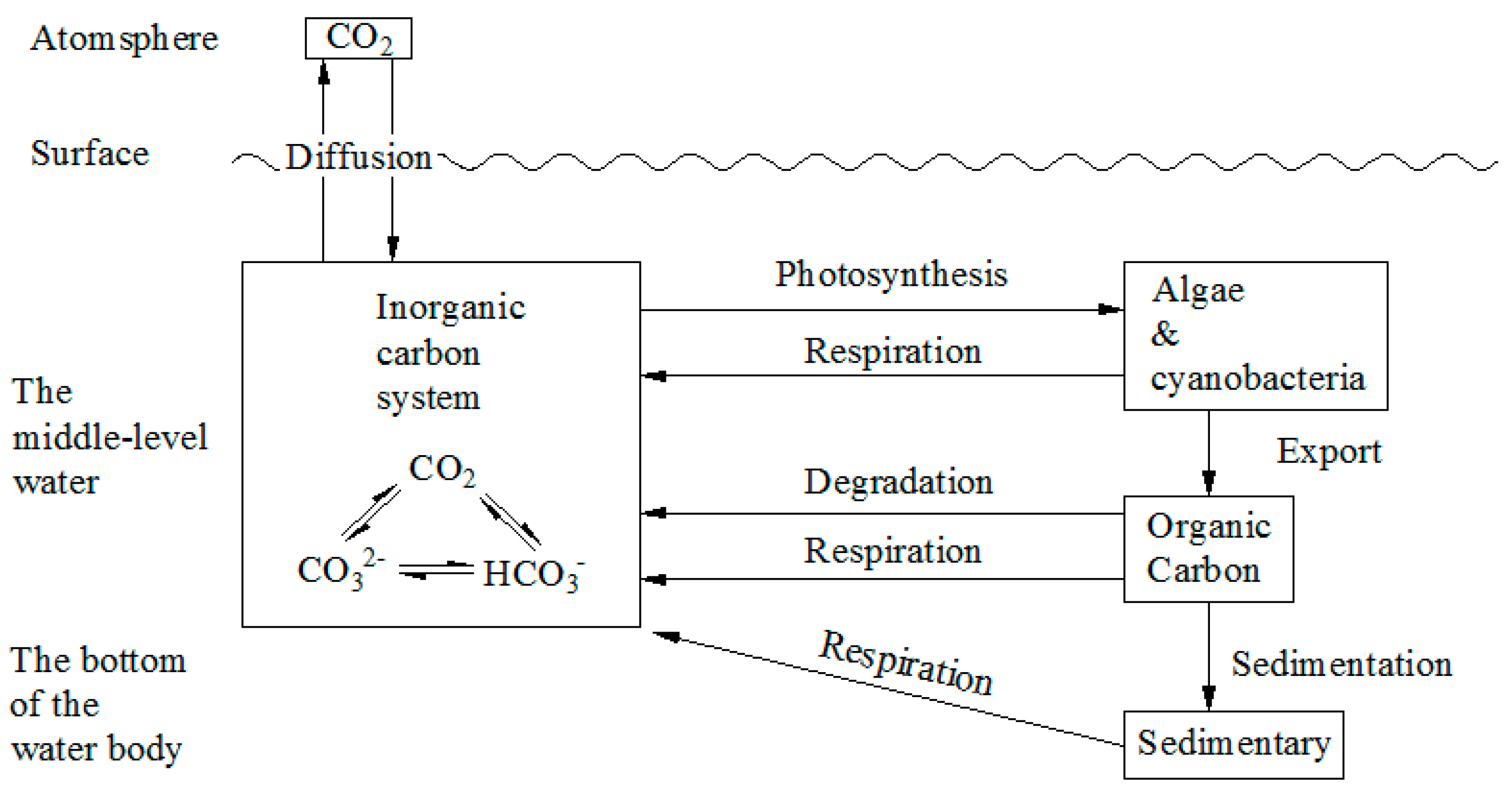
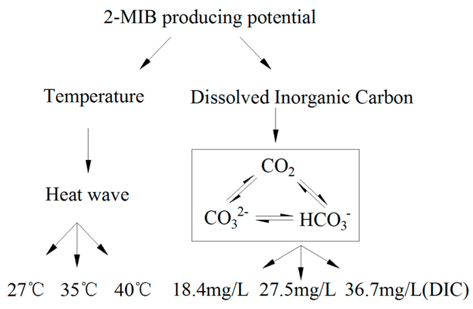
2.3.1. The Technical Pathway

The technical pathway used in this study is shown in Figure 2.

2.3.2. Culture Design

Dolichospermum spiroides (FACHB 498) and Planktothrix sp. (FACHB 1371) were cultured in BG11 medium. Planktothrix sp. was the primary 2-MIB-producing cyanobacteria in Zizhuyuan (ZZY) Park, Beijing, China. The pH of the landscape water in Zizhuyuan Park was approximately 8.0. Therefore, the DIC in the water was primarily HCO3. There were three DIC treatments: 18.4 mg/L DIC (ZZY water DIC concentration), 27.5 mg/L DIC, or 36.7 mg/L DIC, which were adjusted using NaHCO3.
Dolichospermum spiroides (FACHB 498) and Planktothrix sp. (FACHB 1371) were cultured in a climate incubator at 27 °C, 35 °C, or 40 °C under a constant light flux (2000 lx). The control temperature was 27 °C, while 35 °C and 40 °C were the simulated heat-wave temperatures.

2.3.3. Determination

The cultures were harvested every 2 days and the samples were used to monitor changes in the cyanobacteria and off-flavor compounds, including extracellular 2-MIB and intracellular 2-MIB. The algal liquids were centrifuged (8000 rpm, 4450× g) for 15 min, and then the supernatant was removed and used to measure extracellular 2-MIB. After the supernatant samples were cleaned using NaCl (0.8%), a freeze-thaw process (−26 °C and 40 °C, 30 min and 5 min, respectively) was repeated three times followed by ultrasonic treatment. This completely destroyed the algal cells. The supernatant, which was obtained by dissolving the cells in ultrapure water for 2 h followed by centrifuging (8000 rpm, 4450× g) for 10 min, was used to determine the intracellular 2-MIB. At the same time, the cell numbers were counted using a Zeiss microscope (ZEISS-Axio Imager.A2, Jena, Germany). Chlorophyll a (Chla) concentrations in the cyanobacteria were measured after extraction with 90% ethanol for 24 h in the dark. The supernatants were analyzed at 665 nm and 750 nm with a UV-VIS spectrophotometer (Lambda 650S, Perkin Elmer, Waltham, MA, USA). The Dolichospermum spiroides fluorescence intensity (FI) was determined with fluorescence EEMs, which were recorded on a three-dimensional fluorescence spectrometer (Hitachi F-7000, Hitachi, Tokyo, Japan). The parameters in this study were set according to Chen [32]. The EEMs were recorded as a series of emission scans (250–550 nm in 5-nm increments) at set excitation wavelengths (200–450 nm in 5-nm increments). A 5 nm bandpass was used for both the excitation and emission monochromators, and the integration time was 0.25 s. The data were processed with Origin, a plotting software, and characterized by contour maps. Ultra-pure water was used as a blank to correct the Raman scattering of water.

2.3.4. Statistical Analysis

The Planktothrix sp. cell density was more variable than the Dolichospermum spiroides cell density, and Chla can be used to characterize cell growth. Therefore, this study used the 2-MIB/Chla ratio to describe 2-MIB production potential. The average value and standard deviation were calculated for each set of parallel determination data. In the linear regression analysis, the vertical axis shows the intensity values for the fluorescence peaks of the cells in the EEMs, while the corresponding intracellular and extracellular 2-MIB concentrations are shown by the horizontal axis. These data were then used to fit a correlation curve in a period. All graphs were plotted using Origin software.

3. Results

The results showed that the 40 °C treatment killed all the cyanobacteria cells. Therefore, the results and discussion only consider the 27 and 35 °C treatments.

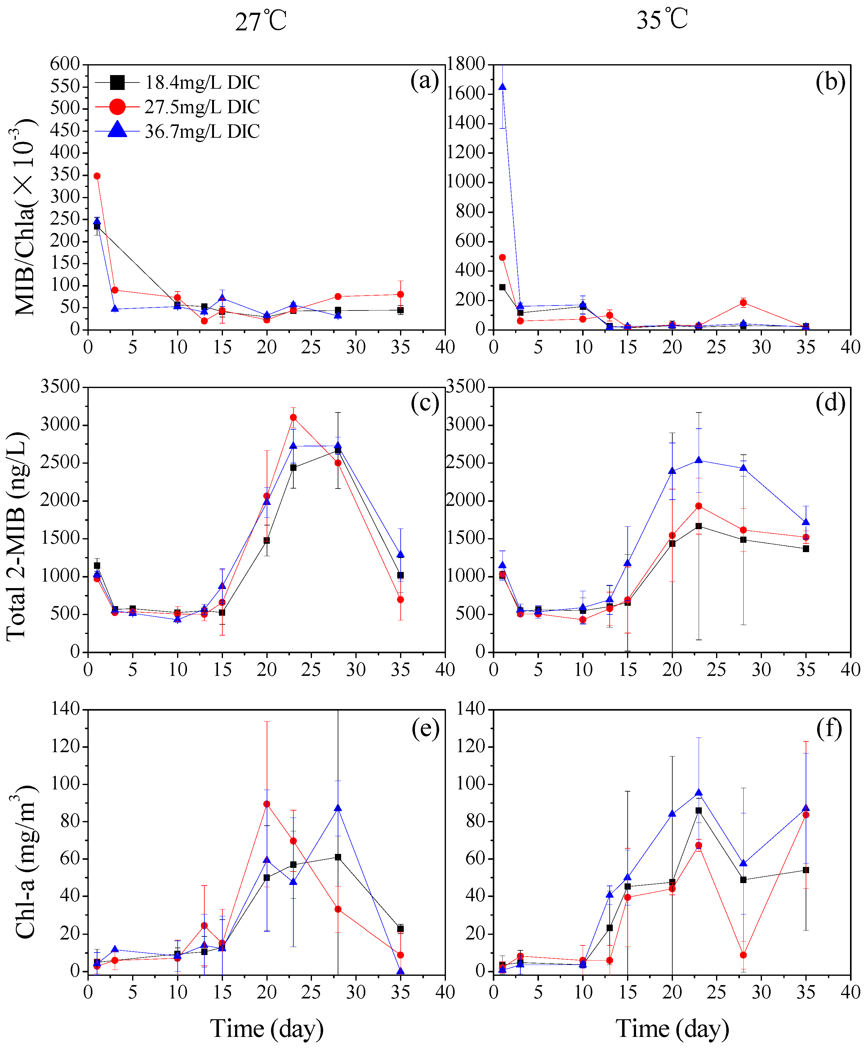
3.1. The Effect of DIC on the Production of 2-MIB by Planktothrix sp.

The 2-MIB production potential of Planktothrix sp. was measured at three different DIC concentrations. Figure 3a,b show that the 2-MIB-producing potentials for Planktothrix sp. under the different DIC conditions varied with time over the growth period, and that they generally had the same change trend. The 2-MIB-producing potential for the Planktothrix sp. determined on the third day was far lower than the potential on the first day. This meant that the 2-MIB-producing potential declined between the first and third day. From the third to the 15th day, there was no uniform rule for 2-MIB-producing potential at the different DIC concentrations. Between the 15th and 35th day, the 2-MIB-producing potential tended to be stable and at a low level for all three concentrations. Above all, the results suggested that the DIC concentration had no significant effect on the 2-MIB-producing potential. Therefore, we analyzed the changes in the total 2-MIB and Chla levels over the growth cycle at the different DIC concentrations.
The 2-MIB production potentials for Planktothrix sp. at the three different DIC concentrations were compared. Figure 3c,d show that at 27 °C, the effect of DIC concentration on the 2-MIB levels was not significant, and that the 2-MIB concentration at 36.7 mg/L DIC reached a peak on the 23rd day. At 35 °C, the total 2-MIB concentration increased as the DIC concentration rose. Figure 3e,f show the change in Chla over time at the different DIC concentrations. At 35 °C, the results showed that the Chla concentration in Planktothrix sp. was highest in the 36.7 mg/L DIC treatment.

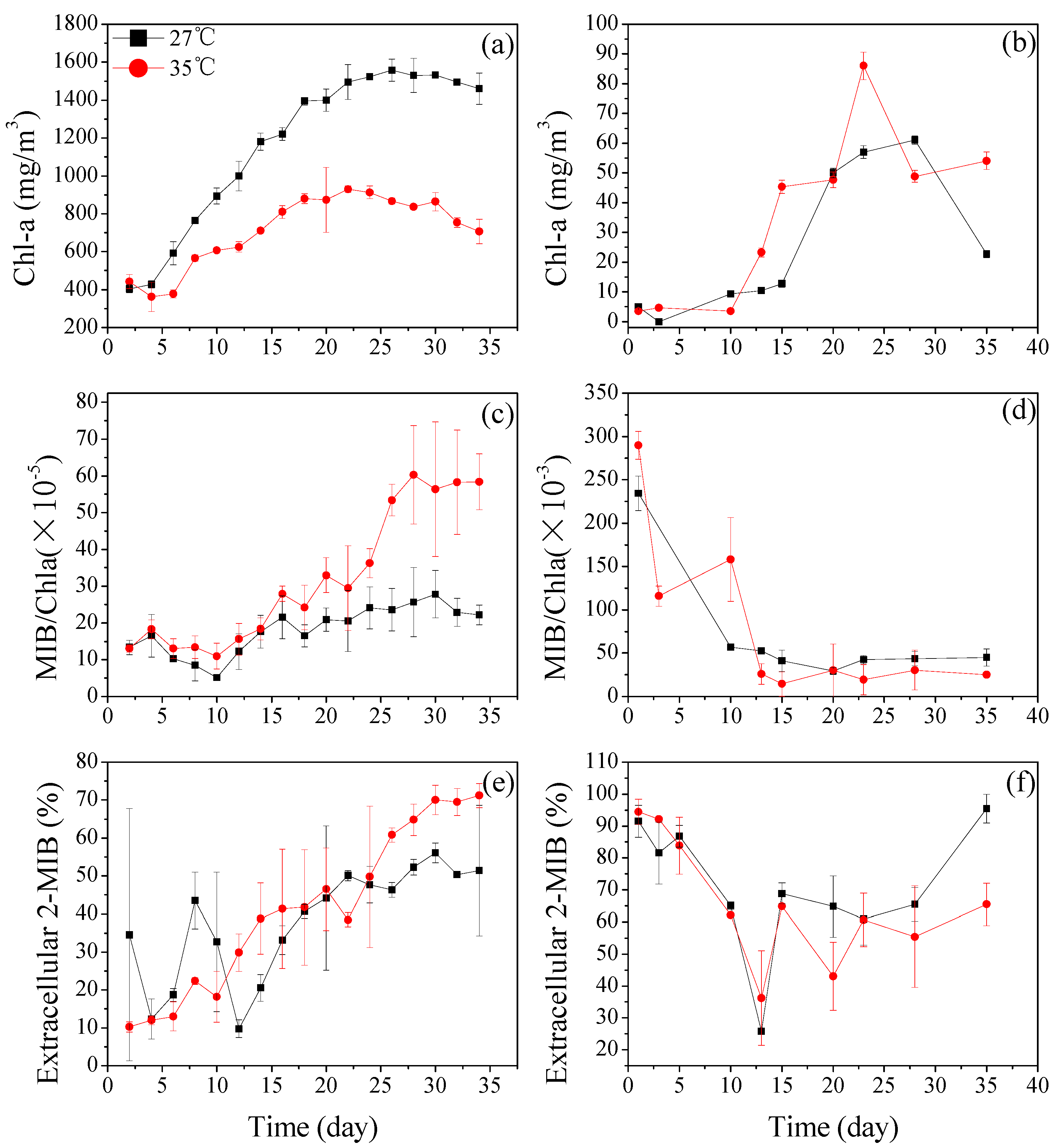
3.2. The Effects of Temperature on Dolichospermum spiroides and Planktothrix sp.

3.2.1. Dolichospermum spiroides and Planktothrix sp. Cell Growth at Different Temperatures

Figure 4a,b show that the Chla concentration in Dolichospermum spiroides was inhibited at 35 °C. The Chla concentration increased slowly with a logarithmic pattern at 35 °C, which indicated that the growth of Dolichospermum spiroides was inhibited at 35 °C. However, this pattern was less obvious for Planktothrix sp., where the highest Chla concentrations varied between the 27 and 35 °C treatments as time progressed. The peak Chla concentration in Dolichospermum spiroides was different from Planktothrix sp. For Dolichospermum spiroides, the Chla concentration at 27 °C reached a peak on the 26th day, but it reached a peak on the 22nd day at 35 °C. For Planktothrix sp., the maximum Chla concentration appeared on the 28th day at 27 °C, but on the 23rd day at 35 °C.

3.2.2. The 2-MIB-Producing Potentials of Dolichospermum spiroides and Planktothrix sp. Under Different Temperature Conditions

At 35 and 27 °C, the Dolichospermum spiroides 2-MIB-producing potential generally increased. Figure 4c shows that the 2-MIB-producing potential was greater at 35 °C than at 27 °C on the 14th day, and it remained higher thereafter. After the 22nd day, the 2-MIB-producing potential at 35 °C increased very rapidly. For Planktothrix sp. (Figure 4d), the 2-MIB-producing potential at the two temperatures declined in general. This was because the Chla concentration was low at the beginning, and its increase led to a dramatic decline in the MIB/Chla ratio.
At the same temperature (35 °C), the Dolichospermum spiroides 2-MIB-producing potential was much smaller than that of Planktothrix sp. (two orders of magnitude) over the same period of time, which was due to the lower absolute 2-MIB concentration, but higher Chla content in Dolichospermum spiroides.

3.2.3. The Extracellular 2-MIB Portion of Dolichospermum spiroides and Planktothrix sp.

2-MIB was divided into two components, which were the extracellular 2-MIB and the intracellular 2-MIB. This study analyzed the variation in extracellular 2-MIB. The proportion of extracellular 2-MIB in Dolichospermum spiroides increased with time at both temperatures. Figure 4e shows that the proportion of extracellular 2-MIB increased at 35 °C, and this increase was more significant than at 27 °C. However, the trend for Planktothrix sp. was different (Figure 4f). Over the growth cycle, the extracellular 2-MIB proportions for Planktothrix sp. were more than 50%, except on the 12th and 20th day.
The results also show that when the MIB/Chla ratio for Dolichospermum spiroides increased rapidly, and there was a concurrent rapid rise in the extracellular 2-MIB proportion (Figure 4c,e). Furthermore, when the MIB/Chla increase was slow, the extracellular 2-MIB proportion curve was more moderate.

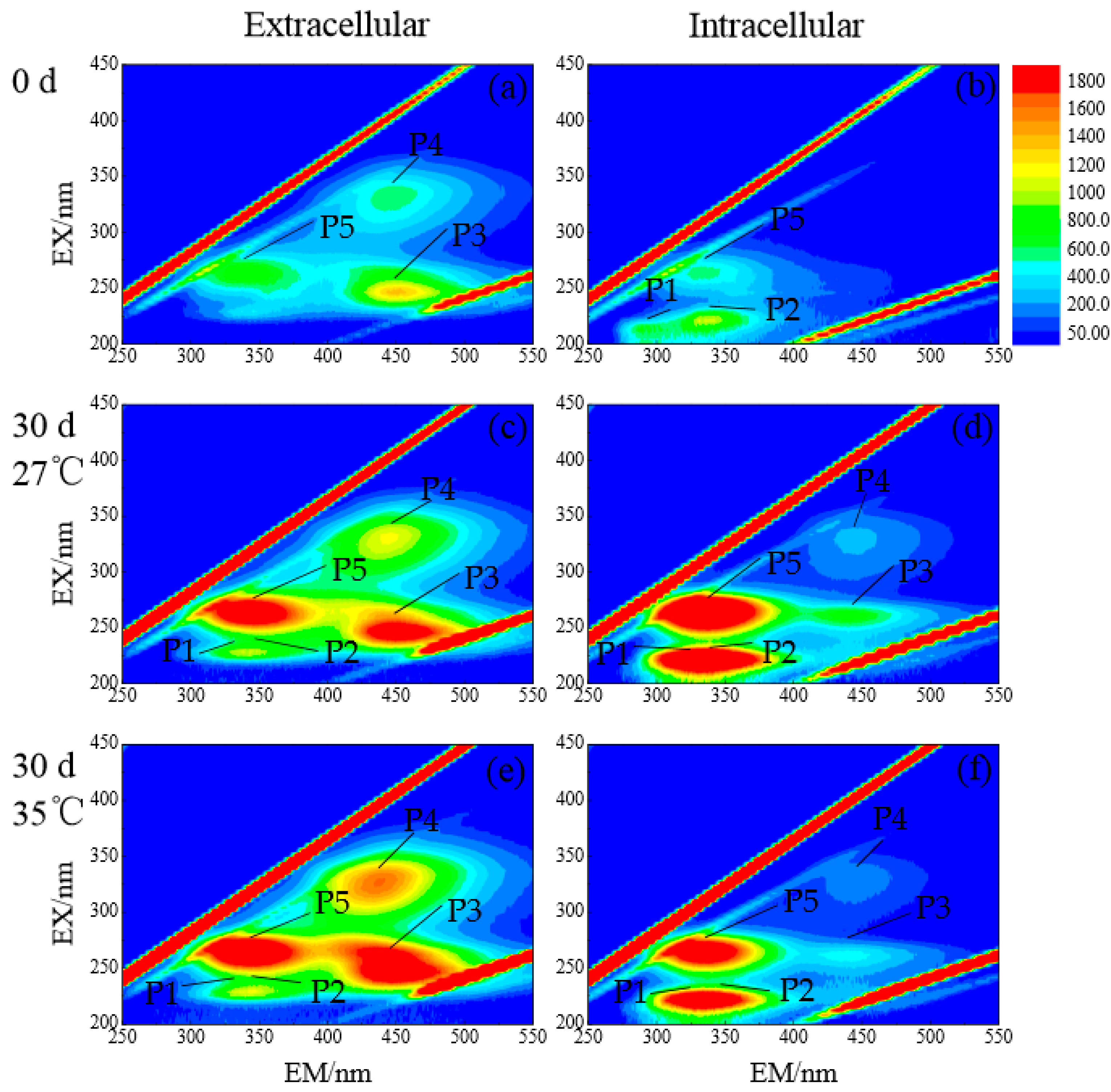
3.2.4. DOM Composition of Dolichospermum spiroides at Different Temperatures

There were three obvious fluorescence regions for the extracellular DOMs (Figure 5a,c,e). These represented the soluble microbial metabolites (left region, P5 represents their peak FI values), fulvic acid (right bottom region, P3 represents its peak FI value), and humic acid (right upper region, P4 represents its peak FI value). These three types of organic matter are major AOMs produced by Dolichospermum spiroides [33]. The intracellular EEM (Figure 5b,d,f) results show that there were obvious differences in DOMs compared to the extracellular 2-MIB. The humic acid and fulvic acid contents were extremely low, and only when the algal cell density was very high (incubated for 30 days at 27 °C), did a small amount of humic acid appear. Intracellular DOMs mainly produced two fluorescence sources. The fluorescence source at the left bottom of Figure 5b,d,f represent two types of aromatic protein, tryptophan (P1 represents its peak FI value) and tyrosine (P2 represents its peak FI value). The phenolic functional groups in tryptophan as well as the indole functional groups in tyrosine were in the excitation/emission (EX/EM) ranges of 200–250 nm and 280–380 nm, respectively, when fluorescence occurred. Therefore the fluorescence region showed the mix of tryptophan and tyrosine aromatic proteins. The left upper fluorescence region represents the phenolic compounds and soluble microbial metabolites. A comparison of the initial states and the 30th day FI for extracellular DOMs (at 27 °C and 35 °C) showed that during the culture period, there was an obvious change in FI values, from 853.4 to 2699 at 27 °C, as well as 3168 at 35 °C for P5; from 1612 to 2495 at 27 °C, as well as 2765 at 35 °C for P3. In contrast, P4 only had a slight change, as its FI value increased from 659.2 to 1129 at 27 °C, as well as 1583 at 35 °C. Furthermore, the fluorescence intensities of the intracellular humus at 27 °C were higher than those at 35 °C. However, the fluorescence intensities for the extracellular humus at 27 °C were lower than those at 35 °C.

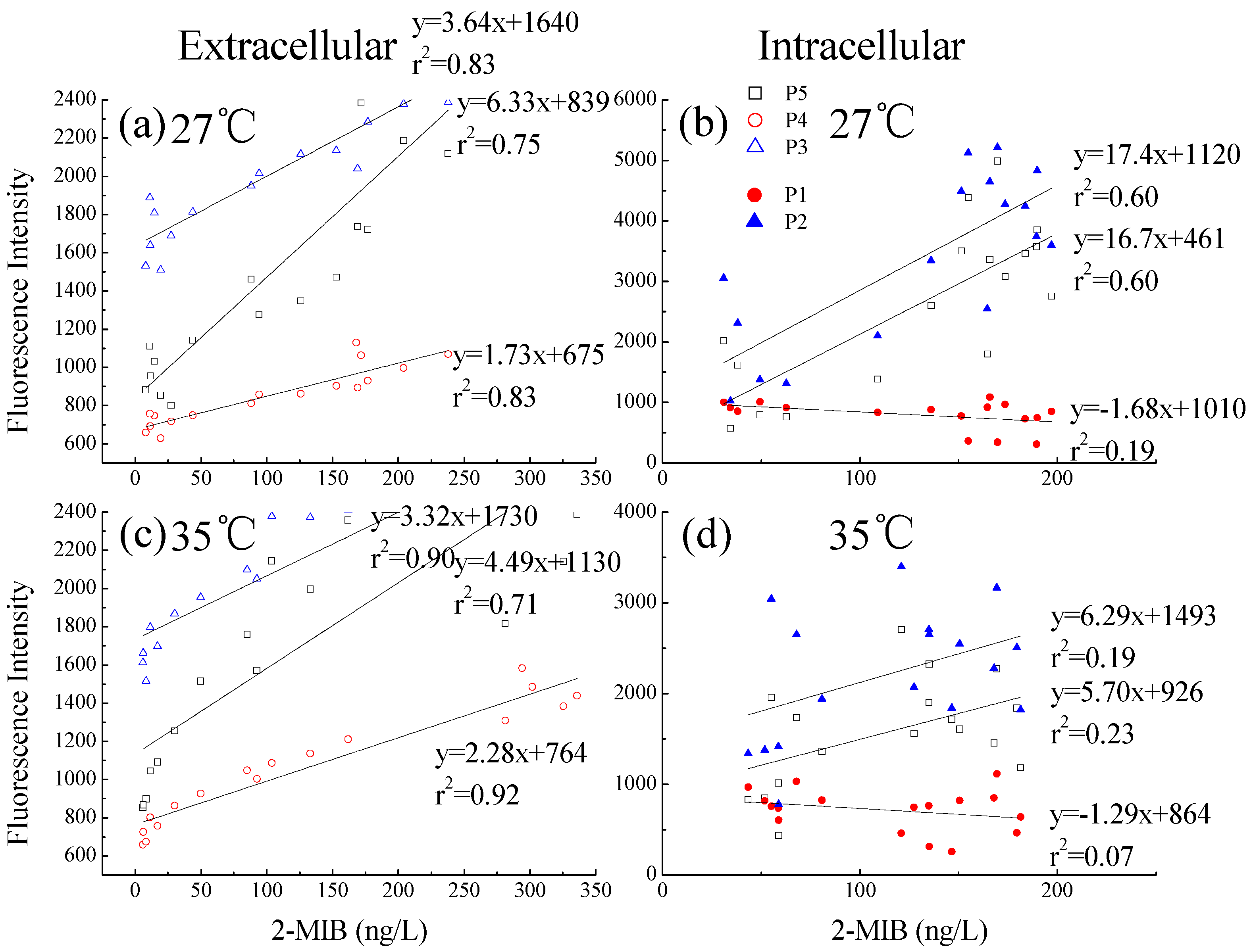
3.2.5. Relationship between Dolichospermum spiroides 2-MIB, and Intra- and Extracellular DOMs

The correlation between FI and 2-MIB concentration was analyzed. Figure 6 shows that the intracellular and extracellular organic matter levels were different. Generally, outside the cells, there were strong correlations between the extracellular organic matter fluorescence peaks and the extracellular 2-MIB concentration (Figure 6a,c). However, this difference was not so obvious for the intracellular 2-MIB (Figure 6b,d). Furthermore, the effect of temperature on the correlations was not the same. The extracellular portion at 35 °C (Figure 6c) produced the highest correlation coefficient for the peak FI values (P3, P4, and P5) and the extracellular 2-MIB concentration. In particular, the P4 and P3 values for FI and the extracellular 2-MIB concentration were highly correlated (r2 ≥ 0.90). However, the correlation between the FI peak values and 2-MIB was less strong at 27 °C.

4. Discussion

None of the three DIC treatments yielded significant differences in 2-MIB-producing potential. At 35 °C, the increase in DIC causes the carbon cycle in the water to generate more algal cells because more carbon would be available for photosynthesis and growth. Figure 3d,f suggest that a high DIC concentration promotes algal growth and leads to increased 2-MIB production. This means that when high temperatures and high DIC concentrations occur simultaneously (heat wave scenario), 2-MIB production by Planktothrix sp. increases quickly. A previous study found that DIC had a similar effect on Chlamydomonas [34]. In their study, the authors found that the photosynthesis of Chlamydomonas moewusii was promoted in a saturated amount of DIC. For the insignificant differences in total 2-MIB and Chla concentration values under the three DIC treatments at 27 °C, it may due to the suppression by nutrient limitation. At 35 °C, the response may become more obvious. In the study of Visser et al. [35], it was found that the response of cyanobacterial growth to rising CO2 concentrations and elevated temperatures can be suppressed by nutrient limitation. When heat wave occurs, measures to prevent algal blooming should be taken.
The results shown in Figure 4a are consistent with those of Wang [23], who reported that the growth rate of Pseudanabaena sp. varied between 10 °C and 35 °C. In this study, Planktothrix sp. growth was not inhibited at 35 °C. The results shown in Figure 4a,b suggest that higher temperatures will shorten the lifecycles of Dolichospermum spiroides and Planktothrix sp. and will the advance the onset of the logarithmic phase in laboratory-scale culture studies.
The 2-MIB-producing potential in Dolichospermum spiroides was higher at 35 °C than at 27 °C. The combined Chla data shows that after the 22nd day, many of the cells began to die and disintegrate, which released 2-MIB and caused a large increase in extracellular 2-MIB. In contrast, temperature had no significant effect on the Planktothrix sp. 2-MIB-producing potential.
Figure 4e,f show that the higher temperature will increase 2-MIB concentrations in water bodies where Dolichospermum spiroides is dominant and that 2-MIB is synthesized in cells. Most of the 2-MIB remains in the cell until the cells die and break down. Then, the 2-MIB is released into the water. In contrast, Planktothrix sp. transport 2-MIB to the extracellular area after the 2-MIB is synthesized. Therefore, extracellular 2-MIB concentrations were always higher in Planktothrix sp. cultures compared to Dolichospermum spiroides cultures.
The results shown in Figure 4c,f also indicate that the contribution made by extracellular 2-MIB to the MIB/Chla ratio over time was more substantial than that made by intracellular 2-MIB. The EEM results suggested that humic acid was not the main component of the Dolichospermum spiroides intracellular fluid. Extracellular concentrations of soluble microbial metabolites and fulvic acid increased significantly, whereas the humic acid concentration increased relatively slowly over time. At 35 °C, the concentrations of intracellular soluble microbial metabolites as well as tyrosine and tryptophan aromatic proteins were lower, while the concentration of extracellular fulvic acid, humic acid, and soluble microbial metabolites were higher than those at 27 °C. This result also suggests that a number of Dolichospermum spiroides cells died at 35 °C and released their humus into the water.
At the higher temperature, there was a stronger correlation between the fluorescence peak intensities of the extracellular DOMs (fulvic acid, humic acid, and soluble microbial metabolites) and 2-MIB concentration. This may be because the extracellular humus substances (including humic acid, fulvic acid, and soluble microbial metabolites) had a similar molecular polarity to 2-MIB, as they are all polar molecules. This suggests that the 2-MIB synthesized in the cells was passively transported to the extracellular environment. Extracellular 2-MIB is strongly related to extracellular humus, and extracellular humus may affect the 2-MIB-producing potential of Dolichospermum sproides by altering the proportion of extracellular 2-MIB.

5. Conclusions

This study used temperature and DIC to simulate the effects of climate change on two 2-MIB-producing cyanobacteria (Dolichospermum spiroides and Planktothrix sp.), and the following conclusions were drawn:
DIC affects 2-MIB production by Planktothrix sp. At 35 °C and 36.7 mg/L DIC, cell growth was promoted, which led to increased 2-MIB production. All the cyanobacteria died when they were exposed to 40 °C. However, 35 °C shortened the growth cycles of Dolichospermum spiroides and Planktothrix sp., but did not kill them. At 35 °C, Dolichospermum spiroides cells entered their death phase early (on the 22nd day), and the 2-MIB-producing potential of Dolichospermum spiroides increased over the whole experimental period. Furthermore, Dolichospermum spiroides at 35 °C had a higher 2-MIB-producing potential than at 27 °C. In contrast, the Planktothrix sp. 2-MIB-producing potential declined in general over time. In addition, temperature had no significant effect on the 2-MIB-producing potential and extracellular 2-MIB proportion released by Planktothrix sp. At 35 °C, there was a stronger correlation between the fluorescence peak intensities produced by the extracellular DOMs and the 2-MIB concentration (fulvic acid (P3 (r2 = 0.90)), humic acid (P4 (r2 = 0.92)), and soluble microbial metabolites (P5 (r2 = 0.71))). The 2-MIB-producing potential of Dolichospermum sproides was affected by extracellular humus.

Acknowledgments

This work was financially supported by the National Natural Science Foundation of China (51408022), Major Science and Technology Program for Water Pollution Control and Treatment (No. 2015ZX07406001).

Author Contributions

Junzhi Zhang and Lijia Qiu conceived and designed the experiments; Lijia Qiu, Xiaoting Wang, Xuanyi Meng, and Yu You performed the experiments; Luwei Li and Lijia Qiu analyzed the data; Junzhi Zhang and Wenlin Ma contributed reagents and materials; Junzhi Zhang, Luwei Li and Lijia Qiu wrote the paper, and Junzhi Zhang, Jianwei Yu, and Wenlin Ma reviewed the paper.

Conflicts of Interest

The authors declare no conflict of interest.

References

  1. Intergovernmental Panel on Climate Change (IPCC). Working Group I Contribution to the IPCC Fifth Assessment Report Climate Change 2013: The Physical Science Basis; IPCC: Geneva, Switzerland, 2013. [Google Scholar]
  2. Shen, Y.; Guoya, W. Key findings and assessment results of IPCC Work Group I fifth assessment report. J. Glaciol. Geocryol. 2013, 35, 1068–1076. [Google Scholar]
  3. Raven, J.; Beardall, J. CO2 concentrating mechanisms and environmental change. Aquat. Bot. 2014, 118, 24–37. [Google Scholar] [CrossRef]
  4. Cao, L.; Caldeira, K. Atmospheric CO2 stabilization and ocean acidification. Geophys. Res. Lett. 2008, 35. [Google Scholar] [CrossRef]
  5. Krüger, G.H.J.; Eloff, J.N. Effect of CO2 and HCO3 on Photosynthetic Oxygen Evolution by Microcystis aeruginosa. Z. Pflanzenphysiol. 1983, 112, 231–236. [Google Scholar] [CrossRef]
  6. Singh, S.P.; Singh, P. Effect of CO2 concentration on algal growth: A review. Renew. Sustain. Energy Rev. 2014, 38, 172–179. [Google Scholar] [CrossRef]
  7. Yu, L.; Kong, F.; Shi, X.; Yang, Z.; Zhang, M.; Yu, Y. Effects of elevated CO2 on dynamics of microcystin-producing and non-microcystin-producing strains during Microcystis blooms. J. Environ. Sci. 2015, 27, 251–258. [Google Scholar] [CrossRef] [PubMed]
  8. O’Neil, J.M.; Davis, T.W.; Burford, M.A.; Gobler, C.J. The rise of harmful cyanobacteria blooms: The potential roles of eutrophication and climate change. Harmful Algae 2012, 14, 313–334. [Google Scholar] [CrossRef]
  9. Winck, F.V.; Melo, D.O.P.; Barrios, A.F.G. Carbon acquisition and accumulation in microalgae Chlamydomonas: Insights from “omics” approaches. J. Proteom. 2013, 94, 207–218. [Google Scholar] [CrossRef] [PubMed]
  10. Andersen, T.; Andersen, F.Ø. Effects of CO2 concentration on growth of filamentous algae and Littorella uniflora in a Danish softwater lake. Aquat. Bot. 2006, 84, 267–271. [Google Scholar] [CrossRef]
  11. Rost, B.; Riebesell, U.; Sültemeyer, D. Carbon acquisition of marine phytoplankton: Effect of the photoperiodic length. Limnol. Oceanogr. 2006, 51, 12–20. [Google Scholar] [CrossRef] [Green Version]
  12. Francis, G. Poisonous Australian Lake. Nature 1878, 18, 11–12. [Google Scholar] [CrossRef]
  13. Gkelis, S.; Papadimitriou, T.; Zaoutsos, N.; Leonardos, I. Anthropogenic and climate-induced change favors toxic cyanobacteria blooms: Evidence from monitoring a highly eutrophic, urban Mediterranean lake. Harmful Algae 2014, 39, 322–333. [Google Scholar] [CrossRef]
  14. Dzialowski, A.R.; Smith, V.H.; Huggins, D.G.; deNoyelles, F.; Lim, N.-C.; Baker, D.S.; Beury, J.H. Development of predictive models for geosmin-related taste and odor in Kansas, USA, drinking water reservoirs. Water Res. 2009, 43, 2829–2840. [Google Scholar] [CrossRef] [PubMed]
  15. Olsen, B.K.; Chislock, M.F.; Wilson, A.E. Eutrophication mediates a common off-flavor compound, 2-methylisoborneol, in a drinking water reservoir. Water Res. 2016, 92, 228–234. [Google Scholar] [CrossRef] [PubMed]
  16. Smith, J.L.; Boyer, G.L.; Zimba, P.V. A review of cyanobacterial odorous and bioactive metabolites: Impacts and management alternatives in aquaculture. Aquaculture 2008, 280, 5–20. [Google Scholar] [CrossRef]
  17. Watson, S.; Charlton, M.; Rao, Y.; Howell, T.; Ridal, J.; Brownlee, B.; Marvin, C.; Millard, S. Off flavours in large waterbodies: Physics, chemistry and biology in synchrony. Water Sci. Technol. 2007, 55, 1–8. [Google Scholar] [CrossRef] [PubMed]
  18. Su, M.; Yu, J.; Zhang, J.; Chen, H.; An, W.; Vogt, R.D.; Andersen, T.; Jia, D.; Wang, J.; Yang, M. MIB-producing cyanobacteria (Planktothrix sp.) in a drinking water reservoir: Distribution and odor producing potential. Water Res. 2015, 68, 444–453. [Google Scholar] [CrossRef] [PubMed]
  19. Niiyama, Y.; Tuji, A.; Takemoto, K.; Ichise, S. Pseudanabaena foetida sp. nov. and P. subfoetida sp. nov.(Cyanophyta/Cya-nobacteria) producing 2-methylisoborneol from Japan. Fottea 2016, 16, 1–11. [Google Scholar] [CrossRef]
  20. Zhang, T.; Zheng, L.; Li, L.; Song, L. 2-Methylisoborneol production characteristics of Pseudanabaena sp. FACHB 1277 isolated from Xionghe Reservoir, China. J. Appl. Phycol. 2016, 28, 3353–3362. [Google Scholar] [CrossRef]
  21. Chou, W.K.W.; Gould, C.A.; Cane, D.E. Incubation of 2-methylisoborneol synthase with the intermediate analog 2-methylneryl diphosphate. J. Antibiot. 2017. [Google Scholar] [CrossRef] [PubMed]
  22. Dickschat, J.S.; Nawrath, T.; Thiel, V.; Kunze, B.; Müller, R.; Schulz, S. Biosynthesis of the Off-Flavor 2-Methylisoborneol by the Myxobacterium Nannocystis exedens. Angew. Chem. Int. Ed. 2007, 46, 8287–8290. [Google Scholar] [CrossRef] [PubMed]
  23. Wang, Z.; Li, R. Effects of light and temperature on the odor production of 2-methylisoborneol-producing Pseudanabaena sp. and geosmin-producing Anabaena ucrainica (cyanobacteria). Biochem. Syst. Ecol. 2015, 58, 219–226. [Google Scholar] [CrossRef]
  24. Lin, T.-F.; Wong, J.-Y.; Kao, H.-P. Correlation of musty odor and 2-MIB in two drinking water treatment plants in South Taiwan. Sci. Total Environ. 2002, 289, 225–235. [Google Scholar] [CrossRef]
  25. Wert, E.C.; Korak, J.A.; Trenholm, R.A.; Rosario-Ortiz, F.L. Effect of oxidant exposure on the release of intracellular microcystin, MIB, and geosmin from three cyanobacteria species. Water Res. 2014, 52, 251–259. [Google Scholar] [CrossRef] [PubMed]
  26. Wang, H.; Liu, D.M.; Wang, P.; Cui, F.Y. Formation of THMs, Microcystin-LR and Odors from Microcystis aeruginosa-Derived Organic Matter. Adv. Mater. Res. 2014, 1051, 348–352. [Google Scholar] [CrossRef]
  27. Ho, L.; Newcombe, G.; Croué, J.-P. Influence of the character of NOM on the ozonation of MIB and geosmin. Water Res. 2002, 36, 511–518. [Google Scholar] [CrossRef]
  28. Li, L.; Gao, N.; Deng, Y.; Yao, J.; Zhang, K. Characterization of intracellular & extracellular algae organic matters (AOM) of Microcystic aeruginosa and formation of AOM-associated disinfection byproducts and odor & taste compounds. Water Res. 2012, 46, 1233–1240. [Google Scholar] [PubMed]
  29. Ulu, F.; Barışçı, S.; Kobya, M.; Sillanpää, M. An evaluation on different origins of natural organic matters using various anodes by electrocoagulation. Chemosphere 2015, 125, 108–114. [Google Scholar] [CrossRef] [PubMed]
  30. Her, N.; Amy, G.; McKnight, D.; Sohn, J.; Yoon, Y. Characterization of DOM as a function of MW by fluorescence EEM and HPLC-SEC using UVA, DOC, and fluorescence detection. Water Res. 2003, 37, 4295–4303. [Google Scholar] [CrossRef]
  31. Tang, Z.; Yu, G.; Liu, D.; Xu, D.; Shen, Q. Different analysis techniques for fluorescence excitation–emission matrix spectroscopy to assess compost maturity. Chemosphere 2011, 82, 1202–1208. [Google Scholar] [CrossRef] [PubMed]
  32. Chen, W.; Westerhoff, P.; Leenheer, J.A.; Booksh, K. Fluorescence Excitation−Emission Matrix Regional Integration to Quantify Spectra for Dissolved Organic Matter. Environ. Sci. Technol. 2003, 37, 5701–5710. [Google Scholar] [CrossRef] [PubMed]
  33. Feng, G.; Dong, B. Study on the Characteristics of Algogenic Organnic Metters. Environ. Sci. Technol. 2016, 39, 144–149. [Google Scholar]
  34. Ghoshal, D.; Husic, H.D.; Goyal, A. Dissolved inorganic carbon concentration mechanism in Chlamydomonas moewusii. Plant Physiol. Biochem. 2002, 40, 299–305. [Google Scholar] [CrossRef]
  35. Visser, P.M.; Verspagen, J.M.; Sandrini, G.; Stal, L.J.; Matthijs, H.C.; Davis, T.W.; Paerl, H.W.; Huisman, J. How rising CO2 and global warming may stimulate harmful cyanobacterial blooms. Harmful Algae 2016, 54, 145–159. [Google Scholar] [CrossRef] [PubMed]
Figure 1. The carbonate cycle in the water environment [9].
Water 09 00859 g001
Figure 2. Technological roadmap.
Water 09 00859 g002
Figure 3. The changes with time in one period of the 2-MIB producing potencial, total concentration of 2-MIB and Chla in three different DIC conditions at 27 and 35 °C: (a) 2-MIB producing potencial at 27 °C; (b) 2-MIB producing potencial at 35 °C; (c) total 2-MIB at 27 °C; (d) total 2-MIB at 35 °C; (e) Chla concentration at 27 °C; (f) Chla concentration at 35 °C.
Water 09 00859 g003
Figure 4. The variations of Chla, 2-MIB-producing potential, and extracellular 2-MIB proportion for Dolichospermum spiroides (a,c,e) and Planktothrix sp. (b,d,f). 2-MIB-producing potential was shown by 2-MIB/Chla.
Water 09 00859 g004
Figure 5. EEMs of Dolichospermum spiroides at two different temperatures: (a,b) EEMs of extra- and intracellular DOMs in the beginning of the culture; (c,d) EEMs of extra- and intracellular DOMs on the 30th day of the culture at 27 °C; (e,f) EEMs of extra- and intracellular DOMs on the 30th day of the culture at 35 °C. P1 represents the peak fluorescence intensity (FI) value of tryptophan aromatic protein; P2 represents the peak FI value of tyrosine aromatic protein; P3 represents the peak FI value of fulvic acid; P4 represents the peak FI value of humic acid; P5 represents the peak FI value of the soluble microbial metabolites.
Water 09 00859 g005
Figure 6. Fitting curves of each DOM’s FI and 2-MIB concentration of Dolichospermum spiroides. (a) Extracellular DOM and 2-MIB at 27 °C; (b) Intracellular DOM and 2-MIB at 27 °C; (c) Extracellular DOM and 2-MIB at 35 °C; (d) Intracellular DOM and 2-MIB at 35 °C.
Water 09 00859 g006

