Article

Coupling Heavy Metal Removal and Biodiesel Production in Chlorella vulgaris: Metal-Specific Regulation of Lipogenic Enzymes and Carbon Allocation


1
School of Resources, Environment and Materials, Guangxi University, No. 100 Daxue Road, Nanning 530004, China
2
Key Laboratory of Environmental Protection, Education Department of Guangxi Zhuang Autonomous Region, Guangxi University, Nanning 530004, China
3
Guangxi Key Laboratory of Emerging Contaminants Monitoring, Early Warning and Environmental Health Risk Assessment, Nanning 530028, China
*
Author to whom correspondence should be addressed.
Water 2026, 18(11), 1306; https://doi.org/10.3390/w18111306
Submission received: 21 April 2026 / Revised: 15 May 2026 / Accepted: 26 May 2026 / Published: 28 May 2026
(This article belongs to the Section Wastewater Treatment and Reuse)

Abstract

Heavy metal pollution poses a serious threat to aquatic ecosystems. Microalgae have attracted considerable attention due to their dual potential for heavy metal removal and lipid recovery. However, studies that simultaneously achieve both heavy metal removal and lipid accumulation remain very limited. The short-term (3 h) and long-term (3 days) effects of single and mixed Cu2+, Zn2+, and Pb2+ stress on Chlorella vulgaris FACHB-8 were investigated for heavy metal removal and lipid recovery. Removal rates varied with metal species, concentration, and single vs. mixed systems. At 3 h, the order was Pb2+ > Cu2+ > Zn2+; at 3 days, Pb2+ ≈ Zn2+ > Cu2+. The Zn2++Pb2+ combination maintained >90% removal across all concentrations, whereas Cu2+ removal was impeded (65–85%). Long-term stress maximized lipid content at 30% under 1 mg/L Cu2+ or 0.5 mg/L Cu2++Zn2+, while Pb2+ restricted it to ≤12.85%. Cu2+ (1 mg/L) produced the highest saturated fatty acids (69.95%, dominated by C16:0 and C18:0), favorable for biodiesel. Highly toxic Pb2+ impaired cellular integrity and suppressed carbon allocation to lipids, whereas moderate Cu2+ or Cu2++Zn2+ stress induced synergistic lipid and SFA accumulation. This metabolic shift was associated with upregulated superoxide dismutase (SOD) and acetyl-CoA carboxylase (ACC) activities, mitigating oxidative damage and redirecting carbon flux toward lipid biosynthesis as a defense strategy.

1. Introduction

Since the industrial revolution, the rapid expansion of industries such as mining, electroplating, and smelting has led to a sustained increase in the discharge of wastewater laden with heavy metals, particularly copper (Cu2+), zinc (Zn2+), and lead (Pb2+) [1]. As highly toxic and persistent pollutants, heavy metals can rapidly bioaccumulate along the food chain, constituting a serious threat to human health and ecosystems [2]. Consequently, the efficient purification of heavy metal-contaminated wastewater has emerged as a critical global environmental challenge. Traditional technologies suffer from high chemical consumption, potential secondary pollution, and elevated costs, making them less suitable for meeting contemporary remediation needs [3,4].
Microalgae have attracted significant attention for heavy metal removal due to their environmental friendliness, cost-effectiveness, and potential for resource recovery [5]. As photosynthetic autotrophic microorganisms, microalgae provide a novel approach to heavy metal remediation [6]. Efficient sequestration of heavy metal ions from aqueous solutions occurs through various mechanisms, including cell wall adsorption, complexation with extracellular polymeric substances (EPS), and intracellular active transport [7]. Removal efficiencies for low-concentration heavy metals can exceed 80%; for example, treatment with Chlorella achieved a 90.29% removal rate for 50 mg/L lead ions [8]. More importantly, the stress effects induced by heavy metals during microalgal accumulation are intricately linked to lipid metabolism, offering a potential pathway for integrating heavy metal wastewater purification with lipid recovery [9].
Moderate heavy metal stress can trigger stress response mechanisms in microalgae, which enhance lipid productivity by modulating carbon flux—inhibiting protein and carbohydrate synthesis while redirecting carbon allocation toward lipid synthesis—and by increasing the activity of lipid-synthesizing enzymes [10]. For example, when cultivated in mixed nickel and chromium wastewater, Tetraselmis sp. achieved a lipid content of 31% [11]. In contrast, excessively high heavy metal concentrations can disrupt the microalgal photosynthetic system and cell membrane structure, leading to reduced activity of lipid synthesis enzymes and ultimately inhibiting lipid accumulation [12]. Heavy metal stress also alters fatty acid profiles, which is a key determinant of biodiesel quality [13,14,15]. The resistance of microalgae to heavy metal damage can be reflected by antioxidant indicators [10]. Heavy metal stress induces the production of reactive oxygen species (ROS), leading to oxidative stress. Superoxide dismutase (SOD) is a key antioxidant enzyme that plays a crucial role in defending against oxidative damage and modulating signaling pathways. Total antioxidant capacity (T-AOC) is a comprehensive indicator of the overall antioxidant capability of microalgae, reflecting their ability to scavenge ROS and mitigate oxidative damage. Malondialdehyde (MDA), a major end product of lipid peroxidation, serves as a classic indicator of oxidative damage by directly reflecting the extent of lipid peroxidation in cell membranes and thus the degree of cellular disruption caused by ROS.
Moreover, the regulatory mechanisms governing microalgal lipid synthesis are considerably more complex in mixed heavy metal systems than in single-metal systems. Competition for adsorption sites on the algal cell surface and interference with intracellular transport channels in mixed systems can indirectly modulate the expression and activity of lipid synthesis-related enzymes [16]. These multi-component, multi-pathway interactive effects imply that lipid regulatory mechanisms identified for single heavy metals cannot be directly extrapolated to mixed systems. Currently, research on how mixed heavy metals regulate lipid synthesis through the cascade of oxidative stress, carbon partitioning, and enzyme activity remains insufficient, thereby limiting the integration of heavy metal remediation with subsequent lipid recovery.
Most current studies have focused on single heavy metals or short-term stress effects, with few systematic investigations incorporating multiple concentration gradients, combined short- and long-term exposure, and mixed systems. As a result, several critical scientific questions remain unresolved. This study focuses on Cu2+, Zn2+, and Pb2+, primarily due to their distinct environmental behaviors and toxicological profiles in aquatic ecosystems and their widespread presence in both industrial wastewater and municipal sewage [17].
Cu2+ and Zn2+ are essential trace elements with low-to-moderate dose-dependent toxicity, whereas Pb2+ is non-essential and highly toxic due to its strong accumulation ability and non-specific protein binding [18]. How these distinct toxicity characteristics differentially regulate oxidative stress, carbon allocation, and lipogenic enzyme activity—and why low-to-moderate toxic metals promote lipid synthesis while highly toxic Pb2+ suppresses it—remain poorly understood.
Accordingly, this study investigates heavy metal removal and lipid accumulation in Chlorella vulgaris under single and combined Cu2+, Zn2+, and Pb2+ stress, focusing on the differential regulation of acetyl-CoA carboxylase (ACC) and glycerol-3-phosphate acyltransferase (GPAT) [19]. We hypothesize that moderately toxic Cu2+/Zn2+ optimizes energy allocation between antioxidant defense and lipid synthesis, whereas highly toxic Pb2+ disrupts this balance. The findings will provide a scientific foundation for integrating heavy metal bioremediation with lipid resource recovery.

2. Materials and Methods

2.1. Microalgae Cultivation

The microalgal strain Chlorella vulgaris (FACHB-8) was obtained from the Institute of Hydrobiology, Chinese Academy of Sciences (Wuhan, China). The BG11 medium used for cultivation had the following composition (mg/L): NaNO3, 1500; K2HPO4, 40; MgSO4·7H2O, 36; CaCl2·2H2O, 75; Na2CO3, 20; citric acid, 6; ferric ammonium citrate, 6; Na2EDTA, 1; MnCl2·4H2O, 1.86; Co(NO3)2·6H2O, 0.05; CuSO4·5H2O, 0.08; NaMoO4·2H2O, 0.39; ZnSO4·7H2O, 0.22; H3BO3, 2.86. Prior to inoculation, the medium was sterilized by autoclaving at 121 °C for 30 min. On a clean bench, the microalgae were inoculated into the sterilized medium and cultivated under continuous aeration at 25 ± 2 °C, with a light intensity of 3500 lx and continuous illumination for 24 h. These cultures were then used for the subsequent heavy metal adsorption experiments. All chemicals used were of analytical grade (AR) and purchased from Tianjin Fengchuan Chemical Reagent Technology Co., Ltd., Tianjin, China.

2.2. Preparation of Heavy Metal Stock Solutions

Calculated amounts of CuSO4·5H2O, ZnSO4·7H2O, and Pb(NO3)2 were dissolved separately in ultrapure water to prepare 1 g/L stock solutions of each heavy metal. The solutions were then filter-sterilized using a 0.22 μm membrane filter (Nantong Hairui Experimental Equipment Co., Ltd., Nantong, China) to avoid microbial contamination. The stock solutions were stored in 1000 mL high-density polyethylene (HDPE) bottles at room temperature (25 ± 2 °C) in the dark to prevent photodegradation. All chemicals used were of analytical grade (AR) and purchased from Tianjin Fengchuan Chemical Reagent Technology Co., Ltd., Tianjin, China.

2.3. Heavy Metal Removal and Lipid Production Experiments

C. vulgaris cells in the logarithmic growth phase were harvested and resuspended in BG11 medium to an OD680 of 0.5 (approximately 0.5 g/L) using a UV-Vis spectrophotometer (UV-8000, Shanghai, China). Heavy metal stock solutions (Cu2+, Zn2+, and Pb2+, singly or in combination) were added to the suspension to achieve the desired initial concentrations. For single-metal systems, concentrations of 0, 2, 4, 8, 16, and 32 mg/L were used; for mixed-metal systems (Cu2++Zn2+, Cu2++Pb2+, or Zn2++Pb2+), concentrations of 0, 1, 2, 4, 8, and 16 mg/L were applied. Higher concentrations were used in the short-term adsorption experiments to evaluate the heavy metal removal capacity of the microalgae, whereas lower concentrations were used in the long-term physiological experiments to analyze heavy metal stress-induced lipid accumulation. The selection of concentration ranges was based on previous studies [20,21].
For the short-term 3 h experiment, 100 mL aliquots of the metal-amended algal suspensions were transferred to 150 mL beakers and continuously stirred at 120 rpm using a magnetic stirrer. After 3 h, a 5 mL sample was collected, diluted 1:1 with ultrapure water, and filtered through a 0.22 μm cellulose acetate membrane. The filtrate was acidified with 5% HNO3 (Chuandong Chemical Group, Chongqing, China) and stored at 4 °C for subsequent analysis. Residual heavy metal concentrations were determined using an atomic absorption spectrophotometer (Shimadzu A6000, Kyoto, Japan). At the end of the 3 h exposure, microalgal lipids were extracted and quantified.
For the long-term 3-day experiment, the metal-amended algal suspensions were cultivated in conical flasks under continuous illumination (3500 lx, photoperiod of 24:0) and aeration at 25 ± 2 °C. Algal growth was monitored daily by measuring OD680. Each day, a 5 mL aliquot was sampled, processed, and analyzed for residual heavy metals as described above. Sampling continued for 3 days. At the end of the 3-day experiment, microalgal lipids were extracted and quantified.

2.4. Investigation of Lipid Accumulation Mechanisms

Based on the phenotypic outcomes from the heavy metal treatments (Section 3.2), four groups representing key physiological scenarios were selected for in-depth comparison: (A) a metal-free control, representing unstressed baseline metabolism; (B) the optimal single-metal condition (1 mg/L Cu2+), which induced the highest lipid accumulation; (C) a representative inhibitory condition (3 mg/L Pb2+), which caused severe physiological stress; and (D) the optimal mixed-metal combination (0.5 mg/L Cu2++Zn2+), which also promoted high lipid yield. Through comparison of these four scenarios, the common and distinct patterns of carbon allocation and antioxidative responses underlying high lipid productivity under different optimal metal pressures were elucidated.
C. vulgaris cells in the logarithmic growth phase were harvested and resuspended in BG11 medium to an OD680 of 0.5, as described in Section 2.3. The cultures were transferred to conical flasks, and heavy metal stock solutions were added to achieve the final concentrations designated for each group. The flasks were then cultivated under continuous illumination and aeration, following the same conditions used for the long-term experiments (Section 2.3). After three days of treatment, samples were collected to determine microalgal lipid content, fatty acid profiles, protein and carbohydrate contents, SOD activities, T-AOC, and the activities of key lipogenic enzymes (GPAT).

2.5. Analytical Methods

2.5.1. Biomass Determination

A 5 mL aliquot of the algal suspension was thoroughly mixed and, if necessary, diluted to ensure the OD680 reading fell within the linear range of the spectrophotometer. The absorbance of the diluted sample at 680 nm was then measured using a UV-Vis spectrophotometer, with deionized water as the blank [22].

2.5.2. Lipid Content Determination

Lipids were extracted from the harvested microalgal biomass using the Bligh–Dyer method [23]. 15 mL of a chloroform–methanol mixture (1:2, v/v) was added to 0.1 g of dry algal powder, and the mixture was ultrasonicated at 85 W for 40 min. Subsequently, 5 mL of chloroform and 10 mL of a 1% (w/v) NaCl solution were added to the extract. After thorough mixing, the mixture was centrifuged at 10,000 rpm for 10 min using a centrifuge (L550, Hunan Xiangyi Laboratory Instrument Development Co., Ltd., Changsha, China). The lower organic phase was carefully collected using a syringe and dried at 60 °C to constant weight. Lipid content (%) was calculated according to the following Formula (1):
C = m L m 0 0.1 × 100 %
where C represents the microalgal lipid content (%); mL is the weight of the weighing dish (g); m0 is the weight of the weighing dish after drying to constant weight (g).

2.5.3. Determination of Fatty Acid Profile

Fatty acid composition was analyzed using gas chromatography–mass spectrometry (GC–MS) (Nexis GC-2030, Shimadzu Corporation, Kyoto, Japan) [24]. The extracted lipids were methylated by adding 2 mL of 14% BF3–methanol solution and incubating in a 60 °C water bath for 30 min. After cooling to room temperature, 1 mL of distilled water and 2 mL of n-hexane were added, and the mixture was vortexed and allowed to separate. The upper organic layer was collected, evaporated to dryness under a nitrogen stream, and redissolved in 1 mL of n-hexane for subsequent GC–MS analysis.
GC–MS analysis was performed on an HP-5MS capillary column (60 m × 0.25 mm, 0.25 μm film thickness). The injector temperature was set to 280 °C, and a 1.0 μL aliquot was injected in split mode (20:1). Helium was used as the carrier gas at a constant flow rate of 1.5 mL/min. The oven temperature program was as follows: initial temperature 120 °C, held for 1 min; increased to 170 °C at 6 °C/min; then to 215 °C at 2.5 °C/min, held for 12 min; subsequently to 230 °C at 4 °C/min, held for 10 min; finally to 280 °C at 10 °C/min, held for 15 min.
Fatty acids were identified by comparing retention times with those of a 37-component fatty acid methyl ester (FAME) standard mixture; quantification was performed using the area normalization method, and results were expressed as percentages of total fatty acids.

2.5.4. Protein Content Determination

Protein content was determined using the Coomassie Brilliant Blue method [25]. A 5 mL aliquot of the algal suspension was transferred into a 10 mL centrifuge tube. After ultrasonication for 5 min, the mixture was centrifuged at 4000 rpm (approximately 3000× g) for 10 min. Subsequently, 2 mL of the supernatant was collected into a new 10 mL centrifuge tube, and a separate tube containing 2 mL of distilled water was prepared as the blank control. Then, 5 mL of Coomassie Brilliant Blue (G250) reagent (Macklin Biochemical Technology Co., Ltd., Shanghai, China) was added to each tube, mixed thoroughly, and allowed to stand for 2 min. Absorbance was measured at 595 nm using a UV-Vis spectrophotometer, with the blank sample as the reference.

2.5.5. Carbohydrate Content Determination

Carbohydrate content was determined using the phenol–sulfuric acid method [26]. A 1 mL aliquot of the algal suspension was centrifuged, and the supernatant was discarded. The pellet was resuspended in 3 mL of 0.5 M NaOH and incubated in a boiling water bath for 10 min to ensure complete dissolution. Then, 2 mL of the resulting solution was transferred into a 10 mL centrifuge tube, followed by the sequential addition of 50 μL of 90% (v/v) phenol solution and 5 mL of concentrated sulfuric acid. The mixture was allowed to react for 30 min and then cooled to room temperature. Absorbance was measured at 485 nm using a spectrophotometer, and carbohydrate content was calculated from a glucose standard curve.

2.5.6. Determination of Oxidative Stress Markers

MDA content, SOD activity, and T-AOC were determined using commercial assay kits (Jiangsu Aidisheng Biotechnology Co., Ltd., Yancheng, China). Algal cells were harvested, washed, and homogenized following the protocols provided with the kits, and the resulting supernatants were used for the assays according to the manufacturer’s instructions to assess the oxidative stress and antioxidant levels.

2.5.7. Determination of Lipogenic Enzyme Activities

The activities of ACC and GPAT were assayed using commercial kits (Jiangsu Aidisheng Biotechnology Co., Ltd., Yancheng, China) to evaluate lipogenic capacity. Algal samples were prepared according to the kit-specific protocols, and the assays were performed following the manufacturer’s instructions.

2.6. Statistical Analysis of Data

The experiments were carried out using three independent biological replicates. Measurements are presented as mean ± standard deviation (SD) of triplicate technical replicates. Statistical analysis was performed using one-way analysis of variance (ANOVA) with Origin 9.4 (OriginLab Corporation, Northampton, MA, USA) and Excel (Microsoft, Redmond, WA, USA) software.

3. Results and Discussion

3.1. Heavy Metal Removal Efficiency

3.1.1. Removal Efficiency of Heavy Metals After 3 h

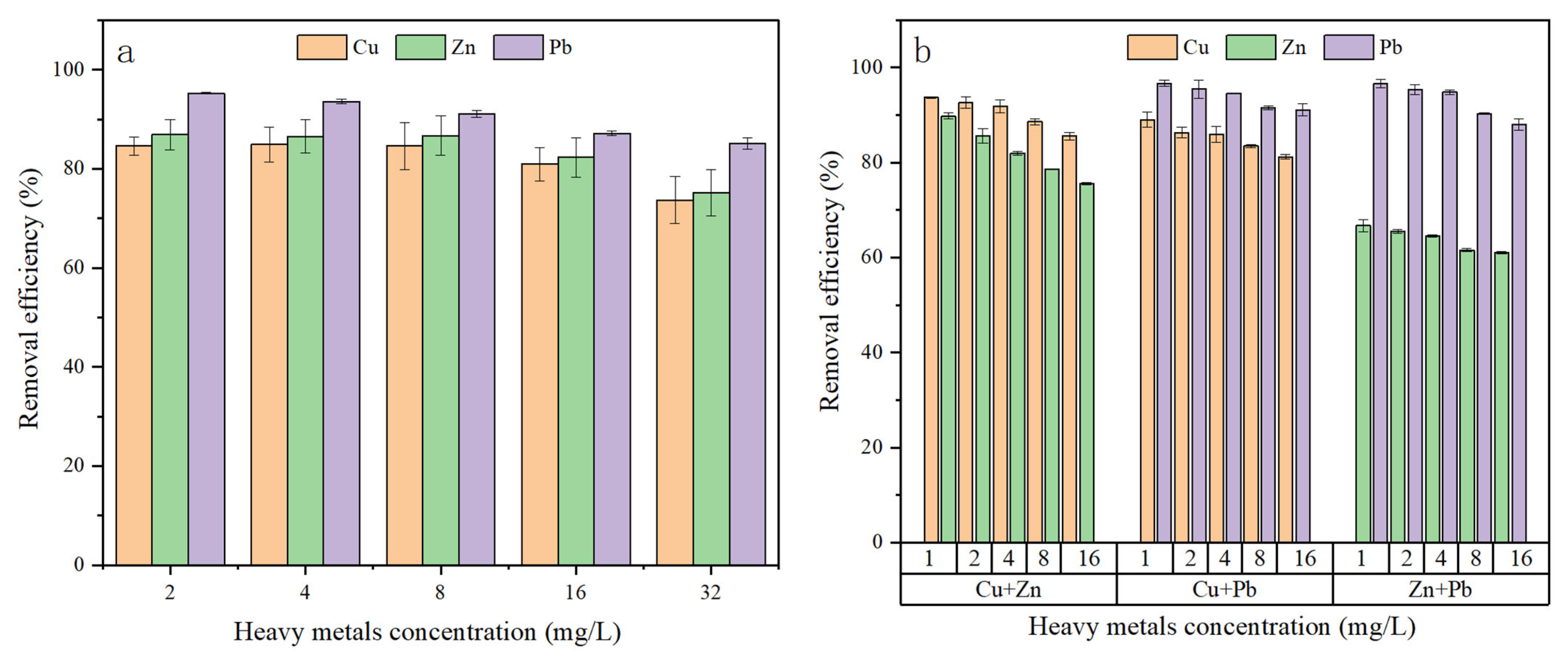
The removal efficiency of single heavy metals (Cu2+, Zn2+, and Pb2+) by C. vulgaris was investigated over a concentration range of 2–32 mg/L following 3 h of exposure. The removal efficiencies of the three heavy metals differed significantly, following the order Pb2+ > Cu2+ > Zn2+. For Pb2+, removal efficiency remained as high as 85%, even at the highest concentration of 32 mg/L, reflecting rapid saturation of extracellular binding sites and strong complexation affinity toward algal surface matrices. In contrast, the removal efficiency for Cu2+ was lower, at 77.74% in the 32 mg/L group, whereas that for Zn2+ was 69.02% under the same condition (Figure 1a). For all three metals, removal efficiency decreased progressively with increasing initial concentration.
Within the experimental concentration range (2–32 mg/L), Pb2+ maintained consistently high removal efficiency, driven by its physicochemical properties that facilitate strong binding with microalgae. The high electronegativity and suitable ionic radius of Pb2+ enable it to readily form stable complexes with EPS and functional groups (e.g., phosphate and carboxyl groups) on the cell surface, thereby enhancing adsorption through chelation and ion exchange, which makes Pb2+ particularly amenable to biosorption [27]. In contrast, the binding affinity of Zn2+ for cell surface groups is weaker than that of Pb2+ and Cu2+, resulting in lower removal efficiencies. The progressive saturation of binding sites on the microalgal cell surface accounts for the observed decline in removal efficiency with increasing initial concentrations, especially for Cu2+ and Zn2+.
The removal efficiency of heavy metals by C. vulgaris after 3 h of exposure was investigated in mixed-metal systems (Cu2++Zn2+, Cu2++Pb2+, and Zn2++Pb2+) over a concentration range of 1–16 mg/L. As illustrated in Figure 1b, the removal efficiency of each heavy metal decreased progressively with increasing initial concentration. In the Cu2++Zn2+ system, the removal efficiency of Cu2+ was consistently higher than that of Zn2+. At 1 mg/L, the efficiency for Cu2+ was 93.77%, compared with 89.77% for Zn2+. At the highest concentration (16 mg/L), Cu2+ removal remained at 85.55%, whereas that of Zn2+ decreased to 75.55%. In the Cu2++Pb2+ system, removal efficiencies ranged from 88.99% to 81.23% for Cu2+ and from 96.68% to 91.06% for Pb2+. In the Zn2++Pb2+ system, the removal efficiency of Zn2+ gradually decreased from 66.68% at 1 mg/L to 61.06% at 16 mg/L, while that of Pb2+ decreased from 96.55% to 88.04%. Particularly, Pb2+ removal remained above 95% in the 1–4 mg/L range and dropped to approximately 90% at 8–16 mg/L.
The overall removal efficiency followed the order Pb2+ > Cu2+ > Zn2+. This order primarily reflects the selective biosorption capacity of microalgae, which is determined by the competitive affinity of different heavy metal ions for active sites on the cell surface. EPS secreted by microalgae, rich in polysaccharides and proteins, can immobilize heavy metals through complexation and precipitation. Studies have shown that the complexation capacity of EPS for Pb2+ is significantly higher than that for Cu2+ and Zn2+ [28]. Furthermore, the microalgal cell wall contains abundant functional groups—such as carboxyl (–COOH), hydroxyl (–OH), and amino (–NH2) groups—that exhibit different affinities for various metal ions. The binding affinity of Pb2+ for carboxyl groups is substantially stronger than that of Zn2+ and Cu2+, accounting for its consistently highest removal efficiency. This competitive affinity also explains the lower removal efficiencies of Cu2+ and Zn2+ in mixed-metal systems compared with single-metal systems, with the effect being most pronounced for Zn2+ due to its weaker binding strength.

3.1.2. Removal Efficiency of Heavy Metals After 3 Days

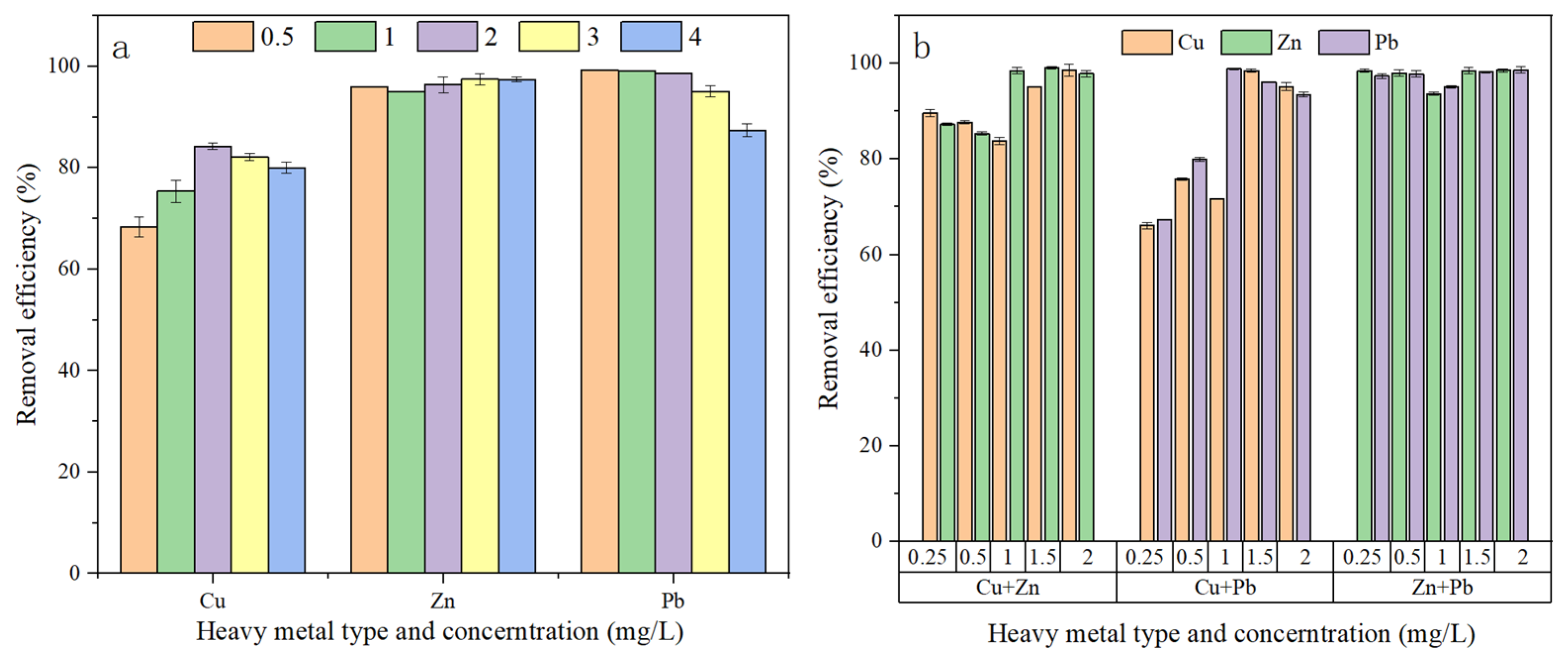
Long-term (3-day) removal of Cu2+, Zn2+, and Pb2+ by C. vulgaris was investigated at initial concentrations of 0.5, 1, 2, 3, and 4 mg/L, as shown in Figure 2a. The concentrations of all three heavy metal ions decreased rapidly on the first day of exposure. Notably, Pb2+ reached near-maximum removal within the first day, with residual concentrations in all groups falling below 0.1 mg/L—a rate considerably faster than that observed for Cu2+ and Zn2+, highlighting the rapid kinetics of Pb2+ surface precipitation.
After three days, significant differences were observed in the removal efficiencies of the three heavy metals, with the overall removal efficiency following the order Pb2+ ≈ Zn2+ > Cu2+. Removal efficiency for Zn2+ ranged from 90% to 100%, reaching nearly 100% in the 4 mg/L group. In contrast, removal efficiency for Cu2+ ranged from 65% to 85%, lower than those for Pb2+ and Zn2+. At low initial concentrations, Pb2+ removal exceeded 95%; however, it declined rapidly at concentrations above 3 mg/L.
The rapid and efficient removal of Pb2+ is largely attributable to its strong complexation with functional groups (e.g., carboxyl and phosphate groups) on the algal cell surface, as well as the formation of surface precipitates [29]. This mechanism underpins the high removal efficiency (>98%) observed at 0.5–2 mg/L. The marked decline in removal efficiency at concentrations above 3 mg/L is likely due to lead-induced toxicity, which causes physiological damage and reduces the availability of binding sites. Nevertheless, even non-viable microalgae can sequester heavy metals to some extent through passive adsorption [30], accounting for the reduced yet sustained removal efficiency at higher concentrations.
As an essential micronutrient, Zn2+ exhibits relatively low toxicity to microalgae. It can also enhance the production of EPS, whose proteins serve as key binding agents for Zn2+ and play a crucial role in its immobilization [31]. Driven by these binding interactions, removal efficiency remained above 90% even at 4 mg/L, demonstrating the excellent remediation potential of microalgae for zinc. Although its binding affinity to cell surface groups is weak—leading to lower removal efficiency in short-term adsorption (3 h)—the removal efficiency after three days reached 90–100% due to time-dependent activation of active uptake and metabolic regulation mechanisms [32]. In contrast, the removal efficiency for Cu2+ after three days was lower than that after three hours, likely due to cumulative copper toxicity and desorption during long-term exposure. Copper toxicity reduces cell viability and impairs the adsorption capacity of surface functional groups, ultimately leading to decreased removal efficiency [33]. In summary, C. vulgaris demonstrates considerable potential for the long-term removal of Pb2+ and Zn2+ from contaminated water, particularly at low to medium concentrations. Remediation of Cu2+-contaminated water may require further optimization. For practical applications, determining the optimal concentration ranges and exposure durations is essential to maximize overall removal efficiency.
Long-term (3 days) removal of Cu2+, Zn2+, and Pb2+ by C. vulgaris was investigated in mixed-metal systems at initial concentrations of 0.25, 0.5, 1, 1.5, and 2 mg/L, as shown in Figure 2b. Across all mixed systems, removal efficiency generally followed the order Pb2+ > Zn2+ > Cu2+, consistent with that observed in single-metal systems, though interactive effects further amplified the differences.
In the Cu2++Zn2+ system, the removal efficiency of Zn2+ was consistently higher than that of Cu2+ across all concentrations, with the difference becoming more pronounced over time. By day 3, removal efficiencies in the 2 mg/L group reached 97.88% for Zn2+ and 83.82% for Cu2+. The disparity between the two metals was greater at lower concentrations, whereas the increase in Cu2+ removal efficiency slowed at higher concentrations. Removal efficiencies in low-concentration mixed groups were not significantly different from those observed in single-metal systems.
In the Cu2++Pb2+ system, the removal efficiency of Pb2+ was significantly higher than that of Cu2+ across all concentrations, surpassing 90% within the first day and approaching 100% by day 3. In the 2 mg/L group, Pb2+ removal remained above 98%, whereas Cu2+ removal was only approximately 86%. High concentrations of Pb2+ exerted a slight inhibitory effect on Cu2+ removal.
In the Zn2++Pb2+ system, both metals exhibited high removal efficiencies, each exceeding 95% by day 3. No significant difference was observed at low concentrations, whereas Pb2+ showed slightly higher efficiency at high concentrations. The coexistence of Zn2+ and Pb2+ did not result in significant inhibition, with both maintaining high removal efficiencies even at elevated concentrations. In contrast, the removal efficiency of Cu2+ decreased with increasing concentration and was markedly affected by competition, whereas the removal efficiencies of Zn2+ and Pb2+ remained stable, demonstrating greater tolerance. Zn2+ is more readily absorbed by microalgae, which accounts for its consistently higher removal efficiency when coexisting with Cu2+. Both metals promote EPS secretion, thereby indirectly enhancing adsorption capacity [31,34]. Owing to its strong complexation capacity, Pb2+ preferentially occupies adsorption sites on the algal cell surface, exerting competitive inhibition on Cu2+. Its strong surface binding capacity renders its removal efficiency less susceptible to variations in concentration or the presence of competing ions. Differences in the binding sites for Zn2+ and Pb2+ alleviate competitive interactions, resulting in superior removal performance in the Zn2++Pb2+ system compared with the Cu2++Pb2+ group [35]. The distinct interaction mechanisms between the three heavy metals and microalgae ultimately account for the observed removal order in mixed systems: Pb2+ > Zn2+ > Cu2+.

3.2. Effects of Heavy Metals on Microalgal Lipid Productivity

3.2.1. Changes in Microalgal Lipid Productivity Under 3 h Cultivation

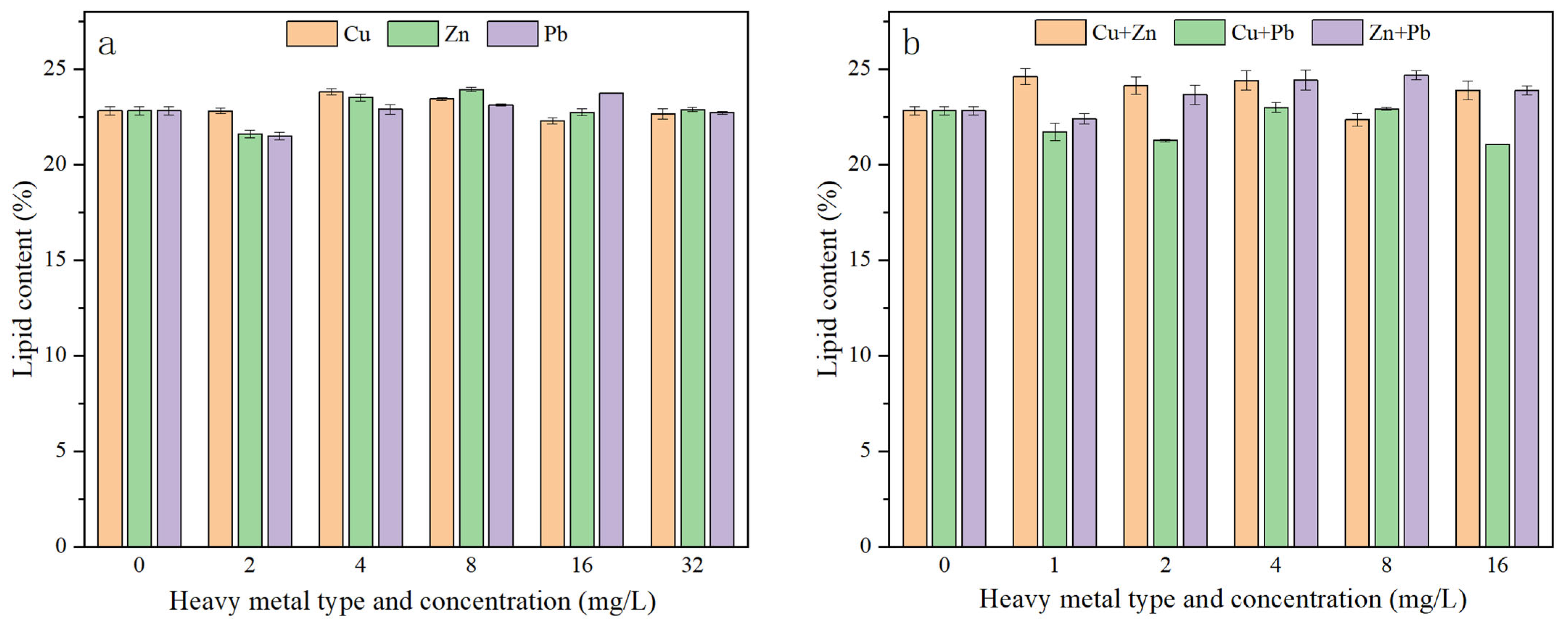
Under short-term (3 h) cultivation, none of the heavy metal treatments significantly affected microalgal lipid metabolism (Figure 3). Lipid productivity ranged from 22.80% to 24.39% for Cu2+, 22.22% to 24.49% for Zn2+, and 22.03% to 23.81% for Pb2+ in single-metal systems, with no significant changes observed across the tested concentrations. Thus, short-term heavy metal stress exerted no statistically significant effect on microalgal lipid productivity, suggesting that initial metal exposure predominantly triggers surface adsorption rather than intracellular metabolic reprogramming, leaving carbon allocation pathways momentarily unaltered.

3.2.2. Effects of Heavy Metals on Lipid Productivity After 3 Days of Cultivation

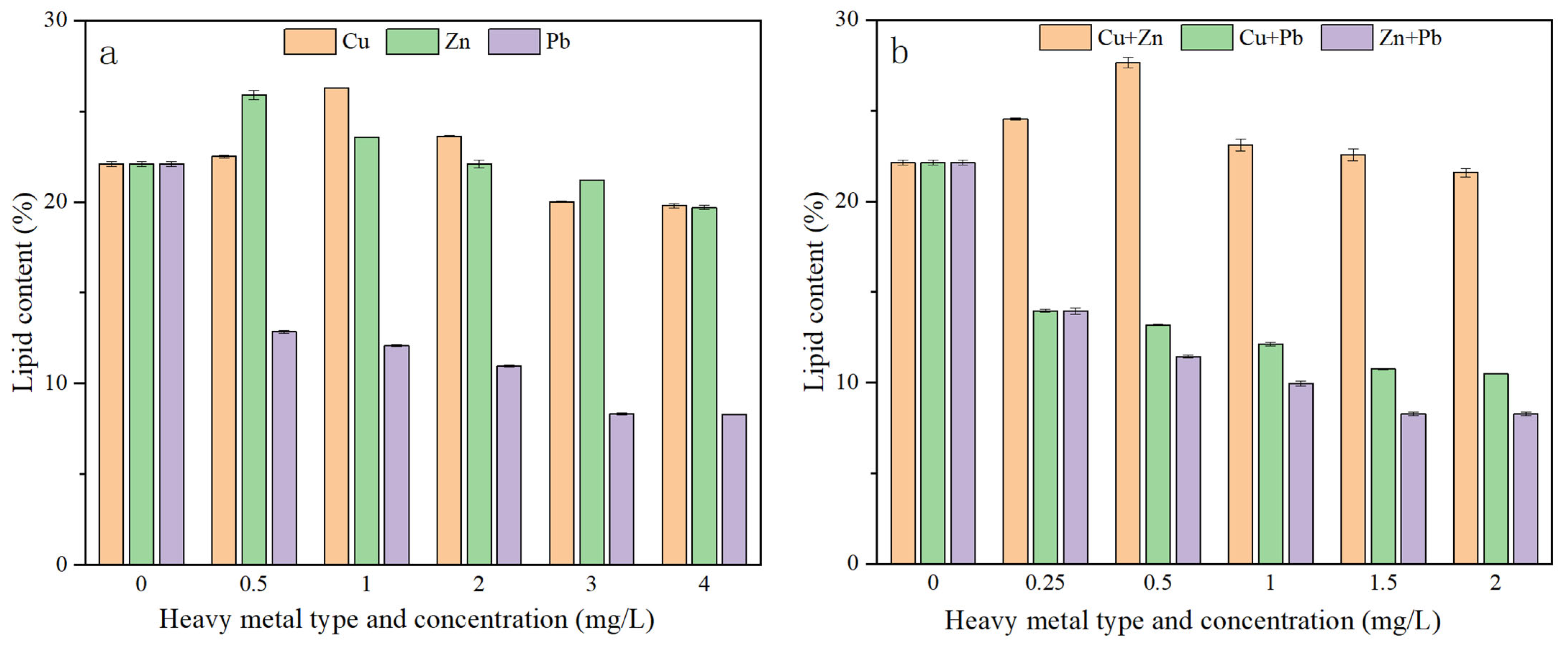
In contrast to short-term exposure, prolonged (3 days) heavy metal stress significantly influenced microalgal lipid productivity (Figure 4), primarily through alterations in growth cycles, metabolic pathways, and stress response mechanisms [13]. As shown in Figure 4a, for Cu2+, lipid productivity was 22.51% at 0.5 mg/L, peaked at 26.29% at 1 mg/L, and gradually decreased to 19.81% at concentrations ≥ 2 mg/L, exhibiting a hormetic response characterized by low-concentration promotion and high-concentration inhibition; this biphasic pattern highlights an initial stress-adaptive lipid biosynthesis followed by metabolic collapse under severe toxicity. For Zn2+, the highest lipid productivity (25.90%) was observed at 0.5 mg/L, followed by a gradual decline with increasing concentration; however, productivity remained at 19.71% even at 4 mg/L; for Pb2+, lipid productivity was only 12.85% at 0.5 mg/L, dropped below 10% at concentrations ≥ 2 mg/L, and fell to 8.29% at 4 mg/L, indicating substantial inhibition. At equivalent concentrations (0.5 mg/L), lipid productivity followed the order Zn2+ (25.90%) > Cu2+ (22.51%) > Pb2+ (12.85%). Within the concentration range of 1–4 mg/L, no significant difference in lipid productivity was observed between Cu2+ and Zn2+ (ranging from 20% to 23.62%), whereas both were significantly higher than that of Pb2+ (≤10.96%).
Prolonged exposure to moderate Cu2+ and Zn2+ levels enhanced lipid accumulation in C. vulgaris, with optimal concentrations of 1 mg/L and 0.5 mg/L, respectively. In contrast, Pb2+ exerted a strong inhibitory effect, likely due to its disruption of cellular structure and interference with metabolic processes. This suggests that during the accumulation of Cu2+ and Zn2+, microalgae activate stress response mechanisms that promote lipid synthesis as an energy-dense cellular sink to mitigate metal-induced oxidative pressure [13]. In contrast, the markedly reduced removal efficiency of Pb2+ aligns with its toxic inhibition of microalgal cells, directly contributing to the decline in lipid productivity under Pb2+ stress.
After three days of exposure, lipid productivity differed markedly among the mixed heavy metal combinations (Figure 4b). The Cu2++Zn2+ combination exhibited the highest lipid productivity, ranging from 20% to 28% and peaking at 27.64% at a total concentration of 0.5 mg/L. In contrast, lipid productivity for the Cu2++Pb2+ and Zn2++Pb2+ combinations remained relatively low, ranging between 10% and 15% with little concentration-dependent variation.
The higher lipid productivity observed under combined Cu2++Zn2+ treatment, relative to single-metal treatments, suggests a synergistic effect of the two metals. At low concentrations, both metals exhibited relatively low toxicity to microalgae and induced lipid accumulation through oxidative stress mechanisms [30]. Compared with the single-metal systems (26.29% for 1 mg/L Cu2+ and 25.90% for 0.5 mg/L Zn2+), the 0.5 mg/L Cu2++Zn2+ mixed group achieved a higher productivity of 27.64%, indicating that this combination represents an optimal condition for maximizing lipid accumulation potential. The low productivities observed in the Cu2++Pb2+ and Zn2++Pb2+ combinations are primarily attributable to the inhibitory effects of Pb2+. The disruptive impact of Pb2+ on cell membrane structure and energy metabolism counteracts the promoting effects of Cu2+ or Zn2+, resulting in lipid productivity that remains constrained by Pb2+ toxicity due to irreversible chloroplast damage and lipogenic enzyme suppression [30,36].
The Cu2++Zn2+ combination achieved optimal productivity at 0.5 mg/L. Although productivity decreased slightly at higher concentrations, it remained above 20%, demonstrating that the combined effect exceeded that of each metal alone. In contrast, combinations containing Pb2+ exhibited consistently low productivity across all concentrations, further confirming the strong inhibitory effect of Pb2+ on lipid accumulation in C. vulgaris.

3.3. Mechanisms of Lipid Accumulation in Microalgae

3.3.1. Changes in Biomass, Lipid, Protein, and Carbohydrate Content of C. vulgaris Under Heavy Metal Stress

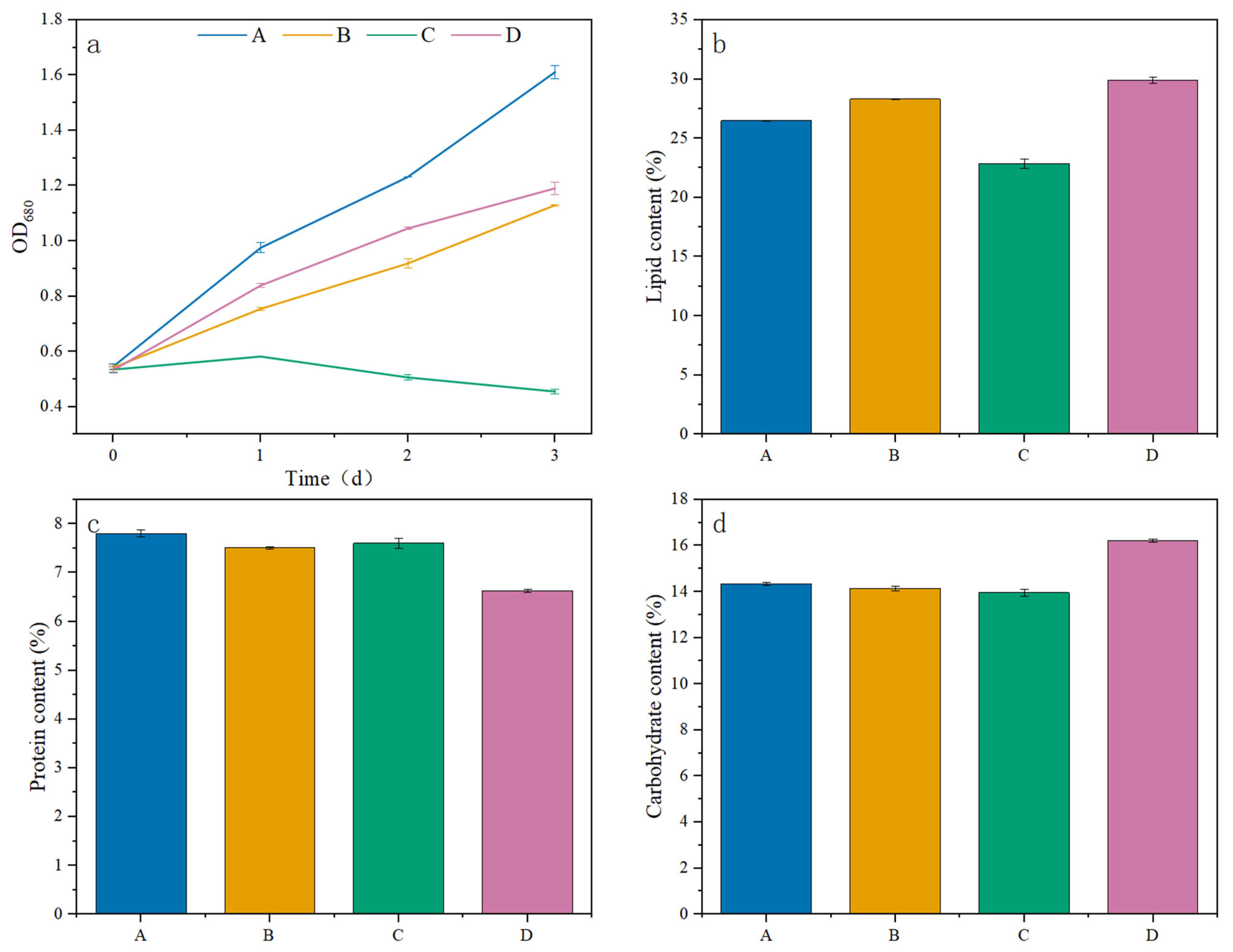
Group A (control) exhibited the highest growth, with rapid biomass accumulation, as the microalgae maintained normal metabolic activity in the absence of heavy metal stress. Throughout the three-day cultivation period, OD680 values increased continuously and stably from 0.5 to 1.6; Groups B and D exhibited similar continuous increases in biomass, though growth rates were significantly slower than that of the control (Figure 5a). Throughout the experiment, biomass in Group D remained consistently slightly higher than in Group B. These findings indicate that microalgal growth was less inhibited by the optimal mixed-metal concentration (0.5 mg/L Cu2++Zn2+) than by the optimal single-metal concentration (1 mg/L Cu2+). Thus, while both Cu2+ and Cu2++Zn2+ treatments moderately suppressed growth, they did not induce substantial cell death; the microalgae maintained metabolic activity and continued to proliferate over the three-day period. Group C was the only treatment in which biomass declined. Following the addition of heavy metals, OD680 increased only slightly on the first day, after which it continuously decreased, reaching approximately 0.45 by day 3—substantially below the initial value. This trend directly demonstrates the marked toxicity of Pb2+ to microalgae, which not only inhibited growth but also induced significant cell death.
Heavy metal treatments differentially regulated carbon allocation among lipids, proteins, and carbohydrates, reflecting the adaptive strategies of microalgae in response to stress (Figure 5a–c). Carbon partitioning toward lipids was selectively promoted under Cu2+-based stress but suppressed under Pb2+ stress, indicating distinct modulation of the lipid synthesis pathway by different metals. In Groups B and D, greater carbon flux was directed toward lipid synthesis, resulting in lipid contents that were 7% and 13% higher than that of the control, respectively. In contrast, single Pb2+ stress in Group C significantly reduced microalgal biomass, with a substantial decrease in intracellular lipid content—approximately 14% lower than the control. Compared with the control, protein content was reduced in all three experimental groups, with Group D having the lowest content (15.38% reduction compared with the control).
C. vulgaris responded to heavy metal stress by regulating carbon allocation: under Cu2+-based stress, carbon was preferentially channeled toward lipid synthesis; under combined Cu2++Zn2+ stress, carbohydrate content was also elevated; and reduced carbon allocation to proteins served as a common adaptive strategy across all heavy metal treatments. By downregulating energy-intensive protein synthesis, microalgae successfully conserved cellular energy to fuel structural lipid assembly and maintain stress-defense cascades under moderate metal pressure.

3.3.2. Fatty Acid Profile Analysis

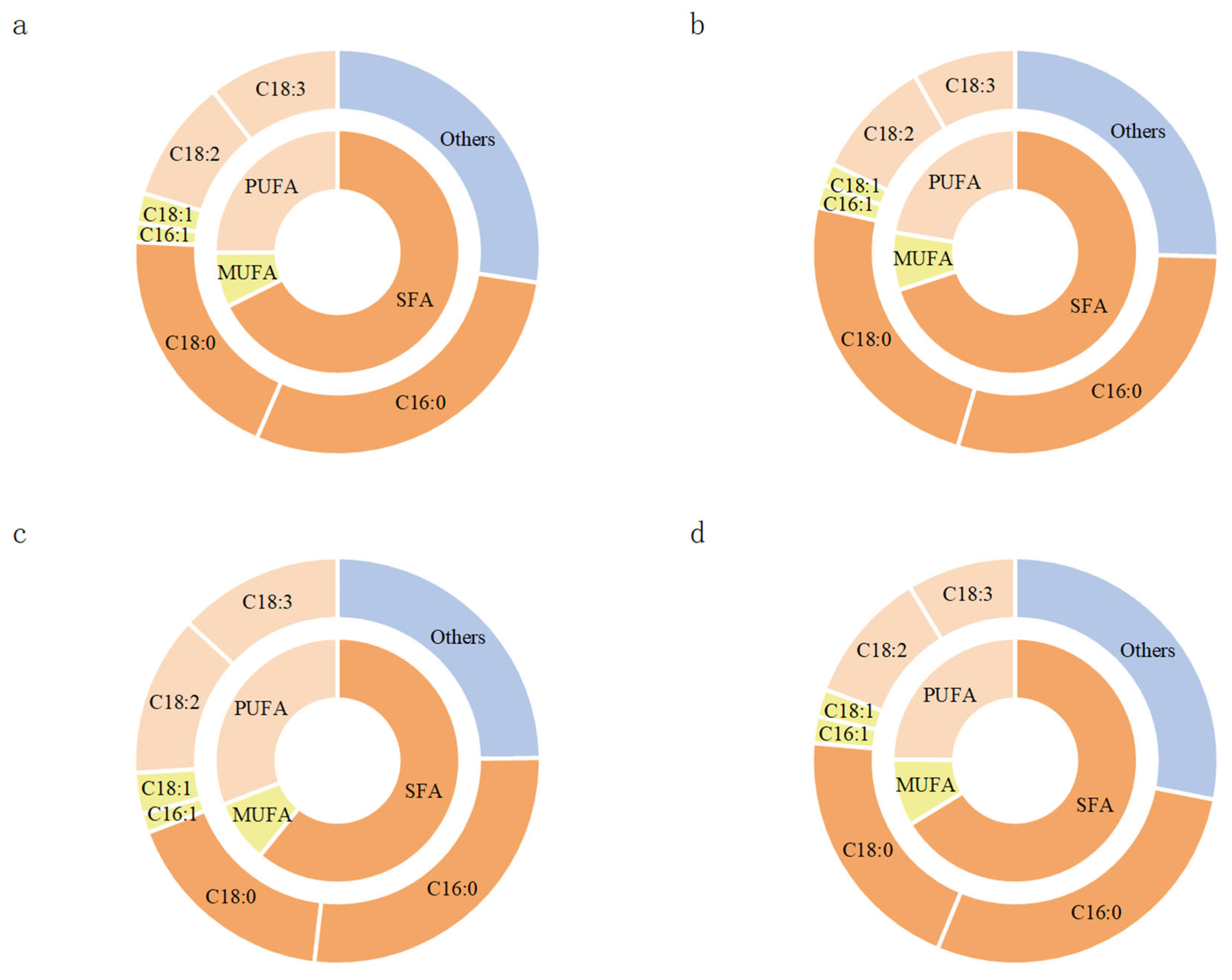
Analysis of the fatty acid profiles enables a more precise assessment of how different heavy metal stresses affect the suitability of microalgal lipids for biodiesel production. The proportion of saturated fatty acids (SFAs) is a critical indicator of fuel oxidative stability [12]. Fatty acid profiles, particularly the C16–C18 composition, are widely recognized as key determinants of biodiesel quality [37]. Fatty acid profiles of different groups are shown in Figure 6. Among the four groups, Group B exhibited the highest SFAs proportion (69.95%), attributable to elevated levels of C16:0 (29%) and C18:0 (24%). These medium- and long-chain SFAs enhance oxidative stability, a critical property for biodiesel shelf life. In addition, lipid productivity under Cu2+ stress exceeded that of the control, indicating that this treatment not only increased lipid yield but also optimized fatty acid composition, making it suitable for biodiesel production. The specific enrichment of SFAs under Cu2+ exposure points to an adaptive remodeling of the cellular membrane, minimizing the availability of vulnerable double bonds to limit heavy metal-induced lipid peroxidation.
Groups A and D exhibited SFAs proportions of 67.65% and 66.28%, respectively, reflecting a balanced composition. While providing oxidative stability, the moderate presence of unsaturated fatty acids (e.g., C18:2 cis and C18:3n3) also contributed to improved low-temperature fluidity. Notably, Group D maintained a stable fatty acid profile while achieving the highest lipid content under combined stress, highlighting its strong potential for industrial application.
Group C showed the lowest SFAs proportion (60.88%), which compromises oxidative stability. Furthermore, the markedly elevated proportion of polyunsaturated fatty acids reflected a severe stress state in the cells, resulting in both the lowest lipid productivity and a fatty acid composition unsuitable for biodiesel production.
The fatty acid compositions of Groups B and D were better aligned with biodiesel quality requirements, whereas Group C exhibited the poorest oil quality, consistent with the trends observed in biomass and lipid content from previous analyses. In Group B, Cu2+—as a relatively mild stress agent—not only enhanced lipid productivity but also promoted the accumulation of medium- and long-chain saturated fatty acids (e.g., C16:0 and C18:0) by modulating fatty acid synthesis pathways, thereby optimizing biodiesel oxidative stability. In Group D, combined stress maintained relatively high lipid productivity while avoiding the pronounced saturation bias observed under single Cu2+ stress, achieving a better balance between oxidative stability and low-temperature fluidity. With slight advantages in biomass and lipid productivity, Group D demonstrated greater flexibility for industrial application. In contrast, in Group C, the strong toxicity of Pb2+ not only caused substantial reductions in biomass and lipid content but also significantly decreased the saturated fatty acid proportion. These findings validate the advantages of Cu2+-based stress for biodiesel production while revealing the negative impact of Pb2+ toxicity on both microalgal lipid yield and quality.

3.3.3. Antioxidant Capacity of Microalgae and Lipid Synthesis Enzyme Activities in Microalgae

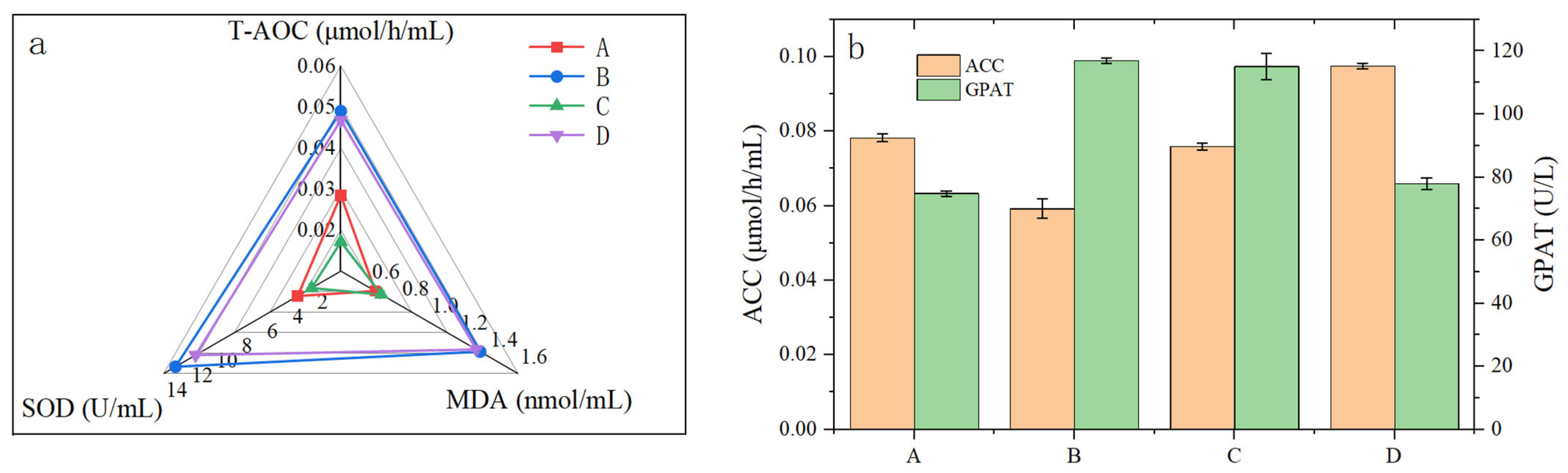
Antioxidant indices of each algal group are shown in Figure 6. SOD activity was significantly increased in Groups B and D, indicating that Cu2+ and Cu2++Zn2+ stress effectively induced SOD synthesis and activated antioxidant defense mechanisms to neutralize excessive superoxide radicals generated within the chloroplasts and mitochondria. In contrast, under Pb2+ treatment, microalgal growth was arrested and the antioxidant system was compromised, preventing cells from counteracting the oxidative damage caused by lead toxicity. MDA levels in Groups B and D were relatively high, followed by Group C, with all three exceeding those of the control group; in Group C, all antioxidant indicators were lower than those of the control. Consistent with the biomass data, this suggests that the high toxicity of Pb2+ induced oxidative damage and led to the inhibition of enzyme activities, including those of antioxidant enzymes [38]. Specifically, both SOD activity and T-AOC were reduced, whereas MDA content showed a slight increase, indicating that microalgae under Pb2+ stress experienced oxidative damage that they were unable to counteract. In Groups B and D, both antioxidant levels and the extent of oxidative damage were significantly elevated relative to the control. The marked increases in SOD activity and T-AOC indicate that under Cu2+ and Cu2++Zn2+ stress, the microalgae mounted an effective defense by upregulating antioxidant enzyme synthesis and accumulating antioxidants to scavenge excess ROS. At the same time, the relatively high MDA levels suggest that despite activation of the antioxidant system, the rate of ROS generation exceeded the scavenging capacity, resulting in sustained lipid peroxidation and a measurable degree of oxidative damage.
Regarding key lipogenic enzymes, compared with the control group (0.07823 μmol/h/mL), ACC activity decreased by approximately 24.3% under 1 mg/L Cu2+ treatment (0.05919 μmol/h/mL), and by approximately 3.0% under 3 mg/L Pb2+ treatment (0.07588 μmol/h/mL). In contrast, under combined 0.5 mg/L Cu2++Zn2+ stress, ACC activity increased by approximately 24.7% relative to the control, reaching 0.09752 μmol/h/mL. For GPAT activity, compared with the control (74.79 U/L), 1 mg/L Cu2+ treatment increased activity to 116.90 U/L (a rise of approximately 56.3%), and 3 mg/L Pb2+ treatment resulted in an activity of 111.76 U/L (an increase of approximately 49.4%). In contrast, the combined 0.5 mg/L Cu2++Zn2+ treatment had no significant effect, yielding a GPAT activity of 77.91 U/L, an increase of only approximately 4.2%.
Overall, both single Cu2+ and Pb2+ treatments significantly enhanced GPAT activity, yet exerted distinct effects on ACC activity: Cu2+ markedly inhibited ACC, whereas Pb2+ caused only a slight reduction. Under the low-concentration Cu2++Zn2+ combined treatment, ACC activity increased substantially, while GPAT activity remained close to the control level. The response patterns of the two enzymes under combined stress thus differed distinctly from those observed under single-metal stress.
When linking these enzyme activities to the final lipid outcomes (as shown in Figure 7b), the following trends emerge: Despite the decrease in ACC activity under Cu2+ treatment, lipid content increased. This can be explained by the combination of enhanced GPAT activity (promoting TAG assembly) and a redirection of carbon flux toward lipid synthesis. In contrast, although Pb2+ treatment also elevated GPAT activity, the final lipid content did not increase. This is likely due to Pb2+-induced stronger oxidative stress, which impaired carbon fixation capacity and led to insufficient fatty acid substrate supply, while cells allocated substantial energy to antioxidant defense rather than lipid accumulation. Under the Cu2++Zn2+ combined treatment, the substantial increase in ACC activity, coupled with near-control GPAT activity, resulted in moderately enhanced lipid production, suggesting that ACC plays a dominant role under this condition.

3.3.4. Mechanisms of Lipid Synthesis in Microalgae

Based on the evaluated biochemical indicators, oxidative stress appears to be closely associated with the distinct lipid accumulation patterns observed across different metal treatments. Under Cu2+ and combined Cu2++Zn2+ stress (Groups B and D), the elevated lipid content and optimized fatty acid profiles coincided with the upregulation of antioxidant defenses, particularly increased SOD activity. This concurrent physiological shift suggests a possible redirection of intracellular carbon flux toward lipid biosynthesis, potentially serving as an adaptive defense strategy to mitigate oxidative damage. The distinct responses of key lipogenic enzymes—where Cu2+ exposure was linked to enhanced GPAT activity alongside ACC inhibition, whereas the addition of Zn2+ stimulated ACC without significantly elevating GPAT—imply that these metals may differentially modulate carbon partitioning pathways. This complementary enzymatic response likely contributed to the combined Cu2++Zn2+ treatment’s ability to maintain relatively high lipid productivity while preserving a favorable fatty acid composition suitable for biodiesel.
Conversely, the profound cellular damage caused by Pb2+ stress (Group C) indicates a fundamentally different physiological outcome. The substantial reductions in biomass and lipid yield, coupled with a poorly optimized fatty acid profile, aligned with a compromised antioxidant system that failed to counteract severe oxidative damage. Rather than reallocating carbon for energy storage, the high toxicity of Pb2+ appears to have overwhelmingly disrupted cellular homeostasis and physiological activity, thereby suppressing lipid accumulation.
The observed correlations among modulated lipogenic enzyme activities, carbon allocation patterns, and antioxidant defenses strongly suggest that microalgae exhibit flexible, metal-specific adaptations. Mild-to-moderate Cu2+-based stress appears to stimulate a biochemical phenotype favoring lipid accumulation, whereas highly toxic Pb2+ induces unrecoverable cellular disruption. Future studies employing advanced molecular and multi-omics approaches are essential to rigorously validate these biochemically derived hypotheses.

4. Conclusions and Prospects

This study investigated the removal efficiencies and lipid productivities of C. vulgaris exposed to various concentrations of single and mixed Cu2+, Zn2+, and Pb2+, along with the mechanisms underlying heavy metal-induced effects on microalgal lipid accumulation. Across all tested concentrations, removal efficiencies in the Pb2+ groups were higher than those in the Cu2+ and Zn2+ groups. In mixed treatments, Pb2+ influenced the removal efficiencies of Cu2+ and Zn2+, whereas its own removal efficiency remained largely unaffected. Cu2+ enhanced microalgal lipid productivity, and the Cu2++Zn2+ combination yielded even higher lipid productivity than Cu2+ alone. In contrast, Pb2+ significantly reduced both microalgal biomass and lipid productivity across all concentrations and combinations tested.
Under Cu2+ and Cu2++Zn2+ treatments, greater carbon allocation toward lipid synthesis was observed, along with sufficient antioxidant capacity to withstand oxidative damage over the three-day period. This enabled the enhancement of lipogenic enzyme activity under moderate ROS pressure, thereby promoting lipid accumulation as a defensive response to oxidative stress. In contrast, even low concentrations of Pb2+ caused severe cellular damage, leading to substantial cell death. Thus, the application of microalgae for treating wastewater containing Cu2+ and Zn2+—while avoiding highly toxic heavy metals such as Pb2+—holds promising potential for achieving both effective heavy metal removal and subsequent microalgal lipid recovery.
This study has several limitations that warrant further investigation, including: the mixed heavy metal experiments were not designed as a full factorial design, which precludes precise evaluation of the interactions between metals; pH dynamics and photosynthetic pigments were not monitored in real time, resulting in insufficient environmental and physiological data; experimental methods were not used to distinguish between passive adsorption and active uptake of heavy metals, was microalgal cell integrity directly verified. To address these limitations, future studies should: (1) adopt a full factorial design to systematically investigate the synergistic or antagonistic effects among multiple heavy metals, thereby accurately identifying the optimal combination and ratio for practical applications; (2) closely monitor dynamic changes in environmental (pH) and microalgal physiological (dry weight, photosynthetic pigments, etc.) parameters; (3) quantify the proportions of heavy metals removed by adsorption versus absorption; and (4) employ methods such as FDA/PI staining or flow cytometry to assess cell membrane integrity and to clarify the respective contributions of living and damaged cells to the removal process.

Supplementary Materials

The following supporting information can be downloaded at https://www.mdpi.com/article/10.3390/w18111306/s1. Figure S1: Heavy metal removal performance of Chlorella vulgaris over 3-day treatment. (a–c) Residual concentration changes of Cu2+, Zn2+, and Pb2+ in single-metal systems with initial concentrations of 0.5, 1, 2, 3, and 4 mg/L, respectively; (d–f) Removal efficiency changes in mixed-metal systems (Cu2++Zn2+, Cu2++Pb2+, and Zn2++Pb2+, respectively) with initial concentrations of 0.25, 0.5, 1, 1.5, and 2 mg/L.; Table S1: Fatty Acid Profile (relative percentage, %).

Author Contributions

B.B.: Methodology, Experimental design, Validation, Formal analysis, Writing—original draft. Q.W.: Conceptualization, Resources, Writing—review & editing. X.M.: Conceptualization, Methodology, Experimental design, Supervision, Writing—original draft, Writing—review & editing. All authors have read and agreed to the published version of the manuscript.

Funding

This research was funded by the National Natural Science Foundation of China grant number [No. 52360008].

Data Availability Statement

The original contributions presented in this study are included in the article/Supplementary Materials. Further inquiries can be directed to the corresponding author.

Acknowledgments

During the preparation of this work the authors used CHATGPT (https://chatgpt.com) in order to polish the language. After using this tool/service, the authors reviewed and edited the content as needed and take full responsibility for the content of the publication.

Conflicts of Interest

The authors declare that they have no known competing financial interests or personal relationships that could have appeared to influence the work reported in this paper.

References

  1. Zhao, K.; Zhao, X.; Gao, T.; Li, X.; Wang, G.; Pan, X.; Wang, J. Dielectrophoresis-assisted removal of Cd and Cu heavy metal ions by using Chlorella microalgae. Environ. Pollut. 2023, 334, 122110. [Google Scholar] [CrossRef]
  2. Qin, Y.; Tao, Y. Pollution status of heavy metals and metalloids in Chinese lakes: Distribution, bioaccumulation and risk assessment. Ecotoxicol. Environ. Saf. 2022, 248, 114293. [Google Scholar] [CrossRef]
  3. Leong, Y.K.; Chang, J.S. Bioremediation of heavy metals using microalgae: Recent advances and mechanisms. Bioresour. Technol. 2020, 303, 122886. [Google Scholar] [CrossRef]
  4. Daneshvar, E.; Zarrinmehr, M.J.; Kousha, M.; Hashtjin, A.M.; Saratale, G.D.; Maiti, A.; Vithanage, M.; Bhatnagar, A. Hexavalent chromium removal from water by microalgal-based materials: Adsorption, desorption and recovery studies. Bioresour. Technol. 2019, 293, 122064. [Google Scholar] [CrossRef] [PubMed]
  5. Fernández, P.M.; Viñarta, S.C.; Bernal, A.R.; Cruz, E.L.; Figueroa, L.I.C. Bioremediation strategies for chromium removal: Current research, scale-up approach and future perspectives. Chemosphere 2018, 208, 139–148. [Google Scholar] [CrossRef] [PubMed]
  6. Goswami, R.K.; Agrawal, K.; Shah, M.P.; Verma, P. Bioremediation of heavy metals from wastewater: A current perspective on microalgae-based future. Lett. Appl. Microbiol. 2022, 75, 701–717. [Google Scholar] [CrossRef] [PubMed]
  7. Priya, A.K.; Jalil, A.A.; Vadivel, S.; Dutta, K.; Rajendran, S.; Fujii, M.; Soto-Moscoso, M. Heavy metal remediation from wastewater using microalgae: Recent advances and future trends. Chemosphere 2022, 305, 135375. [Google Scholar] [CrossRef]
  8. Gu, S.; Lan, C.Q. Biosorption of heavy metal ions by green alga Neochloris oleoabundans: Effects of metal ion properties and cell wall structure. J. Hazard. Mater. 2021, 418, 126336. [Google Scholar] [CrossRef]
  9. Zhou, T.; Wang, J.; Zheng, H.; Wu, X.; Wang, Y.; Liu, M.; Xiang, S.; Cao, L.; Ruan, R.; Liu, Y. Characterization of additional zinc ions on the growth, biochemical composition and photosynthetic performance from Spirulina platensis. Bioresour. Technol. 2018, 269, 285–291. [Google Scholar] [CrossRef]
  10. Xiao, X.; Li, W.; Jin, M.; Zhang, L.; Qin, L.; Geng, W. Responses and tolerance mechanisms of microalgae to heavy metal stress: A review. Mar. Environ. Res. 2023, 183, 105805. [Google Scholar] [CrossRef]
  11. Dammak, M.; Ben Hlima, H.; Tounsi, L.; Michaud, P.; Fendri, I.; Abdelkafi, S. Effect of heavy metals mixture on the growth and physiology of Tetraselmis sp.: Applications to lipid production and bioremediation. Bioresour. Technol. 2022, 360, 127584. [Google Scholar] [CrossRef]
  12. Tan, S.; Wen, F.; Liu, D.; Lu, H.; Li, L.; Zhu, L. Physiological responses and lipid accumulation of freshwater microalgae Chlorella sorokiniana under short-term zinc stress in water solution. Algal Res. 2024, 80, 103528. [Google Scholar] [CrossRef]
  13. Song, X.; Liu, B.F.; Kong, F.; Song, Q.; Ren, N.Q.; Ren, H.Y. Simultaneous chromium removal and lipid accumulation by microalgae under acidic and low temperature conditions for promising biodiesel production. Bioresour. Technol. 2023, 370, 128515. [Google Scholar] [CrossRef]
  14. Kim, J.Y.; Jung, J.M.; Jung, S.; Park, Y.K.; Tsang, Y.F.; Lin, K.Y.A.; Choi, Y.E.; Kwon, E.E. Biodiesel from microalgae: Recent progress and key challenges. Prog. Energy Combust. Sci. 2022, 93, 101020. [Google Scholar] [CrossRef]
  15. Kafil, M.; Berninger, F.; Koutra, E.; Kornaros, M. Utilization of the microalga Scenedesmus quadricauda for hexavalent Chromium bioremediation and biodiesel production. Bioresour. Technol. 2022, 346, 126665. [Google Scholar] [CrossRef] [PubMed]
  16. Zhao, Y.; Song, X.; Zhong, D.-B.; Yu, L.; Yu, X. γ-Aminobutyric acid (GABA) regulates lipid production and cadmium uptake by Monoraphidium sp. QLY-1 under Cadmium stress. Bioresour. Technol. 2020, 297, 1225007. [Google Scholar] [CrossRef]
  17. Davis, A.P.; Shokouhian, M.; Sharma, H.; Minami, C.; Winogradoff, D. Water quality improvement through bioretention: Lead, copper, and zinc removal. Water Environ. Res. 2003, 75, 73–82. [Google Scholar] [CrossRef]
  18. Razzak, S.A.; Faruque, M.O.; Alsheikh, Z.; Alsheikhmohamad, L.; Alkuroud, D.; Alfayez, A.; Hossain, S.M.Z.; Hossain, M.M. A comprehensive review on conventional and biological-driven heavy metals removal from industrial wastewater. Environ. Adv. 2022, 7, 100168. [Google Scholar] [CrossRef]
  19. Chen, H.; Wang, Q. Regulatory mechanisms of lipid biosynthesis in microalgae. Biol. Rev. 2021, 96, 2373–2391. [Google Scholar] [CrossRef]
  20. Mahlangu, D.; Mphahlele, K.; De Paola, F.; Mthombeni, N.H. Microalgae-mediated biosorption for effective heavy metals removal from wastewater: A review. Water 2024, 16, 718. [Google Scholar] [CrossRef]
  21. Tripathi, S.; Poluri, K.M. Heavy metal detoxification mechanisms by microalgae: Insights from transcriptomics analysis. Environ. Pollut. 2021, 285, 117443. [Google Scholar] [CrossRef]
  22. Ummalyma, S.B.; Singh, A. Biomass production and phycoremediation of microalgae cultivated in polluted river water. Bioresour. Technol. 2022, 351, 126948. [Google Scholar] [CrossRef] [PubMed]
  23. Bligh, E.G.; Dyer, W.J. A rapid method of total lipid extraction and purification. Can. J. Biochem. Physiol. 1959, 37, 911–917. [Google Scholar] [CrossRef]
  24. Yasin, N.H.M.; Aziz, N.N.C.; Azmai, M.B.A.; Hanapi, M.F.M. Transesterification method of microalgae biomass to produce fatty acid methyl esters. J. Chem. Technol. Biotechnol. 2023, 98, 2774–2783. [Google Scholar] [CrossRef]
  25. Barbarino, E.; Lourenço, S.O. An evaluation of methods for extraction and quantification of protein from marine macro- and microalgae. J. Appl. Phycol. 2005, 17, 447–460. [Google Scholar] [CrossRef]
  26. DuBois, M.; Gilles, K.A.; Hamilton, J.K.; Rebers, P.A.; Smith, F. Colorimetric method for determination of sugars and related substances. Anal. Chem. 1956, 28, 350–356. [Google Scholar] [CrossRef]
  27. Gu, S.; Lan, C.Q. Effects of culture pH on cell surface properties and biosorption of Pb(II), Cd(II), Zn(II) of green alga Neochloris oleoabundans. Chem. Eng. J. 2023, 468, 143579. [Google Scholar] [CrossRef]
  28. Wang, J.; Chen, R.; Fan, L.; Cui, L.; Zhang, Y.; Cheng, J.; Wu, X.; Zeng, W.; Tian, Q.; Shen, L. Construction of fungi-microalgae symbiotic system and adsorption study of heavy metal ions. Sep. Purif. Technol. 2021, 268, 118689. [Google Scholar] [CrossRef]
  29. Inthorn, D.; Sidtitoon, N.; Silapanuntakul, S.; Incharoensakdi, A. Sorption of mercury, cadmium and lead by microalgae. ScienceAsia 2002, 28, 253–261. [Google Scholar] [CrossRef]
  30. Cavalletti, E.; Romano, G.; Palma Esposito, F.; Barra, L.; Chiaiese, P.; Balzano, S.; Sardo, A. Copper effect on microalgae: Toxicity and bioremediation strategies. Toxics 2022, 10, 527. [Google Scholar] [CrossRef]
  31. Teng, Y.; Wu, Q.; Li, S.; Guan, X.; Zhang, Z.; He, J.; Liao, Y.; Zhang, J.; Zhu, L. Microalgae-fungal consortia immobilized Zn(II) by enhancing secretion of proteins in extracellular polymeric substances: A protective mechanism against excessive zinc uptake. Algal Res. 2026, 93, 104436. [Google Scholar] [CrossRef]
  32. Chan, A.; Salsali, H.; McBean, E. Heavy metal removal (copper and zinc) in secondary effluent from wastewater treatment plants by microalgae. ACS Sustain. Chem. Eng. 2014, 2, 130–137. [Google Scholar] [CrossRef]
  33. Liu, L.; Lin, X.; Luo, L.; Yang, J.; Luo, J.; Liao, X.; Cheng, H. Biosorption of Copper ions through microalgae from piggery digestate: Optimization, kinetic, isotherm and mechanism. J. Clean. Prod. 2021, 319, 128724. [Google Scholar] [CrossRef]
  34. Luo, Y.; Li, X.; Lin, Y.; Wu, S.; Cheng, J.J.; Yang, C. Stress of cupric ion and oxytetracycline in Chlorella vulgaris cultured in swine wastewater. Sci. Total Environ. 2023, 895, 165120. [Google Scholar] [CrossRef]
  35. Cheng, S.Y.; Show, P.L.; Lau, B.F.; Chang, J.S.; Ling, T.C. New prospects for modified algae in heavy metal adsorption. Trends Biotechnol. 2019, 37, 1255–1268. [Google Scholar] [CrossRef] [PubMed]
  36. Hee, C.W.; Shing, W.L.; Chi, C.K. Effect of Lead (Pb) exposure towards green microalgae (Chlorella vulgaris) on the changes of physicochemical parameters in water. S. Afr. J. Chem. Eng. 2021, 37, 252–255. [Google Scholar] [CrossRef]
  37. Muhammad, G.; Potchamyou Ngatcha, A.D.; Lv, Y.; Xiong, W.; El-Badry, Y.A.; Asmatulu, E.; Xu, J.; Alam, M.A. Enhanced biodiesel production from wet microalgae biomass optimized via response surface methodology and artificial neural network. Renew. Energy 2022, 184, 753–764. [Google Scholar] [CrossRef]
  38. Gao, M.; Ling, N.; Tian, H.; Guo, C.; Wang, Q. Toxicity, physiological response, and biosorption mechanism of Dunaliella Salina to copper, lead, and cadmium. Front. Microbiol. 2024, 15, 1374275. [Google Scholar] [CrossRef] [PubMed]
Figure 1. Removal efficiency of heavy metals by C. vulgaris after 3 h of exposure: (a) Single-metal systems (Cu2+, Zn2+, Pb2+) with initial concentrations of 2, 4, 8, 16, and 32 mg/L. (b) Mixed-metal systems (Cu2++Zn2+, Cu2++Pb2+, Zn2++Pb2+) with initial concentrations of 1, 2, 4, 8, and 16 mg/L. Values are means of triplicate measurements with standard error bars (n = 3).
Water 18 01306 g001
Figure 2. Removal efficiency of heavy metals by C. vulgaris after 3 days of exposure. (a) Single-metal systems (Cu2+, Zn2+, Pb2+) with initial concentrations of 0.5, 1, 2, 3, and 4 mg/L. (b) Mixed-metal systems (Cu2++Zn2+, Cu2++Pb2+, Zn2++Pb2+) with initial concentrations of 0.25, 0.5, 1, 1.5, and 2 mg/L. Values are means of triplicate measurements with standard error bars (n = 3).
Water 18 01306 g002
Figure 3. Lipid productivity of C. vulgaris after 3 h in single and mixed heavy metal systems. (a) Single-metal systems (Cu2+, Zn2+, Pb2+) at initial concentrations of 0, 2, 4, 8, 16, and 32 mg/L; (b) mixed-metal systems (Cu2++Zn2+, Cu2++Pb2+, Zn2++Pb2+) at initial concentrations of 0, 1, 2, 4, 8, and 16 mg/L. Values are means of triplicate measurements with standard error bars (n = 3).
Water 18 01306 g003
Figure 4. Lipid productivity of C. vulgaris after 3 days of cultivation in single and mixed heavy metal systems. (a) single-metal systems (Cu2+, Zn2+, Pb2+) at initial concentrations of 0, 0.5, 1, 2, 3, and 4 mg/L. (b) mixed heavy metal systems (Cu2++Zn2+, Cu2++Pb2+, and Zn2++Pb2+) at concentrations of 0, 0.25, 0.5, 1, 1.5, and 2 mg/L. Values are means of triplicate measurements with standard error bars (n = 3).
Water 18 01306 g004
Figure 5. Changes in biomass (a), lipid content (b), protein content (c) and carbohydrate content (d) of C. vulgaris in different treatment groups (A: control; B: 1 mg/L Cu2+; C: 3 mg/L Pb2+; D: 0.5 mg/L Cu2++Zn2+) over 3 days of cultivation. Values are means of triplicate measurements with standard error bars (n = 3).
Water 18 01306 g005
Figure 6. Fatty acid profiles of C. vulgaris from different treatment groups ((a): control; (b): 1 mg/L Cu2+; (c): 3 mg/L Pb2+; (d): 0.5 mg/L Cu2++Zn2+) after 3 days of cultivation.
Water 18 01306 g006
Figure 7. (a) Antioxidant capacity indicators (SOD activity, T-AOC, and MDA content) of C. vulgaris from different treatment groups (A: control; B: 1 mg/L Cu2+; C: 3 mg/L Pb2+; D: 0.5 mg/L Cu2++Zn2+) after 3 days of cultivation. (b) Activities of key lipogenic enzymes (ACC and GPAT) in C. vulgaris from different treatment groups (A: control; B: 1 mg/L Cu2+; C: 3 mg/L Pb2+; D: 0.5 mg/L Cu2++Zn2+) after 3 days of cultivation. Values are means of triplicate measurements with standard error bars (n = 3).
Water 18 01306 g007
Disclaimer/Publisher’s Note: The statements, opinions and data contained in all publications are solely those of the individual author(s) and contributor(s) and not of MDPI and/or the editor(s). MDPI and/or the editor(s) disclaim responsibility for any injury to people or property resulting from any ideas, methods, instructions or products referred to in the content.