Article

Photocatalytic Degradation of Oxytetracycline and Imidacloprid Under Visible Light with Sr0.95Bi0.05TiO3: Influence of Aqueous Matrix

by
Maria J. Nunes
1,*,
Ana Lopes
1,†,
Maria J. Pacheco
1,*,
Paulo T. Fiadeiro
1,
Guilherme J. Inacio
2,
Jefferson E. Silveira
3,
Alyson R. Ribeiro
4,
Wendel S. Paz
2 and
Lurdes Ciríaco
1
1
Fiber Materials and Environmental Technologies (FibEnTech-UBI), Universidade da Beira Interior, R. Marquês de D’Ávila e Bolama, 6200-001 Covilhã, Portugal
2
Department of Physics, Federal University of Espírito Santo, Vitória 29075-910, Brazil
3
Chemical Engineering, Autonomous University of Madrid, Cantoblanco, 28049 Madrid, Spain
4
Department of Preventive Veterinary Medicine, Veterinary School, Federal University of Minas Gerais, Belo Horizonte 31270-901, Brazil
*
Authors to whom correspondence should be addressed.
Deceased.
Water 2025, 17(15), 2177; https://doi.org/10.3390/w17152177
Submission received: 16 June 2025 / Revised: 10 July 2025 / Accepted: 17 July 2025 / Published: 22 July 2025
(This article belongs to the Section Wastewater Treatment and Reuse)

Abstract

In this study, Sr0.95Bi0.05TiO3 was synthesized via solid state reaction, characterized, and applied as a visible-light-active photocatalyst for the degradation of oxytetracycline, imidacloprid, and their mixture. To evaluate the influence of the aqueous matrix on pollutant degradation, photocatalytic experiments were carried out in both distilled water and a real environmental sample (surface water). The Sr0.95Bi0.05TiO3 perovskite showed high photocatalytic performance under visible light, achieving nearly complete degradation of oxytetracycline after 2 h, and significant removal of imidacloprid in river water (60% after 3 h). Enhanced degradation in surface water was attributed to favorable ionic composition and pH. The perovskite oxide maintained its photocatalytic performance over five consecutive cycles, with no significant loss in photocatalytic activity or structural and morphological stability. Ecotoxicological assessment using Daphnia magna confirmed that the treated water was non-toxic, indicating that no harmful byproducts were formed. Complementary Density Functional Theory calculations were conducted to complement experimental findings, providing insights into the structural, electronic, and optical properties of the photocatalyst, enhancing the understanding of the degradation mechanisms involved. This integrated approach, combining experimental photocatalytic performance evaluation in different matrices, ecotoxicity testing, and theoretical modeling, highlights Sr0.95Bi0.05TiO3 as a promising, stable, and environmentally safe photocatalyst for practical wastewater treatment applications.

1. Introduction

In recent years, environmental pollution has become a pressing global concern, necessitating innovative solutions for effective mitigation. Several water decontamination processes have been investigated to remove organic pollutants from water, within which advanced oxidation processes, particularly photocatalysis, have gained significant attention as promising methods to address this challenge [1,2]. This eco-friendly and sustainable technology utilizes energy from radiation, preferably solar energy, to initiate chemical reactions, providing a unique approach to treat contaminated water sources [3]. The vast potential of photocatalysis has sparked extensive research efforts, paving the way for the development of advanced materials and methods to improve pollutant degradation and enhance its efficiency in polluted wastewater treatment [1,3].
Photocatalytic processes have gained attention due to their ability to degrade organic pollutants such as residual antibiotics [4] and insecticide compounds [5,6]. This broad applicability of photocatalysis has driven intense research into the development of novel photocatalytic materials with enhanced efficiency. Semiconductors have been widely explored for this purpose due to their ability to absorb light and generate reactive charge carriers. However, many traditional photocatalysts often suffer from inherent limitations including wide band gaps and low charge-carrier separation efficiency, which restrict their responsiveness to visible light and hinder their overall performance [7]. To overcome these limitations, recent research has increasingly focused on developing visible-light-active photocatalysts. Strategies such as element doping, layered perovskites resulting in heterostructures or composites, and plasmonic enhancement have been employed to broaden light absorption and improve charge separation, thus increasing overall photocatalytic efficiency [8,9]. In this context, a wide array of semiconducting materials has been studied regarding their photocatalytic performance, including titanium dioxide [10,11], sulfides [12], carbon-based composites [13], bismuth-related compounds [14], and ilmenite-based materials [15,16,17], among others [18,19,20,21,22]. Perovskite-type materials, especially in their doped forms, have drawn considerable attention in photocatalysis. Within this class, titanium-based perovskites such as BaTiO3, CaTiO3, PbTiO3, and SrTiO3 (STO) have emerged as attractive photocatalytic materials due to their robust thermal and chemical stabilities [23]. Among these, SrTiO3 offers advantages such as cost-effectiveness and straightforward synthesis methods, including hydrothermal, sol–gel, solvothermal, and combustion techniques [24,25,26]. Nevertheless, undoped STO exhibits limited photocatalytic activity under visible light due to its wide band gap energy of approximately 3.1–3.2 eV [27,28,29]. To address this challenge, doping strategies have been explored to modify the electronic structure of SrTiO3 and improve its photocatalytic performance [30,31]. A recent comprehensive review has summarized the effects of various dopants, including rare-earth, transition-metal, and non-metal elements, on the structural, electronic, and photocatalytic properties of SrTiO3, reinforcing the relevance of targeted doping approaches for enhanced visible-light activity [32]. In particular, the bismuth doping has emerged as an effective approach, as it introduces an n-type doping effect that narrows the band gap and creates beneficial electronic states. These changes enhance light absorption and charge-carrier separation, thereby increasing the material’s responsiveness to visible-light irradiation [33,34]. As a result, Bi-doped SrTiO3 has demonstrated significant potential as a solar-driven photocatalyst for environmental remediation. It offers an effective solution for the degradation of persistent organic contaminants.
Additionally, recent studies have emphasized the need to test photocatalysts under realistic environmental conditions. Recent studies emphasize that matrix components, such as natural organic matter, ionic strength, and pH, can significantly affect degradation pathways, either by inhibiting or enhancing them, thus reinforcing the need to evaluate performance in actual surface waters [35,36].
There is an increase in the presence of emerging pollutants such as oxytetracycline (OTC), a broad-spectrum veterinary antibiotic widely used in animal production facilities to treat various diseases that affect productivity. OTC has attracted increasing attention due to its authorized use in Aquaculture, a rapidly expanding and economically important sector, where the intensification of the production system and high consumption of antibiotics have been reported. During the treatment of aquatic species, OTC is introduced directly to water bodies through medicated feeding, feed leftovers, and animal excretion, with minimal metabolic degradation [37,38]. Another relevant group of contaminants is neonicotinoid pesticides, such as imidacloprid (IMD), which are widely used in agriculture and have been identified as major environmental pollutants in agricultural areas. Their harmful effect on non-target organisms like honeybees, have raised serious ecological concerns. Due to their high toxicity, the European Union banned its use in open fields [39]. Nonetheless, its application persists in closed environments (e.g., greenhouses) and in some countries outside the European Union [40].
In this context, the present study focuses on the development and evaluation of Bi-doped SrTiO3 perovskite oxide (Sr1-xBixTiO3, x = 0.05) as a promising photocatalytic material for environmental remediation. The Sr0.95Bi0.05TiO3 oxide was selected based on a previous study, which showed the highest photocatalytic activity with this Bi content, resulting in complete photocatalytic degradation of Acid Orange 7 after 2 h of visible-light irradiation [34]. The present work aims to investigate the photocatalytic performance of this material in degrading two environmentally relevant contaminants, OTC and IMD, both individually and in mixture. The influence of different aqueous matrices, namely, distilled water and a real environmental sample, on pollutant degradation was evaluated. The stability and reusability of the catalyst was also assessed. Furthermore, acute toxicity assays were performed on the treated solutions using the freshwater crustacean D. magna to evaluate potential ecotoxicological impacts. Such ecotoxicological assessments are essential to ensure that photocatalytic treatment does not produce toxic byproducts, and they align with recent studies that emphasize the need for more comprehensive environmental evaluations of photocatalytic systems [35].
To deepen the understanding of the material’s behavior and support the experimental findings, Density Functional Theory (DFT) calculations were performed. DFT provides a robust framework for analyzing the structural, electronic, and optical properties of materials. In this study, ab initio band calculations based on DFT were performed using the plane-wave pseudopotential method as implemented in the Quantum ESPRESSO code [41] to complement experimental approaches aimed at understanding the photocatalytic behavior of Bi-doped SrTiO3. This combination of theoretical and experimental methods offers valuable insights into the degradation mechanisms of oxytetracycline and imidacloprid under visible light.

2. Materials and Methods

2.1. Bi-Doped SrTiO3 Preparation and Characterization

The Sr1−xBixTiO3 (x = 0 and 0.05) perovskite powders were synthesized via the solid-state method. In this process, stoichiometric amounts of SrCO3 (Merck, Darmstadt, Germany; ≥99.9%), Bi2O3 (Sigma-Aldrich, Steinheim, Germany; 99.9%), and TiO2 (Sigma-Aldrich, Steinheim, Germany; ≥99.5%) were thoroughly ground together in an agate mortar. Subsequently, the mixtures were heated in a tubular furnace equipped with a Carbonite Gero Type 3216 temperature controller (Carbonite Gero, Hope Valley, UK; model STF). The heating process was conducted at 900 °C (x = 0) or 750 °C (x = 0.05) for 24 h. The resulting samples were then reground and subjected to further heating at 1200 °C (x = 0) or 750 °C (x = 0.05) for an additional 24-h period [34,42].
X-ray diffraction (XRD) analysis was performed using a Rigaku (Tokyo, Japan; model DMAX III/C) diffractometer, controlled with APD Philips v3.5B software, and equipped with a monochromatized Cu kα radiation (λ = 0.15406 nm), operating at 40 mA and 30 kV. Scanning electron microscopy (SEM) analysis was conducted with a Hitachi S2700 microscope (Hitachi, Tokyo, Japan) operating at 20 keV. The metal content of the perovskites was determined via Energy Dispersion X-ray (EDX) analysis using a Hitachi S-3400N microscope (Hitachi, Tokyo, Japan) equipped with a Bruker Quantax 400 detector (Bruker, Billerica, MA, USA). Diffuse reflectance spectra were measured using a SPEC STD spectrometer (Sarspec, Vila Nova de Gaia, Portugal), configured with a 25 µm slit, Deuterium Tungsten High Power light source, reflectance probe, and reflectance stand, operating in the ultraviolet-visible range. Bandgap energies (Eg) were calculated from the diffuse reflectance values using the Kubelka–Munk function [34]. X-ray photoelectron spectroscopy (XPS) spectra were acquired on a SPECS spectrometer equipped with a Phoibos 150 9MCD detector (Berlin, Germany), utilizing a non-monochromatic X-ray source (Al and Mg).

2.2. Theoretical Method

The quantum mechanical calculations were performed within the Density Functional Theory framework, as implemented in the Quantum ESPRESSO version 6.8 package [43]. Based on a standard cubic structure of the SrTiO3 perovskite, a unit cell was built using the experimental lattice parameter a = 3.916 Å [44,45,46]. This was followed by the creation of a 3 × 3 × 3 supercell, which contains a total of 135 atoms, serving as the host crystal for the Bi doping.
For our DFT calculations, the Generalized Gradient Approximation (GGA) with the Perdew–Burke–Ernzerhof (PBE) functional, and the Projector Augmented Wave (PAW) [47] method was employed to describe the electron–ion interactions. The plane-wave basis set used a cutoff energy of 65 Ry. To ensure the convergence of the calculations, various tests with different kinetic energy cutoff values were conducted. Because of the number of atoms, the k-mesh used did not exceed a 5 × 5 × 5 grid.
The doping study was performed for different bismuth doping concentrations in Sr1–xBixTiO3, specifically x = 0.037 (3.7%), 0.075 (7.5%), 0.125 (12.5%), and x = 0.25 (25%). The discussion focuses on the first two concentrations to provide a margin to compare with the 5% Bi concentration sample. For each doping concentration, the electronic properties were analyzed and compared with those of the undoped material. The electronic properties were characterized by calculating the density of states (DOS) and evaluating the effects of Bi on the energy gap. By comparing the electronic bands with the DOS, consistency of the Eg estimation was verified in both methods. However, it should be noted that overestimation of band gaps is a recognized limitation of the DFT approach based in GGA–PBE functionals.

2.3. River Water Sample Collection and Characterization

The river water sample (RW) was collected at the Zêzere riverbank (40°15′37.4″ N 7°27′11.6″ W) using clean and inert plastic containers. Upon transportation to the laboratory, the samples were immediately analyzed for carbon content (total carbon (TC), total organic carbon (TOC), and inorganic carbon (IC)), as well as nitrate, chloride, sulfate, sodium, and calcium (Table 1). The samples were stabilized under refrigeration (4 °C). The pH of the sample was 7.02 and the turbidity of 3.20 NTU.

2.4. Photocatalytic Experiments

The photocatalytic performance of Sr0.95Bi0.05TiO3 oxide was evaluated through experiments focusing on the degradation of OTC (Sigma-Aldrich, ≥99%) and IMD (Sigma-Aldrich, analytical standard) under visible-light radiation (Ultra-Vitalux 300 W lamp; Osram, Munich, Germany). In these experiments, a suspension of the perovskite oxide (0.2 g L−1) was dispersed in 100 mL of a solution containing 25 mg L−1 OTC, 5 mg L−1 IMD, or a mixture of both compounds, prepared either in distilled water (dH2O) or RW. The photocatalytic experiments were conducted in bulk aqueous suspensions, where the catalyst powder was dispersed in 100 mL of a solution containing the target pollutant. All suspensions were sonicated for 15 min and then stirred using a magnetic stirrer in the dark for 45 min to establish adsorption–desorption equilibrium between the pollutant molecules and the catalyst surface. Subsequently, the suspensions were irradiated under visible light for 2 to 4 h, while being continuously stirred to ensure homogeneous dispersion and prevent sedimentation. During the photocatalytic tests, 3 mL aliquots were collected at specified time intervals, centrifuged at 5000 rpm for 5 min, and the resulting supernatant was used for further analysis. After each experiment, the photocatalyst was recovered by centrifugation at 5000 rpm, washed several times with distilled water, and dried at 120 °C.
The concentration of OTC and IMD were monitored using reverse-phase high-performance liquid chromatography (RP-HPLC) on a Shimadzu 20A Prominence HPLC system (Shimadzu, Kyoto, Japan) with a DAD-SSPDM20A detector, equipped with a reverse-phase column (Merck Millipore, Darmstadt, Germany; Purosphere STAR RP-18 endcapped, 250 mm × 4 mm (i.d.), 5 µm particles), at 35 °C. A mixture of 10 mM oxalic acid aqueous solution (Fluka, Buchs, Switzerland; 0.1 M) (component A) and acetonitrile (Fisher Chemical, Loughborough, UK; ≥99%) (component B) was used as the mobile phase. The elution was performed isocratically, at a flow rate of 0.7 mL min−1, with a relative percentage of B of 20% (v/v). Detection wavelengths were 270 nm for IMD and 360 nm for OTC. The injection volume for the analysis was set to 20 µL. TOC was measured in a Shimadzu TOC-VCPH analyzer (Shimadzu, Kyoto, Japan), with samples previously filtered with 0.45 µm membrane filters. Chemical oxygen demand (COD) values were obtained by the titrimetric method, according to the standard procedure [48].
Acute toxicity assays with the freshwater crustacean D. magna were performed using the commercial Daphtoxkit FTM Magna Test Kit (MicroBioTests, Ghent, Belgium), following the OECD/OCDE Guideline 202 [49]. The used ephippia batch, DM181120, exhibited a mean EC50 24 h of 1.41 mg L−1 for the reference toxicant potassium dichromate, falling within the acceptable range according to ISO 6341 [50]. Samples from photocatalytic assays conducted over 2 h and 4 h with mixtures of OTC and IMD in dH2O, were centrifuged (5000 rpm for 5 min), and the supernatant was filtered with 0.22 µm membrane filters. Using standard fresh water as control and solvent, a set of dilutions (5.625, 11.25, 22.5, 45 and 90%) was prepared from each filtered solution, and the acute toxicity assays were performed according to the kit’s instructions. Test results were registered after 48 h, and the EC50 was calculated.
The pH was measured with a Mettler Toledo SevenEasy S20 pH meter (Mettler Toledo, Columbus, OH, USA), and the turbidity was measured with a Lovibond TB350 turbidity meter (Lovibond, Dortmund, Germany).

3. Results

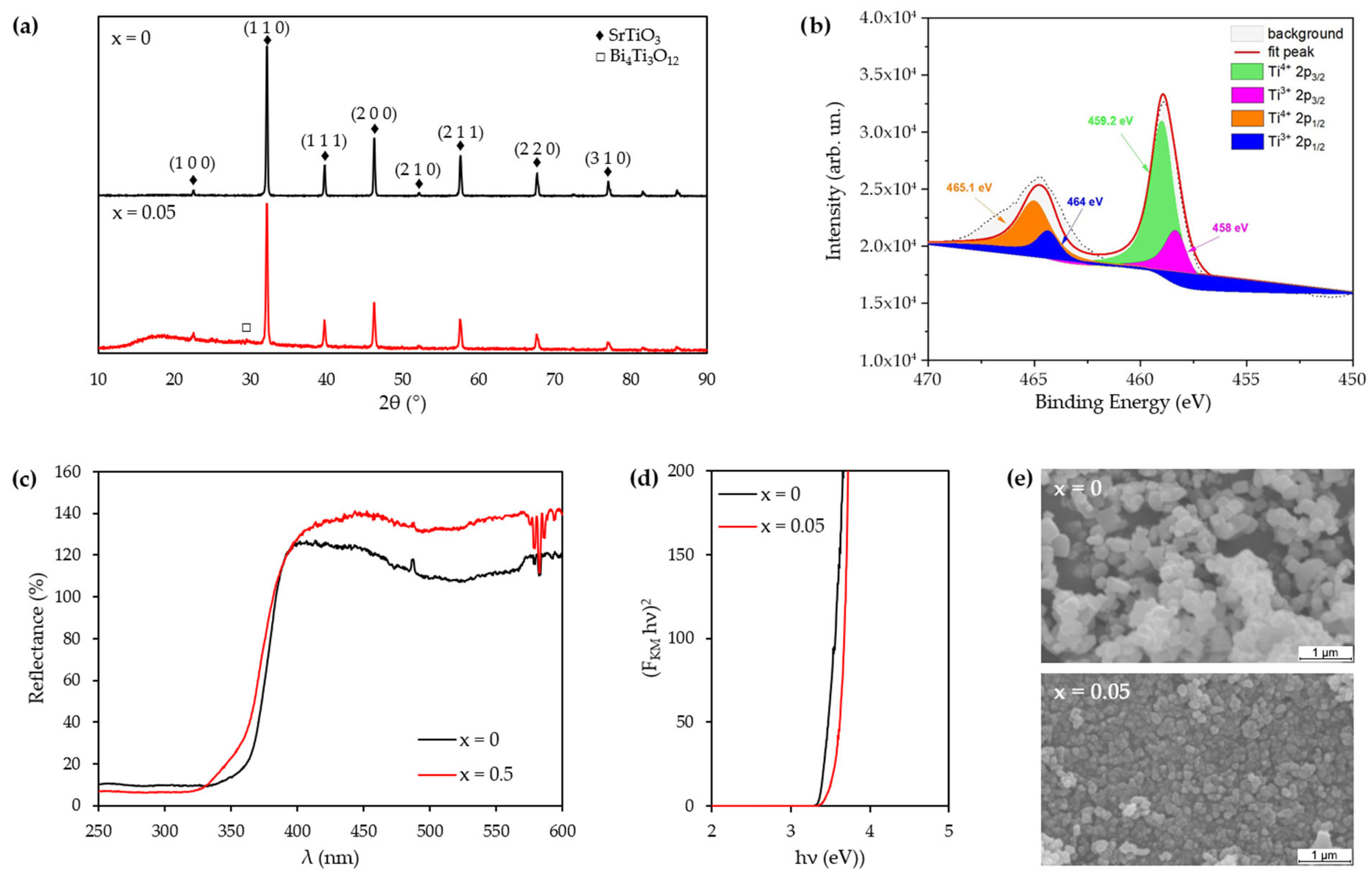
3.1. Perovskite Characterization

Figure 1a presents XRD patterns for the SrTiO3 and Sr0.95Bi0.05TiO3 perovskite powders. The XRD spectra, for both samples, exhibit pronounced peaks at the 2-Theta angles, corresponding to the perovskite symmetry as documented in ICDD file PDF#35-0734. A very weak peak at ~29.90° in the Sr0.95Bi0.05TiO3 sample may be attributed to a minor Bi4Ti3O12 phase (ICDD PDF#47-0398); however, its intensity is very close to the baseline, and no additional characteristic peaks of this phase were observed, suggesting that, if present, such a secondary phase is minimal [34]. EDX analysis, presented in Table 2 and in Table S1 in Supplementary Materials, confirmed the presence of bismuth in the Sr0.95Bi0.05TiO3 sample, with a measured content of 3.4 wt.% and 0.57 at.%, which is consistent with the nominal doping level (5%) and supports the successful incorporation of Bi into the perovskite structure.
Crystallite size determination, based on the full width at half maximum of the (110) peaks consistently indicate smaller values for the Sr0.95Bi0.05TiO3. The estimated crystallite sizes, determined by XRD analysis, are also presented in Table 2. SEM images (Figure 1e) reveal that the SrTiO3 displays more pronounced particle agglomeration, a typical feature of materials synthesized via solid-state routes at high temperatures. In contrast, the Bi-doped material displays less agglomeration, a globular morphology, and distinguishable grains. Measurements of several individual grains in the doped sample yielded diameters between 43 and 120 nm, with most particles below 100 nm, confirming the nanometric character of the material. These morphological observations are in good agreement with the crystallite size values estimated from XRD patterns using the Scherrer equation: 63 nm for Sr0.95Bi0.05TiO3 and 78 nm for SrTiO3. This effect on particle size distribution may be associated not only with the bismuth doping, but also with the differences in temperatures applied during the synthesis process.
Figure 1b shows the XPS spectra of the Ti 2p regions for the Sr0.95Bi0.05TiO3 sample. In the Ti 2p XPS spectra, the doublet corresponding to Ti 2p3/2 and Ti 2p1/2 is observed at binding energies ranging from 457.4 to 458.2 eV and 463.3 to 464.2 eV, respectively. The presence of the Ti3+ state is attributed to the peaks at 458.03 eV, while the Ti4+ is indicated by the peak at 458.99 eV, with a Ti3+/Ti4+ ratio estimated to be 0.35 on the surface [51,52]. Reflectance patterns (Figure 1c) exhibit significant alteration in the wavelengths range of 320 to 400 nm for both the SrTiO3 control and Sr0.95Bi0.05TiO3 samples. This shift corresponds to a decrease in reflectance, highlighting the significant impact of bismuth doping on the optical properties within this range. This change in optical behavior suggests a significant interplay between bismuth doping and material reflectance characteristics. The calculated optical bandgap values (Figure 1d) show a slight increase for the doped sample, ranging from 3.43 eV for SrTiO3 to 3.65 eV for the Sr0.95Bi0.05TiO3. However, the observed bandgap shifts due to bismuth doping highlights potential alterations in the electronic structure, emphasizing the need for further investigation of photocatalytic behavior under visible-light conditions.

3.2. Electronic Structure Calculations

The SrTiO3 exhibits a perovskite structure of the simple cubic type, represented by the general formula ABO3, where sites A and B are occupied by distinct cations. This structure consists of a continuous network of BO6 octahedra interconnected at their corners, extending in all three spatial dimensions to form a highly ordered framework. The larger A-site cations are nestled within the cavities formed by the adjacent eight BO6 octahedra. In this configuration, the A-site cations are 12-fold coordinated with oxygen, while the B-site cations are 6-fold coordinated. The crystalline symmetry of SrTiO3 belongs to the Pm3m space group (number 221), with a lattice parameter of 3.905 Å. Figure 2a, depicting the unit cell of SrTiO3 generated using VESTA version 3.5.8 software [53], shows Sr2+ ions centrally located within an octahedral cage composed of TiO6 units. Considering the ionic radius of Sr2+ (1.32 Å), Ti4+ (0.74 Å), O2− (1.28 Å), and Bi3+ (1.17 Å), it is hypothesized that doping SrTiO3 with bismuth may involve the replacement of Sr2+ with Bi3+ ions due to their comparable ionic sizes. This substitution typically induces a distortion in the ABO3 perovskite structure, owing to the mismatch in ionic sizes and charges. The calculated energy band structure of SrTiO3 shows an indirect band gap of approximately 1.67 eV, which is consistent with previous DFT calculations using the same approximation [44]. Furthermore, the calculated density of states indicates that Ti and O are responsible for the states near the Fermi energy that forms on the gap.
To evaluate the electronic properties and doping effects of Bismuth in SrTiO3 using DFT, two supercells were constructed. Specifically, a 3 × 3 × 3 supercell was created to model two doping concentrations: one with a single Sr atom substituted by Bi, representing a 3.7% doping concentration, and another with two Bi atoms substituting Sr to achieve a 7.4% doping concentration. Additionally, to better match the experimental conditions, a 3 × 3 × 2 supercell was built, with a single Sr atom replaced by Bi to achieve a doping concentration of 5.5%, which is more accurate with the experimental realizations. Analysis of the DOS for varying amounts of doping reveals that the bismuth doped structure exhibits n-type doping behavior (Figure 3a), as evidenced by the Fermi level closer to the conduction band, which introduces additional free carriers. This n-type doping behavior can significantly impact the electronic and optical properties of the material, such as its carrier mobility and light absorption characteristics, which are relevant for photocatalytic applications. Furthermore, with increasing Bi concentration, a notable increment in the band gap energy is observed (Figure 3b). This result could be expected, since doping with other rare-earth metals causes a similar trend, as reported by Zhou et al. [45]. This trend appears to saturate for concentrations greater than 7.4%, where the DOS stabilizes, but the gap decreases. This result is expected, as higher concentrations imply the existence of a BiTiO3 phase with a structure and cell parameter identical to typical SrTiO3 perovskite [34]. Since this is beyond the scope of this study, the discussion will focus on concentrations below the saturation point.
To search for how bismuth doping affects this structure, a detailed DOS analysis was performed on each individual atom (Figure 4). As shown before, the conduction states now cross the Fermi energy. However, it is also possible to verify that the roles of Ti and O are preserved, as they continue to contribute for the states near EFermi. This tuning of the electronic states can be attributed to the Bi states around 2.5 eV. Moreover, the dispersion of the titanium 3d orbitals appears to be broadened by Bi doping compared to other orbitals, which provides a higher density of accessible electronic states at energies that facilitate electron conduction. Consequently, this increased availability of conducting states enhances electron transport across the material’s surface, thereby improving its photocatalytic activity.
The DFT studies show that Bi doping leads to n-type behavior, increasing the free carrier concentrations above the Fermi level, as demonstrated by the computed DOS. This enhancement in carrier density can facilitate improved electron transport and potentially reduce charge recombination under illumination. While doping introduces localized states near the conduction band, these states primarily aid charge-carrier transport rather than substantially extending optical absorption into the visible region. Additionally, the Bi doping shows a linear increase of the energy gap with increasing Bi concentration until saturation, indicating an optimal doping level for enhanced photocatalytic efficiency.

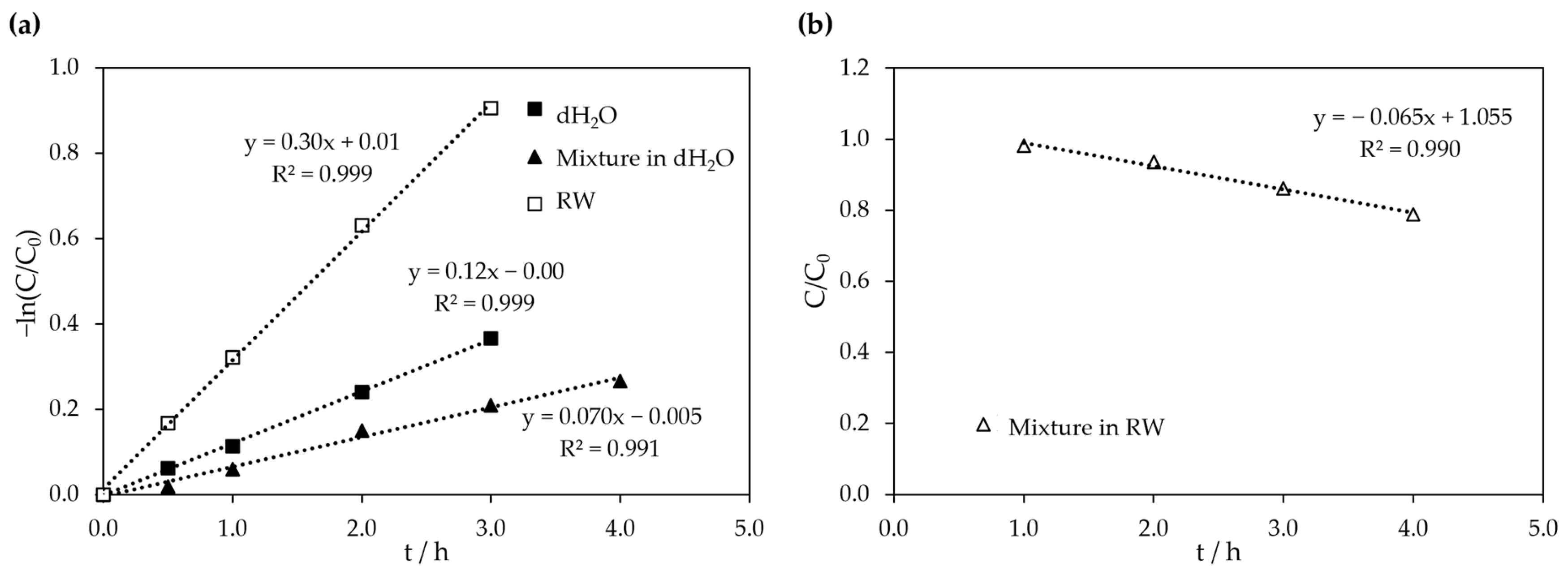
3.3. Photocatalytic Activity of Sr0.95Bi0.05TiO3 Under Visible Light

The photocatalytic activity of Sr0.95Bi0.05TiO3 oxide suspensions (0.2 g L−1) was investigated for the degradation of OTC and IMD solutions (25 and 5 mg L−1, respectively) in two different matrices: dH2O and RW. Control experiments were also performed in the absence of the photocatalyst to assess the influence of the photolytic process towards the pollutants’ degradation. Photolysis experiments conducted in dH2O showed no significant variation in OTC-relative concentration over the duration of the experiment (Figure 5a), indicating that the applied visible-light radiation alone did not contribute significantly to antibiotic degradation through the photolytic process. These findings are consistent with previous studies [54].
In contrast, higher degradation rates were observed in the photolytic experiment in river water (approximately 47% after 2 h) (Figure 5a), which may be attributed to the matrix composition, particularly the presence of metallic ions (Table 1). Wessels et al. [55] demonstrated that the presence of Ca2+ and Mg2+ in solution enhances the absorbance of tetracycline, especially at wavelengths above 350 nm. Xuan et al. [56] reported a substantial improvement in the photolytic degradation of OTC under solar radiation in the presence of Ca2+. This improvement was attributed to the OTC molecule acting as a chelation agent in the presence of multivalent cations, owing to its phenolic and carboxylic groups. The formation of these structures increased OTC absorbance, thereby making the molecules more susceptible to incident radiation. Similarly, another study indicated that the addition of Ca2+, Mg2+, and NH4+ cations to TiO2 suspensions enhances the photocatalytic degradation of two pharmaceuticals [57].
In addition to the chemical composition, physical properties of the river water, such as turbidity, may also influence the efficiency of photolysis and photocatalysis. The river water used in this study presented a turbidity of 3.20 NTU, which is considered relatively low and unlikely to significantly limit light penetration under the experimental conditions. Although suspended materials in natural waters can scatter or absorb light, the enhanced degradation observed in RW compared to dH2O suggests that such optical effects were likely compensated by the positive influence of the matrix composition, namely, the presence of multivalent cations and background ions that may promote pollutant adsorption and photocatalytic reactivity.
The pH of the aqueous matrix significantly influences the direct photolysis of OTC due to the multifunctional behavior of this molecule. OTC, like several other pharmaceutical structures, possesses at least three ionizable centers: pKa1 (3.22) related to the tricarbonyl group protonation, pKa2 (7.46) linked to the deprotonation of the phenolic–diketone group, and pKa3 (8.94) associated with the deprotonation of the dimethyl–amino group [55,58]. As discussed by Jin et al. [59], the four ionic species of OTC (H3OTC+, H2OTC+/−, HOTC, and OTC2−) exhibit distinct absorbance profiles, with higher absorbances at wavelengths > 375 nm at pH > 5.5, directly influencing photolysis kinetic rates. Therefore, the observed difference in the OTC photolysis profile between dH2O (pH 5.5) and RW (pH 7.02) may be attributed to the prevalence of zwitterionic and anionic species in the test solution.
Figure 5a also presents the variation of relative concentration of OTC during the photocatalytic degradation experiments in both matrices. The addition of Sr0.95Bi0.05TiO3 (0.2 g L−1) significantly enhanced degradation, achieving concentration removals above 95% after 1 h of treatment for both samples. The photocatalytic oxidation of OTC using Sr0.95Bi0.05TiO3, under visible-light irradiation, is most likely initiated by the generation of hydroxyl radicals, which subsequently degrade OTC through various oxidation mechanisms. Primary pathways for OTC oxidation, as outlined in the literature, involve the following: hydroxylation, where hydroxyl radicals react with the double bonds in the OTC molecule; decarbonylation, involving the removal of a carbon atom, potentially achieved through various mechanisms such as the oxidation of a secondary alcohol group to form a ketone; and dehydration, which entails the removal of water from the OTC, usually through the elimination of a hydroxyl group and a hydrogen atom [60]. Additionally, Pereira et al. [61] have demonstrated the involvement of singlet oxygen in the photocatalytic oxidation of OTC using TiO2 as a catalyst. Singlet oxygen, a highly reactive oxygen species, is generated as a byproduct of the photocatalytic oxidation of antibiotics, enhancing the understanding of the complex mechanisms involved in this catalytic system.
The same experimental conditions were applied to solutions of IMD (5 mg L−1) in both matrices, with the results presented in Figure 5b. Both photolytic experiments, under visible light, achieved concentration removals of 18% and 22%, after 3 h, for the assays performed in dH2O and in RW, respectively. With the addition of the photocatalyst, degradation rates increased under both experimental conditions, with a more significant improvement in RW, where 60% IMD concentration removal was achieved after 3 h. The matrix composition (i.e., ionic composition and pH) had a positive influence on the IMD photocatalytic degradation profile, although the effect was less pronounced when compared to the one observed with OTC.
The multifunctionality of IMD is less environmentally relevant compared to OTC (Figure S1 in Supplementary Materials). In fact, IMD has two main pKa values (pka1 = 1.56 and pka2 = 11.12). Like other neonicotinoids, their molecular ionization under natural pH conditions does not affect its direct photodegradation, as reported elsewhere [62]. The degradation of IMD is reported to be related to the break of its nitroguanidine functional group, where the role of hydroxyl in this process has been discussed, particularly in the context of UV-C driven photocatalytic reactions [62,63]. These studies indicate that photocatalysis is a promising approach for degrading this recalcitrant compound. Liang et al. [64] propose that the photocatalytic degradation of IMD using bismuth cobalt bimetal oxide under visible light can follow different oxidation pathways, especially under varying pH conditions. Processes such as hydroxylation, oxidation of functional groups, bond cleavage, and the formation of degradation products are involved in the degradation of IMD. The pH variation significantly influences the oxidation pathways, leading to different degradation products. The suggestion of two distinct catalytic mechanisms based on pH implies that IMD degradation may follow diverse pathways depending on environmental conditions. In the present study, the RW pH of 7.02 may have contributed to a comparatively lower degradation efficiency of IMD compared to the OTC results.
Overall, the photocatalytic degradation of both OTC and IMD using Sr0.95Bi0.05TiO3 under visible light is most likely governed by the generation of reactive oxygen species, mainly hydroxyl radicals (OH) and superoxide anions (O2). This is consistent with the behavior reported for similar Bi-doped SrTiO3 systems, where the involvement of these species was confirmed through scavenger assays using terephthalic acid and benzoquinone as selective probes [65]. The slightly alkaline pH of the reaction medium (7.1–7.5) favors the generation and stability of these radicals, supporting efficient oxidative degradation pathways.
To further evaluate the photocatalytic performance of Sr0.95Bi0.05TiO3, the turnover frequency (TOF) was estimated for the degradation of OTC and IMD in dH2O, following the methodology proposed by Chen et al. [66]. The calculated TOF for OTC was 7.95 × 10⁻⁴ min−1, while IMD exhibited a significantly lower value of 5.90 × 10⁻⁵ min−1. This pronounced difference can be attributed to the chemical structure of IMD, which contains electron-withdrawing nitro and chlorine substituents that confer high stability and hinder oxidative degradation [67]. For comparison, Chen et al. [66] reported a TOF of 8.4 × 10⁻⁴ min−1 for TiO2–EDTA in the degradation of azo dyes, highlighting the impact of catalyst design and pollutant structure on degradation kinetics.
The degradation behavior of OTC and IMD in binary mixtures was also evaluated under photolytic and photocatalytic conditions in both dH2O and RW, as shown in Figure 5c. Under photolytic conditions (Figure 5c), both compounds showed no significant degradation in dH2O, where their concentrations remained nearly constant throughout the 4-h irradiation period. In RW, a slight decrease in OTC concentration was observed over time, suggesting that natural constituents in the matrix may enhance photodegradation. IMD, however, was largely unaffected by photolysis in both media. In contrast, under photocatalytic conditions (Figure 5d), OTC was rapidly and almost completely degraded within 1 h in RW and 2 h in dH2O. This confirms that Sr0.95Bi0.05TiO3 maintains strong photocatalytic performance toward OTC, even in the presence of a second contaminant and in complex aqueous matrices. IMD degradation was slower in both matrices.
A comparative analysis between the present work and earlier studies is provided in Table 3. The table presents the results of photodegradation studies for the two organic compounds, OTC and IMD, using different materials with photocatalytic activity (semiconductors) under visible or UV radiation. Although direct comparisons are challenging due to different experimental conditions, the results obtained with the Sr0.95Bi0.05TiO3 perovskite are quite promising.
To complement the assessment of photocatalytic performance, kinetic analyses were conducted. The apparent rate constants (kapp) for the photocatalytic degradation, presented in Table 4, were obtained from the linear regressions of the experimental kinetic data shown in Figure 6 and Figure 7. In most cases, the degradation of both OTC and IMD followed pseudo-first-order kinetics, as indicated by the −ln(C/C0) versus time plots (Figure 6 and Figure 7a). An exception was observed for IMD in the presence of OTC in river water, where the reaction followed pseudo-zero-order kinetics, as evidenced by the linear decay of C/C0 versus time (Figure 7b).
For OTC, the kapp was significantly influenced by the water matrix. In RW, the value of kapp reached 5.58 h−1, which is higher than that observed in dH2O, where kapp was 3.26 h−1. Even under competitive conditions, i.e., in the presence of IMD (5 mg L−1), the OTC degradation kinetics remained similar. In this case, kapp in RW was 4.62 h−1, which is still higher than in dH2O (kapp =3.26 h−1. A similar matrix effect was observed for IMD. The apparent rate constant in RW was 0.30 h−1, which was higher than in dH2O (0.12 h−1), highlighting the role of matrix constituents in enhancing photocatalytic degradation. However, under competitive conditions, when IMD was degraded in the presence of OTC (25 mg L−1), the kinetic observed behavior changed from apparent first order to zero order. The corresponding apparent rate constants under these conditions were 0.06 M h−1 in RW and 0.07 h−1 in dH2O, both slightly lower than the values obtained for the individual degradation of IMD. This reduction may be attributed to a possible preferential adsorption of OTC on the photocatalyst’s surface, potentially blocking active sites and thus limiting the photocatalytic degradation pathway of IMD.

3.4. COD and TOC Removals
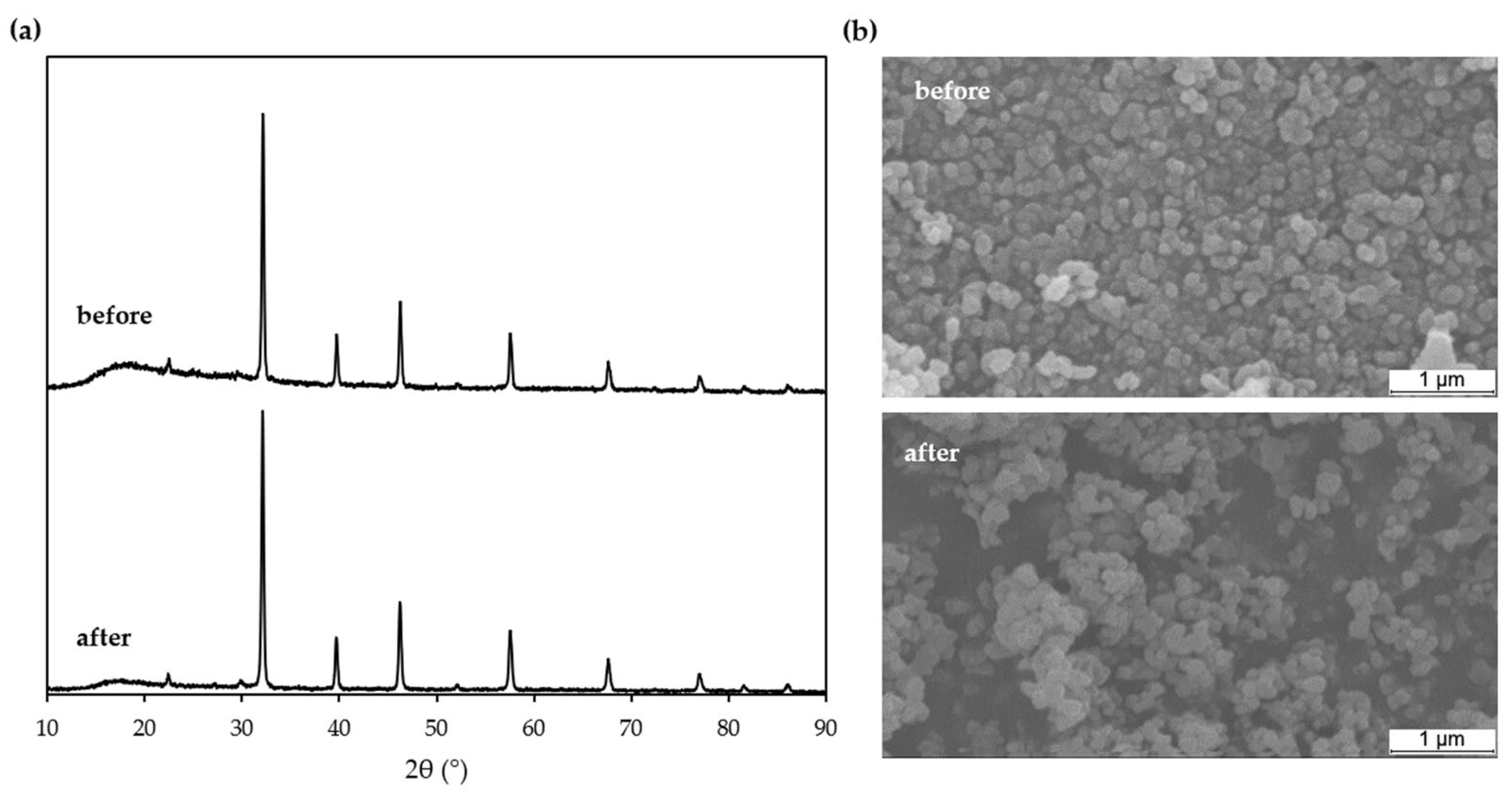
COD and TOC assessments were performed after the photocatalytic degradation of mixtures of OTC and IMD, in both matrices, with results in Table 5. The highest COD and TOC removals were achieved after the photocatalytic experiment performed in the RW matrix, despite slightly higher initial values for both parameters. These higher initial values could be attributed to the presence of humic and fluvic matter in suspension. This suspended matter might contribute to the enhancement of the degradation rate of organic matter by improving the adsorption of pollutant molecules onto the photocatalyst’s surface and reducing the influence of the diffusion process.
Regarding the photocatalytic experiments performed in dH2O, the values of COD and TOC removals were similar, after 4 h, which indicates a good mineralization of the organic matter. In contrast, the photocatalytic experiments in RW showed a COD removal (44.8%) higher than the TOC removal (33.1%). This suggests that the degradation process undergoes some alteration when the aqueous matrix is changed from dH2O to RW, leading to a higher degree of mineralization of organic matter in the RW matrix.

3.5. Reusability

To evaluate the influence of Sr0.95Bi0.05TiO3 reuse on its photocatalytic activity, the oxide sample was recovered after each experiment, washed with distilled water, and dried at 120 °C before being used in the subsequent degradation cycle. This parameter was assessed during photocatalytic degradation of the mixture in dH2O over five consecutive cycles, with an average powder recuperation of 93.8%. The results from all five cycles are shown in Figure 8. A slight decrease in the OTC degradation rate (Figure 8a) was observed from the first to the second photodegradation cycle, followed by a relative consistent performance from the third to the fifth cycles. After five cycles, the percentage of OTC removal decreased only slightly, from 100% in the first cycle to 96.7% in the fifth. This minimal loss of photocatalytic efficiency highlights the excellent reusability and stability of the catalyst. In the case of IMD degradation (Figure 8b), a slight increase in the degradation rate was observed throughout the five photocatalytic cycles.
The behavior observed with both compounds may be attributed to the photocatalyst’s surface modification, such as the accumulation of adsorbed organic matter or changes in the surface charge of the oxide, possibly due to variations in the oxidation states of the metal ions. These changes could influence the adsorption of OTC and IMD molecules, potentially impacting the photocatalytic degradation process.
Moreover, Sr0.95Bi0.05TiO3 powder samples recovered after the five degradation cycles were structural and morphologically characterized by DRX and SEM, respectively (Figure 9). The XRD patterns did not show any significant changes in the crystal structure, nor was any new phase formation observed. This observation suggests that metal ion leaching did not occur during the photocatalytic process [36]. The results indicate that oxide maintains its structural and morphological integrity, with no significant changes being observed after the five cycles. These findings demonstrate that the Sr0.95Bi0.05TiO3 can be effectively reused for at least five cycles without any significant loss in its photocatalytic activity under visiblelight or significant impact on its structural and morphological properties.

3.6. Toxicity

An important aspect of photocatalytic degradation studies is the evaluation of toxicity prior to treatment. In this study, the initial concentrations of the two pollutants used in this study, OTC (25 mg L−1) and IMD (5 mg L−1), were both below their respective EC50,48h (D. magna) values of >102 mg L−1 and 6.029 mg L−1 [72,73]. This suggests that the initial solution is non-toxic towards D. magna, which was confirmed by performing the acute toxicity assay using a solution of OTC (25 mg L−1) and IMD (5 mg L−1) in dH2O. However, considering that some degradation products could potentially exhibit higher acute toxicity than the parent compound, acute toxicity assays, towards D. magna, were also performed with solutions obtained after the photocatalytic degradation of the mixture of the OTC and IMD in dH2O. The samples were collected after 2 and 4 h of irradiation. Both samples showed 0% mortality of D. magna, indicating that they were non-toxic to the test organism. This suggests that the degradation products present in solution are either non-toxic to D. magna, or are present at concentrations below their respective EC50,48h values. These results indicate that the application of Sr0.95Bi0.05TiO3 oxide as a photocatalyst for the degradation of both OTC and IMD under these experimental conditions does not lead to the formation of toxic final solutions towards D. magna.

4. Conclusions

In this study, the potential of Sr0.95Bi0.05TiO3 perovskites as an efficient and eco-friendly photocatalyst for degrading pharmaceuticals and pesticides under visible-light irradiation was investigated. This oxide showed high photocatalytic activity under visible light for degrading oxytetracycline and imidacloprid in both deionized water and river water.
Oxytetracycline was efficiently degraded (100% in 2 h), with higher rates in river water due to the presence of ions and favorable pH. Although IMD is more recalcitrant, it also underwent significant degradation (60% in RW after 3 h), with kinetic behavior affected by the coexistence of OTC. Photocatalysis also led to partial mineralization of organic matter, with better results in river water. The catalyst remained stable and reusable over five cycles without significant loss of activity or structural integrity. Toxicity tests using D. magna confirmed that the solutions treated were non-toxic, indicating that no harmful byproducts were formed. Overall, Sr0.95Bi0.05TiO3 is a promising and reusable photocatalyst for the removal of pharmaceutical and pesticide contaminants.
Complementary DFT calculations provided valuable insights into the underlying mechanics responsible for the success of Sr0.95Bi0.05TiO3. These simulations revealed that bismuth insertion caused n-type doping behavior in the material, as seen by the Fermi level shifting closer to the conduction band and the introduction of more free carriers. This n-type doping has a direct impact on the electrical and optical properties, enhancing carrier mobility and extending the light absorption spectrum, which may explain the reported photocatalytic activity.
These combined experimental and computational findings provide valuable insights into the material’s properties and performance, supporting its further development for photocatalytic water treatment application.

Supplementary Materials

The following supporting information can be downloaded at: https://www.mdpi.com/article/10.3390/w17152177/s1, Figure S1: Anticipated distribution of OTC (a) and IMD (b) ionic species as a function of pH, according to the available pKa values; Table S1. Elemental composition of Sr0.95Bi0.05TiO3 determined by EDX.

Author Contributions

Conceptualization, A.L., J.E.S., W.S.P., and L.C.; data curation, M.J.N., A.R.R., and L.C.; formal analysis, M.J.N., A.L., P.T.F., and L.C.; investigation, M.J.N., J.E.S., and A.R.R.; methodology, M.J.N., J.E.S., and W.S.P.; project administration, A.L.; software, G.J.I., and W.S.P.; supervision, M.J.N., A.L., and L.C.; validation, M.J.N., M.J.P., P.T.F., and G.J.I.; visualization, M.J.P., and L.C.; writing—original draft, M.J.N., and J.E.S.; writing—review and editing, M.J.N., M.J.P., P.T.F., G.J.I., J.E.S., A.R.R., W.S.P., and L.C. All authors have read and agreed to the published version of the manuscript.

Funding

The authors acknowledge the financial support from the Fundação para a Ciência e Tecnologia, FCT, for the project UIDB/00195/2020 of the FibEnTech-UBI Research Unit and for the PhD grant awarded to Maria João Nunes (SFRH/BD/132436/2017 and 10.54499/COVID/BD/151965/2021). The authors are also very grateful for the support granted by the Research Unit of Fiber Materials and Environmental Technologies (FibEnTech-UBI), through the Project reference UIDB/00195/2020, funded by the Fundação para a Ciência e a Tecnologia, IP/MCTES through national funds (PIDDAC) and DOI: 10.54499/UIDB/00195/2020. The authors acknowledge financial support from Brazilian government funding agencies FAPES, CAPES, and CNPq. Wendel S. Paz would also like to thank FAPES (under grants 444/2021, 1044/2022, and 1081/2022 P:2022-8L35F) and CNPq (under grant 409441/2021-0) for their financial support. Jefferson E. Silveira gratefully acknowledges the support from CAPES: Science Without Borders Program, Ministry of Education of Brazil, under grant BEX-1046/13-6, and the support from project PDC-2022-133805-100, funded by the Ministry of Science, Innovation, and Universities of Spain.

Data Availability Statement

The original contributions presented in the study are included in the article/Supplementary Materials, and further inquiries can be directed to the corresponding author.

Conflicts of Interest

The authors declare no conflicts of interest. The funders had no role in the design of the study; in the collection, analyses, or interpretation of data; in the writing of the manuscript; or in the decision to publish the results.

References

  1. Wei, K.; Faraj, Y.; Yao, G.; Xie, R.; Lai, B. Strategies for improving perovskite photocatalysts reactivity for organic pollutants degradation: A review on recent progress. Chem. Eng. J. 2021, 414, 128783. [Google Scholar] [CrossRef]
  2. Madkhali, N.; Prasad, C.; Malkappa, K.; Choi, H.Y.; Govinda, V.; Bahadur, I.; Abumousa, R.A. Recent update on photocatalytic degradation of pollutants in wastewater using TiO2-based heterostructured materials. Results Eng. 2023, 17, 100920. [Google Scholar] [CrossRef]
  3. Liu, H.; Wang, C.; Wang, G. Photocatalytic advanced oxidation processes for water treatment: Recent advances and perspective. Chem. Asian J. 2020, 15, 3239–3253. [Google Scholar] [CrossRef]
  4. Pelosato, R.; Bolognimo, I.; Fontana, F.; Sora, I.N. Applications of heterogeneous photocatalysis to the degradation of oxytetracycline in water: A review. Molecules 2022, 27, 2743. [Google Scholar] [CrossRef]
  5. Zabar, R.; Komel, T.; Fabjan, B.J.; Kralj, B.; Trebse, P. Photocatalytic degradation with immobilised TiO2 of three selected neonicotinoid insecticides: Imidacloprid, thiamethoxam and clothianidin. Chemosphere 2012, 89, 293–301. [Google Scholar] [CrossRef]
  6. Zeshan, M.; Bhatti, I.A.; Mohsin, M.; Iqbal, M.; Amjed, N.; Nisar, J.; AlMasoud, N.; Alomar, T.S. Remediation of pesticides using TiO2 based photocatalytic strategies: A review. Chemosphere 2022, 30, 134525. [Google Scholar] [CrossRef] [PubMed]
  7. Kamat, P.V.; Jin, S. Semiconductor photocatalysis: “Tell us the complete story!”. ACS Energy Lett. 2018, 3, 622–623. [Google Scholar] [CrossRef]
  8. Huang, L.; Huang, X.; Yan, J.; Liu, Y.; Jiang, H.; Zhang, H.; Tang, J.; Liu, Q. Research progresses on the application of perovskite in adsorption and photocatalytic removal of water pollutants. J. Hazard. Mater. 2023, 442, 130024. [Google Scholar] [CrossRef]
  9. Parwaiz, S.; Khan, M.M. Perovskites and perovskite-based heterostructures for photocatalytic energy and environmental applications. J. Environm. Chem. Eng. 2024, 12, 113175. [Google Scholar] [CrossRef]
  10. Silva, P.S.; Pereira, D.; Calisto, V.; Martins, M.A.; Otero, M.; Esteves, V.I.; Lima, D.L.D. Biochar-TiO2 magnetic nanocomposites for photocatalytic solar-driven removal of antibiotics from aquaculture effluents. J. Environ. Manage. 2021, 294, 112937. [Google Scholar] [CrossRef]
  11. Singh, J.; Kumar, S.; Rishikesh; Manna, A.K.; Soni, R.K. Fabrication of ZnO-TiO2 nanohybrids for rapid sunlight driven photodegradation of textile dyes and antibiotic residue molecules. Opt. Mater. 2020, 107, 110138. [Google Scholar] [CrossRef]
  12. Zhang, S.; Ou, X.; Xiang, Q.; Carabineiro, A.A.C.; Fan, J.; Lv, K. Research progress in metal sulfides for photocatalysis: From activity to stability. Chemosphere 2022, 303, 135085. [Google Scholar] [CrossRef]
  13. Ng, Y.H.; Ikeda, S.; Matsumura, M.; Amal, R. A perspective on fabricating carbon-based nanomaterials by photocatalysis and their applications. Energy Environ. Sci. 2012, 5, 9307–9318. [Google Scholar] [CrossRef]
  14. Ajiboye, T.O.; Oyewo, O.A.; Onwudiwe, D.C. The performance of bismuth-based compounds in photocatalytic applications. Surf. Interfaces 2021, 23, 100927. [Google Scholar] [CrossRef]
  15. Silveira, J.E.; de Souza, A.S.; Pasini, F.N.N.; Ribeiro, A.R.; Scopel, W.L.; Zazo, J.A.; Casas, J.A.; Paz, W.S. A comprehensive study of the reduction of nitrate on natural FeTiO3: Photocatalysis and DFT calculations. Sep. Purif. Technol. 2023, 306, 122570. [Google Scholar] [CrossRef]
  16. Silveira, J.E.; Paz, W.S.; Garcia-Muñoz, P.; Zazo, J.A.; Casas, J.A. UV-LED/ilmenite/persulfate for azo dye mineralization: The role of sulfate in the catalyst deactivation. Appl. Catal. B Environ. 2017, 219, 314–321. [Google Scholar] [CrossRef]
  17. Silveira, J.E.; Garcia-Costa, A.L.; Carbajo, J.; Ribeiro, A.R.; Pliego, G.; Paz, W.S.; Zazo, J.A.; Casas, J.A. Nitrate removal in saline water by photo-reduction using natural FeTiO3 as catalyst. Chem. Eng. J. Adv. 2022, 12, 100387. [Google Scholar] [CrossRef]
  18. Xu, W.; Zhang, Q.; Xu, K.; Qiu, L.; Song, J.; Wang, L. Study on visible light photocatalytic performance of BiVO4 modified by graphene analogue boron nitride. Chemosphere 2022, 307, 135811. [Google Scholar] [CrossRef]
  19. Sanei, A.; Dashtian, K.; Seyf, J.Y.; Seidi, F.; Kolvari, E. Biomass derived reduced-graphene-oxide supported α-Fe2O3/ZnO S-scheme heterostructure: Robust photocatalytic wastewater remediation. J. Environ. Manage. 2023, 332, 117377. [Google Scholar] [CrossRef]
  20. Liu, J.; Zhang, Q.; Tian, X.; Hong, Y.; Nie, Y.; Su, N.; Jin, G.; Zhai, Z.; Fu, C. Highly efficient photocatalytic degradation of oil pollutants by oxygen deficient SnO2 quantum dots for water remediation. Chem. Eng. J. 2021, 404, 127146. [Google Scholar] [CrossRef]
  21. Liu, J.; Qu, X.; Zhang, C.; Dong, W.; Fu, C.; Wangan, J.; Zhang, Q. High-yield aqueous synthesis of partial-oxidized black phosphorus as layered nanodot photocatalysts for efficient visible-light driven degradation of emerging organic contaminants. J. Clean. Prod. 2022, 377, 134228. [Google Scholar] [CrossRef]
  22. Reddy, K.R.; Reddy, C.; Nadagouda, M.; Shetti, N.; Jaesool, S.; Aminabhavi, T.M. Polymeric graphitic carbon nitride (g-C3N4)-based semiconducting nanostructured materials: Synthesis methods, properties and photocatalytic applications. J. Environ. Manage. 2019, 238, 25–40. [Google Scholar] [CrossRef] [PubMed]
  23. Mateo, D.; Albero, J.; García, H. Titanium-perovskite-supported RuO2 nanoparticles for photocatalytic CO2 methanation. Joule 2019, 3, 1949–1962. [Google Scholar] [CrossRef]
  24. Mai, X.T.; Bui, D.N.; Pham, V.K.; Pham, T.H.T.; Nguyen, T.T.L.; Chau, H.D.; Tran, T.K.N. Effect of CuO loading on the photocatalytic activity of SrTiO3/MWCNTs nanocomposites for dye degradation under visible light. Inorganics 2022, 10, 211. [Google Scholar] [CrossRef]
  25. Phoon, B.L.; Lai, C.W.; Juan, J.C.; Show, P.-L.; Chen, W.-H. A review of synthesis and morphology of SrTiO3 for energy and other applications. Int. J. Energy Res. 2019, 43, 5151–5174. [Google Scholar] [CrossRef]
  26. Kimijima, T.; Kanie, K.; Nakaya, M.; Muramatsu, A. Solvothermal synthesis of SrTiO3 nanoparticles precisely controlled in surface crystal planes and their photocatalytic activity. Appl. Catal. B Environ. 2014, 144, 462–467. [Google Scholar] [CrossRef]
  27. Li, F.; Yu, K.; Lou, L.-L.; Su, Z.; Liu, S. Theoretical and experimental study of La/Ni co-doped SrTiO3 photocatalyst. Mater. Sci. Eng. B 2010, 172, 136–141. [Google Scholar] [CrossRef]
  28. Jia, A.; Su, Z.; Lou, L.-L.; Liu, S. Synthesis and characterization of highly-active nickel and lanthanum co-doped SrTiO3. Solid State Sci. 2010, 12, 1140–1145. [Google Scholar] [CrossRef]
  29. Huang, S.-T.; Lee, W.W.; Chang, J.-L.; Huang, W.-S.; Chou, S.-Y.; Chen, C.-C. Hydrothermal synthesis of SrTiO3 nanocubes: Characterization, photocatalytic activities, and degradation pathway. J. Taiwan Inst. Chem. Eng. 2014, 45, 1927–1936. [Google Scholar] [CrossRef]
  30. Wu, G.; Li, P.; Xu, D.; Luo, B.; Hong, Y.; Shi, W.; Liu, C. Hydrothermal synthesis and visible-light-driven photocatalytic degradation for tetracycline of Mn-doped SrTiO3 nanocubes. Appl. Surf. Sci. 2015, 333, 39–47. [Google Scholar] [CrossRef]
  31. Hou, D.; Hu, X.; Ho, W.; Hu, P.; Huang, Y. Facile fabrication of porous Cr-doped SrTiO3 nanotubes by electrospinning and their enhanced visible-light-driven photocatalytic properties. J. Mater. Chem. A 2015, 3, 3935–3943. [Google Scholar] [CrossRef]
  32. Xu, Y.; Liang, Y.; He, Q.; Xu, R.; Chen, D.; Xu, X.; Hu, H. Review of doping SrTiO3 for photocatalytic applications. Bull. Mater. Sci. 2023, 46, 6. [Google Scholar] [CrossRef]
  33. Nunes, M.J.; Lopes, A.; Pacheco, M.J.; Ciríaco, L.; Fiadeiro, P.T. Photocatalytic degradation of the AO7 under visible light with Bi-doped SrTiO3. In Textiles, Identity and Innovation: In Touch; CRC Press: London, UK, 2020; pp. 349–355. [Google Scholar]
  34. Nunes, M.J.; Lopes, A.; Pacheco, M.J.; Ciríaco, L. Visible-light-driven AO7 photocatalytic degradation and toxicity removal at Bi-doped SrTiO3. Materials 2022, 15, 2465. [Google Scholar] [CrossRef]
  35. Liu, B.; Zhou, N.; Liu, J.; Wang, X.; Robertson, P.K.J.; Chen, X.; Chen, C.; Luo, J.; Wang, C. Full-spectrum Bi@SrTiO3 for bi-directional promotion effects on photocatalytic redox reaction: Insight into intermediates and mechanism. J. Rare Earths 2024, 42, 917–929. [Google Scholar] [CrossRef]
  36. Mehra, S.; Saroha, J.; Rani, E.; Sharma, V.; Goswami, L.; Gupta, G.; Srivastava, A.K.; Sharma, S.N. Development of visible light-driven SrTiO3 photocatalysts for the degradation of organic pollutants for waste-water treatment: Contrasting behavior of MB & MO dyes. Opt. Mater. 2023, 136, 113344. [Google Scholar] [CrossRef]
  37. EMEA/CVMP (European Agency for the Evaluation of Medicinal Products—Committee for Veterinary Medicinal Products). Oxytetracycline, Tetracycline, Chlortetracycline. Summary Report 3. EMEA/MRL/023/95. 1995. Available online: https://www.ema.europa.eu/en/documents/mrl-report/oxytetracycline-tetracycline-chlortetracycline-summary-report-3-committee-veterinary-medicinal-products_en.pdf (accessed on 10 February 2024).
  38. Suyamud, B.; Chen, Y.; Quyen, D.T.T.; Dong, Z.; Zhao, C.; Hu, J. Antimicrobial resistance in aquaculture: Occurrence and strategies in Southeast Asia. Sci. Total Environ. 2024, 907, 167942. [Google Scholar] [CrossRef] [PubMed]
  39. European Commission (E.U.). Commission Implementing Regulation (EU) 2018/783 of 29 May 2018 amending Implementing Regulation (EU) No 540/2011 as regards the conditions of approval of the active substance imidacloprid. Off. J. Eur. Union 2018, 132, 31–34. [Google Scholar]
  40. Náfrádi, M.; Hlogyik, T.; Farkas, L.; Alapi, T. Comparison of the heterogeneous photocatalysis of imidacloprid and thiacloprid-reaction mechanism, ecotoxicity, and the effect of matrices. J. Environ. Chem. Eng. 2021, 9, 106684. [Google Scholar] [CrossRef]
  41. Silveira, J.E.; Inacio, G.J.; Batista, N.N.; Morais, W.P.; Menezes, M.G.; Zazo, J.A.; Casas, J.A.; Paz, W.P. Franckeite-derived van der Waals heterostructure with highly efficient photocatalytic NOx abatement: Theoretical insights and experimental evidences. J. Environ. Chem. Eng. 2024, 12, 111998. [Google Scholar] [CrossRef]
  42. Nunes, M.J.; Lopes, A.; Pacheco, M.J.; Ciríaco, L. Preparation, characterization and environmental applications of Sr1-x(La,Bi)xTiO3 perovskites immobilized on Ni-foam: Photodegradation of the Acid Orange 7. Environ. Sci. Pollut. Res. 2017, 24, 11102–11110. [Google Scholar] [CrossRef]
  43. Giannozzi, P.; Baroni, S.; Bonini, N.; Calandra, M.; Car, R.; Cavazzoni, C.; Ceresoli, D.; Chiarotti, G.L.; Cococcioni, M.; Dabo, I.; et al. QUANTUM ESPRESSO: A modular and open-source software project for quantum simulations of materials. J. Phys. Condens. Matter. 2009, 21, 395502. [Google Scholar] [CrossRef]
  44. Piskunov, S.; Heifets, E.; Eglitis, R.I.; Borstel, G. Bulk properties and electronic structure of SrTiO3, BaTiO3, PbTiO3 perovskites: An ab initio HF/DFT study. Comput. Mater. Sci. 2004, 29, 165–178. [Google Scholar] [CrossRef]
  45. Zhou, E.; Raulot, J.-M.; Xu, H.; Hao, H.; Shen, Z.; Liu, H. Structural, electronic, and optical properties of rare-earth-doped SrTiO3 perovskite: A first-principles study. Phys. B Condens. Matter. 2022, 643, 414160. [Google Scholar] [CrossRef]
  46. Rizwan, M.; Ali, A.; Usman, Z.; Khalid, N.R.; Jin, H.B.; Cao, C.B. Structural, electronic and optical properties of copper-doped SrTiO3 perovskite: A DFT study. Phys. B Condens. Matter. 2019, 552, 52–57. [Google Scholar] [CrossRef]
  47. Tao, J.; Perdew, J.P.; Tang, H.; Shahi, C. Origin of the size-dependence of the equilibrium van der Waals binding between nanostructures. J. Chem. Phys. 2018, 148, 074110. [Google Scholar] [CrossRef] [PubMed]
  48. American Public Health Association, American Water Works Association, Water Environment Federation. Standard Methods for the Examination of Water and Wastewater, 24th ed.; Lipps, W.C., Braun-Howland, E.B., Baxter, T.E., Eds.; APHA Press: Washington, DC, USA, 2023. [Google Scholar]
  49. OCDE. Test No. 202: Daphnia sp. Acute Immobilisation Test, OECD Guidelines for testing of Chemicals, Section 2; OECD Publishing: Paris, France, 2004. [Google Scholar]
  50. ISO 6341; Water Quality—Determination of the Inhibition of the Mobility of Daphnia magna Straus (Cladocera, Crustacea)—Acute Toxicity Test. International Organization for Standardization: Geneva, Switzerland, 2012.
  51. Sakamoto, K.; Hayashi, F.; Sato, K.; Hirano, M.; Ohtsu, N. XPS spectral analysis for a multiple oxide comprising NiO, TiO2, and NiTiO3. Appl. Surf. Sci. 2020, 526, 146729. [Google Scholar] [CrossRef]
  52. Yu, J.; Li, C.; Li, B.; Zhu, X.; Zhang, R.; Ji, L.; Tang, D.; Asiri, A.M.; Sun, X.; Li, Q.; et al. A perovskite La2Ti2O7 nanosheet as an efficient electrocatalyst for artificial N2 fixation to NH3 in acidic media. Chem. Commun. 2019, 55, 6401–6404. [Google Scholar] [CrossRef] [PubMed]
  53. Momma, K.; Izumi, F. VESTA 3 for three-dimensional visualization of crystal, volumetric and morphology data. J. Appl. Crystallogr. 2011, 44, 1272–1276. [Google Scholar] [CrossRef]
  54. Zhang, H.; Wang, Y.; Zhai, C. Construction of a novel p-n heterojunction CdS QDs/LaMnO3 composite for photodegradation of oxytetracycline. Mater. Sci. Semicond. Process. 2022, 144, 106568. [Google Scholar] [CrossRef]
  55. Wessels, J.M.; Ford, W.E.; Szymczak, W.; Schneider, S. The complexation of tetracycline and anhydrotetracycline with Mg2+ and Ca2+: A spectroscopic study. J. Phys. Chem. B 1998, 102, 9323–9331. [Google Scholar] [CrossRef]
  56. Xuan, R.; Arisi, L.; Wang, Q.; Yates, S.R.; Biswas, K.C. Hydrolysis and photolysis of oxytetracycline in aqueous solution. J. Environ. Sci. Health B 2010, 45, 73–81. [Google Scholar] [CrossRef]
  57. Kudlek, E.; Dudziak, M.; Bohdziewicz, J. Influence of inorganic ions and organic substances on the degradation of pharmaceutical compound in water matrix. Water 2016, 8, 532. [Google Scholar] [CrossRef]
  58. Qiang, Z.; Adams, C. Potentiometric determination of acid dissociation constants (pKa) for human and veterinary antibiotics. Water Res. 2004, 38, 2874–2890. [Google Scholar] [CrossRef]
  59. Jin, X.; Xu, H.; Qiu, S.; Jia, M.; Wang, F.; Zhang, A.; Jiang, X. Direct photolysis of oxytetracycline: Influence of initial concentration, pH and temperature. J. Photochem. Photobiol. A Chem. 2017, 332, 224–231. [Google Scholar] [CrossRef]
  60. Liu, Y.; He, X.; Fu, Y.; Dionysiou, D.D. Degradation kinetics and mechanism of oxytetracycline by hydroxyl radical-based advanced oxidation processes. Chem. Eng. J. 2016, 284, 1317–1327. [Google Scholar] [CrossRef]
  61. Pereira, J.H.O.S.; Reis, A.C.; Queirós, D.; Nunes, O.C.; Borges, M.T.; Vilar, V.J.P.; Boaventura, R.A.R. Insights into solar TiO2-assisted photocatalytic oxidation of two antibiotics employed in aquatic animal production, oxolinic acid and oxytetracycline. Sci. Total Environ. 2013, 463–464, 274–283. [Google Scholar] [CrossRef] [PubMed]
  62. Acero, J.L.; Real, F.J.; Benitez, F.J.; Matamoros, E. Degradation of neonicotinoids by UV irradiation: Kinetics and effect of real water constituents. Sep. Purif. Technol. 2019, 211, 218–226. [Google Scholar] [CrossRef]
  63. Banić, N.D.; Šojić, D.V.; Krstić, J.B.; Abramović, B.F. Photodegradation of neonicotinoid active ingredients and their commercial formulations in water by different advanced oxidation processes. Water Air Soil Pollut. 2014, 225, 1954. [Google Scholar] [CrossRef]
  64. Liang, F.; Zhang, Y.-H.; He, B.; Yang, J.; Shi, Q.; Shi, F.-N. Enhanced photocatalytic degradation of imidacloprid and RhB by the precursor derived Bi12.7Co0.3O19.35 under different pH value. J. Phys. Chem. Solids 2022, 164, 110638. [Google Scholar] [CrossRef]
  65. Garcia, C.R.; Oliva, J.; Chávez, D.; Esquivel, B.; Gómez-Solís, C.; Martínez-Sánchez, E.; Mtz-Enriquez, A.I. Effect of Bismuth Dopant on the Photocatalytic Properties of SrTiO3 under Solar Irradiation. Top. Catal. 2021, 64, 155–166. [Google Scholar] [CrossRef]
  66. Chen, Y.; Wang, X.; Liu, B.; Zhang, Y.; Zhao, Y.; Wang, S. Directional regulation of reactive oxygen species in titanium dioxide boosting the photocatalytic degradation performance of azo dyes. J. Colloid Interface Sci. 2024, 673, 275–283. [Google Scholar] [CrossRef]
  67. Targhan, H.; Rezaei, A.; Aliabadi, A.; Zheng, H.; Cheng, H.; Aminabhavi, T.M. Adsorptive and photocatalytic degradation of imidacloprid pesticide from wastewater via the fabrication of ZIF-CdS/Tpy quantum dots. Chem. Eng. J. 2024, 482, 148983. [Google Scholar] [CrossRef]
  68. Xu, K.; Yang, X.; Ruan, L.; Qi, S.; Liu, J.; Liu, K.; Pan, S.; Feng, G.; Dai, Z.; Yang, X.; et al. Superior adsorption and photocatalytic degradation capability of mesoporous LaFeO3/g-C3N4 for removal of oxytetracycline. Catalysts 2020, 10, 301. [Google Scholar] [CrossRef]
  69. Hernández-Arellano, D.L.; Durán-Álvarez, J.C.; Zanella, R.; López-Juárez, R. Effect of heat treatment on the structure and photocatalytic properties of BiYO3 and BiY0.995Ni0.005O3 ceramic powders. Ceram. Int. 2020, 46, 20291–20298. [Google Scholar] [CrossRef]
  70. Espíndola, J.C.; Cristóvão, R.O.; Mendes, A.; Boaventura, R.A.R.; Vilar, V.J.P. Photocatalytic membrane reactor performance towards oxytetracycline removal from synthetic and real matrices: Suspended vs immobilized TiO2-P25. Chem. Eng. J. 2019, 378, 122114. [Google Scholar] [CrossRef]
  71. Andronic, L.; Vladescu, A.; Enesca, A. Synthesis, characterisation, photocatalytic activity, and aquatic toxicity evaluation of TiO2 nanoparticles. Nanomaterials 2021, 11, 3197. [Google Scholar] [CrossRef]
  72. Safety Data Sheet, Oxytetracycline, Hydrochloride. Available online: https://www.merckmillipore.com/PT/en/product/msds/EMD_BIO-500105?ReferrerURL=https%3A%2F%2Fwww.google.com%2F (accessed on 14 September 2023).
  73. Safety Data Sheet, Imidacloprid. Available online: https://labelsds.com/images/user_uploads/Imidacloprid%202F%20TI%20SDS%202-12-21.pdf (accessed on 14 September 2023).
Figure 1. Characterization of Sr1-xBixTiO3 samples: (a) XRD patterns, (b) XPS of spectra of the Ti 2p regions (x = 0.05), showing deconvoluted peaks assigned to Ti4+ 2p3/2 (green), Ti4+ 2p1/2 (orange), Ti3+ 2p3/2 (magenta), and Ti3+ 2p1/2 (blue); the overall fit is shown in red, and the background as a dashed black line; (c) diffuse reflectance spectra; (d) calculated optical bandgap values; (e) SEM micrographies of SrTiO3 (top) and Sr0.95Bi0.05TiO3 (bottom) (magnification ×20,000).
Water 17 02177 g001
Figure 2. (a) Perovskite structure, (b) DOS of pure SrTiO3. The atoms responsible for the states near Fermi’s energy are the Titanium and Oxygen, as expected. Strontium plays a role in the bulk states, farther from EFermi. (c) Electronic Bands of the SrTiO3 aligned with its density of states. The gap of this structure is indirect and happens between the points R and Γ of the Brillouin zone. With the alignment by the Fermi energy, it is possible to evaluate the energy gap both with the electronic bands or with the DOS. In this case, the gap energy is around 1.67 eV.
Water 17 02177 g002
Figure 3. (a) DOS for different concentrations of Bi in the structure, showing an increase in the gap size. The concentration of Bi shifts the conduction states across EFermi, indicating a n-type doping, and. (b) Value of Energy gap compiled for different concentrations of Bi. There is a clear trend of the energy gap increasing with the concentration of Bi. However, at higher concentrations of Bi, the gap decreases and seems to stabilize.
Water 17 02177 g003
Figure 4. Partial DOS for the structure doped (a) with 3.7% of Bi and (b) with 5.5% Bi. Bismuth shows a highly localized state around 2.5 eV for the 3.7% Bi concentration and around 3.35 eV for the 5.5% Bi concentration. In general, Bi contributes to states on the conduction bands, thus tuning the Fermi energy.
Water 17 02177 g004
Figure 5. Relative concentration variation of (a) OTC (Ci = 25 mg L−1) and (b) IMD (Ci = 5 mg L−1) during photolytic and photocatalytic assays, with Sr0.95Bi0.05TiO3 suspensions (0.2 g L−1), performed in dH2O and RW, under visible light. Relative concentration variation of OTC and IMD during (c) photolytic and (d) photocatalytic assays performed with a mixture of both pollutants in both matrixes and under the same experimental conditions.
Water 17 02177 g005
Figure 6. Kinetic plots for the photocatalytic degradation of OTC under visible light using Sr0.95Bi0.05TiO3 as the photocatalyst. The data are presented as −ln(C/C0) vs. time (t), indicating pseudo-first-order kinetics behavior in different aqueous matrices: dH2O, RW, and binary mixture with IMD in both media.
Water 17 02177 g006
Figure 7. Kinetic plots for the photocatalytic degradation of imidacloprid (IMD) using Sr0.95Bi0.05TiO3 under visible light. (a) Pseudo-first-order kinetics plots (−ln(C/C0) vs. time) for IMD degradation in dH2O, RW, and in a binary mixture with OTC in dH2O. (b) Pseudo-zero-order kinetics plot (C/C0 vs. time) for IMD degradation in a binary mixture with OTC in RW.
Water 17 02177 g007
Figure 8. Reusability of the Sr0.95Bi0.05TiO3 catalyst (0.2 g L−1) on the photocatalytic degradation of a mixture of (a) OTC and (b) IMD in dH2O, under visible light, during five cycles.
Water 17 02177 g008
Figure 9. (a) XRD patterns and (b) SEM micrographies of Sr0.95Bi0.05TiO3 powder samples before (top) and after (bottom) application as photocatalysts (magnification ×20,000).
Water 17 02177 g009
Table 1. River water sample characterization.
ParameterTCTOCICNO3ClSO42−Na+Ca2+
(mg L−1)5.162.143.022.903.702.584.672.80
Table 2. Physical–chemical properties of Sr1-xBixTiO3 (x = 0 and 0.05).
CompoundMetals (wt %)Ti3+/Ti4+ Ratio
(Surface)
Crystallite Size (nm)Eg
(eV)
SrBiTiO
SrTiO347.3-25.027.8-783.43
Sr0.95Bi0.05TiO342.03.424.829.80.35633.65
Table 3. Photocatalytic activity of different materials (photocatalyst) on the degradation of OTC and IMD.
PollutantPhotocatalystCatalyst Dose
(g L−1)
Ci Pollutant
(mg L−1)
t
(min)
Type of
Radiation
Degradation
Efficiency (%)
Ref.
OTCLaFeO30.540120Visible~50[68]
2 wt % LaFeO3/g-C3N4 90
NiY0.995Ni0.005O30.2530300Visible97[69]
TiO2-P250.4530UV90%[70]
Sri0.95Bi0.05TiO30.22560Visible96 (dH2O)[This work]
~100 (RW)
IMDTiO20.65360UV + Visible99[71]
Bi12.7Co0.3O19.351.010240Visible96[64]
Sr0.95Bi0.05TiO30.25180Visible31 (dH2O)[This work]
60 (RW)
Table 4. Apparent kinetic constants for the photocatalytic degradation of OTC, IMD, and mixtures of OTC and IMD, in dH2O and RW, with suspensions of Sr0.95Bi0.05TiO3, under visible-light irradiation.
CompoundAqueous MatrixkappR2
OTCdH2O3.26 h−10.991
RW5.58 h−10.998
Mixture in dH2O3.26 h−10.996
Mixture in RW4.62 h−10.980
IMDdH2O0.12 h−10.999
RW0.30 h−10.999
Mixture in dH2O0.07 h−10.991
Mixture in RW0.06 M h−10.990
Table 5. COD and TOC removals (%) for the photocatalytic degradation of mixtures of OTC and IMD, with Sr0.95Bi0.05TiO3, in dH2O and RW and under visible light.
SampleRemoval (%)
CODTOC
2 h4 h4 h
Mixture in dH2O15.134.531.4
Mixture in RW19.544.833.1
Disclaimer/Publisher’s Note: The statements, opinions and data contained in all publications are solely those of the individual author(s) and contributor(s) and not of MDPI and/or the editor(s). MDPI and/or the editor(s) disclaim responsibility for any injury to people or property resulting from any ideas, methods, instructions or products referred to in the content.

