Article

Exploring the Effects of Fillers and Cultivation Conditions on Microbial-Algal Biofilm Formation and Cattle Wastewater Treatment Efficiency

1
School of Municipal and Environmental Engineering, Jilin Jianzhu University, Changchun 130000, China
2
Key Laboratory of Songliao Aquatic Environment, Ministry of Education, Jilin Jianzhu University, Changchun 130000, China
3
Jilin Academy of Agricultural Sciences, Changchun 130000, China
4
Municipal Engineering Northeast Design and Research Institute Co., Ltd., Changchun 130012, China
5
School of Chemical &Environmental Engineering, University of Mining & Technology, Beijing 100083, China
*
Authors to whom correspondence should be addressed.
Water 2025, 17(12), 1835; https://doi.org/10.3390/w17121835
Submission received: 25 April 2025 / Revised: 11 June 2025 / Accepted: 13 June 2025 / Published: 19 June 2025
(This article belongs to the Special Issue Water Reclamation and Reuse in a Changing World)

Abstract

:
With the rapid development of the livestock farming industry, the treatment of livestock farming wastewater has become increasingly important. The microbial-algal biofilm method has gained widespread attention for cattle wastewater treatment owing to its non-toxic nature, resistance to shock loading, and high treatment efficiency. In this study, three types of substrates—polyurethane sponge, ceramic material, and moving bed biofilm reactor media—were evaluated. The formation of biofilms was assessed through variations in chlorophyll content, microscopic observations, and measurements of biofilm dry weight and attachment rate. Biofilm characterization on the different substrates was conducted via Fourier transform infrared spectroscopy, confocal laser scanning microscopy, and scanning electron microscopy. The results demonstrated that polyurethane sponge was the most effective substrate. Furthermore, a single-factor experiment was conducted to optimize the cultivation conditions for the microbial-algal biofilms and identify the optimal parameters based on the ability of the biofilm to remove COD, TN, TP, and NH4+-N. The optimal conditions were as follows: an illumination intensity of 8000 lux, red light, a temperature of 20 °C, a pH of 7, and an aeration intensity of 8 L/min. Under these conditions, the pollutant removal rates were exceptionally high: ~73.4% for COD, 51.8% for TP, 57.0% for TN, and 75.1% for NH4+-N.

1. Introduction

Livestock farming is a vital component of the global economy, accounting for >40% of the global agricultural output. However, the environmental challenges resulting from large-scale livestock farming practices are substantial, with ~30% of freshwater pollution associated with this industry. Therefore, in-depth research on livestock farming wastewater treatment is critical. The main components of livestock farming wastewater include livestock manure, urine, and farm wash water, all of which contain high concentrations of pollutants and a complex mix of toxic, harmful substances [1]. Traditional biological methods for treating aquaculture and livestock farming wastewater present substantial limitations. These limitations include poor resistance to shock loads due to substantial fluctuations in water quality and quantity, low nitrogen and phosphorus removal efficiency, high energy consumption due to aeration requirements, and the susceptibility of microorganisms to toxic substances. Furthermore, the large scale of modern livestock farming operations increases the volume of wastewater generated, which in turn directly impacts the economic feasibility and long-term sustainability of treatment processes. Consequently, the microbial-algal biofilm method for cattle wastewater treatment has become a primary focus for researchers, owing to its effective synergistic effects, which enhance the treatment efficiency, minimize the carbon and energy consumption, reduce operational costs, and handle complex pollutants, while also providing substantial potential for resource recovery [2,3].
Algae supply oxygen via photosynthesis to support bacterial growth and reproduction, while the carbon dioxide produced via bacterial respiration promotes algal photosynthesis. This mutual relationship enables the symbiotic co-culture of microbial and algal cells, which in turn facilitates material cycling and creates favorable conditions for high pollutant removal rates [4,5]. For example, Zhou et al. compared the treatment effects of algal systems and microbial-algal symbiotic systems on tetracycline-containing wastewater. They observed that tetracycline increased protein and polysaccharide secretion by bacterial cells, which, in turn, protected algal cells. Consequently, the microbial-algal symbiotic system outperformed the algal system in both algal growth and wastewater treatment efficiency [6]. Moreover, algal biomass is rich in valuable components such as proteins and lipids, which enable algae to sustain a high wastewater treatment efficiency while also offering applications in fields such as medicine, health care, and biofuel production. For example, the carbohydrate content in Chlorella species can yield ethanol at rates of up to 40% [7]. Zhang et al. utilized a symbiotic sequencing batch biofilm reactor (SBBR) consisting of activated sludge, photosynthetic bacteria (PSB), and Chlorella to treat high-concentration livestock wastewater. This system achieved the significant removal of pollutants such as COD (Chemical Oxygen Demand), TN (Total Nitrogen), TP (Total Phosphorus), and ammonia nitrogen. Therefore, considering the removal efficiency, economic feasibility, and potential for resource recovery, this study selects Chlorella as the primary algal species for the microbial-algal biofilm system [8].
Currently, microbial-algal symbiotic systems are categorized into three main forms, as follows: suspended, immobilized, and biofilm systems. In suspended algal systems, algae remain mostly in suspension, owing to their small size and their density being similar to that of water. However, this system presents a significant drawback as algal cells are easily lost with the effluent, which in turn negatively impacts wastewater treatment performance [9]. Immobilization addresses the issue of algal cell loss, but the embedding substrates required are often expensive, potentially toxic, and difficult to handle, thus limiting their large-scale application [10]. To address these technical challenges, researchers introduced inert carriers (referred to as fillers) into microbial-algal symbiotic systems, leveraging the directional adsorption characteristics between microbes and algae to form stable microbial-algal biofilms on the surfaces of the fillers. This approach facilitates wastewater treatment and offers an economical solution to the loss of algal cells [11]. Consequently, selecting suitable fillers has become a key research focus, as the appropriate fillers must support microbial physiological activities while also providing strong adsorption capabilities to achieve a balance between hydrophilicity and hydrophobicity [12]. Gross et al. and Sekar et al. investigated the adhesion properties of algal cells on different carrier surfaces. They observed that variations in hydrophilicity and hydrophobicity directly influenced algal cell adhesion and the formation of initial biofilms, with hydrophobic carrier surfaces more readily forming these initial biofilms [13,14]. Thus, this experiment predominantly focuses on biofilm formation on different fillers and their treatment effects on cattle wastewater under varying conditions, with the selected fillers including polyurethane sponge media, ceramic media, and moving bed biofilm reactor (MBBR) media.
For microbial-algal biofilm culture, factors affecting the microbial-algal biofilm system, such as pH, light intensity, temperature, aeration rate, and nutrient availability, must be considered [15]. Given the substantial exchange of materials, information, and energy between microbial and algal cells, their interactions constitute a complex relationship within the system, characterized by mutualism, commensalism, antagonism, and harm [16]. In this process, extracellular organic matter serves as the connection between algae and symbiotic microorganisms, with extracellular polymeric substances (EPSs) being a significant component. EPS is a type of macromolecular polymer secreted collectively by microbial and algal cells and influenced by the external environment. It serves as the main structural component of biofilms [17]. EPS possesses a stable matrix structure and interacts with cells to form a three-dimensional polymer network, which aids in adhesion to surfaces, maintains the stability of microbial aggregates, reduces the entry of toxic substances into cells, and provides carbon and energy to microorganisms when nutrients are insufficient [18,19,20]. This research analyzes the composition and characteristics of EPS through three-dimensional fluorescence spectroscopy.
Most studies lay emphasis only on the physical structure of the filler (e.g., porosity and specific surface area), but no in-depth analysis has been conducted on the interaction mechanism between the surface chemical properties of filler (e.g., functional group composition and electric charge properties) and the functional flora of the biofilm. Moreover, most existing studies focus on the influence of a single environmental factor (e.g., illumination intensity or pH) on the treatment efficiency, without a systematic analysis of the multivariable coupling effects such as light, temperature, pH, and aeration intensity. On this basis, we combine Fourier transform infrared spectroscopy (FTIR), Zeta potential, and confocal laser scanning microscopy to reveal the advantageous mechanism of polyurethane sponge from the three-dimensional perspective of “surface chemistry-cell activity-community structure”. The operation effect of a multivariable coupling effect system such as light-temperature-pH-aeration intensity is explored by a single-factor experiment, so as to provide a solution for the engineering-based application of the algae-bacteria biofilm process.

2. Materials and Methods

This experiment utilized a column-type photobioreactor made of transparent acrylic material. The reactor was equipped with an adjustable smart LED light at the top to modulate the light intensity and wavelength (i.e., to adjust the light color) for the microbial-algal mixture. A comparative study was conducted using three types of fillers (polyurethane sponge media, ceramic spherical media, and MBBR media) to determine the most suitable type for the growth and attachment of microbial-algal biofilms and the removal of pollutants. The polyurethane sponge exhibits a porosity of 95% and a specific surface area of 850 m2/m3; the MBBR (moving bed biofilm reactor) medium possesses a porosity of 85% with a specific surface area of 500 m2/m3; whereas the ceramic medium demonstrates a porosity of 70% and a specific surface area of 450 m2/m3. The structure of the filler determines the adhesion efficiency and spatial distribution of the biofilm. Appropriate fillers can increase the adhesion rate of the biofilm and enhance its activity, thereby improving the efficiency. A mixture of 6.5 L of algal solution and 13 L of activated sludge was prepared in a 1:2 ratio, with the addition of 1 L of culture medium and 1 L of domestic wastewater to acclimatize the activated sludge and Chlorella [21]. Furthermore, 1 L of domestic wastewater was added to the reactor daily, which resulted in a water exchange rate of 5%, to maintain the stability and activity of the system. Table 1 provides the formulations for the culture medium and domestic wastewater. The filler occupied 20% of the effective volume, with the ceramic media fixed in place using a supporting layer positioned two thirds of the way up from the water surface in the reactor. The intensity of the reactor light source was maintained at 5000 lux, with a daylight sedimentation period followed by 12 h of illumination and nighttime aeration at a rate of 70 L/h. The pH was maintained between 7 and 8. Figure 1a shows a comparison of the fillers before and after biofilm formation, where the order from top to bottom is polyurethane sponge media, MBBR media, and ceramic media. Figure 1b shows the reactor setup, where the order from left to right is polyurethane sponge media, MBBR media, and ceramic media.
The microscopic morphology of the biofilm was observed with an SEM microscope (FE-SEM, Regulus 8230, Hitachi, with domestic branches, Shanghai, China). The biofilm on the surface of the filler was taken out and rinsed three times with phosphate-buffer solution to remove the free cells; the gradient dehydration was carried out with ethanol solution, and the residual reagent in the sample was removed using the critical drying method with an amplification factor of 0.4 k. The chemical functional groups of the biofilm were analyzed with a Fourier transform infrared spectrometer (Nicolet iS50, Thermo Fisher Scientific, with domestic branches, Shanghai, China). The biofilm sample was freeze-dried and ground into powder, and the transparent tablets were prepared by the KBr tableting method. Scanning was performed within the wavenumbers ranging from 4000 to 500 cm−1 with a resolution of 4 cm−1. The electric charge characteristics on the surface of the biofilm were determined with a Zeta potential analyzer (Zetasizer Nano ZS90, Malvern Panalytical, with domestic branches, Guangdong, China). The biofilm on the filler was scraped off and dispersed in deionized water. The ultrasonic treatment was carried out for 30 s to form a uniform suspension. After adjusting the turbidity of the suspension, the suspension was transferred to a cuvette, and then measured at a temperature of 25 °C. The chlorophyll content was determined using the spectrophotometry method (APHA2005, Section 10200 H). A volume of 10 mL of the sample was taken and centrifuged at 8000× g rpm for 10 min at 4 °C. The precipitate was taken and 5 mL of 90% acetone solution was added to it. It was shaken in the dark for 24 h for extraction. The extract was centrifuged at 10,000× g rpm for 15 min at 4 °C and the supernatant was taken. A UV-2550 ultraviolet-visible spectrophotometer (Shimadzu, with domestic branches, Beijing, China) was used to measure the absorbance of the supernatant at 665 nm (chlorophyll a) and 649 nm (chlorophyll b). The chlorophyll content was calculated using the formula provided in the standard method. After filler characterization and microbial community structure analysis, the optimal filler was identified. Experiments were conducted using the optimal filler under single-factor conditions by dividing it into three portions and reducing the water volume to one fifth of its initial volume. The experiments utilized pre-treated cattle wastewater from magnetic flocculation, with a hydraulic retention time (HRT) of 72 h and a solids retention time (SRT) of 30 d, and implemented daytime illumination and nighttime aeration. Each working condition was operated for 7 days. The effluent samples were collected on the 4th, 7th, 10th, 13th, and 15th days. Each index was measured three times and repeated, and the average value was taken. After the 30th day of culture, the filler samples were collected. Three duplicate sites were randomly selected from each filler to scrape the biofilm. Water samples were collected before and after daily water intake to determine COD, TN, TP, and NH₄+-N. During the culture period, water samples and sludge samples were collected every 5 days to determine the chlorophyll content.
Regarding the determination method of pollutants, for COD, the potassium dichromate reflux method was adopted [22]. Excess potassium dichromate solution was added to the water sample, and digestion was carried out at 165 °C for 15 min. The absorbance at 600 nm was measured by a spectrophotometer, and the COD concentration was calculated. TN: Potassium persulfate oxidation and ultraviolet spectrophotometry [23]; after the sample was digested with alkaline potassium persulfate, the absorbance at 220 nm and 275 nm was measured, respectively, and the TN content was calculated. TP: Ammonium molybdate spectrophotometric method [24]; after the sample was digested with potassium sulfate, it was reacted with the ammonium molybdate-ascorbic acid reagent to determine the absorbance at 700 nm. NH₄+-N: Nessler’s Reagent spectrophotometry [25]. After adjusting the pH of the sample, it was reacted with Nessler’s reagent and the absorbance at 420 nm was determined.
The influencing factors were varied, while other conditions were kept constant. The duration of both the influent and effluent stages was 30 min, with a flow rate of 500 mL. Considering that the concentrations of the pre-treated cattle wastewater vary owing to differences in batch-to-batch influent, this study did not discuss the influent and effluent concentrations. Instead, the removal rates were used to directly express the effects of different single factors on the pollutant removal efficiency in cattle wastewater.

3. Results

3.1. Characterization Analysis of Biofilms Attached to Fillers

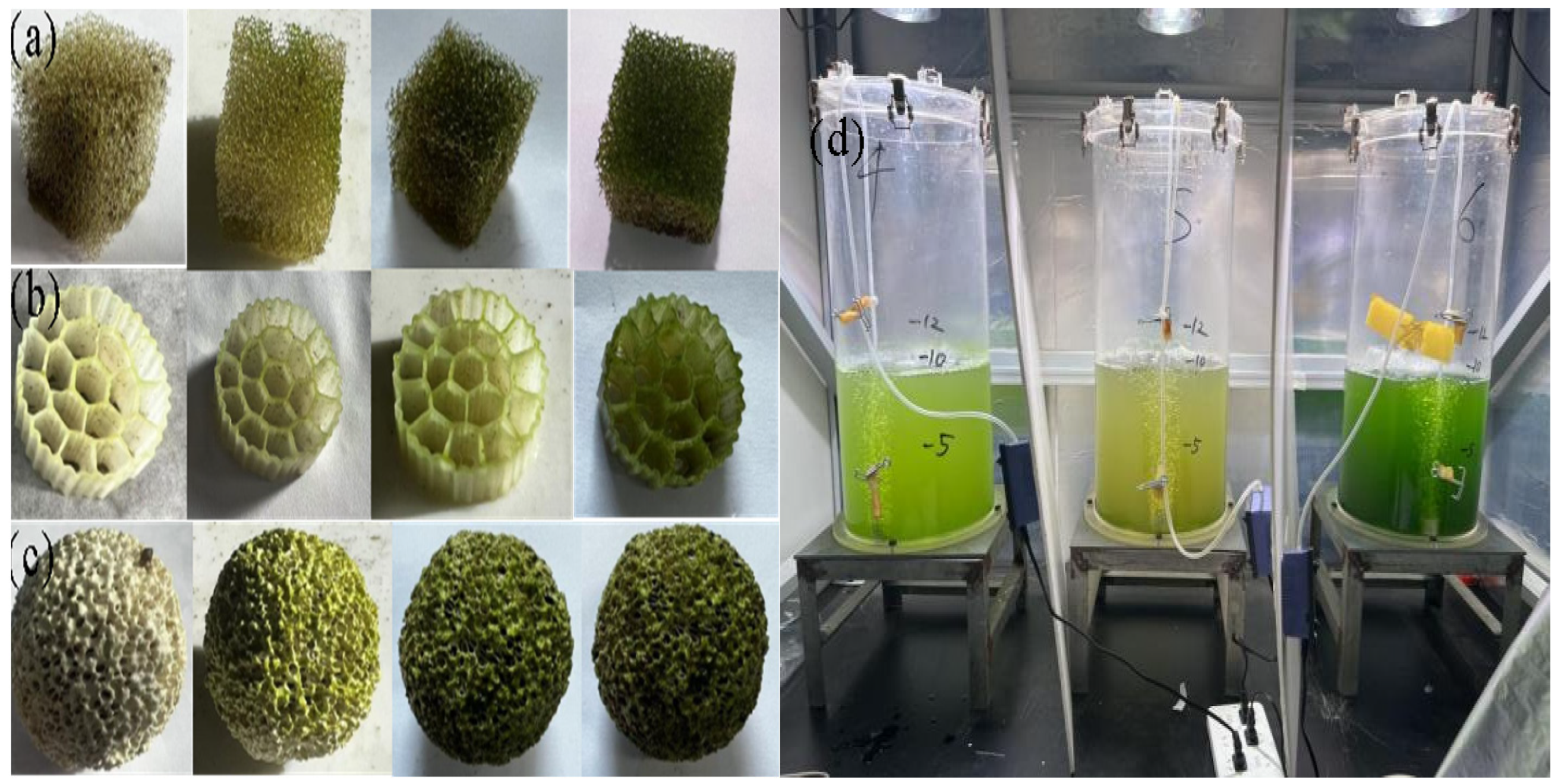
3.1.1. Biomass Dry Weight and Biofilm Formation Rate

After 30 d of continuous cultivation, the biofilm formation phase was essentially complete. A comparative analysis was conducted to evaluate biofilm formation on the different fillers by measuring the biomass weight. Table 1 presents the dry weight of the biofilms and the corresponding biofilm formation rates for the three types of fillers.
According to the Table 1, the polyurethane sponge supported the highest amount of biofilm, with a formation rate of 68.71%. This value was significantly greater than the rates observed for the ceramic filler (10.03%) and the MBBR filler (9.1%). Therefore, based on the biofilm formation rate at equal volumes, the polyurethane sponge exhibited a superior performance compared with both the ceramic and MBBR fillers. This performance could be attributed to the high porosity of the polyurethane sponge, which provided a larger specific surface area for microorganisms to readily adhere to and ample space for physiological activities. Consequently, the adsorption efficiency of both the microbial and algal species was enhanced.

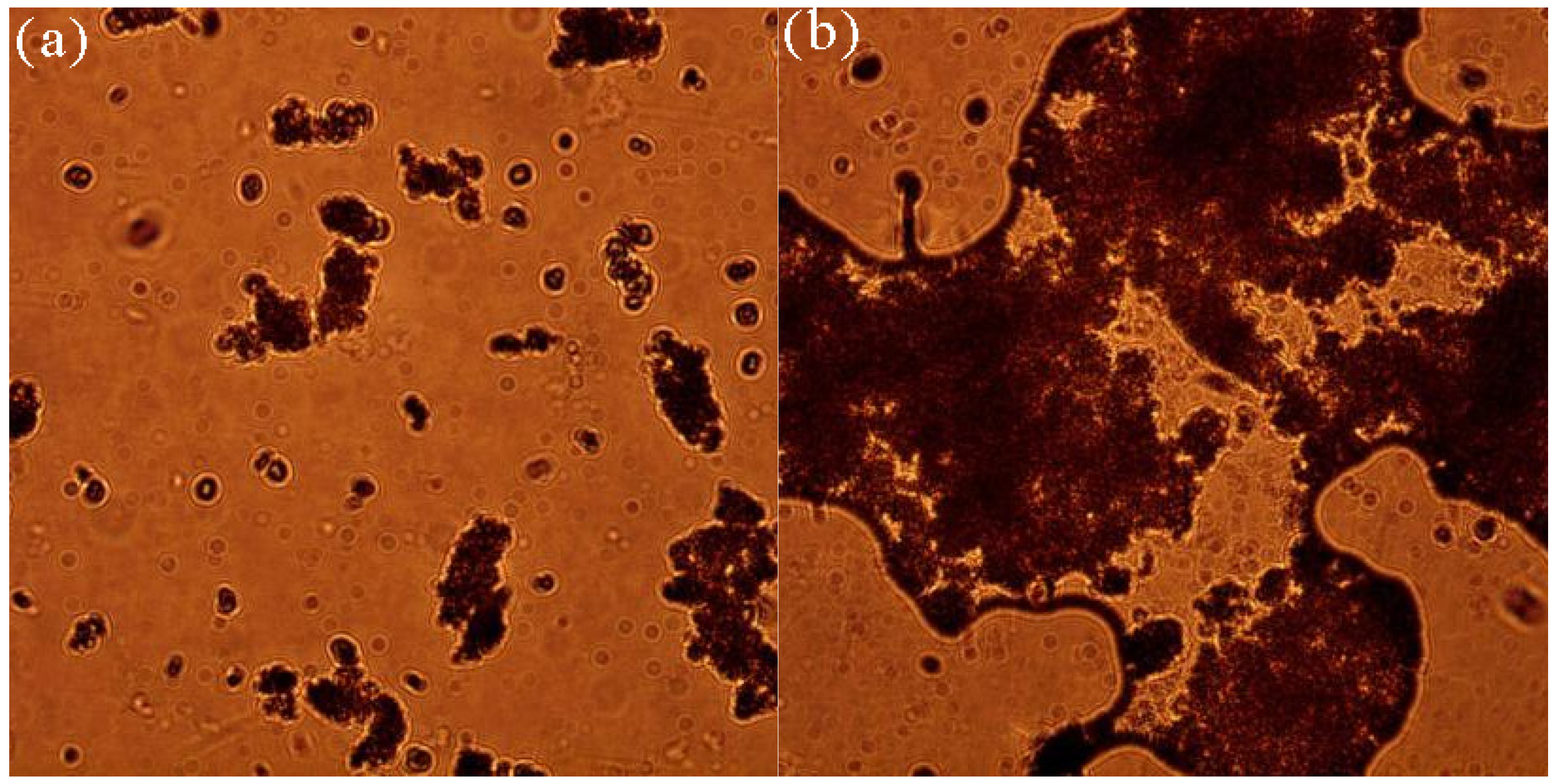
3.1.2. Formation of Microbial-Algal Biofilms Under Microscope

The interaction between Chlorella in water and bacteria in activated sludge is dynamic and complex. The bacteria secrete EPS during the biofilm formation process. EPS has adhesive properties that facilitate the attachment of algae. Moreover, algae, represented by Chlorella, release polysaccharides that further promote bacterial aggregation, which ultimately contributes to the formation of the biofilm.
The magnification of the light microscope was 40×. The integration of microbial and algal cells in water demonstrated a remarkable effect, with numerous clustered areas indicating that these cells adhered to each other through the secretion of EPS and polysaccharides, which ultimately resulted in biofilm formation (Figure 2). This interaction established the basis for mutual attachment, mutual benefit, and a complementary relationship between bacterial and algal cells, which in turn confirmed the successful formation of the biofilm within the reactor. Specific interactions between the microbes and algae were observed, with a visible mucus layer present both around the edges of the clusters and within the matrix (Figure 2). This observation indicated that the adhesion of microbial and algal cells was not coincidental, but rather facilitated by the strong adhesive properties of EPS, which formed structured aggregates. This arrangement provided both structural and functional support for the future application of microbial-algal biofilms in wastewater treatment. The mucus layer adsorbed pollutants from the wastewater to create an environment that promoted cooperative interactions between microbial and algal cells, which in turn supported their synergistic functions.

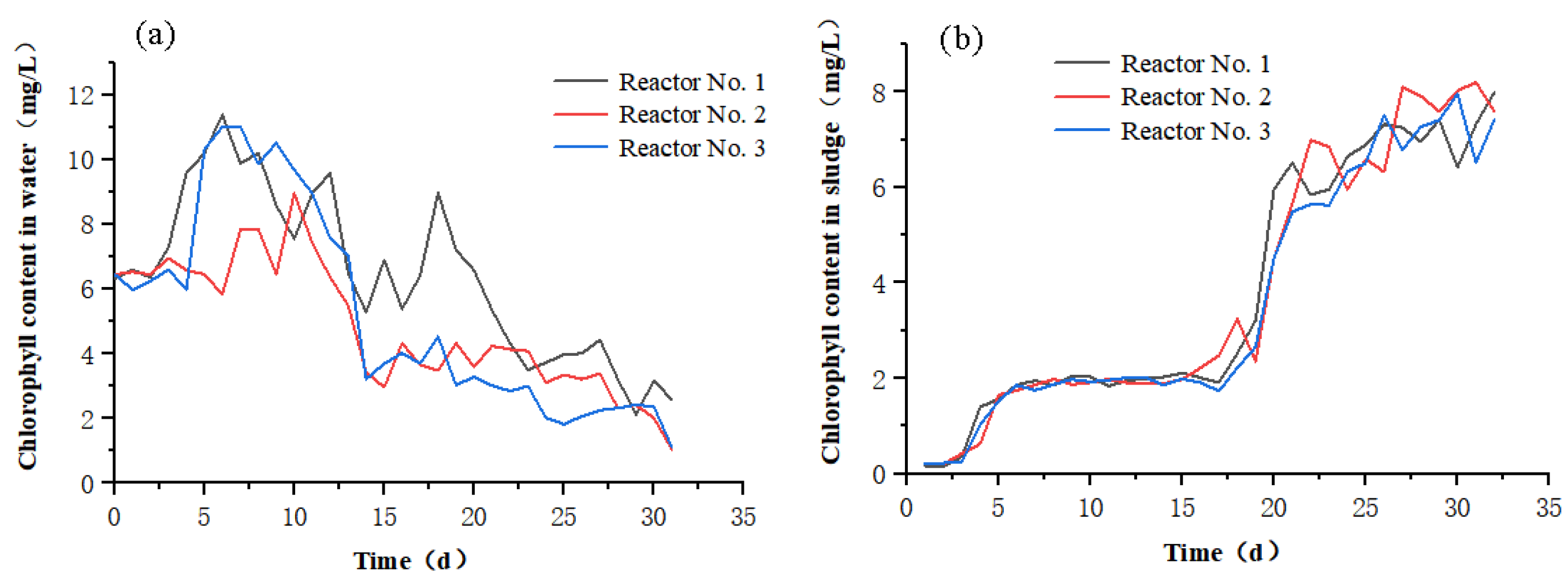
3.1.3. Changes in Chlorophyll Content

Chlorophyll is present within algal cells and is essential for photosynthesis, allowing algae to convert absorbed light energy into chemical energy, which provides nutrients for their growth and reproduction. Therefore, monitoring changes in chlorophyll content in both water and sludge provides a direct assessment of algal population dynamics, algal growth, and the formation of biofilms through interactions between algal and microbial cells. Figure 3 shows the changes in chlorophyll content in both water and sludge over 35 d.
The three reactors exhibited a consistent overall trend (Figure 3a): the initial chlorophyll content in water was 6 mg/L (the fixed initial condition for the introduced Chlorella). The chlorophyll content initially increased, reaching a peak, and then gradually decreased. Between days 15 and 20, fluctuations in chlorophyll content in water were observed in all reactors, followed by a gradual decline. This phenomenon was attributed to the photosynthetic activity of the algae, which resulted in an increased algal population. After a certain population threshold (105–107 CFU/mL) was reached, the algae gradually adhered to each other and formed biofilms that attached to the carrier material.
The overall trend for the three reactors was similarly consistent (Figure 3b): the chlorophyll content in sludge was at 0 mg/L, then it increased slightly and stabilized. By day 20, the chlorophyll content increased significantly, continuing to rise gradually afterward. This pattern was due to the initial mixing of activated sludge with Chlorella, which did not immediately lead to stable adhesion and biofilm formation. Instead, the cells initially formed connections through van der Waals forces, a relatively unstable interaction. After a certain chlorophyll content value was reached, this connection ceased to be dynamic and allowed algal cells to proliferate rapidly. During this phase, both bacteria and algae secreted EPS and polysaccharides, which led to the formation of a stable biofilm with strong adhesion properties and resulted in a substantial increase in the chlorophyll content within the sludge. The fluctuations in chlorophyll content in the water between days 15 and 20 could be attributed to the insufficient and uneven secretion of EPS and polysaccharides during the early stages of biofilm formation. This inadequacy resulted in poor adhesion and an unstable biofilm that was prone to detachment or the formation of large biofilm masses that did not adhere properly to the carrier material, which made them return to the water. The changes in polyurethane foam were substantial, owing to its multi-network hollow structure, which predominantly constituted a mesh framework. Compared with ceramic carriers and MBBR carriers, polyurethane foam was less efficient in adhering to large biofilm masses caused by the uneven and concentrated secretion of EPS and polysaccharides. By day 20, the chlorophyll content in the water steadily decreased. This observation indicated that the algal and bacterial cells formed a “mutual relationship”, which enhanced their adhesion through the secreted EPS and polysaccharides to establish a stable biofilm.

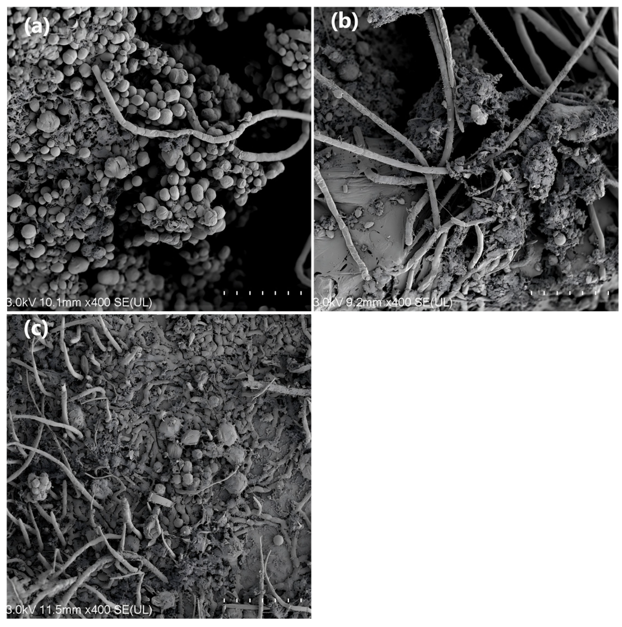
3.1.4. Scanning Electron Microscopy (SEM) Analysis of Microbial-Algal Biofilms

The magnification of the SEM microscope was 400×. Notably, differences were observed in both the quantity and morphology of microbial-algal biofilms on different carriers (Figure 4). The polyurethane foam exhibited a relatively high level of microbial-algal biofilm attachment, with a significantly denser biofilm compared with the other carriers. The formation of a continuous coverage layer with a dense network structure indicated that microbial and algal cells adhered to each other through EPS and polysaccharides, which in turn suggested that this carrier had relatively good biocompatibility.
In contrast, the ceramic carrier had the least amount of attached microbial-algal biofilm, with no extensive biofilm formation and signs of biofilm detachment at the edges. This condition was attributed to the insufficient hydrophilicity of the carrier surface. Owing to the inherent characteristics of the ceramic material, it could not effectively support the adhesion of large biofilm masses. Similarly, the MBBR carrier exhibited suboptimal attachment of the microbial-algal biofilm. Although biofilm was present on the MBBR surface, biofilm formation was inadequate, for reasons similar to those of the ceramic carrier.
The observed differences could be attributed to the three-dimensional network structure of the polyurethane foam, which exhibited a higher porosity compared with the ceramic and MBBR carriers. This structure provided a larger surface area for attachment, better roughness, and improved oxygen supply, all of which contributed to the structural support required for biofilm formation. Consequently, after the secretion of EPS and polysaccharides, the microbial and algal cells could more easily aggregate.

3.1.5. Fourier Transform Infrared Spectroscopy Analysis

Figure 5 shows the Fourier transform infrared (FTIR) spectrum of the biofilm after 30 d of cultivation, from which the functional groups present on the biofilm can be identified. The infrared spectrum of the biofilm on the carriers was analyzed based on the characteristic absorption peaks associated with the major functional groups.
By conducting an FTIR analysis on the biofilm samples from the carriers, we obtained insights into the forms and relative abundances of organic substances, such as proteins, polysaccharides, and lipids, within the biofilm. Notably, absorbance trends for the carriers differed across a wide range of wavenumbers. The polyurethane carrier exhibited a significant absorption peak near 3400 cm−1, potentially associated with O-H stretching vibrations, which in turn indicated a higher presence of hydroxyl groups on its surface. The polyurethane carrier also exhibited a notable absorption peak near 1600 cm−1, potentially associated with C=O or C=C stretching vibrations. In contrast, the absorption peaks for the ceramic carrier near 3400 cm−1 and 1600 cm−1 were relatively weak, while some characteristic peaks appeared in the lower wavenumber region (e.g., 500–1000 cm−1), which potentially correspond to the vibrational modes of inorganic components. The spectral features of the MBBR carrier exhibited unique absorbance variations in certain bands, with an absorption peak near 1500 cm−1, potentially associated with specific functional group vibrations [26].
The O-H stretching vibration peak near 3400 cm−1 in the polyurethane carrier was broader and more pronounced, while the C=O or C=C vibration peak near 1600 cm−1 was also more prominent. In these regions, the absorption in the ceramic carrier was relatively weak, but distinct absorption peaks were present in other bands (e.g., 1000–1500 cm−1), potentially associated with the chemical composition of the carrier surface or the components of the biofilm. The absorbance changes in certain bands for the MBBR carrier demonstrated its unique chemical characteristics.

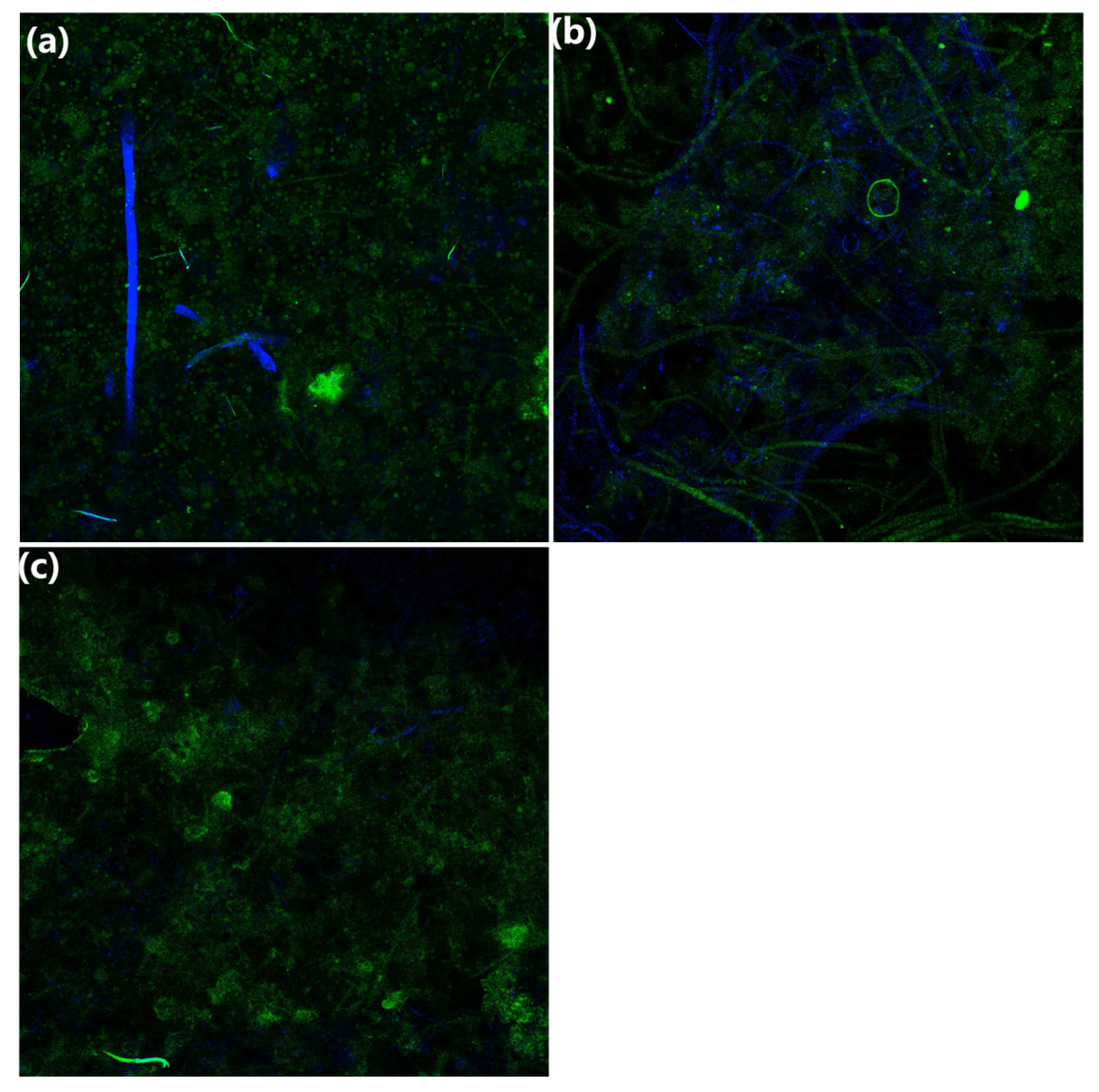
3.1.6. Confocal Laser Scanning Microscopy Analysis

A fluorescence staining method was used to label the cells within the microbial-algal biofilm with green fluorescent protein (GFP) and DNA. The GFP emits green fluorescence at ~530 nm when excited by a 480 nm laser, which in turn indicates protein expression. Propidium iodide (PI) cannot penetrate the intact cell membranes of viable cells. However, PI enters compromised cell membranes and binds to DNA, when excited by a 488 nm laser. Red fluorescence detected at ~660 nm indicates dead cells. After binding to DNA, the DAPI (4′,6-diamidino-2-phenylindole) dye exhibits a maximum absorption peak at 358 nm and a maximum emission peak at 461 nm, emitting blue fluorescence that indicates viable cells. This process shows the distribution of proteins and DNA both inside and outside the cell membranes within the biofilm.
The confocal microscope revealed the composition of the biofilm on the carriers (Figure 6). On the polyurethane foam, the green fluorescence was uniformly distributed and the blue fluorescence was notably present and relatively abundant. This observation indicated a high protein content and the greatest number of viable cells, which in turn demonstrated active microbial metabolism. In contrast, the ceramic carrier exhibited sparse green fluorescence, with fewer and more uniformly distributed blue fluorescence signals. This observation indicated a lower protein content and a reduced number of viable cells compared with the polyurethane foam. This uneven distribution limited its ability to support sufficient microbial metabolic activity for practical applications. The MBBR carrier exhibited the largest and most prominent area of green fluorescence, while the area of blue fluorescence was relatively minimal. This observation indicated the highest protein content and the lowest number of viable cells, which in turn implied poor cell activity and stability. This condition made the MBBR carrier unsuitable for practical applications as well.
For the effective treatment of organic pollutants in wastewater, the presence of viable cells is crucial. Active cell metabolism is essential for achieving effective phosphorus removal and other wastewater treatment outcomes. In this context, the protein content does not contribute significantly to the effectiveness of wastewater treatment. Therefore, protein content is not considered a criterion for evaluating whether the carrier meets the requirements.
In summary, polyurethane foam, characterized by its excellent biofilm formation and the highest number of viable cells, was considered the best carrier in this experiment. Therefore, the polyurethane foam was used as the carrier in subsequent wastewater treatment experiments.

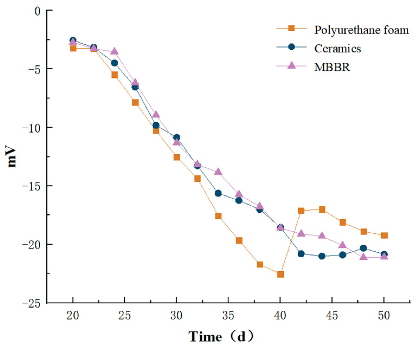
3.1.7. Zeta Potential Analysis

Owing to differences in surface composition and structure among various carriers, the charge magnitude and electrical properties of the biofilms attached to them also vary. The electric potential and properties of microbial surface structures significantly influence their behavior in solution. For example, suspended microorganisms do not exhibit mutual attraction, owing to their overall negative charge, whereas microorganisms with flocculating characteristics can aggregate through the interplay of positive and negative charges [27].
Figure 7 shows the Zeta potential variation curves for the polyurethane foam, ceramic, and MBBR carriers. The initial Zeta potentials of the microbial-algal biofilms on the three carriers were −3.23 mV, −2.56 mV, and −2.74 mV, respectively. The biofilms on all carriers exhibited a negative charge (Figure 7). Related research has shown that traditional biofilm carriers, such as polyethylene (PE), polypropylene (PP), and high-density polyethylene (HDPE), also have negatively charged surfaces, which may lead to electrostatic repulsion between the biofilm carriers and bacterial cells [28].
As the experiment progressed, the Zeta potentials of the biofilms on all carriers exhibited a decreasing trend. The trends for the ceramic and MBBR carriers were similar, while the Zeta potential for the biofilm on the polyurethane foam exhibited a substantial decline. However, in the later stages of biofilm formation, the Zeta potential increased abruptly. This observation might be due to the mesh structure of the polyurethane foam, which could not provide sufficient attachment points for the bulk biofilm. Consequently, although the polyurethane foam had the highest attachment rate, the resulting biofilm structure was predominantly dictated by the characteristics of the carrier, rather than natural processes, as observed with the ceramic and MBBR carriers. In these cases, the biofilm structure was formed through natural binding processes involving EPS and polysaccharides secreted by the microbial cells.
A smaller Zeta potential reduces electrostatic repulsion between microbial flocs, which in turn enhances the flocculation and stability of the microbial-algal biofilm in photobioreactors. Therefore, in the long term, the charge characteristics of the biofilms on the ceramic and MBBR carriers are the most stable, which ultimately indicates the best adsorption performance, followed by polyurethane foam.

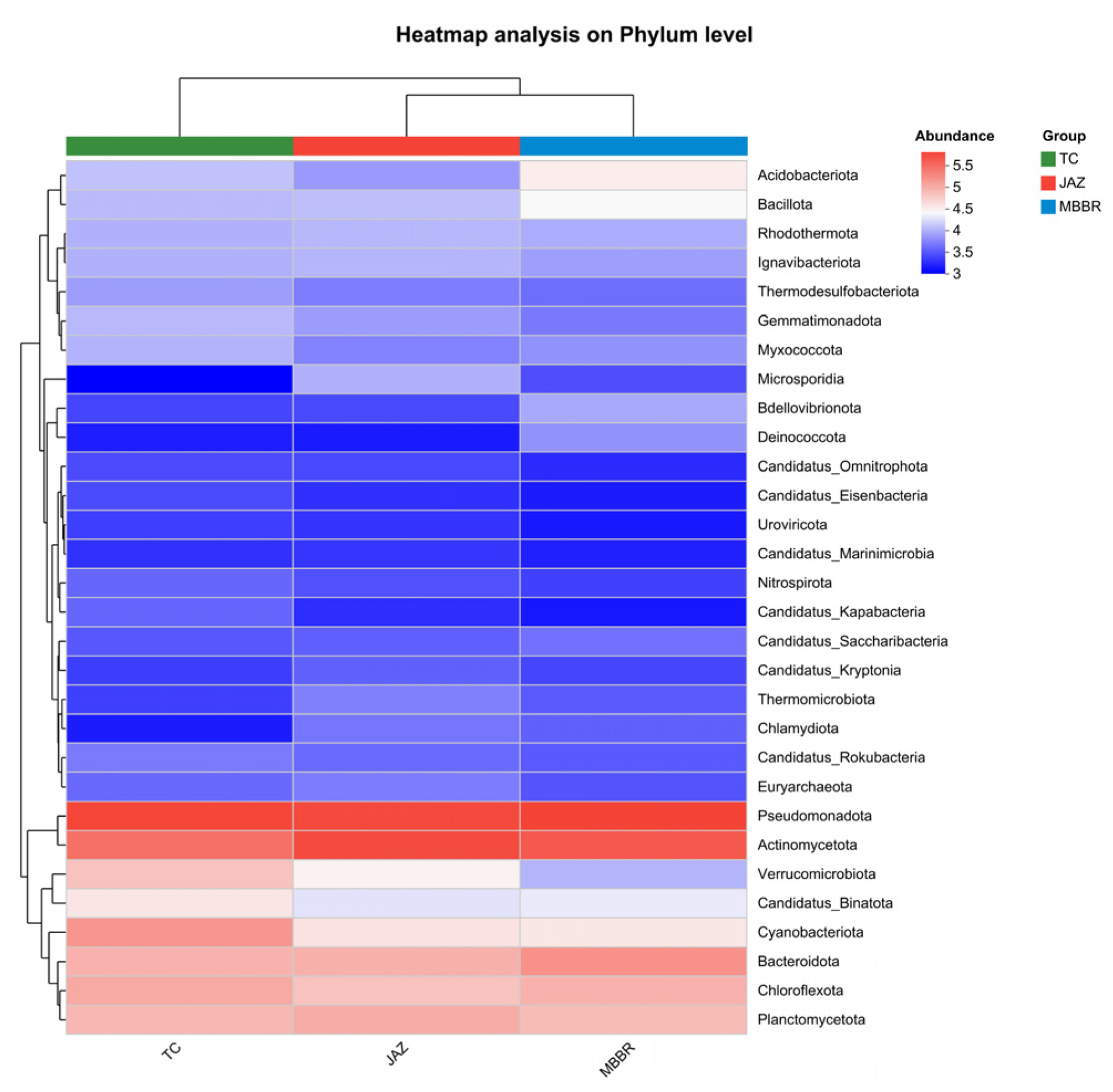
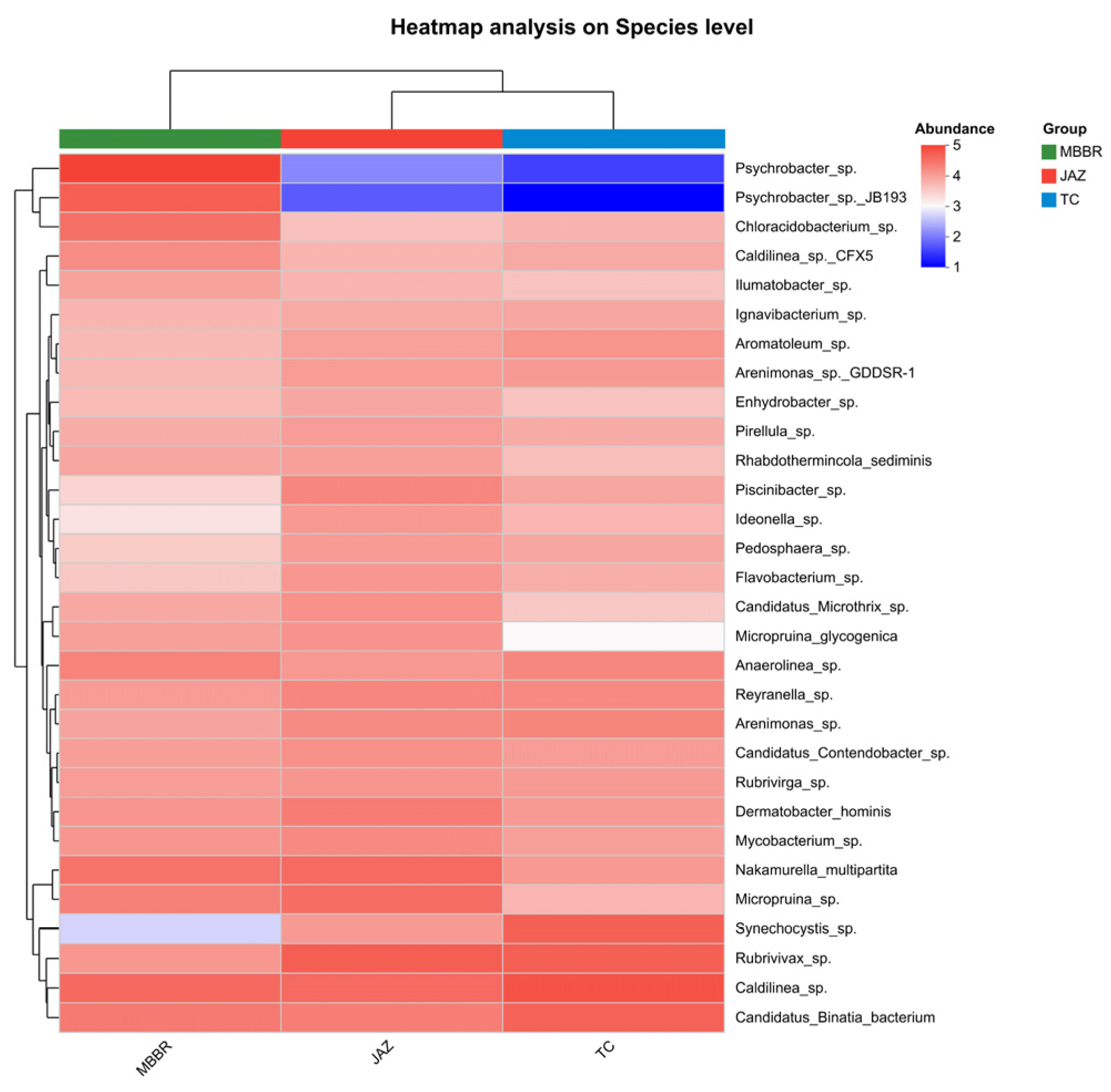
3.2. Analysis of Microbial Community Characteristics

After biofilm formation, the characteristics of microbial community diversity, variability, relative abundance, and biomass on the carrier surface provided insights into the succession patterns of microbial communities on surfaces of different materials and sizes. This information could assist in selecting the most suitable carriers to enhance and maintain the long-term stable growth of microbial-algal biofilms.

3.2.1. Analysis of Community Structural Diversity

Biodiversity is typically described at three levels, as follows: species diversity, genetic diversity, and ecosystem diversity. Microbial diversity encompasses various aspects such as microbial genetic diversity, physiological diversity, species diversity, and ecological diversity [29]. To further explore the structural characteristics of microbial communities at different levels, analyses were conducted at the phylum and species levels (Figure 8 and Figure 9).
The clustering heatmap of microbial communities at the phylum level revealed that phyla such as Proteobacteria and Bacteroidota were highly abundant across all of the carriers (Figure 9). However, the polyurethane sponge (JAZ) carrier exhibited a relatively higher abundance of Gemmatimonadota, which in turn indicated some specificity in its community composition. In contrast, the ceramic (TC) carrier exhibited a higher abundance of Actinomycetota and the MBBR carrier exhibited a relatively higher abundance of Chloroflexota.
The clustering heatmap of microbial communities at the species level revealed that the polyurethane sponge (JAZ) carrier had higher abundances of Psychrobacter sp. and Chloracidobacterium sp. (Figure 9). These species might have a competitive advantage in the environment of the polyurethane sponge (JAZ) carrier and might be involved in specific metabolic processes. In contrast, the ceramic (TC) carrier had a higher abundance of Pseudomonas sp. and Mycobacterium sp., which might play an important role in the ceramic (TC) carrier environment. On the MBBR carrier, a relatively higher abundance of Rubrivirga sp. and Caldilinea sp. was observed.

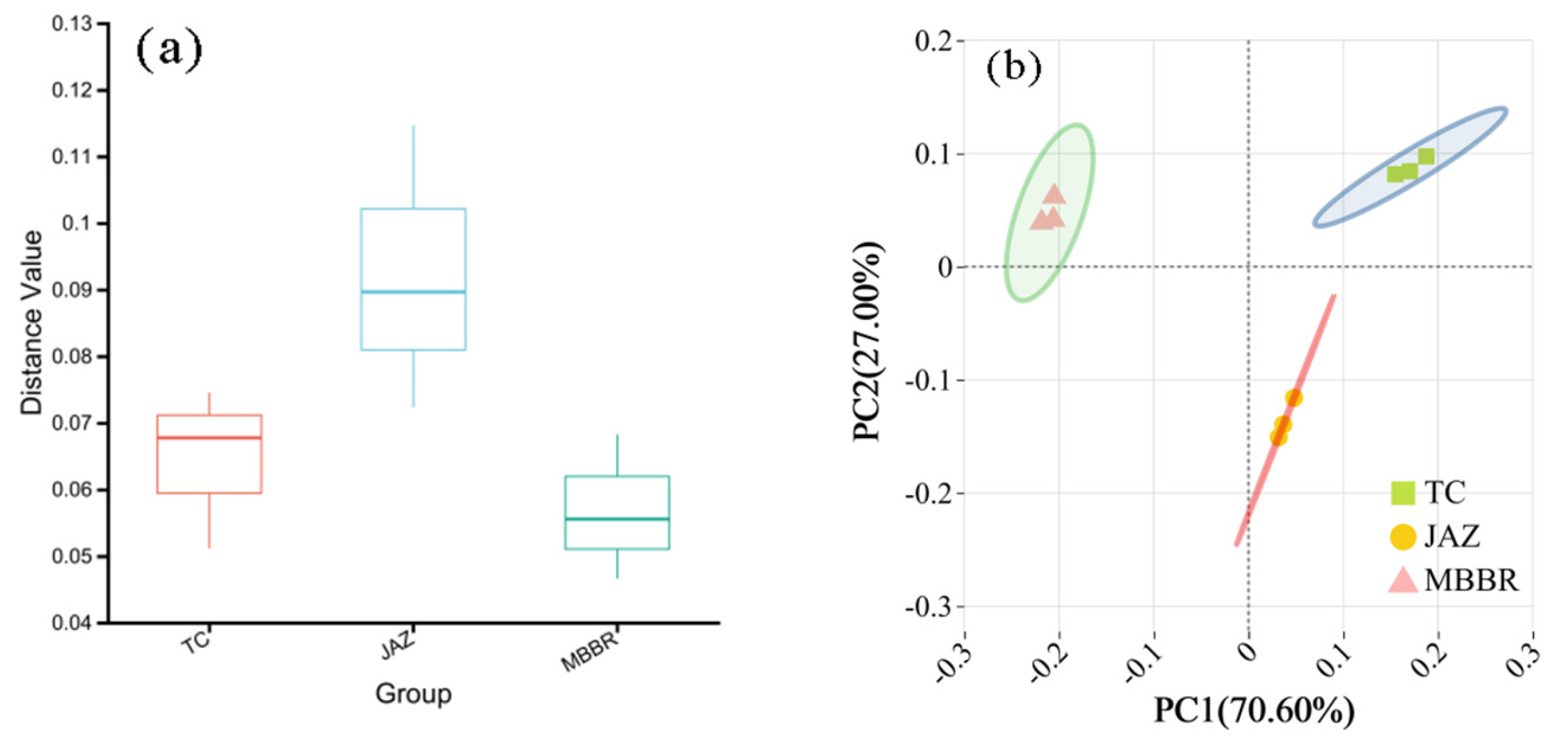
3.2.2. Beta Diversity Analysis

Through a Beta diversity difference analysis (Figure 10a) and PCoA (Principal Coordinates Analysis) analysis (Figure 10b), the microbial community structure of biofilms on different carriers was thoroughly investigated. The results of the Beta diversity analysis indicated that the microbial community on the polyurethane sponge carrier (JAZ) exhibited high diversity, with a wide range of distance values (0.08 to 0.11), which suggested a complex and rich community structure. The community structure on the ceramic carrier (TC) was relatively stable, with distance values concentrated between 0.06 and 0.07. In contrast, the community structure on the MBBR carrier exhibited lower diversity, with distance values mainly ranging from 0.05 to 0.06.
PCoA analysis further revealed the distribution characteristics of microbial communities across different carriers. According to the results (Figure 10a,b), the community structure on the ceramic carrier (TC) was relatively dispersed along the directions of PC1 and PC2, which ultimately indicated a certain degree of variability in its community composition. The community structure on the polyurethane carrier (JAZ) was relatively concentrated, showing higher similarity, with noticeable separation from the ceramic carrier (TC) along the direction of PC1. This observation indicated significant differences in community composition between the carriers. The community structure on the MBBR carrier was distinctly separated from the polyurethane carrier along the direction of PC1. This observation indicated substantial differences in community composition compared with the other carriers.
At the phylum level, the microbial community structures on different carriers exhibited substantial differences. The microbial community on the polyurethane sponge carrier exhibited a higher abundance of Gemmatimonadota, as well as greater diversity and complexity, which resulted in significant separation from the other carriers in the PCoA analysis.
At the species level, significant differences were observed in the microbial community composition across different carriers. The polyurethane sponge carrier had higher abundances of Psychrobacter sp. and Chloracidobacterium sp., while the ceramic carrier had higher abundances of Pseudomonas sp. and Mycobacterium sp. Additionally, the MBBR carrier had relatively higher abundances of Rubrivirga sp. and Caldilinea sp.

3.3. Factors Influencing Microbial-Algal Biofilm Photoreactors

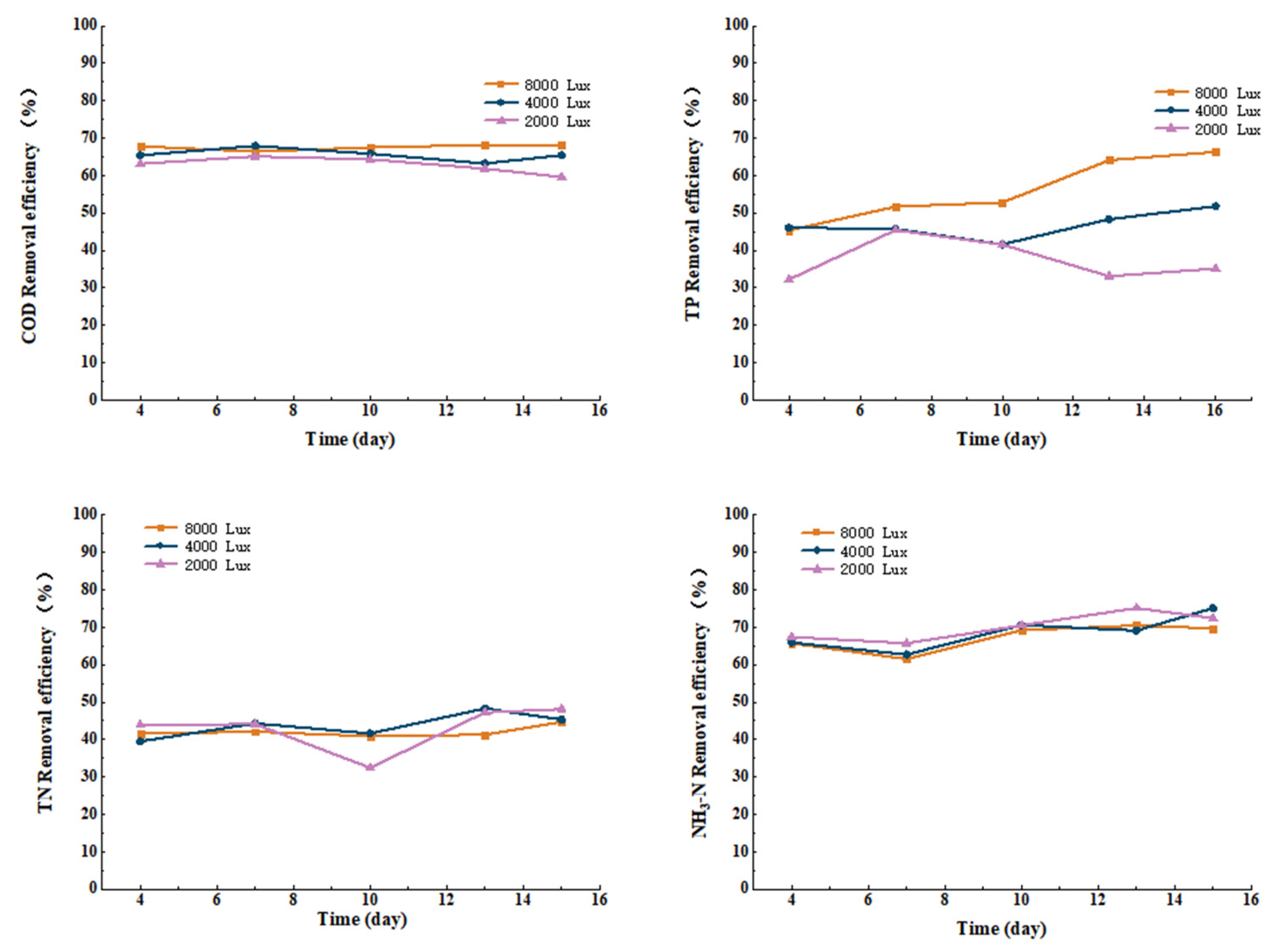
3.3.1. Effect of Light Intensity on COD, TN, TP, and Ammonia Nitrogen Removal from Cattle Wastewater by Microbial-Algal Biofilms

Light plays an important role in algae growth, as light energy directly influences algal development, which in turn affects the overall removal efficiency of organic pollutants by the microbial-algal system [30]. Studies have shown that microbial-algal biofilm systems exposed to light develop more diverse microbial communities compared with those grown in darkness [31]. Under strong light conditions, algae serve as the main “cornerstone” during the initial stages of biofilm formation, while fungi take on this role under low light conditions [32]. Moreover, the effects of light intensity on microbial-algal biofilm systems encompass variables such as the removal rates of organic pollutants, algal photosynthesis, and the nitrification processes of fungal cells [33]. Therefore, identifying the optimal lighting conditions is crucial for the microbial-algal biofilm treatment of wastewater. The color of light (i.e., the wavelength), discussed below, is also essential for exploring the role of light.
Under conditions of similar initial water quality, with a constant light color, pH, temperature, and aeration, the impacts of light intensities of 2000 lux, 4000 lux, and 8000 lux on the COD, TP, TN, and ammonia nitrogen removal were determined. Data were collected at different stages, as follows: D4, D7, D10, D13, and D15. At light intensities of 2000 lux, 4000 lux, and 8000 lux, the maximum COD removal rates were 65.9%, 67.9%, and 71.9%, respectively; the maximum TP removal rates were 46.7%, 51.8%, and 66.3%, respectively; the maximum TN removal rates were 48.1%, 48.3%, and 44.7%, respectively; and the maximum ammonia nitrogen removal rates were 75.1%, 75.1%, and 70.5%, respectively. It can be seen that TN manifests a significant decrease on the 10th day at 2000 lux. Under the low light condition of 2000 lux, the ATP and NADPH produced by the photoreaction of algae are insufficient, resulting in a significant decrease in their carbon fixation capacity. The organic matter synthesized by algae through photosynthesis is the main carbon source for heterotrophic denitrifying bacteria. On the 10th day, the intracellular carbon sources of algae stored in the system may be exhausted, and the amount of newly synthesized organic matter under low light is insufficient to meet the metabolic requirements of denitrifying bacteria, resulting in restricted denitrification processes and a decrease in the TN removal rate. Refer to Figure 11 for details.

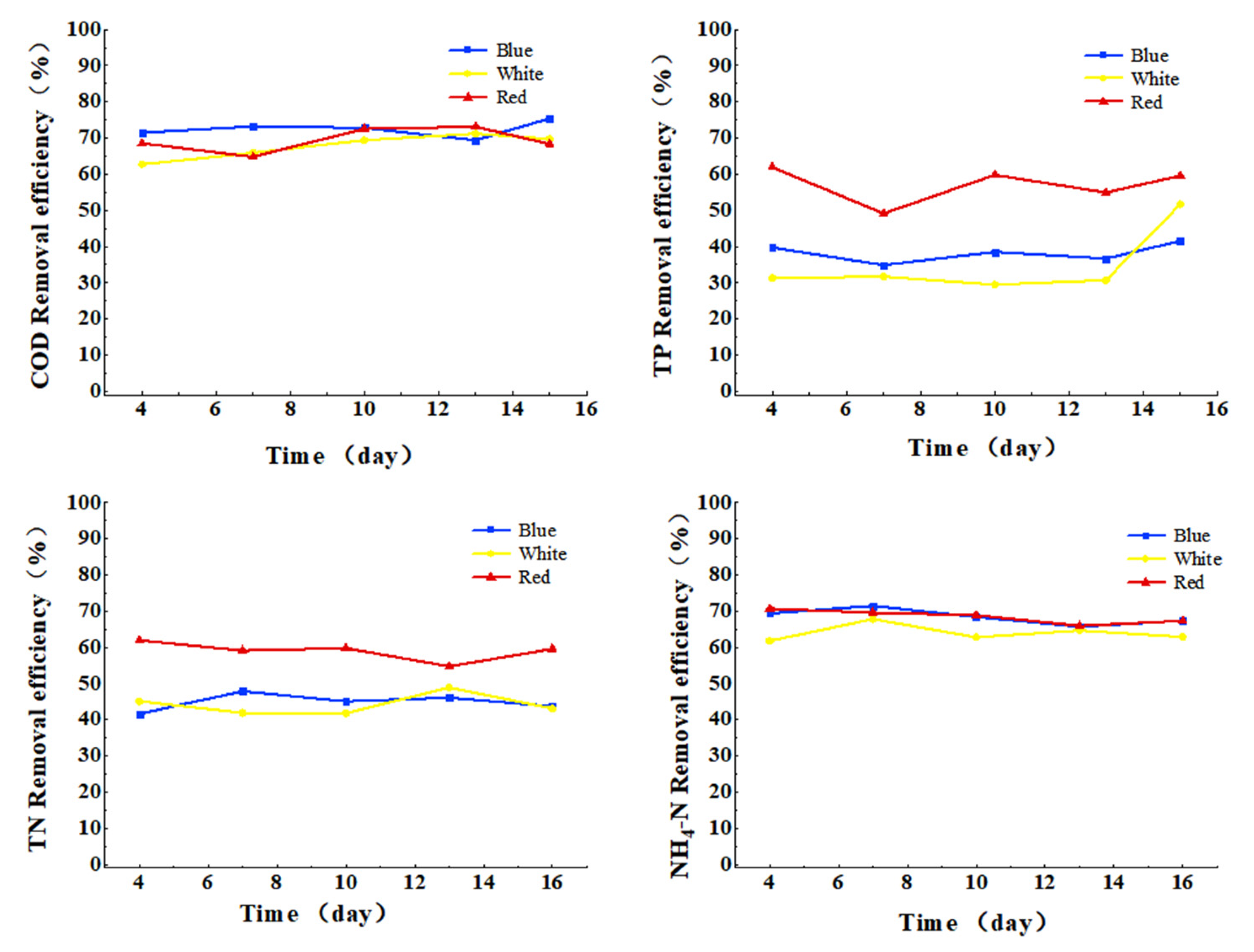
3.3.2. Effect of Light Color on COD, TN, TP, and Ammonia Nitrogen Removal from Cattle Wastewater by Microbial-Algal Biofilms

Under conditions of similar initial water quality, with constant light intensity, pH, temperature, and aeration, data were collected at different stages, as follows: D4, D7, D10, D13, and D15. This was to assess the effects of light colors (blue, white, and red) on the COD, TP, TN, and ammonia nitrogen removal. According to the results of Figure 12, the highest COD removal rate was 73.2% under blue light on D7; the highest TP removal rate was 51.8% under white light on D15; the highest TN removal rate was 61.9% under red light on D4; and the highest ammonia nitrogen removal rate was 71.5% under blue light on D7. These findings indicated that different colors of light had varying effects on the removal rates of COD, TP, TN, and ammonia nitrogen. Specifically, under red light, the COD removal rate was 72.5% and the TN removal rate was 61.9%, both of which were the highest among the three light colors tested. Under blue light, the ammonia nitrogen removal rate was highest at 71.5%. Under white light, the TP removal rate peaked at 51.7%.
It can be seen that different light colors (i.e., illumination wavelengths) play a role in removal of the organic matter, because the key pigments of photosynthesis, such as chlorophyll A (Chl-a), chlorophyll b (Chl-b), and other pigments, have different sensitivities to different illumination wavelengths. Since the microalgae photosystem contains a unique group of photopigments and absorbs special spectra, each microalgae species has a unique spectral composition that is suitable for its own needs. The relevant studies indicate that the illumination color plays a role in the growth of algae [34].

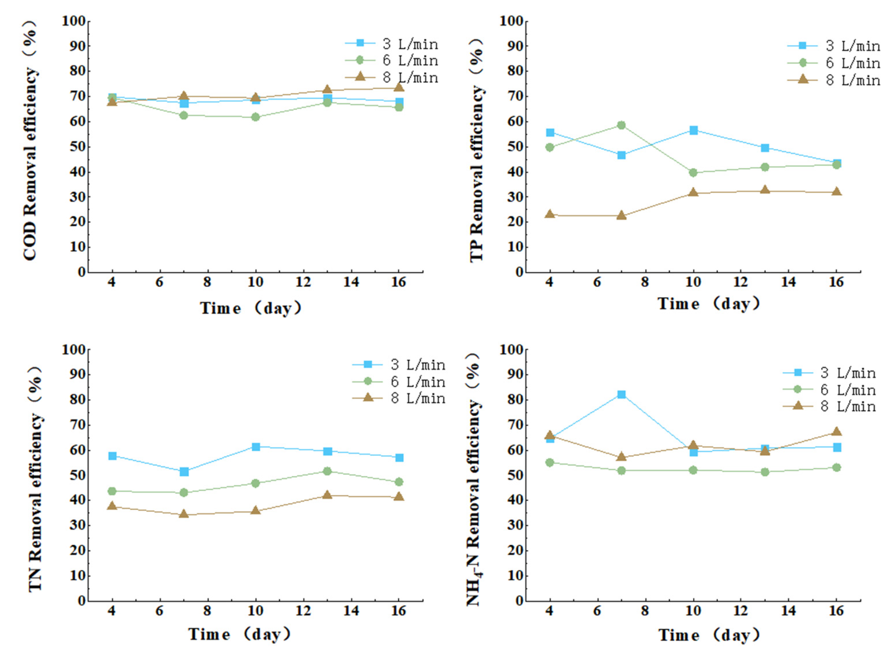
3.3.3. Effect of Aeration Intensity on COD, TN, TP, and Ammonia Nitrogen Removal from Cattle Wastewater by Microbial-Algal Biofilms

Some studies have demonstrated that under non-aeration conditions, the oxygen produced via algal photosynthesis is sufficient to support the oxidation and removal of organic pollutants by fungi, which ultimately achieves good removal efficiency [35]. However, other studies have observed that external aeration can further enhance pollutant removal rates. Additionally, the turbulence created by aeration promotes better substrate exchange and facilitates the formation of well-settling microbial-algal aggregates [36]. Under the same initial water quality conditions, with constant light intensity, light color, pH, and temperature, data were collected at different stages, as follows: D4, D7, D10, D13, and D15. This was to assess the effects of aeration intensities of 3, 6, and 8 L/min on the COD, TP, TN, and ammonia nitrogen removal. According to the results of Figure 13, at D15, when the aeration intensity is 8 L/min, the highest removal rate of COD and ammonia nitrogen is achieved, which is 73.4% and 67.1%, respectively. At D10, when the aeration intensity is 3 L/min, the highest removal rate of TP and TN is achieved, which is 56.7% and 61.5%, respectively. At the aeration intensity of 3 L/min, the content of ammonia nitrogen on the seventh day showed a rising trend, possibly because the low aeration intensity (3 L/min) led to insufficient DO in the reactor in the initial stage (days 1–5), with a thin aerobic layer formed on the outer layer of the biofilm and the inner layer in an anoxic state. As algae grow, the oxygen produced by their photosynthesis temporarily increases the DO on the outer layer of the biofilm, which just meets the activity threshold of ammonia-oxidizing bacteria, while the inner layer still maintains an oxygen-deficient environment. This micro-gradient structure of “external aerobic—internal anoxic” enables shortcut nitrification and denitrification reactions on the biofilm, thereby increasing the removal rate of ammonia nitrogen. It can be seen that with the increase in aeration intensity, the removal rate of COD, TN, TP, and NH4-N does not increase, because the aeration method selected for the present study is that by aeration head. With the increase in aeration intensity, the water bubbles are formed to carry oxygen up and leave the system, resulting in no actual increase in the DO content in the system, which shall be improved in future experiments.

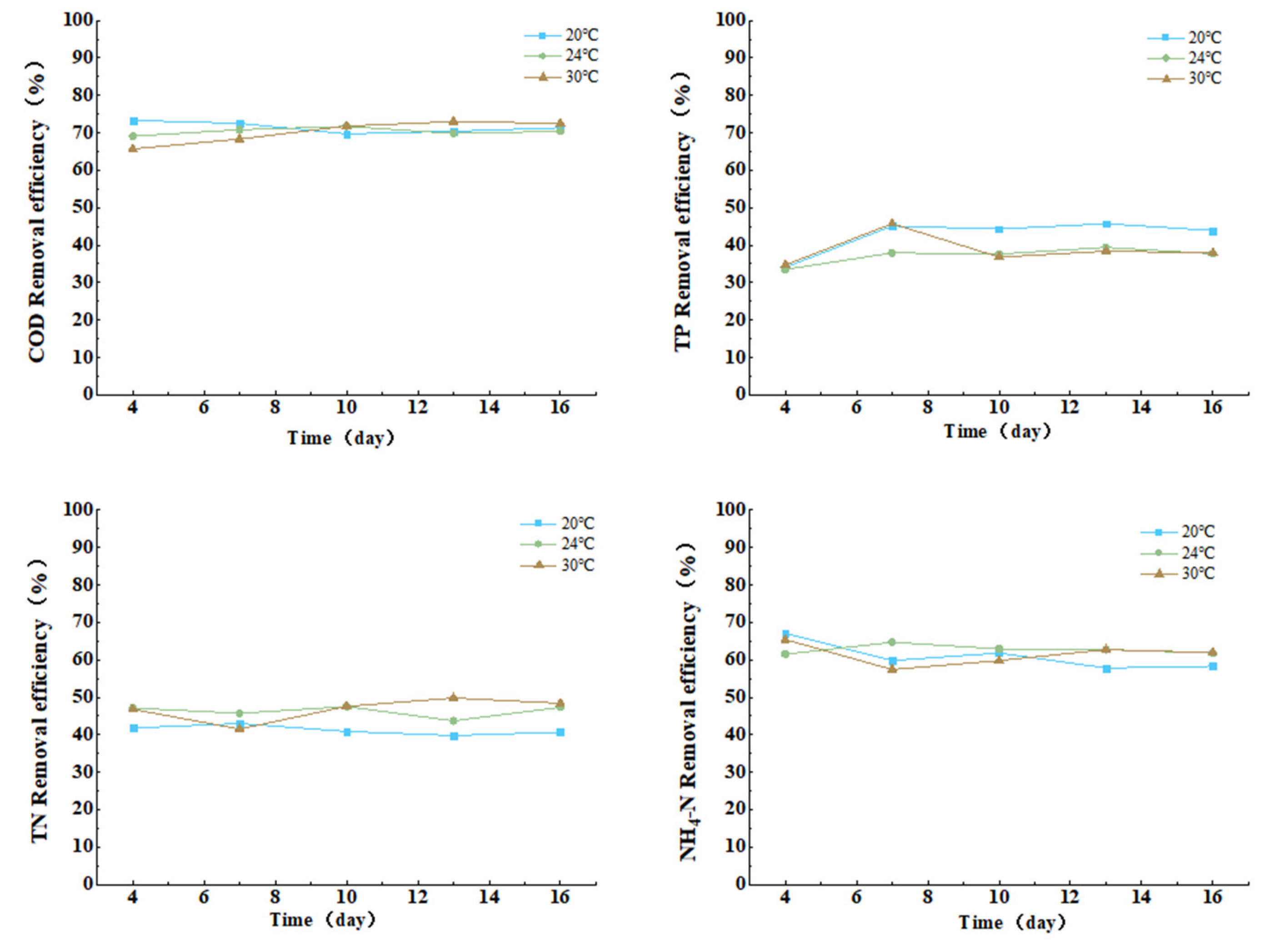
3.3.4. Effect of Temperature on COD, TN, TP, and Ammonia Nitrogen Removal from Cattle Wastewater by Microbial-Algal Biofilms

The effect of temperature on microbial-algal biofilms should not be overlooked, as it significantly influences the performance of the biofilm system [37,38]. Research has demonstrated that temperature can affect algal growth rates, community structure, and photosynthesis. Related studies have also demonstrated that under constant light intensity and optimal nutrient conditions, temperature variations directly impact algal growth rates [39,40]. In this study, the treatment of cattle wastewater using microbial-algal biofilms at different temperatures was investigated. Under conditions of similar initial water quality, with constant light intensity, light color, pH, and aeration, data were collected at different stages, as follows: D4, D7, D10, D13, and D15. This was in order to assess the effects of temperatures of 20 °C, 24 °C, and 30 °C on the COD, TP, TN, and ammonia nitrogen removal. According to Figure 14, the highest removal rate of COD and ammonia nitrogen is 73.3% and 67.1%, respectively, all of which occur on the fourth day at the optimal temperature of 20 °C. The highest removal rate of TN and TP is 47.5% and 45.7%, respectively, all of which occur on the seventh day at the optimal temperatures of 24 °C and 30 °C, respectively.
From the above data, it can be seen that with the rise in temperature, the removal rate of COD, TN, and ammonia nitrogen will show a slight rising trend; however, the removal rate of TP does not increase with the rise in temperature, because the algae used in this experiment is Chlorella with the optimal temperature ranging from 15 °C to 25 °C. Within this temperature range, Chlorella will achieve optimal growth and metabolic activities. When the temperature rises, the algae species grows faster but beyond their most suitable temperature range, resulting in a decrease in their activity. Therefore, the phenomenon shown in the figure occurs.

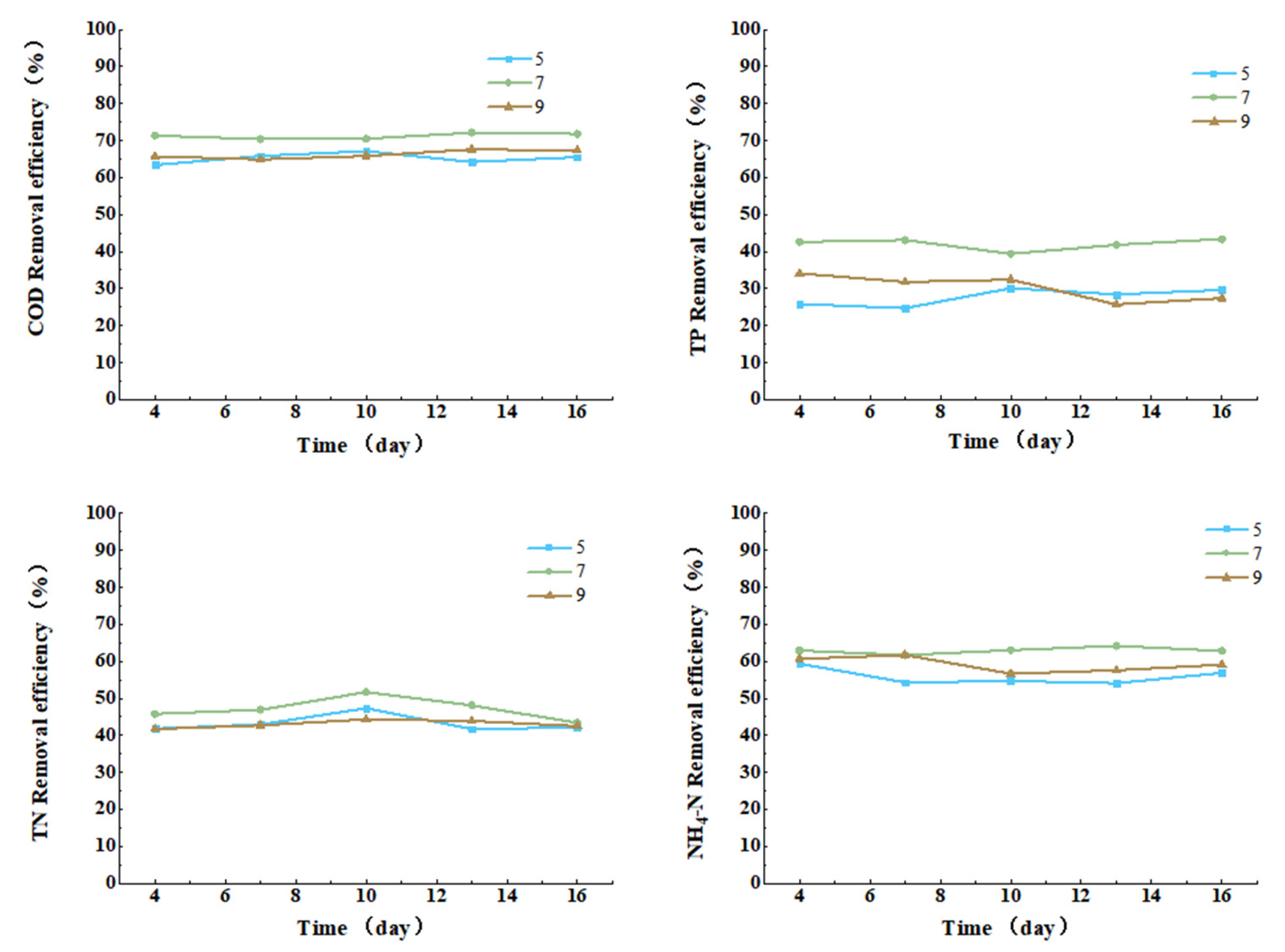
3.3.5. Effect of pH on COD, TN, TP, and Ammonia Nitrogen Removal from Cattle Wastewater by Microbial-Algal Biofilms

The pH value is fundamentally important for any biological system. For example, most microalgal species are buffered within a pH range of 7 to 9 [41]. Therefore, investigating the optimal pH for the microbial-algal biofilm is essential. Under conditions of similar initial water quality, with constant light intensity, light color, temperature, and aeration, data were collected at different stages, as follows: D4, D7, D10, D13, and D15. This was in order to assess the effects of pH levels of 5, 7, and 9 on the COD, TP, TN, and ammonia nitrogen removal. It can be seen from Figure 15 that for the removal of COD, TP, TN and ammonia nitrogen, the optimal pH value is 7. For the removal of COD and TP, the highest removal rates are achieved on the 15th day, which are 71.8% and 43.4%, respectively. For the removal of TN and ammonia nitrogen, the highest removal rate is achieved on the 10th day, which is 51.7% and 63.0%, respectively.
It can be seen that if the pH value is 7, it is possible to achieve the optimal removal rate of COD, TN, TP, and ammonia nitrogen, because at a neutral pH value, the enzyme activity in most microorganisms will reach its peak, so as to promote the absorption and decomposition of organic matter by microorganisms. However, in a too acidic or too alkaline environment, the denaturation of the enzyme protein structure will occur, affecting the removal effect.

4. Discussion and Conclusions

In the present study, the mechanism for high-efficiency treatment of cattle farming wastewater is revealed through the screening of fillers, multi-dimensional characterization, and process optimization of the alga-bacteria biofilm system, and a coupling model of “filler characteristics-community functions-process parameters” is established.
In terms of the filler performance, with its high porosity, high specific surface area, and abundant functional groups on the surface, the polyurethane sponge is used to construct a three-dimensional network biofilm structure, and the dry weight and adhesion rate of its biofilm are significantly better than those of ceramic and MBBR fillers. The confocal laser scanning microscopy shows that the density of living cells on the surface of this filler is the highest, and high-throughput sequencing further confirms its enrichments of Psychrobacter sp. and Chloracidobacterium sp., the former one is resistant to a low temperature and secretes polysaccharides to strengthen the biofilm structure, while the latter one produces oxygen by photic driving to promote nitrification, so as to increase the removal rate of ammonia nitrogen, both of which form the “light energy capture-organic carbon supply-nitrogen-phosphorus conversion” system with the extracellular polymers (EPS), so as to lay the foundation of the community for the removal of the pollutants at low temperature.
The research on the influencing factors on the removal of pollutants shows that under the same influent limitations, it is possible to achieve the optimal overall pollutant removal rate if the illumination intensity is 8000 lux, the illumination color is red, the temperature is 20 °C, the pH value is 7, and the aeration intensity is 8 L/min. Meanwhile, as the concentration of pollutants in the cattle farming wastewater varies with multiple factors, the present study is not limited to finding a single culture condition. Instead, the treatment effect is analyzed through the synergy of multiple factors. Therefore, the alga-bacteria biofilm culture scheme can be adjusted according to the changes in pollutants in the cattle farming wastewater. In the case of the high COD content, the illumination intensity and aeration intensity shall be increased; in the case of the high TN content, the temperature shall be raised and the aeration intensity shall be reduced, and the illumination color shall be adjusted to red; in the case of the high TP content, the illumination intensity shall be increased and the aeration intensity shall be reduced, and the illumination color shall be adjusted to red.
The current literature includes studies on fillers and their surface chemical properties. For instance, Chen et al.’s research has demonstrated the relationship between fillers and biological abundance [42]. There are also studies showing the influence of the physical structure of fillers on treatment efficiency. This study uncovers the key role of the surface chemical properties of the filler in the stability of the biofilm. The study breaks through the limitations of traditional fillers with a focus only on physical structure. Based on the multi-factor collaborative analysis of “light-temperature-aeration”, a correlation model of “light energy utilization efficiency-microbial metabolic activity-pollutant removal” is established, so as to provide the theoretical basis for global optimization of the process parameters.
According to the research results, a technical scheme with low energy consumption and high toughness is made for the treatment of cattle farming wastewater in low-temperature areas. The future research could further analyze the tolerance mechanism of biofilm to extreme pollutants, optimize the utilization efficiency of light energy and the biomass harvesting process, and promote the engineering-based application of this technology. An LED combined light source adapted to the absorption spectrum of algae could be developed, which would enable the limitation of an illumination intensity of 8000 lux to be overcome.

Author Contributions

Conceptualization, investigation, writing—original draft, W.Z. Writing—review and editing, funding acquisition, supervision, L.W. Validation, project administration, M.L. Resources, software, methodology, Y.C. Resources, supervision, C.L. Writing—review and editing, methodology, C.W. Investigation, S.S. All authors have read and agreed to the published version of the manuscript.

Funding

This work was supported by Jilin Province Science and Technology Development Plan Item (20220203106SF).

Data Availability Statement

The data will be made available on reasonable request.

Conflicts of Interest

Author Cong Wang was employed by the company Municipal Engineering Northeast Design and Research Institute Co., Ltd. The remaining authors declare that the research was conducted in the absence of any commercial or financial relationships that could be construed as a potential conflict of interest.

References

  1. Liu, C.; Feng, C.; Duan, Y.; Wang, P.; Peng, C.; Li, Z.; Yu, L.; Liu, M.; Wang, F. Ecological risk under the dual threat of heavy metals and antibiotic resistant Escherichia coli in swine-farming wastewater in Shandong Province, China. Environ. Pollut. 2023, 319, 120998. [Google Scholar] [CrossRef] [PubMed]
  2. Huang, L.; Lu, Z.; Xie, T.; Wang, L.; Mo, C. Nitrogen and phosphorus removal by coupling Anaerobic ammonia oxidation reaction with algal-bacterial symbiotic system. J. Environ. Chem. Eng. 2022, 10, 108905. [Google Scholar] [CrossRef]
  3. Qv, M.; Dai, D.; Liu, D.; Wu, Q.; Tang, C.; Li, S.; Zhu, L. Towards advanced nutrient removal by microalgae-bacteria symbiosis system for wastewater treatment. Bioresour. Technol. 2023, 370, 128574. [Google Scholar] [CrossRef]
  4. Qixin, L.; Xuan, F.; Zhiya, S.; Wenxin, S.; Shuo, W.; Ji, L. Enhanced wastewater treatment performance by understanding the interaction between algae and bacteria based on quorum sensing. Bioresour. Technol. 2022, 354, 127161. [Google Scholar] [CrossRef]
  5. Zhang, B.; Wu, L.; Guo, Y.; Lens, P.N.; Shi, W. Rapid establishment of algal bacterial granular sludge system by applying mycelial pellets in a lab-scale photo-reactor under low aeration conditions: Performance and mechanism analysis. Environ. Pollut. 2023, 322, 121183. [Google Scholar] [CrossRef]
  6. Zhou, Y.; Li, X.; Chen, J.; Wang, F. Treatment of antibiotic containing wastewater with self-suspended algae-bacteria symbiotic particles: Removal performance and reciprocal mechanism. Chemosphere 2023, 323, 138240. [Google Scholar] [CrossRef]
  7. Zhang, Z.; Liu, X.J.; Chen, X.; Yao, L.P.; Zhang, R.Q. Application and prospect of microalgae biotechnology in carbon neutralization. China Biotechnol. 2022, 42 (Suppl. 1), 160–173. [Google Scholar]
  8. Zhang, Z.H.; He, W.H.; Xiang, T.Y.; Shan, Z.D. Algal-bacterial symbiotic SBBR for biogas slurry treatment from pig farm. Technol. Water Treat. 2018, 44, 118–122. [Google Scholar]
  9. Bilad, M.R.; Arafat, H.A.; Vankelecom, I.F.J. Membrane technology in microalgae cultivation and harvesting: A review. Biotechnol. Adv. 2014, 32, 1283–1300. [Google Scholar] [CrossRef]
  10. Wang, R.C.; Cheng, X.; Zeng, X. Mechanisms and applications of bacterial-algal symbiotic systems for pollutant removal from wastewater. Acta Sci. Circumstantiae 2018, 38, 13–22. [Google Scholar]
  11. Zhang, B.; Li, W.; Guo, Y.; Zhang, Z.; Shi, W.; Cui, F.; Lens, P.N.; Tay, J.H. Microalgal-bacterial consortia: From interspecies interactions to biotechnological applications. Renew. Sustain. Energy Rev. 2020, 118, 109563. [Google Scholar] [CrossRef]
  12. Wang, S.W.; Ma, Z.K. Biofilmsewage treatment technology and biofilm carrier. Jiangsu Chem. Ind. 2004, 32, 36–38. [Google Scholar]
  13. Gross, M.; Zhao, X.; Mascarenhas, V.; Wen, Z. Effects of the surface physico-chemical properties and the surface textures on the initial colonization and the attached growth in algal biofilm. Biotechnol. Biofuels 2016, 9, 38. [Google Scholar] [CrossRef]
  14. Sekar, R.; Venugopalan, V.P.; Satpathy, K.K.; Nair, K.V.K.; Rao, V.N.R. Laboratory studies on adhesion of microalgae to hard substrates. Hydrobiologia 2004, 512, 109–116. [Google Scholar] [CrossRef]
  15. Oruganti, R.K.; Katam, K.; Show, P.L.; Gadhamshetty, V.; Upadhyayula, V.K.K.; Bhattacharyya, D. A comprehensive review on the use of algal-bacterial systems for wastewater treatment with emphasis on nutrient and micropollutant removal. Bioengineered 2022, 13, 10412–10453. [Google Scholar] [CrossRef]
  16. Zhou, J.; Lin, G.H.; Cai, Z.H. Roles of microbes in matter cycles in phycosphere niche. Chin. J. Appl. Ecol. 2016, 27, 2708–2716. [Google Scholar]
  17. Xiao, R.; Zheng, Y. Overview of microalgal extracellular polymeric substances (EPS) and their application. Biotechnol. Adv. 2016, 34, 1225–1244. [Google Scholar] [CrossRef]
  18. Pan, M.; Zhu, L.; Chen, L.; Qiu, Y.; Wang, J. Detection Techniques for Extracellular Polymeric Substances in Biofilms: A Review. BioResources 2016, 11, 24. [Google Scholar] [CrossRef]
  19. Tian, Y. Behaviour of bacterial extracellular polymeric substances from activated sludge: A review. Int. J. Environ. Pollut. 2008, 32, 78–89. [Google Scholar] [CrossRef]
  20. Horn, H.; Neu, T.R.; Wulkow, M. Modelling the structure and function of extracellular polymeric substances in biofilms with new numerical techniques. Water Sci. Technol. 2001, 43, 121–127. [Google Scholar] [CrossRef] [PubMed]
  21. Liu, K.; Li, J.; Qiao, H.; Lin, A.; Wang, G. Immobilization of Chlorella sorokiniana GXNN 01 in alginate for removal of N and P from synthetic wastewater. Bioresour. Technol. 2012, 114, 26–32. [Google Scholar] [CrossRef] [PubMed]
  22. Ministry of Environmental Protection of the People’s Republic of China. HJ 828—2017 Water Quality—Determination of the Chemical Oxygen Demand—Dichromate Method; Issued on 2017-03-30, implemented on 2017-05-01; China Environmental Science Press: Beijing, China, 2017.
  23. Ministry of Environmental Protection of the People’s Republic of China. HJ 636—2012 Water Quality—Determination of Total Nitrogen—Alkaline Potassium Persulfate Digestion UV Spectrophotometric Method; Issued on 2012-02-29, implemented on 2012-06-01; China Environmental Science Press: Beijing, China, 2012.
  24. GB/T 11893-1989; Water Quality—Determination of Total Phosphorus—Ammonium Molybdate Spectrophotometric Method. Standards Press of China: Beijing, China, 1989.
  25. Ministry of Environmental Protection of the People’s Republic of China. HJ 535—2009 Water Quality—Determination of Ammonia Nitrogen—Nessler’s Reagent Spectrophotometry; Issued on 2009-12-31, implemented on 2010-04-01; China Environmental Science Press: Beijing, China, 2009.
  26. Li, Y.; Liu, Q.; Chen, W.; Zheng, X.Y.; Xiang, W.; Qu, J.X.; Zhao, Z.L.; Yang, C.F. The FTIR study on the response of extracellular polymer substances to sludge retention time. Ind. Water Treat. 2020, 40, 28–32. [Google Scholar]
  27. Wang, X.H.; Song, R.H.; Teng, S.X.; Gao, M.M.; Ni, J.Y.; Liu, F.F.; Wang, S.G.; Gao, B.Y. Characteristics and mechanisms of Cu(II) biosorption by disintegrated aerobic granules. J. Hazard. Mater. 2010, 179, 431–437. [Google Scholar] [CrossRef]
  28. Mao, Y.; Quan, X.; Zhao, H.; Zhang, Y.; Chen, S.; Liu, T.; Quan, W. Accelerated startup of moving bed biofilm process with novel electrophilic suspended biofilm carriers. Chem. Eng. J. 2017, 315, 364–372. [Google Scholar] [CrossRef]
  29. Shu, W.-S.; Huang, L.-N. Microbial diversity in extreme environments. Nat. Rev. Microbiol. 2022, 20, 219–235. [Google Scholar] [CrossRef]
  30. Kesaano, M.; Sims, R.C. Algal biofilm based technology for wastewater treatment. Algal Res. 2014, 5, 231–240. [Google Scholar] [CrossRef]
  31. Sekar, R.; Nair, K.V.K.; Rao, V.N.R.; Venugopalan, V.P. Nutrient dynamics and successional changes in a lentic freshwater biofilm. Freshw. Biol. 2002, 47, 1893–1907. [Google Scholar] [CrossRef]
  32. Roeselers, G.; van Loosdrecht, M.C.M.; Muyzer, G. Heterotrophic pioneers facilitate phototrophic biofilm development. Microb. Ecol. 2007, 54, 578–585. [Google Scholar] [CrossRef]
  33. Huesemann, M.; Crowe, B.; Waller, P.; Chavis, A.; Hobbs, S.; Edmundson, S.; Wigmosta, M. A validated model to predict microalgae growth in outdoor pond cultures subjected to fluctuating light intensities and water temperatures. Algal Res. 2016, 13, 195–206. [Google Scholar] [CrossRef]
  34. Maltsev, Y.; Maltseva, K.; Kulikovskiy, M.; Maltseva, S. Influence of light conditions on microalgae growth and content of lipids, carotenoids, and fatty acid composition. Biology 2021, 10, 1060. [Google Scholar] [CrossRef]
  35. Foladori, P.; Petrini, S.; Andreottola, G. How suspended solids concentration affects nitrification rate in microalgal-bacterial photobioreactors without external aeration. Heliyon 2020, 6, e03088. [Google Scholar] [CrossRef]
  36. Tang, C.C.; Zuo, W.; Tian, Y.; Sun, N.; Wang, Z.W.; Zhang, J. Effect of aeration rate on performance and stability of algal-bacterial symbiosis system to treat domestic wastewater in sequencing batch reactors. Bioresour. Technol. 2016, 222, 156–164. [Google Scholar] [CrossRef] [PubMed]
  37. Werker, A.G.; Dougherty, J.M.; McHenry, J.L.; Van Loon, W.A. Treatment variability for wetland wastewater treatment design in cold climates. Ecol. Eng. 2002, 19, 1–11. [Google Scholar] [CrossRef]
  38. Zimmo, O.R.; Van der Steen, N.P.; Gijzen, H.J. Nitrogen mass balance across pilot-scale algae and duckweed-based wastewater stabilisation ponds. Water Res. 2004, 38, 913–920. [Google Scholar] [CrossRef]
  39. Goldman, J.C.; Carpenter, E.J. A kinetic approach to the effect of temperature on algal growth 1. Limnol. Oceanogr. 1974, 19, 756–766. [Google Scholar] [CrossRef]
  40. Raven, J.A.; Geider, R.J. Temperature and algal growth. New Phytol. 1988, 110, 441–461. [Google Scholar] [CrossRef]
  41. Huang, Q.; Jiang, F.; Wang, L.; Yang, C. Design of photobioreactors for mass cultivation of photosynthetic organisms. Engineering 2017, 3, 318–329. [Google Scholar] [CrossRef]
  42. Chen, X.H.; Zheng, H.D.; Qiu, D.G.; Lin, Y.Q.; Xu, Y.B.; Chen, C.Z. Analysis of bacterial community structure in biofilters with different fillers using high-throughput sequencing. Mar. Environ. Sci. 2023, 42, 550–557. [Google Scholar]
Figure 1. (a) Comparison of polyurethane sponge filler, (b) MBBR filler, (c) ceramic spherical filler before and after hanging film. (d) Photobioreactor device.
Water 17 01835 g001
Figure 2. Microscopic view of a microbial-algal biofilm. (a) Before cultivation; (b) After cultivation.
Water 17 01835 g002
Figure 3. Chlorophyll content in water (a); chlorophyll content in sludge (b).
Water 17 01835 g003
Figure 4. Scanning electron micrograph: (a) polyurethane foam, (b) ceramics, and (c) MBBR.
Water 17 01835 g004
Figure 5. FTIR spectra of bacterial and algal biofilms on different fillers.
Water 17 01835 g005
Figure 6. Confocal microscope image: (a) polyurethane foam, (b) ceramics, and (c) MBBR.
Water 17 01835 g006
Figure 7. Zeta potential of bacterial and algal biofilms on different fillers.
Water 17 01835 g007
Figure 8. Microbial community analysis: Heat map of microbial community clustering for the top 30 species at the phylum level.
Water 17 01835 g008
Figure 9. Microbial community analysis: Heat map of microbial community clustering for the top 30 species at the species level.
Water 17 01835 g009
Figure 10. Beta diversity analysis of different attached fillers (a); PCoA analysis of microbial communities at the species level for different attached fillers (b).
Water 17 01835 g010
Figure 11. Effect of light intensity on the removal of COD, TN, TP, and NH3-N from cattle wastewater.
Water 17 01835 g011
Figure 12. Effect of light color on the removal of COD, TN, TP, and NH3-N from cattle wastewater.
Water 17 01835 g012
Figure 13. Effect of aeration strength on the removal of COD, TN, TP, and NH3-N from cattle wastewater.
Water 17 01835 g013
Figure 14. Effect of temperature on the removal of COD, TN, TP, and NH3-N from cattle wastewater.
Water 17 01835 g014
Figure 15. Effect of pH on the removal of COD, TN, TP, and NH3-N from cattle wastewater.
Water 17 01835 g015
Table 1. Biofilm dry weight and formation rate.
FillerBiofilm Dry Weight (mg)Biofilm Formation Rate (%)
Polyurethane sponge92.4168.71
Ceramic filler103.410.03
MBBR filler1572.19.1
Disclaimer/Publisher’s Note: The statements, opinions and data contained in all publications are solely those of the individual author(s) and contributor(s) and not of MDPI and/or the editor(s). MDPI and/or the editor(s) disclaim responsibility for any injury to people or property resulting from any ideas, methods, instructions or products referred to in the content.

