Article

Stability Enhancement of Microalgae–Fungal Pellets

1
School of Civil Engineering, Architecture and Environment, Hubei University of Technology, Wuhan 430068, China
2
Hubei Key Laboratory of Ecological Restoration for River-Lakes and Algal Utilization, Hubei University of Technology, Wuhan 430068, China
3
Key Laboratory of Intelligent Health Perception and Ecological Restoration of Rivers and Lakes, Ministry of Education, Hubei University of Technology, Wuhan 430068, China
*
Author to whom correspondence should be addressed.
Water 2025, 17(12), 1766; https://doi.org/10.3390/w17121766
Submission received: 21 April 2025 / Revised: 5 June 2025 / Accepted: 11 June 2025 / Published: 12 June 2025
(This article belongs to the Section Wastewater Treatment and Reuse)

Abstract

:
Microalgae–fungal pellets (MFPs) effectively degrade pollutants in high-density aquaculture wastewater; however, their structural instability limits their long-term applicability. This study evaluated the effects of three crosslinking agents, sodium alginate (SA), chitosan (CTS), and polyvinyl alcohol (PVA), on enhancing the stability of MFPs. The results demonstrated that the initial 20 g/L SA-crosslinked MFP sample (SMFP0) exhibited significantly improved structural stability, maintaining superior mechanical hardness (57.05 g) after 9 days. Further analysis revealed that SMFP0 exhibited a more negative absolute Zeta potential (−13.05 mV), increased fluorescence intensity (0.020) in its tightly bound extracellular polymeric substances (TB-EPSs), and significantly higher protein (PN, 64.22 mg/L) and polysaccharide (PS, 56.99 mg/L) concentrations compared with the control (p < 0.05). These findings suggest that SMFP0 possesses physicochemical properties that are conducive to microalgae–fungal aggregation. A scanning electron microscopy (SEM) analysis confirmed that the SA gel network enhanced the system’s stability by strengthening the microalgae–fungal interfacial adhesion and maintaining a porous, light-permeable structure. In practical wastewater treatment, SMFP0 achieved superior removal rates for COD (84.19%), ammonia nitrogen (95.29%), total nitrogen (89.50%), and total phosphorus (93.46%) compared with non-crosslinked MFPs (p < 0.05). After 9 days of continuous operation (SMFP9), the pollutant removal efficiencies remained comparable to those observed in the initial stage of the non-crosslinked system, indicating improved structural durability for extended practical application.

1. Introduction

Microalgae–fungal pellets MFPs are biological treatment units that integrate fungi and microalgae, with the fungi providing physical support and protection while decomposing organic matter to supply nutrients for the microalgae. In turn, the microalgae generate oxygen through photosynthesis to support fungal growth [1]. During microalgal metabolism, nitrogenous pollutants in wastewater can be converted into starch and triglycerides, providing essential material and energy resources for algal growth [2]. Traditional wastewater treatment methods suffer from drawbacks such as operational complexity and sludge generation. Moreover, the high cost of equipment, intricate procedures, and reliance on skilled personnel contribute to their economic disadvantages [3]. Microalgae and fungi, through their metabolic collaboration and symbiotic spatial relationship, can efficiently degrade pollutants in wastewater while also facilitating the subsequent separation of microalgae, demonstrating significant potential for practical applications [4]. Furthermore, the addition of microalgal–fungal pellets containing electron acceptors (e.g., NO3, NO2, SO42−, and Fe3⁺) to wastewater can promote the oxidation of CH4 to CO2 while simultaneously enhancing the internal cycling of redox-active species, thereby effectively reducing methane emissions [5]. However, the symbiotic relationship between microalgae and fungi is not static; instead, it evolves dynamically in response to the cultivation time, nutritional conditions, and environmental stresses [6], posing challenges to its stability. In the existing research on using MFPs for wastewater treatment, the operational durations were all relatively short: Muradov et al. [7] applied MFPs to treat swine biogas slurry over a 2-day period; Yang et al. [8] used a co-culture system of Aspergillus sp. and Chlorella sp. for molasses wastewater treatment for 5 days; Song et al. [9] utilized a Penicillium sp.–Chlorella vulgaris co-culture system for soy sauce wastewater treatment over 6 days; Wang et al. [10] studied the treatment of starch wastewater using an Aspergillus oryzaeChlorella pyrenoidosa co-culture system for 3 days. Although MFPs have demonstrated excellent pollutant removal capacity in short-term applications, there is a lack of investigations into the stability issues of this system for long-term wastewater treatment.
Currently, there is limited research on the stability of MFPs, with only a few reports suggesting that the adhesion strength between fungi and microalgae is crucial for maintaining the pellet structure. In MFPs, macromolecules such as proteins and polysaccharides within fungal extracellular polymeric substances (EPSs) adsorb microalgae via intermolecular interactions involving aromatic rings, thereby facilitating their attachment to the surface of fungal mycelial pellets [11]. Most of the research on the stability of algal–bacterial systems focuses on systems that originate from granular sludge [12,13,14,15,16]. In the field of granular sludge stability research, Liu et al. [17] proposed a novel strategy to optimize the particle size distribution of granular sludge by regulating the hydraulic shear force and oxygen mass transfer processes, thereby improving its stability. This stability can also be improved by adjusting key operational parameters, such as the organic loading rate and superficial gas velocity [18,19]. Numerous studies have also shown that increases in the number of EPSs facilitate aggregation between algae and bacteria, forming algae–bacteria aggregates. This not only improves the pollutant removal efficiency but also enhances the resistance to adverse environmental conditions, thereby promoting the overall stability of the algae–bacteria symbiotic system [14,20,21]. Immobilization techniques using crosslinking agents have also been widely applied to maintain the structural stability and biological activity of pellets. Zeng et al. [22] incorporated iron–manganese sludge particles into three-dimensional hydrogel beads composed of chitosan (CTS) and sodium alginate (SA). These composite beads demonstrated a high removal efficiency for arsenic (III). Mao et al. [23] prepared granules using the polyvinyl alcohol (PVA)–sulfate method for wastewater treatment. Their results indicated that both the mechanical stability and biological activity of the activated sludge granules reached a steady state, with a service life exceeding 30 days, thus demonstrating excellent overall stability. Evidently, factors such as the particle size distribution of the sludge, hydraulic conditions, organic loading, superficial gas velocity, EPSs, and use of crosslinking agents influence the physical structure, chemical properties, and microbial metabolism of granular sludge, thereby affecting its stability.
This study utilizes immobilization techniques involving crosslinking agents, as applied in granular sludge stability research, to develop a strategy for enhancing the stability of MFPs, thereby offering technical support for their long-term application in wastewater treatment.

2. Materials and Methods

2.1. Experimental Material

2.1.1. MFPs

Chlorella sp. FACHB9 was obtained from the Institute of Hydrobiology, Chinese Academy of Sciences. The algal strain was cultured in a sterilized BG11 medium (autoclaved at 121 °C for 20 min). The microalgae were maintained in a plant growth chamber at 25 °C under a light intensity of 80 μmol·m−2·s−1 and with a 12 h:12 h light–dark cycle. Aspergillus niger was obtained from the Microbiology Laboratory of Central China Normal University. Aspergillus niger spores were cultured on Potato Dextrose Agar (PDA) plates at 30 °C until sporulation. The spores were harvested and inoculated into a BG11 medium, followed by incubation on a rotary shaker at 130 rpm and 30 °C for 48 h to induce fungal pellet formation. MFPs were formed by co-cultivating Aspergillus niger mycelial pellets and Chlorella sp. FACHB9 at 30 °C, with an initial pH of 4.0, 130 rpm, and a 1:1 (Aspergillus niger mycelial pellets/Chlorella sp. FACHB9) dry weight ratio.

2.1.2. Crosslinking Agents

SA and PVA were obtained from Shanghai Be pharm Science & Technology Co., Ltd., Shanghai, China, while CTS was sourced from Shanghai Ri ji Biotechnology Development Co., Ltd., Shanghai, China.

2.1.3. Wastewater

The wastewater from indoor high-density fish farming at the Institute of Agro-Products Processing and Nuclear-Agricultural Technology, Hubei Academy of Agricultural Sciences, was collected, allowed to stand for 1 h, and then filtered through a 250-mesh plankton net for subsequent use. The initial water quality parameters were as follows: total soluble nitrogen: 14–15 mg/L; ammonia nitrogen: 11–12 mg/L; nitrate nitrogen: 1.25–1.33 mg/L; nitrite nitrogen: 0.75–0.79 mg/L; total soluble phosphorus: 1.87–1.92 mg/L; chemical oxygen demand: 180–185 mg/L.

2.2. Preparation of MFPs Strengthened with Crosslinking Agents

The MFP0 samples, prepared as described in Section 2.1.1, were individually introduced into SA (5, 20, and 35 g/L), CTS (20, 30, and 40 g/L), and PVA (50, 100, and 150 g/L) solutions at varying concentrations. After thorough mixing and standing, the SA-MFP (SMFP-1, SMFP-2, SMFP-3), CTS-MFP (CMFP-1, CMFP-2, CMFP-3), and PVA-MFP (PMFP-1, PMFP-2, PMFP-3) samples were successfully prepared.

2.3. Analytical Method

2.3.1. Structural Stability Analysis of MFPs

A 250 mL Erlenmeyer flask was filled with 100 mL of BG11 medium, which was adjusted to a chemical oxygen demand COD concentration of 120 mg/L and a total nitrogen concentration of 15 mg/L. Ten types of MFP (MFP0, SMFP-1, SMFP-2, SMFP-3, CMFP-1, CMFP-2, CMFP-3, PMFP-1, PMFP-2, and PMFP-3)were added to the flask, which was subsequently placed on a horizontal shaker and agitated at 130 rpm and 30 °C.
Every 2 days, 5–6 MFPs of each type were taken, and their hardness (N) was measured using a P0.5 cylindrical probe of a texture analyzer. The specific measurement parameters were set as follows: pre-test speed of 1 mm/s, test speed of 0.50 mm/s, return speed of 1 mm/s, compression ratio of 50%, time interval between two compressions of 5 s, and trigger force of 5 g.
On the 1st and 9th days, 0.20 g of each type of MFP were ground, mixed with 0.90% NaCl solution, and centrifuged at 10,000 rpm for 10 min. The supernatant was then collected. The Zeta potential was then measured using a Zeta potential analyzer (Brookhaven Instruments Corporation, New York, NY, USA) to characterize the internal charge of the pellets. Simultaneously, the content and composition (PS and PN) of the EPSs in various pellet types were analyzed. The thermal extraction method was employed for analysis and extraction. For each type of MFP, 0.50 g were taken and added to 10 mL of a 0.05% NaCl solution. The mixture was then centrifuged at 10,000 rpm for 10 min, and the supernatant was discarded. Next, 10 mL of a 0.05% NaCl solution were added and heated to 70 °C. Ultrasonic treatment was performed for 2 min, followed by shaking on a horizontal shaker at 150 rpm for 10 min. The ultrasonic treatment was repeated for 2 min, and the mixture was finally centrifuged at 10,000 rpm for 10 min, with the supernatant being discarded. Next, the sample was diluted to 10 mL with a 0.05% NaCl solution, subjected to ultrasonic treatment for 3 min, and incubated in a water bath at 60 °C for 30 min. The mixture was then centrifuged at 10,000 rpm for 10 min. After passing the supernatant through a 0.45 μm filter membrane, TB-EPS was obtained. The fluorescence characteristics of the extracted EPS were analyzed using three-dimensional excitation–emission matrix (3D-EEM) fluorescence spectroscopy. The excitation wavelength range was set to 200–500 nm, with an emission wavelength range of 200–600 nm. The gain voltage was set to 700 V, the scanning speed was 1200 nm/min, the wavelength increment was 5 nm, and both the excitation and emission slits were set to 5 nm. Ultrapure water was used as the blank sample, and the fluorescence values of all samples were corrected by subtracting the blank value to reduce errors. The polysaccharides (PSs) and proteins (PNs) in the TB-EPS were quantified using the phenol–sulfuric acid method and the Coomassie Brilliant Blue spectrophotometric method, respectively, with glucose and bovine serum albumin being used as the standard substances. Additionally, the internal structures of the pellets were observed using a scanning electron microscope (SEM, SU8000, Hitachi, Tokyo, Japan) on the 1st and 9th days.
Based on the aforementioned measurement results, we analyzed and selected the crosslinked MFP that exhibited the most significant stability enhancement.

2.3.2. Water Quality Determination

The experiment was conducted in culture cylinders, each containing 700 mL of wastewater. A total of four groups were set up: the blank group, the MFP0 group (0.70 g of MFP0 pellets added), the group with the newly prepared crosslinked microalgae–fungal pellets showing the best stability enhancement effect (inoculum amount: 0.70 g), and the group with the crosslinked MFP after being stored for 9 days (inoculum amount: 0.70 g).
During the 3-day water treatment process, water samples were collected once a day. After filtration through a 0.45 μm filter membrane, the filtrate was used to determine the concentrations of chemical oxygen demand (COD), ammonia nitrogen (NH3-N), nitrate nitrogen (NO3-N), nitrite nitrogen (NO2-N), total soluble nitrogen (TSN), and total soluble phosphorus (TSP) in the water. The analysis method for ammonia nitrogen (NH3-N) was Nessler’s reagent spectrophotometric method (HJ 535-2009) [24]; for nitrite nitrogen (NO2-N), the spectrophotometric method (GB 7493-87) was used [25]; for nitrate nitrogen (NO3-N), the ultraviolet spectrophotometric method (HJ/T 346-2007) was adopted [26]; for total soluble nitrogen (TSN), the alkaline potassium persulfate digestion UV spectrophotometric method (HJ 636-2012) was applied [27]; for total soluble phosphorus (TSP), the ammonium molybdate spectrophotometric method (GB 11893-89) was used [28]; for chemical oxygen demand (COD), the dichromate method (GB 11914-89) was employed [29].

2.4. Statistical Analysis

All experiments in this study were conducted with three parallel replicates. Figures were generated using Origin 2018, with error bars representing the standard deviations. One-way analysis of variance (ANOVA) was performed using IBM SPSS 26.0 Statistics, with the least significant difference (LSD) being used for multiple comparisons. The three-dimensional fluorescence intensities of each MFP were visualized as bubble charts using Origin 2024.

3. Results and Discussion

3.1. The Impact of Crosslinking Agents on the Structural Stability of MFPs

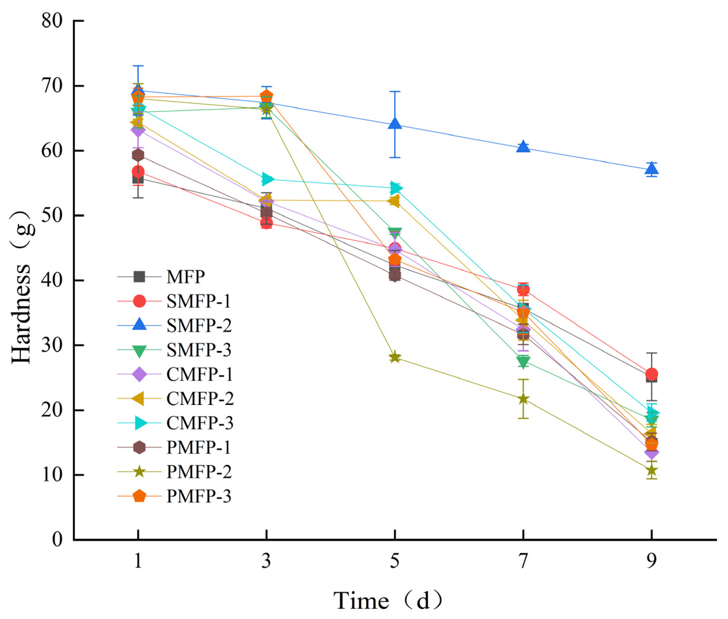
3.1.1. The Impact of Crosslinking Agents on the Mechanical Hardness of Microalgal–Fungal Pellets

As shown in Table 1, the initial hardness of each crosslinked MFP was significantly higher than that of the non-crosslinked group (p < 0.05), and the hardness was notably influenced by the type and concentration of crosslinkers. In the SMFP group, the hardness initially increased and then declined as the concentration increased. In the CMFP group, the hardness consistently increased with rising concentrations. In the PMFP group, the hardness increased with increasing concentrations but plateaued at higher levels. Over time (Figure 1), the hardness of all groups gradually declined, but SMFP-2 exhibited the slowest decrease, with significantly higher hardness levels than all other groups on day 9 (Table 1).
The addition of crosslinking agents enhances the hardness of MFPs. Notably, the MFPs that were crosslinked with a medium SA concentration exhibited significantly improved hardness and structural stability, which is consistent with previous research findings. Lai et al. [30] reported that the hardness of SA/PVA hydrogels is closely related to their concentration, with the hardness of SA hydrogels first increasing and then decreasing as the concentration increases. Studies have shown [31,32] that both low and excessively high concentrations of SA fail to form microbial pellets with a compact structure and high mechanical strength. This may be due to insufficient crosslinking at low SA concentrations, affecting the stability of the immobilization [33]; conversely, excessively high concentrations tend to produce incompatible polymer networks [34]. Therefore, the “medium-concentration optimum” phenomenon observed in this study may be attributed to the three-dimensional network structure that is formed by an intermediate concentration of SA, which provides an optimal balance between pellet integrity and structural flexibility. Additionally, the crosslinking-induced structural stability not only enhances the mechanical shear resistance of the MFP but also provides a more stable microenvironment that facilitates internal microalgal–fungal synergistic metabolism. These effects help extend the operational lifespan and reduce the risk of structural failure in actual wastewater treatment systems, highlighting its promising potential for practical application.

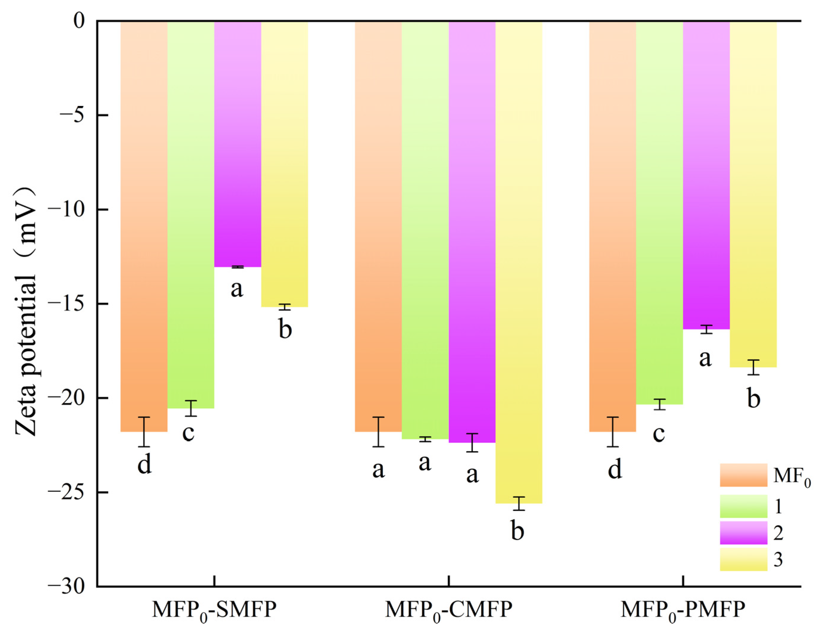
3.1.2. The Impact of Crosslinking Agents on the Zeta Potential of MFP

As shown in Figure 2, on day 9, all MFPs, with or without crosslinkers, exhibited negative Zeta potentials, and the crosslinkers significantly influenced their values. In the SMFP group, the Zeta potentials at all tested concentrations were significantly greater than that of the non-crosslinked group (MFP0) (p < 0.05). Among these, SMFP-2 (20 g/L) had the highest Zeta potential (−13.05 mV), which was significantly greater than those of MFP0, SMFP-1, and SMFP-3. In the CMFP group, CMFP-1 and CMFP-2 showed no significant difference compared with MFP0 (p > 0.05), whereas CMFP-3 (40 mg/L) exhibited the lowest potential (−25.60 mV), which was significantly lower than those of MFP0, CMFP-1, and CMFP-2 (p < 0.05). In the PMFP group, significant differences were observed among all concentrations when compared with MFP0 (p < 0.05). PMFP-2 (100 g/L) exhibited the highest Zeta potential (−16.36 mV), which was significantly greater than those of MFP0, PMFP-1, and PMFP-3.
The Zeta potential is a key indicator of the internal charge and dispersion stability in MFPs. Highly dispersed pellets typically exhibit absolute Zeta potentials above 30 mV, which confer resistance to aggregation. Conversely, lower absolute Zeta potentials favor coagulation or flocculation as attractive forces, which exceed repulsive forces [35]. Studies have demonstrated [36] that absolute Zeta potentials above the 30 mV coagulation threshold generate sufficient repulsive forces to maintain a stable suspension. In this study, the control group pellets exhibited absolute Zeta potentials below 30 mV, meeting the criteria for coagulation. Szekalska et al. [37] reported that crosslinking SA with divalent ions significantly reduced the microsphere Zeta potential. In the SMFP-2 group (20 g/L SA), the absolute Zeta potential remained low (13.05 mV), significantly below those of all other groups, even after 9 days. This suggests that these conditions enhance pellet aggregation, which is consistent with the SMFP-2 group’s superior hardness and structural integrity after 9 days. During nutrient adsorption, the charge magnitude and stability critically determine the adsorption efficiency [38,39]. The SMFP-2 group’s lower Zeta potential and stronger attractive forces may be behind its enhanced nutrient adsorption capacity. Furthermore, charge stability is essential for applying MFPs in wastewater treatment. Over long-term operation, charge stability ensures a sustained adsorption performance and prevents degradation due to charge loss.

3.1.3. The Impact of Crosslinking Agents on the 3D Fluorescence of the Extracellular Polymeric Substances in MFPs

Three-dimensional excitation–emission matrix (3D-EEM) fluorescence spectroscopy was performed on the extracellular polymeric substances (EPSs) of the MFPs on days 1 and 9, and the results are shown in Figure 3. The primary fluorophores in TB-EPS were identified as soluble microbial metabolites (Ex/Em = 250–280 nm/300–350 nm), likely tryptophan and tyrosine [40]. However, within 9 days of pellet formation, the fluorescence intensity decreased from 0.15 to 0.077, indicating a decline in this product.
Previous studies have demonstrated that crosslinking agents influence the formation and release of EPSs. High-molecular-weight crosslinkers, such as CTS, are known to form dense network structures that restrict microbial metabolite release [41]. Agudelo et al. [42] reported that CTS addition reduced β-lactoglobulin’s fluorescence intensity. Our experimental results indicated that although crosslinking did not alter the primary TB-EPS composition relative to the non-crosslinked control, the fluorescence intensity decreased across all groups except PMFP-2, which exhibited a slight increase (Figure 4, left column). Fluorescence spectroscopy of the PMFP variants revealed that the initial fluorescence intensity increased with a higher PVA concentration. This trend may result from PVA-enhanced adsorption site availability [43,44] and biofilm formation [45], which collectively stimulate microbial EPS production and lead to a higher fluorescence intensity.
Over the 9-day period following crosslinking, the fluorescence intensity of the extracellular polymeric substances (EPSs) in all crosslinked microalgae–fungal pellet groups continued to decline. The CMFP group exhibited the most pronounced decrease, particularly in CMFP-3, where the fluorescence intensity dropped from 0.082 to 0.026. This finding aligns with those by Adav et al. [46], who reported that nutrient limitations or environmental stress during cultivation may reduce microbial metabolite secretion, thereby decreasing the fluorescence intensity. In contrast, the medium-concentration SA group (SMFP-2) showed an increase in fluorescence intensity from 0.11 to 0.13 (Figure 4, right column), which is consistent with previous findings that SA promotes the retention and accumulation of EPSs [47]. SA is a polysaccharide polymer and rich in reactive functional groups, including hydroxyl and carboxyl groups [48]. These groups interact with microbial metabolites, offering abundant adsorption sites for EPS formation [49]. Dou et al. [50] found that 6% SA promotes fibrous structure formation in soy protein-based meat analogs, while excessive amounts have adverse effects. The observed decreases in TB-EPS fluorescence in SMFP-1 and SMFP-3, along with the increase in SMFP-2, support this conclusion. Previous studies have indicated that low concentrations of SA provide limited adsorption sites, while higher concentrations increase site availability, enhancing microbial metabolite adsorption and EPS formation. However, excessive SA encapsulation may hinder effective adsorption, thereby reducing the adsorption capacity [51]. Consequently, the addition of a high SA concentration (35 g/L) in the SMFP-3 group led to a decline in the TB-EPS fluorescence intensity.

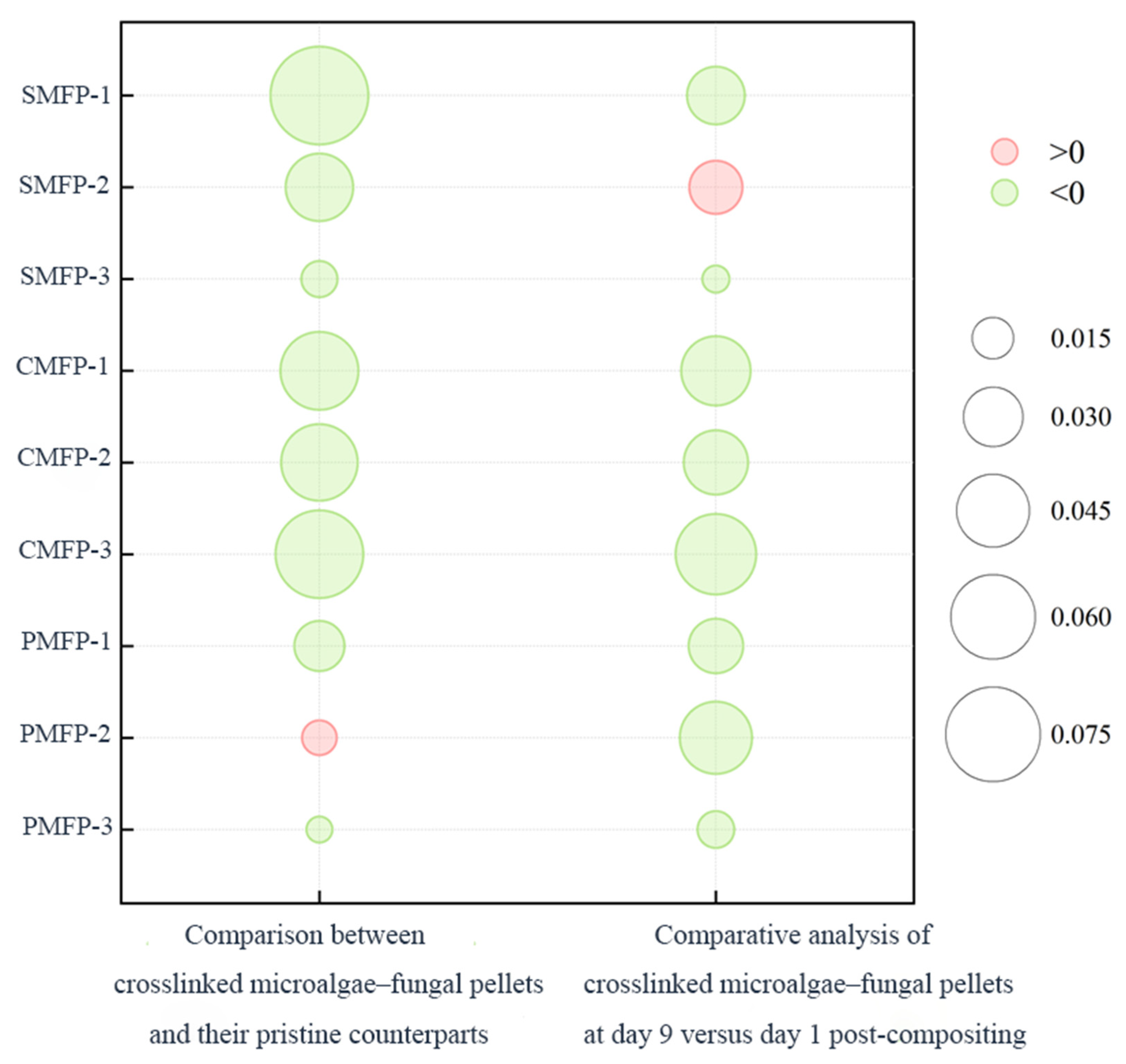
3.1.4. The Impact of Crosslinking Agents on the Protein and Polysaccharide Contents of EPSs in Various MFPs on Day 1 and Day 9

As shown in Figure 5a, on day 1, the PN content in the TB-EPSs of the low-concentration SA group (SMFP-1) was lower than that of the non-crosslinked control (MFP0), but the PN contents of SMFP-2 and SMFP-3 increased significantly and exceeded that of MFP0 with the increase in SA concentration; the PN content of the PMFP group showed a similar trend of changes, while there was no significant difference in PN contents among the different concentrations in the CMFP group. After nine days, the PN contents of all groups decreased: in MFP0, it decreased from 54.75 mg/L to 18.25 mg/L, while SMFP-2 (20 g/L) exhibited the smallest decrease in PN content (only from 75.62 mg/L to 64.22 mg/L). In terms of PS (Figure 5b), the initial PS contents of all the crosslinked groups were significantly higher than that of MFP0; after nine days, the PS contents of all groups except SMFP-2 decreased, while the PS content of SMFP-2 increased and was significantly higher than those of all other groups (p < 0.05).
SA crosslinking exerts a stabilizing effect on the components of TB-EPSs in MFPs, similar to its impact on the pellet hardness and Zeta potential. At an SA concentration of 20 g/L (SMFP-2 group), the resulting three-dimensional network structure not only effectively delayed PN loss but also significantly enhanced PS accumulation. This aligns with the findings of Cao et al. [52], who reported that the “egg-box” structure that is formed by SA carboxyl groups and divalent cations (e.g., Ca2⁺) in microbial extracellular polymeric substances not only enhances the EPS matrix stability but also reduces the diffusion loss of extracellular enzymes.
As the SA concentration increased, the PN contents in SMFP-2 and SMFP-3 significantly increased, indicating that SA increases the PN content in MFPs. This may be because sodium alginate, a polysaccharide, provides additional carbon sources for the MFPs [53]. At higher concentrations, MFPs can utilize increased carbon sources for metabolism, promoting protein synthesis. Additionally, SA forms a more compact gel structure [54], providing a stable growth environment for the MFPs and reducing interference from environmental fluctuations in microalgal growth, thereby promoting protein synthesis.
However, CTS had a minimal impact on the PN contents of the MFPs, likely due to its weaker biological activity compared with sodium alginate. The linear polysaccharide structure of SA easily binds to cell surface receptors, promoting adsorption and signal transduction, whereas the branched structure of CTS may limit its effective interaction with cell surfaces [55,56]. This limits its regulatory effects on metabolic enzyme activity and protein synthesis pathways within MFPs, resulting in weaker interactions with the pellets.
After 9 days, the PN contents in all microalgae–fungal pellet groups decreased. This may have been due to the consumption of PN for cell growth and metabolic activities during the growth of the MFPs [57]. The SMFP-2 group exhibited the smallest decrease in PN content, indicating that the gel structure that is formed by SA at 20 g/L provides a stable microenvironment for MFPs, reducing any external environmental interference with cell metabolism and helping maintain a higher PN content.
The results of this experiment also indicated that increasing the concentrations of sodium alginate, CTS, and PVA promoted an increase in PS contents in the SMFP, CMFP, and PMFP samples, likely due to interactions between biological and chemical processes. High-molecular-weight polymers alter the physicochemical properties of microalgae–fungal pellet cell surfaces through surface adsorption, such as increasing negative charges on the cell surface [58], which may enhance the activity of enzymes that are involved in PS biosynthesis pathways in the cell wall [59,60,61]. As biological macromolecules, these polymers may provide nutrients that are usable by MFPs [62], or they may influence PS synthesis by regulating the microbial community structure [63,64], thereby promoting nutrient transformation and utilization. However, after 9 days of storage, the PS contents in all groups, except SMFP-2, decreased compared with the initial value. This may be due to the consumption of PS, an important component of the cell wall, during the growth of the MFPs for cell construction and repair [65].

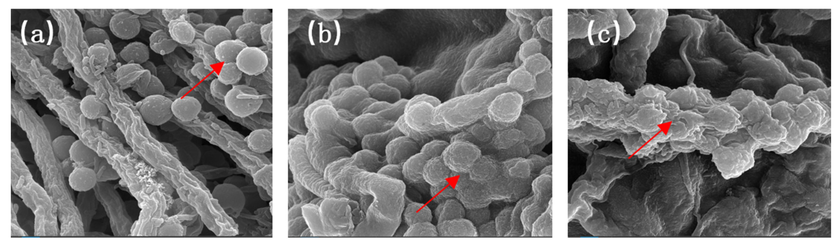
3.2. The Impact of Crosslinking Agents on the Microstructure of MFPs

A scanning electron microscopy analysis (10,000× magnification; Figure 6) revealed distinct structural differences among the microalgae–fungal pellets under different treatments. In the freshly prepared MFP (MFP0), Chlorella cells were primarily immobilized on the surface of Aspergillus niger hyphae via physical adsorption, forming a loosely bound microalgae–fungal composite. Abundant internal pores facilitated mass transfer and light transmittance (Figure 6a). After crosslinking treatment with 20 g/L SA, a gel layer formed at the hyphae–algal cell interface in the initial sample (SMFP0), significantly enhancing the encapsulation and immobilization of Chlorella cells (Figure 6b). Studies have shown [66,67] that SA can form an ECM hydrogel through ionotropic crosslinking, thereby enhancing structural stability. Similarly, studies have demonstrated [68] that the gel structure of SA possesses sufficient porosity to immobilize microorganisms without obstructing their internal pores. Thus, the addition of SA in this study not only enhanced the physical connection between fungal hyphae and microalgal cells, thus improving the compactness of the structure but also supported the physiological activities of microorganisms through its internal pores, facilitating the growth and maintenance of normal physiological functions in SMFP0.
Nine days after with 20 g/L SA crosslinked MFPs (SMFP9), microalgae cell shrinkage slightly increased the surface roughness; however, the gel network structure remained intact, continuing to maintain close encapsulation of the hyphae and microalgae cells (Figure 6c). This is consistent with the findings of Eckert et al. [69], who reported that microcapsules that are prepared with SA can maintain good mechanical properties for up to three months. In summary, microstructural modification via crosslinkers not only enhances the compactness of the microalgae–fungal association and the spatial stability but also improves the microenvironmental controllability and light transmittance, thereby establishing a structural foundation for stable system operation and sustained efficient pollutant removal. Such structural stability is critical for long-term operation and resistance to shock loads in practical engineering applications.

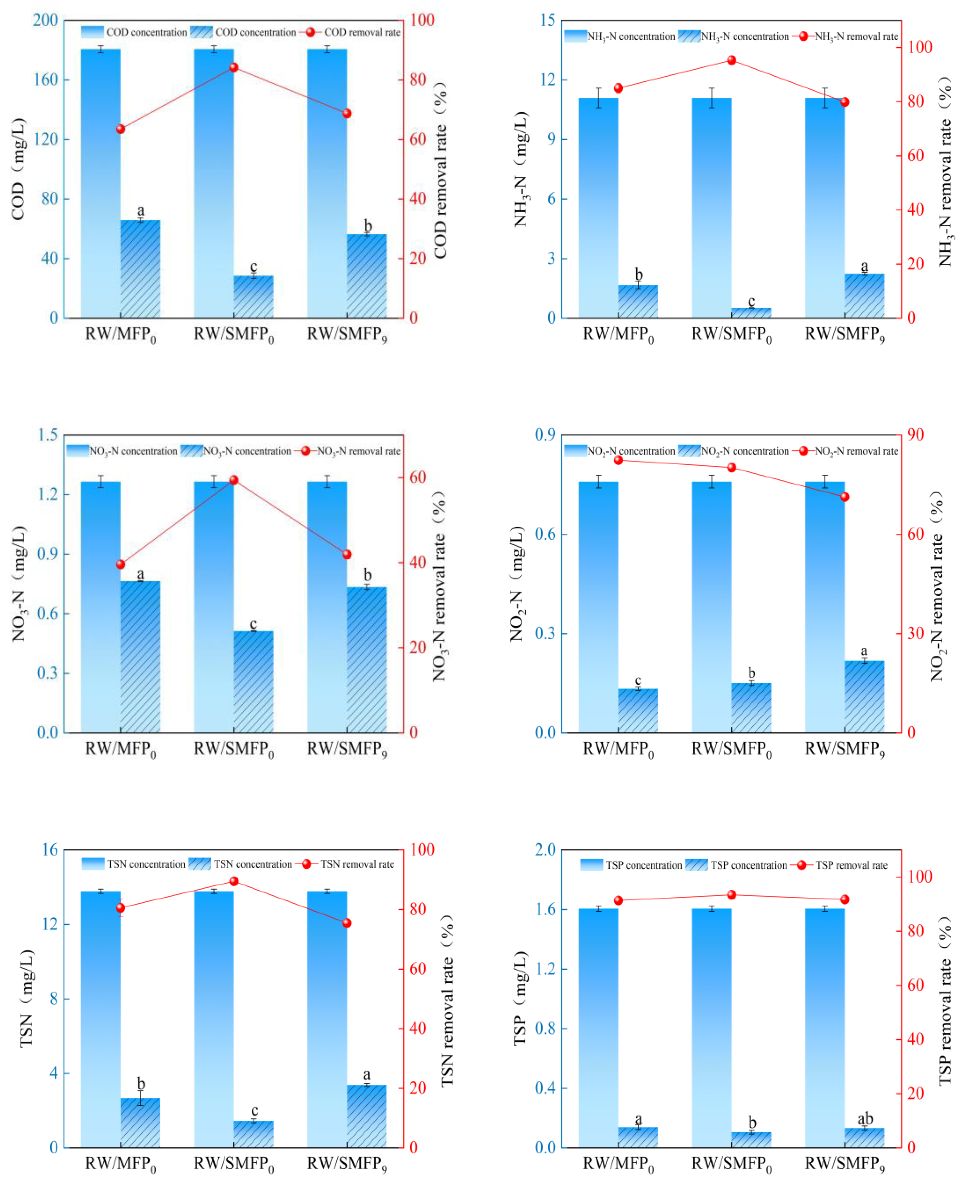
3.3. Treatment Efficiency of SA-Crosslinked MFPs for High-Density Aquaculture Wastewater

After 3 days of treatment with various MFPs, the high-density aquaculture wastewater met the Discharge Standard for Pollutants in Aquaculture Tail Water in Hubei Province [70]. As shown in Figure 7, a three-day comparison of the pollutant removal efficiencies of MFP0, SMFP0, and SMFP9 in high-density aquaculture wastewater indicated that, with the exception of NO2-N, for which MFP0 showed the highest removal rate (82.42%), the SMFP0 group exhibited the best overall performance in removing COD, NH3-N, NO3-N, TSN, and TSP. Specifically, the removal rates of COD (84.19%), NH3-N (95.29%), NO3-N (59.42%), and TSN (89.50%) by SMFP0 were significantly higher than those achieved by both MFP0 and SMFP9 (p < 0.05). For TSP, SMFP9 achieved a removal rate of 91.73%, while SMFP0 achieved the highest rate of 93.46%, significantly outperforming MFP0. This result can be attributed to the fact that sodium alginate, as a polysaccharide, contains multiple carboxyl and hydroxyl groups within its molecular structure. These groups can form complexes with phosphate ions, thereby enhancing the phosphorus adsorption capacity of MFPs [71]. The high NH3-N removal rate in the SMFP0 group is also consistent with the findings of Liu et al. [72], who achieved an NH3-N removal rate of 96.60% ± 0.10% using SA-immobilized microalgal pellets to treat nitrogen-containing and phosphorus wastewater. These results further confirm that the crosslinking effect of SA provides a protective biofilm structure for the MFP [73]. This structure shields internal microorganisms from external environmental shocks, endowing the pellets with higher hardness levels (69.28 ± 3.80 g) and enhancing their wastewater treatment capacity.
The removal rates of COD and NO3-N by SMFP9 were 68.76% and 41.94%, respectively, significantly higher than those of MFP0. After 3 days of treatment using the SMFP9 group, the TSP concentration in the wastewater decreased to 0.13 mg/L, with no significant difference compared with the other two groups. However, MFP0 outperformed SMFP9 in the removal of NH3-N and TSN. This may be due to the decreased microbial activity in SMFP9 following long-term cultivation. During the 9-day cultivation period, microorganisms may have consumed nutrients, leading to a reduction in microbial quantity or activity [74]. These results indicate that SMFP0 can maintain effective wastewater treatment capacity after 9 days of storage, further confirming the excellent biocompatibility of SA hydrogels in both internal and external environments [75,76]. Additionally, SA microgels are non-cytotoxic and facilitate nutrient exchange with the environment, enabling microbial cells to maintain a healthy growth state within the microgel over an extended period [77,78] and thereby allowing the MFPs to sustain high-efficiency nutrient removal capacity. However, in terms of NH3-N and TSN removal, the removal rates of 79.82% and 75.48% in the SMFP9 group were significantly lower than those in the MFP0 group and the lowest among the three groups. This may be attributed to the reduction in microbial quantity or activity in SMFP9 due to long-term storage [74].
A systematic comparison of key performance indicators (Table 2) of MFP0, SMFP0, and SMFP9 focusing on their hardness, EPS composition, and pollutant removal rates revealed that the SMFP0 group exhibited the best overall performance. The highest mechanical strength and TB-EPS content in the microalgae–fungal pellets correlated with their superior removal efficiencies for COD, TSP, and multiple nitrogen species (NH3-N, NO3-N, and TSN), underscoring their advantages in terms of structural stability and pollutant purification capacity. The SMFP9, which was crosslinked with SA for 9 days, maintained comparable mechanical strength to that of MFP0 and a higher TB-EPS content, resulting in higher removal rates of COD and NO3-N compared with the MFP0 group, as well as a comparable TSP removal rate. Although the NH3-N and TSN removal rates of SMFP9 were lower than those of MFP0 due to the reduced microbial activity resulting from long-term cultivation, the overall data suggest that SA-crosslinked MFPs at 20 g/L offer clear advantages in maintaining their structural stability and nutrient removal potential, thereby supporting their sustained application in water treatment.

3.4. Economic Benefits and Operational Parameter Analysis of SA-Crosslinked MFP

The application of microalgae–fungal pellets in wastewater treatment demonstrates not only excellent pollutant removal efficiency but also substantial potential for economic and resource benefits. Under high-density aquaculture wastewater conditions, the SA-crosslinked microalgae–fungal pellets (SMFP0) exhibited strong nutrient removal capacity, highlighting their high efficiency in nutrient control. The microalgae–fungal biomass that is generated during the treatment can serve as a feedstock for animal feed, biofertilizers, or bioenergy, offering significant potential for resource recovery. Assuming a daily treatment capacity of 10 tons and 330 days of annual operation, the estimated annual biomass production would be approximately 3.3 tons. If valued at the market price of aquaculture feed (approximately CNY 150 per kilogram), the estimated annual biomass revenue could reach approximately CNY 495,000. Moreover, the significant reduction in COD contributes to carbon sequestration. Previous studies have demonstrated that microalgae–bacteria consortia not only mitigate nitrogen pollution but also promote carbon fixation, thereby providing a material and energy basis for adaptation to extreme environmental conditions [79]. Overall, this system integrates pollution control, resource recovery, and ecological benefits, presenting strong potential for practical implementation.
In practical applications, the light intensity is a key factor driving algal photosynthesis. In rainy regions, reduced natural light availability may lower the efficiency of algal oxygen production, necessitating supplemental artificial lighting (e.g., LED sources) to maintain metabolic equilibrium. Light intensity sensors can be employed to dynamically adjust the supplemental lighting duration, thereby minimizing energy consumption and operational costs.

4. Conclusions

This study successfully developed an MFP crosslinked with 20 g/L SA and systematically evaluated its physical properties and wastewater treatment performance. The results demonstrated that the crosslinked system exhibited remarkable structural stability, as reflected in its enhanced hardness, Zeta potential, EPS composition, and microalgae–fungal interactions. On the ninth day after crosslinking, the hardness of the microalgal–fungal pellets remained comparable to that of the initial non-crosslinked sample. A comparative analysis of the MFP0, SMFP0, and SMFP9 samples’ efficiency in treating high-density aquaculture wastewater revealed that SMFP0 exhibited a significantly higher pollutant removal efficiency than the non-crosslinked group and retained a high treatment capacity, even on the ninth day post-crosslinking.
These findings suggest that the crosslinked MFPs possess both structural durability and long-term treatment efficacy, making them suitable for extended operation in real-world engineering applications. Their enhanced mechanical integrity offers a potential solution to the disintegration issue that has been observed in conventional microalgae–fungal systems, extending the operational lifespan while reducing the replacement frequency and maintenance costs. Moreover, the system exhibited excellent removal efficiency for key pollutants for the advanced treatment of aquaculture effluents, thus supporting the promotion of sustainable aquaculture practices. The strong EPS secretion and low Zeta potential further indicate favorable microalgae–fungal aggregation and environmental adaptability, enhancing the system’s potential for engineering-scale applications. Notably, in addition to pollutant removal, the system exhibits carbon capture potential, offering a pathway for biomass valorization and bioenergy development. This aligns with the ongoing transition toward green, low-carbon wastewater treatment technologies under the national “carbon peak and carbon neutrality” strategy.
Although this study validated the practical application potential of SA-crosslinked MFPs in improving system stability and treatment performance, several critical aspects warrant further investigation. Future research should focus on pilot-scale and full-scale validation to comprehensively evaluate this system’s long-term removal efficiency, operational stability, and economic feasibility, thereby providing a solid theoretical and technical foundation for large-scale implementation.

Author Contributions

G.Z.: Formal analysis, investigation, methodology, validation, writing—original draft, and writing—review and editing. H.M.: Conceptualization, writing—review and editing, and funding acquisition. K.C.: Writing—review and editing and supervision. All authors have read and agreed to the published version of the manuscript.

Funding

This research was supported by the National Natural Science Foundation of China under Grants 32170383 and 52179065, as well as the Ministry of Education’s Chunhui Program Collaborative Research Project (Grant No. 202201970).

Data Availability Statement

The data will be made available on request.

Acknowledgments

We thank the Microalgae Bioenergy Laboratory (HBUT) for providing valuable information.

Conflicts of Interest

The authors declare no conflicts of interest.

References

  1. Jin, Y.; Zhan, W.; Wu, R.; Han, Y.; Yang, S.; Ding, J.; Ren, N. Insight into the roles of microalgae on simultaneous nitrification and denitrification in microalgal-bacterial sequencing batch reactors: Nitrogen removal, extracellular polymeric substances, and microbial communities. Bioresour. Technol. 2023, 379, 129038. [Google Scholar] [CrossRef] [PubMed]
  2. An, X.; Yousif Abdellah, Y.A.; Wang, L.; Elsheikh, E.A.E.; Wang, Y.; Yang, J.; Hu, G. NaCl as an excellent trigger-induced biodiesel production and phenol-containing wastewater treatment in a novel salt-tolerant microalgae Ankistrodesmus sp. ACC. Bioresour. Technol. 2025, 429, 132515. [Google Scholar] [CrossRef] [PubMed]
  3. Adewuyi, S.; Babatunde, T.; Mmuoegbulam, A.; Abimbade, S.; Abiodun, O.; Klink, M.J.; Nelana, S.; Malomo, D.; Ayanda, O. Toxicity and health implications of pesticides and the need to remediate pesticide-contaminated wastewater through the advanced oxidation processes. Water Conserv. Manag. WCM 2024, 8, 97–108. [Google Scholar] [CrossRef]
  4. Zhou, W.G.; Cheng, Y.L.; Li, Y.; Wan, Y.Q.; Liu, Y.H.; Lin, X.Y.; Ruan, R. Novel Fungal Pelletization-Assisted Technology for Algae Harvesting and Wastewater Treatment. Appl. Biochem. Biotechnol. 2012, 167, 214–228. [Google Scholar] [CrossRef]
  5. Chu, Y.; Zhang, X.; Tang, X.; Jiang, L.; He, R. Uncovering anaerobic oxidation of methane and active microorganisms in landfills by using stable isotope probing. Environ. Res. 2025, 271, 121139. [Google Scholar] [CrossRef]
  6. Ai, D.; Wu, T.; Ge, Z.; Ying, Z.; Sun, S.; Huang, D.; Zhang, J. The coupling effect promotes superoxide radical production in the microalgal-fungal symbiosis systems: Production, mechanisms and implication for Hg(II) reduction. J. Hazard. Mater. 2024, 477, 135347. [Google Scholar] [CrossRef]
  7. Muradov, N.; Taha, M.; Miranda, A.F.; Wrede, D.; Kadali, K.; Gujar, A.; Stevenson, T.; Ball, A.S.; Mouradov, A. Fungal-assisted algal flocculation: Application in wastewater treatment and biofuel production. Biotechnol. Biofuels 2015, 8, 24. [Google Scholar] [CrossRef] [PubMed]
  8. Yang, L.M.; Li, H.K.; Wang, Q. A novel one-step method for oil-rich biomass production and harvesting by co-cultivating microalgae with filamentous fungi in molasses wastewater. Bioresour. Technol. 2019, 275, 35–43. [Google Scholar] [CrossRef]
  9. Song, H.; Qian, J.; Fan, L.; Toda, T.; Li, H.; Sekine, M.; Song, P.; Takayama, Y.; Koga, S.; Li, J.; et al. Enhancing biomass yield, nutrient removal, and decolorization from soy sauce wastewater using an algae-fungus consortium. Algal Res. 2022, 68, 102878. [Google Scholar] [CrossRef]
  10. Wang, S.-K.; Yang, K.-X.; Zhu, Y.-R.; Zhu, X.-Y.; Nie, D.-F.; Jiao, N.; Angelidaki, I. One-step co-cultivation and flocculation of microalgae with filamentous fungi to valorize starch wastewater into high-value biomass. Bioresour. Technol. 2022, 361, 127625. [Google Scholar] [CrossRef]
  11. Li, Y.; Xu, Y.T.; Liu, L.; Li, P.; Yan, Y.; Chen, T.; Zheng, T.L.; Wang, H.L. Flocculation mechanism of Aspergillus niger on harvesting of Chlorella vulgaris biomass. Algal Res.-Biomass Biofuels Bioprod. 2017, 25, 402–412. [Google Scholar] [CrossRef]
  12. Meng, F.S.; Xi, L.M.; Liu, D.F.; Huang, W.W.; Lei, Z.F.; Zhang, Z.Y.; Huang, W.L. Effects of light intensity on oxygen distribution, lipid production and biological community of algal-bacterial granules in photo-sequencing batch reactors. Bioresour. Technol. 2019, 272, 473–481. [Google Scholar] [CrossRef]
  13. Huang, W.; Liu, D.; Huang, W.; Cai, W.; Zhang, Z.; Lei, Z. Achieving partial nitrification and high lipid production in an algal-bacterial granule system when treating low COD/NH4–N wastewater. Chemosphere 2020, 248, 126106. [Google Scholar] [CrossRef]
  14. Liu, L.; Fan, H.Y.; Liu, Y.H.; Liu, C.X.; Huang, X. Development of algae-bacteria granular consortia in photo-sequencing batch reactor. Bioresour. Technol. 2017, 232, 64–71. [Google Scholar] [CrossRef]
  15. Zhao, Z.W.; Liu, S.; Yang, X.J.; Lei, Z.F.; Shimizu, K.; Zhang, Z.Y.; Lee, D.J.; Adachi, Y. Stability and performance of algal-bacterial granular sludge in shaking photo-sequencing batch reactors with special focus on phosphorus accumulation. Bioresour. Technol. 2019, 280, 497–501. [Google Scholar] [CrossRef] [PubMed]
  16. He, Q.L.; Chen, L.; Zhang, S.J.; Chen, R.F.; Wang, H.Y.; Zhang, W.; Song, J.Y. Natural sunlight induced rapid formation of water-born algal-bacterial granules in an aerobic bacterial granular photo-sequencing batch reactor. J. Hazard. Mater. 2018, 359, 222–230. [Google Scholar] [CrossRef]
  17. Liu, Y.-Q.; Tay, J.-H. Cultivation of aerobic granules in a bubble column and an airlift reactor with divided draft tubes at low aeration rate. Biochem. Eng. J. 2007, 34, 1–7. [Google Scholar] [CrossRef]
  18. Moy, B.Y.P.; Tay, J.H.; Toh, S.K.; Liu, Y.; Tay, S.T.L. High organic loading influences the physical characteristics of aerobic sludge granules. Lett. Appl. Microbiol. 2002, 34, 407–412. [Google Scholar] [CrossRef] [PubMed]
  19. Chen, Y.; Jiang, W.; Liang, D.T.; Tay, J.H. Structure and stability of aerobic granules cultivated under different shear force in sequencing batch reactors. Appl. Microbiol. Biotechnol. 2007, 76, 1199–1208. [Google Scholar] [CrossRef]
  20. Zhang, B.; Lens, P.N.L.; Shi, W.X.; Zhang, R.J.; Zhang, Z.Q.; Guo, Y.; Bao, X.; Cui, F.Y. Enhancement of aerobic granulation and nutrient removal by an algal-bacterial consortium in a lab-scale photobioreactor. Chem. Eng. J. 2018, 334, 2373–2382. [Google Scholar] [CrossRef]
  21. Peng, T.; Wang, Y.; Wang, J.; Fang, F.; Yan, P.; Liu, Z. Effect of different forms and components of EPS on sludge aggregation during granulation process of aerobic granular sludge. Chemosphere 2022, 303, 135116. [Google Scholar] [CrossRef] [PubMed]
  22. Zeng, H.; Wang, F.; Xu, K.; Zhang, J.; Li, D. Optimization and regeneration of chitosan-alginate hybrid adsorbent embedding iron-manganese sludge for arsenic removal. Colloids Surf. A Physicochem. Eng. Asp. 2020, 607, 125500. [Google Scholar] [CrossRef]
  23. Mao, Y.; Wang, J. Immobilization of activated sludge in PVA matrix using innovative methods. Huanjing Kexue Xuebao/Acta Sci. Circumstantiae 2013, 33, 370–376. [Google Scholar]
  24. HJ 535-2009; Water Quality―Determination of Ammonia Nitrogen―Nessler’s Reagent Spectrophotometry. Standardization Administration of China: Beijing, China, 2009. (In Chinese)
  25. GB 7493-87; Water Quality-Determination of Nitrogen (Nitrite)-Spectrophotometric Method. Standardization Administration of China: Beijing, China, 1987. (In Chinese)
  26. HJ/T 346-2007; Water Quality—Determination of Nitrate-Nitrogen—Ultraviolet Spectrophotometry. Standardization Administration of China: Beijing, China, 2007. (In Chinese)
  27. HJ 636-2012; Water Quality-Determination of Total Nitrogen-Alkaline Potassium Persulfate Digestion UV Spectrophotometric. Standardization Administration of China: Beijing, China, 2012. (In Chinese)
  28. GB 11893-89; Water Quality-Determination of Total Phosphorus-Ammonium Molybdate Spectrophotometric Method. Standardization Administration of China: Beijing, China, 1989. (In Chinese)
  29. GB 11914-89; Water Quality-Determination of the Chemical Oxygen Demand-Dichromate Method. Standardization Administration of China: Beijing, China, 1989. (In Chinese)
  30. Lai, Z.; Cui, Y.; Yu, Y.; Wang, J. The Hardness of Sodium Alginate-Polyvinyl Alcohol Hydrogel. Mater. Rep. 2010, 24, 32–34. (In Chinese) [Google Scholar]
  31. Moreno-Garrido, I. Microalgae immobilization: Current techniques and uses. Bioresour. Technol. 2008, 99, 3949–3964. [Google Scholar] [CrossRef]
  32. Huebner, F.R.; Wall, J.S. Polysaccharide Interactions with Wheat Proteins and Flour Doughs. Cereal Chem. 1979, 56, 68–72. [Google Scholar]
  33. Rodrigues, R.; Ortiz, C.; Berenguer-Murcia, A.; Torres Sáez, R.; Fernández-Lafuente, R. Modifying enzyme activity and selectivity by immobilization. Chem. Soc. Rev. 2012, 42, 6290–6307. [Google Scholar] [CrossRef] [PubMed]
  34. Wang, Q.; Du, Y.; Hu, X.; Yang, J.; Fan, L.; Feng, T. Preparation of alginate/soy protein isolate blend fibers through a novel coagulating bath. J. Appl. Polym. Sci. 2006, 101, 425–431. [Google Scholar] [CrossRef]
  35. Wang, X.; Bao, K.; Cao, W.; Zhao, Y.; Hu, C.W. Screening of microalgae for integral biogas slurry nutrient removal and biogas upgrading by different microalgae cultivation technology. Sci. Rep. 2017, 7, 5426. [Google Scholar] [CrossRef]
  36. Wang, J.; Yang, X.; Klemeš, J.J.; Tian, K.; Ma, T.; Sunden, B. A review on nanofluid stability: Preparation and application. Renew. Sustain. Energy Rev. 2023, 188, 113854. [Google Scholar] [CrossRef]
  37. Szekalska, M.; Sosnowska, K.; Czajkowska-Kośnik, A.; Winnicka, K. Calcium Chloride Modified Alginate Microparticles Formulated by the Spray Drying Process: A Strategy to Prolong the Release of Freely Soluble Drugs. Materials 2018, 11, 1522. [Google Scholar] [CrossRef] [PubMed]
  38. Xu, Y.; Curtis, T.; Dolfing, J.; Wu, Y.; Rittmann, B.E. N-acyl-homoserine-lactones signaling as a critical control point for phosphorus entrapment by multi-species microbial aggregates. Water Res. 2021, 204, 117627. [Google Scholar] [CrossRef] [PubMed]
  39. Hu, Q.; Zhou, N.; Rene, E.R.; Wu, D.; Sun, D.; Qiu, B. Stimulation of anaerobic biofilm development in the presence of low concentrations of toxic aromatic pollutants. Bioresour. Technol. 2019, 281, 26–30. [Google Scholar] [CrossRef]
  40. Zhang, B.; Shi, J.; Shi, W.; Guo, Y.; Lens, P.N.L.; Zhang, B. Effect of different inocula on the granulation process, reactor performance and biodiesel production of algal-bacterial granular sludge (ABGS) under low aeration conditions. Chemosphere 2023, 345, 140391. [Google Scholar] [CrossRef]
  41. Rinaudo, M. Chitin and chitosan: Properties and applications. Prog. Polym. Sci. 2006, 31, 603–632. [Google Scholar] [CrossRef]
  42. Agudelo, D.; Nafisi, S.; Tajmir-Riahi, H.-A. Encapsulation of Milk β-Lactoglobulin by Chitosan Nanoparticles. J. Phys. Chem. B 2013, 117, 6403–6409. [Google Scholar] [CrossRef] [PubMed]
  43. Daza, J.; Ramirez, M.; Henquín, E.; Rintoul, I. Modelling of swelling of PVA hydrogels considering non-ideal mixing behaviour of PVA and water. J. Mater. Chem. B 2019, 7, 4049–4054. [Google Scholar] [CrossRef]
  44. Sun, H.; Qu, Z.; Yu, J.; Ma, H.; Li, B.; Sun, D.; Ge, Y. Asymmetric 5-sulfosalicylic acid-PVA catalytic pervaporation membranes for the process intensification in the synthesis of ethyl acetate. Sep. Purif. Technol. 2022, 282, 120113. [Google Scholar] [CrossRef]
  45. Ruiz-Marin, A.; Mendoza-Espinosa, L.G.; Stephenson, T. Growth and nutrient removal in free and immobilized green algae in batch and semi-continuous cultures treating real wastewater. Bioresour. Technol. 2010, 101, 58–64. [Google Scholar] [CrossRef]
  46. Adav, S.S.; Lee, D.-J. Extraction of extracellular polymeric substances from aerobic granule with compact interior structure. J. Hazard. Mater. 2008, 154, 1120–1126. [Google Scholar] [CrossRef]
  47. Zhang, J.; Liu, L.; Jiang, Y.; Shah, F.; Xu, Y.; Wang, Q. High-moisture extrusion of peanut protein-/carrageenan/sodium alginate/wheat starch mixtures: Effect of different exogenous polysaccharides on the process forming a fibrous structure. Food Hydrocoll. 2020, 99, 105311. [Google Scholar] [CrossRef]
  48. Gao, X.; Guo, C.; Hao, J.; Zhao, Z.; Long, H.; Li, M. Adsorption of heavy metal ions by sodium alginate based adsorbent-a review and new perspectives. Int. J. Biol. Macromol. 2020, 164, 4423–4434. [Google Scholar] [CrossRef]
  49. Sajjad, M.; Aziz, A.; Kim, K. Biosorption and Binding Mechanisms of Ni2+ and Cd2+ with Aerobic Granules Cultivated in Different Synthetic Media. Chem. Eng. Technol. 2017, 40, 2179–2187. [Google Scholar] [CrossRef]
  50. Dou, W.; Zhang, X.; Zhao, Y.; Zhang, Y.; Jiang, L.; Sui, X. High moisture extrusion cooking on soy proteins: Importance influence of gums on promoting the fiber formation. Food Res. Int. 2022, 156, 111189. [Google Scholar] [CrossRef] [PubMed]
  51. Ociński, D.; Jacukowicz-Sobala, I.; Kociołek-Balawejder, E. Alginate beads containing water treatment residuals for arsenic removal from water—Formation and adsorption studies. Environ. Sci. Pollut. Res. 2016, 23, 24527–24539. [Google Scholar] [CrossRef] [PubMed]
  52. Cao, L.; Lu, W.; Mata, A.; Nishinari, K.; Fang, Y. Egg-box model-based gelation of alginate and pectin: A review. Carbohydr. Polym. 2020, 242, 116389. [Google Scholar] [CrossRef]
  53. Li, C.; Wang, H.; Yan, G.; Dong, W.; Chu, Z.; Wang, H.; Chang, Y.; Ling, Y.; Zhang, Y. Initial carbon release characteristics, mechanisms and denitrification performance of a novel slow release carbon source. J. Environ. Sci. 2022, 118, 32–45. [Google Scholar] [CrossRef]
  54. Li, A.; Gong, T.; Yang, X.; Guo, Y. Interpenetrating network gels with tunable physical properties: Glucono-δ-lactone induced gelation of mixed Alg/gellan sol systems. Int. J. Biol. Macromol. 2020, 151, 257–267. [Google Scholar] [CrossRef]
  55. Thakur, V.K.; Thakur, M.K. Recent Advances in Graft Copolymerization and Applications of Chitosan: A Review. ACS Sustain. Chem. Eng. 2014, 2, 2637–2652. [Google Scholar] [CrossRef]
  56. Verma, A.; Thakur, S.; Mamba, G.; Prateek; Gupta, R.K.; Thakur, P.; Thakur, V.K. Graphite modified sodium alginate hydrogel composite for efficient removal of malachite green dye. Int. J. Biol. Macromol. 2020, 148, 1130–1139. [Google Scholar] [CrossRef]
  57. Wang, S.; Huang, X.; Liu, L.; Yan, P.; Chen, Y.; Fang, F.; Guo, J. Insight into the role of exopolysaccharide in determining the structural stability of aerobic granular sludge. J. Environ. Manag. 2021, 298, 113521. [Google Scholar] [CrossRef] [PubMed]
  58. Xin, X.; Zhao, F.; Rho, J.Y.; Goodrich, S.L.; Sumerlin, B.S.; He, Z. Use of polymeric nanoparticles to improve seed germination and plant growth under copper stress. Sci. Total Environ. 2020, 745, 141055. [Google Scholar] [CrossRef]
  59. Rabe, K.; Müller, J.; Skoupi, M.; Niemeyer, C. Cascades in Compartments: EnRoute to Machine-Assisted Biotechnology. Angew. Chem. Int. Ed. 2017, 56, 13574–13589. [Google Scholar] [CrossRef]
  60. Lancaster, L.; Banta, S.; Wheeldon, I. Engineering enzyme microenvironments for enhanced biocatalysis. Chem. Soc. Rev. 2018, 47, 5177–5186. [Google Scholar] [CrossRef]
  61. Sweetlove, L.J.; Fernie, A.R. The role of dynamic enzyme assemblies and substrate channelling in metabolic regulation. Nat. Commun. 2018, 9, 2136. [Google Scholar] [CrossRef]
  62. Azam, F. Microbial Control of Oceanic Carbon Flux: The Plot Thickens. Science 1998, 280, 694–696. [Google Scholar] [CrossRef]
  63. Zhalnina, K.; Louie, K.B.; Hao, Z.; Mansoori, N.; da Rocha, U.N.; Shi, S.; Cho, H.; Karaoz, U.; Loqué, D.; Bowen, B.P.; et al. Dynamic root exudate chemistry and microbial substrate preferences drive patterns in rhizosphere microbial community assembly. Nat. Microbiol. 2018, 3, 470–480. [Google Scholar] [CrossRef]
  64. Doornbos, R.F.; van Loon, L.C.; Bakker, P.A.H.M. Impact of root exudates and plant defense signaling on bacterial communities in the rhizosphere. A review. Agron. Sustain. Dev. 2012, 32, 227–243. [Google Scholar] [CrossRef]
  65. Arshad, Z.; Maqbool, T.; Shin, K.H.; Kim, S.-H.; Hur, J. Using stable isotope probing and fluorescence spectroscopy to examine the roles of substrate and soluble microbial products in extracellular polymeric substance formation in activated sludge process. Sci. Total Environ. 2021, 788, 147875. [Google Scholar] [CrossRef]
  66. Hoque, M.; Babu, R.P.; McDonagh, C.; Jaiswal, S.; Tiwari, B.K.; Kerry, J.P.; Pathania, S. Pectin/sodium alginate-based active film integrated with microcrystalline cellulose and geraniol for food packaging applications. Int. J. Biol. Macromol. 2024, 271, 132414. [Google Scholar] [CrossRef]
  67. Gothard, D.; Smith, E.; Kanczler, J.; Black, C.; Wells, J.; Roberts, C.; White, L.; Qutachi, O.; Peto, H.; Rashidi, H.; et al. In Vivo Assessment of Bone Regeneration in Alginate/Bone ECM Hydrogels with Incorporated Skeletal Stem Cells and Single Growth Factors. PLoS ONE 2015, 10, e0145080. [Google Scholar] [CrossRef] [PubMed]
  68. Martinsen, A.; Skjåk-Bræk, G.; Smidsrød, O. Alginate as immobilization material: I. Correlation between chemical and physical properties of alginate gel beads. Biotechnol. Bioeng. 1989, 33, 79–89. [Google Scholar] [CrossRef]
  69. Eckert, C.; Agnol, W.D.; Dallé, D.; Serpa, V.G.; Maciel, M.J.; Lehn, D.N.; Volken de Souza, C.F. Development of alginate-pectin microparticles with dairy whey using vibration technology: Effects of matrix composition on the protection of Lactobacillus spp. from adverse conditions. Food Res. Int. 2018, 113, 65–73. [Google Scholar] [CrossRef]
  70. DB42/2330-2024; Discharge Standard of Pollutants for Aquaculture Tailwater in Hubei Province. Standardization Administration of China: Beijing, China, 2024. (In Chinese)
  71. Lu, H.; Zhang, W.; Yang, Y.; Huang, X.; Wang, S.; Qiu, R. Relative distribution of Pb2+ sorption mechanisms by sludge-derived biochar. Water Res. 2012, 46, 854–862. [Google Scholar] [CrossRef] [PubMed]
  72. Liu, X.; Wang, K.; Zhang, J.; Wang, J.; Wu, J.; Peng, F. Ammonium removal potential and its conversion pathways by free and immobilized Scenedesmus obliquus from wastewater. Bioresour. Technol. 2019, 283, 184–190. [Google Scholar] [CrossRef] [PubMed]
  73. Hu, B.; Han, L.; Ma, R.; Phillips, G.; Nishinari, K.; Fang, Y. All-Natural Food-Grade Hydrophilic-Hydrophobic Core-Shell Microparticles: Facile Fabrication Based on Gel-Network-Restricted Antisolvent Method. ACS Appl. Mater. Interfaces 2019, 11, 11936–11946. [Google Scholar] [CrossRef]
  74. Kuppusamy, S.; Thavamani, P.; Venkateswarlu, K.; Lee, Y.B.; Naidu, R.; Megharaj, M. Remediation approaches for polycyclic aromatic hydrocarbons (PAHs) contaminated soils: Technological constraints, emerging trends and future directions. Chemosphere 2017, 168, 944–968. [Google Scholar] [CrossRef]
  75. Orive, G.; Carcaboso, A.; Hernandez, R.; Gascón, A.; Pedraz, J. Biocompatibility Evaluation of Different Alginates and Alginate-Based Microcapsules. Biomacromolecules 2005, 6, 927–931. [Google Scholar] [CrossRef] [PubMed]
  76. Zimmermann, U.; Klöck, G.; Federlin, K.; Hannig, K.; Kowalski, M.; Bretzel, R.G.; Horcher, A.; Entenmann, H.; Sieber, U.; Zekorn, T. Production of mitogen-contamination free alginates with variable ratios of mannuronic acid to guluronic acid by free flow electrophoresis. Electrophoresis 1992, 13, 269–274. [Google Scholar] [CrossRef]
  77. Akay, S.; Heils, R.; Trieu, H.K.; Smirnova, I.; Yesil-Celiktas, O. An injectable alginate-based hydrogel for microfluidic applications. Carbohydr. Polym. 2017, 161, 228–234. [Google Scholar] [CrossRef]
  78. Liao, Q.Q.; Zhao, S.K.; Cai, B.; He, R.X.; Rao, L.; Wu, Y.; Guo, S.S.; Liu, Q.Y.; Liu, W.; Zhao, X.Z. Biocompatible fabrication of cell-laden calcium alginate microbeads using microfluidic double flow-focusing device. Sens. Actuators A Phys. 2018, 279, 313–320. [Google Scholar] [CrossRef]
  79. Geng, Y.; Lian, C.-A.; Yang, L.; Pavlostathis, S.G.; Qiu, Z.; Qiao, X.; Xiong, Z.; Dong, N.; Hu, J.; Luo, X.; et al. Self-adaptation of tolerant microalgae-bacterial consortia in landfill leachate: Simultaneous achievement of efficient nitrogen removal and value-added utilization. Chem. Eng. J. 2025, 504, 158912. [Google Scholar] [CrossRef]
Figure 1. Hardness levels of different microalgae–fungal pellets (MFPs).
Water 17 01766 g001
Figure 2. Zeta potentials of different MFPs. Note: Different lowercase letters denote significant differences (p < 0.05).
Water 17 01766 g002
Figure 3. Three-dimensional fluorescence spectroscopy analysis of extracellular polymeric substances (TB-EPSs) in MFPs: (a) 1st day of non-crosslinked group (MFP0); (b) 9th day of non-crosslinked group (MFP9).
Water 17 01766 g003
Figure 4. Comparison of 3D fluorescence intensities of extracellular polymeric substances in various MFPs (left column: initial fluorescence differences in extracellular polymeric substances between crosslinked and non-crosslinked MFPs; right column: fluorescence differences in extracellular polymeric substances between 9th day and initial day for various MFPs).
Water 17 01766 g004
Figure 5. Impact of crosslinking agents on the components of extracellular polymeric substances in MFPs: (a) protein content; (b) polysaccharide content. Note: Different lowercase letters denote significant differences (p < 0.05).
Water 17 01766 g005
Figure 6. Microstructural evolution of SA-crosslinked MFPs. (a) Freshly prepared MFP (MFP0); (b) after treatment with 20 g/L SA crosslinked MFPs, i.e., the initial sample (SMFP0); (c) nine days after with 20 g/L SA crosslinked MFPs (SMFP9). Note: The area indicated by the arrow(s) in the figure corresponds to the Results section of the text.
Water 17 01766 g006
Figure 7. Treatment efficiencies of SA-crosslinked MFPs for high-density aquaculture wastewater (RW: high-density aquaculture wastewater). Note: Different lowercase letters denote significant differences (p < 0.05).
Water 17 01766 g007
Table 1. Textural properties of different microalgae–fungal pellets (MFPs) after 9 days of culture.
Sample1st-Day Hardness (g)9th-Day Hardness (g)
MFP55.76 ± 3.03 d25.13 ± 3.69 b
SMFP-156.80 ± 2.17 d25.55 ± 0.41 b
SMFP-269.28 ± 3.80 a57.05 ± 1.05 a
SMFP-365.94 ± 2.47 abc18.51 ± 1.13 cd
CMFP-163.21 ± 2.76 c13.52 ± 1.43 e
CMFP-264.36 ± 0.50 bc16.38 ± 1.41 de
CMFP-366.74 ± 0.28 abc19.60 ± 1.30 c
PMFP-159.32 ± 0.26 abc15.09 ± 1.37 e
PMFP-268.02 ± 2.33 ab10.76 ± 1.36 f
PMFP-368.27 ± 1.37 ab14.71 ± 0.41 e
Note: Different lowercase letters denote significant differences (p < 0.05).
Table 2. Summary of key performance metrics of different MFP treatment groups.
Performance MetricMFP0SMFP0SMFP9
Hardness (g)55.76 ± 3.03 b69.28 ± 3.80 a57.05 ± 1.05 b
PN (mg/L)54.75 ± 3.38 c75.62 ± 2.48 a64.22 ± 3.05 b
PS (mg/L)21.40 ± 1.95 b52.97 ± 2.67 a56.99 ± 2.54 a
COD removal rate (%)63.52 ± 0.87 c84.19 ± 1.03 a68.76 ± 0.62 b
NH3-N removal rate (%)84.98 ± 1.80 b95.29 ± 0.09 a79.82 ± 0.66 c
NO3-N removal rate (%)39.58 ± 0.16 c59.42 ± 0.16 a41.94 ± 1.05 b
NO2-N removal rate (%)82.42 ± 0.65 a80.17 ± 0.98 b71.30 ± 1.06 c
TSN removal rate (%)80.58 ± 2.88 b89.50 ± 0.78 a75.48 ± 0.60 c
TSP removal rate (%)91.38 ± 0.95 b93.46 ± 0.80 a91.73 ± 0.87 ab
Note: Different lowercase letters denote significant differences (p < 0.05).
Disclaimer/Publisher’s Note: The statements, opinions and data contained in all publications are solely those of the individual author(s) and contributor(s) and not of MDPI and/or the editor(s). MDPI and/or the editor(s) disclaim responsibility for any injury to people or property resulting from any ideas, methods, instructions or products referred to in the content.

