Article

The Influence of Triphenyltin Exposure on the Osmoregulatory Capacity of Marine Medaka (Oryzias melastigma) at Different Salinities

Marine College, Shandong University, Weihai 264209, China
*
Author to whom correspondence should be addressed.
Water 2024, 16(7), 921; https://doi.org/10.3390/w16070921
Submission received: 16 February 2024 / Revised: 14 March 2024 / Accepted: 17 March 2024 / Published: 22 March 2024
(This article belongs to the Section Water Quality and Contamination)

Abstract

:
Triphenyltin (TPT) is an organotin pollutant widely found in the aquatic environment. It has endocrine-disrupting and osmotic pressure toxicity. In this study, the physiological and biochemical effects of TPT and various salinities were investigated in different tissues (gut, gill, and brain) of marine medaka. The exposure experiments were conducted for 42 days in different salinities (0, 15, and 30 ppt) without TPT exposure and in different salinity groups with TPT exposure concentrations of 100 ng/L, respectively. The results showed that the Na+-K+-ATPase (NKA) and Ca2+ATPase activity had significant tissue-specific differences, with the highest activity observed in the gills, indicating their major contribution to osmoregulation. Changes in salinity also resulted in significant alterations in the ion concentrations of the gut and gill tissues in the 0-C and 15-C groups. While the changes in Na+ and Cl were relatively stable, the presence of TPT disrupted the regulation of Ca2+ and K+. In conclusion, substantial variations were observed in the osmoregulatory capacity of marine medaka tissues. Environmental concentrations of TPT had little effect on osmotic enzyme activity but interfered with the regulation of Ca2+ and K+ concentrations in the tissues. This study provides valuable insights into the osmotic toxicity of TPT in aquatic environments with different salinities.

Graphical Abstract

1. Introduction

Triphenyltin (TPT) has been widely employed as a biocide in marine antifouling coatings on a global scale for the eradication of marine organism attachment and growth on ship hulls [1,2]. However, extensive research has confirmed TPT to be a significant endocrine-disrupting pollutant with adverse effects on the reproductive and thyroid functions of aquatic organisms [3], resulting in reproductive toxicity and developmental disturbances [4,5,6,7]. Consequently, the use of TPT in biocides was banned in 2008 due to its detrimental impact on aquatic life. Nevertheless, the presence of TPT can still be detected in water bodies within China. For instance, studies have identified seasonal variations in TPT content within fish livers obtained from Taiwanese fishing harbors [8]. Additionally, the average TPT concentration measured in China’s Bohai Bay was found to be 6.9 ng/L [9]. Furthermore, the surrounding waters of Hong Kong exhibited TPT concentrations ranging from 3.8 to 11.7 ng/L [10]. Although research on TPT in freshwater and seawater ecosystems is still limited, there is a clear need for further investigations to understand its response to different salinity environmental conditions.
Salinity fluctuations represent a significant environmental factor capable of influencing the growth of aquatic organisms [11,12]. When subjected to suitable salinity conditions, osmoregulation remains stable, allowing fish to allocate a majority of their energy towards growth and development [13]. However, alterations in salinity levels can disrupt osmotic pressure regulation, leading to unfavorable impacts on organ functionality. For example, acute salt stress induces a significant decrease in hepatic glycogen content in Mozambique tilapia [14], while declining salinity during the spawning season of Chinese perch may impede steroid hormone metabolism, ultimately resulting in reproductive dysfunction [15]. In the osmoregulation of fish, ion channel proteins such as Na+-K+-ATPase (NKA) and Ca2+ATPase play vital roles by regulating dynamic changes in various ion concentrations to adjust to varying salinity environments, thereby, maintaining the homeostasis of the fish, including improving intestinal water absorption [16,17] and plasma ion concentrations [18]. Additionally, changes in salinity indirectly affect the toxicity of pollutants. Existing research has shown that marine organisms are more susceptible to TPT [9,19]. Therefore, it is evident that the synergistic impacts of alterations in salinity and exposure to pollutants in aquatic systems warrant attention.
The aim of this study was to investigate the osmotic toxicity of TPT in aquatic organisms under different salinity conditions. Marine medaka (Oryzias melastigma) was adopted as a model to examine the changes in ion transporting proteins (NKA, Ca2+ATPase) and ion concentrations in the gut, gills, and brain tissues following combined exposure to TPT and different salinities. This study aims to provide a theoretical basis for assessing the osmotic toxicity effects induced by TPT exposure in medaka under varying salinity environments.

2. Materials and Methods

2.1. Experimental Animals and Test Chemicals

Six-month-old healthy adult medaka cultured in the laboratory were chosen as the experimental subjects. They were maintained in aerated artificial seawater with a temperature of 24 ± 0.5 °C, salinity of 30 ± 1 ‰, and a 14 h light:10 h dark photoperiod housed in a recirculating water tank. Enriched brine shrimp was provided as food twice daily at 9:00 am and 3:00 pm.
TPT with a purity of 96% was obtained from Aladdin Company (Shanghai, China). A stock solution of TPT chloride with a concentration of 100 ng/μL was prepared using DMSO (Solarbio Science Technology Co., Ltd. Bejing, China) as a solvent.

2.2. Experimental Design and Sample Collection

Randomly selected medaka were temporarily housed in 18 square glass tanks of equal size for 14 d. After the temporary housing, salinity acclimation was initiated. The 18 glass tanks were divided into 3 groups, with 6 tanks in each group. Except for the control group maintained at 30 ppt salinity, the remaining two groups underwent daily reduction of salinity by 3 ppt until reaching the target salinities of 0 ppt and 15 ppt, respectively.
Following the salinity adjustment, a chronic exposure experiment was conducted. Within each group of 6 tanks with the same salinity, 3 tanks were designated as the untreated group, while the other 3 tanks were exposed to TPT [tris (4-nonylphenyl) tin)] at a concentration of 100 ng/L. The experimental groups consisted of the following: 0-C group (0 ppt salinity, 0 ng/L TPT), 0-T group (0 ppt salinity, 100 ng/L TPT), 15-C group (15 ppt salinity, 0 ng/L TPT), 15-T group (15 ppt salinity, 100 ng/L TPT), 30-C group (30 ppt salinity, 0 ng/L TPT), and 30-T group (30 ppt salinity, 100 ng/L TPT). The artificial seawater used in this study was prepared according to standard procedures.
To achieve significant toxic responses, the exposure duration of TPT was set to 42 days based on previous study [20,21]. After 42 d of exposure, marine medaka was anesthetized using 0.03% MS-222 (Sigma Aldrich Co., Ltd., Saint Louis, MO, USA), followed by dissection to obtain gut, gill, and brain tissues. The tissues were promptly flash-frozen in liquid nitrogen and then stored at −80 °C for future physiological and biochemical analyses. All procedures in this study complied with the guidelines of the Chinese Association for Laboratory Animal Sciences and the ethical principles for animal experimentation at Shandong University.

2.3. Biochemical Analysis

Biochemical indices and ion concentrations in the gut, gills, and brain tissues were determined using commercial assay kits obtained from Jiangsu Biotech Research Institute Co., Ltd. in Nanjing, China), following the instructions provided by the manufacturer. The assays included NKA (A070-2), Ca2+ATPase (A070-4), Na+ (C002-1), Cl (C003-2), Ca2+ (C004-2), and K+ (C001-2). The protein concentration was quantified using the Bradford assay [22], employing bovine serum albumin (BSA) as a standard for calibration

2.4. Quantitative Real-Time PCR (qPCR) Assay

Gut and gill tissues were combined into one experimental sample for every three fish, whereas brain tissues were combined into one experimental sample for every six fish samples. Total RNA extraction was performed using the Trizol method. The RT-PCR procedure followed the steps described in the previous study. 18S was used as the reference gene for normalization in this study, the primer sequences used in the experiments was listed in Table 1. The 2−△△Ct method was employed for qPCR data analysis.

2.5. Statistical Analysis

Statistical significance was determined using a one-way analysis of variance (ANOVA) and three-way ANOVA followed by Tukey’s HSD multiple range tests if the data were normally distributed and homogeneity of variance was met. The normality was assessed using the Shapiro–Wilk test, and the homogeneity of variances was evaluated using the Levene test. Principal component analysis (PCA) was employed to determine important parameters as key factors contributing to individual differences. Data are presented as mean ± standard error (SE). The probability score of p < 0.05 was used to determine significance. In addition, the Pearson correlation coefficient was calculated to determine the relationships between all parameters. All statistical analyses were performed using SPSS software (version 27).

3. Results and Discussion

3.1. Changes in Ion-Transporting Proteins

In all treatment groups, significant differences were observed in NKA activity (p < 0.05, Figure 1A) and Ca2+ATPase activity (p < 0.05, Figure 1B) (Table 2) between different organs. The gill tissue exhibited the highest NKA activity, followed by the gut, while the brain tissue showed the lowest NKA activity. A similar trend was also observed after the addition of 100 ng/L TPT. For Ca2+ATPase, there were no significant differences in tissue activity expression trends before and after the addition of TPT (p < 0.05, Figure 1B) (Table 2). The expression of channel proteins also displayed evident tissue-specific differences.
The NKA enzyme plays a crucial role in ion transport [23], and Ca2+ATPase, as a membrane-bound enzyme, controls the flow of Ca2+ ions [24]. Prior research has indicated that changes in salinity can interfere with the activity of NKA enzymes in Oreochromis niloticus [25,26], Siganus rivulatus [20], and Etroplus suratensis [21]. Similarly, the activity of Ca2+ATPase also varies with salinity fluctuations [27]. However, such changes are not significant in Oreochromis mossambicus [28], Oryzias dancena [29], and Acipenser medirostris [30]. This suggests that the activity of the NKA enzyme and Ca2+ATPase may differ among species with different osmoregulatory abilities. Hence, in this experiment, there were no significant distinctions observed among the various treatment groups, which could be attributed to the medaka’s stronger osmoregulatory capacity. In another study conducted on Nile tilapia [31], TPT exposure did not interfere with Ca2+ATPase activity, which is consistent with the findings of this study. Furthermore, the tissue-specific differences indicated variations in the contribution of the gut, gills, and brain to osmoregulation, with gills playing a more direct role in osmotic regulation.

3.2. Disturbances in the Concentration of Different Ions

The changes in ion concentrations in different tissues under exposure to different salinities and 100 ng/L TPT are shown in Figure 2. There were significant differences in the expression of Na+ among different tissues, with the gills showing the highest Na+ concentration (p < 0.05, Figure 2A) (Table 2). The addition of TPT (30-T) resulted in a slight decrease in the relative concentration of Na+ in the intestine (Figure 2A), but there was no significant overall change in trend. For Cl, there was a similar overall trend in the concentration changes in various tissues under different salinity conditions, and no significant differences were observed with the addition of TPT (p < 0.05, Figure 2B) (Table 2). However, for Ca2+ and K+, significant differences were found among different tissues (p < 0.05, Figure 2C) (Table 2). At 0 salinity, the addition of TPT did not cause noticeable changes. However, compared to the 15-T group, the gill Ca2+ concentration significantly decreased in the 15-C group (p < 0.05, Figure 2C) (Table 2), indicating a change in concentration trend. After the addition of TPT, compared to the 30-C group, the relative expression of Ca2+ in the gills and brain were affected, resulting in a significant change in concentration trend (p < 0.05, Figure 2C) (Table 2). For K+, there were similar tissue concentration changes at 0 and 15 salinity, and the addition of TPT did not cause significant alterations. However, at 30 salinity, this change resulted in noticeable differences (p < 0.05, Figure 2C) (Table 2).
In summary, the research findings indicate that the changes in Na+, Cl, and K+ concentrations were relatively stable across all treatment groups. However, following the addition of TPT, there was a significant fluctuation observed in Ca2+ concentration. Previous studies have reported that TPT can disrupt Ca2+ homeostasis in mammalian organs, such as cardiac myocytes, Refs. [32,33], and pancreatic β-cells [34]. Consistent with these results, this study demonstrates that TPT inhibits the regulation of Ca2+ concentration in the gut and gills, leading to disrupted Ca2+ levels. This effect is less pronounced in the brain, possibly due to the protective role of the blood-brain barrier [35,36,37], while the gut and gills are directly exposed to TPT.

3.3. Differences in Expression Levels of Related Genes

Significant fluctuations in gene expression at the genetic level are observed, which can assist in deciphering molecular mechanisms [38]. Taking NKCC1α as an example, compared to the 30-C group, all tissue gene expression levels increased in the 15-C group, and significant tissue differences were observed, with the highest expression in the gill tissue. Upon addition of TPT, the expression of NKCC1α in the gut and brain tissues was inhibited (p < 0.05, Figure 3A) (Table 3). For FXYD5, the expression decreased gradually with increasing salinity in the gills, and a similar trend was observed after the addition of TPT, but with a relative decrease in expression (p < 0.05, Figure 3B) (Table 3). Similarly, there was also remarkable tissue variation in the expression of this gene, with the utmost level in the gills in the 0-C and 0-T groups, and the highest level in the brain in the 15-C and 15-T groups (p < 0.05, Figure 3C) (Table 3). The expression of cacna1c also showed similar tissue differences, with the highest gene expression in the gills (p < 0.05, Figure 3C) (Table 3).
The expression level of NKCC1α in the gut is the lowest, and based on the comparison of previous experimental results, it can be inferred that this gene may contribute less to the regulation of osmotic pressure in the gut. This finding is also supported by Cutler’s study, which suggests a possible correlation with the location of the gut (anterior, middle, or posterior segment) [39]. NKCC1α is also involved in regulating the transport of K+ in the gill intercellular space [40,41]. However, in this study, this regulatory role was disrupted at 30 ppt salinity, suggesting that NKCC1α may not play a dominant role under high salinity conditions. The expression of FXYD5 can alter the Vmax of NKA and serves as a homeostatic mechanism to regulate Na+ and K+ concentrations [42,43]. This study showed a slight increase in NKA activity and a significant downregulation of FXYD5 in the gill, which may indicate a compensatory mechanism between the two due to the strong osmoregulatory capacity of medaka. Additionally, TPT exhibited inhibitory effects on the expression of cacna1c, further confirming its disruptive impact on Ca2+ regulation.

3.4. PCA and Correlation Analysis

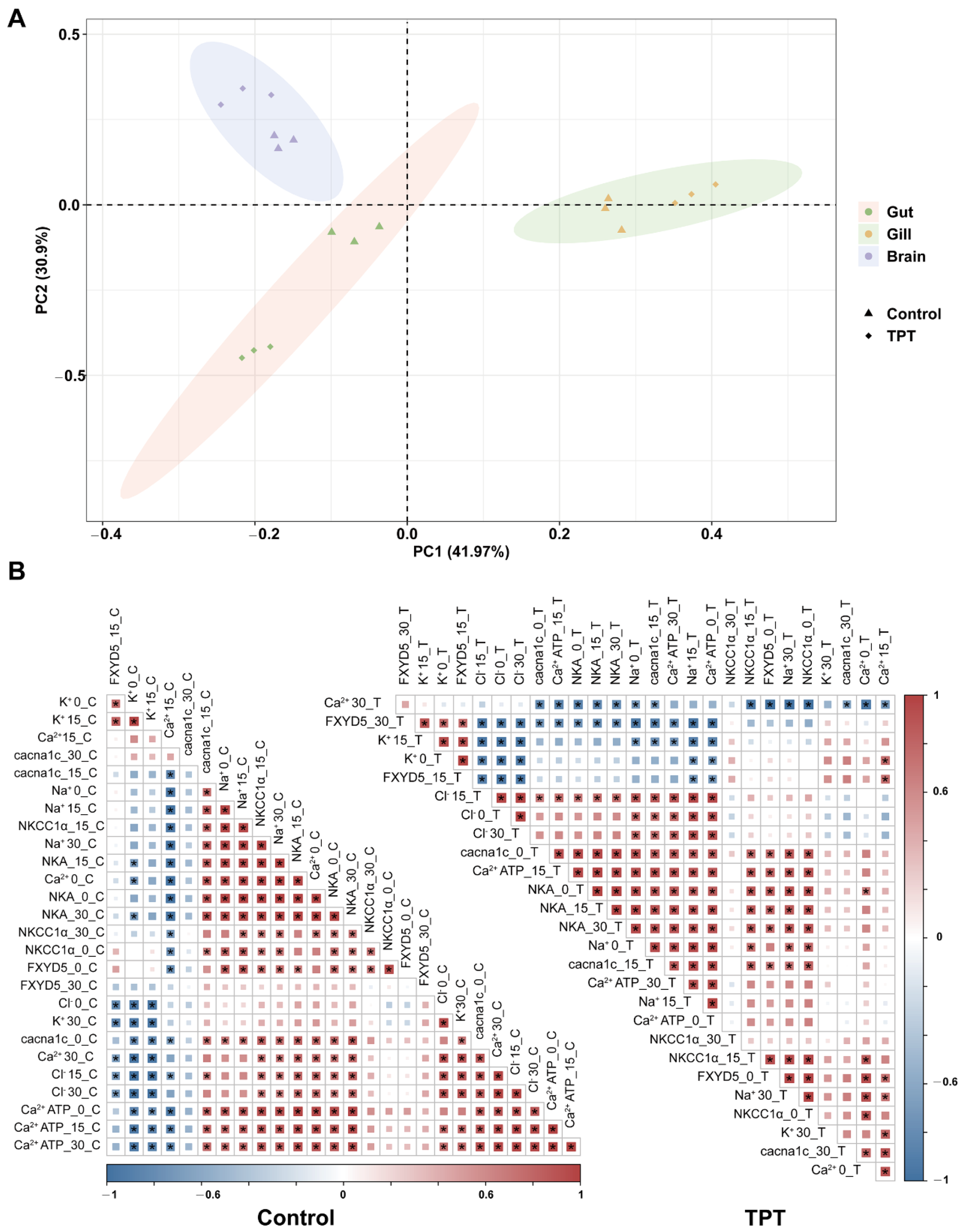
There were significant differences in physiological indicators among the gut, gill, and brain tissues in the co-exposure group of salinity and TPT, as indicated by PCA (Figure 4A). The contributions of PC1 and PC2 were 41.97% and 30.9%, respectively. Furthermore, significant variations were noted between the control group and the TPT group within different tissues, suggesting that the physiological changes in the gut, gill, and brain vary under salinity fluctuations, and these changes are influenced to different extents by TPT exposure. To elucidate the relationships between various parameters, this study visualized the data using a heatmap (Figure 4B). Significant positive correlations (p < 0.05) were observed between NKA enzyme activity and Na+ content in all treatment groups, as revealed by Pearson correlation analysis. In the 30-C group, the expression level of NKCC1a was positively correlated with NKA activity (p < 0.05). Similarly, in the 0-C treatment group, FXYD5 showed a noteworthy positive correlation with NKA activity (p < 0.05). In the 0-C and 15-C treatment groups, Ca2+ATPase exhibited significant positive correlations with cacna1c expression (p < 0.05). Similar results were observed in the co-exposure of TPT and salinity.
Furthermore, a three-way ANOVA analysis was conducted to assess the interplay among tissue type, salinity, and TPT exposure. These factors showed varying degrees of influence on enzyme activity, ion concentrations, and the expression of related genes (Table 4 and Table 5).

4. Conclusions

Overall, the current results indicated that there were substantial variations in NKA enzyme and Ca2+ ATPase activities in different tissues under different salinity conditions, with the enzyme activities associated with gill tissue being higher than those of the gut and brain. Among the different tissues, there were also significant changes in ion concentrations. Correlation analysis showed a significant positive correlation between changes in Na+ ion concentration and NKA activity. After exposure to TPT, the changes in ion concentration within the tissue were disturbed, resulting in a significant decrease in the expression levels of osmoregulation-related genes. This may indicate that the presence of TPT interfered with the osmoregulatory function of marine medaka. This study provides recent biochemical data on the effects of salinity and TPT exposure on osmoregulation in marine medaka. It is essential for understanding the risk assessment of TPT in different stress environments.

Author Contributions

Conceptualization, Z.-H.L.; methodology, T.-Z.L. and C.-Z.C.; validation, T.-Z.L. and C.-Z.C.; formal analysis, L.L.; investigation, S.-Y.X. and T.-Z.L.; resources, Z.-H.L.; data curation, P.L.; writing—original draft preparation, T.-Z.L.; writing—review and editing, C.-Z.C. and Z.-H.L.; supervision, Z.-H.L. and P.L.; project administration, Z.-H.L.; funding acquisition, Z.-H.L. All authors have read and agreed to the published version of the manuscript.

Funding

This research was funded by National Natural Science Foundation of China, 42277269.

Data Availability Statement

Data available on request due to restrictions.

Conflicts of Interest

The authors declare no conflict of interest.

References

  1. He, S.W.; Li, P.; Li, Z.H. Review on endocrine disrupting toxicity of triphenyltin from the perspective of species evolution: Aquatic, amphibious and mammalian. Chemosphere 2021, 269, 128711. [Google Scholar] [CrossRef]
  2. Li, P.; Li, Z.H. Environmental co-exposure to TBT and Cd caused neurotoxicity and thyroid endocrine disruption in zebrafish, a three-generation study in a simulated environment. Environ. Pollut. 2020, 259, 113868. [Google Scholar] [CrossRef]
  3. Li, P.; Li, Z.H.; Zhong, L.Q. Parental exposure to triphenyltin inhibits growth and disrupts thyroid function in zebrafish larvae. Chemosphere 2020, 240, 124936. [Google Scholar] [CrossRef]
  4. Zhang, Z.B.; Hu, J.Y.; Zhen, H.J.; Wu, X.Q.; Huang, C. Reproductive Inhibition and Transgenerational Toxicity of Triphenyltin on Medaka (Oryzias latipes) at Environmentally Relevant tip Levels. Environ. Sci. Technol. 2008, 42, 8133–8139. [Google Scholar] [CrossRef]
  5. Sun, L.B.; Zhang, J.L.; Zuo, Z.H.; Chen, Y.X.; Wang, X.H.; Huang, X.; Wang, C.G. Influence of triphenyltin exposure on the hypothalamus-pituitary-gonad axis in male Sebastiscus marmoratus. Aquat. Toxicol. 2011, 104, 263–269. [Google Scholar] [CrossRef]
  6. Yi, X.L.; Leung, K.M.Y. Assessing the toxicity of triphenyltin to different life stages of the marine medaka Oryzias melastigma through a series of life-cycle based experiments. Mar. Pollut. Bull. 2017, 124, 847–855. [Google Scholar] [CrossRef] [PubMed]
  7. Li, Z.H.; Li, P. Effects of the tributyltin on the blood parameters, immune responses and thyroid hormone system in zebrafish. Environ. Pollut. 2021, 268, 115707. [Google Scholar] [CrossRef] [PubMed]
  8. Lee, C.C.; Wang, T.; Hsieh, C.Y.; Tien, C.J. Organotin contamination in fishes with different living patterns and its implications for human health risk in Taiwan. Environ. Pollut. 2005, 137, 198–208. [Google Scholar] [CrossRef] [PubMed]
  9. Hu, J.Y.; Zhen, H.J.; Wan, Y.; Gao, J.M.; An, W.; An, L.H.; Jin, F.; Jin, X.H. Trophic magnification of triphenyltin in a marine food web of Bohai Bay, north China: Comparison to tributyltin. Environ. Sci. Technol. 2006, 40, 3142–3147. [Google Scholar] [CrossRef] [PubMed]
  10. Sham, R.C.T.; Ho, K.K.Y.; Zhou, G.J.; Li, Y.Y.; Wang, X.H.; Leung, K.M.Y. Occurrence, ecological and human health risks of phenyltin compounds in the marine environment of Hong Kong. Mar. Pollut. Bull. 2020, 154, 111093. [Google Scholar] [CrossRef] [PubMed]
  11. Islam, M.J.; Slater, M.J.; Kunzmann, A. What metabolic, osmotic and molecular stress responses tell us about extreme ambient heatwave impacts in fish at low salinities: The case of European seabass, Dicentrarchus labrax. Sci. Total Environ. 2020, 749, 141458. [Google Scholar] [CrossRef]
  12. Liu, D.R.; Zhang, Z.W.; Song, Y.K.; Yang, J.Y.; Lu, Y.Y.; Lai, W.J.; Wu, Z.Y.; Zhao, D.D.; Lin, H.R.; Zhang, Y.; et al. Effects of salinity on growth, physiology, biochemistry and gut microbiota of juvenile grass carp (Ctenopharyngodon idella). Aquat. Toxicol. 2023, 258, 106482. [Google Scholar] [CrossRef]
  13. Laiz-Carrión, R.; Sangiao-Alvarellos, S.; Guzmán, J.M.; del Río, M.P.M.; Soengas, J.L.; Mancera, J.M. Growth performance of gilthead sea bream Sparus aurata in different osmotic conditions: Implications for osmoregulation and energy metabolism. Aquaculture 2005, 250, 849–861. [Google Scholar] [CrossRef]
  14. Zhu, J.H.; Wang, X.D.; Bu, X.Y.; Wang, C.L.; Pan, J.Y.; Li, E.C.; Shi, Q.C.; Zhang, M.L.; Qin, J.G.; Chen, L.Q. Relationship between myo-inositol synthesis and carbohydrate metabolism changes in Mozambique tilapia (Oreochromis mossambicus) under acute hypersaline stress. Aquaculture 2021, 532, 736005. [Google Scholar] [CrossRef]
  15. Fang, Z.R.; Li, X.J.; Wang, Y.P.; Lu, W.; Hou, J.C.; Cheng, J. Steroidogenic Effects of Salinity Change on the Hypothalamus-Pituitary-Gonad (HPG) Axis of Male Chinese Sea Bass (Lateolabrax maculatus). Int. J. Mol. Sci. 2022, 23, 10905. [Google Scholar] [CrossRef]
  16. Hedén, I.; Sundell, K.; Jönsson, E.; Sundh, H. The role of environmental salinity on Na+-dependent intestinal amino acid uptake in rainbow trout (Oncorhynchus mykiss). Sci. Rep. 2022, 12, 10. [Google Scholar] [CrossRef]
  17. Cao, Q.Q.; Blondeau-Bidet, E.; Lorin-Nebel, C. Intestinal osmoregulatory mechanisms differ in Mediterranean and Atlantic European sea bass: A focus on hypersalinity. Sci. Total Environ. 2022, 804, 150208. [Google Scholar] [CrossRef]
  18. da Silva, A.O.F.; Martinez, C.B.R. Acute effects of cadmium on osmoregulation of the freshwater teleost Prochilodus lineatus: Enzymes activity and plasma ions. Aquat. Toxicol. 2014, 156, 161–168. [Google Scholar] [CrossRef]
  19. Wen, J.J.; Cui, X.Y.; Gibson, M.; Li, Z.Y. Water quality criteria derivation and ecological risk assessment for triphenyltin in China. Ecotoxicol. Environ. Saf. 2018, 161, 397–401. [Google Scholar] [CrossRef]
  20. Saoud, I.P.; Kreydiyyeh, S.; Chalfoun, A.; Fakih, M. Influence of salinity on survival, growth, plasma osmolality and gill Na+-K+-ATPase activity in the rabbitfish Siganus rivulatus. J. Exp. Mar. Biol. Ecol. 2007, 348, 183–190. [Google Scholar] [CrossRef]
  21. Chandrasekar, S.; Nich, T.; Tripathi, G.; Sahu, N.P.; Pal, A.K.; Dasgupta, S. Acclimation of brackish water pearl spot (Etroplus suratensis) to various salinities: Relative changes in abundance of branchial Na+/K+-ATPase and Na+/K+/2Cl co-transporter in relation to osmoregulatory parameters. Fish Physiol. Biochem. 2014, 40, 983–996. [Google Scholar] [CrossRef]
  22. Bradford, M.M. Rapid and Sensitive Method for Quantitation of Microgram Quantities of Protein Utilizing Principle of Protein-Dye Binding. Anal. Biochem. 1976, 72, 248–254. [Google Scholar] [CrossRef]
  23. Marshall, W.S. Na+, Cl, Ca2+ and Zn2+ transport by fish gills: Retrospective review and prospective synthesis. J. Exp. Zool. 2002, 293, 264–283. [Google Scholar] [CrossRef] [PubMed]
  24. Pattnaik, S.; Chainy, G.B.N.; Jena, J.K. Characterization of Ca2+-ATPase activity in gill microsomes of freshwater mussel, Lamellidens marginalis (Lamarck) and heavy metal modulations. Aquaculture 2007, 270, 443–450. [Google Scholar] [CrossRef]
  25. Loro, V.L.; Wood, C.M. The roles of calcium and salinity in protecting against physiological symptoms of waterborne zinc toxicity in the euryhaline killifish (Fundulus heteroclitus). Comp. Biochem. Physiol. C-Toxicol. Pharmacol. 2022, 261, 109422. [Google Scholar] [CrossRef]
  26. Kulac, B.; Atli, G.; Canli, M. Response of ATPases in the osmoregulatory tissues of freshwater fish Oreochromis niloticus exposed to copper in increased salinity. Fish Physiol. Biochem. 2013, 39, 391–401. [Google Scholar] [CrossRef]
  27. Sinha, A.K.; Limbaugh, N.; Renukdas, N.; Bishop, W.M.; Romano, N. Modulating effect of elevated water hardness on growth performance, ammonia dynamics and ion-regulatory capacity in channel catfish (Ictalurus punctatus) following chronic challenge with high environmental ammonia and salinity stress. Aquaculture 2022, 560, 738489. [Google Scholar] [CrossRef]
  28. Sardella, B.A.; Brauner, C.J. The effect of elevated salinity on ‘California’ Mozambique tilapia (Oreochromis mossambicus × O-urolepis hornorum) metabolism. Comp. Biochem. Physiol. C-Toxicol. Pharmacol. 2008, 148, 430–436. [Google Scholar] [CrossRef] [PubMed]
  29. Yang, W.K.; Hsu, A.D.; Kang, C.K.; Lai, I.P.; Liao, P.S.; Lee, T.H. Intestinal FXYD12 and sodium-potassium ATPase: A comparative study on two euryhaline medakas in response to salinity changes. PLoS ONE 2018, 13, 16. [Google Scholar] [CrossRef]
  30. Allen, P.J.; McEnroe, M.; Forostyan, T.; Cole, S.; Nicholl, M.M.; Hodge, B.; Cech, J.J. Ontogeny of salinity tolerance and evidence for seawater-entry preparation in juvenile green sturgeon, Acipenser medirostris. J. Comp. Physiol. B-Biochem. Syst. Environ. Physiol. 2011, 181, 1045–1062. [Google Scholar] [CrossRef] [PubMed]
  31. Xing, S.Y.; Li, P.; He, S.W.; Cao, Z.H.; Wang, X.; Cao, X.Q.; Liu, B.; Chen, C.Z.; You, H.; Li, Z.H. Physiological responses in Nile tilapia (Oreochromis niloticus) induced by combined stress of environmental salinity and triphenyltin. Mar. Environ. Res. 2022, 180, 105736. [Google Scholar] [CrossRef] [PubMed]
  32. Brown, R.A.; Jefferson, L.; Sudan, N.; Lloyd, T.C.; Ren, J. Acetaldehyde depresses myocardial contraction and cardiac myocyte shortening in spontaneously hypertensive rats role of intracellular Ca2+. Cell. Mol. Biol. 1999, 45, 453–465. [Google Scholar] [PubMed]
  33. Sandmann, S.; Min, J.Y.; Meissner, A.; Unger, T. Effects of the calcium channel antagonist mibefradil on haemodynamic parameters and myocardial Ca2+-handling in infarct-induced heart failure in rats. Cardiovasc. Res. 1999, 44, 67–80. [Google Scholar] [CrossRef] [PubMed]
  34. Miura, Y.; Matsui, H. Effects of triphenyltin on cytosolic Na+ and Ca2+ response to glucose and acetylcholine in pancreatic β-cells from hamster. Toxicol. Appl. Pharmacol. 2001, 174, 1–9. [Google Scholar] [CrossRef] [PubMed]
  35. Jeong, J.Y.; Kwon, H.B.; Ahn, J.C.; Kang, D.; Kwon, S.H.; Park, J.A.; Kim, K.W. Functional and developmental analysis of the blood-brain barrier in zebrafish. Brain Res. Bull. 2008, 75, 619–628. [Google Scholar] [CrossRef]
  36. Diao, X.Z.; Han, H.; Li, B.L.; Guo, Z.; Fu, J.; Wu, W.H. The Rare Marine Bioactive Compounds in Neurological Disorders and Diseases: Is the Blood-Brain Barrier an Obstacle or a Target? Mar. Drugs 2023, 21, 406. [Google Scholar] [CrossRef]
  37. Kennedy, C.J.; Tierney, K.B.; Mittelstadt, M. Inhibition of P-glycoprotein in the blood-brain barrier alters avermectin neurotoxicity and swimming performance in rainbow trout. Aquat. Toxicol. 2014, 146, 176–185. [Google Scholar] [CrossRef]
  38. Li, Z.H.; Xu, H.Y.; Zheng, W.L.; Lam, S.H.; Gong, Z.Y. RNA-sequencing analysis of TCDD-induced responses in zebrafish liver reveals high relatedness to in vivo mammalian models and conserved biological pathways. PLoS ONE 2013, 8, e77292. [Google Scholar] [CrossRef] [PubMed]
  39. Cutler, C.P.; Cramb, G. Two isoforms of the Na+/K+/2CI cotransporter are expressed in the European eel (Anguilla anguilla). Biochim. Biophys. Acta-Biomembr. 2002, 1566, 92–103. [Google Scholar] [CrossRef]
  40. Horng, J.L.; Yu, L.L.; Liu, S.T.; Chen, P.Y.; Lin, L.Y. Potassium Regulation in Medaka (Oryzias latipes) Larvae Acclimated to Fresh Water: Passive Uptake and Active Secretion by the Skin Cells. Sci. Rep. 2017, 7, 14. [Google Scholar] [CrossRef]
  41. Kang, C.K.; Tsai, H.J.; Liu, C.C.; Lee, T.H.; Hwang, P.P. Salinity-dependent expression of a Na+, K+, 2Cl cotransporter in gills of the brackish medaka Oryziasdancena: A molecular correlate for hyposmoregulatory endurance. Comp. Biochem. Physiol. A-Mol. Integr. Physiol. 2010, 157, 7–18. [Google Scholar] [CrossRef] [PubMed]
  42. Gotliv, I.L. FXYD5: Na+/K+-ATPase Regulator in Health and Disease. Front. Cell. Dev. Biol. 2016, 4, 26. [Google Scholar] [CrossRef]
  43. Miller, T.J.; Davis, P.B. FXYD5 modulates Na+ absorption and is increased in cystic fibrosis airway epithelia. Am. J. Physiol.-Lung Cell. Mol. Physiol. 2008, 294, L654–L664. [Google Scholar] [CrossRef] [PubMed]
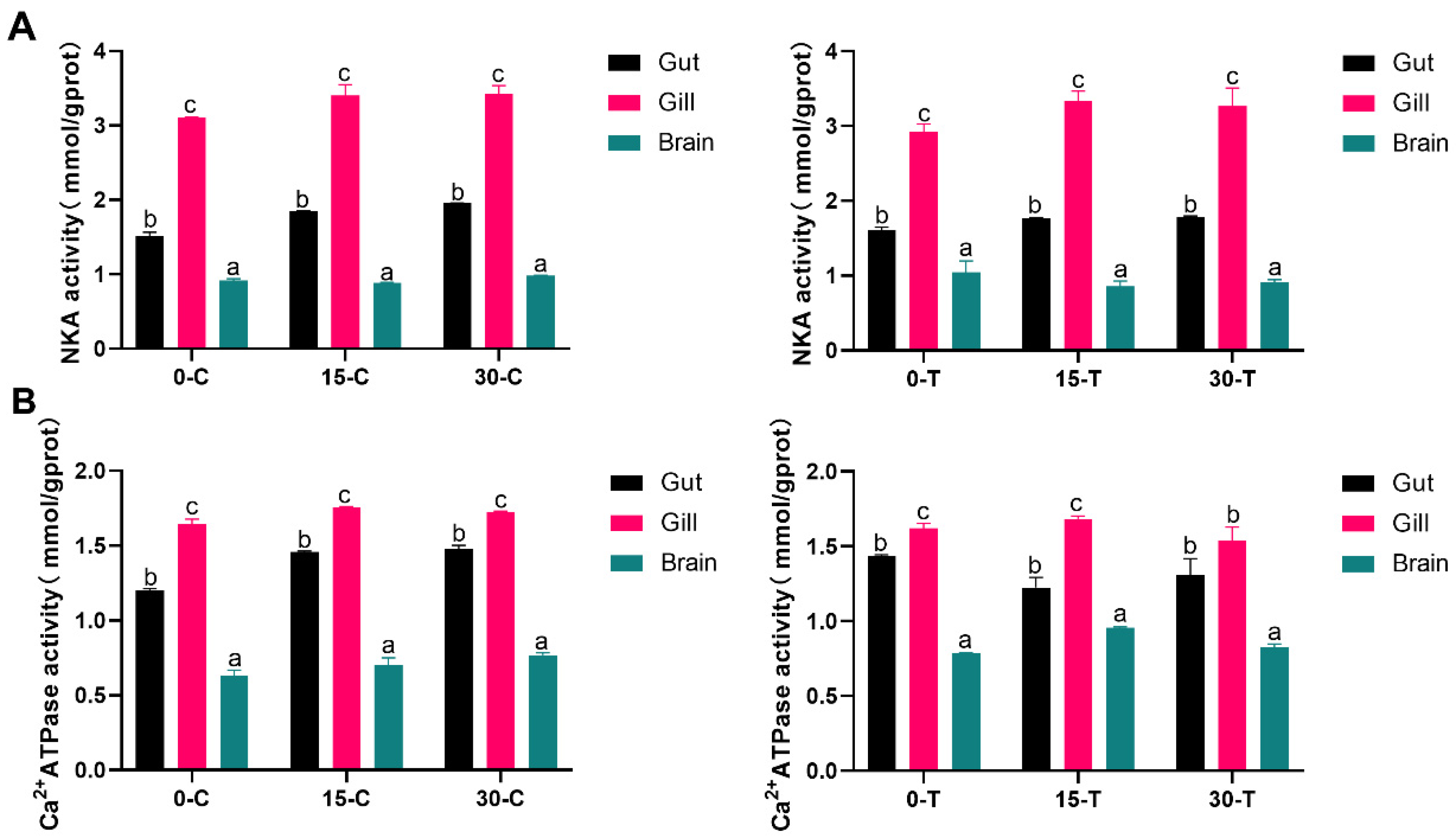
Figure 1. Changes in tissue-specific enzyme activities under different salinities (0, 15, 30) and exposure to tributyltin (C: 0 ng/L, T: 100 ng/L) are shown. (A) NKA enzyme activity; (B) Ca2+ATPase activity. Vertical bars represent the mean ± SE (n = 3). Letters indicate significant differences (p < 0.05).
Water 16 00921 g001
Figure 2. Changes in ion concentrations within tissues under different salinities (0, 15, 30) and TPT exposure (C: 0 ng/L, T: 100 ng/L) are shown. (A) Na+ concentration; (B) Cl concentration; (C) Ca2+ concentration; (D) K+ concentration. Vertical bars represent the mean ± SE (n = 3). Letters indicate significant differences (p < 0.05).
Water 16 00921 g002
Figure 3. Changes in gene expression levels of NKCC1α, FXYD5, and cacna1c under different salinities (0, 15, 30) and TPT exposure (C: 0 ng/L, T: 100 ng/L) are shown. (A) NKCC1α level; (B) FXYD5 level; (C) cacna1c level. Vertical bars represent the mean ± SE (n = 3). Letters indicate significant differences (p < 0.05).
Water 16 00921 g003
Figure 4. Chemometric analysis is presented. (A) PCA analysis showing the association between tissue, salinity (0, 15, 30), and TPT (C: 0 ng/L, T: 100 ng/L). (B) Heatmap analysis displaying the correlation among various factors measured in the experiment, with values ranging from −1 to 1, where red indicates positive correlation and blue indicates negative correlation. “*” denotes a significant difference (p < 0.05).
Water 16 00921 g004
Table 1. The primer sequences used in the experiments.
Gene NameSequences of Primers (5′−3′)
NKCC1αFTCAAACCAGTGAGGGAGAATACAAC
NKCC1αRTTCTTGTTCATTTTAAGGGCGTCGG
FXYD5FCCAATGATAGAAGGCAAAGAGT
FXYD5RATGGAGCTTGTTCACAGGA
cacna1cFGTTTGTGGCAATCAGGACCAT
cacna1cRAAAACTTCCCCTTAAAGAGTTGCA
18SFGACAAATCGCTCCACCAACT
18SRCCTGCGGCTTAATTTGACCC
Table 2. ANOVA analysis was conducted on the relevant biochemical parameters among different treatments, resulting in F-values, degrees of freedom (DF), and p-values.
GroupCa2+ClK+Na+NKACa2+ATP
FDFpFDFpFDFpFDFpFDFpFDFp
0-C95.292; 6<0.0194.272; 6<0.01132.252; 6<0.01104.412; 6<0.01975.5272; 6<0.01272.4592; 6<0.01
15-C95.292; 6<0.01718.862; 6<0.01418.942; 6<0.01372.632; 6<0.01224.3822; 6<0.01374.4822; 6<0.01
30-C171.442; 6<0.011790.962; 6<0.0141.222; 6<0.0192.482; 6<0.01384.0362; 6<0.011049.7852; 6<0.01
0-T418.112; 6<0.01465.382; 6<0.01111.612; 6<0.01160.272; 6<0.0177.2292; 6<0.01448.8702; 6<0.01
15-T101.482; 6<0.012226.262; 6<0.0145.242; 6<0.01388.672; 6<0.01194.2682; 6<0.0184.8782; 6<0.01
30-T114.772; 6<0.01905.142; 6<0.012.992; 60.126767.512; 6<0.0168.4042; 6<0.0119.3592; 60.02
Note: salinity: 0, 15, and 30; C: control, TPT 0 ng/L; T: TPT 100 ng/L; DF: degrees of freedom for the main factor or interaction effects; p < 0.05 indicates significance effects.
Table 3. The relative mRNA expression levels among different treatments were analyzed using ANOVA, resulting in F-values, degrees of freedom (DF), and p-values.
GroupNKCC1αFXYD5cacna1c
FDFpFDFpFDFp
0-C55.222; 6<0.0177.392; 6<0.0112.572; 60.07
15-C53.392; 6<0.0112.292; 60.080.232; 60.013
30-C3.662; 60.910.682; 60.5430.012; 60.429
0-T200.652; 6<0.0150.862; 6<0.012.752; 6<0.01
15-T24.292; 6<0.01152.032; 6<0.010.402; 6<0.01
30-T1.022; 60.41641.442; 6<0.010.152; 60.092
Note: salinity: 0, 15, and 30; C: control, TPT 0 ng/L; T: TPT 100 ng/L; DF: degrees of freedom for the main factor or interaction effects; p < 0.05 indicates significance effects.
Table 4. Three-way ANOVA analysis of the interaction effects of tissue, salinity, and TPT on enzyme activities and ion concentrations.
Factors/Interactions NKACa2+ ATPaseNa+ClCa2+K+
DFFpFpFpFpFpFp
Tissue2928.950.000666.150.0001151.390.0003115.980.00068.94 0.000 223.270.000
Salinity27.900.0015.260.01010.480.000167.840.000334.82 0.000 23.0830.000
TPT11.890.1770.010.9451.580.21710.200.00310.80 0.002 41.0630.000
Tissue * Salinity43.880.101.900.13158.580.000115.550.000168.37 0.000110.460.000
Tissue * TPT20.950.39614.950.00025.900.00037.780.000186.79 0.0005.140.011
Salinity * TPT20.940.40110.470.00039.820.00026.750.0001.73 0.192 48.860.000
Tissue * Salinity * TPT40.47 0.7605.780.00151.060.00085.390.000139.03 0.000 17.720.000
Note: tissue: gut, gill, and brain; salinity: 0, 15, and 30; TPT: 0 and 100 ng/L; DF: degrees of freedom for the main factor or interaction effects; p < 0.05 indicates significance effects. * indicates interactions between different factors.
Table 5. Gene expression levels under stress conditions analyzed using three-way ANOVA.
Factors/Interactions NKCC1αFXYD5cacna1c
DFFpFpFp
Tissue2176.790.00072.0560.00064.430.000
Salinity239.880.0004.120.00018.440.000
TPT10.270.60748.790.02410.460.003
Tissue * Salinity433.0730.00043.360.00021.370.000
Tissue * TPT24.960.01312.270.00017.600.000
Salinity * TPT210.480.000130.70.0005.3350.009
Tissue * Salinity * TPT47.740.00012.470.0008.3380.000
Note: tissue: gut, gill, and brain; salinity: 0, 15, and 30; TPT: 0 and 100 ng/L; DF: degrees of freedom for the main factor or interaction effects; p < 0.05 indicates significance effects; * indicates interactions between different factors.
Disclaimer/Publisher’s Note: The statements, opinions and data contained in all publications are solely those of the individual author(s) and contributor(s) and not of MDPI and/or the editor(s). MDPI and/or the editor(s) disclaim responsibility for any injury to people or property resulting from any ideas, methods, instructions or products referred to in the content.