Article

Effects of Norflurazon and UV Radiation on Symbiotic and Free-Living Hydra

by
Goran Kovačević
1,
Ana Matijević
2,
Petra Korać
1,*,
Davor Želježić
3,
Siegfried Reipert
4,
Katarina Caput Mihalić
1,
Damir Sirovina
1,
Petra Peharec Štefanić
1 and
Martina Ivšić
1
1
Department of Biology, Faculty of Science, University of Zagreb, Horvatovac 102a, HR-10000 Zagreb, Croatia
2
Department of Laboratory Diagnostics, University Hospital Centre Zagreb, Kišpatićeva 12, HR-10000 Zagreb, Croatia
3
Unit for Mutagenesis, Institute for Medical Research and Occupational Health, HR-10000 Zagreb, Croatia
4
Cell Imaging and Ultrastructure Research (CIUS), University of Vienna, Djerassiplatz 1, A-1030 Vienna, Austria
*
Author to whom correspondence should be addressed.
Water 2024, 16(5), 645; https://doi.org/10.3390/w16050645
Submission received: 4 December 2023 / Revised: 15 February 2024 / Accepted: 17 February 2024 / Published: 22 February 2024

Abstract

:
In this study, we aimed to document the freshwater symbiotic interactions along with the impact of the abiotic environment and anthropogenic effects on the functionality of freshwater organisms. Symbiotic green hydra (Z) and free-living brown hydra (S), either separately or both species together, were treated with the herbicide norflurazon in concentrations of 2 × 10⁻⁶ mol/L (N6) and 2 × 10⁻⁷ mol/L (N7) for 72 h. Also, hydras were treated with both norflurazon and UV radiation at a wavelength of 254 nm for 2 min or were irradiated only. The next part of the experiment was performed in the same way but with added suspensions of isolated endosymbiotic alga, free-living alga, or both algae together. Mortality, migration, tentacle and tissue damage, changes in the thickness of the mesoglea of hydras, and clustering of algae were monitored. Green hydra generally showed lower rates of migration, and mortality was observed only in green hydra exposed to UV radiation. Tentacle damage was more pronounced in green hydra and included a specific fork-like structure. The use of cryofixation and TEM enabled us to partly elucidate the effect of clustering of algae. In summary, our study provides new insights into the influence of different environmental stressors and their combination on symbiotic and free-living freshwater hydras and algae and a better understanding of interactions in freshwater ecosystems.

1. Introduction

Hydra is a cosmopolitan freshwater invertebrate (Cnidaria, Hydrozoa). Common species include Hydra viridissima (green hydra) (Z) and Hydra oligactis (brown hydra) (S). The hydra body is composed of two cellular layers with a third in-between gel-like layer, the mesoglea. This layer has a gelatinous structure and contains collagen, lipids, proteins, and carbohydrates. The mesoglea enables the passage of food as well as the migration of cells during regeneration [1]. Both the ectoderm and gastroderm contribute to the formation of the mesoglea [2]. Water in large quantities is an integral part of the mesoglea, and it serves as a support for hydra. In green hydra, algal endosymbionts are located in endodermal epithelial cells. A significant point in studying algal symbiotic systems in green hydra is that photosynthetic symbionts provide nutrients for the polyps, enabling hydra to survive extended periods of starvation. The rate of photosynthesis in symbiotic Chlorella is much higher than in their free-living relatives [3]. Th endosymbiotic alga is the stronger partner in this symbiotic relationship [4] and can be isolated from its host [5]. Compared to the free-living related algae, isolated endosymbiotic algae are less adapted to the new microenvironmental conditions [6,7]. This points to an incomplete process of symbiogenesis between green hydra and endosymbiotic algae [5]. The hydra holobiont also includes bacteria as ectosymbionts [8,9].
Hydra has been studied as a suitable experimental organism for more than 250 years, especially in evolutionary and ecotoxicological research [10,11,12,13,14,15], and in research on the mechanisms and evolution of endosymbiotic relationships in particular, the green hydra itself is used [16]. The hydra–algae symbiosis represents an important model for studying the specificity between the host and the symbiont in the invertebrate–algae system [16]. Green hydra has been shown to have a higher tolerance to certain harmful substances than brown hydra [12]. Hydras are commonly used organisms in biological research because of their small body size; easy manipulation; quick reproduction by budding, by which in a short period, a high number of genetically identical organisms can be available; and easily measurable morphological, behavioral, and reproductive changes. Hydras have a huge regeneration capability [17]. Since the life processes of this sessile organism take place by diffusion, the impacts of changes in the surrounding medium can be easily monitored in hydras. Research on hydra has been performed including the effects of pesticides, heavy metals, irradiation, and antibiotics [11,13,18]. They share evolutionary and symbiotic relationships. The described specificity of green hydra symbiosis can be used to potentially trace the reestablishment of symbiosis with its endosymbionts, i.e., tracing the course of symbiosis is possible [19]. Free-living brown hydra was used for comparison.
Norflurazon is a selective preemergent herbicide. In plants, norflurazon acts by inhibiting carotenoid biosynthesis [20]. The mobility and absorption of norflurazon in the soil depends on the physical and chemical properties of the soil. It is relatively weakly mobile in mineral soils, less so in soils with a lot of organic matter [21]. Its slow mobility in the soil is associated with poor solubility [22]. The half-life is 38–731 days, depending on the soil characteristics. It belongs to the fluorinated pyridazinones. It blocks the enzyme phytoene-desaturase, leading to the destruction of the structure and bleaching of newly formed chloroplasts in bright light (high-light conditions), during which there is suppression of thylakoid development and shrinkage, but it does not affect already existing differentiated chloroplasts [23,24,25,26,27,28], which leads to photooxidation and depletion of chlorophyll, the result of which is the aforementioned bleaching of chloroplasts and inactivation of the photosynthetic apparatus. Primary carotenoids are auxiliary antioxidant pigments necessary for the absorption of excess light in photosynthetic organisms and thus for the prevention of photooxidation of chlorophyll and photodestruction of plastids [27,29].
It acts on weeds during germination, causes the so-called “bleaching effect”, after which the plant dies. Norflurazon is absorbed by the root and is transported by the xylem elements of the plant’s conduction system to the plant leaves, i.e., to the target parts of the organism that carry chloroplasts. Given the possibility that by washing treated surfaces in nature, the herbicide reaches the deep layers of the soil, surface water, and underground water [11,22,30,31], which are a significant source of drinking water and have a potential effect on aquatic primary producers, whereby they can change the species composition of algal communities [32,33] and thus the composition of food chains in ecosystems, the potential environmental danger of norflurazon for organisms and aquatic ecosystems was assessed.
Due to such characteristics, there is a risk of norflurazon reaching groundwater [34]. Studies of this herbicide have been carried out on various organisms: cyanobacteria [24,25], Euglena gracilis [35], algae [36], plants [28,37,38,39,40], hydra [41], and planarians [42], and a low category III of toxicity was established [41,42]. Norflurazon is classified as a possible human carcinogen. ECHA has classified it as very toxic to aquatic life with long-lasting effects [42].
In humans, ultraviolet radiation causes sunburn and darkening and thickening of the outer layer of the skin and is associated with the development of melanoma and other types of skin cancer. It may also contribute to eye and immune system disorders. Moreover, UV light might cause damage to DNA by the formation of pyrimidine dimers and affect the nucleotide repair mechanism [43]. UV radiation in hydra causes damage to the tentacles, hypostome, and gastral region [44,45]; initiates ectopic foot formation in regenerating hydra; and promotes budding [46].
The influence of UV light on living organisms has been researched extensively, especially since we became aware of the reduction in the ozone layer. The effects of UV light, mainly from the UV-B spectrum, have been investigated in many organisms. Silva et al. (2022) analyzed the effects of UV radiation on a soil microbial community [47], namely, on the dinoflagellate Pelagodinium béii exposed to ultraviolet (UV) radiation. Sonntag, Summerer and Sommaruga (2011) tested resistance to solar UV radiation in mixotrophic and heterotrophic ciliates [48], Parajuli et al. (2023) observed how UV-A and UV-B affects the different stages of the small insect Diaphorina citri [49], and Stábile et al. (2021) and Eshun-Wilson et al. (2020) investigated the fitness costs associated with UV radiation in planktonic crustaceans [50,51]. Alves et al. investigated the harmful effects on the fish Sparus aurata [52]. Furthermore, the effects of UV light on herbicide toxicity have been investigated, and some studies have shown the effects of radiation of different wavelengths from the UV-A, UV-B, and UV-C spectrum [53]. Recently, as aquatic habitats have changed, Hydra has been shown to exhibit different periodical appearances in known localities [44]. We are witnessing the degradation of the atmosphere and non-compliance with agreements and protocols, which should regulate pollution and damage to the atmosphere. That could lead to major changes in the atmosphere that could result in an increased amount of UV radiation and perhaps even a significant penetration of UV-C radiation. Since hydra is a common research organism and inhabits very shallow waters that allow the penetration of UV radiation, it can serve as a good model for researching the consequences of increased penetration of UV radiation through the atmosphere and a thin layer of water.
The aim of this study was to investigate effects of exposure to norflurazon and/or UV radiation that can potentially cause uncontrolled cell proliferation in hydra. Two species of hydra, one symbiotic and one free-living, were used, as were two species of algae, one symbiotic and one free-living. We aimed to determine the following: (i) whether norflurazon treatment and UV radiation simultaneously cause higher damage in terms of morphological and behavioral changes in hydra or mortality compared to the effects of a single agent; (ii) the effects on the co-existence of the two species of hydra when they are exposed together to norflurazon/UV radiation and separately; and (iii) whether behavioral or morphological changes occur in these two species of hydra when exposed to norflurazon and/or UV radiation in the presence of algae. For clearer analysis, we observed the algae by using cryofixation and transmission electron microscopy (TEM). In general, our results show that environmental stresses can strongly affect different, namely, symbiotic and free-living, hydra species. Such findings point to the need of further examination of symbiotic and interspecific relationships and their value in adaptation processes.

2. Materials and Methods

2.1. Organisms and Treatments

Brown hydra was collected from lakes in Maksimir forest park (Zagreb, NW Croatia), while green hydra was obtained from cultures at the Division of Zoology, Faculty of Science. Animals were grown in laboratory conditions of 22.4 °C in daylight, kept in aerated water and fed with Artemia salina (Linnaeus, 1758) larvae until the experiments were conducted. During the treatment, animals were not fed. The free-living alga C. vulgaris Beij. [K&H, 1992] (CV) and the endosymbiotic alga Mychonastes homosphaera (Chlorophyceae) (Skuja) Kalina et Punčochářová (CZ) were obtained from cultures at the Division of Zoology, Faculty of Science [5]. The experiments were conducted on three test groups and corresponding controls (K). The first test group included 10 green hydras in one container, the second group was composed of 10 brown hydras in one container, and the third test group consisted of 5 green and 5 brown hydras in one container. All the test groups were (1) treated with norflurazon; (2) irradiated with UV radiation (using UV cross-linker CL-508 (Uvitec, Cambridge, UK)) (R); and (3) treated simultaneously with both norflurazon and UV radiation. The control group animals were grown in aerated water. The experiments were performed in triplicate, and hydras were monitored for 72 h using a stereomicroscope.
The individuals of green hydra (Hydra viridissima Pallas, 1766) and brown hydra (Hydra oligactis Pallas, 1766), either separately or jointly, were treated with norflurazon in concentrations of 2 × 10⁻⁶ mol/L (N6) or 2 × 10⁻⁷ mol/L (N7) for 72 h, which is in accordance with [13,14,54]. In addition, hydras were treated simultaneously with both norflurazon and UV radiation of a wavelength of 254 nm, 0.023 mW/cm2 for 2 min, which is in accordance with [44], or were irradiated only. Additionally, experiments were conducted with hydras in the presence of algae, by adding suspensions of either the free-living alga Chlorella vulgaris Beij. [K&H, 1992] (CV) or the endosymbiotic alga Mychonastes homosphaera (Chlorophyceae) (Skuja) Kalina et Punčochářová (CZ) or both algae combined. The test groups in this part of the study consisted of five animals of each hydra species separately and two animals of each species in the same container. Equivalent amounts of monocultures of the free-living alga C. vulgaris or the endosymbiotic alga M. homosphaera were added to the experimental dishes with hydras separately or as a combination of both species together. Norflurazon treatment and irradiation were conducted as in the first part of the study. Control samples were hydras with specific algae species or with both species together that were neither treated with norflurazon nor irradiated. The following changes in hydra were monitored: mortality, migration, tentacle and tissue damage, and thickness of the mesoglea. Clustering of algae was observed, and cryofixation and TEM were used for analysis of algae.

2.2. Histological Changes

Histological changes caused by norflurazon treatment and/or irradiation were analyzed under a light microscope. After 72 h, hydras were placed in Bouin’s fixative (15 mL of saturated aqueous solution of picric acid, 5 mL of 40% formalin, and 1 mL of ice acetic acid) and then embedded in paraplast. Paraplast blocks were cut using a microtome to slide sections of four μm in thickness. Paraplast was then removed by incubation in xylene, and sections were dehydrated in an alcohol series (70%, 80%, 90%, 100%) followed by washing with distilled water (3 × 10 min). After rinsing with water, the sections were again dehydrated in an alcohol series (70%, 80%, 90% and 100%) and treated with xylene (2 × 15 min). The sections were stained with hemalaun and eosin solutions and covered using Canada balsam. Micrographs of histological preparations were taken by using an Olympus microscope (Tokyo, Japan) equipped with a digital camera. Morphometry of the mesoglea was performed using ImageJ software. According to a per section length of 100 μm, the thickness of the mesoglea was measured for 30 randomly selected segments on the histological slides, per particular treatment.

2.3. Transmission Electron Microscopy

For TEM, algae from the algal cultures were cryoimmobilized by high-pressure freezing (HPF) followed by low-temperature dehydration, fixation, and embedding in epoxy resin as described recently [45]. The algae were transferred into 1-hexadecene-coated (Merck, Sharp & Dome, Kenilworth, NJ, USA) 3 mm carriers, type B, 300 µm in depth (Leica Microsystems, Vienna, Austria) filled up with 20% bovine serum albumin and covered with the flat surface of another carrier, type B. The samples in the carriers were frozen at ca. 2000 bar in a high-pressure HPM100 freezer (Leica Microsystems, Austria) and transferred to an automated freeze substitution system, namely, AFS2 (Leica Microsystems, Austria), that was precooled to −140 °C and equipped with an agitation module (Cryomodultech e.U., Vienna, Austria) [45,55,56]. FS with 1% OsO4 in acetone was agitated (15 V) for 44 h at −85 °C to ensure sufficient dehydration within microbody-like organelles of the freshwater algae. After warming to room temperature, the samples were infiltrated with epoxy resin Agar 100 (Agar Scientific Ltd., Stansted, UK). Polymerization was performed in an oven at 65 °C for ca. 36 h. Ultrathin sections (70–90 nm in thickness) were cut with an Ultracut S (LEICA Microsystems, Austria) ultramicrotome by using an oscillating diamond knife, Diatome V7 (Diatome, Nidau, Switzerland). They were placed on Formvar-coated copper grids and contrasted with 4% neodymium(III)-acetate [57] and lead citrate prior to analyses in a TEM ZEISS 900N (Oberkochen, Germany) at 80 kV. Images were acquired by using a digital TRS (4 megapixel) camera and ImageSp-professional software (Tröndle, Moorenweis, Germany). For Enlargement of figure details the GIMP image editor (v2.10.36) was used.

3. Results

In this study, we investigated and detected the effects of the herbicide norflurazon/UV radiation on mortality, migration, tentacles, tissues, and mesoglea of hydra. In addition, we detected the effect on the mentioned hydra setups with the presence of two species of microalgae. Moreover, we observed the clustering of algae added to the containers with hydras.
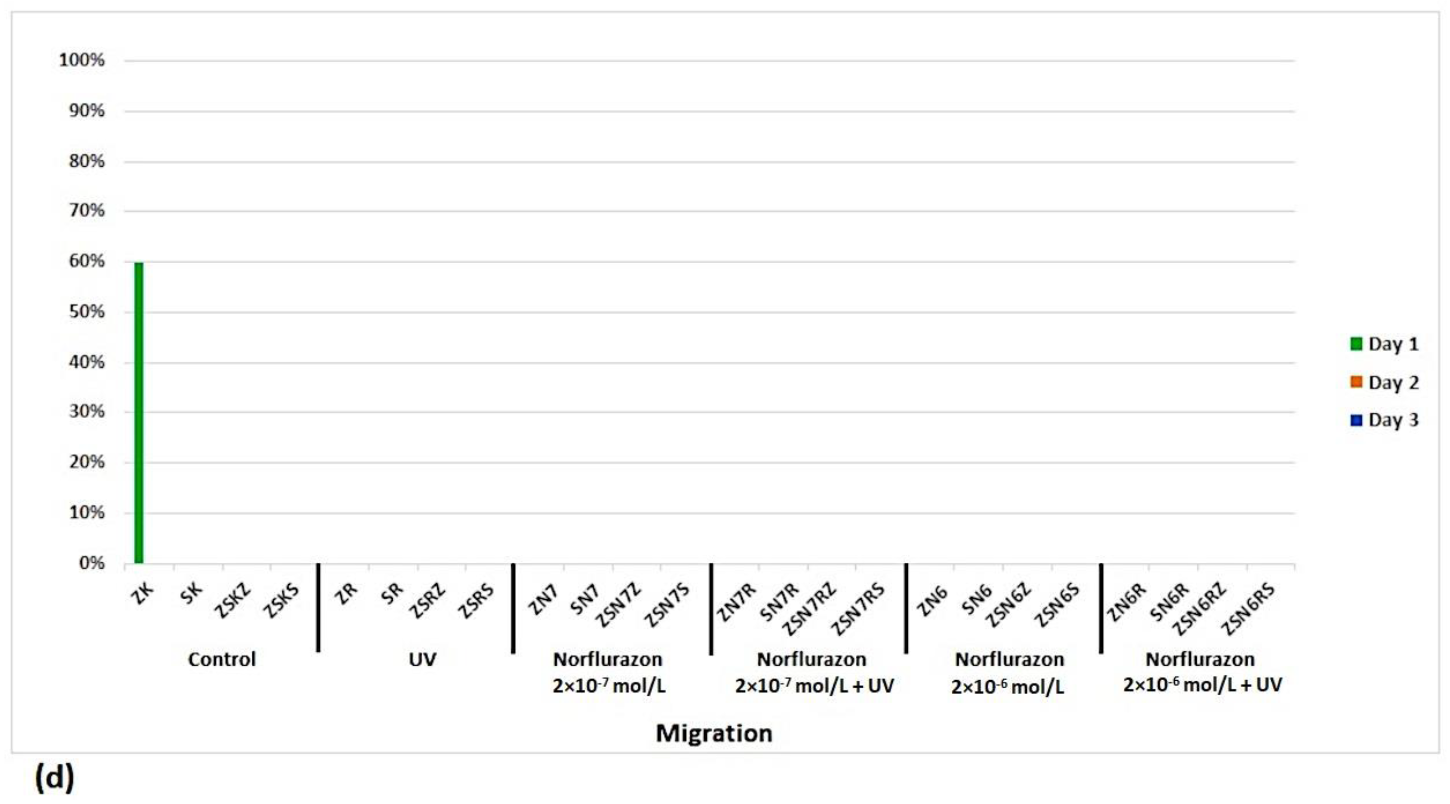
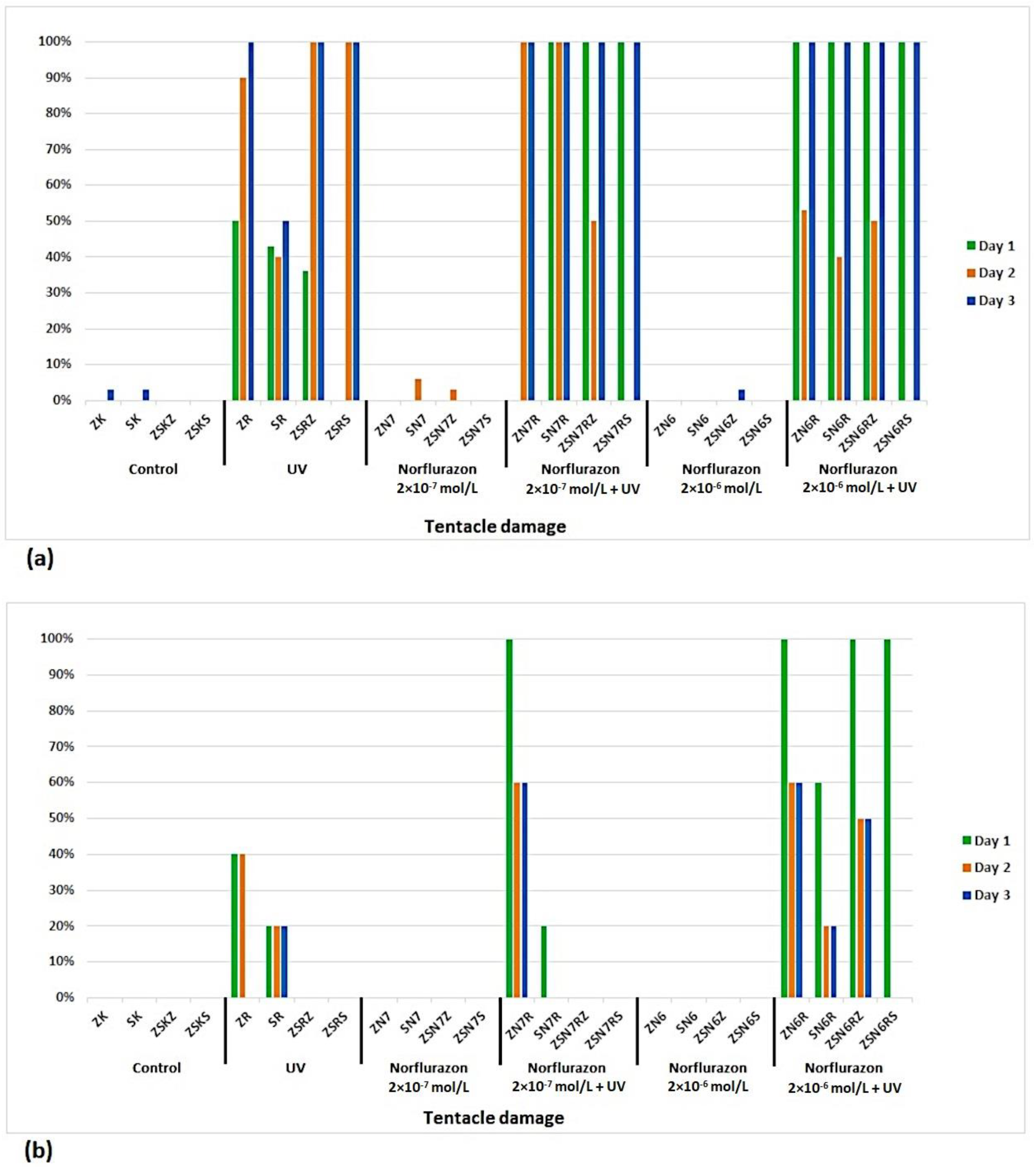
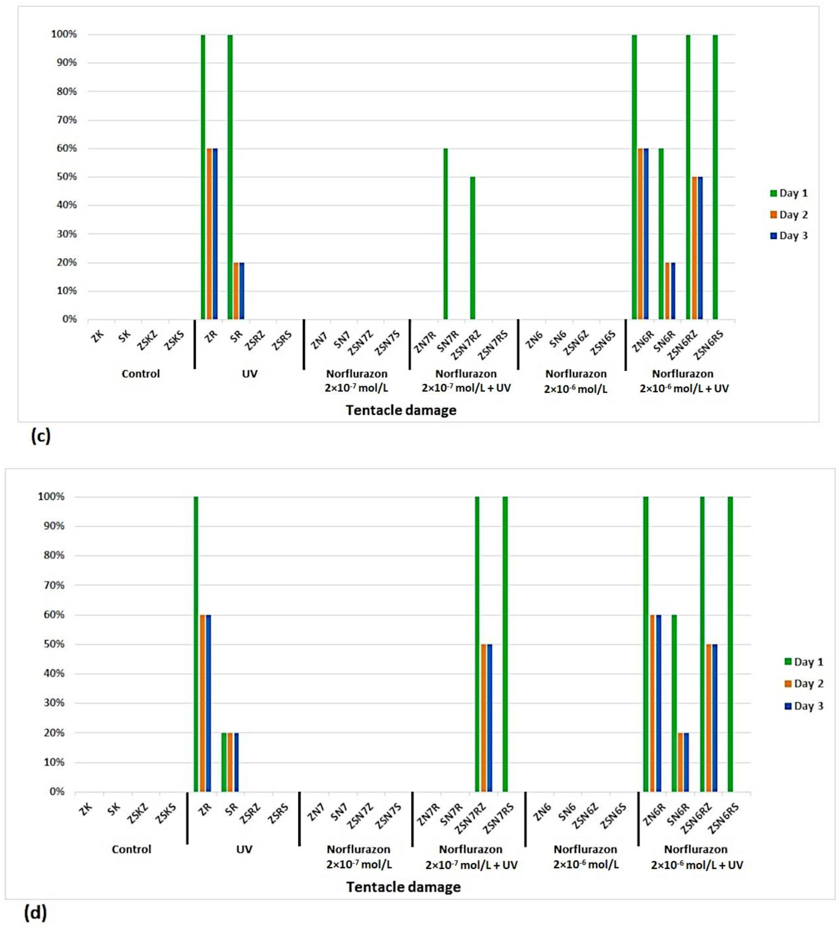
Mortality was present only in irradiated green hydra, and the added algae, in part, affected the mortality of green hydra. Overall mortality is shown in Figure 1. Brown hydra, alone and when exposed together with green hydra, showed greater fluctuations in migration. The highest migrations were recorded in the control and in norflurazon treatment. Brown hydra migrated more than green hydra. Green hydra generally had a low rate of migration, but in the presence of algae, migration of green hydra partially increased. Migration was enhanced in the control of green hydra by the presence of C. vulgaris. With the addition of both algae, migration was present only in the green hydra control. Here, algae possibly created a more favorable environment. However, algae could also cause inhibition of migration by hindering the movement of hydra. Overall migrations are shown in Figure 2. Tentacles were more affected by irradiation than norflurazon. Green hydra suffered from higher tentacle damage. The presence of algae diminished the induction of damage. Overall tentacle damage is shown in Figure 3. Such results may suggest that the effect of the two environmental factors, norflurazon and UV radiation is more deleterious to green hydra compared with brown hydra.
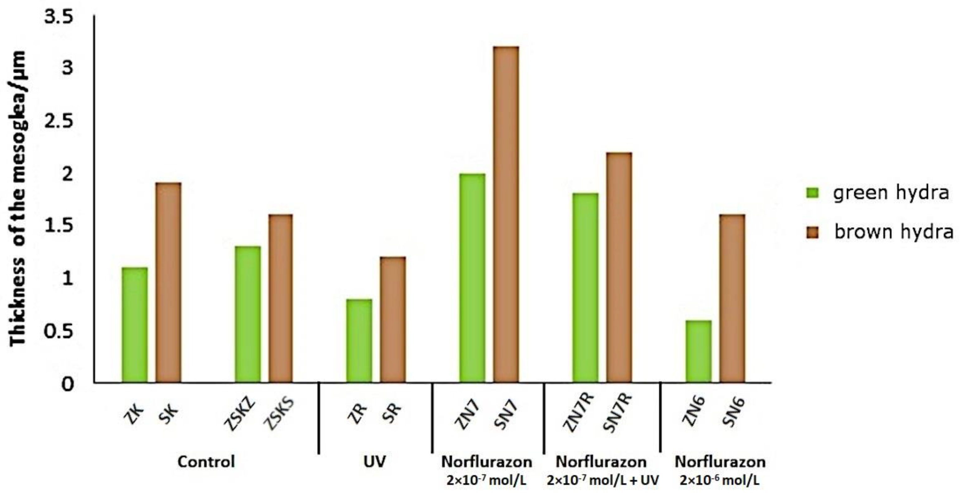
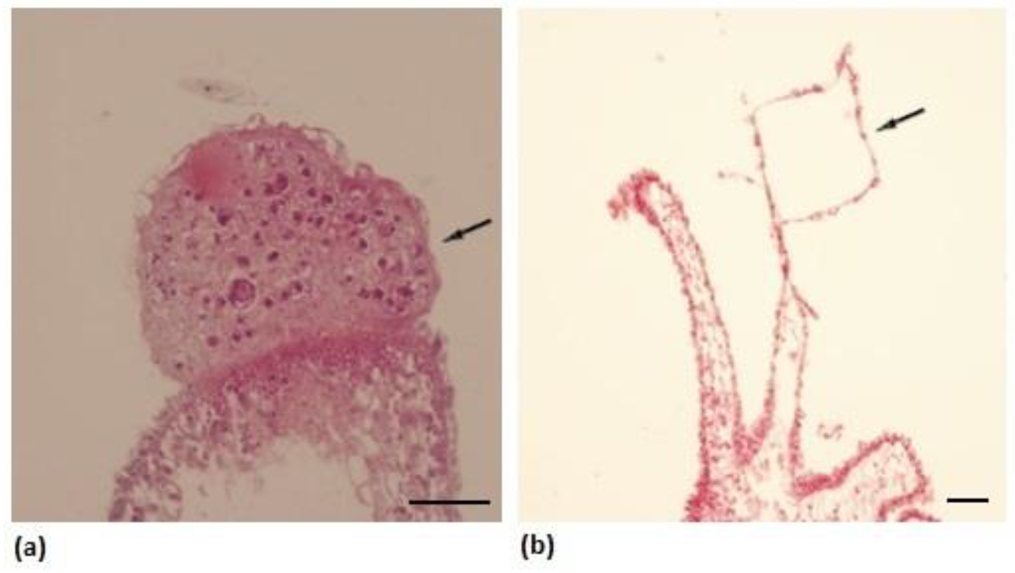
Histological analysis revealed that the mesoglea was least thick in green hydra exposed to norflurazon 2 × 10⁻⁶ mol/L and brown hydra exposed to UV radiation, while the greatest thickness was noted in both species treated with norflurazon 2 × 10⁻⁷ mol/L and was more pronounced in non-irradiated animals (Figure 4). The results show that environmental stressors could have an impact on alterations of the mesoglea thickness in hydra. In UV-irradiated green hydra, radiation might have induced uncontrolled cell proliferation/tissue damage or irregular budding, and norflurazon caused the formation of specific fork-like tentacles (Figure 5). Likewise, irradiation induced increased cell proliferation in the brown hydra body and tentacles.
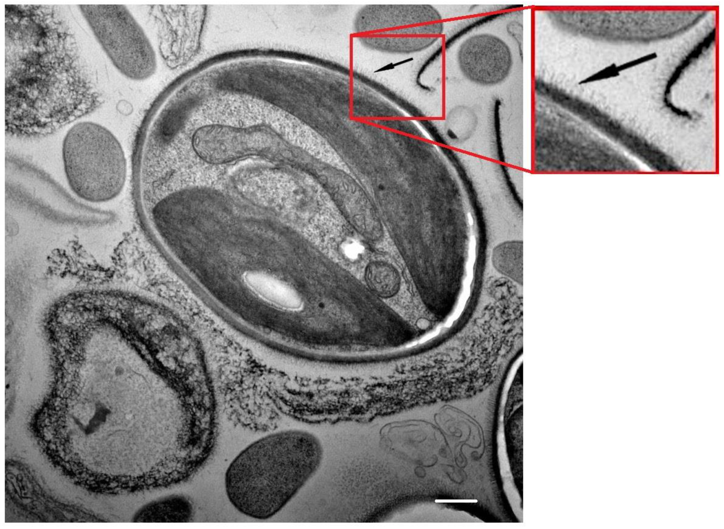
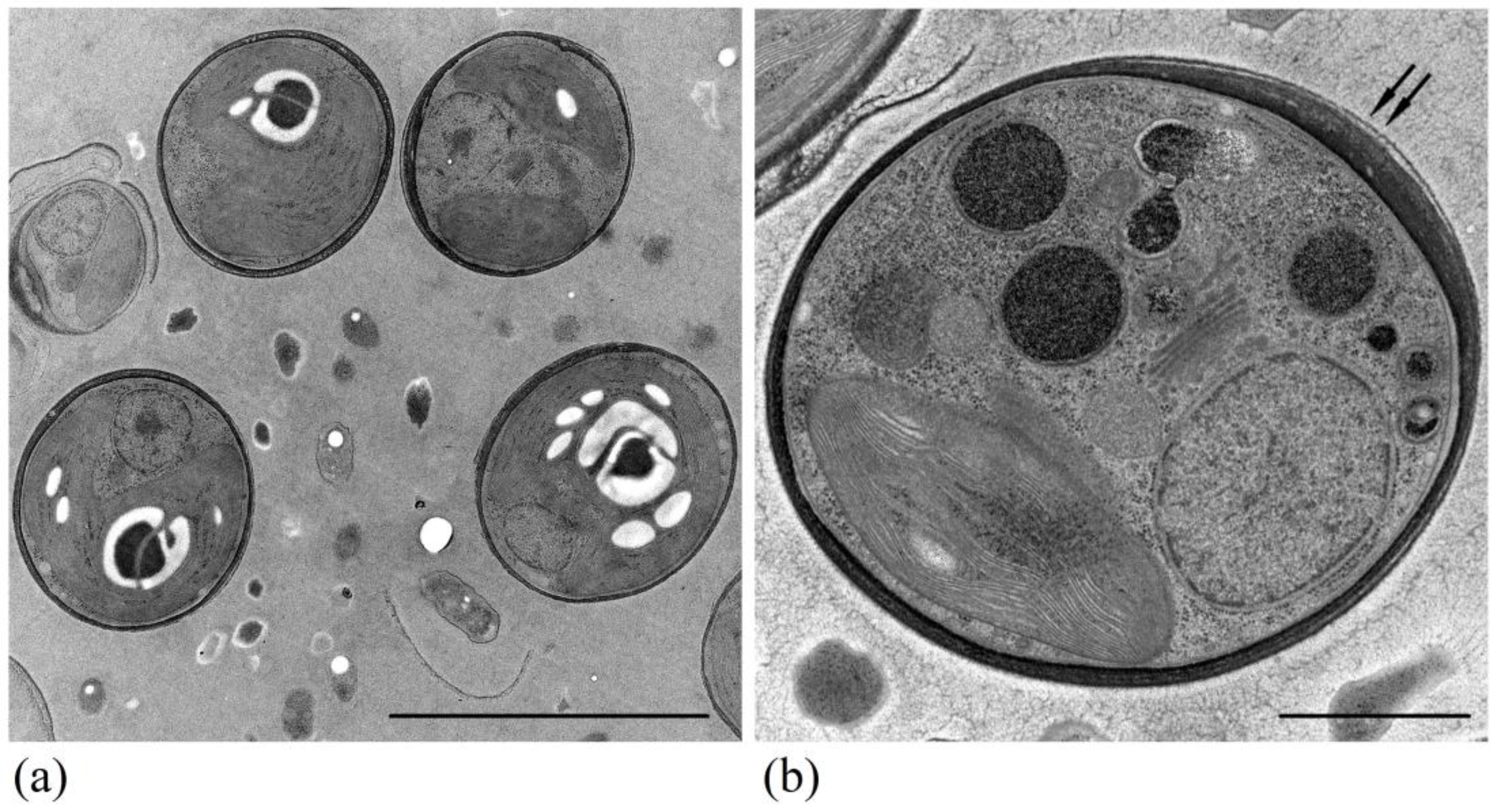
Clustering of both algal species was present, but no regularity in the clustering was noted. Observed changes in algal clustering were uniformly present, i.e., they did not change during the second and third day. Added algae in particular containers homogenously colored the solutions/controls in green. In the containers with both algae together, this phenomenon was not noted. TEM was applied to observe microalgal characteristics and tried to explain the phenomenon of clustering. Besides an excellent overall preservation of the algae (Figure 6 and Figure 7), the multilamellar nature of the cell wall was noted for M. homosphaera (Figure 7b). In C. vulgaris no multilamellar structure of thecell wall was observed, and the cell wall was thick, but cryofixation and TEM provided a clue possibly related to clustering of algae by observing the extracellular polymeric substances (EPS) on the cell surface (Figure 6). Clustering was the most pronounced in containers with both species of algae, and containers with only one species of algae had equal clustering (Table 1).

4. Discussion

In this study, the effects of two concentrations of an aqueous solution of norflurazon and UV radiation on symbiotic green hydra and free-living brown hydra were investigated. The responses to norflurazon treatment and UV radiation were traced when isolated endosymbiotic and free-living algae were added to the containers with hydras, separately or combined. Responses to the microenvironmental conditions were observed as follows: the effect on hydra behavior, damage, mortality, and algal clustering.
Three days of treatment is sufficient to allow the tested agents to act [58,59], and this finding regarding exposing the hydras for 3 days has been confirmed in papers published by other authors [60,61,62,63].
Retention of norflurazon in the soil can damage crops after the application of this agent. The occurrence of sublethal effects is related to the issue of norflurazon persistence. Means with a long half-life represent a special risk [64]. In the paper of Savin and Amador [21], a concentration of 0.57 mg/kg of soil was determined. Due to high mobility of the pesticide, similar concentrations are expected in water. In the EPA’s Memorandum on Risks from Human Exposure to Norflurazon and its Degradate Desmethylnorflurazon in Groundwater [65], it was determined that the concentration of norflurazon in groundwater is 1.5 mg/mL, which is even lower than our concentration meaning that it is much higher in the surface waters that drain into those waters through agricultural soil. Since we applied acute toxicity testing, according to principles of toxicology testing, we used the worst possible scenario (according to highest dose we found in the literature), namely, as found in the environment, 548.0 µg/L [54]—0.61 mg/L of norflurazon was the higher dose and the lower dose was 0.061 mg/L, which was far below doses reported in papers on environmental analyses by competent regulatory agencies. Other studies used similar concentrations of norflurazon 0.51 mg/L [66]. As stated by Wilson and Koch, due to its water solubility (28 mg/L), norflurazon use on agricultural lands has the potential to leach to groundwater and surface water [36]. Wilson et al. reported that 8.480 mg of norflurazon escaped from the applied soil. Moreover, around 6–8 months were required for norflurazon to be degraded to 50% of its original concentration in aquatic environments [54].
Silva et al. (2022) analyzed the effects of UV radiation (UVR) on a soil microbial community and observed an increase in microbial catabolic activity as well as small alternations in the composition of the soil bacterial communities, although the microbial diversity was not affected. Investigated was the physiological response of the symbiotic dinoflagellate Pelagodinium béii to exposure to ultraviolet (UV) radiation, and P. béii was isolated from the foraminifer Globigerinoides sacculifer. Decreases in growth rate, cellular chlorophyll a content, and photosynthetic activity were observed within 2 days [47]. Symbiotic ciliates (P. bursaria) receive protection against UV damage from their algae; by contrast, aposymbiotic P. bursaria are highly sensitive to UV radiation. Already after 2 h of exposure, damaged cells and motionless ciliates were observed. Sonntag, Summerer, and Sommaruga (2011) tested resistance to solar UVR of mixotrophic and heterotrophic ciliates and found that one of the mixotrophic ciliates, Vorticella chlorellata, was more resistant to UVR than the heterotrophic species [48]. Parajuli et al. (2023) observed how UV-A and UV-B light affected the different stages of the small insect Diaphorina citri and discovered that eggs, early instar nymphs, and adults were little affected by UV-A light, but the survival of adults was reduced at the higher doses used. Egg hatch and the survival times of early and late instar nymphs declined in proportion to the UV-B dose, and higher UV-B doses reduced the survival time of only adult females [49]. Stábile et al. (2021) and Eshun-Wilson et al. (2020) investigated the fitness costs associated with UVR in planktonic crustaceans [50]. Stábile et al. (2021) observed that the individuals exposed to fluctuating UV radiation had lower fitness than the individuals exposed to the same but constant UVR dose and concluded that the responses of the investigated organisms may depend on variability of the stressor in nature [50]. In addition, Eshun-Wilson et al. (2020) concluded that their results suggest potential detrimental effects on fitness and survival of D. pulex subjected to UVR stress [51]. Alves et al. investigated the harmful effects on the fish Sparus aurata (2020; 2022) and concluded that the results indicate that short- and long-term exposure to UVB retarded growth and decreased survival rates, induced various physiological and behavioral changes, a reduction in appetite, and skin problems [52]. Furthermore, the effects of UV light on herbicide toxicity have also been investigated, and some studies have shown the effects of radiation of different wavelengths of the UV-A, UV-B, and UV-C spectrum, concluding that UV-C irradiation results in a greater reduction in toxicity of the active herbicide ingredient glyphosate [N-(phosphonomethyl)glycine] than UV-B irradiation at the same doses, while irradiation with rays from the UV-A spectrum did not affect glyphosate toxicity [53].
Bioindicator species and biomonitoring are important in assessing the health of an ecosystem. By monitoring the living component of the ecosystem, we obtain a broader understanding of its state and the processes taking place [67]. Sublethal effects are much more interesting and significant and indicate the possibility of real long-term harm of the compound to the organism [64] and may have long-term consequences for species, populations, and ecosystems. Damages are the result of the impossibility of timely and complete defense against changes in the surrounding medium. For green hydra, it has been proven that it is less sensitive to the harmful effect of some substances, such as iron, compared to brown hydra [40,58,59]. However, the results of the research on the sensitivity of different types of hydra to toxic substances showed that in some cases green hydra is still more susceptible to the harmful effects of these substances. The reason for this may be the effect of toxicants on algae. For example, cadmium and copper cause damage to algae, so in such cases, green hydra loses the advantages of the endosymbiotic relationship. In the case of other substances, such as zinc, which are not as toxic to algae, the harmful effect on green hydra is less than that on non-symbiotic hydra species [68]. Also, endosymbiotic algae in green hydra are not bleached by norflurazon, i.e., the “bleaching effect” does not take place [40].
Our results showed the following, migration is a way of avoiding unfavorable conditions in the environment [69]. Likewise, herein hydras also migrated towards the surface or, in case of severe morphological damage, remained at the very bottom of the container. Without the addition of algae, the highest level of migration towards the surface was observed in brown hydra following treatment with norflurazon. The animals that were irradiated showed a diminished percentage of migration. Migration was observed in hydras in the presence of microalgae. Green hydras avoided the neighborhood of algae and stuck to the vertical wall of the container, while the algae were more or less clustered at the bottom. Diminished motility following UV irradiation has been observed in other animal organisms. It has been suggested that the effect is a result of the damage of mitochondria by UV light, which undermines their function by impairing the membrane permeability and ATP [70]. Due to the deleterious effect of norflurazon on mitochondria [40,71], partially, the reduction in mobility might be a consequence of the changes in the redox potential of the hydra cells. The results suggest that symbiotic algae have a beneficiary effect on the protection of green hydras. It may be due to maltose and oligo-maltose species, ascorbic acid, mycrosporine-like amino acids, and mucus excreted from algae into the cytoplasm of hydra [72,73]. Additionally, continuous excretion of the respiratory products of algae in hydra’s cytoplasm may result in significantly higher basal levels of non-enzymatic and enzymatic mechanisms of oxidative stress defense in the cytoplasm of green hydra’s cells compared to brown hydra [74]. Both beneficiary effects of symbiotic association between green hydra and alga may result in hydra’s lower susceptibility to oxidative stress as the primary toxic mechanism of action of norflurazon in animal cells. Green hydra might have higher resistance to the adverse effect of UV radiation. The in hospite habitat of endosymbiotic algae is the endodermal cell of the hydra’s epithelium with stabile homeostatic intracellular conditions. In the natural environment, they are easily stressed by different mechanisms due to changes in chemical composition, physical parameters of the environment or presence of potential predators, such as brown hydra, that excrete kairomones and other cues [75]. Stressed algae release different volatile compounds, toxins, and defense metabolites [76,77]. For Chlorella sp., it has been proven that it releases more than 100 different volatile compounds among which are sulfuric compounds (e.g., dimethyl sulfide), aldehydes, alkanes, fatty acids, terpenes, and alcohols [78,79], and some of these compounds may exhibit adverse effects in other organisms [78] and are released to increase resistance to stress and as defense against predators [77]. According to our knowledge, there are no data published in regard to the effect of volatile metabolites on hydra. However, some of them are toxic and genotoxic, such as alcohols, aldehydes, and terpenes. Algae could recognize cues as being released from potential host and predator at the same time. A defense strategy of single-cell algae is to form clusters in the presence of predators as a reaction to the kairomones released by them [75]. When in the colony, due to size-mismatch, they are harder-to-approach prey. The ability of C. vulgaris to form colonies in the presence of predators has been reported [80,81], and microalgal aggregations and nets are present in microcosm conditions [45]. This phenomenon of microalgal clustering was observed in the present study. Most intensive cluster formation was observed when endosymbiotic and free-living algae were jointly present. Colony formation might also be triggered by interalgal communication by the exchange of certain chemical signaling molecules [82]. Such an increase in group size when two different species of algae are combined has been reported by Kapsetaki et al. [83]. In the case of microalgal aggregations, hydras can produce more mucus and therefore stick firmly to the experimental dish [45]. Here, hydra migration could have been inhibited.
TEM showed that algae in our experiment do not possess any rod-like structures at the surface [45] that could support the explanation for the clustering of algae. Instead, the trilaminate structure of the cell wall of both C. vulgaris and M. homosphaera was observed with TEM based on conventional chemical fixation [84]. Herein, in C. vulgaris, the cell wall was thick, and cryofixation and TEM helped us to track the extracellular polymeric substances (EPS) in the form of brush-like structures on the C. vulgaris cell surface. Microalgae can excrete EPS into their immediate living environment during their life cycle to form a hydrated biofilm matrix made of a complex high-molecular-weight mixture of biopolymers (polysaccharides, proteins, nucleic acids, and lipids) [85]. EPS retain their stable matrix structure and form a 3-D polymer network in which cells can interact with each other and mediate their adhesion to surfaces. EPS are versatile, natural biopolymers that act as anticoagulants, protect cells from dehydration and toxic substances, and serve as energy and carbon sinks in times of stress [85]. In our experiment, they could have supported the clustering of algae. For M. homosphaera, here, cryofixation and TEM confirmed the multilamellar nature of the cell wall, observed in conventional sample preparation [84]. Perhaps such a structure could support the clustering as well, if the outer layers of the multilamellar cell wall are glyco-coated. Such a coating, however, might have got lost during sample preparation.
Our results suggest that hydra species do not influence each other’s adaptation to stressors, when placed in a common container. Hydras are surface water organisms that are capable of developing tumor tissue [86]. In addition, the present research comprises a histopathological analysis of the hydra’s tissue and mesoglea. In fish, as vertebrates, norflurazon at the concentration of 1.5 mg/mL induced adverse effects. At the same concentration, it may cause chronic effects on aquatic invertebrate survival and offspring production [65]. It has been recorded that at a concentration of 2 and 0.2 μM, norflurazon induces a significant increase in primary DNA damage in planarian cells, while at the concentration of 200 μM, it results in severe adverse conditions in Polycelis felina (Daly.) [41]. The present study also contributes to the understanding of symbiotic and free-living hydras as pollution indicators. Different types of hydra, as an important component of the benthic communities of stagnant waters, represent a good model organism in ecotoxicological research due to easily measurable morphological and behavioral changes [6].
Based on the present results, we may deduce that UV radiation induced more severe tissue damage compared to herbicide exposure. It is plausible that a higher adverse effect of UV light is due to its potential to directly damage DNA. Namely, norflurazon acts adversely by inducing oxidative stress in the cells and the formation of ROS impairs intracellular macromolecules and cellular structures. However, norflurazon does not interact directly with DNA; thus, its adverse effect may be diminished by de novo synthesis of damaged macromolecules (i.e., proteins) and by an increase in intracellular non-enzymatic molecules and enzymes that protect from reactive oxygen species by upregulating expression of genes protein products that are essential for cell physiology and antioxidative protection. UV radiation, by inducing pyrimidine dimers, 6-4 photoproducts, and strand breaks, disrupts the integrity of DNA. It impairs gene expression and may induce apoptosis [87]. Cells of the tentacles are small and plate-like. Consequentially, herein, we recorded more severe damage to the tentacles of both hydra species following irradiation. Also, after treatment with norflurazon or UV radiation on tentacles, new growths were recorded or tentacles took a forked shape [44]. Tentacles, as body parts that are the most protruded, are most exposed to the environment and adverse effects of stressors. They are formed of a thin layer of cells, and due to their accessibility, UV radiation may penetrate into the majority of cells and induce adverse effects. Our results show that they were damaged by UV radiation, while here, norflurazon mostly did not exhibit a deleterious effect. This suggests that the direct DNA damage induced by UV radiation plays a more significant role in triggering apoptosis than induction of oxidative stress by the herbicide. The size of an organism is one of the key characteristics that influences the response of that organism to UV radiation because it is absorbed per unit of surface area. Because of this, it was to be expected that UV treatment would produce much greater damage and cause greater mortality in green hydra because it is smaller than brown hydra.
According to the energy that it transmits, on the electromagnetic scale, UV radiation falls between ionizing and non-ionizing radiation. In general, it is more often considered as a non-ionizing radiation. Yet, UV light is able to transfer enough energy to molecules to break covalent bonds. In reaction with water molecules, it cleaves them by photolysis or even radiolysis [88]. Either of them result in the formation of high levels of different ROS [89]. It is well documented that tissues with a high water content are the most damaged ones in interactions with radiation due to ionization of the water molecules [90]. When hydra tissue is damaged by the stressor and some body parts are missing (tentacles), interstitial cells, as pluripotent stem cells, begin to divide, and their daughter cells differentiate to lead to regeneration of hydra [91]. Exposure to UV radiation and norflurazon induces a significant number of lesions in DNA and triggers apoptosis [92]. Similar, cells with upregulated gene transcription are also prone to DNA damage since, for transcription to occur, gene regions must be unwound and proteins involved in chromatin organization detached [93]. Zymogene cells are pluripotent in their character, and when some parts of the hydra body are damaged or lost, they migrate, divide, and differentiate to replace missing body parts [94]. On the other hand, environmental stressors can induce DNA changes that can lead to uncontrolled cell proliferation and finally tumor development [86,95]. To date, research on the histomorphological changes induced by norflurazon has shown that brown hydra is much more damaged than green hydra, i.e., green hydra is more viable [33]; morphological damage to the tentacles is a known reaction of hydras to xenobiotics [96] and that the mesoglea is partly missing [33]. It also seems that radiation increases the number and volume of algae in gastrodermal cells [97]. Unlike UV radiation, norflurazon does not cleave water molecules; thus, it does not exhibit direct adverse effects on the mesoglea. Furthermore, it has been reported that the mesoglea plays a role in buffering the intercellular environment [98]. Thus, penetration of toxic substances such as norflurazon might lead to uptake of an additional amount of water to diminish the effect of the pesticide. It may be a consequence of the toxic effect of the herbicide mediated by induction of oxidative stress. Unlike in UV-radiated hydras (controls), here, in animals exposed to norflurazon only (2 × 10⁻⁷ mol/L), a thickening of the mesoglea was recorded. It is a reversed effect compared to the one following UV irradiation. The mesoglea plays a significant role in regeneration of damaged body parts and replacement of areas where cells undergo apoptosis or necrosis. It contains filaments (integrin, fibronectin, and laminin) that enable migration of the cells to the area of the hydra that needs to be regenerated [99,100,101]. To enable cell migration, the mesoglea undergoes reorganization, which includes swelling of the mesoglea [102].

5. Conclusions

The effects of norflurazon and UV irradiation were observed both on green and brown hydra. Changes and damage appeared during separate or simultaneous action of norflurazon/UV radiation. Mortality was present only in irradiated green hydra. Migration was more pronounced in brown hydra. In the presence of algae, green hydra migrated more intensively than brown hydra. Tentacle damage was more pronounced in green hydra and included a specific fork-like structure. In hydras treated with UV radiation, a possible uncontrolled tissue growth was observed. The use of cryofixation and TEM enabled us to partly explain the clustering of microalgae. Our results suggest that hydra species do not influence each other regarding adaptation to stressors. To better understand the mutual interactions between the symbiotic and free-living hydras and algae as constituents of freshwater ecosystems, further studies with special emphasis on the composition of algae in the (micro)environment should be conducted.

Author Contributions

Conceptualization, G.K., A.M., P.K. and D.Ž.; methodology, G.K., A.M., P.K., S.R. and K.C.M.; validation, G.K., A.M., P.K., S.R., K.C.M., D.S. and P.P.Š.; formal analysis, G.K., A.M., P.K., S.R., K.C.M., D.S., D.Ž., P.P.Š. and M.I.; investigation, G.K., A.M., P.K., D.Ž., S.R., K.C.M. and P.P.Š.; resources, G.K., A.M., P.K., D.Ž., S.R., K.C.M., D.S., P.P.Š. and M.I.; writing—original draft preparation, G.K., A.M., P.K., D.Ž., S.R., K.C.M., D.S. and M.I.; writing—review and editing, G.K., A.M., P.K., K.C.M. and S.R.; visualization, G.K., A.M., P.K., D.Ž., S.R., K.C.M., D.S., P.P.Š. and M.I.; supervision, G.K., P.K. and K.C.M. All authors have read and agreed to the published version of the manuscript.

Funding

This research was funded by the University of Zagreb, support number 20282310 and the European Union—Next Generation EU (Program Contract Class: 643-02/23-01/00016, Reg. no. 533-03-23-0006).

Data Availability Statement

The data presented in this study are available in this article.

Conflicts of Interest

The authors declare no conflicts of interest.

Abbreviations

  • For easier understanding of the abbreviations, in the figures and the table, we used combinations of the below listed terms. Abbreviations ending in Z or S indicate the observation of Z or S in the mixed sample of Z and S.
CVChlorella vulgaris Beij. [K&H, 1992]
CZMychonastes homosphaera (Chlorophyceae) (Skuja) Kalina et Punčochářová
ZGreen hydra
SBrown hydra
KControl
N6Norflurazon 2 × 10⁻⁶ mol/L
N7Norflurazon 2 × 10⁻⁷ mol/L
RIrradiated
EPSExtracellular polymeric substances
TEMTransmission electron microscopy
HPFHigh-pressure freezing
FSFreeze substitution

References

  1. Žnidarić, D. Comparison of the regeneration of the hypostome with the budding process in Hydra littoralis. Roux Arch. 1970, 166, 45–53. [Google Scholar] [CrossRef]
  2. Zhang, X.; Zhang, J.; Li, L.; Sarras, M.P., Jr. Biogenesis of hydra mesoglea and its functions in regeneration. In Abstract Book of the International Workshop Hydra and the Molecular Logic of Regeneration; Bosch, T.C.G., Holstein, T.W., David, C.N., Eds.; DFG: Tutzing, Germany, 2005; p. 22. [Google Scholar]
  3. Kremer, B.P. Carbon metabolism of endosymbiotic algae. In Endocytobiology. Endosymbiosis and Cell Biology. Vol. I. A Synthesis of Recent Research; Schwemmler, W., Schenk, H.E.A., Eds.; WdeG: Berlin, Germany; New York, NY, USA, 1980; pp. 89–96. [Google Scholar]
  4. Kovačević, G.; Kalafatić, M.; Jelenčić, B.; Franjević, D. Endosymbiotic alga as the stronger evolutionary partner in green hydra symbiosis. J. Endocyt. Cell Res. 2010, 20, 13–15. [Google Scholar]
  5. Kovačević, G.; Franjević, D.; Jelenčić, B.; Kalafatić, M. Isolation and cultivation of endosymbiotic algae from green hydra and phylogenetic analysis of 18S rDNA sequences. Folia Biol. 2010, 58, 135–143. [Google Scholar] [CrossRef]
  6. Kovačević, G. Value of the Hydra model system for studying symbiosis. Int. J. Dev. Biol. 2012, 56, 627–635. [Google Scholar] [CrossRef] [PubMed]
  7. Ivšić, M.; Kovačević, G. Evaluation of Algae Farming Using the Chlorella Bioassay. Croat. J. Fish. 2018, 76, 99–106. [Google Scholar] [CrossRef]
  8. Bosch, T.C.G. Understanding complex host-microbe interactions in Hydra. Gut Microbes 2012, 3, 345–351. [Google Scholar] [CrossRef]
  9. He, J.; Bosch, T.C.G. Hydra’s Lasting Partnership with Microbes: The Key for Escaping Senescence? Microorganisms 2022, 10, 774. [Google Scholar] [CrossRef] [PubMed]
  10. Beach, M.J.; Pascoe, D. The role of Hydra vulgaris (Pallas) in assessing the toxicity of freshwater pollutants. Water Res. 1998, 32, 101–106. [Google Scholar] [CrossRef]
  11. Kalafatić, M.; Kopjar, N. Response of Green Hydra to the Treatment with Different Pesticides under Laboratory Conditions. Z. Angew. Zool. 1994, 2, 213–223. [Google Scholar]
  12. Kalafatić, M. Regeneration and asexual reproduction of Hydra oligactis treated with different pesticides. Biologia 1997, 52, 475–480. [Google Scholar]
  13. Kovačević, G.; Kalafatić, M.; Ljubešić, N.; Šunjić, H. The effect of chloramphenicol on the symbiosis between alga and hydra. Biologia 2001, 56, 605–610. [Google Scholar]
  14. Arkhipchuk, V.V.; Blaise, C.; Malinovskaya, M.V. Use of hydra for chronic toxicity assessment of waters intended for human consumption. Environ. Pollut. 2006, 142, 200–211. [Google Scholar] [CrossRef]
  15. Rachamim, T.; Sher, D. What Hydra can teach us about chemical ecology—How a simple, soft organism survives in a hostile aqueous environment. Int. J. Dev. Biol. 2012, 56, 605–611. [Google Scholar] [CrossRef] [PubMed]
  16. Yonge, C.M. Ecology and physiology of reef building corals. In Perspectives in Marine Biology; Buzzati-Traverso, A., Ed.; University California Press: Berkeley, CA, USA; Los Angeles, CA, USA, 1958; pp. 117–136. [Google Scholar]
  17. Bode, H.R. The interstitial cell lineage of hydra: A stem cell system that arose early in evolution. J. Cell Sci. 1996, 109 Pt 6, 1155–1164. [Google Scholar] [CrossRef]
  18. Fradkin, M.; Kakis, H.; Campbell, R. Effects of γ irradiation on hydra: Elimination of interstitial cells from viable hydra. Radiat. Res. 1978, 76, 187–197. [Google Scholar] [CrossRef]
  19. Corbett, J.R.; Wright, K.; Baillie, A.C. The Biochemical Mode of Action of Pesticides; Academic Press Inc.: London, UK, 1984; pp. 240–245. [Google Scholar]
  20. Anderson, I.C.; Robertson, D.S. Role of carotenoids in protecting chlorophyll from photodestruction. Plant Physiol. 1960, 35, 531–534. [Google Scholar] [CrossRef] [PubMed]
  21. Savin, M.C.; Amador, J.A. Mineralization of Norflurazon in a Cranberry Bog Soil: Laboratory Evaluations of Management Practices. J. Environ. Qual. 1998, 27, 1234–1239. [Google Scholar] [CrossRef]
  22. Giry, G.; Ayele, J. Study of norflurazon behavior in soils-Influence of formulation additives upon sorption parameters. Agronomie 2002, 22, 467–478. [Google Scholar] [CrossRef]
  23. Böger, P. Mode of action of herbicides affecting carotenogenesis. J. Pestic. Sci. 1996, 21, 473–478. [Google Scholar] [CrossRef]
  24. Chamovitz, D.; Pecker, I.; Sandmann, G.; Böger, P.; Hirschberg, J. Cloning a gene coding for norflurazon resistance in cyanobacteria. Z. Naturforsch. C J. Biosci. 1990, 45, 482–486. [Google Scholar] [CrossRef]
  25. Chamovitz, D.; Sandman, G.; Hirschberg, J. Molecular and biochemical characterization of herbicide-resistant mutants of cyanobacteria reveals that phytoene desaturation is a rate-limiting step in carotenoid biosynthesis. J. Biol. Chem. 1993, 268, 17348–17353. [Google Scholar] [CrossRef] [PubMed]
  26. Sandman, G.; Vique, A. Phylogeny of carotenogenic genes and enzymes in cyanobacteria. In The Phototrophyc Prokaryotes, Proceedings of the 9th International Symposium on Phototrophyc Prokaryotes: Structural and Functional Analyses of Cyanobacterial Photosystem, Vienna, Austria, 6–12 September 1997; Peschek, A., Löffelhardt, W., Schwetterer, G., Eds.; Kluwer Academic/Plenum Publishers: Vienna, Austria, 1997. [Google Scholar]
  27. Wrischer, M.; Ljubešić, N.; Modrušan, Z. Development of calcelaria chromoplasts in the presence of herbicides affecting carotenoid biosyn-thesis. Acta Bot. Croat. 1991, 50, 23–30. [Google Scholar]
  28. Lichtenthaler, H.K.; Burkard, G.; Grumbach, K.H.; Meier, D. Physiological effects of photosystem II-herbicides on the development of the photosynthetic apparatures. Photosynth. R 1980, 1, 29–43. [Google Scholar] [CrossRef]
  29. Thurman, E.M.; Bastian, K.C.; Mollhagen, T. Occurrence of cotton herbicides and insecticides in playa lakes of the High Plains of West Texas. Sci. Total Environ. 2000, 248, 189. [Google Scholar] [CrossRef]
  30. Troiano, J.; Marade, J.; Spurlock, F. Empirical Modeling of Spatial Vulnerability Applied to a Norflurazon Retrospective Well Study in California. J. Environ. Qual. 1999, 28, 397–403. [Google Scholar] [CrossRef]
  31. Ma, J. Differential Sensitivity to 30 Herbicides among Populations of Two Green Algae Scenedesmus obliquus and Chlorella pyrenoidosa. Bull. Environ. Contam. Toxicol. 2002, 68, 275–281. [Google Scholar] [CrossRef]
  32. Ma, J.; Xu, L.; Wang, S.; Zheng, R.; Jin, S.; Huang, S.; Huang, Y. Toxicity of 40 Herbicides to the Green Alga Chlorella vulgaris. Ecotoxicol. Environ. Saf. 2002, 51, 128–132. [Google Scholar] [CrossRef]
  33. Maceljski, M.; Hrlec, G.; Ostojić, Z.; Cvjetković, B. Pregled sredstava za zaštitu bilja u Republici Hrvatskoj za 1997. godinu. Glas. Zašt. Bilja 1997, 3–4, 93–212. [Google Scholar]
  34. Tschiersh, H.; Ohomann, E.; Döge, M. Modification of the thylacoid structure of Euglena gracilis by norflurazon treatment: Consequences for fluorescence quenching. Environ. Exp. Bot. 2002, 47, 259–270. [Google Scholar] [CrossRef]
  35. Merzlyak, M.N.; Khozin, I.; Cohen, Z. Spectro-photometric Analysis of Carotenoids in Plant Extracts Based on Elimination of Chlorophyll Absorption. Phytochem. Anal. 1996, 7, 294–299. [Google Scholar] [CrossRef]
  36. Wilson, P.C.; Koch, R. Influence of Exposure Concentration and Duration on Effects and Recovery of Lemna minor Exposed to the Herbicide Norflurazon. Arch. Environ. Contam. Toxicol. 2013, 64, 228–234. [Google Scholar] [CrossRef]
  37. Abrous, O.; Benhassaine-Kesri, G.; Tremolieres, A.; Mazijak, P. Effect of norflurazon on lipid metabolism in soya seedlings. Phytochemistry 1998, 49, 979–985. [Google Scholar] [CrossRef]
  38. Salopek, B.; Ljubešić, N. The fine structure of pepper chloroplasts: The effect of bleaching herbicides. Acta Bot. Croat. 1994, 53, 7–13. [Google Scholar]
  39. Vecchia, F.D.; Barbato, R.; Rocca, N.L.; Moro, I.; Rascio, N. Responses to bleaching herbicides by leaf chloroplasts of maize plants grown at different temperatures. J. Exp. Bot. 2001, 357, 811–820. [Google Scholar] [CrossRef]
  40. Kovačević, G.; Kalafatić, M.; Ljubešić, N. Effects of Norflurazon on Green and Brown Hydra. Folia Biol. 2009, 57, 91–96. [Google Scholar] [CrossRef]
  41. Horvat, T.; Kalafatić, M.; Kopjar, N.; Kovačević, G. Toxicity testing of herbicide norflurazon on an aquatic bioindicator species—The planarian Polycelis felina (Daly.). Aquat. Toxicol. 2005, 73, 342–352. [Google Scholar] [CrossRef]
  42. ECHA. Available online: https://echa.europa.eu/information-on-chemicals/cl-inventory-database/-/discli/notification-details/2240/1767154 (accessed on 17 November 2023).
  43. Friedberg, E.C. DNA Damage and Repair. Nature 2003, 421, 436–439. [Google Scholar] [CrossRef] [PubMed]
  44. Kalafatić, M.; Kovačević, G.; Zupan, I.; Franjević, D. Effect of repeated UV-irradiation on Hydra oligactis Pallas. Period. Biol. 2003, 105, 171–175. [Google Scholar]
  45. Kovačević, G.; Petrinec, D.; Tramontana Ljubičić, P.; Reipert, S.; Sirovina, D.; Špoljar, M.; Peharec Štefanić, P.; Želježić, D. Formation of Microalgal Hunting Nets in Freshwater Microcosm Food Web: Microscopic Evidence. Water 2023, 15, 3448. [Google Scholar] [CrossRef]
  46. Ghaskadbi, S.S.; Shetye, L.; Chiplonkar, S.; Ghaskadbi, S. Ultraviolet irradiation initiates ectopic foot formation in regenerating hydra and promotes budding. J. Biosci. 2005, 30, 177–182. [Google Scholar] [CrossRef] [PubMed]
  47. Silva, I.; Alves, M.; Malheiro, C.; Silva, A.R.R.; Loureiro, S.; Henriques, I.; González-Alcaraz, M.N. Short-Term Responses of Soil Microbial Communities to Changes in Air Temperature, Soil Moisture and UV Radiation. Genes 2022, 13, 850. [Google Scholar] [CrossRef]
  48. Sonntag, B.; Summerer, M.; Sommaruga, R. Are freshwater mixotrophic ciliates less sensitive to solar ultraviolet radiation than heterotrophic ones? J. Eukaryot. Microbiol. 2011, 58, 196–202. [Google Scholar] [CrossRef]
  49. Parajuli, S.; Beattie, G.A.C.; Holford, P.; Yang, C.; Cen, Y. Susceptibility of Diaphorina citri to Irradiation with UV-A and UV-B and the Applicability of the Bunsen-Roscoe Reciprocity Law. Insects 2023, 14, 445. [Google Scholar] [CrossRef]
  50. Franca, S.; Christer, B.; Lars-Anders, H.; Marcus, L. Fitness cost from fluctuating ultraviolet radiation in Daphnia magna. Biol. Lett. 2021, 17, 20210261. [Google Scholar] [CrossRef]
  51. Eshun-Wilson, F.; Wolf, R.; Andersen, T.; Hessen, D.O.; Sperfeld, E. UV radiation affects antipredatory defense traits in Daphnia pulex. Ecol. Evol. 2020, 10, 14082–14097. [Google Scholar] [CrossRef]
  52. Alves, R.N.; Mahamed, A.H.; Alarcon, J.F.; Al Suwailem, A.; Agustí, S. Adverse Effects of Ultraviolet Radiation on Growth, Behavior, Skin Condition, Physiology, and Immune Function in Gilthead Seabream (Sparus aurata). Front. Mar. Sci. 2020, 7, 306. [Google Scholar] [CrossRef]
  53. Papagiannaki, D.; Medana, C.; Binetti, R.; Calza, P.; Roslev, P. Effect of UV-A, UV-B and UV-C irradiation of glyphosate on photolysis and mitigation of aquatic toxicity. Sci. Rep. 2020, 10, 20247. [Google Scholar] [CrossRef] [PubMed]
  54. Wilson, P.C.; Boman, B.; Foos, J.F. Norflurazon and Simazine Losses in Surface Runoff Water from Flatwoods Citrus Production Areas. Bull. Environ. Contam. Toxicol. 2007, 78, 341–344. [Google Scholar] [CrossRef] [PubMed]
  55. Reipert, S.; Goldammer, H.; Richardson, C.; Goldberg, M.W.; Hawkins, T.J.; Hollergschwandtner, E.; Kaufmann, W.A.; Antreich, S.; Stierhof, Y.D. Agitation modules: Flexible means to accelerate automated freeze substitution. J. Histochem. Cytochem. 2018, 66, 903–921. [Google Scholar] [CrossRef]
  56. Goldammer, H.; Hollergschwandtner, E.; Elisabeth, N.H.; Frade, P.R.; Reipert, S. Automatized freeze substitution of algae accelerated by a novel agitation module. Protist 2016, 167, 369–376. [Google Scholar] [CrossRef] [PubMed]
  57. Kuipers, J.; Giepmans, B.N.G. Neodymium as an alternative contrast for uranium in electron microscopy. Histochem. Cell Biol. 2020, 153, 271–277. [Google Scholar] [CrossRef]
  58. Kovačević, G.; Želježić, D.; Horvatin, K.; Kalafatić, M. Morphological features and comet assay of green and brown hydra treated with aluminium. Symbiosis 2009, 44, 145–152. [Google Scholar]
  59. Kovačević, G.; Gregorović, G.; Matijević, A.; Kalafatić, M. Toxic effects of iron on green and brown hydra. Curr. Sci. 2016, 110, 502–504. [Google Scholar]
  60. Hyne, R.V.; Rippon, G.D.; White, J.; Ellender, G. Accumulation of uranium by freshwater hydra into discharged nematocysts. Aquat. Toxicol. 1992, 23, 231–246. [Google Scholar] [CrossRef]
  61. Vimalkumar, K.; Sangeetha, S.; Felix, L.; Kay, P.; Pugazhendhi, A. A systematic review on toxicity assessment of persistent emerging pollutants (EPs) and associated microplastics (MPs) in the environment using the Hydra animal model. Comp. Biochem. Physiol. Part C Toxicol. Pharmacol. 2022, 256, 109320. [Google Scholar] [CrossRef]
  62. Lum, K.T.; Huebner, H.J.; Li, Y.; Phillips, T.D.; Raushel, F.M. Organophosphate nerve agent toxicity in Hydra attenuata. Chem. Res. Toxicol. 2003, 16, 953–957. [Google Scholar] [CrossRef] [PubMed]
  63. Lekamge, S.; Miranda, A.F.; Abraham, A.N.; Li, V.V.; Shukla, R.; Bansal, V.; Nugegoda, D. The Toxicity of Silver Nanoparticles (AgNPs) to Three Freshwater Invertebrates with Different Life Strategies: Hydra vulgaris, Daphnia carinata, and Paratya australiensis. Front. Environ. Sci. 2018, 6, 152. [Google Scholar] [CrossRef]
  64. Walker, C.H. Organic Pollutants—An Ecotoxicological Perspective; Taylor & Francis: London, UK, 2001. [Google Scholar]
  65. Available online: https://www3.epa.gov/pesticides/chem_search/reg_actions/reregistration/fs_PC-105801_1-Jul-96.pdf (accessed on 19 January 2024).
  66. Wilson, P.C.; Wilson, S.B.; Haunert, D. Toxicity of the norflurazon to the aquatic macrophyte Vallisneria americana (Michx.). J. Toxicol. Environ. Health Part A 2006, 69, 1167–1179. [Google Scholar] [CrossRef] [PubMed]
  67. Holt, E.A.; Miller, S.W. Bioindicators: Using organisms to measure environmental impacts. Nat. Educ. Knowl. 2011, 2, 8. [Google Scholar]
  68. Karntanut, W.; Pascoe, D. The toxicity of copper, cadmium and zinc to four different Hydra (Cnidaria: Hydrozoa). Chemosphere 2002, 47, 1059–1064. [Google Scholar] [CrossRef] [PubMed]
  69. Burnett, A.L. Biology of Hydra; Academic Press: New York, NY, USA, 1973. [Google Scholar] [CrossRef]
  70. Hoh Kam, J.; Hogg, C.; Fosbury, R.; Shinhmar, H.; Jeffery, G. Mitochondria are specifically vulnerable to 420 nm light in drosophila which undermines their function and is associated with reduced fly mobility. PLoS ONE 2021, 16, e0257149. [Google Scholar] [CrossRef]
  71. Kovačević, G.; Kalafatić, M.; Ljubešić, N. New Observations on Green Hydra Symbiosis. Folia Biol. 2007, 55, 77–79. [Google Scholar] [CrossRef]
  72. Downs, C.A.; Woodley, C.M.; Fauth, J.E.; Knutson, S.; Burtscher, M.M.; May, L.A.; Avadanei, A.R.; Higgins, J.L.; Ostrander, G.K. A survey of environmental pollutants and cellular-stress markers of Porites astreoides at six sites in St. John, U.S. Virgin Islands. Ecotoxicology 2011, 20, 1914–1931. [Google Scholar] [CrossRef] [PubMed]
  73. Zuo, Z. Why Algae Release Volatile Organic Compounds—The Emission and Roles. Front. Microbiol. 2019, 10, 491. [Google Scholar] [CrossRef]
  74. Dizdaroglu, M.; Coskun, E.; Jaruga, P. Repair of oxidatively induced DNA damage by DNA glycosylases: Mechanisms of action, substrate specificities and excision kinetics. Mutat. Res. Rev. Mutat. Res. 2017, 771, 99–127. [Google Scholar] [CrossRef] [PubMed]
  75. Pohnert, G.; Steinke, M.; Tollrian, R. Chemical cues, defence metabolites and the shaping of pelagic interspecific interactions. Trends Ecol. Evol. 2007, 22, 198–204. [Google Scholar] [CrossRef] [PubMed]
  76. Leflaive, J.; Ten-Hage, L. Algal and cyanobacterial secondary metabolites in freshwaters: A comparison of allelopathic compounds and toxins. Freshw. Biol. 2007, 52, 199–214. [Google Scholar] [CrossRef]
  77. Zhou, Y.; Ma, W.; Wang, L.; Sun, W.; Li, M.; Zhang, W.; Liu, Y.; Song, X.; Fan, Y. Characterization and antioxidant activity of the oligo-maltose fraction from Polygonum cillinerve. Carbohydr. Polym. 2019, 226, 115307. [Google Scholar] [CrossRef]
  78. Abdel-Baky, H.H.; Shallan, M.; Baroty, G.E.; El-Baz, F.K. Volatile Compounds of the Microalga Chlorella vulgaris and Their Phytotoxic Effect. Pak. J. Biol. Sci. 2002, 5, 61–65. [Google Scholar] [CrossRef]
  79. Lafarge, C.; Cayot, N. Insight on a comprehensive profile of volatile compounds of Chlorella vulgaris extracted by two “green” methods. Food Sci. Nutr. 2019, 7, 918–929. [Google Scholar] [CrossRef]
  80. Fisher, R.M.; Bell, T.; West, S.A. Multicellular group formation in response to predators in the alga Chlorella vulgaris. J. Evol. Biol. 2016, 29, 551–559. [Google Scholar] [CrossRef]
  81. Herron, M.D.; Borin, J.M.; Boswell, J.C.; Walker, J.; Chen, I.K.; Knox, C.A.; Boyd, M.; Rosenzweig, F.; Ratcliff, W.C. De novo origins of multicellularity in response to predation. Sci. Rep. 2019, 9, 2328. [Google Scholar] [CrossRef]
  82. Venuleo, M.; Raven, J.A.; Giordano, M. Intraspecific chemical communication in microalgae. New Phytol. 2017, 215, 516–530. [Google Scholar] [CrossRef] [PubMed]
  83. Kapsetaki, S.E.; Fisher, R.M.; West, S.A. Predation and the formation of multicellular groups in algae. Evol. Ecol. Res. 2016, 17, 651–669. [Google Scholar]
  84. Kević, N.; Radić Brkanac, S.; Vincek, N.; Peharec Štefanić, P.; Faraguna, F.; Kovačević, G.; Kalafatić, M.; Franjević, D. Endosymbiotic green algae in European Hydra strains show quantitative difference on morphological and isoenzyme level. Symbiosis 2019, 77, 161–175. [Google Scholar] [CrossRef]
  85. Xiao, R.; Zheng, Y. Overview of Microalgal Extracellular Polymeric Substances (EPS) and Their Applications. Biotechnol. Adv. 2016, 34, 1225–1244. [Google Scholar] [CrossRef]
  86. Boutry, J.; Buysse, M.; Tissot, S.; Cazevielle, C.; Hamede, R.; Dujon, A.M.; Ujvari, B.; Giraudeau, M.; Klimovich, A.; Thomas, F.; et al. Spontaneously occurring tumors in different wild-derived strains of hydra. Sci. Rep. 2023, 13, 7449. [Google Scholar] [CrossRef]
  87. Rastogi, R.P.; Richa; Kumar, A.; Tyagi, M.B.; Sinha, R.P. Molecular Mechanisms of Ultraviolet Radiation-Induced DNA Damage and Repair. J. Nucleic Acids 2010, 2010, 592980. [Google Scholar] [CrossRef] [PubMed]
  88. Yousry, C.; Saber, M.M.; Abd-Elsalam, W.H. Cosmeceutical Topical Water-in-Oil Nanoemulsion of Natural Bioactives: Design of Experiment, in vitro Characterization, and in vivo Skin Performance Against UVB Irradiation-Induced Skin Damages. Int. J. Nanomed. 2022, 17, 2995–3012. [Google Scholar] [CrossRef]
  89. Jin, F.; Wei, M.; Liu, C.; Ma, Y. The mechanism for the formation of OH radicals in condensed-phase water under ultraviolet irradiation. Phys. Chem. Chem. Phys. 2017, 19, 21453–21460. [Google Scholar] [CrossRef]
  90. Häder, D.P.; Williamson, C.E.; Wängberg, S.; Rautio, M.; Rose, K.C.; Gao, K.; Helbling, E.W.; Sinha, R.P.; Worres, R. Effects of UV radiation on aquatic ecosystems and interactions with other environmental factors. Photochem. Photobiol. Sci. 2015, 14, 108–126. [Google Scholar] [CrossRef]
  91. King, R.S.; Newmark, P.A. The cell biology of regeneration. J. Cell Biol. 2012, 196, 553–562. [Google Scholar] [CrossRef]
  92. Chatterjee, N.; Walker, G.C. Mechanisms of DNA damage, repair, and mutagenesis. Environ. Mol. Mutagen. 2017, 58, 235–263. [Google Scholar] [CrossRef] [PubMed]
  93. Dickey, J.S.; Baird, B.J.; Redon, C.E.; Avdoshina, V.; Palchik, G.; Wu, J.; Kondratyev, A.; Bonner, W.M.; Olga, A. Martin Susceptibility to bystander DNA damage is influenced by replication and transcriptional activity. Nucleic Acids Res. 2012, 40, 10274–10286. [Google Scholar] [CrossRef] [PubMed]
  94. Žnidarić, D.; Lui, A. Regeneration of hydra damaged by zolone PM insecticide. Z. Mikrosk. Anat. Forsch. 1983, 97, 1046–1054. [Google Scholar] [PubMed]
  95. Domazet-Lošo, T.; Klimovich, A.; Anokhin, B.; Anton-Erxleben, F.; Hamm, M.J.; Lange, C.; Bosch, T.C.G. Naturally occurring tumours in the basal metazoan Hydra. Nat. Commun. 2014, 5, 4222–4230. [Google Scholar] [CrossRef]
  96. Kalafatić, M.; Kovačević, G.; Ljubešić, N.; Šunjić, H. Effects of ciprofloxacin on green hydra and endosymbiotic alga. Period. Biol. 2011, 103, 267–272. [Google Scholar]
  97. Žnidarić, D.; Ante, L.; Kalafatić, M. Regeneration and reproduction of irradiated green hydra (Cnidaria). Period. Biol. 1992, 2, 99–104. [Google Scholar]
  98. Lyndby, N.H.; Murray, M.C.; Trampe, E.; Meibom, A.; Kühl, M. The mesoglea buffers the physico-chemical microenvironment of photosymbionts in the upside-down jellyfish Cassiopea sp. Front. Ecol. Evol. 2023, 11, 1112742. [Google Scholar] [CrossRef]
  99. Sarras, M.P., Jr.; Yan, L.; Grens, A.; Zhang, X.; Agbas, A.; Huff, J.K.; St John, P.L.; Abrahamson, D.R. Cloning and biological function of laminin in Hydra vulgaris. Dev. Biol. 1994, 164, 312–324. [Google Scholar] [CrossRef]
  100. Stidwill, R.P.; Christen, M. Alteration of fibronectin affinity during differentiation modulates the in vitro migration velocities of Hydra nematocytes. Cell Motil. Cytoskelet. 1998, 41, 68–73. [Google Scholar] [CrossRef]
  101. Reber-Muller, S.; Studer, R.; Muller, P.; Yanze, N.; Schmid, V. Integrin and talin in the jellyfish Podocoryne carnea. Cell Biol. Int. 2001, 25, 753–769. [Google Scholar] [CrossRef] [PubMed]
  102. Parisi, M.G.; Grimaldi, A.; Baranzini, N.; La Corte, C.; Dara, M.; Parrinello, D.; Cammarata, M. Mesoglea Extracellular Matrix Reorganization during Regenerative Process in Anemonia viridis (Forskål, 1775). Int. J. Mol. Sci. 2021, 22, 5971. [Google Scholar] [CrossRef] [PubMed]
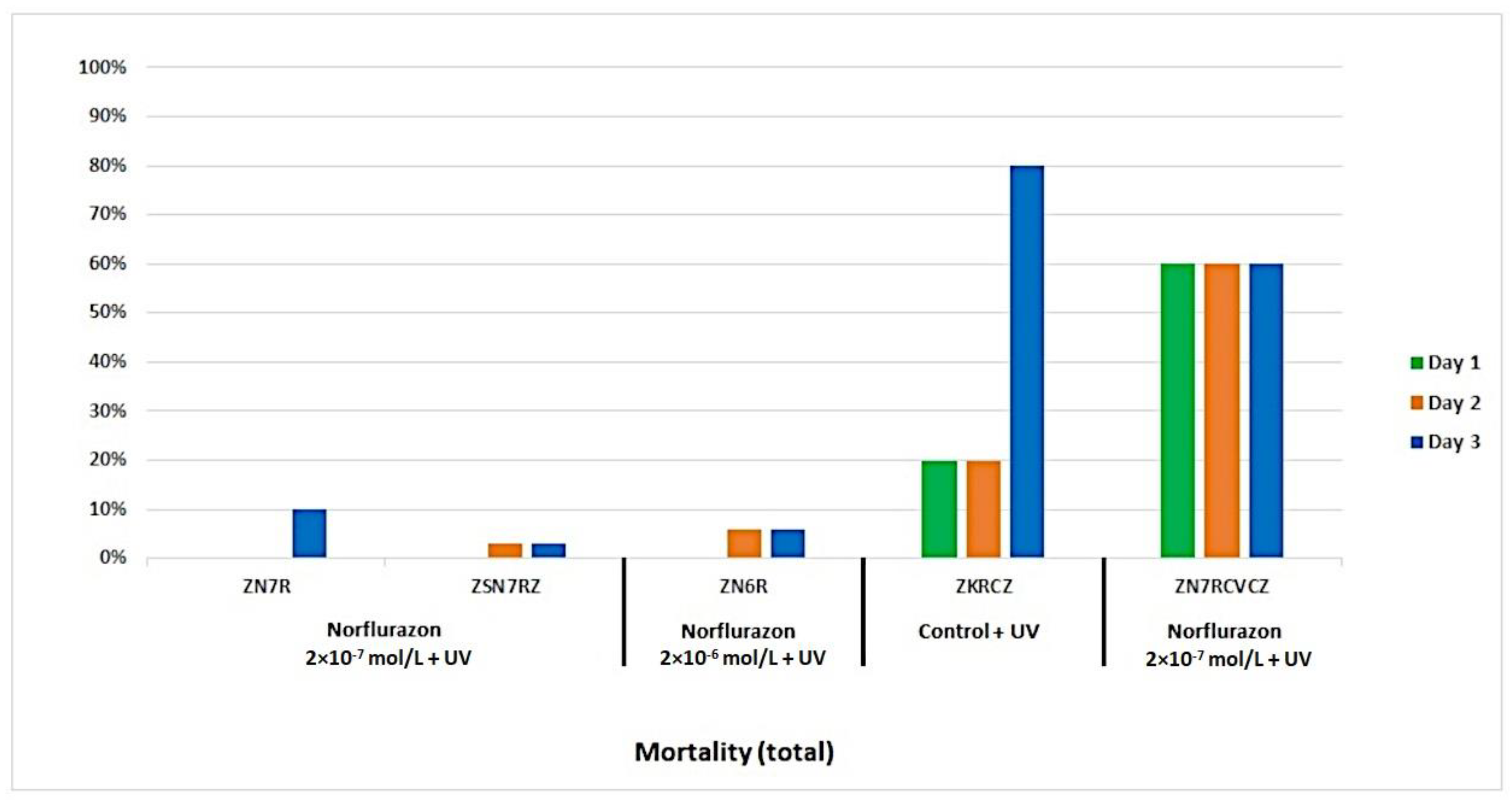
Figure 1. Mortality during the experiment. (CV—Chlorella vulgaris Beij. [K&H, 1992]; CZ—Mychonastes homosphaera (Chlorophyceae) (Skuja) Kalina et Punčochářová; Z—green hydra; S—brown hydra; K—control; N6—norflurazon 2 × 10⁻⁶ mol/L; N7—norflurazon 2 × 10⁻⁷ mol/L; and R—irradiated).
Water 16 00645 g001
Figure 2. Migration during the experiment. (a) Without addition of algae; (b) with addition of CV; (c) with addition of CZ; and (d) with addition of CV and CZ. (CV—Chlorella vulgaris Beij. [K&H, 1992]; CZ—Mychonastes homosphaera (Chlorophyceae) (Skuja) Kalina et Punčochářová; Z—green hydra; S—brown hydra; K—control; N6—norflurazon 2 × 10⁻⁶ mol/L; N7—norflurazon 2 × 10⁻⁷ mol/L; and R—irradiated).
Water 16 00645 g002aWater 16 00645 g002b
Figure 3. Tentacle damage during the experiment. (a) Without addition of algae; (b) with addition of CV; (c) with addition of CZ; and (d) with addition of CV and CZ. (CV—Chlorella vulgaris Beij. [K&H, 1992]; CZ—Mychonastes homosphaera (Chlorophyceae) (Skuja) Kalina et Punčochářová; Z—green hydra; S—brown hydra; K—control; N6—norflurazon 2 × 10⁻⁶ mol/L; N7−norflurazon 2 × 10⁻⁷ mol/L; and R—irradiated).
Water 16 00645 g003aWater 16 00645 g003b
Figure 4. Thickness of the mesoglea in samples of green and brown hydra treated with norflurazon and UV radiation. (CV—Chlorella vulgaris Beij. [K&H, 1992]; CZ—Mychonastes homosphaera (Chlorophyceae) (Skuja) Kalina et Punčochářová; Z—green hydra; S—brown hydra; K—control; N6—norflurazon 2 × 10⁻⁶ mol/L; N7—norflurazon 2 × 10⁻⁷ mol/L; and R—irradiated).
Water 16 00645 g004
Figure 5. (a) Green hydra irradiated with UV radiation, third day of the experiment. Potential un-controlled cell proliferation in the body of hydra or possible attempt of budding (arrow); (b) green hydra, in a mixed sample with brown hydra, treated with norflurazon (2 × 10⁻⁶ mol/L), third day of the experiment. Fork-like damage to tentacles (arrow). Scale bars represent 50 µm.
Water 16 00645 g005
Figure 6. C. vulgaris from cultures preserved for transmission electron microscopy (TEM) by high-pressure freezing/freeze substitution (HPF/FS). Extracellular polymeric substances (EPS), marked with an arrow, displayed in more detail within a boxed area (red). Scale bar 250 nm.
Water 16 00645 g006
Figure 7. Isolated endosymbiotic M. homosphaera from cultures, preserved for TEM by HPF/FS. (a) Overview. Scale bar 5 µm; (b) Enlarged. Multilamellar cell wall marked with two arrows. Scale bar 1 µm.
Water 16 00645 g007
Table 1. Overview of algal clustering in the experiment.
SampleDay 1Day 2Day 3
CVCZCVCZCVCZCVCZCVCZCVCZ
ControlZ+++++++++
S+green −++green −++green −+
ZS+++++++++
IrradiatedZ+++++++++++++++
S+++++++++
ZS++++++++++++
Norflurazon
2 × 10⁻⁷ mol/L
Zgreen −++++green −++++green −++++
S+++++++++
ZS+++++++++
Norflurazon
2 × 10⁻⁶ mol/L
Z++++++++++++
Sgreen +green ++green +green ++green +green ++
ZS+++++++++++++++
Norflurazon
2 × 10⁻⁷ mol/L
Irradiated
Z+++++++++
S++++++++++++
ZS++++++++++++
Norflurazon
2 × 10⁻⁶ mol/L
Irradiated
Z+++++++++
S+++++++++
ZS+++++++++
− no clustering; + presence of clustering; ++ large presence of clustering; green = solution was homogenously green.
Disclaimer/Publisher’s Note: The statements, opinions and data contained in all publications are solely those of the individual author(s) and contributor(s) and not of MDPI and/or the editor(s). MDPI and/or the editor(s) disclaim responsibility for any injury to people or property resulting from any ideas, methods, instructions or products referred to in the content.