Article

Quantitative Detection of VBNC State Pseudomonas aeruginosa Contributing to Accurate Assessment of Microbial Inactivation in Drinking Water Disinfection

School of Environmental and Chemical Engineering, Shanghai University, Shanghai 200444, China
*
Authors to whom correspondence should be addressed.
These authors contributed equally to this work.
Water 2024, 16(2), 236; https://doi.org/10.3390/w16020236
Submission received: 28 November 2023 / Revised: 21 December 2023 / Accepted: 22 December 2023 / Published: 10 January 2024

Abstract

:
In harsh environments, bacteria often enter a viable but nonculturable (VBNC) state, which cannot be detected using heterotrophic plate counting (HPC). Importantly, VBNC bacteria can potentially resuscitate under favorable conditions, posing a risk to drinking water safety. This study introduces an innovative approach, combining improved quantitative polymerase chain reaction (qPCR) with propidium monoazide (PMA) dye and HPC to accurately quantify VBNC Pseudomonas aeruginosa (P. aeruginosa). The method was applied to assess the ability of various disinfection techniques to induce P. aeruginosa into the VBNC state. Different disinfection methods, including ultraviolet radiation (UV), sodium hypochlorite (NaClO), and peracetic acid (PAA), significantly reduced bacterial culturability (>99.9%), with the majority entering the VBNC state. Notably, under favorable conditions, UV-induced VBNC cells were resuscitated faster than those induced by NaClO. VBNC P. aeruginosa exhibited relatively high intracellular adenosine triphosphate (ATP) levels, indicating ongoing metabolic activity. Scanning electron microscopy (SEM) reveals that some bacteria maintained cellular integrity for UV and PAA treatment, while evident membrane disruption was observed after NaClO disinfection. This study represents a significant advancement in quantitatively detecting VBNC state P. aeruginosa, contributing to an accurate assessment of microbial inactivation during drinking water disinfection.

1. Introduction

Pseudomonas aeruginosa (P. aeruginosa), a prevalent opportunistic pathogen in drinking water distribution systems [1], poses a potential risk of causing human infections such as non-tuberculosis lung disease [2]. The conventional culture-based heterotrophic plate count (HPC) method is commonly used to evaluate disinfection efficacy against P. aeruginosa [3]. However, existing disinfection techniques often induce a viable but nonculturable (VBNC) state [3,4,5], rendering the HPC method ineffective [6]. Consequently, traditional culture-based approaches are likely to significantly underestimate the presence of VBNC bacteria that may pose a threat to drinking water safety [7].
Various activity-based techniques have been developed to detect bacteria in the VBNC state, including measurements of respiratory and metabolic activity, fluorescent-based hybridization, and staining techniques based on cell membrane integrity [8,9,10]. However, these methods face limitations in their broad applicability. Dye-based flow cytometry is widely used for assessing the viability of foodborne pathogens, but it faces challenges in accurately distinguishing between viable and dead bacteria. For instance, Pilar Truchado et al. used flow cytometry to determine the ratio of live to dead Listeria monocytogenes (L. monocytogenes) cells before and after chlorine treatment, and the inability to precisely distinguish between viable and dead cells resulted in an overestimation of the proportion of live bacteria compared to the actual values [11].
Fortunately, a quantitative polymerase chain reaction (qPCR) method coupled with HPC has been developed to detect VBNC bacteria. This approach involves qPCR for identifying viable cells and HPC for culturable cell detection. Subtracting HPC-detected cells from those identified via qPCR yields the VBNC population. While HPC methodologies have achieved a state of maturity, the application of qPCR in drinking water disinfection remains an evolving area. In the majority of studies, the pretreatment of bacteria with the dye propidium monoazide (PMA), wherein PMA can penetrate damaged cell membranes that hinder DNA amplification [10], was used for the detection of viable cells using qPCR. However, it is imperative to acknowledge that not all dead cells necessarily experience membrane damage; some may die due to internal DNA damage [12,13,14]. Recent research suggests that longer gene segments are more susceptible to damage, potentially increasing the likelihood of PCR amplification termination [4]. Building on this advancement, we developed an innovative method using PMA-qPCR with longer gene segments to selectively identify bacteria with intact cell membranes, excluding false positives from short-chain DNA amplification. To the best of our knowledge, this is the first time we have developed a methodology that combines PMA-qPCR with longer gene segments and HPC for viable P. aeruginosa detection.
Building upon our developed methodology, we aim to investigate whether P. aeruginosa is directly eliminated or induced into VBNC states and how disinfection methods influence cell membrane morphology and bioactivity. This investigation is critical, as pathogenic bacteria in the VBNC state may retain toxicity [15] and can regrowth in a suitable environment [16,17]. For instance, the revival of P. aeruginosa in a VBNC state can be observed during incubation in Luria–Bertani (LB) medium after UV treatment [18]. The regrowth of Achromobacter reached 103 CFU/mL after 2 mg/L chlorine treatment, followed by 25 h of cultivation in sterile drinking water [19]. Furthermore, it is worth noting that the resuscitation of VBNC P. aeruginosa in secondary water supply tanks has been reported [20], representing a “hidden source of contamination” in the drinking water supply system. Therefore, a comprehensive understanding of the behavior and potential threats of bacteria in the VBNC state is significant for ensuring drinking water safety.
In this study, we quantitatively analyzed VBNC P. aeruginosa using PMA-qPCR and HPC. The effectiveness of UV radiation, NaClO, and PAA disinfection on microbial inactivation in drinking water was comprehensively assessed. We compared the revival potential of VBNC P. aeruginosa induced by different disinfection methods. Additionally, we examined the intracellular adenosine triphosphate (ATP) levels and cell membrane morphology of induced VBNC cells. This study could mark an important step toward understanding the induced VBNC state of bacteria in different disinfection methods.

2. Materials and Methods

2.1. Cultivation and Preparation of P. aeruginosa Cells

P. aeruginosa (Collection No. CMCC(B)10104) obtained from HuanKai Biology Co., Ltd. (Guangzhou, China) was cultured aseptically in Luria–Bertani (LB) medium. After cultivation, the bacterial culture in its growth phase was obtained by centrifugation at 6869× g for 10 min. This centrifugation step effectively facilitated the separation of cells in the logarithmic growth phase. The resulting cell pellet underwent washing with phosphate-buffered saline (PBS, pH = 7.0 ± 0.1) and dual-phase centrifugation, leading to bacterial cells suspended in PBS to create a stock solution with an initial concentration of approximately 3.36 × 10⁸ CFU/mL. The stock solution was diluted 1:10 in high-pressure sterilized ultrapure water, yielding a final concentration of 3.36 × 10⁷ CFU/mL.

2.2. Inactivation and Regrowth Experiments

UV irradiation was sourced from a commercially available mercury lamp (Heraeus, Hanau, Germany) with a wavelength of 245 nm. Previous studies have documented that UV irradiation at 245 nm has a strong capacity to disrupt the DNA of microorganisms, which enables us to examine the disinfection efficiency of P. aeruginosa [4]. Initially, in order to mix the water samples sufficiently and the disinfection effect accurately, 40 mL of the water samples from Section 2.1 were introduced into a sterile Petri dish with a diameter of 100 mm and exposed to UV irradiation at room temperature. Samples were retrieved from the Petri dishes at specified intervals (0 min, 1 min, 3 min, 5 min, 7 min, 10 min, 15 min, 20 min, 25 min, and 30 min). UV intensity was determined using the iodide/iodate spectrophotometric method [21].
To ensure sufficient contact between P. aeruginosa and the disinfectant, the chlorination disinfection experiments were conducted within a sterile Petri dish with a diameter of 100 mm containing 40 mL of water samples. NaClO (Rhawn, Shanghai, China) was added to the Petri dishes in concentrations of 0.5 mg/L, 1 mg/L, 2 mg/L, and 3 mg/L, respectively. The free chlorine concentration was measured using the N, N-diethyl-pphynylenediamine (DPD) method. Petri dishes were positioned on a magnetic stirrer for thorough mixing, and samples were collected at distinct exposure time points (0 min, 2 min, 3 min, 4 min, 5 min, 10 min, 15 min, 20 min, 25 min, and 30 min). Sodium thiosulfate solution (Na2S2O3, 0.1 mM) was used to quench the chlorination process.
To assess the disinfection effectiveness of commercial PAA (17.6% PAA, 9.8% H2O2, and 72.6% acetic acid), 40 mL water samples containing P. aeruginosa were placed in a Petri dish with a diameter of 100 mm and thoroughly mixed. The final concentration of the reaction solution was adjusted to 40 μM by adding PAA stock solution. Continuous stirring using a magnetic stirrer ensured uniform mixing of the reaction solution. Samples were periodically withdrawn from the reactor and immediately quenched with an excess of Na2S2O3 solution.
The regrowth potential of VBNC P. aeruginosa was investigated under favorable conditions. A suspension containing 10 mL of VBNC P. aeruginosa was combined with 10 mL of LB medium in a 50 mL centrifuge tube and subjected to cultivation under conditions of 37 °C and 150 rpm/min for 16 h. Then, aliquots of 1 mL each were collected bihourly and dispensed onto nutrient agar medium, followed by an incubation period of 24 h at 37 °C, facilitating the ascertainment of viable bacterial growth. The resuscitation efficacy was quantitatively assessed using the HPC method.

2.3. PMA-qPCR and qPCR Standard Curve

A standard curve linking the copy number of the target gene to Ct values was established using plasmid DNA carrying the specific gene (Figure S1). Dilution experiments with P. aeruginosa bacterial cultures were conducted to establish a quantitative relationship between the copy number of the specific gene and the number of bacterial cells. In this study, a P. aeruginosa bacterial suspension with an initial concentration of 4.9 × 10⁷ CFU/mL was selected. This suspension underwent a tenfold dilution gradient. Subsequently, 500 μL of the diluted water samples were taken, and an appropriate amount of PMA (Biotium, Fremont, CA, USA) was added and thoroughly mixed. The samples were then incubated under dark conditions for 5 min, followed by exposure to a 650 W halogen light source on ice for 5 min. After light exposure, the samples were filtered through a 0.22 μm membrane, and the membrane was excised with sterile scissors. Bacterial DNA was then extracted using the DNeasy® PowerSoil® Pro Kit (Qiagen, Hilden, Germany). Quantification of viable bacterial cells was achieved through qPCR using long primers (Table 1, opr-L) to construct the PMA-qPCR standard curve. Additionally, DNA was extracted from 500 μL of water samples without PMA treatment, and qPCR was performed using short primers (Table 1, opr-S) to determine the total bacterial count; therefore, the qPCR standard curve was established.

2.4. Evaluation of PMA-qPCR Method Precision

To evaluate the precision of the PMA-qPCR method, a bacterial suspension (3.39 × 10⁷ CFU/mL) was prepared by subjecting a 10 mL aliquot to a 100 °C treatment for 30 min, yielding a heat-killed bacterial suspension. Live and heat-killed bacterial suspensions were then combined in predetermined ratios, forming a composite bacterial solution. The extraction of DNA was conducted from the PMA-treated composite bacterial solution. DNA extraction from the untreated composite bacterial solution was performed, and the samples were subjected to conventional qPCR reactions to ascertain the total bacterial count. Furthermore, the determination of the actual viable cell count within the composite bacterial solution was accomplished using the HPC method. Through comparative analysis and assessment of the viable cell counts yielded by PMA-qPCR and qPCR methods against the established actual viable cell count, the precision, accuracy, and sensitivity of the PMA-qPCR approach for quantifying viable P. aeruginosa cells were substantiated.

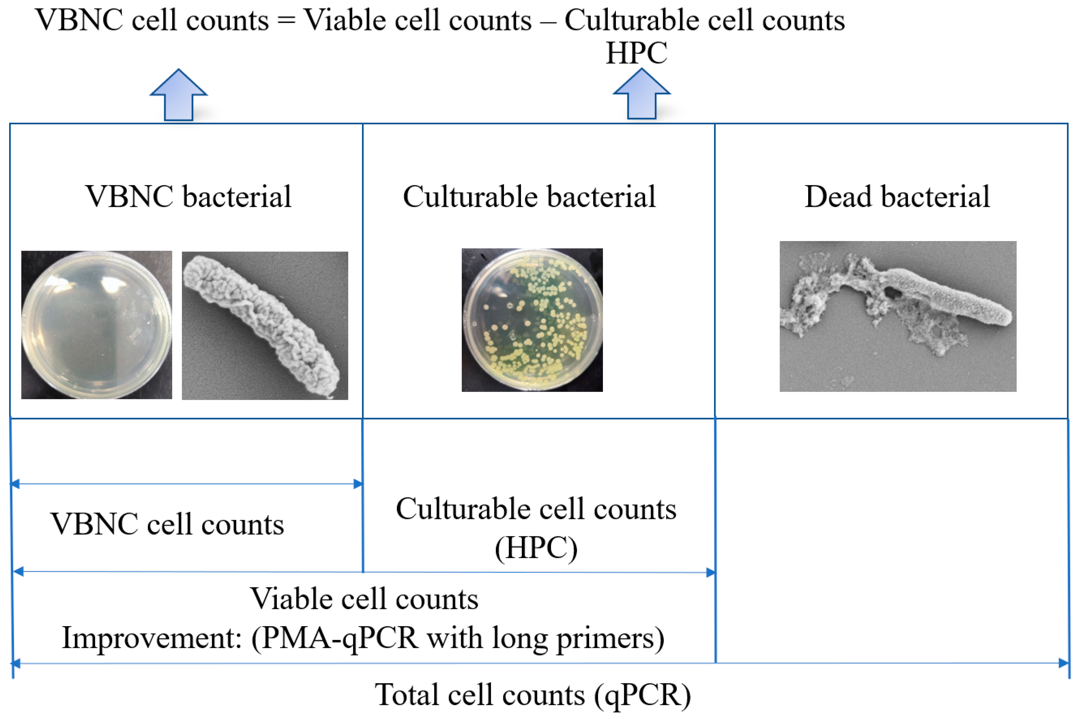
2.5. Quantification of VBNC State P. aeruginosa

DNA extracted from the reaction mixture was analyzed by the PMA-qPCR approach. The PMA-qPCR standard curve was employed to determine the count of viable P. aeruginosa. The count of culturable bacteria was determined using the HPC method. Initially, 1 mL samples were drawn from the treated specimens and underwent a sequence of dilutions using ultrapure water. Appropriate dilution factors were selected from these samples for inoculation onto nutrient agar plates. Plating was carried out in triplicate at each gradient of dilution, followed by an incubation period of 24 h at 37 °C. Quantification of VBNC state P. aeruginosa was conducted as outlined in Scheme 1. The enumeration of VBNC cells was determined by calculating the disparity between the viable cell count established by the PMA-qPCR method and the count of culturable cells determined by the HPC method.

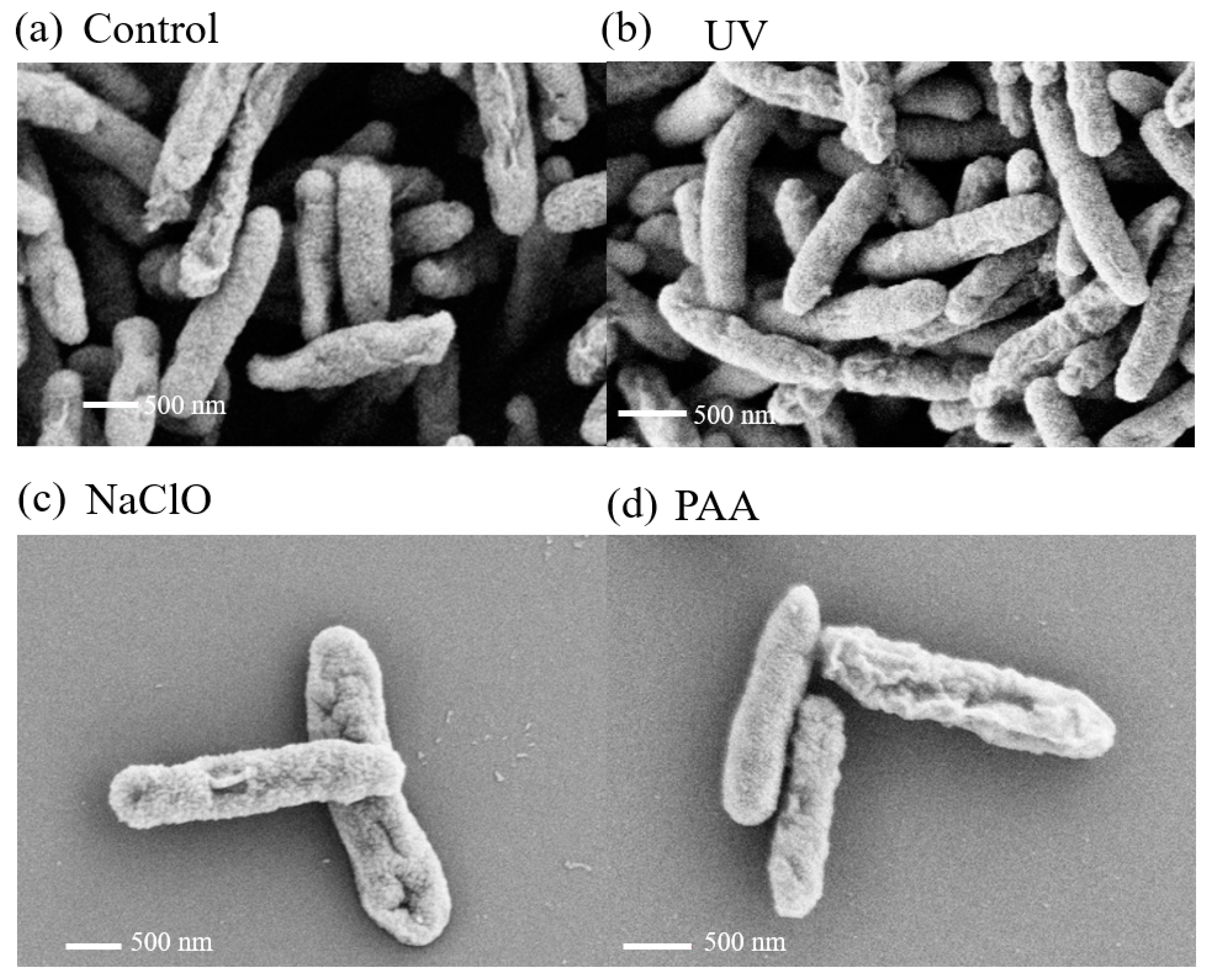
2.6. Assessment of Cell Membrane Morphology and Metabolic Activity

The investigation of cell membrane morphology and metabolic activity in the VBNC state was carried out. The fine-scale structure and morphological features of the bacteria were examined using a scanning electron microscope (SEM, Hitachi SU-8010, Tokyo, Japan). The intracellular ATP content of the bacteria was quantified using the BacTiter-Glo Microbial Cell Viability Assay Kit (Promega, Madison, WI, USA) to precisely evaluate cellular metabolic activity.

3. Results and Discussion

3.1. Integration of Modified qPCR and HPC for Accurate Quantification of VBNC P. aeruginosa

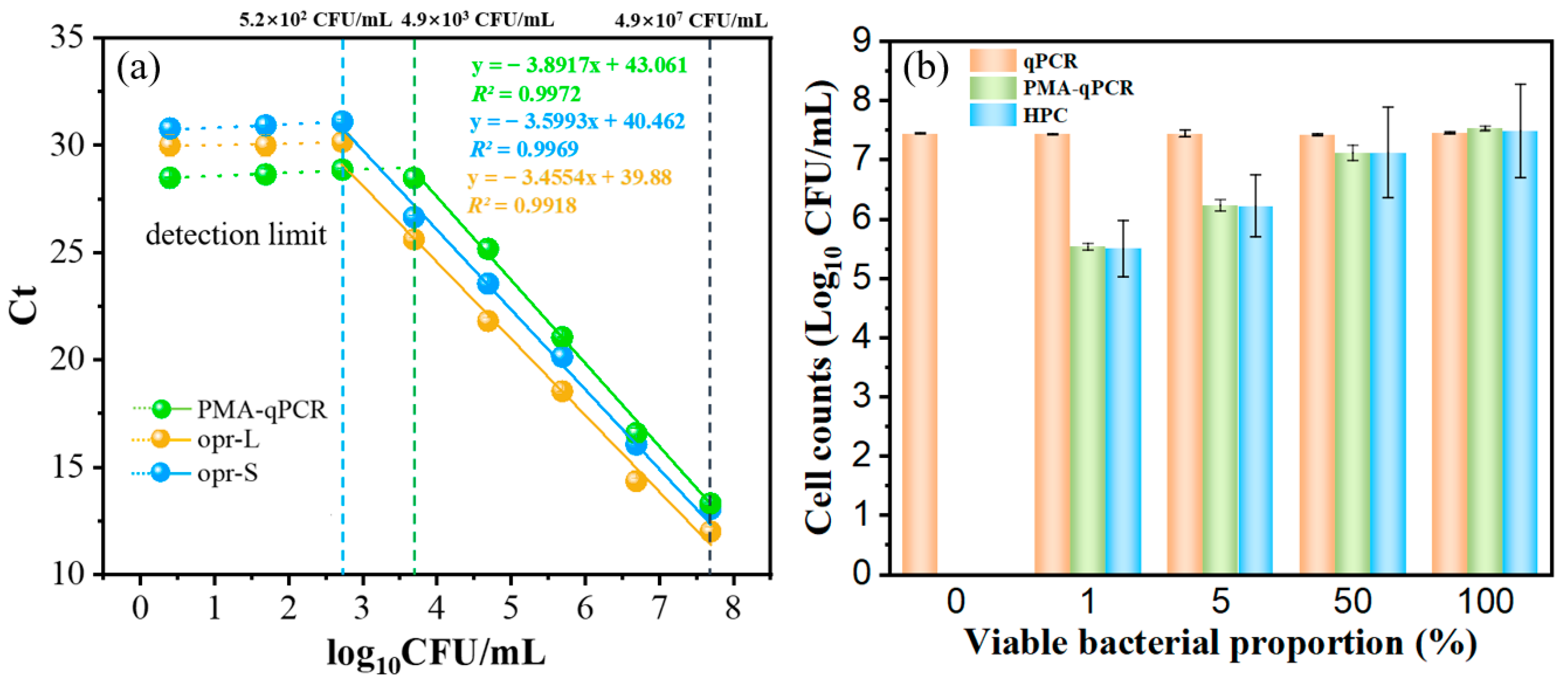
3.1.1. Validation of Long Primers for Accurate Detection of Viable P. aeruginosa in qPCR

Figure 1 illustrates the outcomes of gel electrophoresis, delineating the efficacy of specific primers (opr-S, opr-L) in probing P. aeruginosa. Figure 1a delineates the results of short primer amplification (opr-S, 99 bp), while Figure 1b elucidates the ramifications of long primer amplification (opr-L, 504 bp). Notably, the gel profile exhibits clear, bright, and uniform bands at both 99 bp and 504 bp, without evidence of primer dimers, indicating the specificity of the selected primers for amplifying the P. aeruginosa opr gene.
To further validate the accuracy of the long primer in detecting viable bacteria, a tenfold gradient dilution of the initial concentration of P. aeruginosa bacterial suspension (4.9 × 10⁷ CFU/mL) underwent qPCR reactions using both opr-L and opr-S primers. Standard curves correlating P. aeruginosa concentration with Ct values were established, as depicted in Figure 2a. The standard coefficients of both curves (R2 ˃ 0.99) indicate excellent linear correlation. Importantly, the two qPCR standard curves almost completely overlap, suggesting similar PCR efficiency and copy numbers for opr-L and opr-S primers during target sequence amplification. This further confirms the accuracy of the long primer in qPCR for determining viable bacteria.

3.1.2. Negligible Effect of PMA on qPCR Detection of Viable P. aeruginosa

PMA is well-known for its ability to penetrate damaged cell membranes and inhibit PCR amplification of bacterial DNA from dead cells. It is also critical to assess whether PMA affects the DNA amplification of viable bacteria in qPCR. In this section, we investigated the effect of PMA on qPCR detection of viable P. aeruginosa. A series of known concentrations of viable P. aeruginosa samples were prepared and exposed to PMA. Employing the validated long primer, PMA-qPCR experiments were conducted using the PMA binding method. A series of PMA-qPCR optimization experiments addressing parameters such as PMA concentration, dark incubation time, and exposure time were initially conducted (Figure S2). Under optimal conditions, we successfully established a standard curve correlating viable P. aeruginosa cell counts with Ct values. As shown in Figure 2a, the PMA-qPCR method (opr-L) demonstrated excellent linearity within the range of live cell concentrations from 4.9 × 10³ CFU/mL to 4.9 ×10⁷ CFU/mL, with a correlation coefficient (R2) of 0.99. This confirms that PMA does not penetrate the cell membrane of viable bacteria, thereby not affecting the determination of viable bacteria. The PMA-qPCR method, combined with the long primer, proved to be suitable for accurate quantification of viable P. aeruginosa cell counts.

3.1.3. PMA-qPCR for Selective Quantification of Viable P. aeruginosa in Viable–Dead Mixed Samples

To further validate the precision of the PMA-qPCR method in quantifying viable bacteria, quantification of P. aeruginosa samples with varying ratios of viable and dead cells was conducted. Classical qPCR and HPC methods were employed for comparative analyses. When qPCR was used to determine the bacterial count in mixtures with different ratios of viable and dead cells, the Ct values fluctuated between 13.64 and 13.74 (Table S1), corresponding to bacterial counts of 7.53–7.56 log10 CFU/mL, showing no significant variation (Figure 2b). This indicates that the traditional qPCR method is unable to distinguish between viable and dead bacteria, leading to potential false-positive results in detecting viable bacteria.
Furthermore, the cell counts determined by the PMA-qPCR method were compared to those obtained through the standard HPC method. The results, as shown in Figure 2b, revealed an increase in the count of viable bacteria with a rising proportion of viable cells. Importantly, these results closely matched those obtained using the traditional HPC method. This suggests that PMA-qPCR can selectively amplify the DNA of viable bacteria, effectively distinguishing viable and dead cells in mixed samples during qPCR detection.

3.2. Assessment of Various Disinfection Methods for Inducing a VBNC State in P. aeruginosa

3.2.1. Inactivation of Viable P. aeruginosa in UV Method

Different doses of UV radiation were applied to evaluate the effectiveness of disinfection against P. aeruginosa. Results demonstrated that culturable P. aeruginosa was readily inactivated under UV treatment. Under a UV dose of 26.1 mJ/cm2, no culturable bacteria were detectable. However, there was no significant change in the number of viable P. aeruginosa at this time (7.11 log10 CFU/mL, Figure 3a), indicating that all P. aeruginosa entered the VBNC state. Even with higher UV doses (50–261 mJ/cm2), samples continued to harbor P. aeruginosa in a VBNC state, with a presence of 6.05 log10 CFU/mL (Figure 3a). It is important to note that our study employed a maximum UV dose (261 mJ/cm2), significantly exceeding typical drinking water treatment levels. Despite a considerable reduction in culturable bacteria, a fraction of P. aeruginosa displayed resilience to high UV doses, maintaining viability and transitioning into a VBNC state. Consequently, relying solely on UV disinfection may inadequately mitigate the risk of P. aeruginosa contamination in water systems.

3.2.2. Inactivation of Viable P. aeruginosa in NaClO Method

The impact of varying concentrations of NaClO solution on P. aeruginosa inactivation was evaluated. As depicted in Figure S3a, low residual chlorine levels (0.5 mg/L) exhibited negligible impact on the viability of P. aeruginosa. Elevating the residual chlorine concentration to 1 mg/L resulted in a significant reduction in the culturable bacterial count of P. aeruginosa from an initial 7.75 log10 CFU/mL to 2.95 log10 CFU/mL within 30 min. Under residual chlorine concentrations of 2 mg/L and 3 mg/L, the culturable bacterial count of P. aeruginosa declined precipitously from 7.75 log10 CFU/mL to 0 CFU/mL within 10 and 4 min, respectively. Increasing chlorine doses led to a faster transition to the VBNC state. This clearly demonstrates the effective reduction in culturability of P. aeruginosa through chlorination.
Furthermore, it is worth noting that after treatment with 0.5, 1, 2, and 3 mg/L residual chlorine for 30 min, the VBNC cell count showed negligible change. This suggests that, within a certain range, increasing residual chlorine concentration does not significantly enhance the bactericidal effect against P. aeruginosa. Consequently, in practical water treatment processes, relying solely on single chlorine disinfection measures may not achieve complete bacterial eradication.

3.2.3. Inactivation of Viable P. aeruginosa in PAA Method

The disinfection performance of PAA for P. aeruginosa was assessed at 40 μM PAA with a 30-min exposure time. As shown in Figure 3c, the count of culturable bacteria decreased from 7.52 log10 CFU/mL to 3.59 log10 CFU/mL, achieving a 99.98% removal efficiency. However, viable bacteria decreased only from 7.52 log10 CFU/mL to 7.01 log10 CFU/mL. After 30 min of PAA treatment, a portion of P. aeruginosa entered a VBNC state, while some retained culturability and others were completely inactivated. These results indicate that PAA treatment partially eliminates the activity of P. aeruginosa. Practical applications may require adjusting the disinfectant concentration and treatment time for optimal sterilization.

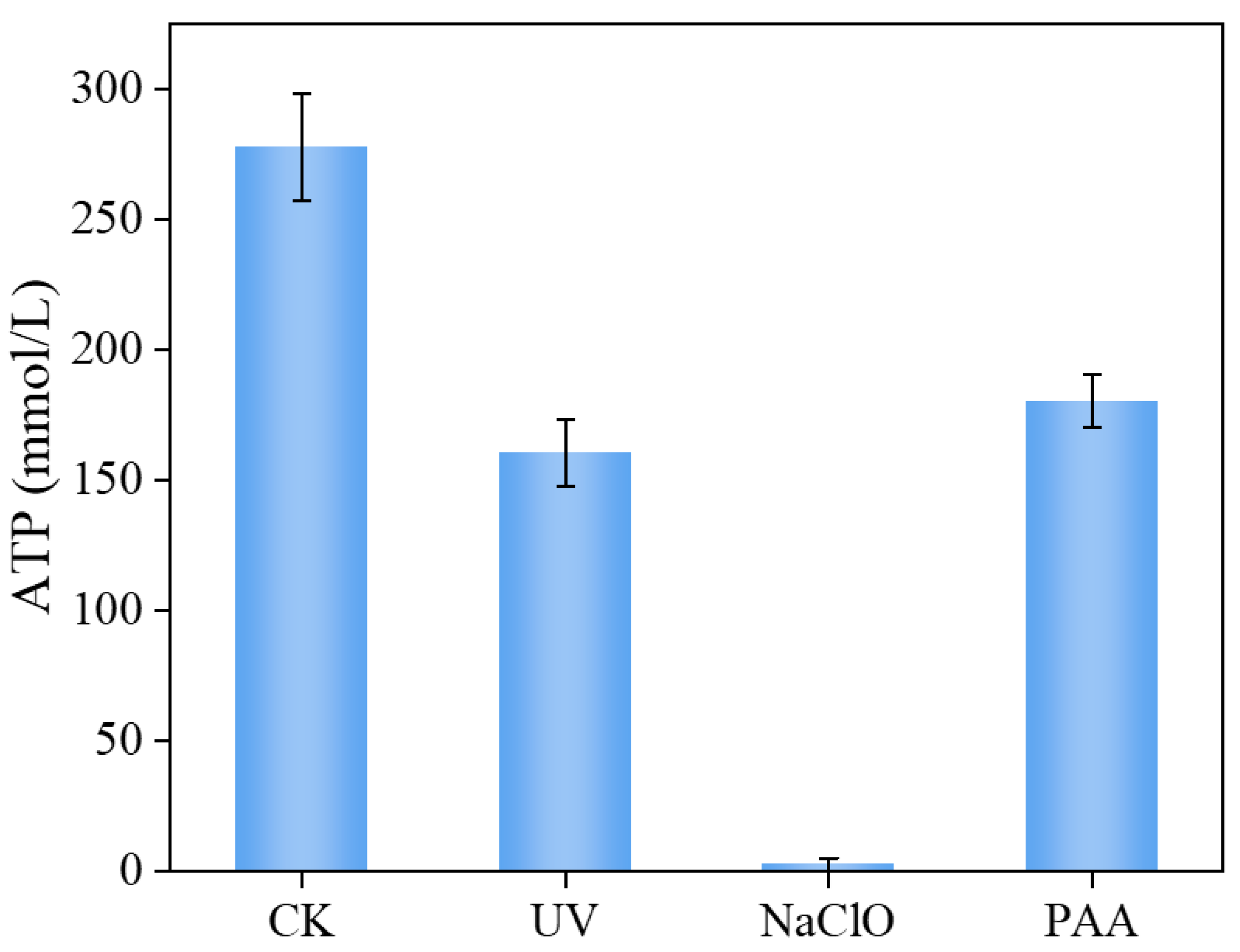
3.2.4. Comparison of Inactivation Effects of Four Disinfection Methods

Results from UV radiation, NaClO, and PAA disinfection demonstrate that even under high doses of UV radiation (261 mJ/cm²), NaClO (3 mg/L), and PAA (40 μM), single disinfection methods are inadequate for completely eradicating viable P. aeruginosa. To further evaluate the effectiveness of various disinfection approaches on P. aeruginosa elimination, we applied the Chick–Watson model to characterize its inactivation kinetics:
log ( N t N 0 ) = k · t
where N0 is the concentration of P. aeruginosa in the sample before disinfection (in CFU/mL), Nt is the concentration of P. aeruginosa in the sample after t-time disinfection (in CFU/mL), k is the disinfection rate constant (in min−1), and t is the disinfection treatment time (in min). From Figure 3d, UV disinfection exhibited the highest rate of reduction in viable and culturable bacteria, followed by NaClO and PAA disinfection. This suggests a significant advantage of UV disinfection in eradicating culturable bacteria and inducing entry into a VBNC state at a relatively faster rate.
This difference in disinfection efficiency may be attributed to variations in the disinfection mechanisms. NaClO and PAA act as oxidative disinfectants, inactivating bacteria through oxidation processes [22], while UV disinfection deactivates bacteria by damaging their genetic material [23]. It is possible that bacterial antioxidant mechanisms are less effective in resisting UV disinfection [24]. Furthermore, the reaction conditions significantly influence disinfection outcomes. During UV disinfection, microorganisms are exposed to intense radiation, leading to quicker deactivation. In contrast, the rate of deactivation in chemical disinfection depends on various factors, including disinfectant concentration, environmental temperature, and reaction time. Typically, achieving the same level of deactivation may require more time and higher concentrations in chemical disinfection processes.

3.3. Resuscitation of VBNC P. aeruginosa

Under specific conditions, VBNC bacteria may exhibit resuscitate potential [25,26,27]. For VBNC bacteria in drinking water treatment, the absence of disinfectants and other complex components in water supply pipes may lead to the resuscitation of VBNC bacteria. To assess the potential health risk of VBNC P. aeruginosa induced by different disinfection methods, resuscitation experiments of VBNC bacteria were conducted under laboratory conditions. Since P. aeruginosa treated with 40 μM PAA for 30 min did not fully enter the VBNC state, reaction solutions treated with 261 mJ/cm2 UV radiation and 3 mg/L chlorine for 30 min were selected for investigation. Results in Figure 4a showed that UV-induced VBNC bacteria underwent resuscitation at 10 h, with a resuscitation count of 2.59 log10 CFU/mL. In contrast, the resuscitation lag phase of NaClO-induced VBNC P. aeruginosa reached 14 h (Figure 4b), with no bacterial growth in 8 of the batches, indicating the probabilistic nature of resuscitation [3]. After 16 h of cultivation, resuscitation occurred in all batches. These research results indicate that the disinfection method has a significant impact on the resuscitation ability of VBNC bacteria.
In previous studies, various methods have been used to determine whether VBNC cells have truly undergone resuscitation. However, these studies did not conclusively demonstrate that the appearance of culturable cells was the result of the resuscitation of VBNC bacteria [4]. The observed difference in bacterial resuscitation time (Figure 4a, 10 h) compared to the normal generation time of P. aeruginosa (Figure S4, 2 h) suggests that the observed bacteria are the result of the resuscitation of VBNC state bacteria. This finding provides crucial evidence for the genuine resuscitation of VBNC state bacteria. Furthermore, compared to NaClO disinfection, UV-induced VBNC P. aeruginosa has a shorter resuscitation lag phase. This difference may be related to the extent of damage to the cell membrane morphology and bioactivity of P. aeruginosa caused by UV and NaClO disinfection.
To further understand how the bioactivity of VBNC P. aeruginosa affects resuscitation in different disinfection methods, the intracellular ATP levels within cells were measured. ATP serves as a ubiquitous cellular energy carrier in cells [28]. In cell biology, intracellular ATP levels reflect the state of cellular energy metabolism, growth, and division capabilities [29]. Consequently, intracellular ATP content is considered a pivotal indicator of cellular bioactivity [30]. According to the results presented in Figure 5, the intracellular ATP concentration of UV disinfection-induced VBNC state cells was significantly lower than that of non-induced cells (p < 0.01) but still maintained a relatively high level. This indicates that although these bacteria have lost culturability, they still require energy to maintain basic metabolic activity. Furthermore, the intracellular ATP level after NaClO disinfection was significantly lower than that after UV and PAA disinfection. This may be one of the reasons for the longer time required for the resuscitation of VBNC bacteria induced by NaClO disinfection. These results further illustrate the impact of the bioactivity of VBNC state bacteria on their resuscitation.
It is also interesting to explore how the cell membrane morphology of VBNC bacteria is associated with the resuscitation process and bioactivity of VBNC P. aeruginosa. The microscopic structure of the cell surface of P. aeruginosa before and after treatment with different disinfection methods was analyzed using SEM. In its initial state (Figure 6a), untreated P. aeruginosa exhibited a clear cell outline with a smooth and intact surface, showing no signs of damage or wrinkles. After UV radiation treatment, the cell morphology remained largely intact, with only slight wrinkles or depressions observed on the surface of some cells. The preservation of the intact cell morphology may contribute to faster resuscitation in a suitable environment (Figure 4a). In contrast, after NaClO treatment, substantial damage and depressions were evident on the surface of P. aeruginosa, indicating compromised integrity of the cell wall and cell membrane. The damage to the cell membrane suggests a reaction between chlorine and various cell components, directly leading to the breakdown of the protective layer of the cell membrane [31]. Importantly, the damage to the cell membrane may result in a longer time required for the resuscitation of VBNC P. aeruginosa (Figure 4b). Furthermore, studies suggest that chlorine disinfection-induced cell death first manifests as surface damage, subsequently affecting internal cell structures [3]. Damage to internal structures hinders the synthesis of ATP molecules, thereby inhibiting the synthesis of large molecules such as proteins and nucleic acids [32]. This could lead to significantly lower intracellular ATP levels after NaClO disinfection (Figure 5).
In addition, after PAA treatment, the surface morphology of P. aeruginosa became slightly rougher, differing from the common surface perforations observed in chlorine disinfection [33]. In fact, previous studies indicate that PAA has a minimal impact on the cell membrane, and at PAA doses below 60 mg/L, most cells remain intact [34,35]. Overall, PAA disinfection did not cause significant damage to the cell membrane morphology of P. aeruginosa. In conclusion, these changes in the surface morphology of P. aeruginosa may be one of the reasons for the different resuscitation behaviors and intracellular ATP contents of the bacteria.

4. Conclusions

This study has established a PMA-qPCR method with a broad range and high accuracy for quantifying the viable cell count of P. aeruginosa. By integrating this method with HPC, we achieved precise quantification of VBNC bacteria with various disinfection methods. These results revealed that individual disinfection methods, such as UV, NaClO, and PAA, cannot completely eliminate P. aeruginosa, instead inducing its transition into a VBNC state. In the VBNC state, intracellular ATP experienced a slight reduction but maintained relatively high levels. Concurrently, morphological changes, including wrinkles or depressions, were observed on the bacterial surface. Importantly, we confirmed the resuscitation capability of VBNC bacteria, with UV-induced VBNC P. aeruginosa exhibiting a shorter resuscitation lag phase compared to NaClO-induced VBNC forms. Overall, this comprehensive study, facilitated by the newly established PMA-qPCR technique, provides a valuable approach for quantifying VBNC P. aeruginosa. Additionally, it sheds light on the induction, characteristics, and resuscitation dynamics of the VBNC state. These research findings contribute to a deeper understanding of the impact of different disinfection methods on VBNC bacteria, offering more reliable scientific support for addressing microbial safety concerns in drinking water treatment.
This study serves as a step toward refining the identification of viable but nonculturable (VBNC) cell counts. However, it is essential to acknowledge that the VBNC challenge remains complex and not entirely resolved. Current methodologies for determining the VBNC cell count rely on indirect approaches, specifically the subtraction of culturable cells from viable cells. While the measurement of culturable cells is well established, the main challenge remains the identification of viable cells. There is still an ongoing need for improvement in determining the viable cell count, particularly in addressing uncertainties in PMA-qPCR, such as the detection limit for low levels of viable cell counts. Further investigation is also needed to understand the microbial state on a single-cell level, given that the individual cell may exhibit distinct behavior under identical conditions. Future research directions may involve selective binding of Au nanoparticles (with an appropriate aptamer) based on the different activities of VBNC bacteria and normal live bacteria, followed by enhanced Raman spectroscopy or single-cell ICP–MS testing.

Supplementary Materials

The following supporting information can be downloaded at: https://www.mdpi.com/article/10.3390/w16020236/s1, Figure S1: Standard curve of gene copy number versus Ct value, opr-S primer (a); opr-L primer (b); Figure S2: Optimization of PMA treatment for P. aeruginosa; Figure S3: Inactivation of P. aeruginosa by NaClO. (a) NaClO at 0.5 mg/L, (b) NaClO at 1 mg/L, (c) NaClO at 2 mg/L; Figure S4: Growth curve of P. aeruginosa (a); relationship between OD600 and bacterial load (b); Table S1: Detection of different proportions of live bacteria using three methods.

Author Contributions

Conceptualization, Z.F., H.Z. and C.T.; methodology, H.Z., C.T., N.D. and X.H.; investigation, Z.F., H.Z. and C.T.; resources, N.D. and X.H.; data curation, Z.F., H.Z. and C.T.; writing—original draft preparation, Z.F., H.Z. and C.T.; writing—review and editing, H.Z., N.D. and X.H.; supervision, X.H. All authors have read and agreed to the published version of the manuscript.

Funding

This research was funded by the National Natural Science Foundation of China, grant number No. 51678351.

Data Availability Statement

All data were published in the main text and Supplementary Materials.

Acknowledgments

This work was supported by the National Natural Science Foundation of China (No. 51678351).

Conflicts of Interest

The authors declare no conflicts of interest.

References

  1. Moreira, N.F.F.; Ribeirinho-Soares, S.; Viana, A.T.; Graça, C.A.L.; Ribeiro, A.R.L.; Castelhano, N.; Egas, C.; Pereira, M.F.R.; Silva, A.M.T.; Nunes, O.C. Rethinking water treatment targets: Bacteria regrowth under unprovable conditions. Water Res. 2021, 201, 117374. [Google Scholar] [CrossRef] [PubMed]
  2. Wang, H.; Edwards, M.A.; Falkinham, J.O.I.; Pruden, A. Probiotic Approach to Pathogen Control in Premise Plumbing Systems? A Review. Environ. Sci. Technol. 2013, 47, 10117–10128. [Google Scholar] [CrossRef] [PubMed]
  3. Chen, S.; Li, X.; Wang, Y.; Zeng, J.; Ye, C.; Li, X.; Guo, L.; Zhang, S.; Yu, X. Induction of Escherichia coli into a VBNC state through chlorination/chloramination and differences in characteristics of the bacterium between states. Water Res. 2018, 142, 279–288. [Google Scholar] [CrossRef] [PubMed]
  4. Zhang, S.; Ye, C.; Lin, H.; Lv, L.; Yu, X. UV Disinfection Induces a Vbnc State in Escherichia coli and Pseudomonas aeruginosa. Environ. Sci. Technol. 2015, 49, 1721–1728. [Google Scholar] [CrossRef] [PubMed]
  5. Yin, W.; Yang, L.; Zhou, X.; Liu, T.; Zhang, L.; Xu, Y.; Li, N.; Chen, J.; Zhang, Y. Peracetic acid disinfection induces antibiotic-resistant E. coli into VBNC state but ineffectively eliminates the transmission potential of ARGs. Water Res. 2023, 242, 120260. [Google Scholar] [CrossRef] [PubMed]
  6. Dietersdorfer, E.; Kirschner, A.; Schrammel, B.; Ohradanova-Repic, A.; Stockinger, H.; Sommer, R.; Walochnik, J.; Cervero-Aragó, S. Starved viable but non-culturable (VBNC) Legionella strains can infect and replicate in amoebae and human macrophages. Water Res. 2018, 141, 428–438. [Google Scholar] [CrossRef] [PubMed]
  7. Oliver, J.D. The Public Health Significance of Viable but Nonculturable Bacteria. In Nonculturable Microorganisms in the Environment; Colwell, R.R., Grimes, D.J., Eds.; Springer: Boston, MA, USA, 2000; pp. 277–300. ISBN 978-1-4757-0273-6. [Google Scholar]
  8. Guo, L.; Ye, C.; Cui, L.; Wan, K.; Chen, S.; Zhang, S.; Yu, X. Population and single cell metabolic activity of UV-induced VBNC bacteria determined by CTC-FCM and D2O-labeled Raman spectroscopy. Environ. Int. 2019, 130, 104883. [Google Scholar] [CrossRef]
  9. Ma, X.; Wang, L.; Dai, L.; Kwok, L.-Y.; Bao, Q. Rapid Detection of the Activity of Lacticaseibacillus casei Zhang by Flow Cytometry. Foods 2023, 12, 1208. [Google Scholar] [CrossRef]
  10. Ogayar, E.; Larrañaga, I.; Lomba, A.; Kaberdin, V.R.; Arana, I.; Orruño, M. Efficiency and specificity of CARD-FISH probes in detection of marine vibrios. Environ. Microbiol. Rep. 2021, 13, 928–933. [Google Scholar] [CrossRef]
  11. Zhong, J.; Zhao, X. Detection of viable but non-culturable Escherichia coli O157:H7 by PCR in combination with propidium monoazide. 3 Biotech 2017, 8, 28. [Google Scholar] [CrossRef]
  12. Eischeid, A.C.; Linden, K.G. Molecular Indications of Protein Damage in Adenoviruses after UV Disinfection. Appl. Environ. Microbiol. 2011, 77, 1145–1147. [Google Scholar] [CrossRef] [PubMed]
  13. Eischeid, A.C.; Meyer, J.N.; Linden, K.G. UV Disinfection of Adenoviruses: Molecular Indications of DNA Damage Efficiency. Appl. Environ. Microbiol. 2009, 75, 23–28. [Google Scholar] [CrossRef] [PubMed]
  14. Zhou, X.; Ren, X.; Chen, Y.; Feng, H.; Yu, J.; Peng, K.; Zhang, Y.; Chen, W.; Tang, J.; Wang, J.; et al. Bacteria inactivation by sulfate radical: Progress and non-negligible disinfection by-products. Front. Environ. Sci. Eng. 2023, 17, 29. [Google Scholar] [CrossRef]
  15. Liao, X.; Liu, D.; Ding, T. Nonthermal Plasma Induces the Viable-but-Nonculturable State in Staphylococcus aureus via Metabolic Suppression and the Oxidative Stress Response. Appl. Environ. Microbiol. 2020, 86, e02216-19. [Google Scholar] [CrossRef] [PubMed]
  16. Yoon, J.-H.; Lee, S.-Y. Characteristics of viable-but-nonculturable Vibrio parahaemolyticus induced by nutrient-deficiency at cold temperature. Crit. Rev. Food Sci. Nutr. 2020, 60, 1302–1320. [Google Scholar] [CrossRef] [PubMed]
  17. Mizunoe, Y.; Wai, S.N.; Ishikawa, T.; Takade, A.; Yoshida, S. Resuscitation of viable but nonculturable cells of Vibrio parahaemolyticus induced at low temperature under starvation. FEMS Microbiol. Lett. 2000, 186, 115–120. [Google Scholar] [CrossRef] [PubMed]
  18. Wang, L.; Ye, C.; Guo, L.; Chen, C.; Kong, X.; Chen, Y.; Shu, L.; Wang, P.; Yu, X.; Fang, J. Assessment of the UV/Chlorine Process in the Disinfection of Pseudomonas aeruginosa: Efficiency and Mechanism. Environ. Sci. Technol. 2021, 55, 9221–9230. [Google Scholar] [CrossRef]
  19. Hu, Z.; Bai, X. Self-repair and resuscitation of viable injured bacteria in chlorinated drinking water: Achromobacter as an example. Water Res. 2023, 245, 120585. [Google Scholar] [CrossRef]
  20. Rui, J.; Zhang, L.; Zhao, Y.; Huang, X. Potential Risks of Pseudomonas aeruginosa in Secondary Water Supply System. China Water Wastewater 2022, 38, 45–50. [Google Scholar]
  21. Rahn, R.O. Potassium Iodide as a Chemical Actinometer for 254 nm Radiation: Use of lodate as an Electron Scavenger. Photochem. Photobiol. 1997, 66, 450–455. [Google Scholar] [CrossRef]
  22. Ding, N.; Li, Z.; Jiang, L.; Liu, H.; Zhang, Y.; Sun, Y. Kinetics and mechanisms of bacteria disinfection by performic acid in wastewater: In comparison with peracetic acid and sodium hypochlorite. Sci. Total Environ. 2023, 878, 162606. [Google Scholar] [CrossRef] [PubMed]
  23. Sun, W.; Jing, Z.; Zhao, Z.; Yin, R.; Santoro, D.; Mao, T.; Lu, Z. Dose–Response Behavior of Pathogens and Surrogate Microorganisms across the Ultraviolet-C Spectrum: Inactivation Efficiencies, Action Spectra, and Mechanisms. Environ. Sci. Technol. 2023, 57, 10891–10900. [Google Scholar] [CrossRef] [PubMed]
  24. Xu, Y.-Q.; Wu, Y.-H.; Luo, L.-W.; Huang, B.-H.; Chen, Z.; Wang, H.-B.; Liu, H.; Ikuno, N.; Koji, N.; Hu, H.-Y. Inactivation of chlorine-resistant bacteria (CRB) via various disinfection methods: Resistance mechanism and relation with carbon source metabolism. Water Res. 2023, 244, 120531. [Google Scholar] [CrossRef] [PubMed]
  25. Cheng, S.; Su, R.; Song, L.; Bai, X.; Yang, H.; Li, Z.; Li, Z.; Zhan, X.; Xia, X.; Lü, X.; et al. Citral and trans-cinnamaldehyde, two plant-derived antimicrobial agents can induce Staphylococcus aureus into VBNC state with different characteristics. Food Microbiol. 2023, 112, 104241. [Google Scholar] [CrossRef] [PubMed]
  26. Pasquaroli, S.; Zandri, G.; Vignaroli, C.; Vuotto, C.; Donelli, G.; Biavasco, F. Antibiotic pressure can induce the viable but non-culturable state in Staphylococcus aureus growing in biofilms. J. Antimicrob. Chemother. 2013, 68, 1812–1817. [Google Scholar] [CrossRef] [PubMed]
  27. Zhang, Y.; Liu, J.; Cao, M.; Zhang, Y.; Zhang, X. Role of Resuscitation Promoting Factor-like Protein from Nocardiopsis halophila. Microorganisms 2023, 11, 485. [Google Scholar] [CrossRef]
  28. Chaffey, N.; Alberts, B.; Johnson, A.; Lewis, J.; Raff, M.; Roberts, K.; Walter, P. Molecular biology of the cell. 4th edn. Ann. Bot. 2003, 91, 401. [Google Scholar] [CrossRef]
  29. Zimmermann, H. 5′-Nucleotidase: Molecular structure and functional aspects. Biochem. J. 1992, 285, 345–365. [Google Scholar] [CrossRef]
  30. Venkateswaran, K.; Hattori, N.; La Duc, M.T.; Kern, R. ATP as a biomarker of viable microorganisms in clean-room facilities. J. Microbiol. Methods 2003, 52, 367–377. [Google Scholar] [CrossRef]
  31. Guo, L.; Ye, C.; Yu, X.; Horn, H. Induction of bacteria in biofilm into a VBNC state by chlorine and monitoring of biofilm structure changes by means of OCT. Sci. Total Environ. 2023, 891, 164294. [Google Scholar] [CrossRef]
  32. Venkobachar, C.; Iyengar, L.; Prabhakara Rao, A.V.S. Mechanism of disinfection: Effect of chlorine on cell membrane functions. Water Res. 1977, 11, 727–729. [Google Scholar] [CrossRef]
  33. Lindmark, M.; Cherukumilli, K.; Crider, Y.S.; Marcenac, P.; Lozier, M.; Voth-Gaeddert, L.; Lantagne, D.S.; Mihelcic, J.R.; Zhang, Q.M.; Just, C.; et al. Passive In-Line Chlorination for Drinking Water Disinfection: A Critical Review. Environ. Sci. Technol. 2022, 56, 9164–9181. [Google Scholar] [CrossRef] [PubMed]
  34. Lee, S.H.I.; Cappato, L.P.; Corassin, C.H.; Cruz, A.G.; Oliveira, C.A.F. Effect of peracetic acid on biofilms formed by Staphylococcus aureus and Listeria monocytogenes isolated from dairy plants. J. Dairy Sci. 2016, 99, 2384–2390. [Google Scholar] [CrossRef] [PubMed]
  35. Zhang, C.; Brown, P.J.B.; Hu, Z. Higher functionality of bacterial plasmid DNA in water after peracetic acid disinfection compared with chlorination. Sci. Total Environ. 2019, 685, 419–427. [Google Scholar] [CrossRef]
Scheme 1. Quantification of the number of P. aeruginosa in VBNC status. PMA is used to exclude dead bacteria with damaged cell membranes, while long-length primers are employed to exclude dead bacteria with DNA breakage.
Water 16 00236 sch001
Figure 1. Electrophoretic validation of standard plasmids: opr-S primer (a); opr-L primer (b). Note: Well No. 1 is the DNA Marker, with bands from bottom to top corresponding to 100 bp, 250 bp, 500 bp, 750 bp, 1000 bp, and 2000 bp. Well No. 2 serves as the negative control with dd H2O. Wells No. 3 to 9 represent standard plasmid concentrations ranging from 2.84 × 10⁹ to 2.84 × 10³ copies/μL, respectively.
Water 16 00236 g001
Figure 2. The standard curve of specific gene Ct values after consecutive dilutions (a) and the detection of varying proportions of viable bacteria using three different methods (b). qPCR is employed for quantifying the total bacterial count; PMA-qPCR is utilized for assessing the count of viable bacteria; and HPC is employed for enumerating the culturable bacterial population. Note: The vertical dashed lines represent the detection limit.
Water 16 00236 g002
Figure 3. The inactivation of P. aeruginosa by UV, NaClO, and PAA, along with the rate of reduction in viable and culturable bacterial counts, are among these three disinfection methods. (a) UV disinfection; (b) NaClO at 3 mg/L; (c) PAA at 40 μΜ; and (d) reduction rate.
Water 16 00236 g003
Figure 4. Reactivation of P. aeruginosa after 30 min of (a) UV and (b) NaClO treatment. Each blue dot represents a sample, and each treatment group has 10 samples in parallel (n = 10). The star symbol represents the maximum and minimum value. Orange square represents the average value. Conditions: [NaClO]0 = 3 mg/L, the UV fluence rate is 0.145 mW/cm2, and the initial concentration of P. aeruginosa is ∼3.32 × 10⁷ CFU/mL.
Water 16 00236 g004
Figure 5. The intracellular ATP level of P. aeruginosa in CK group before disinfection treatment and after UV, NaClO, and PAA treatment. Note: error bars represent the standard deviation of n = 3 samples.
Water 16 00236 g005
Figure 6. SEM images of P. aeruginosa before and after inactivation by (a) Control, (b) UV, (c) NaClO, and (d) PAA. Conditions: [NaClO]0 = 3 mg/L, [PAA] = 40 μM, the UV fluence rate is 0.145 mW/cm2, and the initial concentration of P. aeruginosa is ∼3.32 × 10⁷ CFU/mL.
Water 16 00236 g006
Table 1. The primers for the amplification of opr gene.
Primer NamePrimer Sequence (5′ → 3′) Amplification Product Size
opr-SFGACGTACACGCGAAAGACCT99 bp
opr-SRGCCCAGAGCCATGTTGTACT
opr-LFATGGAAATGCTGAAATTCGGC504 bp
opr-LRCTTCTTCAGCTCGACGCGACG
Disclaimer/Publisher’s Note: The statements, opinions and data contained in all publications are solely those of the individual author(s) and contributor(s) and not of MDPI and/or the editor(s). MDPI and/or the editor(s) disclaim responsibility for any injury to people or property resulting from any ideas, methods, instructions or products referred to in the content.

