Article

Individual and Combined Toxic Effects of Nano-ZnO and Polyethylene Microplastics on Mosquito Fish (Gambusia holbrooki)

1
Aquaculture Department, Faculty of Natural Resources and the Environment, Behbahan Khatam Alanbia University of Technology, Behbahan 6361663973, Iran
2
Department of Animal Science, School of Life Sciences, Central University of Himachal Pradesh, Kangra 176206, India
3
Department of Chemical, Biological, Pharmaceutical and Environmental Sciences, University of Messina, Viale Ferdinando Stagno, d’Alcontres 31, 98166 Messina, Italy
*
Authors to whom correspondence should be addressed.
Water 2023, 15(9), 1660; https://doi.org/10.3390/w15091660
Submission received: 26 March 2023 / Revised: 16 April 2023 / Accepted: 20 April 2023 / Published: 24 April 2023

Abstract

:
The omnipresence of microplastics and nanoparticles has led to their entry into the fresh and marine aquatic systems and affected the biota. The present study aims to evaluate the impact of the interaction of polyethylene microplastic (PE-MPs) and zinc oxide nanoparticles (ZnO-NPs) in mosquito fish, Gambusia holbrooki. For this, fish were exposed to 100 µg L−1 PE-MPs (group 2), 200 µg L−1 PE-MPs (group 3), 50 µg L−1 ZnO-NPs (group 4), 50 µg L−1 ZnO-NPs combined with 100 µg L−1 PE-MPs (group 5), and 200 µg L−1 PE-MPs (group 6) and control (group 1) for 14 days. The assessment was made through accumulation studies (MPs and Zn) and antioxidant assay. Significant elevation in the activity of catalase, superoxide dismutase, glutathione peroxidase, and glutathione reductase levels was observed in ZnO-NPs alone and in combination with PE-MPs (100 and 200 µg L−1) groups only. High malondialdehyde levels were observed in all the exposed groups. Concordantly total antioxidant (TAN) levels displayed a significant reduction in all treated groups compared to control. Accumulation study on microplastic suggested liver-targeted accumulation of PE-MPs, while for ZnO-NPs, observed PE-MPs assisted accumulation. The study affirms the induction of oxidative stress and ZnO-NPs-induced toxicity facilitated by PE-MPs in fish.

1. Introduction

Plastic polymers are used in various industries, such as paint, fabric, pipes, packaging, etc. Multifaceted utilization increased the amount of plastic waste in the environment. Statistical data show the annual entry of about 14 million tons of plastic debris into aquatic ecosystems [1]. Plastic leftovers in water bodies or sediments can break down into smaller pieces through mechanical, physicochemical, and biological agents [2].
Plastic lower than 5 mm in size are called microplastic (MPs). This is used in cosmetic production, textile, and packaging as an ingredient [3,4,5]. Thus, MPs can enter the environment through various pathways. They are found in urban sewages, industrial effluents, and surface runoff [2,6]. Furthermore, MPs constitute a critical pollutant in marine ecosystems [1,7]. Studies have shown that MPs in marine ecosystems could affect varied aquatic organisms [8,9]. As a case of concern, the aquatic organism may swallow these MPs or absorb them through gills that can enter the digestive system. Thus, MPs may be distrusted in different tissues after entering the blood and penetrating vital organs [7].
The literature review displayed that MPs can affect the growth, survival, and reproduction rate of aquatic organisms [10,11,12]. Damaged DNA, oxidative stress, histopathology, and changes in the blood biochemical parameters are reported in the finfish and shellfish exposed to MPs [7,10,13,14]. Moreover, exposure to MPs could induce immunotoxicity and change the intestinal microbiota [15]. MPs can act like Trojan horses that can transit various environmental contaminations [16,17,18,19]. These act as absorbers of other chemical and biological pollutants by virtue of their physical and chemical properties. Heavy metals are known pollutants that MPs can carry in water ecosystems [15,20,21,22,23]. Thus, MPs can affect the toxicity and bioavailability of other xenobiotics.
Nano-ZnO has antibacterial and antifungal properties [24,25,26,27,28]. Therefore, it can be used in the synthesis of disinfectants. Due to its physicochemical properties, nano-ZnO is used in various industries, such as cosmetic, paint, and ceramic manufacturing. ZnO can enter surface water via the discharge of wastewater into aquatic environments and was shown to be toxic to aquatic animals [29,30,31]. Studies showed that ZnO could induce oxidative stress, immunotoxicity, and changes in the behavior of fish. Histopathological damage and alterations in the blood biochemical parameters were observed in aquatic organisms exposed to ZnO [28,30,32].
Previous studies indicated that metal nanoparticles in combination with MPs, could induce reactive oxygen species production, the imbalance between ROS and cellular antioxidant capacity, developing oxidative stress [15]. Endocrine dysfunctions, decreasing growth rate, increasing mortality rate, and disruption in reproductive systems were reported in various aquatic organisms exposed to nanoparticle mixture with MPs [33].
In the present study, the mosquito fish (Gambusia holbrooki) was selected as a laboratory animal to assay the toxic effects of ZnO in combination with MPs. Gambusia is a viviparous fish found in freshwater habitats. This species often feed on mosquito larvae. Therefore, the mosquito fish is often introduced to surface waters to biologically control the larvae of vector insects. The species is easily adapted to experimental circumstances. Its life cycle is very short; it reaches sexual maturity quickly and gets pregnant in the aquarium. Therefore, this fish is a suitable candidate for toxicity studies.

2. Materials and Methods

2.1. Chemicals

Chemicals such as hydrogen peroxide, nitric acid, perchloric acid, and phosphate buffer solution were purchased from Merk, Germany.

2.2. Microplastics

High-density polyethylene microplastic powder (HDPE-MPs, (C2H4)n, density: 0.963 ± 0.012 g cm−3; thermoplastic polymer, melting point: 120–140 °C and average size: 200–250 µm) powder was brought from Karunkara Co., (Shiraz, Iran).

2.3. Nano-Particles ZnO (ZnO-NPs)

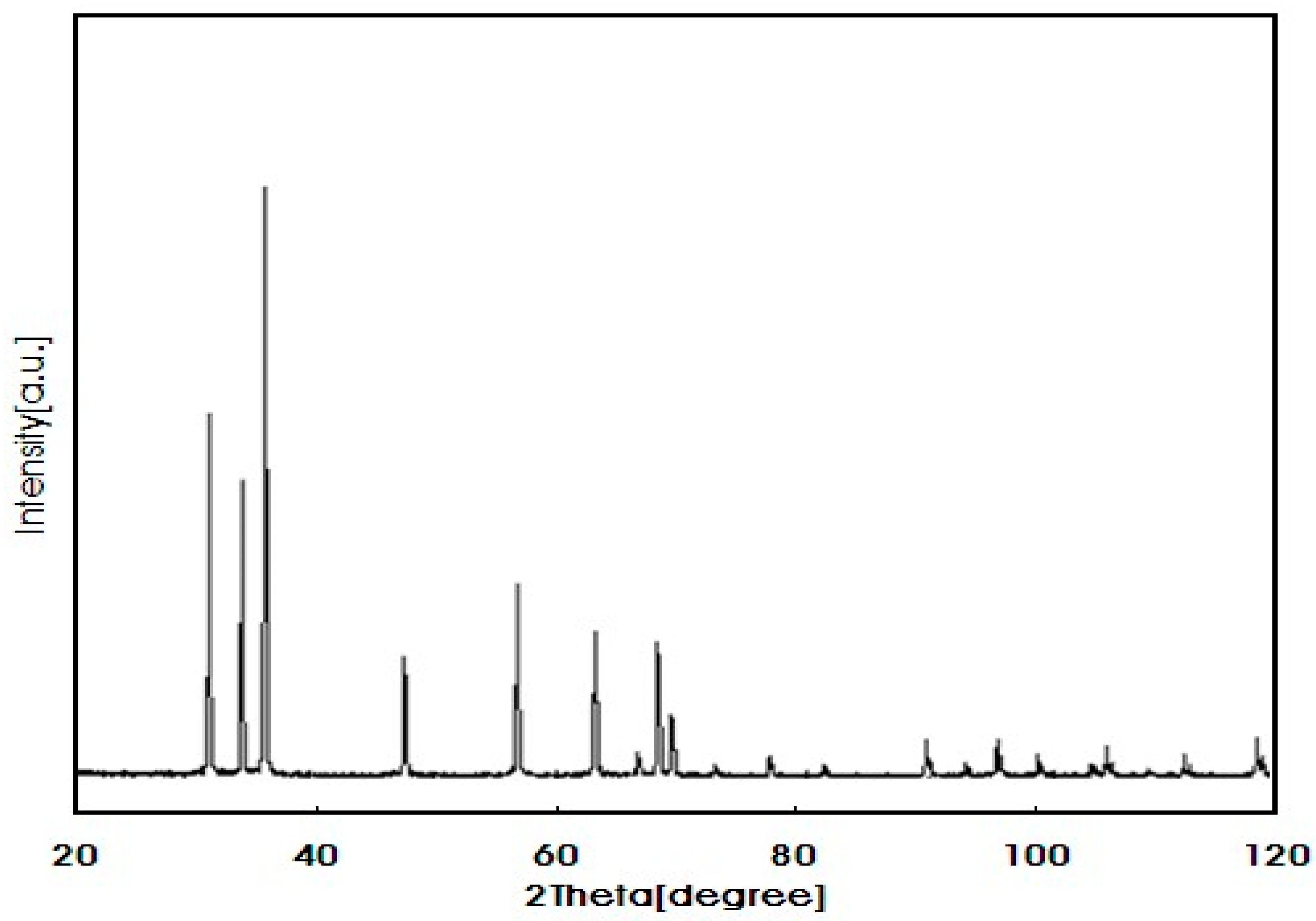
Nano-ZnO was purchased from the Iranian Nano-materials Pioneers Company (Mashhad, Iran). ZnO-nanoparticles solution was prepared using distilled water, followed by ultra-sonication (10 min, 35 KHz, 100/400 W) in an ultrasound bath (Elma, Germany) for 30 min. The prepared solution had the characteristic size of 10–30 nm as confirmed by electron microscopy (Figure 1) and other physicochemical properties described in Figure 2 and Table 1.

2.4. Fish

Mosquito fish (Gambusia holbrooki), with an average weight of 2.7 ± 0.1 g and length of 4.20 ± 0.35 cm were captured from a local waterbody (Zargan, Iran). Followed by the immersion of fish in the NaCl salt solution bath (1%) for 30 s for disinfection, they were finally transferred into aquariums equipped with aerators. Fish were allowed to adapt to laboratory circumstances for ten days (temperature: 23 ± 2 °C, dissolved oxygen: 6 mg L−1, pH: 7.4 ± 0.2, photoperiods: 14 light/10 dark). Water quality was monitored regularly, metabolic waste was siphoned, and 50% of water exchanges were conducted daily to maintain the physicochemical qualities of the water. Fish were fed with a commercial diet (Beyza Feed Mill, Shiraz, Iran) at the rate of 3% of body weight (metabolizable energy: 4 Kcal g−1; crude protein: 43.5%; crude lipid: 20%; crude fiber: 2%).

2.5. Experimental Design

A total of three hundred and sixty fish were allocated to six experimental groups (with two replicates) in twelve separate 80 L aquariums. PE-MPs treatment solutions were prepared by adding a required concentration of MPs in 250 mL of distilled water and 0.1% Tween 20 and ultra-sonicated (10 min, 35 KHz, 100/400 W) for 30 min to prepare a homogeneous suspension. Next, defined concentrations of nano-ZnO were mixed with corresponding suspensions and placed in an ultrasonic bath for 30 min. Both PE-MPs and nano-ZnO suspensions were placed in a bath shaker for 24 h at 200 rpm and 25 °C. All suspensions were maintained in a refrigerator at 4 °C. Furthermore, stock solutions were ultra-sonicated before use to acquire uniform suspensions.
In the experiment, fish (n = 30) were exposed to 100 µg L−1 PE-MPs (group 2), 200 µg L−1 PE-MPs (group 3), 50 µg L−1 ZnO-NPs (group 4), 50 µg L−1 ZnO-NPs combined with 100 µg L−1 PE-MPs (group 5), and 50 µg L−1 ZnO-NPs combined with 200 µg L−1 PE-MPs (group 6), respectively. Control fish (group 1) was maintained in de-chlorinated water. After 14 days of treatment, fish from each treatment were captured and anesthetized with clove powder (100 mg L−1). Then, the fish were autopsied, and their livers were removed and placed in liquid nitrogen. Samples were maintained at a temperature of −26 °C (2–3 days) until before homogenization.

2.6. Bioaccumulation of MPs

The fish’s liver (n = 12) extracted from each MPs exposed experimental group was dried with a freeze dryer and processed to detect the presence of MPs using a Fourier transform infrared spectroscopy (FTIR) apparatus (JASCO, FT/IR-4600) [34]. The basis of the FTIR spectroscopy technique is to measure the chemical composition of materials by measuring how they interact with infrared light.

2.7. Bioaccumulation of Zn

Six fish liver from Zn treatment groups were dried in the oven at 70 °C for 8 h. Next, 1 g of dried samples were mixed with 5 mL H2O2, then were digested with 5 mL nitric acid (0.1 mol L−1) and 5 mL (0.1 mol L−1) perchloric acid to obtain a clear solution. Then, this solution was filtered with the Whatman paper (0.45 μm). Zn concentrations from each sample were measured using inductively coupled plasma optical emission spectrometry (ICP-OES, Optima 7000 DV, PerkinElmer, Waltham, MA, USA). The bio-concentration of Zn was expressed as the metal mass (μg)/dry tissue mass (g).
The bio-concentration factor (BCF) was calculated using the following equations:
B C F = C f i s h C w a t e r
where C f i s h and C w a t e r are the Zn concentrations in the fish body and water (µg g−1 dry weight and µ L−1), respectively.

2.8. Oxidative Biomarkers

From fish (n = 24) 1 g of the liver tissue sample was mixed with a 0.1 M phosphate buffer solution containing 300 mM sucrose and 0.1 mM ethylene diamine tetra-acetic acid with pH 7.8 in a ratio of 1 to 10. Then, samples were homogenized by a homogenizer. Homogenized tissue was centrifuged (15,000 rpm) at a temperature of 4 °C for 15 min.
The activity of superoxide dismutase (SOD), catalase (CAT), glutathione peroxidase (GPx), glutathione reductase (GR), malondialdehyde (MDA), and total antioxidant (TAN) contents were measured using the antioxidant biomarker kit according to the instructions provided by the manufacturer (Kiazist Pishro Barman Co., Hamadan, Iran).

2.9. Evaluation of the Interaction of MPs on the Toxicity of ZnO-NPs

The following mathematical formulas were used to estimate the effect of MPs on the toxicity of ZnO nanoparticles [35]. The synergism rate only occurs when the expected effect is greater than the observed effect:
  • Predicted effect of the endpoints of fish exposed to MPs and ZnO-NPs, alone
Predicted   effect   = MPs Control × ZnONPs Control
2.
The observed effect of the endpoints of fish exposed to MPs and ZnO-NPs, in combination
Observed   effect   = The   combination   of   MPs   and   ZnONPs Control
3.
The synergistic effect
Synergy   ratio = Predicted   effect Observed   effect .

2.10. Data Analysis

After checking the normality of data by the Kolmogorov–Smirnov test, statistical analyses were carried out using GraphPad Prism 8.0.2. software. Data analysis was performed using a two-way analysis of variance (two-way ANOVA) at a 95% confidence level (p < 0.05), and the means were compared with Duncan’s test. The results are presented as mean ± standard deviation (SD).

3. Results

3.1. FTIR Assay

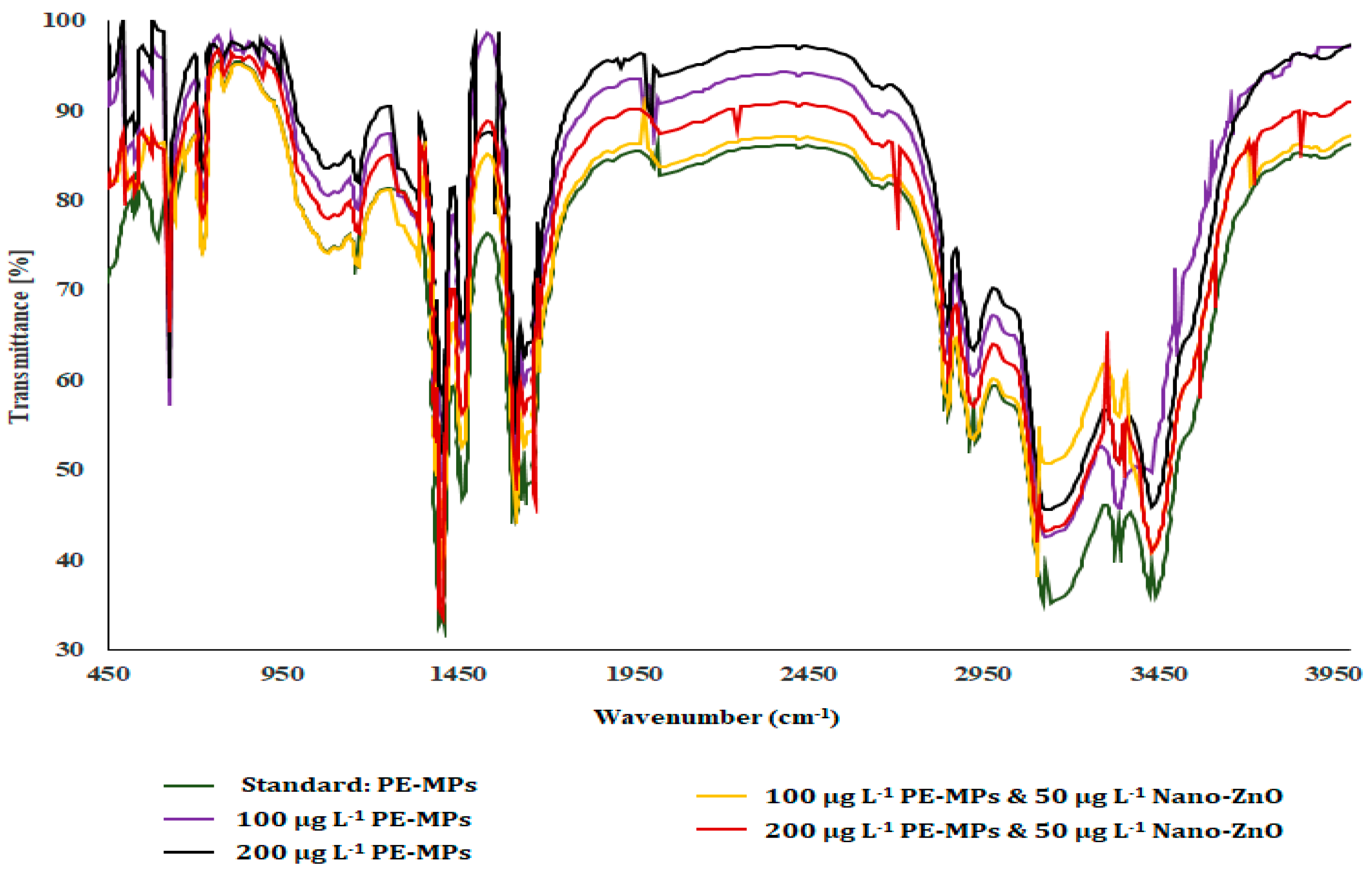
FTIR graphs obtained for different treatment groups revealed the presence of MPs, suggesting MP accumulation in the fish liver compared to the standard graph (Figure 3).
The most prominent peaks in the FTIR spectrum of polyethylene are typically associated with vibrations of the CH2 groups in the polymer backbone. Polyethylene has several characteristic peaks in its FTIR spectrum, including a strong peak at approximately 2916 cm−1 corresponding to CH2 stretching vibrations and a weaker peak at approximately 1460 cm−1 corresponding to CH2 bending vibrations. Other peaks may also be present depending on the specific type of polyethylene and any additives present. Additionally, the low-density polyethylene (LDPE) typically peaks at around 720 cm−1, which is associated with rocking vibrations of the CH2 groups. On the other hand, high-density polyethylene (HDPE) shows a peak at around 1375 cm−1, which is attributed to CH3 deformation vibrations.

3.2. Bioaccumulation of Zn

Data obtained after ICP- OES analysis indicated that the bioaccumulation rate and bioaccumulation factor of Zn for G. holbrooki was increased following co-exposure of PE-MPs and ZnO-NPs, i.e., groups 5 and 6. However, the rate of Zn bioaccumulation remained unaffected by the concentration or amount of microplastic (Table 2).

3.3. Antioxidant Enzyme Assay

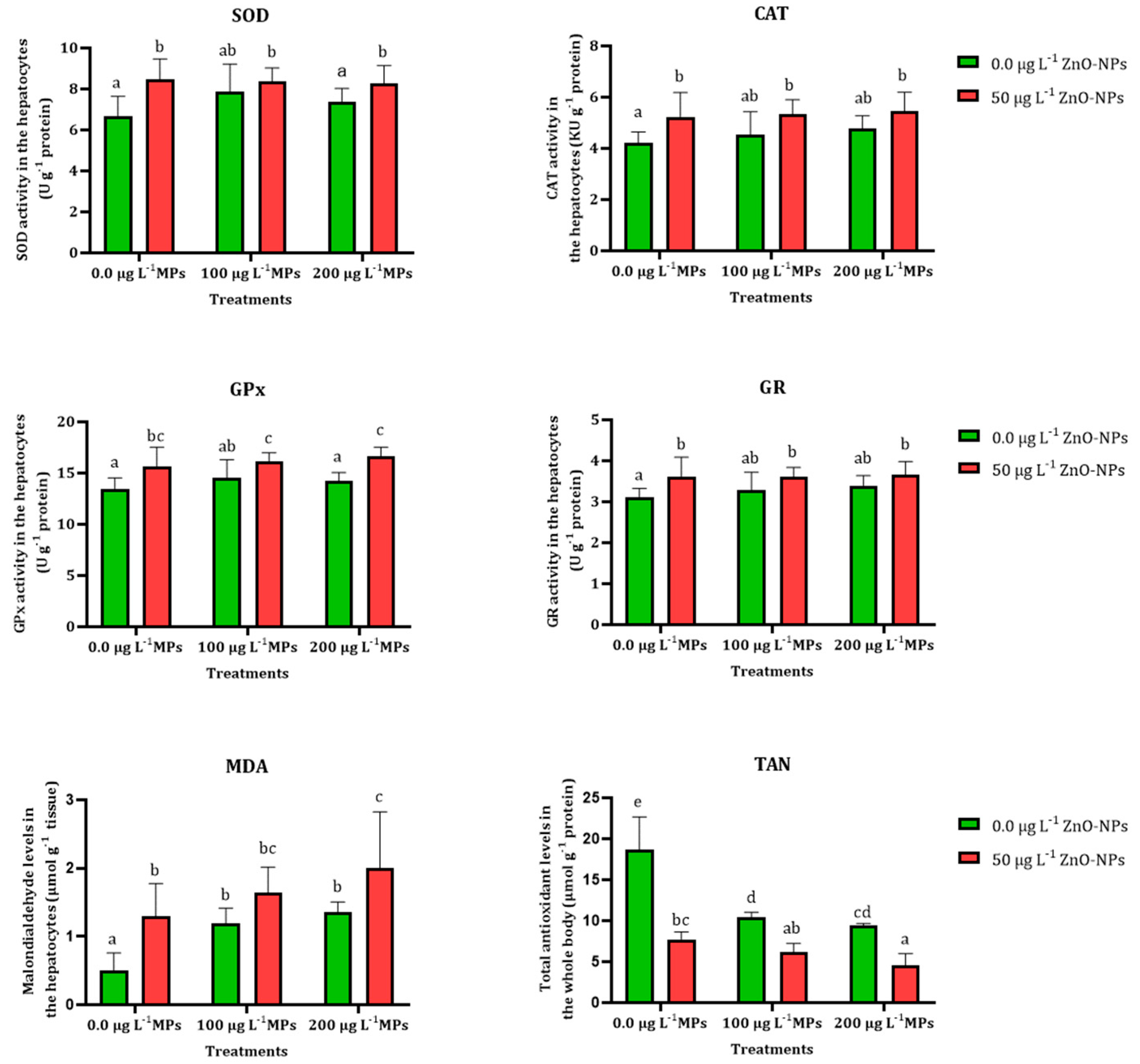
There was no significant difference in SOD activity (U/protein) in the hepatocytes of G. holbrooki exposed to 100 and 200 µg L−1 PE-MPs, and the control group after 14-day exposure, whereas a significant (p < 0.05) increase was observed in the hepatocytes of fish challenged with 50 µg L−1 ZnO-NPs alone and in combination with 100 and 200 µg L−1 PE-MPs (Figure 4).
However, the activity of GPx (U/protein) significantly (p < 0.05) increased in the hepatocytes of G. holbrooki after exposure to 50 µg L−1 ZnO-NPs alone and in combination with 100 and 200 µg L−1 PE-MPs. Lack of significance was determined in the GPx activity in the liver cells of fish after treatment with 100 and 200 µg L−1 PE-MPs compared with the control group after statistical analysis (Figure 4).
Although a significant increase (p < 0.05) was found in the CAT activity (KU/g protein) in the hepatocytes of G. holbrooki following exposure to 50 µg L−1 ZnO-NPs alone and in combination with 100 and 200 µg L−1 PE-MPs, lack of significance was determined by statistical analysis in fish exposed to 100 and 200 µg L−1 PE-MPs compared with the reference group.
In this study, the activity of GR (U/protein) was significantly (p < 0.05) enhanced after exposure to 50 µg L−1 ZnO-NPs alone and in combination with 100 and 200 µg L−1 PE-MPs, while GR activity was not statistically significant (p > 0.05) in the treated fish with 100 and 200 µg L−1 PE-MPs compared with the control group (Figure 4).
Results show elevated MDA contents (µmol g−1 tissue) in the hepatocytes after 14-day exposure to 50 µg L−1 ZnO-NPs, and 100 and 200 µg L−1 PE-MPs, alone and combined, thus exhibiting affected tissue in all exposure groups. There was a significant (p < 0.05) decrease in the TAN levels (µmol g−1 protein) of the treated fish with ZnO-NPs and PE-MPs alone and in combined compared to the control. The lowest values of TAN were found in the fish exposed to 50 µg L−1 ZnO-NPs alone and in combination with 100 and 200 µg L−1 PE-MPs (Figure 4).

3.4. Interaction Study between MPs and ZnO-NPs

Mathematical calculation conducted to determine the impact of interaction of PE-MPs and ZnO-NPs manifested synergistic (additive) impact on the antioxidant enzyme activity (Table 3).

4. Discussion

With the omnipresence of small-sized particles, such as microplastics in everyday products (ingredients of many daily use items, cosmetics, packaging, etc.) and nanoparticles in drugs, the biomedical field, and different formulations, there are raising concerns about their adverse effect on aquatic life. Due to their small size, these can be easily ingested by a wide range of organisms, from zooplankton to larger marine mammals. Thus, it has become crucial to understand how these interact and affect the aquatic biota in their natural environment because these pollutants may directly or indirectly enter the environment and contaminate air, water, and soil.
In an organism, the liver serves as the site of detoxification, and the bioaccumulation of the microplastic was observed in the liver tissues, which corresponds to the reports of the liver as the target organ of MPs toxicity [36]. Following PE-MPs ingestion, these may reach other sites through circulation as evidenced in liver organoids (model) and fish [36,37]. MPs can even cross the intestinal barrier to reach the liver tissue [38]. The accumulation can influence the organism by blocking the digestive passage and physiology of the cell [39,40,41]. Additionally, a significantly enhanced Zn bioaccumulation was observed in the microplastic co-exposed groups compared to the individual Zn group and control group. Concordant studies have reported microplastics as a carrier of heavy metals, as they provide a surface for adsorption and thus act for the co-transportation of the metal [15]. This co-transportation as evidenced in the present study enhances the bioavailability of Zn, which may lead to a range of adverse health effects, including cellular damage and organ dysfunction. Exposure to Zn has been reported to develop histopathological alterations in the liver of the fish [42]. Therefore, increasing MPs in the environment and their ability to transport heavy metals ascertain their potential to enter the food chain.
Present data indicate an altered antioxidant system in accordance with the studies in ragworms and fish on exposure to MPs and nanoparticles [31,43,44]. Exposure to MPs and nanoparticles is reported to be associated with the production of reactive oxygen species (ROS) [45,46,47]. ROS are regularly produced as part of metabolism but are counterbalanced by an effective antioxidant system present in the organism [13,48,49]. If the level of ROS increases in the body, it can lead to conditions such as oxidative stress, inflammation, and cellular damage. The antioxidant system comprises both enzymatic and non-enzymatic regulation. The enzymatic class of the antioxidant system has CAT, SOD, and GPx, while non-enzymatic has major GR, along with vitamin C and proteins. The catalase enzyme acts to effectively scavenge the H2O2, while SOD acts to decompose the superoxide radical. A significant increase in SOD and CAT activities suggests the high activity of these enzymes against the overproduction of ROS.
In the present study, the antioxidant assay revealed no significant differences in the SOD, CAT, GPx, and GR activity in 100 and 200 µg L−1 PE-MPs groups compared with the control group. While significant (p < 0.05) increase was evident upon exposure to nanoparticles (ZnO NPs) alone and in combination with both lower (100 µg L−1) and higher (200 µg L−1) concentrations of PE-MPs. Significant elevation in the activity of SOD, CAT, GPx, and GR suggests an activated defense mechanism. Similar increased levels of SOD activity were also reported on exposure to nanoparticles [31,38,50,51]. In contrast to this, studies have reported a decreased level of SOD and CAT upon exposure to NPs (Al2O3, CuO, and TiO2), which may be associated with the inhibitory effect of ROS [50].
Malondialdehyde (MDA) is a byproduct of lipid peroxidation (LPO). In the process, reactive oxygen species (ROS) attack polyunsaturated fatty acids in cell membranes forming lipid hydroperoxides. These hydroperoxides can then break down into various products, including MDA. Therefore, significantly higher levels of MDA in the tissue indicate damage caused by ROS. The elevated level of MDA was also evident in the white sucker fish exposed to ZnO-NPs at 1 mg L−1 [29]. Similar observations were made by Farkas et al., where they reported a similar increase in LPO level in MPs and Au-NPs while the maximum value was reported in the combination (Au-NPs + MPs) group [45].
Along with regular antioxidant assay comprising of CAT, SOD, and GR, to assess the development of oxidative stress, an alternative approach was used, i.e., estimation of total antioxidant levels [52]. This approach prevents the chances of error and ambiguity in cases determining individual antioxidants and is less time-consuming. This method measures the concentration of total antioxidants using UV/visible spectrophotometrically [53]. The lower value of total antioxidants recorded represents their utilization against the enormous number of reactive radicals produced in the fish in response to both MPs and ZnO-NPs. The study found that ZnO-NPs act as respiratory toxicants in white suckers at environmentally relevant concentrations [29]. They reported induced oxidative stress, malondialdehyde, heat shock proteins (HSPs), and caspases 3/7 activity along with increased membrane permeability in the gill. They also found that high HSP expression levels could increase cell survival chance and resistance to oxidative stress induced damage. Analysis of the synergistic interaction (ZnO-NPs and PE-MPs) on oxidative stress parameters illustrated a synergist effect in all the studied parameters (Table 3). Concordantly, they reported the suppressive effect of the association on the total antioxidant levels. This supported the hypothesis of the development of oxidative stress by the studied pollutants and the negative impact of the association. The outcomes of the synergism study corroborate the findings [29,45].
The present study demonstrates this association to aggravate and raise severe health issues in aquatic forms. So, the release of these particles needs to be regulated for environmental safety. Further, studies are required to understand the toxicity mechanism of the interaction in fish.

Author Contributions

M.B.: supervisor; investigation, project administration, validation, formal analysis, writing—original draft. A.Z.: investigation, project administration. C.F.: original draft, review and editing. R.S.: revised and drafted the manuscript. All authors have read and agreed to the published version of the manuscript.

Funding

This study was supported by a grant from Behbahan Khatam Alanbia University of Technology, Iran (3-2-6771 BKATU).

Institutional Review Board Statement

International, national, and institutional guidelines for the care and use of animals were followed. Experimental protocols were done following the Iranian animal ethics framework under the supervision of the Iranian Society for the Prevention of Cruelty to Animals and Behbahan Khatam Alanbia University of Technology (6-5-107401/01-11-08).

Data Availability Statement

The data that support the findings of this study are available from Behbahan Khatam Alanbia University of Technology but restrictions apply to the availability of these data, which were used under license for the current study, and so are not publicly available. Data are however available from the authors upon reasonable request and with permission of Behbahan Khatam Alanbia University of Technology.

Conflicts of Interest

The authors declare no conflict of interest.

References

  1. IUCN. Marine Plastic Pollution. Available online: https://www.iucn.org/resources/issues-brief/marine-plastic-pollution (accessed on 25 February 2023).
  2. Buwono, N.R.; Risjani, Y.; Soegianto, A. Spatio-Temporal Patterns of Occurrence of Microplastics in the Freshwater Fish Gambusia affinis from the Brantas River, Indonesia. Environ. Pollut. 2022, 311, 119958. [Google Scholar] [CrossRef] [PubMed]
  3. Multisanti, C.R.; Merola, C.; Perugini, M.; Aliko, V.; Faggio, C. Sentinel Species Selection for Monitoring Microplastic Pollution: A Review on One Health Approach. Ecol. Indic. 2022, 145, 109587. [Google Scholar] [CrossRef]
  4. Prokić, M.D.; Gavrilović, B.R.; Radovanović, T.B.; Gavrić, J.P.; Petrović, T.G.; Despotović, S.G.; Faggio, C. Studying Microplastics: Lessons from Evaluated Literature on Animal Model Organisms and Experimental Approaches. J. Hazard. Mater. 2021, 414, 125476. [Google Scholar] [CrossRef] [PubMed]
  5. Schirinzi, G.F.; Pedà, C.; Battaglia, P.; Laface, F.; Galli, M.; Baini, M.; Consoli, P.; Scotti, G.; Esposito, V.; Faggio, C. A New Digestion Approach for the Extraction of Microplastics from Gastrointestinal Tracts (GITs) of the Common Dolphinfish (Coryphaena hippurus) from the Western Mediterranean Sea. J. Hazard. Mater. 2020, 397, 122794. [Google Scholar] [CrossRef]
  6. Manbohi, A.; Mehdinia, A.; Rahnama, R.; Hamzehpour, A.; Dehbandi, R. Sources and Hotspots of Microplastics of the Rivers Ending to the Southern Caspian Sea. Mar. Pollut. Bull. 2023, 188, 114562. [Google Scholar] [CrossRef]
  7. Liu, Y.; Zhang, K.; Xu, S.; Yan, M.; Tao, D.; Chen, L.; Wei, Y.; Wu, C.; Liu, G.; Lam, P.K.S. Heavy Metals in the “Plastisphere” of Marine Microplastics: Adsorption Mechanisms and Composite Risk. Gondwana Res. 2022, 108, 171–180. [Google Scholar] [CrossRef]
  8. Burgos-Aceves, M.A.; Abo-Al-Ela, H.G.; Faggio, C. Physiological and Metabolic Approach of Plastic Additive Effects: Immune Cells Responses. J. Hazard. Mater. 2021, 404, 124114. [Google Scholar] [CrossRef]
  9. Pagano, M.; Vazzana, I.; Gentile, A.; Caracappa, G.; Faggio, C. Hematological and Biochemical Parameters in Sea Turtles (Caretta caretta) after Stranding. Reg. Stud. Mar. Sci. 2019, 32, 100832. [Google Scholar] [CrossRef]
  10. Mallik, A.; Xavier, K.A.M.; Naidu, B.C.; Nayak, B.B. Ecotoxicological and Physiological Risks of Microplastics on Fish and Their Possible Mitigation Measures. Sci. Total Environ. 2021, 779, 146433. [Google Scholar] [CrossRef]
  11. Tsai, S.C.; Tseng, Y.-J.; Wu, S.M. Reproductive and Developmental Alterations on Zebrafish (Danio rerio) Upon Long-Term Exposure of Di-Ethyl Phthalate (DEP), Di-Isononyl Phthalate (DINP) and Di (2-Ethylhexyl) Phthalate (DEHP). Bull. Environ. Contam. Toxicol. 2023, 110, 49. [Google Scholar] [CrossRef]
  12. Yu, Y.; Zheng, T.; Li, H.; Hou, Y.; Dong, C.; Chen, H.; Wang, C.; Xiang, M.; Hu, G.; Dang, Y. Growth Inhibition of Offspring Larvae Caused by the Maternal Transfer Effects of Tetrabromobisphenol A in Zebrafish. Environ. Pollut. 2023, 322, 121143. [Google Scholar] [CrossRef] [PubMed]
  13. Gholamhosseini, A.; Banaee, M.; Sureda, A.; Timar, N.; Zeidi, A.; Faggio, C. Physiological Response of Freshwater Crayfish, Astacus leptodactylus Exposed to Polyethylene Microplastics at Different Temperature. Comp. Biochem. Physiol. Toxicol. Pharmacol. CBP 2023, 267, 109581. [Google Scholar] [CrossRef]
  14. Possatto, F.E.; Barletta, M.; Costa, M.F.; do Sul, J.A.I.; Dantas, D.V. Plastic Debris Ingestion by Marine Catfish: An Unexpected Fisheries Impact. Mar. Pollut. Bull. 2011, 62, 1098–1102. [Google Scholar] [CrossRef]
  15. Hoseini, S.M.; Sinha, R.; Fazel, A.; Khosraviani, K.; Hosseinpour Delavar, F.; Arghideh, M.; Sedaghat, M.; Paolucci, M.; Hoseinifar, S.H.; Van Doan, H. Histopathological Damage and Stress- and Immune-related Genes’ Expression in the Intestine of Common Carp, Cyprinus carpio Exposed to Copper and Polyvinyl Chloride Microparticle. J. Exp. Zool. Part Ecol. Integr. Physiol. 2022, 337, 181–190. [Google Scholar] [CrossRef] [PubMed]
  16. Alimba, C.G.; Faggio, C.; Sivanesan, S.; Ogunkanmi, A.L.; Krishnamurthi, K. Micro(Nano)-Plastics in the Environment and Risk of Carcinogenesis: Insight into Possible Mechanisms. J. Hazard. Mater. 2021, 416, 126143. [Google Scholar] [CrossRef]
  17. Banaee, M. Alkaline Phosphatase Activity as a Biochemical Biomarker in Aqua-Toxicological Studies. Int. J. Aquat. Biol. 2021, 8, 143–147. [Google Scholar]
  18. Banihashemi, E.A.; Soltanian, S.; Gholamhosseini, A.; Banaee, M. Effect of Microplastics on Yersinia ruckeri Infection in Rainbow Trout (Oncorhynchus mykiss). Environ. Sci. Pollut. Res. 2022, 29, 11939–11950. [Google Scholar] [CrossRef]
  19. Lombardo, J.; Solomando, A.; Cohen-Sánchez, A.; Pinya, S.; Tejada, S.; Ferriol, P.; Mateu-Vicens, G.; Box, A.; Faggio, C.; Sureda, A. Effects of Human Activity on Markers of Oxidative Stress in the Intestine of Holothuria tubulosa, with Special Reference to the Presence of Microplastics. Int. J. Mol. Sci. 2022, 23, 9018. [Google Scholar] [CrossRef]
  20. Banaee, M.; Soltanian, S.; Sureda, A.; Gholamhosseini, A.; Haghi, B.N.; Akhlaghi, M.; Derikvandy, A. Evaluation of Single and Combined Effects of Cadmium and Micro-Plastic Particles on Biochemical and Immunological Parameters of Common Carp (Cyprinus carpio). Chemosphere 2019, 236, 124335. [Google Scholar] [CrossRef]
  21. Hodkovicova, N.; Hollerova, A.; Svobodova, Z.; Faldyna, M.; Faggio, C. Effects of Plastic Particles on Aquatic Invertebrates and Fish—A Review. Environ. Toxicol. Pharmacol. 2022, 96, 104013. [Google Scholar] [CrossRef]
  22. Impellitteri, F.; Curpăn, A.-S.; Plăvan, G.; Ciobica, A.; Faggio, C. Hemocytes: A Useful Tool for Assessing the Toxicity of Microplastics, Heavy Metals, and Pesticides on Aquatic Invertebrates. Int. J. Environ. Res. Public. Health 2022, 19, 16830. [Google Scholar] [CrossRef] [PubMed]
  23. Savuca, A.; Nicoara, M.N.; Faggio, C. Comprehensive Review Regarding the Profile of the Microplastic Pollution in the Coastal Area of the Black Sea. Sustainability 2022, 14, 14376. [Google Scholar] [CrossRef]
  24. Iswarya, A.; Vaseeharan, B.; Anjugam, M.; Gobi, N.; Divya, M.; Faggio, C. β-1, 3 Glucan Binding Protein Based Selenium Nanowire Enhances the Immune Status of Cyprinus carpio and Protection against Aeromonas hydrophila Infection. Fish Shellfish. Immunol. 2018, 83, 61–75. [Google Scholar] [CrossRef] [PubMed]
  25. Jeyavani, J.; Sibiya, A.; Sivakamavalli, J.; Divya, M.; Preetham, E.; Vaseeharan, B.; Faggio, C. Phytotherapy and Combined Nanoformulations as a Promising Disease Management in Aquaculture: A Review. Aquac. Int. 2022, 30, 1071–1086. [Google Scholar] [CrossRef]
  26. Paulpandian, P.; Beevi, I.S.; Somanath, B.; Kamatchi, R.K.; Paulraj, B.; Faggio, C. Impact of Camellia sinensis Iron Oxide Nanoparticle on Growth, Hemato-Biochemical and Antioxidant Capacity of Blue Gourami (Trichogaster trichopterus) Fingerlings. Biol. Trace Elem. Res. 2023, 201, 412–424. [Google Scholar] [CrossRef] [PubMed]
  27. Rashidian, G.; Lazado, C.C.; Mahboub, H.H.; Mohammadi-Aloucheh, R.; Prokić, M.D.; Nada, H.S.; Faggio, C. Chemically and Green Synthesized ZnO Nanoparticles Alter Key Immunological Molecules in Common Carp (Cyprinus carpio) Skin Mucus. Int. J. Mol. Sci. 2021, 22, 3270. [Google Scholar] [CrossRef] [PubMed]
  28. Vijayakumar, S.; Vaseeharan, B.; Sudhakaran, R.; Jeyakandan, J.; Ramasamy, P.; Sonawane, A.; Padhi, A.; Velusamy, P.; Anbu, P.; Faggio, C. Bioinspired Zinc Oxide Nanoparticles Using Lycopersicon esculentum for Antimicrobial and Anticancer Applications. J. Clust. Sci. 2019, 30, 1465–1479. [Google Scholar] [CrossRef]
  29. Bessemer, R.A.; Butler, K.M.A.; Tunnah, L.; Callaghan, N.I.; Rundle, A.; Currie, S.; Dieni, C.A.; MacCormack, T.J. Cardiorespiratory Toxicity of Environmentally Relevant Zinc Oxide Nanoparticles in the Freshwater Fish Catostomus commersonii. Nanotoxicology 2015, 9, 861–870. [Google Scholar] [CrossRef]
  30. Khan, G.B.; Akhtar, N.; Khan, M.F.; Ullah, Z.; Tabassum, S.; Tedesse, Z. Toxicological Impact of Zinc Nano Particles on Tilapia Fish (Oreochromis mossambicus). Saudi J. Biol. Sci. 2022, 29, 1221–1226. [Google Scholar] [CrossRef]
  31. Saddick, S.; Afifi, M.; Abu Zinada, O.A. Effect of Zinc Nanoparticles on Oxidative Stress-Related Genes and Antioxidant Enzymes Activity in the Brain of Oreochromis niloticus and Tilapia zillii. Saudi J. Biol. Sci. 2017, 24, 1672–1678. [Google Scholar] [CrossRef]
  32. Saha, S.; Chukwuka, A.V.; Mukherjee, D.; Dhara, K.; Saha, N.C.; Faggio, C. Behavioral and Physiological Toxicity Thresholds of a Freshwater Vertebrate (Heteropneustes fossilis) and Invertebrate (Branchiura sowerbyi), Exposed to Zinc Oxide Nanoparticles (NZnO): A General Unified Threshold Model of Survival (GUTS). Comp. Biochem. Physiol. Part C Toxicol. Pharmacol. 2022, 262, 109450. [Google Scholar] [CrossRef]
  33. Ferreira, P.; Fonte, E.; Soares, M.E.; Carvalho, F.; Guilhermino, L. Effects of Multi-Stressors on Juveniles of the Marine Fish Pomatoschistus Microps: Gold Nanoparticles, Microplastics and Temperature. Aquat. Toxicol. 2016, 170, 89–103. [Google Scholar] [CrossRef] [PubMed]
  34. Zeidi, A.; Rezaei, M.R.; Sayadi, M.H.; Gholamhosseini, A.; Banaee, M. Evaluation of Polyethylene Microplastic Bio-Accumulation in Hepatopancreas, Intestine and Hemolymph of Freshwater Crayfish, Astacus leptodactylus. Int. J. Aquat. Biol. 2022, 10, 273–279. [Google Scholar]
  35. Banaee, M.; Akhlaghi, M.; Soltanian, S.; Sureda, A.; Gholamhosseini, A.; Rakhshaninejad, M. Combined Effects of Exposure to Sub-Lethal Concentration of the Insecticide Chlorpyrifos and the Herbicide Glyphosate on the Biochemical Changes in the Freshwater Crayfish Pontastacus leptodactylus. Ecotoxicol. Lond. Engl. 2020, 29, 1500–1515. [Google Scholar] [CrossRef] [PubMed]
  36. Cheng, W.; Li, X.; Zhou, Y.; Yu, H.; Xie, Y.; Guo, H.; Wang, H.; Li, Y.; Feng, Y.; Wang, Y. Polystyrene Microplastics Induce Hepatotoxicity and Disrupt Lipid Metabolism in the Liver Organoids. Sci. Total Environ. 2022, 806, 150328. [Google Scholar] [CrossRef]
  37. Shen, R.; Yang, K.; Cheng, X.; Guo, C.; Xing, X.; Sun, H.; Liu, D.; Liu, X.; Wang, D. Accumulation of Polystyrene Microplastics Induces Liver Fibrosis by Activating CGAS/STING Pathway. Environ. Pollut. Barking Essex 2022, 300, 118986. [Google Scholar] [CrossRef]
  38. Lu, Y.; Zhang, Y.; Deng, Y.; Jiang, W.; Zhao, Y.; Geng, J.; Ding, L.; Ren, H. Uptake and Accumulation of Polystyrene Microplastics in Zebrafish (Danio rerio) and Toxic Effects in Liver. Environ. Sci. Technol. 2016, 50, 4054–4060. [Google Scholar] [CrossRef]
  39. Azadikhah, D.; Yalsuyi, A.M.; Saha, S.; Saha, N.C.; Faggio, C. Biochemical and Pathophysiological Responses in Capoeta capoeta under Lethal and Sub-Lethal Exposures of Silver Nanoparticles. Water 2023, 15, 585. [Google Scholar] [CrossRef]
  40. Cole, M.; Lindeque, P.; Fileman, E.; Halsband, C.; Goodhead, R.; Moger, J.; Galloway, T.S. Microplastic Ingestion by Zooplankton. Environ. Sci. Technol. 2013, 47, 6646–6655. [Google Scholar] [CrossRef] [PubMed]
  41. Kannan, M.; Bojan, N.; Swaminathan, J.; Zicarelli, G.; Hemalatha, D.; Zhang, Y.; Ramesh, M.; Faggio, C. Nanopesticides in Agricultural Pest Management and Their Environmental Risks: A Review. Int. J. Environ. Sci. Technol. 2023. [Google Scholar] [CrossRef]
  42. Abdel-Warith, A.A.; Younis, E.; Al-Asgah, N.A.; Wahbi, M.O. Effect of Zinc Toxicity on Liver Histology of Nile tilapia, Oreochromis niloticus. Sci. Res. Essays 2011, 6, 3760–3769. [Google Scholar] [CrossRef]
  43. Gomiero, A.; Strafella, P.; Pellini, G.; Salvalaggio, V.; Fabi, G. Comparative Effects of Ingested PVC Micro Particles With and Without Adsorbed Benzo(a)Pyrene vs. Spiked Sediments on the Cellular and Sub Cellular Processes of the Benthic Organism Hediste diversicolor. Front. Mar. Sci. 2018, 5, 99. [Google Scholar] [CrossRef]
  44. Modesto, K.A.; Martinez, C.B.R. Roundup® Causes Oxidative Stress in Liver and Inhibits Acetylcholinesterase in Muscle and Brain of the Fish Prochilodus lineatus. Chemosphere 2010, 78, 294–299. [Google Scholar] [CrossRef]
  45. Farkas, J.; Christian, P.; Urrea, J.A.G.; Roos, N.; Hassellöv, M.; Tollefsen, K.E.; Thomas, K.V. Effects of Silver and Gold Nanoparticles on Rainbow Trout (Oncorhynchus mykiss) Hepatocytes. Aquat. Toxicol. Amst. Neth. 2010, 96, 44–52. [Google Scholar] [CrossRef]
  46. Kim, J.-H.; Yu, Y.-B.; Choi, J.-H. Toxic Effects on Bioaccumulation, Hematological Parameters, Oxidative Stress, Immune Responses and Neurotoxicity in Fish Exposed to Microplastics: A Review. J. Hazard. Mater. 2021, 413, 125423. [Google Scholar] [CrossRef] [PubMed]
  47. Umamaheswari, S.; Priyadarshinee, S.; Kadirvelu, K.; Ramesh, M. Polystyrene Microplastics Induce Apoptosis via ROS-Mediated P53 Signaling Pathway in Zebrafish. Chem. Biol. Interact. 2021, 345, 109550. [Google Scholar] [CrossRef]
  48. Martyniuk, V.; Khoma, V.; Matskiv, T.; Yunko, K.; Gnatyshyna, L.; Stoliar, O.; Faggio, C. Combined Effect of Microplastic, Salinomycin and Heating on Unio tumidus. Environ. Toxicol. Pharmacol. 2023, 98, 104068. [Google Scholar] [CrossRef] [PubMed]
  49. Zicarelli, G.; Romano, C.; Gallo, S.; Valentino, C.; Pepe Bellomo, V.; Leonetti, F.L.; Giglio, G.; Neri, A.; Marsili, L.; Milazzo, C.; et al. Diet and Plastic Ingestion in the Blackmouth Catshark Galeus melastomus, Rafinesque 1810, in Italian Waters. Animals 2023, 13, 1039. [Google Scholar] [CrossRef] [PubMed]
  50. Canli, E.G.; Canli, M. Effects of Aluminum, Copper and Titanium Nanoparticles on the Liver Antioxidant Enzymes of the Nile Fish (Oreochromis niloticus). Energy Rep. 2020, 6, 62–67. [Google Scholar] [CrossRef]
  51. Liu, Z.; Malinowski, C.R.; Sepúlveda, M.S. Emerging Trends in Nanoparticle Toxicity and the Significance of Using Daphnia as a Model Organism. Chemosphere 2022, 291, 132941. [Google Scholar] [CrossRef]
  52. Słowińska, M.; Nynca, J.; Cejko, B.I.; Dietrich, M.A.; Horváth, Á.; Urbányi, B.; Kotrik, L.; Ciereszko, A. Total Antioxidant Capacity of Fish Seminal Plasma. Aquaculture 2013, 400–401, 101–104. [Google Scholar] [CrossRef]
  53. Banaee, M.; Sagvand, S.; Sureda, A.; Amini, M.; Haghi, B.N.; Sopjani, M.; Faggio, C. Evaluation of Single and Combined Effects of Mancozeb and Metalaxyl on the Transcriptional and Biochemical Response of Zebrafish (Danio rerio). Comp. Biochem. Physiol. Toxicol. Pharmacol. CBP 2023, 268, 109597. [Google Scholar] [CrossRef] [PubMed]
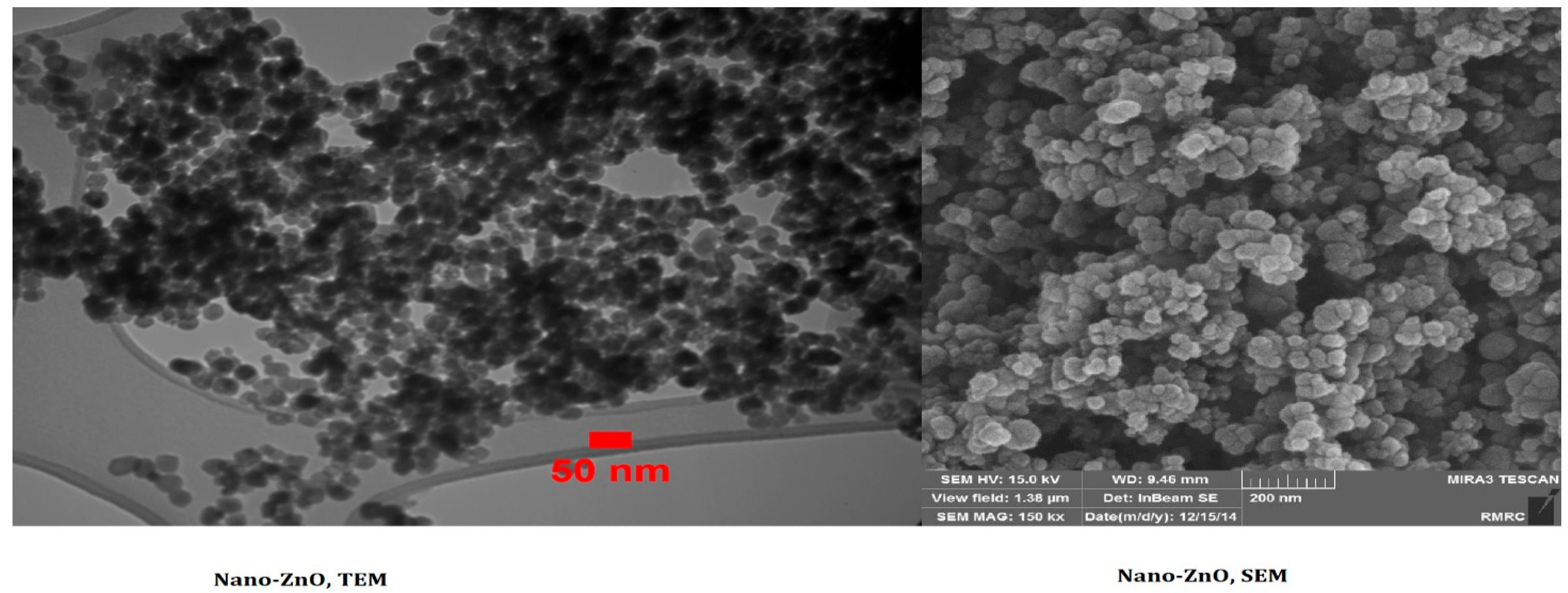
Figure 1. TEM and SEM micrographs of the nano-ZnO powders (adapted from the Iranian Nano-materials Pioneers Company’s catalog).
Water 15 01660 g001
Figure 2. The X-ray powder diffraction (XRD) curves of nano-crystalline ZnO (adapted from the Iranian Nano-materials Pioneers Company’s catalog).
Water 15 01660 g002
Figure 3. Fourier infrared spectroscopic spectrum related to liver samples to detect PE-MPs (n = 12).
Water 15 01660 g003
Figure 4. Effects of PE-MPs and ZnO-NPs on oxidative biomarkers of G. holbrooki (n = 24). Data are presented in mean ± S.D. Different superscript represents significant (p < 0.05) change.
Water 15 01660 g004
Table 1. Physicochemical properties of Zinc oxide nanoparticles, adopted from the Iranian Nano-materials Pioneer’s manufacturer.
Physicochemical PropertiesRange
Purity+99.9%
Average primary particle size (D50)10–30 nm
Specific surface area (SSA)60 m2 g−1
ColorMilky white
Bulk density5.606 g cm−3
Crystal phaseSingle
Crystal morphologyNearly spherical
Table 2. The bioaccumulation of Zn in the body of G. holbrooki co-exposed to PE-MPs and ZnO-NPs (n = 6).
Microplastics (MPs)
0.0 µg L−1100 µg L−1200 µg L−1
ZnO
Nano-Particles
0.0 µg L−150 µg L−10.0 µg L−150 µg L−10.0 µg L−150 µg L−1
Bioaccumulation
(µg g−1 dried tissue)
0.0 ± 0.0 a10.48 ± 0.51 b0.0 ± 0.0 a13.56 ± 1.12 c0.0 ± 0.0 a14.17 ± 0.60 c
Bioaccumulation factor 0.0 ± 0.0 a0.21 ± 0.01 b0.0 ± 0.0 a0.27 ± 0.02 c0.0 ± 0.0 a0.28 ± 0.01 c
Notes: Results are presented in mean ± S.D. Different alphabetical superscript in a row represent significant (p < 0.05) change.
Table 3. Evaluation of synergism of ZnO-NPs and PE-MPs concerning the oxidative biomarkers after 14 days.
Oxidative BiomarkerTreatmentsPredicated EffectObserved EffectSynergy RatioCombined Effect
SOD
(U g−1 protein)
50 µg L−1 ZnO-NPs and 100 µg L−1 PE-MPs1.491.301.15S
50 µg L−1 ZnO-NPs and 200 µg L−1 PE-MPs1.401.241.13S
GPx
(U g−1 protein)
50 µg L−1 ZnO-NPs and 100 µg L−1 PE-MPs1.251.191.04S
50 µg L−1 ZnO-NPs and 200 µg L−1 PE-MPs1.221.230.99A
CAT
(KU g−1 protein)
50 µg L−1 ZnO-NPs and 100 µg L−1 PE-MPs1.331.261.05S
50 µg L−1 ZnO-NPs and 200 µg L−1 PE-MPs1.401.281.09S
GR
(U g−1 protein)
50 µg L−1 ZnO-NPs and 100 µg L−1 PE-MPs1.221.161.05S
50 µg L−1 ZnO-NPs and 200 µg L−1 PE-MPs1.261.151.09S
MDA
(µmol g−1 tissue)
50 µg L−1 ZnO-NPs and 100 µg L−1 PE-MPs6.143.31.86S
50 µg L−1 ZnO-NPs and 200 µg L−1 PE-MPs1.7440.43A
TAN
(µmol g−1 protein)
50 µg L−1 ZnO-NPs and 100 µg L−1 PE-MPs0.230.320.70A
50 µg L−1 ZnO-NPs and 200 µg L−1 PE-MPs0.200.240.85A
Notes: Synergistic effect: S; suppressive effect: A (A < 1 < S); synergistic impact refers to the combined effect of two or more things that is greater than the sum of their individual effects; a suppressive effect on biochemical parameters refers to the ability of a substance or intervention to reduce the levels or activity of specific biomolecules in the body; and oxidative stress is a biological process that occurs when cellular and molecular components of the body are exposed to high levels of reactive oxygen species (ROS) and free radicals.
Disclaimer/Publisher’s Note: The statements, opinions and data contained in all publications are solely those of the individual author(s) and contributor(s) and not of MDPI and/or the editor(s). MDPI and/or the editor(s) disclaim responsibility for any injury to people or property resulting from any ideas, methods, instructions or products referred to in the content.