Article

Natural Bioactive Phytocompounds to Reduce Toxicity in Common Carp Cyprinus carpio: A Challenge to Environmental Risk Assessment of Nanomaterials

1
Department of Biology, Virtual University of Pakistan, Faisalabad 38000, Pakistan
2
Department of Zoology, Government College Women University, Faisalabad 38000, Pakistan
3
Department of Biotechnology, Balochistan University of Information Technology, Engineering and Management Sciences, Quetta 87300, Pakistan
4
Department of Physics, Virtual University of Pakistan, Faisalabad 38000, Pakistan
5
Department of Molecular Biology, Virtual University of Pakistan, Faisalabad 38000, Pakistan
6
Department of Chemical, Biological, Pharmaceutical and Environmental Sciences, University of Messina, 98166 Messina, Italy
*
Authors to whom correspondence should be addressed.
Water 2023, 15(6), 1152; https://doi.org/10.3390/w15061152
Submission received: 15 February 2023 / Revised: 10 March 2023 / Accepted: 13 March 2023 / Published: 16 March 2023
(This article belongs to the Special Issue Climate Impact on Sustainability of Aquatic Organisms)

Abstract

:
Nanomaterials, due to their large aspect-to-size ratio and reactive surfaces that facilitate their access through biological barriers, can induce oxidative stress in host cells. Therefore, there is a growing concern about the biological risks of nanomaterials. This study investigated the biological effects of copper (1.5 mg/L) as CuO or nanoparticles (Cu-NPs) in common carp Cyprinus carpio along with the beneficial effects of Myristiga fragrans seed extract (MFSE) administrated as post-treatment at different doses (4 or 8 or 12 mg/L) for 28 days. The MFSE exhibited a protective role by reducing in a dose-dependent manner the bioaccumulation of Cu level in CuO (from 2.46 to 1.03 µg/Kg in gills; from 2.44 to 1.06 µg/Kg in kidney) and Cu-NPs treated carps (from 2.44 to 1.23 µg/Kg in gills; from 2.47 to 1.09 µg/Kg in kidney) as well as modulating different blood parameters. A mitigation of the histological alterations induced by CuO and Cu-NPs exposure in carp gills (i.e., primary and secondary lamellar degeneration, lamellar fusion, necrosis) and kidneys (i.e., abnormal glomerulus, tubular injury, necrosis) was also observed after MFSE administration. The dietary supplementation of MFSE modulated also the antioxidant defense of carps with respect to the elevated levels of lipid peroxidation (LPO) and glutathione (GST) and the reduced catalase (CAT) induced by CuO and Cu-NPs. Overall, the CuO and Cu-NPs-induced toxicity in C. carpio was mitigated by using MFSE. Further studies are suggested to determine the optimum dose and delivery method of MFSE to guarantee a sustainable conservation of aquatic species.

1. Introduction

Particles with a size less than 100 nm are recognized as nanoparticles (NPs), which may be classified based on their properties, shapes or sizes [1,2,3]. Nowadays, among modern technologies, the nanotechnology is rapidly advancing, and it has a pivotal role in our daily life enabling the improvement of products from many sectors such as drug delivery [4,5], nano-fertilizers (NFs) [6,7], paints [8,9] and electronics [10,11]. Moreover, nanotechnology is employed for nutrient balance in agronomic systems through NFs at a large scale [7].
NFs are nutrient delivery systems with an extraordinary large surface area compared with conventional chemical fertilizers, and they have high sorption capability at specific active sites that increase their absorption rates by plants [12]. However, ultimately, due to their increasing use, NFs have become an important source of environmental and aquatic pollution [6,13]. Micronutrients such as silica [14], zinc [15], copper [16], iron [17], ammonium [18] and potassium [19] have been synthesized in a nanoscale manner and used in the management of plant growth. Alongside this, some nutrients of agronomical importance are transferred to plants with specialized nano-carriers including titanium oxide, silica and chitosan NPs [20], since the nanoparticulate size makes them a significant carrier for bio-agents and enhances penetration into the plant surfaces [21]. In addition, nano-carriers could provide a favorable environment for the bioagents to remain viable and active for a longer time [22].
Nano-copper fertilizers (NCuFs) have a great ability to activate the enzymes involved in the photosynthetic processes of plants, besides being essential also for respiration [23] and the metabolism of carbohydrates and proteins in plants [24]. In addition, NCuFs possess one of the components of plastocyanin, a copper-containing protein [25], which mediates electron transfer and that is found in a variety of plants [26], where it participates in photosynthesis. Apart from all the positive aspects on the use of NCuFs, the distinctive properties of NFs on the other hand adversely affect human and ecosystem health [27,28]. Indeed, NPs have a smaller size than cells and cell components, so they can easily enter in the small biological structures and in turn interrupt their normal structure and function [1,29]. Aquatic systems are the ultimate sinks for all contaminants, including NFs, which pose unknown threats to the aquatic environmental quality [29]. Once accumulated by biota, NFs are able to induce the direct generation of harmful free radical oxygen species (FROS) in living systems. These FROS produce several injuries into the cells, including also DNA damage [30]. Aquatic species are at risk of NFs exposure, and a body of literature is emerging concerning the chemical behavior of NFs in aquatic systems, including their accumulation and toxicity in aquatic species [14]. Overall, about NPs, it is known that they tend to accumulate into cells, especially in macrophages and hepatocytes [29]. Moreover, several toxic effects induced by NPs have been documented in different aquatic organisms, such as phytoplankton [25], sea urchins [31], mollusks [32], and fish [33,34].
The World Health Organization (WHO) encourages the use of medicinal herbs and plants as drugs [35]. Plants have antioxidant properties because of the presence of active phytochemical compounds including vitamins, flavonoids, terpenoids, carotenoids, cumarins, curcumins, lignins, saponins, and plant sterols [36,37,38]. The nutmeg (Myristica fragrans), whose seed is widely used as a spice, is a tropical evergreen tree native of Indonesia [39,40]. The nutmeg has a characteristic pleasant fragrance and a slightly warm taste. It is used to flavor many kinds of baked goods, confections, puddings, meats, sausages, sauces, vegetables, and beverages [41]. It is also used as a component of curry powder, teas and soft drinks, or mixed in milk and alcohol [42]. In Pakistan, M. fragrans is commonly known as “Jaiphal”, and it is used for slight illnesses [35]. M. fragrans is also rich in natural antioxidants such as phenolics, saponins, flavonoids and tannins, which act as antioxidants [43,44]. Indeed, numerous epidemiological studies have showed the ability of nutmeg to reduce oxidative stress-related diseases [45]. Overall, M. fragrans possesses strong antioxidant and anti-inflammatory activities, acts as a good preservative agent and offers benefits in some medical treatments [40], acting as a soothing [46], for muscle pain [47] and cardiovascular diseases [48], having antithrombic [49] and anti-aging properties [50].
Therefore, this study was designed to evaluate the potential of natural bioactive phytocompounds extracted from the nutmeg in mitigating the impact of nanomaterials on aquatic organisms. In detail, different dosages of M. fragrans seed extract (MFSE) were administrated as post-treatment to common carp Cyprinus carpio waterborne exposed to copper in its bulk counterpart (copper II oxide, CuO) and as nanoparticle (Cu-NPs) in order to elucidate the ameliorative effects of nutmeg on the health status of fish, as assessed by tissue metal accumulation, investigation of the hematological profile, histology of fish gills and kidney, and analysis of oxidative stress enzymes.

2. Materials and Methods

2.1. Procurement and Acclimatization of Fish

Common carp C. carpio of similar weight (35–40 g) were obtained from the Department of Fisheries Seed Hatchery Faisalabad, in Punjab, Pakistan. Fish were then transferred to the research laboratory of the Department of Zoology, Government College Women University in Faisalabad (GCUF), Pakistan, and were acclimatized for two weeks in a 100 L tank prior to the experiment. During the acclimatization period, a normal photoperiod (12 h light:12 h dark) was maintained, and carps were fed twice daily using commercial fish feed (Super Nova, SKU103892996_PK-1250174609). The water was changed regularly daily, and dead fish were removed. About water quality parameters, pH, temperature, dissolved oxygen, ammonia, total hardness and total dissolved solids were monitored and maintained constant by using a multiparameter apparatus (HI 9828, Hanna Instruments, Keison Co., Chelmsford, UK) throughout the entire experiment, which consisted of semi-static exposures. The parameters of water quality are reported in Table 1.
Prior to run the experiments, risk assessment was conducted including general risks e.g., availability of water, proper cleanliness of fish aquaria to minimize the deposition of CuO and Cu-NPs, use of electric air pumps and the risks associated with the handling of Cu-NPs.

2.2. Characterization of CuO and Cu-NPs

All the chemicals used in this study were of high-quality analytical and molecular grade. The CuO (copper II oxide, <10 µm; CAS No. 1317-38-D) and Cu-NPs (60–80 nm; CAS No. 7440-50-8; 99% pure) were acquired as a powder from Sigma-Aldrich. The Cu-NPs were characterized by using an X-ray diffraction (XRD), which is a rapid analytical technique primarily used for the phase identification of a crystalline material and to provide information on unit cell dimensions. The XRD pattern for Cu-NPs was recorded by a Bruker D8 Advance X-ray diffractometer, using Cu-Kα radiation of wavelength λ = 1.5406 A° in the scan range 2θ = 10°–80° degrees with a step of 0.02 degrees and the generator settings of 30 mA and 40 kV. The indexing process of the powder diffraction pattern was performed, and Miller Indices (hkl) were assigned to each peak at the first step. The diffractogram of the entire data is shown in Figure 1. Three main characteristic diffraction peaks for Cu were observed at around 2θ = 42.292°, 49.859° and 73.644°, which correspond to the (999), (450) and (220) crystallographic planes of the face-centered cubic (FCC) Cu phase.
Moreover, considering the three characteristic diffraction peaks for Cu, the average particle size of NPs has been estimated by using the Debye–Scherrer formula:
D = 0.89λ/(β∗cosθ)
where
  • D = particle diameter (average crystallite size);
  • Β = Full Width at Half Maximum (FWHM);
  • θ = Bragg angle;
  • λ = X-ray wavelength, Cu-Kα emission (λ = 1.54056 A°).
As reported in Table 2, the average crystallite size of Cu-NPs was in the range of 78.33 nm.
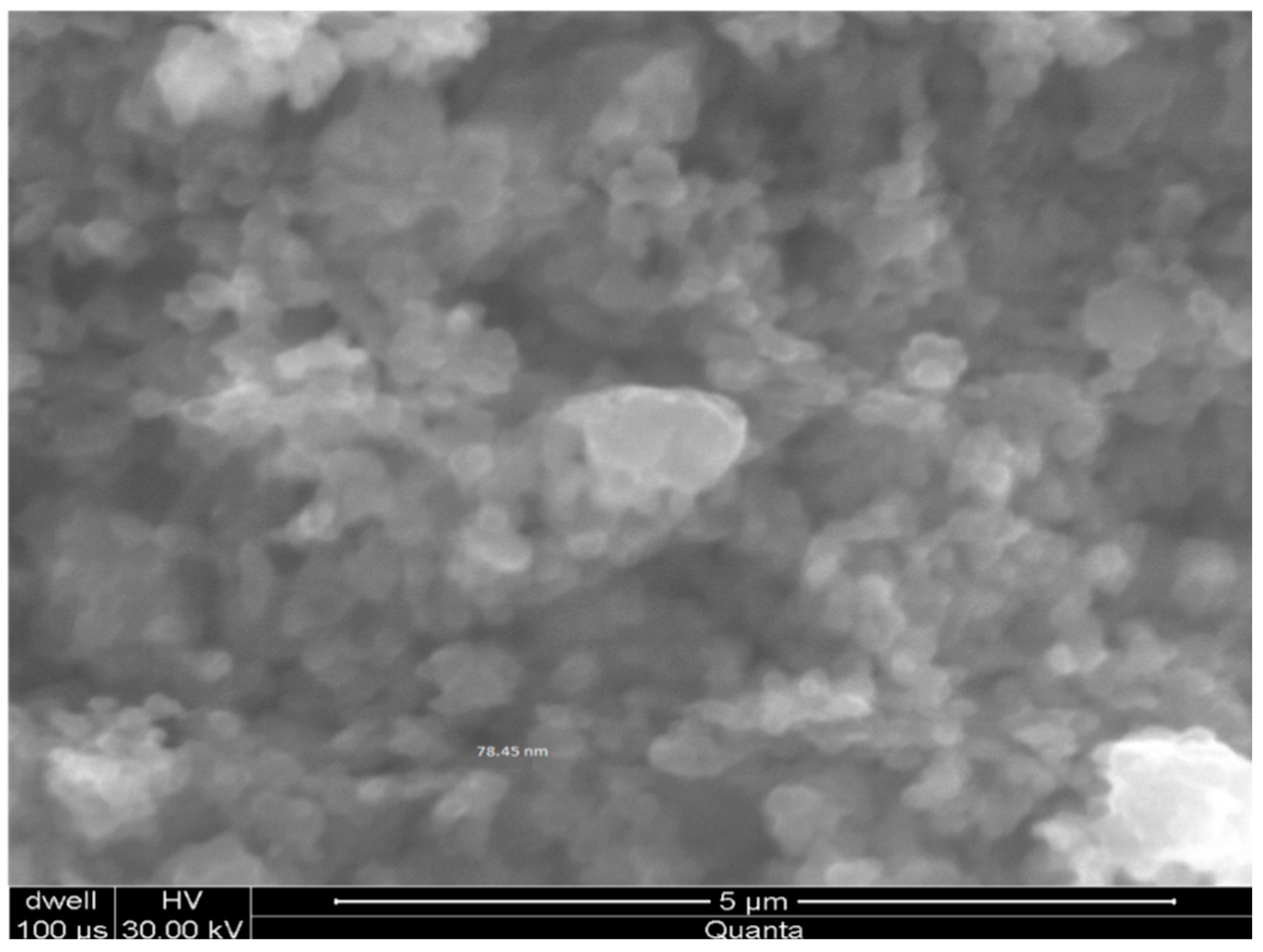
Additionally, the scanning electron microscopy (SEM) was also used to estimate the homogeneous dispersion of Cu-NPs particle size. The sample of Cu-NPs was imaged by an FEI Helios Dual-Beam SEM-based measuring system, which was equipped with a high-performance electron beam column and sample stage. The best spatial resolution of the system is 1 nm at optimum settings at 15 kV accelerating voltage. For this study, the best results were obtained with 86 pA beam current and 30 μs beam dwell-time for each image pixel, and with the sample at 3.5 mm working distance. The samples were imaged at 20, 000× and 30,000× magnification at a 1.2 μm field of view, which provides a good balance between high spatial details and particle density. The results revealed the uniform dispersion of Cu-NPs with cubical morphology. The dispersion of particles was homogeneous, and the average particle size was in the range of 65–90 nm, as shown in Figure 2.
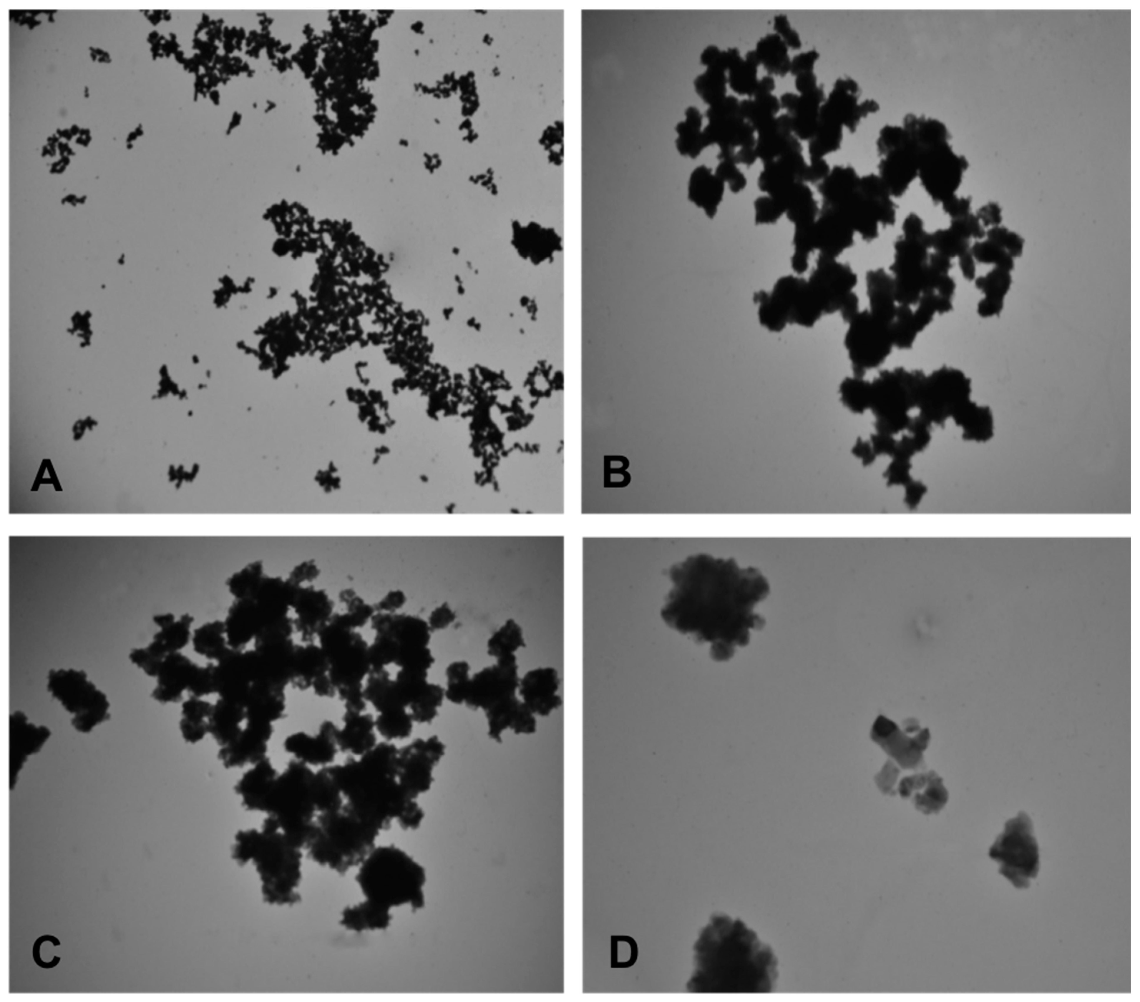
Moreover, the transmission electron microscopy (TEM) performed by using the Electron Microscope, model JEM-1010 (JEOL, Tokyo, Japan) revealed the individual particle size of Cu-NPs of 70–90 nm having irregular morphology, with more particles having the cubical crystalline form and some particles observed in clumps (Figure 3). These characterization results were also presented in our previous papers [34,51].
For preparation of the stock solutions, the required amounts of CuO and Cu-NPs were separately weighed by using a laboratory weight balance (Model HC series), placed into polypropylene tubes (Pyrex) and dispersed in deionized/ultra-pure water (Millipore, 18.2 Mcm resistance, ThermoFisher Scientific, Waltham, MA, USA) without any solvent. The suspension was then vortexed (Vortex Genie-2T, ThermoFisher Scientific, Waltham, MA, USA) for 3–4 h at 2200 rpm in order to obtain the maximum dispersion, and then, it was ultrasonicated (Jp-031) for 2 h prior to the immediate transfer into glass aquaria to run the experiments.

2.3. Preparation of the Nutmeg Extract

Seeds of M. fragrans were procured from authenticated stores and were ground, sieved, and stored in an air-tight jar for extract preparation. The preparation of the plant extract was performed according to the standard method of Morsy et al. [52] and slightly modified. Briefly, after collection of the powdered plant material, it was mixed with methanol in a ratio of 1:1, kept for one week at room temperature, and then filtered by Whatman filter paper No. 1. The filtrate was then dried, weighed, and stored for successive analysis. The determination of the biochemical composition of MFSE was performed in accordance with the Association of Official Analytical Chemists (AOAC) [53]. In addition, for exploration of the secondary metabolites, standard protocols were applied for the total soluble phenolics, tannins, flavonoids [54], alkaloids [55] and saponins [56].

2.4. LC50 of MFSE for Common Carp

The 96 h median lethal concentration (LC50) value of MFSE for C. carpio was evaluated as follows. Firstly, fish were exposed (3 replicates) to different doses (8 or 16 or 32 or 64 or 128 mg/L) of MFSE separately. The trial was conducted in glass aquaria of 40 L water capacity, with maintenance of the same physico-chemical parameters applied during the period of acclimatization (see Table 1). During the 96 h time period, the test water was not changed, and no feed was administrated to the exposed fish. Dead fish were removed on a daily basis in the early morning and late evening. The 96 h LC50 was calculated by the use of Probit Analysis (Minitab®17.1.0 software, ©2013 Minitab Inc., State College, PA, USA).

2.5. Experimental Design

A total of 135 common carps of similar weight were distributed into 9 treatment groups (n = 15), including two positive control groups (CuO and Cu-NPs) treated, respectively, with a sub-acute selected dose (1.5 mg/L) of CuO or Cu-NPs, three treated groups (CuO1, CuO2, and CuO3) reared with three different dosages of MFSE (4 or 8 or 12 mg/L, respectively) along with the selected dose of CuO through waterborne exposure, three other treated groups (Cu-NP1, Cu-NP2, and Cu-NP3) reared with three different dosages of MFSE (4 or 8 or 12 mg/L, respectively) along with the selected dose of Cu-NPs through waterborne exposure, and one negative control group (C or control) with fish exposed to no treatment with free access to food. The selected dose of 1.5 mg/L of CuO and Cu-NPs was selected in accordance with our previous studies by Noureen et al. [34,51], which were conducted in order to establish the 96 h LC50 of Cu-NPs in C. carpio. The experimental exposures were conducted for 28 days in triplicates. The water physico-chemical parameters such as temperature, pH, total dissolved solids, and oxygen were determined by using a multiparameter apparatus (HI 9828, Hanna Instruments) and maintained in the same conditions as used during the acclimatization period (see Table 1).
The fish were anesthetized with clove oil in bucket [51]. Blood samples were collected from the caudal vein by the use of a sterile syringe to be then transferred in EDTA microtubes [34] in order to conduct the hematological analysis. Fish tissues, including gills and kidneys, were excised and stored at −20 °C until the determination of metal accumulation and oxidative stress enzymes. Additionally, small pieces of carp gills and kidneys were collected and immediately fixed in a Bouin solution to conduct the histological analysis.
All the experiments conducted on carps herein described were approved by the Animal Ethics Committee, GCUF, Pakistan and carried out according to the Animal Ethics Committee’s guidelines on the use of animals for research purposes, using the minimum possible number of fish.

2.6. Biochemical Analysis

2.6.1. Tissue Metal Analysis

For tissue metal analysis, fish gills and kidney sub-samples (1 g) were transferred to a flask and mixed with nitric acid (HNO3) (10 mL) and perchloric acid (HClO) (2 mL) for sample digestion. The mixture was then heated on a hot plate (6796-620D) by the use of a fuming hood at 100 °C until the color disappeared. The digested samples were then cooled and diluted with deionized water (50 mL) and filtered with Whatman filter paper No. 1 [57]. The absorbance was detected by an atomic absorption spectrometer (AI 1200, Aurora Instruments LTD, ThermoFisher Scientific, Waltham, MA, USA) and expressed as g/kg wet weight (w.w.).

2.6.2. Hematological Analysis

Evaluation of hemoglobin (Hb), hematocrit (Hct), red blood cells (RBC), white blood cells (WBC), mean cell volume (MCV) and platelets (PLT) was performed by a hematology automated analyzer (Beckman Coulter UniCel DxH, ThermoFisher Scientific, Waltham, MA, USA), following the manufacturer’s guidelines.

2.6.3. Histological Analysis

Fish tissues, previously fixed, were further processed using standard protocols mentioned previously [58] and stained with hematoxylin and eosin (H&E staining). Observations of histological slides were then conducted on five fields of one section per sample at 200× magnification by the use of a light microscope (MEIJI, Model: MT4300H, Saitama, Japan), which was equipped with a Canon digital camera (EOS 1300D) for the acquisition of images.

2.6.4. Analysis of Oxidative Stress Enzymes

For the analysis of oxidative stress enzymes (OSEs), fish tissues (gills and kidney) were firstly processed by a Potter–Elvejham homogenizer (ThermoFisher Scientific, Waltham, MA, USA). The obtained homogenates were centrifuged (Z32-HK) at 10,000 rpm for 10 min, and the supernatants were collected and kept at −20 °C until the analysis of OSEs [58], namely lipid peroxidation (LPO) [59], glutathione (GSH) [60], and catalase (CAT) [34]. In order to evaluate LPO, malondialdehyde (MDA; nmol/mg protein) was measured by using the thiobarbituric acid reactive (TBARS) method [59]. The level of reduced GSH (µM/g) was assessed by the colorimetric Ellman’s method, treating the tissue homogenate with trichloroacetic acid (TCA) 10%, and adding 0.2 M Tris EDTA buffer to prevent GSH oxidation [60]. Furthermore, CAT enzymatic activity (mol/min/mg protein) was measured by the dismutation of H2O2 at 240 nm for 90 s using a colorimetric technique [34].

2.7. Statistical Analysis

Data were expressed as mean ± standard error (SE). By the use of Minitab17 software, data were analyzed for comparative purposes by application of one-way ANOVA and the Tukey’s post-hoc test. In addition, the Principal Component Analysis (PCA), an unsupervised statistical technique, was applied by the use of MATLAB to assess the influence of CuO, Cu-NPs and the different dosages of MFSE on the different biomarker parameters tested on carps. Data were considered statistically significant at p < 0.05.

3. Results

3.1. LC50, Phytochemical and Proximate Composition of MFSE

The 96 h LC50 value of the nutmeg M. fragrans for C. carpio was 31.64 ± 4.77 mg/L. This was baseline information that was helpful to establish the appropriate dosage of MFSE extracts to be used against CuO or Cu-NPs-induced toxicity in fish.
The analysis of bioactive phytocompounds present in MFSE revealed a higher percentage of alkaloids (365.6 ± 10.15 mg/100 g of MFSE) and tannins (2.53 ± 0.02 mg/100 g of MFSE), as clearly reported in Table 3. The flavonoids, total phenolics and saponins were also reported in significant percentages (Table 3).
Similarly, in regard to the proximate composition of MFSE, it was found that it is characterized by a significant presence of macro and micro components of various biochemical composition in the following order: carbohydrates > oil contents > fiber > moisture > proteins > ash, as reported in Table 3. Additionally, the results revealed also that MFSE exhibited significant percentages of a variety of minerals including K (432.7 ± 10.20), Mg (40.34 ± 1.03), Ca (79.78 ± 4.02), Zn (2.99 ± 0.02), Mn (0.7 ± 0.20), Cu (1.27 ± 0.21), Fe (26.70 ± 1.50) and Na (96.32 ± 3.02).

3.2. Bioaccumulation of Cu in Fish Tissues

After 28-day exposure, it was observed that the administration of MFSE significantly (p < 0.05) decreased the concentration of Cu accumulated in carp tissues in all the different treatment groups with respect to the positive control groups (CuO and Cu-NPs), as reported in Table 4. Moreover, not significant (p > 0.05) differences were observed among the different treated groups of MSFE along with CuO and Cu-NPs (Table 3). Moreover, the level of ion concentration of Cu (µg/Kg w.w.) in fish tissues (gills and kidney) was found to be not significantly different among the CuO and Cu-NPs treatment, while there were significant (p < 0.05) differences between and within each treatment group in respect to the MFSE dosage applied.

3.3. Hematological Assessment

Different blood parameters, including Hb, Hct, RBC, WBC, MCV and PLT, were measured in common carps from all the different experimental groups, as reported in Table 5. In respect to the normal hematological profile observed in fish from the negative control group (C), a significant (p < 0.05) reduction in almost all the hematological parameters was noticed in the positive control groups because of the toxicity induced by CuO or Cu-NPs, except for WBC and PLT that were instead significantly (p < 0.05) increased in respect to C. It is noteworthy that the administration of MSFE induced a mitigative effect in the common carps against the toxicity induced by CuO or Cu-NPs, since the levels recorded for all the blood parameters tended to be comparable with those measured in fish from the negative control group (Table 4).

3.4. Histological Assessment

The gills of common carps from all the experimental groups are shown in Figure 4. The histological structure of the gills of carps from the group C presented a regular morphology of the branchial tissue, as shown in Figure 4a. Contrarily, severe histological alterations including secondary lamellar degeneration (sld), primary lamellar degeneration (pln), lamellar fusion (lf), and necrosis (n) were noticed in the gills of carps from the positive control groups after exposure to 1.5 mg/L of CuO (Figure 4b) or Cu-NPs (Figure 4c). The same alterations, but at a moderate extent, were also observed in the gills of carps treated with CuO or Cu-NPs along with different dosages of MFSE (Figure 4d–i), which showed a mitigative effects toward the toxicity of CuO and Cu-NPs.
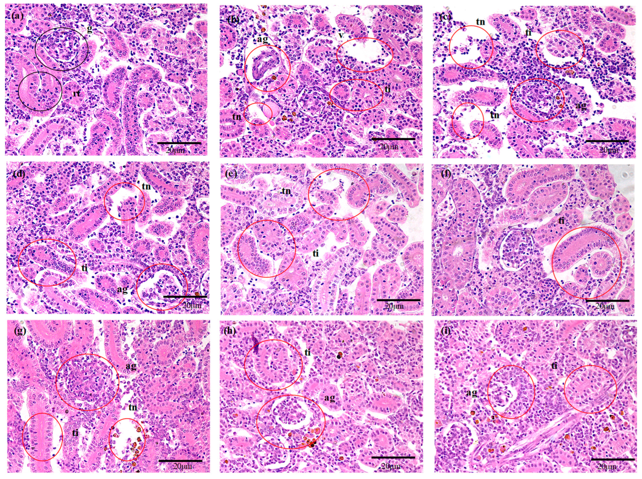
The kidney of common carps from all the experimental groups are shown in Figure 5. The histological structure of the kidney of carps from the group C presented a regular morphology of the tissue, as shown in Figure 5a. Contrarily, histological abrasions including the presence of abnormal glomerulus (ag), tubular injury (ti) and tubular necrosis (tn) were noticed in the kidney of carps from the positive control groups after exposure to 1.5 mg/L of CuO (Figure 5b) or Cu-NPs (Figure 5c) as well as moderately in the kidney of carps treated with CuO or Cu-NPs along with different dosages of MFSE (Figure 5d–i), which partly suppressed the histological abrasions in a dose-dependent manner after 28 days of exposure.

3.5. Oxidative Stress Enzymes (OSEs)

Table 6 presents the concentration of OSEs (namely LPO, GSH and CAT) measured in the kidney and gills of the common carp C. carpio from the different treatment groups. It was found that the levels of LPO and GSH in carps exposed to CuO or Cu-NPs increased significantly (p < 0.05) with respect to the negative control group, whereas a significant (p < 0.05) decline was noticed in the activity of CAT. Interestingly, the administration of MSFE was able to reduce in a dose-dependent manner the values of both LPO and GSH in respect to those measured after exposure to CuO or Cu-NPs, and it concomitantly enhanced the level of CAT, whose values were similar to those recorded in common carps from the negative control group (Table 6).

3.6. Principal Component Analysis (PCA)

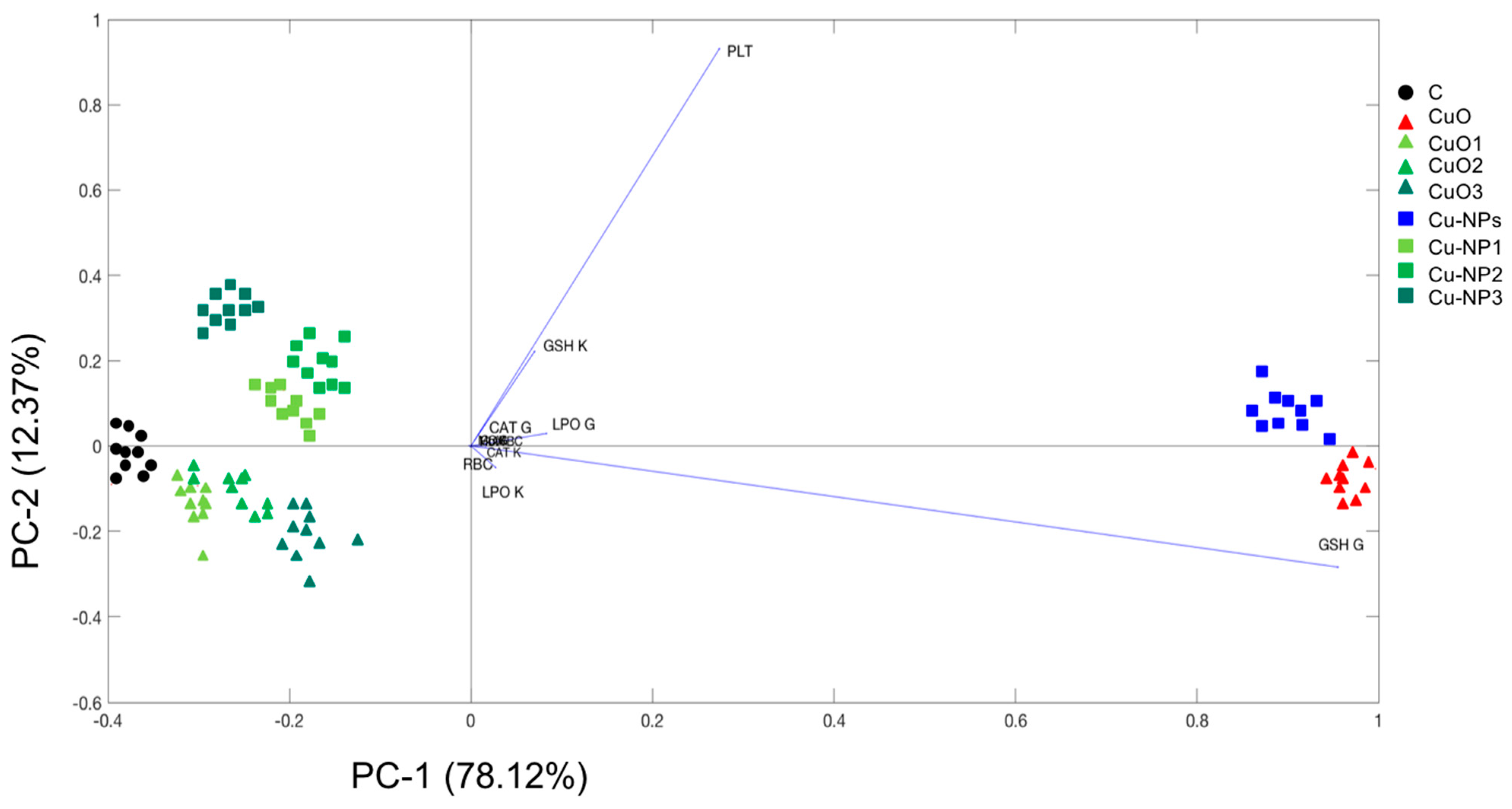
The results obtained from the PCA using multiple biomarkers, such as data of Cu bioaccumulation in gills and kidney, blood parameters such as Hb, Hct, RBC, WBC, MCV and PLT, and oxidative enzymes such as LPO, GSH and CAT in gills and kidney, is depicted in Figure 6. It was found that the CuO and Cu-NPs groups were close in the score plot and clearly separated from the control and MFSE treatment groups, which were separated from each other but almost overlapped. In detail, the first axis (78.12%) of PCA was influenced by almost all the tested biomarkers, which overlapped except for the level of GSH measured in carp gills and the amount of PLT.

4. Discussion

Nanotechnology has significant and promising implications in modern society, even if recently concerns have been raised about its impact on human and ecosystem health [1,2,3]. The NPs released in the environment are able to accumulate into biota and, due to their physico-chemical properties, induce the generation of FROS, which produce several injuries into the cells that may result in the progression of numerous diseases [20]. Usually, the organism reacts to ROS through stimulating their intracellular antioxidant mechanisms [40], resulting in an imbalance between ROS and antioxidant defense system-induced cell damage [17]. In the aquatic ecosystems, the main sources of CuO and Cu-NPs are mostly geological deposits, volcanic activity, and the weathering and erosion of rocks and soils, but there are also anthropogenic sources, including mining activities, agriculture, metal, electrical manufacturing, sludge and pesticides [61]. In addition, the antifouling paints that are used for coatings on ship bodies are one of the major sources of nano copper in the marine environment [62]. The distinctive physico-chemical properties of Cu-NPs may enhance their toxicological behavior in vivo by aiding in the cellular uptake and translocation of the particles in the body [24], where they trigger the production of ROS.
Recently, the researchers are focusing on discovering plant antioxidants as protecting agents against toxins [38], which may be represented by the various active compounds generally present in plants, such as alkaloids, flavanoids, pigments, phenolics, terpenoids, steroids and essential oils [41]. The current research highlighted the ameliorative effects of M. fragrans against CuO and Cu-NPs induced toxicity in C. carpio. Indeed, it is evident from the present study that MFSE exerted significant antioxidant potential against the impact provoked by CuO and Cu-NPs exposure in fish, as it was confirmed by metal accumulation, hematological, histological and oxidative stress assessment. The antioxidant potential of nutmeg was reported in many previous studies. For instance, Olaleye et al. [63] documented the antioxidant properties of M. fragrans in rats by investigating the histology of different organs. However, it was also found that a prolonged administration of M. fragrans may cause a variety of tissue abnormalities. Bamidele et al. [64] reported that M. fragrans improved the hematological indices in albino rats. Nasreen et al. [65] documented also the ameliorative potential of the nutmeg on oxidative stress produced by diabetes mellitus in mice. Therefore, all these findings are in agreement with the results obtained in the present study, in which M. fragrans effectively mitigated the CuO and Cu-NPs toxicity in exposed fish.
The 96 h LC50 value of the M. fragrans for the common carp C. carpio was herein determined in order to establish the appropriate doses of MFSE to be administrated in fish to mitigate the CuO and Cu-NPs-induced toxicity. Interestingly, in the current investigation, the chemical composition of M. fragrans showed significant amount of bioactive compounds, especially alkaloids and tannins, and high levels of nutrients and minerals. Similar findings were previously reported by Olaleye et al. [63], Li et al. [43], Anaduaka et al. [66] and Rancy et al. [56]. However, as herein reported, it is particularly worthwhile to note that nutmeg contains significant amounts of K, Ca, Na and Mn. Indeed, the presence of these minerals in such a high percentage was not documented before for M. fragrans. Moreover, the chemical analysis conducted in the current study revealed that the nutmeg is characterized by the presence of proteins, fats, total carbohydrates, minerals, saponins, flavonoids, alkaloids, tannins and total phenolics, which make the nutmeg suitable for fish to enhance their growth. This was specifically demonstrated by Bhavan et al. [67], who reported the effects of M. fragrans, Glycyrrhiza glabra and Quercus infectoria on the growth performance of the prawn Macrobrachium rosenbergi.
Among the beneficial effects of using nutmeg in fish, it is relevant to mention that the administration of MFSE reduced significantly in a dose-dependent manner the tissue concentration of Cu among the different treatment groups with respect to the positive control groups (CuO and Cu-NPs). Recently, natural coagulants including M. fragrans were used for the purification of heavy metals-contaminated water [68,69]. To date, the research studies regarding the treatment of heavy metals are restricted mainly to some chelating agents to be used alone or in combination with few antioxidants, even if most of the conventional metal chelating agents and antioxidants have been reported to possess toxic side effects such as headache, nausea, hypertension, hepatotoxicity, and nephrotoxicity [70]. Generally, the chelating agents accomplish their binding with metal ions forming complex structures, which might be toxic for organisms and therefore are excreted from the body [71]. As demonstrated in the current study, the MFSE possesses a number of molecules acting as antioxidants (i.e., total phenolics, alkaloids and tannins) that can contribute to reducing the level of metals accumulated in fish, as herein observed in the experimental common carps exposed to CuO and Cu-NPs.
A valuable tool to evaluate the health status of fish and monitor potential responses of stress is represented by investigation of the hematological profile [72,73]. Interestingly, further significant protective effects exercised by MFSE were revealed by analyzing the hematological profile of the experimental common carps. Indeed, it was noticed that MFSE was equally effective in mitigating the toxicity induced in fish by exposures to both CuO and Cu-NPs, by increasing the levels of Hb, Hct, RBC and MCV, as well as reducing the values of WBC and PLT count to levels comparable with those of fish from the negative control group. Many previous studies also reported the beneficial effects of MFSE on hematological parameters. Rashidain et al. [74] reported that M. fragrans dietary supplementation exhibited protective effects on the growth and hematological indices of common carps against Aeromonas hydrophila. In addition, Bachri et al. [75] documented that the sub-chronic administration of MFSE induced protective effects on the hematological parameters in rats, similarly to what was documented in albino rats by Bamidele et al. [62], who found that extracts of M. fragrans stimulated hemopoiesis at high doses.
In addition, histopathological alterations are commonly used in biomonitoring programs as a biomarker of the stress induced by different pollutants in fish [58]. Indeed, based on the histological evaluation performed in the present study, the damage provoked by CuO and Cu-NPs exposure was evident in fish gills and kidney as well as the recovery tendency in the morphology of both tissues induced in a dose-dependent manner by the administration of MSFE. In fish, gills perform a number of vital functions including gas exchange, osmoregulation, acid–base balance, and show the effects of contaminants very early as compared to other organs [51]. Conversely, the kidney is the primary excretory and osmoregulatory organ in fish [58]. In the current study, both gills and kidney showed severe histological abnormalities after exposure to CuO and Cu-NPs, resulting in alteration of their physiological functions, as also previously reported by Noureen et al. [34] in C. carpio exposed to commercial Cu-NPs. However, these morphological changes were notably reduced in the presence of MFSE, which was likely due to its antioxidant and chelating properties. Accordingly, similar findings were reported by Rashidien et al. [74] that documented the protective effects of M. fragrans against the toxicity induced by Aeromonas hydrophila in the gills and kidney of carp C. carpio.
Finally, it was also observed in the current study that the combined treatments of CuO or Cu-NPs with different concentrations of MSFE induced ameliorative antioxidant effects in common carps, as demonstrated by the level of OSEs found at values comparable to those recorded in fish from the negative control group. As a matter of fact, the phytochemical studies proved that the extracts of the nutmeg M. fragrans have potential antioxidant activity [54,76], and indeed, the results herein obtained suggested that MSFE could act as an effective source of antioxidants. Orhan et al. [77] also reported that the supplementation of MFSE to diabetic rats regularized the elevated level of LPO and enhanced susceptibility to oxidative stress associated with depletion of antioxidants in liver, kidney and pancreas. Overall, there is still limited research focused on the ameliorative effects of M. fragrans against pollutant toxicity in fish. However, nutmeg is already used for medicinal purposes in different countries for treatment of a variety of diseases [52]. Recently, the radio-protective and immune-modulatory effects of nutmeg extracts have been reported in mammalian cells [63], which is likely attributed to the presence in M. fragrans of macelignan and lignan compounds with antioxidant biological activities [61].
However, as described by the PCA, the influence of CuO and Cu-NPs exposure in common carps was revealed in the level of GSH measured in gills and the amount of PLT, which may be considered as the main factor responsible for sample groupings. As stated above, the administration of different dosages of MSFE was able to mitigate the CuO and Cu-NPs effects, and in fact, all the other treatment groups were plotted by PCA closely each other and to the control group, but oppositely to CuO and Cu-NPs treatments.
It Is worthy to highlight that in the last decades, researchers have been focusing on innovative strategies designed to remove nanowastes from industrial wastewater in order to convert it into a nontoxic bulk material and to recycle the deactivated nanomaterials [78,79]. This will surely contribute to minimizing the release of NPs in aquatic ecosystems and their adverse effects to biota [25,31,34,51,80,81,82], thus providing important benefits for environmental and human health.

5. Conclusions

In this study, it was highlighted that MFSE was able to ameliorate the general health status of CuO and Cu-NPs treated carps in a dose-dependent manner through reducing the accumulation of Cu, restoring the hematological indices and histopathological lesions, as well as modulating the activity of oxidative stress enzymes. All these documented ameliorative effects of MFSE against CuO and Cu-NPs toxicity may be attributed to the significant amount of natural bioactive phytocompounds, especially alkaloids and tannins, and high levels of nutrients and minerals, which are found in the nutmeg extracts. Overall, these findings support the use of nutmeg extracts as a dietary supplement, especially in aquaculture, and they highlight the need for further studies to determine the optimum dose and delivery method of MFSE to guarantee a sustainable conservation of aquatic species. Moreover, the applicative potential of the use of nutmeg extracts to improve the wellness of freshwater fish, as herein demonstrated, encourages us to test the natural bioactive phytocompounds of MFSE also in marine fish species exposed to classes of contaminants different from nanomaterials.

Author Contributions

Conceptualization, A.N.; Data curation, F.J. and T.C.; Formal analysis, A.N., A.W., M.Z.K. and N.S.; Investigation, A.N.; Methodology, A.N., F.J., A.W., M.Z.K., N.S. and T.C.; Supervision, T.C.; Validation, F.J. and T.C.; Visualization, A.N., A.W., M.Z.K. and N.S.; Writing—original draft, A.N.; Writing—review and editing, T.C. All authors have read and agreed to the published version of the manuscript.

Funding

This research received no external funding.

Data Availability Statement

The data presented in this study are available upon request from the corresponding author.

Conflicts of Interest

The authors declare no conflict of interest.

References

  1. Khan, I.; Saeed, K.; Khan, I. Nanoparticles: Properties, applications and toxicities. Arab. J. Chem. 2019, 12, 908–931. [Google Scholar] [CrossRef]
  2. Jeevanandam, J.; Barhoum, A.; Chan, Y.S.; Dufresne, A.; Danquah, M.K. Review on nanoparticles and nanostructured materials: History, sources, toxicity and regulations. Beilstein J. Nanotechnol. 2018, 9, 1050–1074. [Google Scholar] [CrossRef] [PubMed] [Green Version]
  3. Iftikhar, M.; Noureen, A.; Uzair, M.; Jabeen, F.; Abdel Daim, M.; Cappello, T. Perspectives of nanoparticles in male infertility: Evidence for induced abnormalities in sperm production. Int. J. Environ. Res. Public Health 2021, 18, 1758. [Google Scholar] [CrossRef] [PubMed]
  4. Makvandi, P.; Josic, U.; Delfi, M.; Pinelli, F.; Jahed, V.; Kaya, E.; Ashrafizadeh, M.; Zarepour, A.; Rossi, F.; Zarrabi, A.; et al. Drug delivery (nano) platforms for oral and dental applications: Tissue regeneration, infection control, and cancer management. Adv. Sci. 2021, 8, 2004014. [Google Scholar] [CrossRef] [PubMed]
  5. Sahu, T.; Ratre, Y.K.; Chauhan, S.; Bhaskar, L.V.K.S.; Nair, M.P.; Verma, H.K. Nanotechnology based drug delivery system: Current strategies and emerging therapeutic potential for medical science. J. Drug Delivery Sci. Technol. 2021, 63, 102487. [Google Scholar] [CrossRef]
  6. Hassanisaadi, M.; Barani, M.; Rahdar, A.; Heidary, M.; Thysiadou, A.; Kyzas, G.Z. Role of agrochemical-based nanomaterials in plants: Biotic and abiotic stress with germination improvement of seeds. Plant Growth Regul. 2022, 97, 375–418. [Google Scholar] [CrossRef]
  7. Mayaya, T. Nanotechnology applications in crop production and food systems. Int. J. Plant Breeding 2020, 7, 624–634. [Google Scholar]
  8. Chen, M.C.; Koh, P.W.; Ponnusamy, V.K.; Lee, S.L. Titanium dioxide and other nanomaterials based antimicrobial additives in functional paints and coatings. Progr. Organ. Coat. 2022, 163, 106660. [Google Scholar] [CrossRef]
  9. Zhu, Q.; Chua, M.H.; Ong, P.J.; Lee, J.J.C.; Chin, K.L.O.; Wang, S.; Kai, D.; Ji, R.; Kong, J.; Dong, Z.; et al. Recent advances in nanotechnology-based functional coatings for the built environment. Mater. Today Adv. 2022, 15, 100270. [Google Scholar] [CrossRef]
  10. Pandey, P.P. Role of nanotechnology in electronics: A review of recent developments and patents. Recent Patents Nanotechnol. 2022, 16, 45–66. [Google Scholar]
  11. Kumar, P.M.; Saminathan, R.; Sumayli, A. Experimental analysis of a heat sink for electronic chipset cooling using a nano improved PCM (NIPCM). Mater. Today Proceed. 2022, 56, 1527–1531. [Google Scholar] [CrossRef]
  12. Quezada, G.A.; Ingle, A.; Golińska, P.; Rai, M. Strategic applications of nano-fertilizers for sustainable agriculture: Benefits and bottlenecks. Nanotechnol. Rev. 2022, 11, 2123–2140. [Google Scholar] [CrossRef]
  13. Joshi, H.; Somdutt; Choudhary, P.; Mundra, S.L. Future prospects of nanotechnology in agriculture. Int. J. Chem. Stud. 2019, 7, 957–963. [Google Scholar]
  14. Kah, M.; Kookana, R.S.; Gogos, A.; Bucheli, T.D. A critical evaluation of nanopesticides and nanofertilizers against their conventional analogues. Nature Nanotechnol. 2018, 13, 677–684. [Google Scholar] [CrossRef]
  15. Adil, M.; Bashir, S.; Bashir, S.; Aslam, Z.; Ahmad, N.; Younas, T.; Asghar, R.M.A.; Alkahtani, J.; Dwiningsih, Y.; Elshikh, M.S. Zinc oxide nanoparticles improved chlorophyll contents, physical parameters, and wheat yield under salt stress. Front. Plant Sci. 2022, 13, 932861. [Google Scholar] [CrossRef]
  16. Kapoor, P.; Dhaka, R.K.; Sihag, P.; Mehla, S.; Sagwal, V.; Singh, Y.; Langaya, S.; Balyan, P.; Singh, K.P.; Xing, B.; et al. Nanotechnology-enabled biofortification strategies for micronutrients enrichment of food crops: Current understanding and future scope. NanoImpact 2022, 26, 100407. [Google Scholar] [CrossRef]
  17. Kalwani, M.; Chakdar, H.; Srivastava, A.; Pabbi, S.; Shukla, P. Effects of nanofertilizers on soil and plant-associated microbial communities: Emerging trends and perspectives. Chemosphere 2022, 287, 132107. [Google Scholar] [CrossRef] [PubMed]
  18. Zahra, Z.; Habib, Z.; Hyun, H.; Shahzad, H.M.A. Overview on recent developments in the design, application, and impacts of nanofertilizers in agriculture. Sustainability 2022, 14, 9397. [Google Scholar] [CrossRef]
  19. Beig, B.; Niazi, M.B.K.; Sher, F.; Jahan, Z.; Malik, U.S.; Khan, M.D.; Américo-Pinheiro, J.H.P.; Vo, D.-V.N. Nanotechnology-based controlled release of sustainable fertilizers. A review. Environ. Chem. Lett. 2022, 20, 2709–2726. [Google Scholar] [CrossRef]
  20. Elsayed, A.A.; El-Gohary, A.; Taha, Z.K.; Farag, H.M.; Hussein, M.S.; AbouAitah, K. Hydroxyapatite nanoparticles as novel nano-fertilizer for production of rosemary plants. Sci. Hortic. 2022, 295, 110851. [Google Scholar] [CrossRef]
  21. Chen, H.; Yada, R. Nanotechnologies in agriculture: New tools for sustainable development. Trends Food Sci. Technol. 2011, 22, 585–594. [Google Scholar] [CrossRef]
  22. Nasr, M. Nanotechnology application in agricultural sector. In Nanobiotechnology in Bioformulations; Springer: Berlin/Heidelberg, Germany, 2019; pp. 317–329. [Google Scholar]
  23. Abbasifar, A.; Shahrabadi, F.; ValizadehKaji, B. Effects of green synthesized zinc and copper nano-fertilizers on the morphological and biochemical attributes of basil plant. J. Plant Nutr. 2020, 43, 1104–1118. [Google Scholar] [CrossRef]
  24. Cota-Ruiz, K.; Ye, Y.; Valdes, C.; Deng, C.; Wang, Y.; Hernández-Viezcas, J.A.; Duarte-Gardea, M.; Gardea-Torresdey, J.L. Copper nanowires as nanofertilizers for alfalfa plants: Understanding nano-bio systems interactions from microbial genomics, plant molecular responses and spectroscopic studies. Sci. Total Environ. 2020, 742, 140572. [Google Scholar] [CrossRef]
  25. De Marchi, L.; Rocha, R.J.M.; Rodrigues, A.C.M.; Soares, A.M.V.M.; Pretti, C.; Chiellini, F.; Freitas, R. Environmental fate of multistressors on carpet shell clam Ruditapes decussatus: Carbon nanoparticles and temperature variation. Sustainability 2020, 12, 4939. [Google Scholar] [CrossRef]
  26. Yang, Y.; Guo, Y.; O’Brien, A.M.; Lins, T.F.; Rochman, C.M.; Sinton, D. Biological responses to climate change and nanoplastics are altered in concert: Full-factor screening reveals effects of multiple stressors on primary producers. Environ. Sci. Technol. 2020, 54, 2401–2410. [Google Scholar] [CrossRef] [PubMed]
  27. Behl, T.; Kaur, I.; Sehgal, A.; Singh, S.; Sharma, N.; Bhatia, S.; Al-Harrasi, A.; Bungau, S. The dichotomy of nanotechnology as the cutting edge of agriculture: Nano-farming as an asset versus nanotoxicity. Chemosphere 2022, 288, 132533. [Google Scholar] [CrossRef] [PubMed]
  28. Verma, S.K.; Suar, M.; Mishra, Y.K. Green perspective of nano-biotechnology: Nanotoxicity horizon to biomedical applications. Front. Bioeng. Biotechnol. 2022, 10, 919226. [Google Scholar] [CrossRef]
  29. Liu, Y.; Zhu, S.; Gu, Z.; Chen, C.; Zhao, Y. Toxicity of manufactured nanomaterials. Particuology 2022, 69, 31–48. [Google Scholar] [CrossRef]
  30. Guerriero, G.; Trocchia, S.; Abdel-Gawad, F.K.; Ciarcia, G. Roles of reactive oxygen species in the spermatogenesis regulation. Front. Endocrinol. 2014, 5, 56. [Google Scholar] [CrossRef] [Green Version]
  31. Cappello, T.; Vitale, V.; Oliva, S.; Villari, V.; Mauceri, A.; Fasulo, S.; Maisano, M. Alteration of neurotransmission and skeletogenesis in sea urchin Arbacia lixula embryos exposed to copper oxide nanoparticles. Comp. Biochem. Physiol. C 2017, 199, 20–27. [Google Scholar] [CrossRef]
  32. D’Agata, A.; Fasulo, S.; Dallas, L.J.; Fisher, A.S.; Maisano, M.; Readman, J.W.; Jha, A.N. Enhanced toxicity of ‘bulk’ titanium dioxide compared to ‘fresh’ and ‘aged’ nano-TiO2 in marine mussels (Mytilus galloprovincialis). Nanotoxicology 2014, 8, 549–558. [Google Scholar] [CrossRef]
  33. Vandhana, S.; Nithya, M.; Deepak, A. Review on nano toxic effects in living organisms (mice and zebrafish). Int. J. Innov. Res. Sci. Technol. 2015, 1, 134–137. [Google Scholar]
  34. Noureen, A.; Jabeen, F.; Tabish, T.A.; Yaqub, S.; Ali, M.; Chaudhry, A.S. Assessment of copper nanoparticles (Cu-NPs) and copper (II) oxide (CuO) induced hemato-and hepatotoxicity in Cyprinus carpio. Nanotechnology 2018, 29, 144003. [Google Scholar] [CrossRef] [PubMed] [Green Version]
  35. Pormohammad, A.; Hansen, D.; Turner, R.J. Antibacterial, antibiofilm, and antioxidant activity of 15 different plant-based natural compounds in comparison with ciprofloxacin and gentamicin. Antibiotics 2022, 11, 1099. [Google Scholar] [CrossRef] [PubMed]
  36. Belhadj, S.; Gargouri, M.; Guerriero, G.; Hentati, O. Polysaccharides from the green alga Ulva lactuca improve antioxidant balance and bone mineral density in diabetic rats. Biomed. Environ. Sci. 2021, 34, 637–640. [Google Scholar]
  37. Khalil, W.K.B.; Bassem, S.M.; Sabry, N.M.; Eltawab, M.I.A.; Enshasy, H.E.; Atemraz, T.; Guerriero, G.; Abdel-Gawad, F.K. The prevention impact of the green algal extract against genetic toxicity and antioxidant enzyme alteration in the Mozambique tilapia. Egypt. J. Aquat. Biol. Fish. 2022, 26, 1103–1118. [Google Scholar] [CrossRef]
  38. Budiman, A.; Aulifa, D.L.; Kusuma, A. Antibacterial and antioxidant activity of black mulberry (Morus nigra L.) extract for acne treatment. Pharm. J. 2017, 9, 611–614. [Google Scholar] [CrossRef] [Green Version]
  39. Tan, K.; Khoo, H.; Azrina, A. Comparison of antioxidant components and antioxidant capacity in different parts of nutmeg (Myristica fragrans). Int. Food Res. J. 2013, 20, 1049–1052. [Google Scholar]
  40. Ginting, B.; Nurdin, N. Evaluation of antioxidant and anticancer activity of Myristica fragrans Houtt. Bark. Pharmac. J. 2021, 13, 780–786. [Google Scholar] [CrossRef]
  41. Nikolic, V.; Nikolic, L.; Dinc, A.; Gajic, I.; Urosevic, M.; Stanojevic, L.; Stanojevic, J.; Danilovic, B. Chemical composition, antioxidant and antimicrobial activity of nutmeg (Myristica fragrans Houtt.) seed essential oil. J. Essent. Oil Bearing Plants 2021, 24, 218–227. [Google Scholar] [CrossRef]
  42. Din, M.U.; Ali, A.; Yasir, M.; Jilani, M.I.; Shoaib, S.; Latif, M.; Ahmad, A.; Naz, S.; Aslam, F.; Iqbal, M.; et al. Chemical composition and in vitro evaluation of cytotoxicity, antioxidant and antimicrobial activities of essential oil extracted from Myristica Fragrans Houtt. Pol. J. Environ. Stud. 2021, 30, 1585–1590. [Google Scholar] [CrossRef]
  43. Li, C.-W.; Chu, Y.-C.; Huang, C.-Y.; Fu, S.-L.; Chen, J.-J. Evaluation of antioxidant and anti-α-glucosidase activities of various solvent extracts and major bioactive components from the seeds of Myristica fragrans. Molecules 2020, 25, 5198. [Google Scholar] [CrossRef]
  44. Sulaiman, S.F.; Ooi, K.L. Antioxidant and anti food-borne bacterial activities of extracts from leaf and different fruit parts of Myristica fragrans Houtt. Food Control 2012, 25, 533–536. [Google Scholar] [CrossRef]
  45. Reddy, B.C.; Noor, A.; Sarada, N.C.; Vijayalakshmi, M.A. Antioxidant properties of Cordyline terminalis (L.) Kunth and Myristica fragrans Houtt. encapsulated separately into casein beads. Curr. Sci. 2011, 101, 416–420. [Google Scholar]
  46. Lee, J.Y.; Park, W. Anti-inflammatory effect of myristicin on RAW 264.7 macrophages stimulated with polyinosinic-polycytidylic acid. Molecules 2011, 16, 7132–7142. [Google Scholar] [CrossRef] [PubMed] [Green Version]
  47. Lima, R.K.; Cardoso, M.d.G.; Andrade, M.A.; Guimarães, P.L.; Batista, L.R.; Nelson, D.L. Bactericidal and antioxidant activity of essential oils from Myristica fragrans Houtt and Salvia microphylla HBK. J. Am. Oil Chem. Soc. 2012, 89, 523–528. [Google Scholar] [CrossRef]
  48. Dang, Z.; Su, S.; Jin, G.; Nan, X.; Ma, L.; Li, Z.; Lu, D.; Ge, R. Tsantan Sumtang attenuated chronic hypoxia-induced right ventricular structure remodeling and fibrosis by equilibrating local ACE-AngII-AT1R/ACE2-Ang1-7-Mas axis in rat. J. Ethnopharmacol. 2020, 250, 112470. [Google Scholar] [CrossRef]
  49. Nurjanah, S.; Putri, I.L.; Sugiarti, D.P. Antibacterial activity of nutmeg oil. KnE Life Sci. 2017, 2, 563–569. [Google Scholar] [CrossRef] [Green Version]
  50. Sripanidkulchai, B.; Suttajit, M.; Ratanavalachai, T. Anti-aging strategies, plant bioactives, and drug development: Current insights. In Plant Bioactives as Natural Panacea Against Age-Induced Diseases; Pandey, K.B., Suttajit, M., Eds.; Elsevier: Amsterdam, The Netherlands, 2023; pp. 23–48. [Google Scholar]
  51. Noureen, A.; Jabeen, F.; Tabish, T.A.; Zahoor, M.K.; Ali, M.; Iqbal, R.; Yaqub, S.; Chaudhry, A.S. Ameliorative effects of Moringa oleifera on copper nanoparticle induced toxicity in Cyprinus carpio assessed by histology and oxidative stress markers. Nanotechnology 2018, 29, 464003. [Google Scholar] [CrossRef] [PubMed] [Green Version]
  52. Morsy, N.F.S. A comparative study of nutmeg (Myristica fragrans Houtt.) oleoresins obtained by conventional and green extraction techniques. J. Food Sci. Technol. 2016, 53, 3770–3777. [Google Scholar] [CrossRef] [PubMed] [Green Version]
  53. Ben-Gigirey, B.; Rodríguez-Velasco, M.L.; Gago-Martínez, A. Extension of the validation of AOAC official method SM 2005.06 for dc-GTX2, 3: Interlaboratory study. J. AOAC Int. 2012, 95, 111–121. [Google Scholar] [CrossRef] [PubMed]
  54. Assa, J.R.; Kusnadi, J.; Berhimpon, S. Antioxidant potential of flesh, seed and mace of nutmeg (Myristica fragrans Houtt). Int. J. Chem. Tech. Res. 2014, 6, 2460–2468. [Google Scholar]
  55. Hayfaa, A.A.-S.; Sahar, A.M.A.-S.; Awatif, M.A.-S. Evaluation of analgesic activity and toxicity of alkaloids in Myristica fragrans seeds in mice. J. Pain Res. 2013, 6, 611–615. [Google Scholar] [CrossRef] [Green Version]
  56. Rancy, A.; Krishnakumari, S. Phytochemical profiling of Myristica fragrans seed extract with different organic solvents. Asian J. Pharma. Clin. Res. 2015, 8, 303–307. [Google Scholar]
  57. Uddin, A.B.M.H.; Khalid, R.S.; Alaama, M.; Abdualkader, A.M. Comparative study of three digestion methods for elemental analysis in traditional medicine products using atomic absorption spectrometry. J. Anal. Sci. Technol. 2016, 7, 1–7. [Google Scholar] [CrossRef] [Green Version]
  58. Noureen, A.; De Marco, G.; Rehman, N.; Jabeen, F.; Cappello, T. Ameliorative hematological and histomorphological effects of dietary Trigonella foenum-graecum seeds in common carp (Cyprinus carpio) exposed to copper oxide nanoparticles. Int. J. Environ. Res. Public Health 2022, 19, 13462. [Google Scholar] [CrossRef]
  59. Niki, E. Biomarkers of lipid peroxidation in clinical material. Biochim. Biophys. Acta (BBA)-Gen. Subjects 2014, 1840, 809–817. [Google Scholar] [CrossRef] [PubMed]
  60. Lakritz, J.; Plopper, C.G.; Buckpitt, A.R. Validated high-performance liquid chromatography-electrochemical method for determination of glutathione and glutathione disulfide in small tissue samples. Anal. Biochem. 1997, 247, 63–68. [Google Scholar] [CrossRef]
  61. Olaleye, M.; Akinmoladun, C.; Akindahunsi, A. Antioxidant properties of Myristica fragrans (Houtt) and its effect on selected organs of albino rats. Afr. J. Biotechnol. 2006, 5, 1274–1278. [Google Scholar]
  62. Bamidele, O.; Akinnuga, A.M.; Alagbonsi, I.A.; Ojo, O.A.; Olorunfemi, J.O.; Akuyoma, M.A. Effects of ethanolic extract of Myristica fragrans Houtt.(nutmeg) on some heamatological indices in albino rats. Int. J. Med. Med. Sci. 2011, 3, 215–218. [Google Scholar]
  63. Nasreen, W.; Sarker, S.; Sufian, M.A.; Opo, F.A.D.M.; Shahriar, M.; Akhter, R.; Halim, M.A. A possible alternative therapy for type 2 diabetes using Myristica fragrans Houtt in combination with glimepiride: In vivo evaluation and in silico support. Zeitschrift für Naturforschung C J. Biosci. 2020, 75, 103–112. [Google Scholar] [CrossRef]
  64. Anaduaka, E.G.; Uchendu, N.O.; Ezeanyika, L.U.S. Mineral, amino acid and fatty acid evaluations of Myristica fragrans seeds extracts. Scient. Afr. 2020, 10, e00567. [Google Scholar] [CrossRef]
  65. Bhavan, P.S.; Devi, N.N.; Muralisankar, T.; Manickam, N.; Radhakrishnan, S.; Srinivasan, V. Effects of Myristica fragrans, Glycyrrhiza glabra and Quercus infectoria on growth promotion in the prawn Macrobrachium rosenbergii. Int. J. Life Sci. Biotech. Pharm. Res. 2013, 2, 169–182. [Google Scholar]
  66. Willis, B.E.; Bishop, W.M. Understanding fate and effects of copper pesticides in aquatic systems. J. Geosci. Environ. Protect. 2016, 4, 37–42. [Google Scholar] [CrossRef] [Green Version]
  67. Lotz, A. Marine coatings: Making sense of US, state, and local mandates of copper-based antifouling regulations. JCT Coatingstech 2016, 13, 50–54. [Google Scholar]
  68. Zhang, Y.; Xie, P.; Guo, X.; Kang, W. Procoagulant substance and mechanism of Myristica fragrans. J. Med. Food 2016, 19, 1065–1073. [Google Scholar] [CrossRef] [PubMed]
  69. Abdel-Gawad, F.K.; Khalil, W.K.B.; Bassem, S.M.; Kumar, V.; Parisi, C.; Inglese, S.; Temraz, T.A.; Nassar, H.F.; Guerriero, G. The duckweed, Lemna minor modulates heavy metal-induced oxidative stress in the Nile tilapia Oreochromis niloticus. Water 2020, 12, 2983. [Google Scholar] [CrossRef]
  70. Balakrishnan, S.; Sivaji, S.; Kandasamy, S.; Duraisamy, S.; Kumar, N.S.; Gurusubramanian, G. Biosynthesis of silver nanoparticles using Myristica fragrans seed (nutmeg) extract and its antibacterial activity against multidrug-resistant (MDR) Salmonella enterica serovar Typhi isolates. Environ. Sci. Pollut. Res. 2017, 24, 14758–14769. [Google Scholar] [CrossRef]
  71. Lei, L.; Xue, Y.-B.; Liu, Z.; Peng, S.-S.; He, Y.; Zhang, Y.; Fang, R.; Wang, J.-P.; Luo, Z.-W.; Yao, G.-M.; et al. Coumarin derivatives from Ainsliaea fragrans and their anticoagulant activity. Sci. Rep. 2015, 5, 13544. [Google Scholar] [CrossRef] [Green Version]
  72. Abdel Salam, R.G.; Bassem, S.M.; Abdel-Reheim, E.S.; Mahmoud, A.-L.; Guerriero, G.; Abdel-Gawad, F.K. Role of effective microorganisms on hematological and biochemical indices of cultured Oreochromis niloticus exposed to lead, copper, and cadmium under temperature variations. J. Appl. Biol. Biotechnol. 2023, 11, 153–160. [Google Scholar]
  73. Parrino, V.; De Marco, G.; Minutoli, R.; Lo Paro, G.; Giannetto, A.; Cappello, T.; De Plano, L.M.; Cecchini, S.; Fazio, F. Effects of pesticides on Chelon labrosus (Risso, 1827) evaluated by enzymatic activities along the north eastern Sicilian coastlines (Italy). Eur. Zool. J. 2021, 88, 540–548. [Google Scholar]
  74. Rashidian, G.; Shahin, K.; Elshopakey, G.E.; Mahboub, H.H.; Fahim, A.; Elabd, H.; Prokić, M.D.; Faggio, C. The dietary effects of nutmeg (Myristica fragrans) extract on growth, hematological parameters, immunity, antioxidant status, and disease resistance of common carp (Cyprinus carpio) against Aeromonas hydrophila. J. Mar. Sci. Eng. 2022, 10, 325. [Google Scholar] [CrossRef]
  75. Bachri, M.; Yuliani, S.; Sari, A. Effect of subchronic administration of nutmeg (Myristica fragrans Houtt) ethanolic extract to hematological parameters in rat. IOP Conf. Ser. Mater. Sci. Eng. 2017, 259, 012009. [Google Scholar] [CrossRef] [Green Version]
  76. Kapoor, I.; Singh, B.; Singh, G.; de Heluani, C.S.; Catalan, C.A. Chemical composition and antioxidant activity of essential oil and oleoresins of nutmeg (Myristica fragrans Houtt.) fruits. Int. J. Food Propr. 2013, 16, 1059–1070. [Google Scholar] [CrossRef] [Green Version]
  77. Orhan, I.E.; Belhattab, R.; Deniz, F.S.S.; Gulpinar, A.R.; Coskun, S.H.; Kartal, M. Profiling of cholinesterase inhibitory and antioxidant activities of Artemisia absinthium, A. herba-alba, A. fragrans, Marrubium vulgare, M. astranicum, Origanum vulgare subsp. glandulossum and essential oil analysis of two Artemisia species. Ind. Crops Prod. 2010, 32, 566–571. [Google Scholar]
  78. Liu, W.; Huang, F.; Liao, Y.; Zhang, J.; Ren, G.; Zhuang, Z.; Zhen, J.; Lin, Z.; Wang, C. Treatment of CrVI-containing Mg(OH)2 nanowaste. Angewandte Int. Ed. Chemie 2008, 47, 5619–5622. [Google Scholar] [CrossRef]
  79. Liu, W.; Huang, F.; Wang, Y.J.; Zou, T.; Zheng, J.; Lin, Z. Recycling Mg(OH)2 nanoadsorbent during treating the low concentration of CrVI. Environ. Sci. Technol. 2011, 45, 1955–1961. [Google Scholar] [CrossRef]
  80. De Marco, G.; Oliveri Conti, G.; Giannetto, A.; Cappello, T.; Galati, M.; Iaria, C.; Pulvirenti, E.; Capparucci, F.; Mauceri, A.; Ferrante, M.; et al. Embryotoxicity of polystyrene microplastics in zebrafish Danio rerio. Environ. Res. 2022, 208, 112552. [Google Scholar] [CrossRef]
  81. Maisano, M.; Cappello, T.; Catanese, E.; Vitale, V.; Natalotto, A.; Giannetto, A.; Barreca, D.; Brunelli, E.; Mauceri, A.; Fasulo, S. Developmental abnormalities and neurotoxicological effects of CuO NPs on the black sea urchin Arbacia lixula by embryotoxicity assay. Mar. Environ. Res. 2015, 111, 121–127. [Google Scholar] [CrossRef]
  82. Giannetto, A.; Cappello, T.; Oliva, S.; Parrino, V.; De Marco, G.; Fasulo, S.; Mauceri, A.; Maisano, M. Copper oxide nanoparticles induce the transcriptional modulation of oxidative stress-related genes in Arbacia lixula embryos. Aquat. Toxicol. 2018, 201, 187–197. [Google Scholar] [CrossRef]
Figure 1. XRD pattern of Cu-NPs.
Water 15 01152 g001
Figure 2. Photomicrograph of SEM analysis of Cu-NPs.
Water 15 01152 g002
Figure 3. Electron micrographs of Cu-NPs at different magnifications, namely at (A) 8K DSC_2273, (B) 30K DSC_2278, (C) 50K DSC_2280, and (D) 80K DSC_2286.
Water 15 01152 g003
Figure 4. Comparative histological images of gills of the common carp C. carpio from the different experimental exposures. (a) Regular gill morphology of carps from the control negative group; (b) Gills of carps from the control positive group exposed to 1.5 mg/L of CuO; (c) Gills of carps from the control positive group exposed to 1.5 mg/L of Cu-NPs; (df) Gills of carps exposed to 1.5 mg/L of CuO in addition to MSFE administrated at 4 mg/L (d), 8 mg/L (e) and 12 mg/L (f); (gi) Gills of carps exposed to 1.5 mg/L of Cu-NPs in addition to MSFE administrated at 4 mg/L (g), 8 mg/L (h) and 12 mg/L (i). Overall, it is possible to notice the primary gill lamellae (pgl), secondary gill lamellae (sgl), secondary lamellar degeneration (sld), primary lamellar degeneration (pln), lamellar fusion (lf), and necrosis (n). The scale bar represents 20 µm; five fields of one histological section per sample were analyzed. (H&E; 200× magnification).
Water 15 01152 g004
Figure 5. Comparative histological images of kidney of the common carp C. carpio from the different experimental exposures. (a) Regular kidney morphology of carps from the control negative group; (b) Kidney of carps from the control positive group exposed to 1.5 mg/L of CuO; (c) Kidney of carps from the control positive group exposed to 1.5 mg/L of Cu-NPs; (df) Kidney of carps exposed to 1.5 mg/L of CuO in addition to MSFE administrated at 4 mg/L (d), 8 mg/L (e) and 12 mg/L (f); (gi) Kidney of carps exposed to 1.5 mg/L of Cu-NPs in addition to MSFE administrated at 4 mg/L (g), 8 mg/L (h) and 12 mg/L (i). Overall, it is possible to notice normal glomerulus (g), normal renal tubule (rt), abnormal glomerulus (ag), tubular injury (ti), and tubular necrosis (tn). The scale bar represents 20 µm; five fields of one histological section per sample were analyzed (H&E; 20× magnification).
Water 15 01152 g005
Figure 6. PCA of multiple biomarker parameters (Cu G and Cu K: bioaccumulation of Cu in gills and kidney, respectively; Hb: hemoglobin; Hct: hematocrit, RBC: red blood cells; WBC: white blood cells; MCV: mean cell volume; PLT: platelets; LPO G and LPO K: lipid peroxidation in gills and kidney, respectively; GSH G and GSH K: glutathione in gills and kidney, respectively; CAT G and CAT K: catalase in gills and kidney, respectively) measured in common carps exposed to CuO (red triangles) and Cu-NPs (blue squares), CuO and three different dosages of MFSE (light, medium and dark green triangles for CuO1, CuO2, and CuO3, respectively), Cu-NPs and three different dosages of MFSE (light, medium and dark green squares for Cu-NP1, Cu-NP2, and Cu-NP3, respectively), and the control group (C, black circles).
Water 15 01152 g006
Table 1. Water quality parameters maintained during the acclimatization period.
Water Quality Parameters
pH6.9–7.2
Temperature (°C)25
Dissolved oxygen (mg/L)6.8–7.4
NH3 (ppm)0.4–0.6
Total hardness (ppm)47–52
Total dissolved solids (ppt)6.8–7.5
Table 2. Calculation of the crystallite size of Cu-NPs.
D
2 Θ
Degree
FWHM
Rad
K
Constant
D
nm
2.0860043.3410.2940.8984
1.8065050.4790.3560.8972
1.2774074.1730.3880.8979
Table 3. Mean (± SE) of bioactive compounds and proximate composition of M. fragrans.
Bioactive compounds Total phenolics (mg/100 g)0.41 ± 0.01
Tannins (mg/100 g)2.53 ± 0.02
Alkaloids (mg/100 g)365.6 ± 10.15
Saponins (%)0.03 ± 0.01
Flavonoids (mg/100 g)0.33 ± 0.02
BiochemistryMoisture (%)10.12 ± 1.21
Ash (%)3.17 ± 0.32
Oil contents (%)29.54 ± 2.07
Carbohydrates (%)59.07 ± 5.06
Proteins (%)8.23 ± 1.22
Fiber (%)10.87 ± 1.01
Table 4. Concentration (mean ± SE) of Cu (µg/Kg w.w.) in gills and kidney of the common carp C. carpio experimentally treated with 1.5 mg/L of CuO or Cu-NPs alone, and with different doses of MSFE, besides the control group (C). Means with different superscripts (a, b, c, d) presented significant differences.
GroupsCuO or Cu-NPs (mg/L)MFSE
(mg/L)
Gills
(µg/Kg w.w.)
Kidney
(µg/Kg w.w.)
C00.00.11 ± 0.004 d0.14 ± 0.008 d
CuO1.50.02.46 ± 0.006 a2.44 ± 0.006 a
CuO11.54.01.23 ± 0.010 b1.24 ± 0.010 b
CuO21.58.01.10 ± 0.010 c1.13 ± 0.015 b
CuO31.512.01.03 ± 0.010 c1.06 ± 0.010 c
Cu-NPs1.50.02.44 ± 0.006 a2.47 ± 0.006 a
Cu-NP11.54.01.22 ± 0.010 b1.27 ± 0.010 b
Cu-NP21.58.01.33 ± 0.010 c1.16 ± 0.010 c
Cu-NP31.512.01.23 ± 0.010 b1.09 ± 0.010 b
Table 5. Value of different hematological parameters (mean ± SE) measured in the common carp C. carpio experimentally treated with 1.5 mg/L of CuO or Cu-NPs alone, and with different doses of MSFE, besides the control group (C) (Hb: hemoglobin; Hct: hematocrit; RBC: red blood cells; WBC: white blood cells; MCV: mean cell volume; PLT: platelets).
GroupsCuO or Cu-NPs (mg/L)MFSE (mg/L)Hb (g/dL)Hct
(%)
RBC (×106/M)WBC (×103/µL)MCV (fl)PLT (×103/µL)
C00.09.2124.622.823.34139.41181.22
CuO1.50.05.8119.731.1845.85130.421252.34
CuO11.54.06.0318.151.575.95134.21171.55
CuO21.58.06.2318.971.544.45135.23167.32
CuO31.512.06.2119.151.583.55137.22156.53
Cu-NPs1.50.05.6218.161.0868.98129.421272.33
Cu-NP11.54.06.1118.161.565.95135.21799.36
Cu-NP21.58.06.1219.261.585.85136.22792.31
Cu-NP31.512.06.3120.151.644.45137.24789.54
Table 6. Concentration of oxidative stress enzymes (mean ± SE) measured in gills and kidney of the common carp C. carpio experimentally treated with 1.5 mg/L of CuO or Cu-NPs alone, and with different doses of MSFE, besides the control group (C). Means with different superscripts (a, b, c, d) presented significant differences. (LPO: lipid peroxidation; GSH: glutathione; CAT: catalase).
GroupsCuO or Cu-NPs (mg/L)LPO
(nmol/mg of Protein)
GSH
(µM/g)
CAT
(mol/min/mg)
KidneyGillsKidneyGillsKidneyGills
C0319.11 ± 18.08 d423 ± 19.02 d1235 ± 28.11 d2239 ± 41.08 d2.37 ± 0.22 a2.54 ± 11.02 a
CuO1.5517.70 ± 19.05 a718.7 ± 21.09 a1455 ± 25.01 b5587 ± 45.08 a1.13 ± 0.41 d1.57 ± 0.24 d
CuO11.5472.10 ± 17.06 c433.4 ± 18.08 d1564 ± 27.05 c2465 ± 41.08 b2.17 ± 0.22 b2.08 ± 0.13 b
CuO21.5480.40 ± 16.04 c487.8 ± 19.02 c1591 ± 28.03 c2484 ± 40.09 b2.12 ± 0.19 b2.01 ± 11.04 c
CuO31.5570.70 ± 19.07 b505.1 ± 18.01 b1598 ± 28.13 c2489 ± 41.81 b2.08 ± 0.17 c2.03 ± 0.13 c
Cu-NPs1.5545.80 ± 20.03 a759.4 ± 22.05 a2040 ± 31.04 a5407 ± 56.11 a1.11 ± 0.21 d1.93 ± 0.05 d
Cu-NP11.5397.70 ± 17.08 c472.1 ± 16.08 c1562 ± 27.09 c2455 ± 40.12 c2.17 ± 0.23 b2.08 ± 0.12 b
Cu-NP21.5409.70 ± 18.03 c487.8 ± 15.09 c1601 ± 28.03 b2488 ± 40.23 b2.11 ± 0.22 b2.03 ± 0.11 c
Cu-NP31.5503.40 ± 18.08 b530.6 ± 20.03 b1617 ± 28.11 b2527 ± 41.08 c2.06 ± 0.15 c2.03 ± 0.11 c
Disclaimer/Publisher’s Note: The statements, opinions and data contained in all publications are solely those of the individual author(s) and contributor(s) and not of MDPI and/or the editor(s). MDPI and/or the editor(s) disclaim responsibility for any injury to people or property resulting from any ideas, methods, instructions or products referred to in the content.