Article

Carotenogenic Activity of Two Hypersaline Greek Dunaliella salina Strains under Nitrogen Deprivation and Salinity Stress

by
Xanthi Chantzistrountsiou
1,*,
Alexandros Ntzouvaras
1,
Sofia Papadaki
2,
Amersa Tsirigoti
1,
Ioannis Tzovenis
1 and
Athena Economou-Amilli
1
1
Sector of Ecology & Systematics, Department of Biology, School of Science, National and Kapodistrian University of Athens, Panepistimiopolis Zografou, 15784 Athens, Greece
2
School of Chemical Engineering, National Technical University of Athens, Zografou Campus, 9 Iroon Polytechneiou,15780 Athens, Greece
*
Author to whom correspondence should be addressed.
Water 2023, 15(2), 241; https://doi.org/10.3390/w15020241
Submission received: 2 December 2022 / Revised: 23 December 2022 / Accepted: 28 December 2022 / Published: 5 January 2023

Abstract

:
The culture growth and carotenogenic activity of two Greek Dunaliella salina strains (AthU-Al D30 and AthU-Al D31) under stress conditions are investigated herein, with emphasis on β-carotene production as well as on lutein and zeaxanthin. In particular, the strains were cultivated in “standard” conditions (60 ‰ salinity and 1.18 M of NaNO3), under salinity stress conditions (160 ‰ salinity and 1.18 M of NaNO3) and under nitrogen deprivation conditions (60 ‰ salinity and 0 M of NaNO3). In addition to the two Greek strains, the D. salina CCAP 19/18 strain, which has been extensively studied regarding carotenogenesis, is included in this study as a reference. All three strains were found to produce increased amounts of β-carotene when cultivated under nitrogen deprivation conditions, while the AthU-Al D31 strain also produced high amounts of carotenoids under salt stress. The HPLC carotenoid profiles of the strains revealed reduced production of lutein in nitrogen deprivation conditions, coupled with a high production of β-carotene. The strains exhibited various responses in terms of carotenogenic activity, indicating an intraspecific variation in the metabolic reactions related to carotenogenesis.

1. Introduction

The technological and commercial exploitation of microalgal biomass for production of secondary metabolites and high-value products has been explored for more than 50 years [1,2]. A variety of compounds are commercially produced from microalgae [3]; these products include essential nutritional fatty acids such as docosahexaenoic acid (DHA) and eicosapentahexaenoic acid (EPA) [4], as well as high-value carotenoids such as astaxanthin and β-carotene [5,6]. The unicellular flagellate microalgal genus Dunaliella (Chlorophyceae), first described by Teodoresco [7], has been largely investigated with regards to its carotenoid content and the ability of some of its representatives to produce high amounts of β-carotene under the influence of certain environmental conditions [8,9,10].
Dunaliella is one of the most widespread eukaryotic genera of phytoplankton in marine environments, with a prominent presence in hypersaline habitats [11,12]. Dunaliella cells are motile and may appear green and/or orange and from ovoid to pyriform (9 to 15 μm), characterised by the lack of a rigid cell wall [13]. This genus encompasses a variety of marine, halotolerant and halophilic species, growing in a variety of habitats, with one of the most prominent species being D. salina; the latter occurs in extremely variable salinities, from ~ 50 ‰ to saturation, often often exhibiting an optimum growth rate when cultivated in 200–250 ‰, exhibiting a remarkable capacity for environmental adaptation [8,11,13]. In order to survive in such extreme environments, cells of certain Dunaliella species synthesise high concentrations of β-carotene and glycerol as means of protection against intense light and osmotic pressure [14,15]. In marine environments, Dunaliella cells usually appear green; however, when grown under “stress” conditions (sensu Larcher [16]), they may turn orange–red due to accumulation of carotenoids within the chloroplast [17]. Excessive production of β-carotene in Dunaliella has been shown to result from a variety of stress factors that disrupt the balance of the cell, including increased light intensity [18,19], as well as factors related to growth rate reduction, such as high salinity, extreme pH values and nutrient deprivation [8,11,20].
It has been suggested that one of the possible roles of carotenogenesis in Dunaliella is cell protection against oxidative stress [21,22,23]. Oxidative stress in marine plants has been directly linked with varying environmental conditions such as salinity increase and nutrient limitation [24,25]. Those conditions have been linked to increased production of β-carotene, which has been shown to be a potent antioxidant [26,27] Carotenogenic strains of Dunaliella are regarded as an alternative and more sustainable natural source of β-carotene, and recent research has focused on innovative approaches for industrial-scale cultivation of such strains such as algal biorefineries [28,29]. Furthermore, there is an increasing demand for natural food additives and colourants [30]. Hence, identification of highly productive isolates of Dunaliella is of great importance [31,32], and a significant number of scientific reports have focused on the search for local carotenogenic strains of Dunaliella worldwide [33,34,35,36].
The aim of the present study was to investigate two Dunaliella salina strains, isolated from coastal areas of Greece, through means of a preliminary evaluation of different cultivation conditions on carotenogenesis, with emphasis on production of β-carotene.

2. Materials and Methods

2.1. Sample Collection and Strain Isolation

The studied strains were isolated from the ‘Megalon Emvolon’ salt marsh in Northern Greece. This salt marsh, located close to the city of Thessaloniki (40°29′ N 22°50′ E), belongs to the wider Angelochori lagoon system, which is a protected area within the Natura 2000 framework. The salt marsh area extends to 700 × 103 m2 and includes salt pans that have been active for more than 110 years. Sampling took place during the period between late winter and midspring of 2000. Water samples of high salinity (60–140‰) were collected and transferred to the laboratory in sterile isothermal containers and subsequently cultured in sterilised modified saltwater of adjusted salinity (according to that of the original samples), enriched with Walne’s medium [37,38] (see Supplement S1), for development of natural multialgal blooms. Salinity was adjusted using analytical-grade ΝaCl salt (Sigma-Aldrich, St. Louis, MO, USA). The resulting blooms were observed under a light microscope for strain selection. Single-strain isolation was carried out using the micropipette technique [39] on droplets of all blooms and under a light microscope (×100 magnification). Single cells were obtained using capillary action with a sterile glass pipette. The cells were placed in droplets of sterile salt water with adjusted salinity and were subsequently cultivated in conditions that corresponded to sampling-site conditions. Following acclimation to the lab-culture conditions, the obtained nonaxenic single-strain cultures were submitted to the AthU-Al (Athens University Algae) Strain Bank (in the Section of Ecology and Systematics, Department of Biology, National and Kapodistrian University of Athens, under the supervision of Prof. Athena Economou-Amilli). This collection is preserved in a closed culture chamber (21–23 °C temperature, light intensity of 25–42 μmole photons m−2·s−1, 12 h/12 h day/night period, relative humidity of approximately 35%).
The strains included in the present study are two Greek strains isolated from ‘Megalon Emvolon’, namely AthU-Al D30 and AthU-Al D31, henceforth referred to as D30 and D31, respectively. Both strains were identified as representatives of the species D. salina (Figure 1) using classic taxonomy methods, biochemical criteria and molecular data [40]. The D. salina 19/18 CCAP strain (purchased from the Culture Collection of Algae and Protozoa, United Kingdom)—a typical model system regarding carotenoid production [10,29,41]—was also included.

2.2. Experimental Design

Strain selection was conducted through a series of screening experiments; during these experiments, the strains were cultivated in 11 different NaCl concentrations (40–240‰, with 20‰ intervals), and measurements of cell density, dry biomass, β-carotene content and productivity were performed. A NaCl concentration of 160‰ was selected for the induction of salinity stress in these strains; when cultivated in the NaCl concentration of 160‰, all strains used in this study demonstrated elevated β-carotene productivity, while culture viability was not greatly affected (in higher salinities, cell viability was challenged, resulting in reduced biomass and β-carotene productivity). Furthermore, a number of other studies indicate this salinity level to be ideal for induction of salinity stress in D. salina strains [9,42,43].
Experiments were performed using aerated liquid cultures. Cultures of all strains were gradually scaled up from 50 mL to 2 L of rigorously aerated, autoclaved artificial salt water of adjusted salinity (Tropic Marin® PRO-REEF formula), enriched with a variation of Walne’s medium nutrient solution, in Erlenmeyer flasks. Artificial illumination was provided through cool-daylight fluorescent lamps at 150–200 μmol photons m−2·s−1, 12:12 h L/D, while temperature was kept constant at 22 ± 1 °C. All inocula came from the exponential/post-exponential phase (Day 3) of aerated cultures preserved in 60 ‰ salinity. The inoculum cells were centrifuged and resuspended twice in a nitrogen-free culture medium in order to remove any residual nitrogen. The strains were cultivated for 10 days in glass Erlenmeyer flasks (culture volume of 2 L), in aerated (05-1 vvm flow rate, air only)-batch culture conditions.
All strains were cultivated in three different conditions: (a) 60 ‰ salinity (preservation salinity after acclimation in the lab) and 1.18 M of NaNO3 (100% nitrate content according to Walne’s medium), (b) salinity stress conditions (160 ‰ salinity and 1.18 M of NaNO3) and (c) nitrogen deprivation conditions (60 ‰ salinity and 0 M of NaNO3). In (c), the culture medium recipe was modified to be identical to the original Walne’s medium recipe but without the addition of NaNO3 (nitrogen source). Henceforth, (a) will be referred to as “standard conditions”, as it corresponds to the preservation conditions of the strains in the lab.

2.3. Analytical Procedures

Culture cell density (Nt, cells·mL−1) was measured daily under a Zeiss KF2 light microscope (Carl Zeiss AG, Jena, Germany), using a Neubauer Improved Haemocytometer (Marienfeld Superior, Colonia, Germany), to obtain each strain’s growth curve and determine the maximum exponential growth rates (μmax = [ln(Nt/N0)]/Δt).
Photosynthetic pigments, i.e., chlorophyll-a (Chl a) and chlorophyll-b (Chl b) as well as total carotenoids (TCs), were also measured daily with a spectrophotometer (ONDA UV-21, Giorgio Bormac srI, Carpi, Italy). Samples of 12 mL were taken in triplicate for each culture and centrifuged at 3000 rpm for 10 min in a Sorvall Dupont Superspeed RC-2B centrifuge (Thermo Fisher Scientific, Waltham, MA, USA), and after the supernatant was discarded, the cell pellet was incubated in 5 mL of chilled 80% acetone in −18 °C overnight before optical density (OD) measurements were made. The equations used to determine final pigment concentration in the samples (expressed in μg/mL) were those proposed by Borovkov et al. [44]:
Chl a = 11.75 × (A662) − 2.35 × (A645)
Chl b = 18.61 × (A645) − 3.96 × (A662)
TC = (1000 × A470 − 2.27 × Chl a − 81.4 × Chl b)/227
where A is absorbance.
Moreover, determination of the Chl a, Chl b and TC concentrations was also calculated via equations initially proposed by Wellburn [45], expressed in μg/mL:
Chl a = 12.21 × (A663) − 2.81 × (A644)
Chl b = 20.13 × (A646) − 5.03 × (A663) (6)
TC = (1000 × A470 − 3.27 × Chl a − 104 × Chl b)/198
Additionally, a modified spectrophotometric method was applied in this study for daily estimation of β-carotene production. Pigment extraction was carried out as described before, and measurements were made at 443, 492 and 505 nm. This method was corrected using three standard curves (one for each wavelength), which were constructed using pure, analytical-grade β-carotene (Sigma-Aldrich, St. Louis, MO, USA). Solutions of known concentration of pure analytical-grade β-carotene of were scanned in the whole UV-Vis spectrum, and standard curves were run for the three wavelengths that presented the highest absorption. Subsequently, a system of three equations and three factors was solved in order to estimate the highest possible percentage of b-carotene in comparison with the existing equations that used only one wavelength in the literature. This procedure resulted in the development of Equation (7) for the spectrophotometric estimation of β-carotene that was applied in the present study:
β-carotene (g/L) = 0.0017(A443) + 0.0054(A492) + 0.0031 (A505)
On Day 10, the total biomass of all of the cultures was harvested via centrifugation in 1500 rpm, the liquid supernatant was discarded and the wet pellets were freeze-dried. The freeze-dried biomass samples were extracted using 80% acetone in a solvent-to-solid ratio of 10 mL/200 mg of dry biomass; the mixture was vigorously vortexed for 30 sec and left overnight at 4 °C. After being vigorously vortexed for 15 s, the extracts were centrifuged at 3500 rpm for 15 min. The supernatants were collected and filtered using syringe filters with a pore size of 0.45 μm and a filter size of 25 mm, then stored in dark glass vials in a refrigerator until the implementation of carotenoid analysis using HPLC and UV-Vis spectrophotometric analysis.
Identification and quantification of individual carotenoids was based on their chromatographic behaviours in HPLC. Analysis was performed with an HPLC device (Shimadzu HP 1100 Series, Shimadzu, Columbia, USA) equipped with a diode array detector. Carotenoid compounds were analysed with a YMC C30 (Germany) analytical column (5 m, 250 × 4.6 mm I.D.). The solvents consisted of methanol, t-butylmethylether and 1% aqueous phosphoric-acid solution, and the flow rate was 1 mL·min−1. The linear gradient was as follows: 0 min, 15%; 15 min, 30%; 23 min, 80%; 27 min, 80%; 27.1 min, 15%; and 35 min, 15%. Concentration of 1% phosphoric acid solution was constant, at 4%. Detection of carotenoids was accomplished using a diode array system at a wavelength of 458 nm. β-carotene, lutein and zeaxanthin were identified via comparison to internal standards and quantified with the use of a standard curve. Resolution of the detected carotenoids was made through creation of absorption spectra and comparison of λmax values with those referred to in the literature [46].
All sample groups were tested statistically using a one-way ANOVA and a post hoc Tukey’s HSD (honestly significant difference) test [47].

3. Results

3.1. Growth and Pigments in Daily Basis

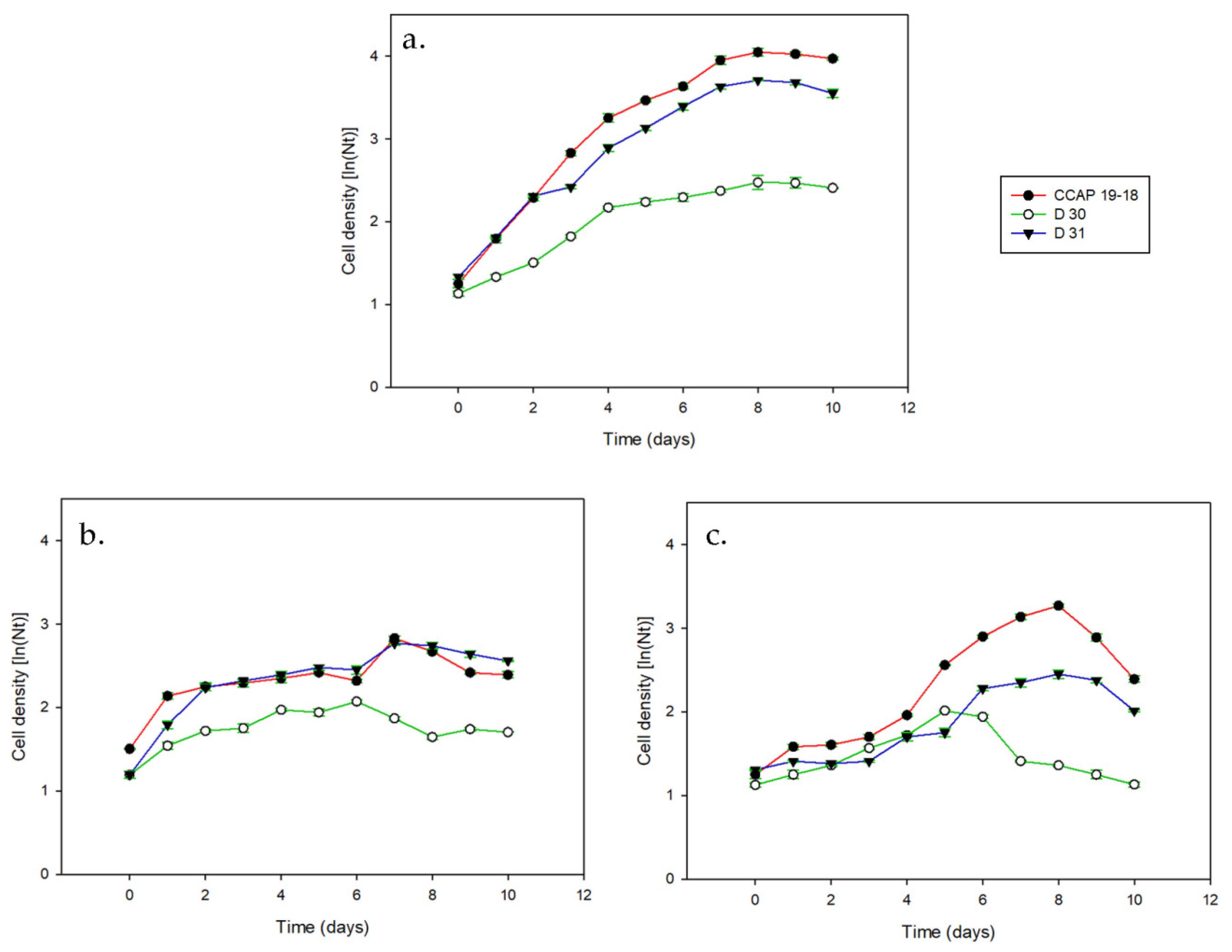
Growth curves for all of the strains in the different culture conditions are shown in Figure 2, while the respective μmax values are shown in Table 1. Daily measurements were taken from Day 0 (inoculation day) to Day 10. All of the strains showed expected growth development in standard conditions (Figure 2a), achieving maximum growth rates (exponential phase) on Day 4 and maximum cell yields (cells per mL of culture) on Days 8–9. The reference strain, CCAP 19/18, had the highest μmax in standard conditions (0.513 d−1 ± 0.010), followed by D31 (0.390 d−1 ± 0.004) and D30 (0.255 d−1 ± 0.006). The growth of the strains was significantly decreased under stress conditions (p < 0.05). When cultivated under nitrogen deprivation (Figure 2b), all three strains reached maximum growth on Day 1, with growth rates decreasing from then on; the D31 strain had the highest μmax (0.288 d−1 ± 0.011). When cultivated under salt stress (Figure 2c), the CCAP 19/18 and D31 strains showed a clear lag phase in growth from Day 0 to Day 5, while the D30 strain showed no lag in growth; under salt stress conditions, all three strains showed the lowest μmax values compared to the standard values.
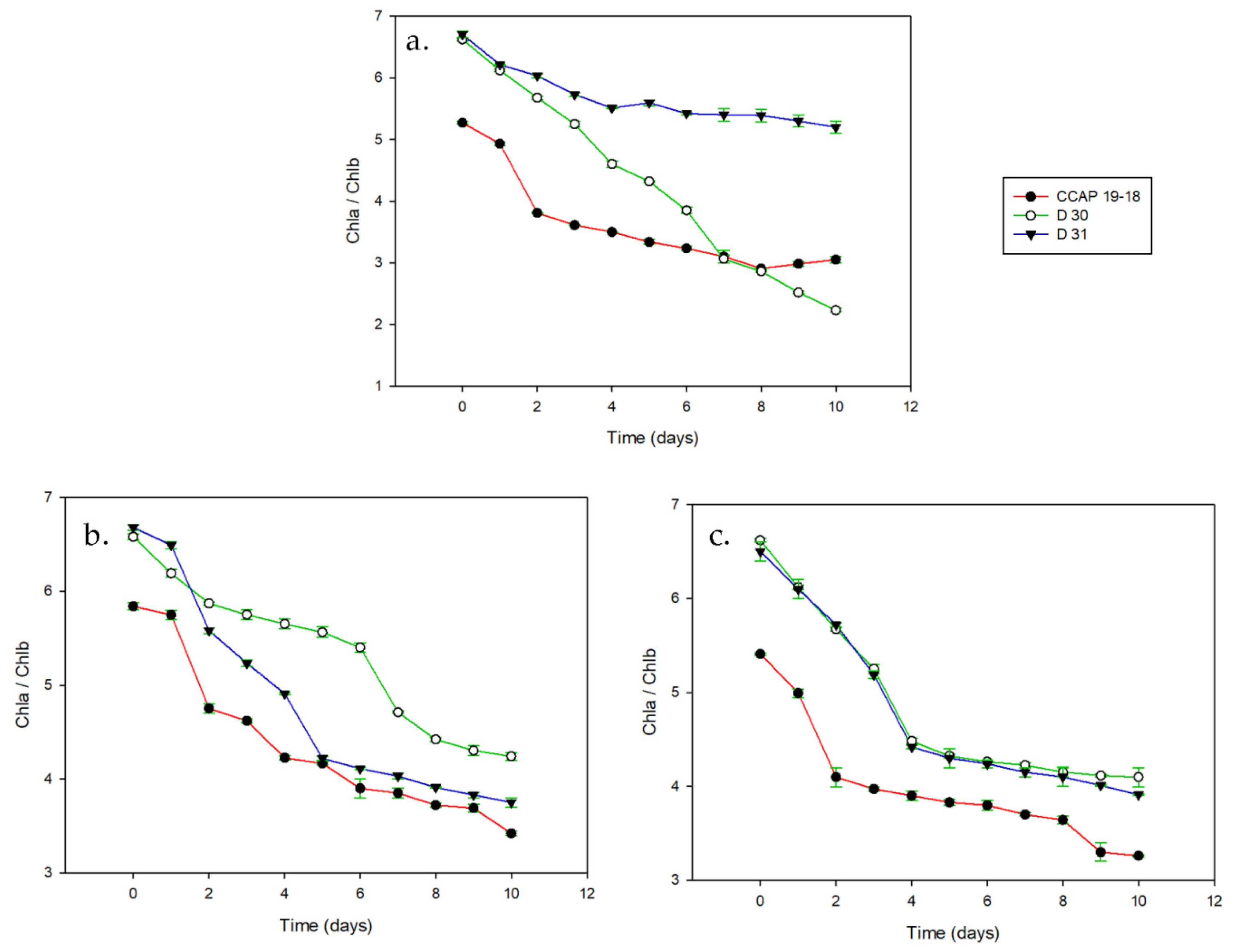
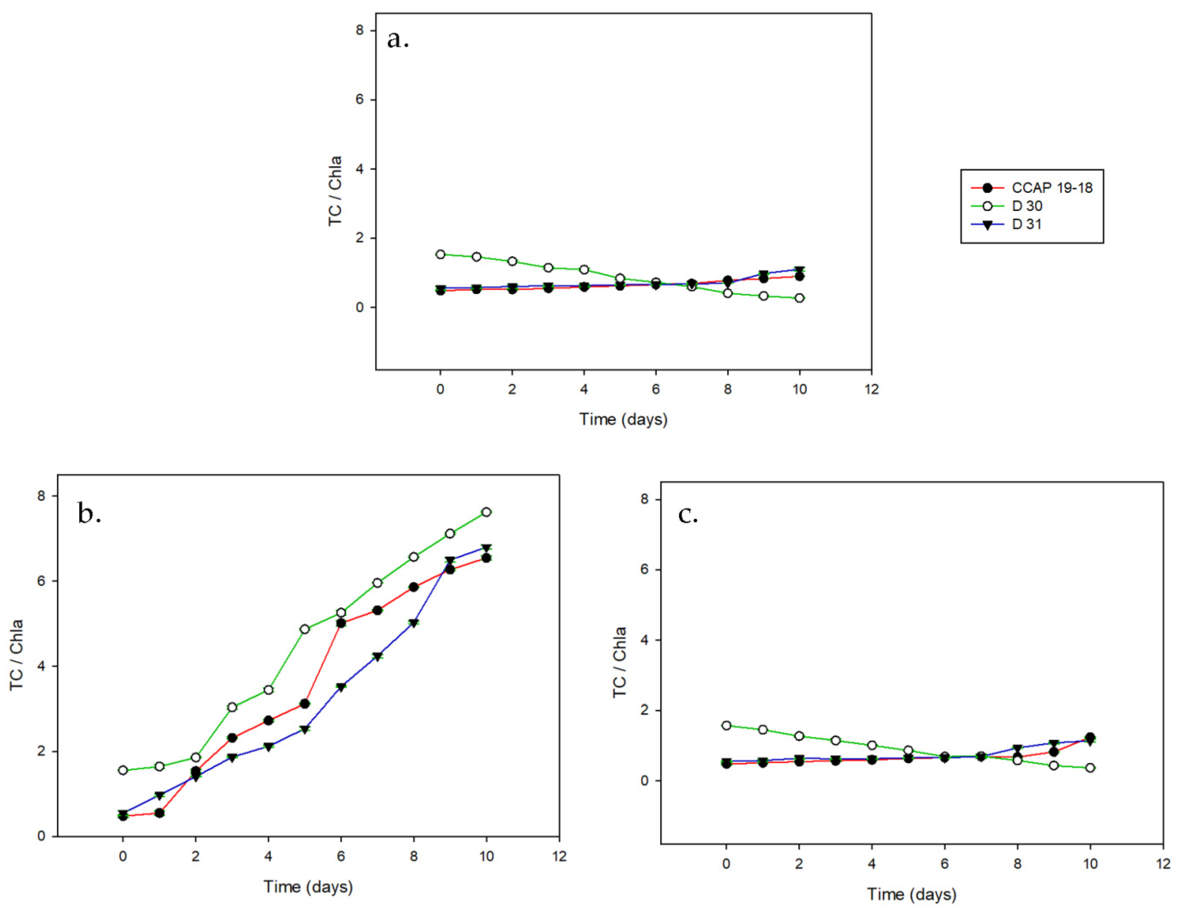
The chlorophyll ratio (Chl a/Chl b) of all of the strains is shown in Figure 3. This ratio showed a clear decreasing tendency in all of the strains and conditions; however, the decrease was much steeper in some cases. More specifically, the CCAP 19/18 and D30 strains showed a relatively smooth transition of their chlorophyll ratios to lower values with time, in all conditions, with the standard conditions (Figure 3a) exhibiting lower chlorophyll ratio values. On the contrary, the D31 strain had the lowest values under both of the stress conditions (salt stress and nitrogen deprivation, Figure 3b,c). The TC/Chl a ratio is shown in Figure 4. All strains demonstrated a logistic increase in the TC/Chl a ratio when cultivated under nitrogen deprivation conditions (Figure 4b). This indicates an increase in carotenoid production coupled with a decrease in production of Chl a, as also indicated by the Chl a/Chl b ratio (Figure 3). The maximum TC/Chl a ratios for all strains were also achieved when they were cultivated in nitrogen deprivation conditions.
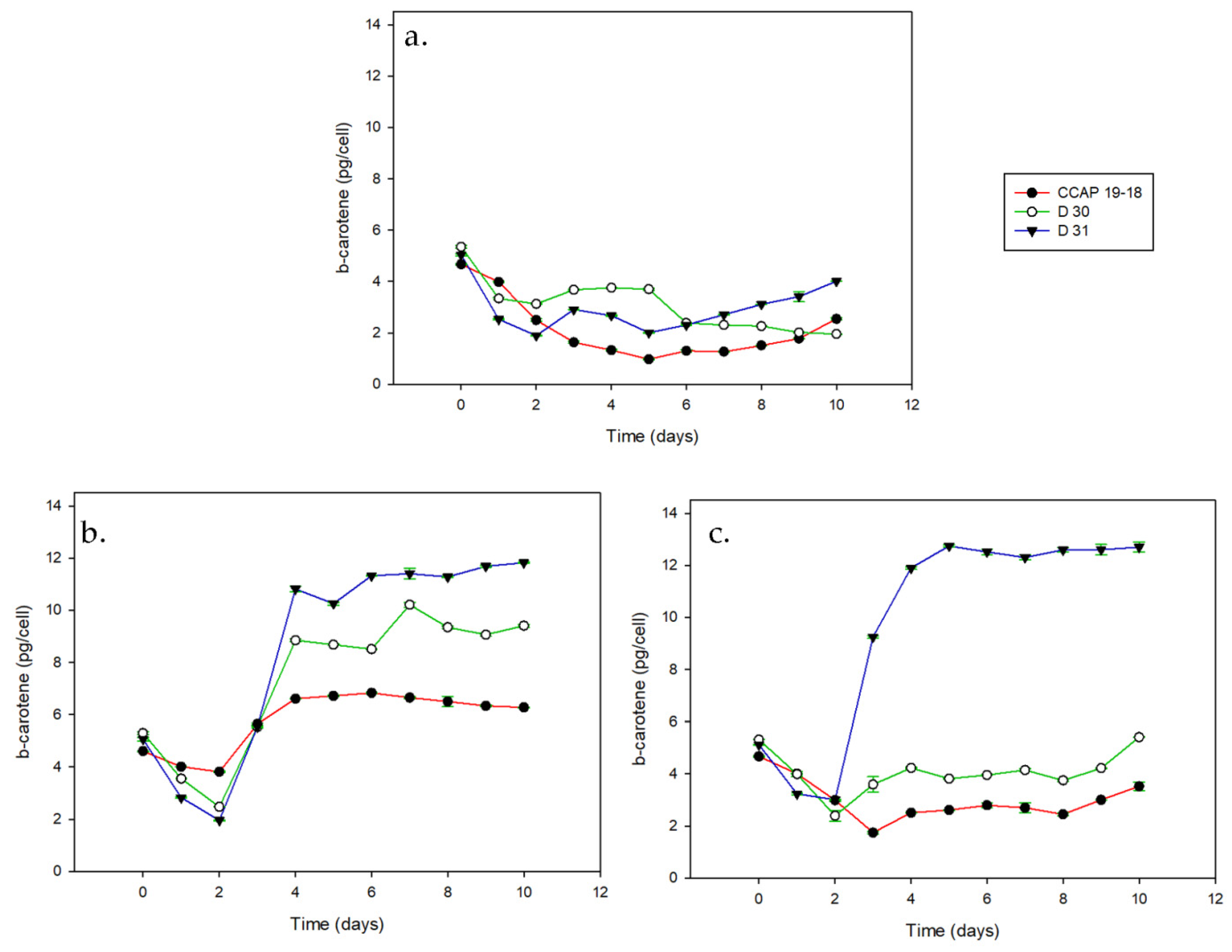
The daily fluctuation in β-carotene cell content (pg·cell−1) is shown in Figure 5. All of the strains showed an initial decrease in β-carotene content within their cells when those cells were actively growing (up to Day 2 after inoculation) and an increase when growth was reduced. When cultivated under stress conditions, the β-carotene content of the cells increased after the first few days of growth before stabilizing at high values when entering the stationary phase. The increase was much greater in the case of nitrogen deprivation (Figure 5b), reaching up to 6.94 pg·cell−1 and 10.16 pg·cell−1 for the CCAP 19/18 and D30 strains, respectively. In standard conditions, the D31 strain exhibited a very small increase in β-carotene content after the initial decrease; however, the highest value was achieved when the strain was cultivated under salt stress (13.1 pg·cell−1, Figure 5c).

3.2. Carotenoid Content on a Dry Basis

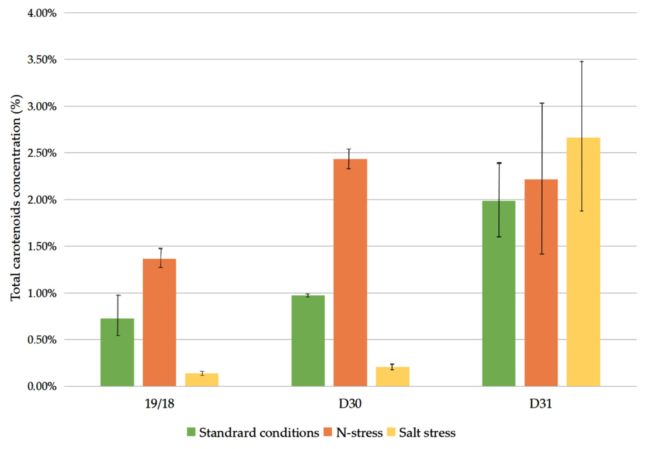
In Figure 6, the % TC content of total biomasson a dry basis (db) is shown (p < 0.05). In the CCAP 19/18 and D30 strains, the TC content increased when cultivated under nitrogen deprivation conditions reaching up to 1.36% of the db (13.64 mg·g−1) and 2.43% of the db (24.34 mg·g−1), respectively. The total carotenoids decreased when the two strains were cultivated under salt stress with values under 1% of the db. On the contrary, the D31 strain—which had the highest TC values, while showing a carotenoid-content increase, under nitrogen deprivation, reaching up to 2.22% of the db (22.16 mg·g−1)—also exhibited increased TC content when cultivated under salt stress conditions, reaching up to 2.67% of the db (26.65 mg·g−1).

3.3. Carotenoids Composition

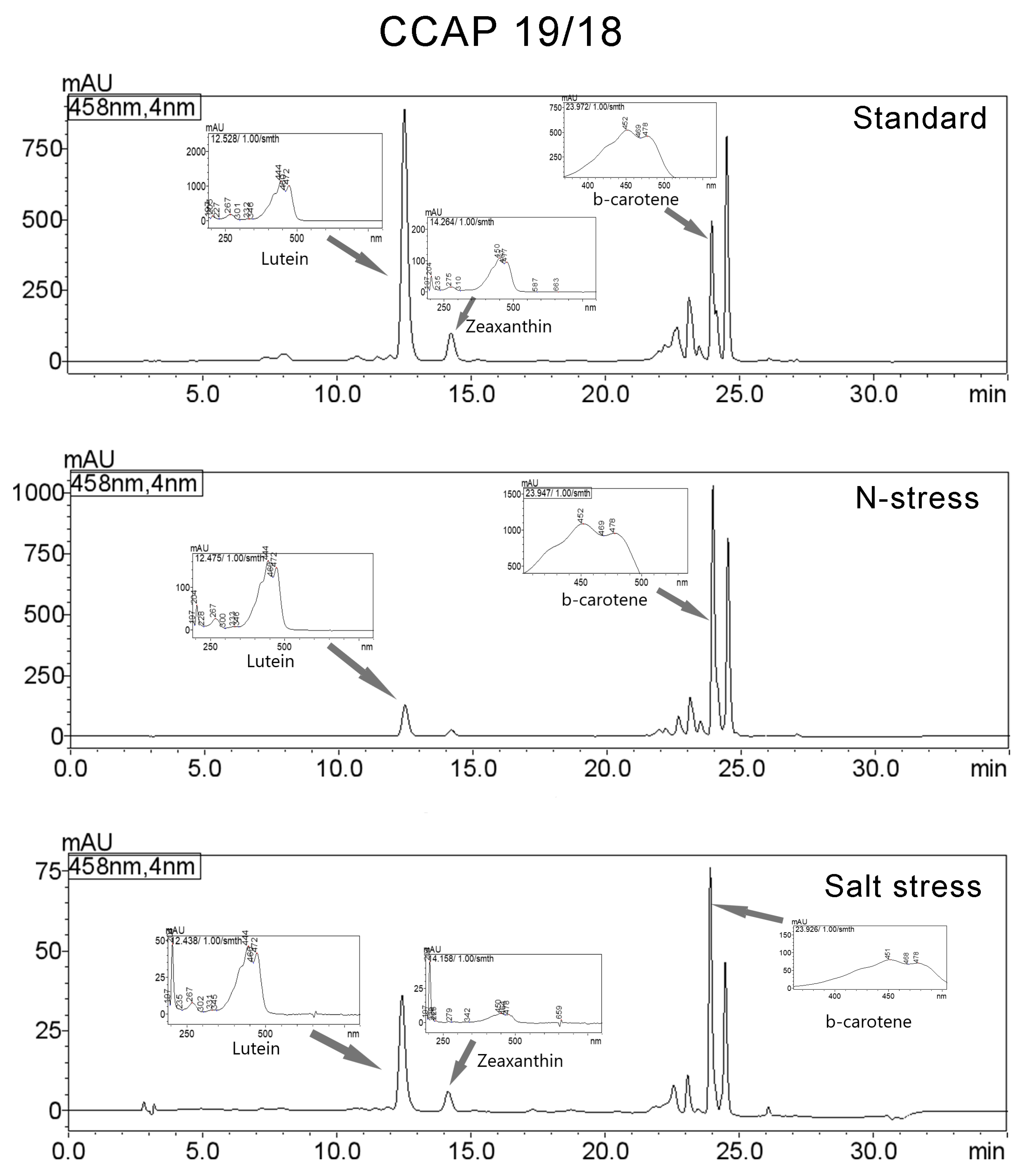
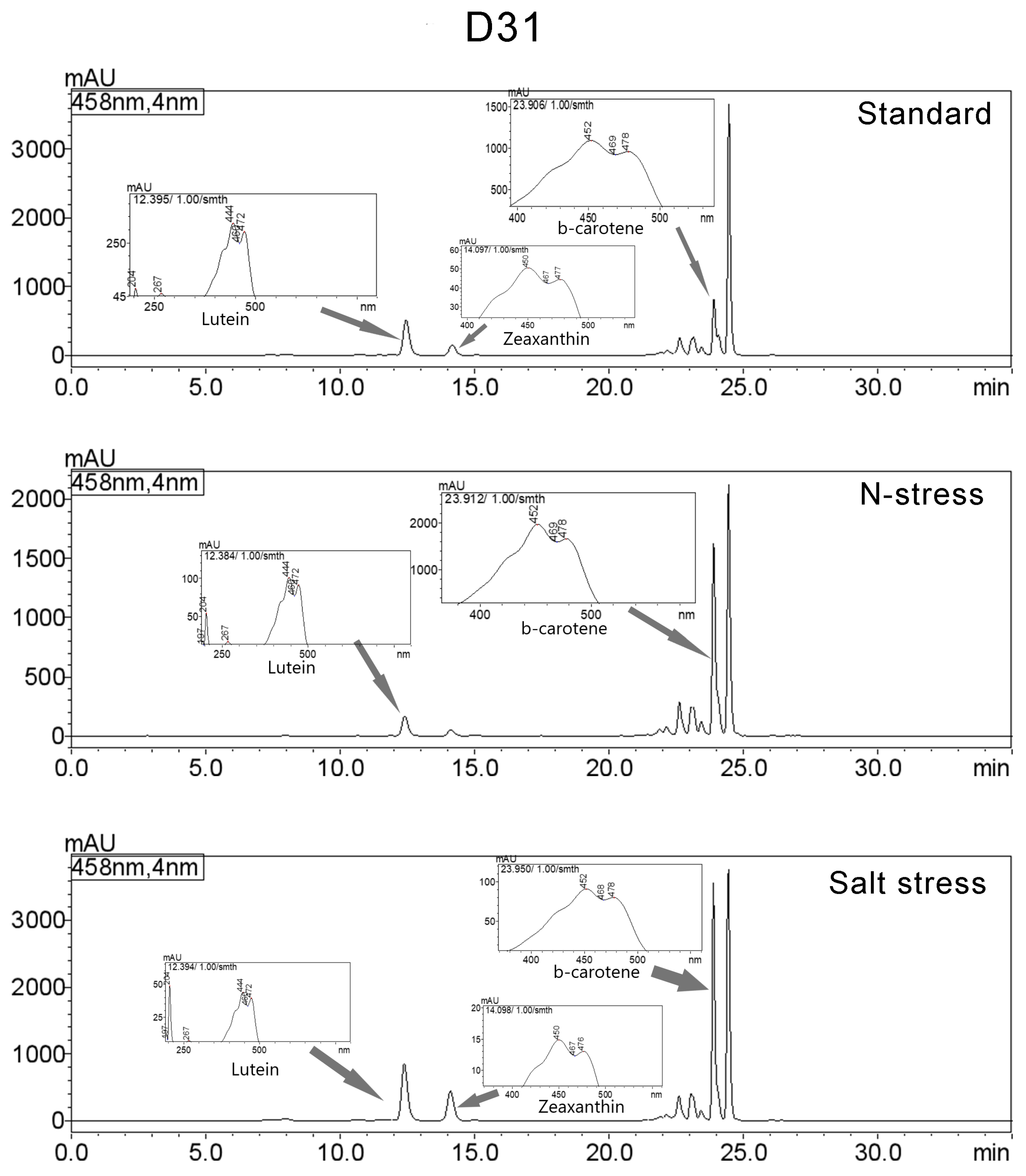
The carotenoid compositions of the strains were chromatographically determined on Day 10 (Figure 7, Figure 8 and Figure 9, Table 2, full chromatographic data available in Supplement S2). For the quality characterisation of lutein, zeaxanthin and β-carotene, internal standards were used. The rest of the carotenoids detected were categorised into seven groups (Car1-Car7) based on the resemblance of their spectra. The carotenoids that were detected in all of the strains were Car2, Car3 and Car7. Based on the literature, the spectra of these carotenoid groups resemble those of β,γ-carotene, β-Apo-12’-carotenal and β-carotene di-epoxide, respectively [46]. According to Table 2, lutein, zeaxanthin and β-carotene were detected in all studied strains. In the CCAP 19/18 (Figure 7) strain, lutein was the major carotenoid in standard conditions (36.84% of the TCs). When cultivated in nitrogen deprivation, there was a major increase in β-carotene content (39.42%) along with a major decrease in lutein (8.29%), while no zeaxanthin was detected. When cultivated under salt stress conditions, lutein (24.29%) and β-carotene (27.87%) represented half of the TCs, along with a small amount of zeaxanthin (4.31%). In D30 (Figure 8), β-carotene was increased under nitrogen deprivation (45.55%) but was also one of the major carotenoids in standard conditions and under salt stress (32.48% and 27.08%, respectively). Similarly to CCAP 19/18, lutein was majorly decreased under nitrogen deprivation (3.86%) compared to in standard conditions (15.10%); however, it was slightly increased under salt stress conditions (20.05%). In the D31 strain (Figure 9), β-carotene was majorly increased under nitrogen deprivation (32.49%) and salt stress conditions (28.07%) compared to in standard conditions (11.48%), and lutein decreased under nitrogen deprivation conditions (5.77%). Zeaxanthin represented a small percentage of the TCs in most cases (Table 2), while it was again absent in the nitrogen deprivation of the CCAP 19/18 and D31 strains but detected in the D30 strain. In general, nitrogen deprivation seemed to drastically affect the content of all previously mentioned major carotenoids. Specifically, lutein and zeaxanthin content were lowered or absent under nitrogen stress conditions, while β-carotene content was drastically increased. According to our results, an increase in salinity favoured production of β-carotene in all of the strains.

4. Discussion

Carotenoid content in Dunaliella is known to increase to up to 15% of its dry weight under various stress conditions (high light, temperature, salinity and nitrogen limitation being some of them), revealing a well-documented antioxidant function [33,48,49,50]. Nitrogen deprivation has been consistently correlated with high β-carotene productivities in Dunaliella strains [8,10,51,52,53]. According to our results, limiting nitrogen could be positively correlated with increased total carotenoids (TCs) and β-carotene content in all three strains (Figure 4 and Figure 5). Nitrogen depletion is one of the stress factors that disrupt cell division and have been associated with the production of reactive oxygen species (ROS) in microalgae, linking increased production of carotenoids to the cellular response to oxidative stress [2,8,54,55]. Therefore, even though nitrogen deprivation enhances carotenogenesis in Dunaliella, if applied long-term, it can lead to a steep decline in cell density as the cell death rate increases [10,56]. In the present study, cultivation of the strains in nitrogen deprivation conditions yielded low cell densities compared to in standard conditions (Figure 2), suggesting that the limitation of this nutrient hindered culture productivity without, however, halting it; this is also supported by the low growth rates previously observed when Dunaliella saline was cultivated under nitrogen limitation by Pisal and Lele [57]. Additionally, low Chl a/Chl b and high TC/Chl a ratios, along with an exponential daily increase in β-carotene cell content, support gradual limitation of photosynthetic activity and chlorophyll content in favour of carotenogenesis. This is in accordance with previous research on Dunaliella cells grown under nitrogen limitation conditions [57,58,59]. Phadwal and Singh [59] found that D. salina and another strain of Dunaliella sp. showed an increase in cellular β-carotene content from 0.24 and 0.5 pg·cell−1 to 3.4 and 3.94 pg·cell−1, respectively, whereas Pisal and Lele [57] observed an increase from 2.3 pg·cell−1 to 6.4 pg·cell−1 in nitrogen limitation. However, that increase was followed by a decrease in growth rate in both aforementioned cases.
In the present study, cultivation under salinity stress caused an increase in carotene content only in the D31 strain, while the total carotenoid and β-carotene content of the CCAP 19/18 and D30 strains remained lower than in the control conditions. Although high salinity has been successfully used in enhancement of carotenogenic activity of Dunaliella [51,52,53,54,55,56,57,58,59,60], there are cases where it has not yielded analogous results (e.g., [56]). Differential responses against salinity stress between different Dunaliella species have also been reported previously [25]. High salinity hampered the growth in all strains tested, similarly to in previous studies [60,61], as it is known to alter the metabolism of Dunaliella cells and restrict their growth by implementing oxidative stress as well [25]. Our results are in accordance with previous observations that indicated an inverse relationship between β-carotene content and the specific growth rates in D. salina cultures [8,33].
Chlorophyll content has been previously used as an index for the presence of physiological stress [62,63]. Chl a/Chl b ratios have also been correlated to increased light-harvesting efforts in vascular plants [64]. Each of the three studied strains showed an important decrease in the Chl a/Chl b ratio in all cultivating conditions (Figure 3), indicating the presence of physiological stress, which is in accordance with previous findings. Both Lv et al. [62] and Mirshekari et al. [65] reported an acute decrease in Chl a/b ratios under nitrogen starvation. However, Yound and Beardall [66] found that Chl a maintenance was favoured over Chl b in nitrogen-starved Dunaliella tertiolecta cells. In the present experiment, this decrease differed between the strains; CCAP 19/18 showed a similar decrease in all culture conditions and D30 showed a greater decrease under standard conditions, while D31 showed a greater decrease under both stress conditions (nitrogen deprivation and salt stress). These results indicate a variety of response intensity to stress factors in different strains of the same species and even between strains of the same locality (D30 and D31).
Another indicator of the changes leading to carotenogenesis, that occurred within the cells under stress conditions,,is the ratio of TC/Chl a. It has been reported that carotenoid content is favoured over chlorophyll content when under stress conditions, especially under nitrogen starvation in D. tertiolecta [56,66]. All of the studied strains exhibited a great increase in the aforementioned ratio under nitrogen deficiency (tenfold for D31 and CCAP 19/18 and fivefold for D30) when compared to those in standard conditions (Figure 4). Under salt stress, D31 and CCAP 19/18 each showed a threefold increase in the total TC/Chl a ratio. These results are comparable to those of previous research; Srinivasan et al. [61] achieved a sixfold increase, Lv et al. [62] a fivefold increase and Borovkov et al. [67] a fivefold increase under nitrogen deprivation conditions. Fazeli et al. [68], while studying the CCAP 19/18 strain in a salt-concentration increase protocol, achieved a threefold increase in the total carotenoid/chlorophyll ratio as well. Hadi et al. [69], using culture conditions and a salinity increase like those presented herein, achieved an increase of 2.5 times in this ratio. Cifuentes et al. [70], using milder salt stress (an increase from 2.14 M to 4.28 M of NaCl), achieved a 3.7- to 4.5-fold change in seven out of the eight strains used. These results indicate a clear relationship between carotenoid production and the reduction in photosynthetic activity under stress conditions, which is supported by the data presented herein.
Although salinity and nitrogen deprivation are known to be related to carotenogenic activity in Dunaliella strains, the specifics of those relationships are yet to be clarified. In 1990, Borowitzka et al. [60] concluded that the lutein biosynthesis path in Dunaliella salina must be sensitive to osmotic stress; therefore, high salinities favour synthesis of β-carotene at the expense of lutein production. However, according to the results presented herein (Table 2), the production of lutein did not seem to be affected under high-salinity conditions in any of the studied strains. It has been previously shown that lutein production in Dunaliella salina can be adversely affected by specific combinations of environmental stressors [71], and different strains of D. salina may respond differently to salinity stress regarding lutein production; that response has a genetic background that remains to be elucidated [72].
On the other hand, according to our results, the production of lutein in the studied strains was consistently limited only under nitrogen deprivation conditions (Table 2). Although the metabolite was present, the amounts produced as a percentage of the TCs were much smaller compared to in the standard conditions, as opposed to the percentage of β-carotene, which was the major carotenoid produced under nitrogen starvation in the studied strains. Previous research of microalgae has indicated that nitrogen limitation enhances accumulation of lutein [73,74,75]. Nevertheless, it was shown that cultivation of D. tertiolecta in a medium that completely lacked any nitrogen source (as applied in the present study) resulted in low lutein production [58,76]. Yeh et al. [77] concluded that a certain amount of nitrogen in the initial culture medium is required before repletion can lead to high production of lutein in a Desmodesmus sp. strain. Therefore, it is possible that complete lack of a nitrogen source might be the reason behind reduced lutein production in the studied strains.
It is notable that despite the clear tendencies revealed from the results presented herein (i.e., reduced growth rates and enhanced carotenogenic activity under stress conditions), there was an apparent diversification in the specific responses among the studied strains under stress conditions. The D31 strain deviated from the other two strains, as it responded with higher β-carotene accumulation under salt stress compared to that under nitrogen deprivation. Additionally, all three strains showed varied responses in photosynthetic and carotenogenic activity, as well as carotenoid profiles, in all culture conditions. This indicates that intraspecific diversity that has been recently suggested by molecular data [78] could be readily reflected in the physiological and metabolic characteristics of different hypersaline Dunaliella strains, and that this diversity could be apparent even in strains of the same locality (such as D30 and D31). Another key observation is that the Greek strains studied here showed comparable and, in most cases, higher TCs and β-carotene contents compared to the reference strain, CCAP 19/18, thus constituting possible candidates for further exploitation in the carotenoid-production industry.
Research of local representatives of Dunaliella could reveal highly carotenogenic strains, and thorough study of their carotenogenic characteristics could not only help elucidate the intraspecific relationships within the genus but also aid in improved fine-tuning of their cultivation for high-value secondary metabolites such as carotenoids. This study is the first report of carotenogenic Dunaliella salina strains from the salt marsh of Angelochori and the wider area of Greece, revealing a potential untapped natural source of biotechnological interest.

Supplementary Materials

The following supporting information can be downloaded at: https://www.mdpi.com/article/10.3390/w15020241/s1, Supplement S1: Chemical Composition of Walne’s Medium, Supplement S2: HPLC data of the studied strains. References [37,38] are cited in the supplementary materials.

Author Contributions

Conceptualization and methodology, X.C., I.T. and A.N.; validation, I.T. and A.E.-A.; formal analysis and investigation, X.C., A.N., A.T. and S.P.; data curation, X.C., A.N. and S.P.; writing—original draft preparation, X.C.; writing—review and editing, I.T., A.T. and A.E.-A.; visualization, X.C. and A.N.; supervision, A.E.-A.; project administration, A.E.-A.; funding acquisition, A.E.-A. All authors have read and agreed to the published version of the manuscript.

Funding

This research was cofinanced by Greece and the European Union (European Maritime, Fisheries and Aquaculture Fund—EMFAF) through the Operational Programme «Fisheries and Maritime 2014–2020» in the context of the project “Culturing microalga Dunaliella salina towards the production of nanomaterials for innovative applications in technology and biomedicine” (MIS 5033613).

Data Availability Statement

Data are available as Supplementary Materials.

Conflicts of Interest

The authors declare no conflict of interest. The funders had no role in the design of this study; in the collection, analyses or interpretation of these data; in the writing of this manuscript; or in the decision to publish these results.

References

  1. Borowitzka, L.J.; Moulton, T.P.; Borowitzka, M.A. The mass culture of Dunaliella salina for fine chemicals: From laboratory to pilot plant. In Proceedings of the Eleventh International Seaweed Symposium, Developments in Hydrobiology, Qingdao, China, 19–25 June 1983. [Google Scholar] [CrossRef]
  2. Saha, S.K.; Murray, P. Exploitation of microalgae species for nutraceutical purposes: Cultivation aspects. Fermentation 2018, 4, 46. [Google Scholar] [CrossRef] [Green Version]
  3. Borowitzka, M.A. Commercial-scale production of microalgae for bioproducts. In Blue Biotechnology: Production and Use of Marine Molecules; La Barre, S., Bates, S.S., Eds.; Wiley-VCH: Weinheim, Germany, 2018; Volume 1, pp. 33–65. [Google Scholar] [CrossRef]
  4. Winwood, R.J. Recent developments in the commercial production of DHA and EPA rich oils from micro-algae. Ocl 2013, 20, D604. [Google Scholar] [CrossRef] [Green Version]
  5. Novoveská, L.; Ross, M.E.; Stanley, M.S.; Pradelles, R.; Wasiolek, V.; Sassi, J.F. Microalgal carotenoids: A review of production, current markets, regulations, and future direction. Mar. Drugs 2019, 17, 640. [Google Scholar] [CrossRef] [Green Version]
  6. Raja, R.; Hemaiswarya, S.; Rengasamy, R. Exploitation of Dunaliella for β-carotene production. Appl. Microbiol. Biotechnol. 2007, 74, 517–523. [Google Scholar] [CrossRef] [PubMed]
  7. Teodoresco, E.C. Organisation et développement du Dunaliella, nouveau genre de Volvocacée-Polyblepharidée. Beih. Z Bot. Centralbl. 1905, 18, 215–232. [Google Scholar]
  8. Ben-Amotz, A.; Avron, M. On the factors which determine massive β-carotene accumulation in the halotolerant alga Dunaliella bardawil. Plant. Physiol. 1983, 72, 593–597. [Google Scholar] [CrossRef] [Green Version]
  9. Hashemi, A.; Moslemi, M.; Pajoum Shariati, F.; Delavari Amrei, H. Beta-carotene production within Dunaliella salina cells under salt stress condition in an indoor hybrid helical-tubular photobioreactor. Can. J. Chem. Eng. 2020, 98, 69–74. [Google Scholar] [CrossRef] [Green Version]
  10. Lamers, P.P.; Janssen, M.; De Vos, R.C.; Bino, R.J.; Wijffels, R.H. Carotenoid and fatty acid metabolism in nitrogen-starved Dunaliella salina, a unicellular green microalga. J. Biotechnol. 2012, 162, 21–27. [Google Scholar] [CrossRef]
  11. Borowitzka, M.A.; Siva, C.J. The taxonomy of the genus Dunaliella (Chlorophyta, Dunaliellales) with emphasis on the marine and halophilic species. J. Appl. Phycol. 2007, 19, 567–590. [Google Scholar] [CrossRef]
  12. Oren, A. The ecology of Dunaliella in high-salt environments. J. Biol. Res. (Thessalon) 2014, 21, 23. [Google Scholar] [CrossRef] [Green Version]
  13. Massjuk, N.P. Morphology, Systematics, Ecology, Geographic Distribution of Genus Dunaliella Teod, and Perspectives of Its Practical Use; Nauk, Dumka Press: Kiev, Ukraine, 1973. [Google Scholar]
  14. Ben-Amotz, A.; Avron, M. The role of glycerol in the osmotic regulation of the halophilic alga Dunaliella parva. Plant. Physiol. 1973, 51, 875–878. [Google Scholar] [CrossRef] [PubMed] [Green Version]
  15. Montazeri-Najafabady, N.; Negahdaripour, M.; Salehi, M.H.; Morowvat, M.H.; Shaker, S.; Ghasemi, Y. Effects of osmotic shock on production of β-carotene and glycerol in a naturally isolated strain of Dunaliella salina. J. Appl. Pharm. Sci. 2016, 8, 160–163. [Google Scholar] [CrossRef] [Green Version]
  16. Larcher, W. Streß bei Pflanzen. Naturwissenschaften 1987, 74, 158–167. [Google Scholar] [CrossRef]
  17. Ben-Amotz, A.; Katz, A.; Avron, M. Accumulation of β-carotene in halotolerent algae: Purification and characterization of β-carotene-rich globules from Dunaliella bardawil (Chlorophyceae). J. Phycol. 1982, 18, 529–537. [Google Scholar] [CrossRef]
  18. Ben-Amotz, A.; Shaish, A.; Avron, M. Mode of action of the massively accumulated β-carotene of Dunaliella bardawil in protecting the alga against damage by excess irradiation. Plant Physiol. 1989, 91, 1040–1043. [Google Scholar] [CrossRef] [Green Version]
  19. Loeblich, L.A. Action spectra and effect of light-intensity on growth, pigments and photosynthesis in Dunaliella salina. J. Protozool. 1974, 21, 420. [Google Scholar] [CrossRef] [Green Version]
  20. Loeblich, L.A. Growth limitation of Dunaliella salina by CO2 at high salinity. J. Phycol. 1970, 6, 9. [Google Scholar]
  21. Shaish, A.; Avron, M.; Pick, U.; Ben-Amotz, A. Are active oxygen species involved in induction of β-carotene in Dunaliella bardawil? Planta. 1993, 190, 363–368. [Google Scholar] [CrossRef]
  22. Xi, Y.; Kong, F.; Chi, Z. ROS induce β-carotene biosynthesis caused by changes of photosynthesis efficiency and energy metabolism in Dunaliella salina under stress conditions. Front. Bioeng. Biotechnol. 2021, 8, 1447. [Google Scholar] [CrossRef]
  23. Zhang, X.; Tang, X.; Wang, M.; Zhang, W.; Zhou, B.; Wang, Y. ROS and calcium signaling mediated pathways involved in stress responses of the marine microalgae Dunaliella salina to enhanced UV-B radiation. J. Photochem. Photobiol. B Biol. 2017, 173, 360–367. [Google Scholar] [CrossRef]
  24. Dring, M.J. Stress resistance and disease resistance in seaweeds: The role of reactive oxygen metabolism. Adv. Bot. Res. 2006, 43, 175–207. [Google Scholar] [CrossRef]
  25. Tammam, A.A.; Fakhry, E.M.; El-Sheekh, M. Effect of salt stress on antioxidant system and the metabolism of the reactive oxygen species in Dunaliella salina and Dunaliella tertiolecta. Afr. J. Biotechnol. 2011, 10, 3795–3808. [Google Scholar]
  26. Mueller, L.; Boehm, V. Antioxidant activity of β-carotene compounds in different in vitro assays. Molecules 2011, 16, 1055–1069. [Google Scholar] [CrossRef] [PubMed] [Green Version]
  27. Stahl, W.; Sies, H. Antioxidant activity of carotenoids. Mol. Aspects Med. 2003, 24, 345–351. [Google Scholar] [CrossRef]
  28. Harvey, P.J.; Ben-Amotz, A. Towards a sustainable Dunaliella salina microalgal biorefinery for 9-cis β-carotene production. Algal Res. 2020, 50, 102002. [Google Scholar] [CrossRef]
  29. Wolf, L.; Cummings, T.; Müller, K.; Reppke, M.; Volkmar, M.; Weuster-Botz, D. Production of β-carotene with Dunaliella salina CCAP19/18 at physically simulated outdoor conditions. Eng. Life Sci. 2021, 21, 115–125. [Google Scholar] [CrossRef]
  30. Neves, M.I.; Silva, E.K.; Meireles, M.A. Trends and challenges in the industrialization of natural colorants. Food Public Health 2019, 9, 33–44. [Google Scholar] [CrossRef] [Green Version]
  31. Kleinegris, D.M.; Janssen, M.; Brandenburg, W.A.; Wijffels, R.H. The selectivity of milking of Dunaliella salina. Mar. Biotechnol. 2010, 12, 14–23. [Google Scholar] [CrossRef] [Green Version]
  32. Morowvat, M.H.; Ghasemi, Y. Culture medium optimization for enhanced β-carotene and biomass production by Dunaliella salina in mixotrophic culture. Biocatal. Agric. Biotechnol. 2017, 7, 217–223. [Google Scholar] [CrossRef]
  33. Gallego-Cartagena, E.; Castillo-Ramírez, M.; Martínez-Burgos, W. Effect of stressful conditions on the carotenogenic activity of a Colombian strain of Dunaliella salina. Saudi J. Biol. Sci. 2019, 26, 1325–1330. [Google Scholar] [CrossRef]
  34. Gómez, P.I.; González, M.A. Genetic polymorphism in eight Chilean strains of the carotenogenic microalga Dunaliella salina Teodoresco (Chlorophyta). Biol. Res. 2001, 34, 23–30. [Google Scholar] [CrossRef] [PubMed]
  35. Gómez, P.I.; González, M.A. Genetic variation among seven strains of Dunaliella salina (Chlorophyta) with industrial potential, based on RAPD banding patterns and on nuclear ITS rDNA sequences. Aquaculture 2004, 233, 149–162. [Google Scholar] [CrossRef]
  36. Guevara, M.; Lodeiros, S.; Gómez, O.; Lemus, N.; Núñez, P.; Romero, L.; Vásquez, A.; Rosales, N. Carotenogénesis de cinco cepas del alga Dunaliella sp. (Chlorophyceae) aisladas de lagunas hipersalinas de Venezuela. Rev. Biol. Trop. 2005, 53, 331–337. [Google Scholar] [CrossRef] [PubMed] [Green Version]
  37. Walne, P.R. Studies on the food value of nineteen genera of algae to juvenile bivalves of the genera Ostrea, Crassostrea, Mercenaria and Mytilus. Fish. Invest. Ser. 2 1970, 26, 5. [Google Scholar]
  38. Guillard, R.R.L.; Hargraves, P.E. Stichochrysis immobilis is a diatom, not a chrysophyte. Phycologia 1993, 32, 234–236. [Google Scholar] [CrossRef]
  39. Andersen, R.A.; Kawachi, M. Traditional Microalgae Isolation Techniques. In Algal Culturing Techniques; Andersen, R.A., Ed.; Elservier Academic Press: Oxford, UK, 2005; pp. 90–92. [Google Scholar]
  40. Koletti, K.; Chantzistrountsiou, X.; Ntzouvaras, A.; Tzovenis, I.; Flemetakis, E.; Economou-Amilli, A. Identification and phylogeny of microalgae strains from Greek coastal lagoons (tentative title). A. EconomoU-Amilli, Sector of Ecology & Systematics, Department of Biology, School of Science, National and Kapodistrian University of Athens, Panepistimiopolis Zografou, 15784 Athens, Greece. 2022; manuscript in preparation. [Google Scholar]
  41. Kleinegris, D.M.; Janssen, M.; Brandenburg, W.A.; Wijffels, R.H. Continuous production of carotenoids from Dunaliella salina. Enzyme Microb. Technol. 2011, 48, 253–259. [Google Scholar] [CrossRef] [Green Version]
  42. Hashemi, A.; Pajoum Shariati, F.; Delavari Amrei, H.; Heydari Nasab, A. The Effect of Instantaneous and Slow-Release Salt Stress Methods on Beta-Carotene Production within Dunaliella Salina Cells. IJCCE. 2021, 40, 1642–1652. [Google Scholar] [CrossRef]
  43. He, Q.; Lin, Y.; Tan, H.; Zhou, Y.; Wen, Y.; Gan, J.; Zhang, Q. Transcriptomic profiles of Dunaliella salina in response to hypersaline stress. BMC Genomics 2020, 21, 115. [Google Scholar] [CrossRef]
  44. Borovkov, A.B.; Gudvilovich, I.N.; Avsiyan, A.L.; Memetshaeva, N.O.A.; Lelekov, A.S.; Novikova, T.M. Production characteristics of Dunaliella salina at two-phase pilot cultivation (Crimea). Turkish J. Fish. Aquat. Sci. 2019, 20, 401–408. [Google Scholar] [CrossRef]
  45. Wellburn, A.R. The spectral determination of chlorophylls a and b, as well as total carotenoids, using various solvents with spectrophotometers of different resolution. J. Plant Physiol. 1994, 144, 307–313. [Google Scholar] [CrossRef]
  46. Britton, G.; Liaanen-Jensen, S.; Pfander, H. Carotenoids. Handbook; Springer Science & Business Media: Berlin, Germany, 2004. [Google Scholar] [CrossRef]
  47. Tukey, J.W. Comparing individual means in the analysis of variance. Biometrics 1949, 99, 114. [Google Scholar] [CrossRef]
  48. Ben-Amotz, A. Effect of irradiance and nutrient deficiency on the chemical composition of Dunaliella bardawil Ben-Amotz and Avron (Volvocales, Chlorophyta). J. Plant Physiol. 1987, 131, 479–487. [Google Scholar] [CrossRef]
  49. Lamers, P.P.; van de Laak, C.C.W.; Kaasenbrood, P.S.; Lorier, J.; Janssen, M.; De Vos, R.C.H.; Bino, R.J.; Wijffels, R.H. Carotenoid and fatty acid metabolism in light-stressed Dunaliella salina. Biotechnol. Bioeng. 2010, 106, 638–648. [Google Scholar] [CrossRef] [PubMed]
  50. Semenenko, V.E.; Abdullayev, A.A. Parametric control of β-carotene biosynthesis in Dunaliella salina cells under conditions of intensive cultivation. Fiz. Rast. 1980, 27, 31–41. [Google Scholar]
  51. Bonneford, H.; Moelants, N.; Talec, A.; Mayzaud, P.; Bernard, O.; Sciandra, A. Coupling and uncoupling of triglyceride and beta-carotene production by Dunaliella salina under nitrogen limitation and starvation. Biotechnol. Biofuels 2017, 10, 1–10. [Google Scholar] [CrossRef] [Green Version]
  52. Chen, Y.; Tang, X.; Kapoore, R.V.; Xu, C.; Vaidyanathan, S. Influence of nutrient status on the accumulation of biomass and lipid in Nannochloropsis salina and Dunaliella salina. Energy Convers. Manag. 2015, 106, 61–72. [Google Scholar] [CrossRef]
  53. Marín, N.; Morales, F.; Lodeiros, C.; Tamigneaux, E. Effect of nitrate concentration on growth and pigment synthesis of Dunaliella salina cultivated under low illumination and preadapted to different salinities. J. Appl. Phycol. 1998, 10, 405–411. [Google Scholar] [CrossRef]
  54. Jimenez, C.; Pick, U. Differential reactivity of [beta]-carotene isomers from Dunaliella bardawil toward oxygen radicals. Plant Physiol. 1993, 101, 385–390. [Google Scholar] [CrossRef] [Green Version]
  55. Zhekisheva, M.; Boussiba, S.; Khozin-Goldberg, I.; Zarka, A.; Cohen, Z. Accumulation of oleic acid in Haematococcus pluvialis (Chlorophyceae) under nitrogen starvation or high light is correlated with that of astaxanthin esters1. J. Phycol. 2002, 38, 325–331. [Google Scholar] [CrossRef] [Green Version]
  56. Gómez, P.I.; Barriga, A.; Cifuentes, A.S.; Gonzalez, M.A. Effect of salinity on the quantity and quality of carotenoids accumulated by Dunaliella salina (strain CONC-007) and Dunaliella bardawil (strain ATCC 30861) Chlorophyta. Biol. Res. 2003, 36, 185–192. [Google Scholar] [CrossRef] [Green Version]
  57. Pisal, D.S.; Lele, S.S. Carotenoid production from microalga, Dunaliella salina. Indian J. Biotechnol. 2005, 4, 476–483. [Google Scholar]
  58. Geider, R.J.; Macintyre, H.L.; Graziano, L.M.; McKay, R.M.L. Responses of the photosynthetic apparatus of Dunaliella tertiolecta (Chlorophyceae) to nitrogen and phosphorus limitation. Eur. J. Phycol. 1998, 33, 315–332. [Google Scholar] [CrossRef]
  59. Phadwal, K.; Singh, P.K. Effect of nutrient depletion on β-carotene and glycerol accumulation in two strains of Dunaliella sp. Bioresour. Technol. 2003, 90, 55–58. [Google Scholar] [CrossRef] [PubMed]
  60. Borowitzka, M.A.; Borowitzka, L.J.; Kessly, D. Effects of salinity increase on carotenoid accumulation in the green alga Dunaliella salina. J. Appl. Phycol. 1990, 2, 111–119. [Google Scholar] [CrossRef]
  61. Cifuentes, A.S.; Gonzalez, M.A.; Parra, O.O. The effect of salinity on the growth and carotenogenesis in two Chilean strains of Dunaliella salina Teodoresco. Biol. Res. 1996, 29, 227–236. [Google Scholar]
  62. Lv, H.; Cui, X.; Wahid, F.; Xia, F.; Zhong, C.; Jia, S. Analysis of the physiological and molecular responses of Dunaliella salina to macronutrient deprivation. PLoS ONE 2016, 11, e0152226. [Google Scholar] [CrossRef]
  63. Srinivasan, R.; Mageswari, A.; Subramanian, P.; Suganthi, C.; Chaitanyakumar, A.; Aswini, V.; Gothandam, K.M. Bicarbonate supplementation enhances growth and biochemical composition of Dunaliella salina V-101 by reducing oxidative stress induced during macronutrient deficit conditions. Sci. Rep. 2018, 8, 6972. [Google Scholar] [CrossRef] [Green Version]
  64. Dale, M.P.; Causton, D.R. Use of the Chlorophyll a/b Ratio as a Bioassay for the Light Environment of a Plant. Funct. Ecol. 1992, 6, 190. [Google Scholar] [CrossRef]
  65. Mirshekari, M.; Einali, A.; Valizadeh, J. Metabolic changes and activity pattern of antioxidant enzymes induced by salicylic acid treatment in green microalga Dunaliella salina under nitrogen deficiency. J. Appl. Phycol. 2019, 31, 1709–1719. [Google Scholar] [CrossRef]
  66. Young, E.B.; Beardall, J. Photosynthetic function in Dunaliella tertiolecta (Chlorophyta) during a nitrogen starvation and recovery cycle. J. Phycol. 2003, 5, 897–905. [Google Scholar] [CrossRef]
  67. Borovkov, A.B.; Gudvilovich, I.N.; Avsiyan, A.L. Scale-up of Dunaliella salina cultivation: From strain selection to open ponds. J. Appl. Phycol. 2020, 32, 1545–1558. [Google Scholar] [CrossRef]
  68. Fazeli, M.R.; Tofighi, H.; Samadi, N.; Jamalifar, H.; Fazeli, A. Carotenoids accumulation by Dunaliella tertiolecta (Lake Urmia isolate) and Dunaliella salina (ccap 19/18 & wt) under stress conditions. DARU J. Pharm. Sci. 2006, 14, 146–150. [Google Scholar]
  69. Hadi, M.R.; Shariati, M.; Afsharzadeh, S. Microalgal biotechnology: Carotenoid and glycerol production by Dunaliella sp. algae isolated from the Gave khooni salt marsh, Iran. Biotech. Bioproc. Eng. 2008, 13, 540–544. [Google Scholar] [CrossRef]
  70. Cifuentes, A.S.; González, M.; Conejeros, M.; Dellarossa, V.; Parra, O. Growth and carotenogenesis in eight strains of Dunaliella salina Teodoresco from Chile. J. Appl. Phycol. 1992, 4, 111–118. [Google Scholar] [CrossRef]
  71. Fu, W.; Paglia, G.; Magnúsdóttir, M.; Steinarsdóttir, E.A.; Gudmundsson, S.; Palsson, B.Ø.; Andrésson, O.S.; Brynjólfsson, S. Effects of abiotic stressors on lutein production in the green microalga Dunaliella salina. Microb. Cell Factories 2014, 13, 1–9. [Google Scholar] [CrossRef]
  72. Liang, M.H.; Liang, Z.C.; Chen, H.H.; Jiang, J.G. The bifunctional identification of both lycopene β-and ε-cyclases from the lutein-rich Dunaliella bardawil. Enzyme Microb. Technol. 2019, 131, 109426. [Google Scholar] [CrossRef]
  73. Del Campo, J.A.; Moreno, J.; Rodríguez, H.; Vargas, M.A.; Rivas, J.; Guerrero, M.G. Carotenoid content of chlorophycean microalgae: Factors determining lutein accumulation in Muriellopsis sp. (Chlorophyta). J. Biotechnol. 2000, 76, 51–59. [Google Scholar] [CrossRef]
  74. Ho, S.H.; Chan, M.C.; Liu, C.C.; Chen, C.Y.; Lee, W.L.; Lee, D.J.; Chang, J.S. Enhancing lutein productivity of an indigenous microalga Scenedesmus obliquus FSP-3 using light-related strategies. Bioresour. Technol. 2014, 152, 275–282. [Google Scholar] [CrossRef]
  75. Xie, Y.; Ho, S.H.; Chen, C.N.N.; Chen, C.Y.; Ng, I.S.; Jing, K.J.; Chang, J.S.; Lu, Y. Phototrophic cultivation of a thermo-tolerant Desmodesmus sp. for lutein production: Effects of nitrate concentration, light intensity and fed-batch operation. Bioresour. Technol. 2013, 144, 435–444. [Google Scholar] [CrossRef]
  76. Kim, S.H.; Liu, K.H.; Lee, S.Y.; Hong, S.J.; Cho, B.K.; Lee, H.; Lee, C.G.; Choi, H.K. Effects of light intensity and nitrogen starvation on glycerolipid, glycerophospholipid, and carotenoid composition in Dunaliella tertiolecta culture. PLoS ONE 2013, 8, e72415. [Google Scholar] [CrossRef] [Green Version]
  77. Yeh, T.J.; Tseng, Y.F.; Chen, Y.C.; Hsiao, Y.; Lee, P.C.; Chen, T.J.; Chen, C.Y.; Cao, C.Y.; Chang, J.S.; Chen, J.C.; et al. Transcriptome and physiological analysis of a lutein-producing alga Desmodesmus sp. reveals the molecular mechanisms for high lutein productivity. Algal Res. 2017, 21, 103–119. [Google Scholar] [CrossRef]
  78. Highfield, A.; Ward, A.; Pipe, R.; Schroeder, D.C. Molecular and phylogenetic analysis reveals new diversity of Dunaliella salina from hypersaline environments. J. Mar. Biol. Assoc. UK 2021, 101, 27–37. [Google Scholar] [CrossRef]
Figure 1. Living cells of the studied strains: (a) Dunaliella salina CCAP 19/18, (b) AthU-Al D30 (D30) and (c) AthU-Al D31 (D31). The red colouration of the cells is a result of carotenoid accumulation (Scale bar = 10 μm).
Water 15 00241 g001
Figure 2. Growth curves of the studied strains cultivated in different conditions: (a) standard conditions, (b) nitrogen deprivation and (c) salinity stress. Cell yield is shown as cells/mL·105 (SE, n = 3).
Water 15 00241 g002
Figure 3. Daily changes in the chlorophyll-a/chlorophyll-b ratio (Chl a/Chl b) of the studied strains cultivated in different conditions: (a) standard conditions, (b) nitrogen deprivation and (c) salinity stress (SE, n = 3).
Water 15 00241 g003
Figure 4. Daily changes in the total carotenoids to chlorophyll-a ratio (TC/Chl a) of the studied strains cultivated in different conditions: (a) standard conditions, (b) nitrogen deprivation and (c) salinity stress (SE, n = 3).
Water 15 00241 g004
Figure 5. Daily changes in the β-carotene content of the cells (pg·cell−1) of the studied strains cultivated in different conditions: (a) standard conditions, (b) nitrogen deprivation and (c) salinity stress (SE, n = 3).
Water 15 00241 g005
Figure 6. Total carotenoid content of the Dunaliella salina CCAP 19/18, AthU-Al D30 (D30) and AthU-Al D31 (D31) strains cultivated in standard conditions, under nitrogen deprivation (N-stress) and under high salinity (salt stress), expressed as % of total biomass. [STDEV, n = 3].
Water 15 00241 g006
Figure 7. HPLC chromatographs of the Dunaliella salina CCAP 19/18 strain cultivated in standard conditions, under nitrogen deprivation (N-stress) and under high salinity (salt stress). Lutein, zeaxanthin and β-carotene peaks are marked on the chromatographs.
Water 15 00241 g007
Figure 8. HPLC chromatographs of the Dunaliella salina AthU-Al D30 (D30) strain cultivated in standard conditions, under nitrogen deprivation (N-stress) and under high salinity (salt stress). Lutein, zeaxanthin and β-carotene peaks are marked on the chromatographs.
Water 15 00241 g008
Figure 9. HPLC chromatographs of the Dunaliella salina AthU-Al D31 (D31) strain cultivated in standard conditions, under nitrogen deprivation (N-stress) and under high salinity (salt stress). Lutein, zeaxanthin and β-carotene peaks are marked on the chromatographs.
Water 15 00241 g009
Table 1. Maximum growth rates (μmax) of the Dunaliella salina CCAP 19/18, AthU-Al D30 and AthU-Al D31 strains in different culture conditions.
Strainμmax (d−1)
Standard ConditionsNitrogen DeprivationSalinity Shock
CCAP 19\180.513 a,b (0.010)0.197 a,c (0.013)0.165 a,b,c (0.007)
AthU-Al D300.255 a (0.006)0.188 a,c (0.013)0.162 a,b,c (0.017)
AthU-Al D310.390 b (0.004)0.288 b (0.011)0.112 a,b,c (0.025)
Note: Data: mean (STDEV, n = 3) a,b,c; statistically significant differences (p < 0.05).
Table 2. Chromatographic features of the studied strains, regarding lutein, zeaxanthin and β-carotene content, in different culture conditions.
SampleCarotenoidsRet. Time (min)AreaHeightArea%Lamda Max
CCAP 19/18
StandardLutein12.5281482189989077836.844444/472/267/205/332
Zeaxanthin14.2641826232997074.54450/477/204/275/663
β-carotene23.972419495549721010.428452/478/204/273/659
N-StressLutein12.47520868901273778.285444/472/204/267/333
β-carotene23.9479928467103159339.416452/478/204/273/601
Salt StressLutein12.4386107843631824.293204/444/472/267/331
Zeaxanthin14.15810832362454.308204/450/478/659/225
β-carotene23.9267006347745327.867204/451/478/270/659
AthuAl D30
StandardLutein12.395423825125654615.098444/472/204/267/333
Zeaxanthin14.097806390464742.873204/450/477/274/659
β-carotene23.9069118255103698732.483452/478/204/273/659
N-StressLutein12.3841301617797943.86444/472/204/267/332
Zeaxanthin14.08439293255641.303204/450/476/274/630
β-carotene23.91215360003184687545.55452/478/204/273/659
Salt Stress Lutein12.3945796703478720.048204/444/472/267/335
Zeaxanthin14.098230176137727.961204/450/476/273/629
β-carotene23.957829768762327.079204/452/478/272/659
AthuAl D31
StandardLutein12.457852733351945314.461444/472/267/204/333
Zeaxanthin14.17326696671525104.527450/477/204/274/659
β-carotene23.908677063581278011.482452/478/204/273/659
N-StressLutein12.41127352421664835.774444/472/204/267/333
β-carotene23.89615389583162950332.485451/478/204/273/630
Salt StressLutein12.3931379888485081714.126444/472/267/205/332
Zeaxanthin14.10577179164418177.901450/477/204/274/659
β-carotene23.89127417197357270628.067449/478/273/204/659
Disclaimer/Publisher’s Note: The statements, opinions and data contained in all publications are solely those of the individual author(s) and contributor(s) and not of MDPI and/or the editor(s). MDPI and/or the editor(s) disclaim responsibility for any injury to people or property resulting from any ideas, methods, instructions or products referred to in the content.