Article

Mussel Shell-Supported Yttrium-Doped Bi2MoO6 Composite with Superior Visible-Light Photocatalytic Performance

1
College of Food and Pharmacy, Zhejiang Ocean University, Zhoushan 316022, China
2
Ocean College, Zhejiang University, Zhoushan 316021, China
3
National Marine Facilities Aquaculture Engineering Technology Research Center, Zhejiang Ocean University, Zhoushan 316022, China
*
Author to whom correspondence should be addressed.
Water 2023, 15(19), 3478; https://doi.org/10.3390/w15193478
Submission received: 17 August 2023 / Revised: 17 September 2023 / Accepted: 29 September 2023 / Published: 2 October 2023

Abstract

:
A series of Yttrium (Y)-doped Bi2MoO6 composites with calcined mussel shell powder (CMS) as supports were synthesized using a solvothermal method. The as-prepared samples were analyzed using multiple techniques to investigate their microscopic morphology, composition structure, and optical properties. The photocatalytic performance of the as-prepared samples was assessed via examining their capacity to degrade Rhodamine B (RhB) under visible-light irradiation. The photocatalytic data showed that the Y-doped Bi2MoO6/CMS composites exhibited better photocatalytic activity compared to pure Bi2MoO6 and undoped Bi2MoO6/CMS samples. Among the samples, the 0.5%Y-doped Bi2MoO6/CMS (0.5%Y-BC) showed the highest photocatalytic activity, achieving a maximum degradation rate of 99.7% within 60 min. This could be attributed to highly reactive sites due to Y doping, a narrower band gap, and a lower recombination rate of photoinduced electron–hole pairs. Additionally, the 0.5%Y-BC photocatalyst exhibited excellent stability and reusability properties even after four cycles, making it suitable for practical applications. The findings provided a feasible synthesis of nanocomposite photocatalysts with outstanding properties for organic pollutant removal from the solution system.

1. Introduction

Environmental pollution caused by organic dyes has been a longstanding and pressing concern, prompting researchers and scientists to seek effective wastewater treatment methods [1]. Among the various technologies available, photocatalytic technology has emerged as a promising method for pollution control due to its numerous advantages, including its ease in preparation, cost-effectiveness, minimal energy consumption, remarkable degradation efficiency, and outstanding safety and stability [2,3,4,5,6,7]. As a result, research on photocatalytic materials has become a central focus [8,9].
Bismuth molybdate (Bi2MoO6) has garnered significant attention as a photocatalyst due to its unique layered structure. The structure consists of [Bi2O2]2+ slices and MoO6 octahedra that share corner oxygen atoms [10]. The conduction band in Bi2MoO6 is primarily composed of Mo3d orbitals, while the valence band is formed via the hybridization of Bi 6s and O2p orbitals [11]. This special band structure results in Bi2MoO6 having a relatively narrow band gap ranging from 2.5 to 2.9 eV [12]. Additionally, Bi2MoO6 exhibits exceptional chemical stability, non-toxicity, and good corrosion resistance, making it a promising candidate in the field of photocatalysis [13,14]. However, its high recombination rate of photo-generated electron and hole pairs leads to poor catalytic performance, limiting its application [15]. To enhance the efficiency of Bi2MoO6 in photocatalytic reactions, various modification techniques have been utilized, including morphological control, semiconductor combination, and element doping [16]. Among these approaches, element doping is a commonly used method to reduce the band gap energy and inhibit the recombination of photo-generated electrons and holes [17,18].
Rare earth ions possess a rich 4f electron configuration, and exhibit unique optical properties that have the potential to improve the photocatalytic activity of semiconductors [19,20]. Due to their similar ionic radii to bismuth ions, rare earth metal ions can be easily dispersed in Bi2MoO6, making them an effective approach to improve photocatalytic activity [21]. Several experiments have been carried out to evaluate the photocatalytic efficiency of Bi2MoO6 doped with rare earth metals such as Eu, Zr, Ce, Ho, Yb, Gd, Lu, Tb, Er, and Sm [22,23,24,25]. Among these metals, yttrium has gained attention due to its favorable characteristics, including increased photo absorption capacity and the ability to trap photo-excited electrons. These attributes can assist in reducing the rate of photogenerated electron–hole pair recombination in the photocatalyst [26,27]. For instance, in a study conducted by Vaddi et al., it was shown that doping ZnO nanorods with yttrium resulted in a significant reduction in their bandgap, which in turn led to an improvement in their photodegradation activity [28]. Similarly, Vadivel et al. used a solvothermal method to synthesize Yttrium-doped BiOF/RGO, and found that Y3+ ions had a synergistic effect in reducing the electron–hole recombination rate of BiOF with RGO [29]. However, to the best of our knowledge, no study has been carried out on Yttrium-doped Bi2MoO6.
In recent years, the utilization of waste biomass materials for the preparation of photocatalyst supports has become a central research topic due to their low cost, high efficiency, and renewability [30,31,32,33,34], such as egg shell [35], coconut shell [36], and mussel shell [37,38,39]. Among these, mussel shell, a major byproduct of the mussel aquaculture industry, is usually discarded, leading to environmental pollution. However, mussel shell is naturally biomineralized material, characterized by a substantial presence of calcium carbonate and a distinctive “brick-mud” structure [40,41]. Moreover, they contain various trace metal elements, rendering them well-suited for the development of high-value-added products [37,42]. Several studies have indicated that shells could serve as carriers for photocatalysts due to their special microstructure [37,38,39]. The trace metal elements in the shell can act as modifiers to enhance the catalyst’s performance [37]. Our previous study has demonstrated that the mussel shell can effectively disperse the photocatalyst and prevent its aggregation, to enhance electron transport and improve the photocatalytic activity [43]. We intended to further enhance the photocatalytic performance of the composite catalyst by doping rare earth elements.
In this study, we prepared a novel ternary Y-doped Bi2MoO6/CMS composite using the solvothermal method. We examined the effect of yttrium content on the chemical composition, morphology, and optical properties. Additionally, we evaluated the photocatalytic activities, stability, and reusability of samples via degrading RhB under visible light irradiation. Finally, we explored the photocatalytic degradation mechanism through active species trapping experiments.

2. Materials and Methods

2.1. Materials

The mussel shells were collected from Shengsi Shunda Seafood Co., Ltd. in Zhejiang Province, Zhoushan, China. They were washed thoroughly with distilled water to remove any surface impurities.
Bismuth nitrate pentahydrate (Bi(NO3)3·5H2O, 99%), sodium molybdate (Na2MoO4, 99%), Yttrium Nitrate Hexahydrate (Y(NO3)3·6H2O, 99%), ethanol (>99.7%), glycol and hydrochloric acid were purchased from Sinopharm Chemical Reagent Co., Ltd. (Shanghai, China). Rhodamine B (RhB, C28H31N2O3Cl) was obtained from Beijing Chemical Reagent Co., Ltd. (Beijing, China). All chemicals were analytical grade and used without further purification.

2.2. Preparation of Y-Doped Bi2MoO6/CMS Photocatalyst

Initially, the pretreated mussel shells were immersed with 0.1% HCl for 24 h, washed with distilled water until neutral, and dried at 80 °C in the oven for 4 h. The dried mussel shells were calcined at 900 °C for 3 h in a tube furnace with high-purity nitrogen (N2, 99.99%) flowing into the furnace at a rate of 100 mL/min. The resulting calcined sample was cooled, ground into powder (100 mesh), and named CMS.
Next, 0.363 g Bi(NO3)·5H2O and 0.0907 g Na2MoO4·2H2O were dissolved in a 7 mL ethylene glycol solution via sonication for 30 min. Different amounts of Y(NO3)3·6H2O were dissolved in 10 mL absolute ethanol, and slowly added dropwise to the Bi-Mo solution to obtain a mixed solution I with a molar ratio of Y to Bi of 0.063%, 0.125%, 0.25%, 0.5%, and 1%, respectively. Simultaneously, 0.1306 g CMS powder was dispersed in 10 mL of absolute ethanol for 1 h using ultrasonic treatment. The prepared solution I was then slowly added to the CMS suspension under continuous stirring for 2 h to obtain a mixed solution II. The mixed solution II was transferred into a 50 mL Teflon-lined stainless-steel autoclave, heated at 160 °C for 12 h, and cooled to room temperature. Finally, the as-prepared sample was filtered, washed with absolute ethanol and deionized water several times. Then, the material was dried in an oven at 60 °C for 12 h, and further pyrolyzed at 400 °C for 120 min in a tube furnace under a nitrogen atmosphere, while the heating rate was set at 10 °C/min at a nitrogen flow rate of 100 mL/min to produce the Y-doped Bi2MoO6/CMS composite. Different molar ratios of Y to Bi were prepared using a similar method and named 0.063%Y-BC, 0.125%Y-BC, 0.25%Y-BC, 0.5%Y-BC, and 0.1%Y-BC, accordingly. As a reference, pure Bi2MoO6 was synthesized under a similar procedure without the addition of CMS and Y(NO3)3·6H2O, and Bi2MoO6/CMS was synthesized without the use of Y(NO3)3·6H2O.

2.3. Characterization

The surface morphologies of the samples were examined using scanning electron microscopy (SEM, Hitachi S-4800, Tokyo, Japan). The specific surface areas and pore size distribution of the samples were analyzed using a Micromeritics ASAP 2010 instrument (Micromeritics Instrument Ltd., Atlanta, GA, USA) and calculated via the BET method. To characterize the functional groups and crystal structure of the samples, Perkin Elmer Fourier transform infrared spectrometer (FTIR, Thermo Scientific Nicolet IS 50, Waltham, MA, USA) and X-ray diffraction (XRD, Rigaku Ultima IV, Tokyo, Japan) with Cu Ka radiation (V = 30 kV, I = 25 mA) were employed, respectively. The X-ray photoelectron spectroscopy (XPS, ThermoScientific Escalab 250 Xi, Waltham, MA, USA) with Al Ka X-ray radiation was used to analyze the existence of elements and their chemical states on the surface of the sample. To monitor the recombination rate of electron–hole pairs, photoluminescence spectra (PL, FLS980 Series of Fluorescence Spectrometers, Edinburgh Instruments, UK) were recorded. Additionally, the optical properties of the samples were analyzed using a UV-vis spectrophotometer and its integral sphere accessories (UV, Shimadzu UV 2600, Kyoto, Japan).

2.4. Photocatalytic Activity Test

The photocatalytic activity of Y-doped Bi2MoO6/CMS photocatalyst was tested by conducting the degradation of RhB in a photochemical reactor (PhchemIII, Beijing, China). A 300 W Xenon lamp, equipped with a 420 nm cut-off filter, was employed as the simulative visible light source, and the entire reaction device was maintained at room temperature using a cooling-water-cycle system. Briefly, 20 mg of the as-prepared photocatalyst was mixed with 40 mL of 6 mg/L RhB in a 50 mL glass beaker under magnetic stirring conditions. Before illuminating the visible light, the reaction system of photocatalyst and RhB was kept in the dark for 3 h to establish an adsorption–desorption equilibrium, and illuminated and kept for 1 h. We utilized UV-visible spectroscopy (UV, Shimadzu UV 2600, Kyoto, Japan) to measure the degradation rate of RhB by the photocatalysts, based on the absorbance values of RhB at a wavelength of 554 nm. The degradation efficiency was calculated from Equation (1). The reaction rate was estimated using the Langmuir–Hinshelwood kinetic equation, as shown in Equation (2) [44].
Photocatalytic degradation = [(A0At)/A0] × 100%,
where A0 is an initial photocatalytic degradation of RhB and At represents RhB adsorption measured at a definite time.
Ln   ( C 0 C t ) = K   t ,
C0 represents the initial concentration of RhB, while Ct represents the concentration of RhB during the photocatalytic process at the time t. Moreover, K is the reaction rate constant. The direct decomposition of RhB without any catalyst under visible irradiation was omitted.

2.5. Cycle Experiment

To evaluate the chemical stability and practical applicability of the as-prepared photocatalyst, we performed a four-cycle degradation of RhB using Y-doped Bi2MoO6/CMS. After each cycle of photocatalytic degradation, the catalyst was collected through centrifugation, washed with deionized water, and dried in an 80 °C oven. The collected photocatalysts were then reused for subsequent cycles of degradation under the same conditions.

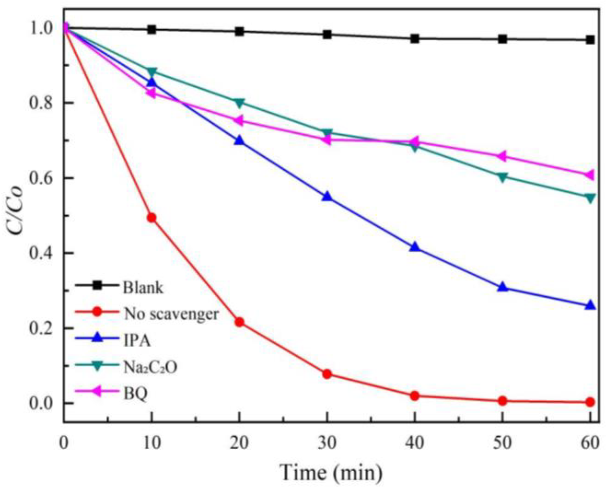
2.6. Active Species Trapping Experiments

Briefly, 2 mL of isopropanol (IPA, 0.1 mol/L), sodium oxalate (Na2C2O4, 0.1 mol/L) and p-benzoquinone (BQ, 0.1 mol/L) were prepared as scavengers for ·OH, h+, and ·O2 species. These scavengers were added to the reaction solution to be degraded before the illumination process, respectively. Through this setup, the active species participating in the photocatalytic reaction were analyzed.

3. Results

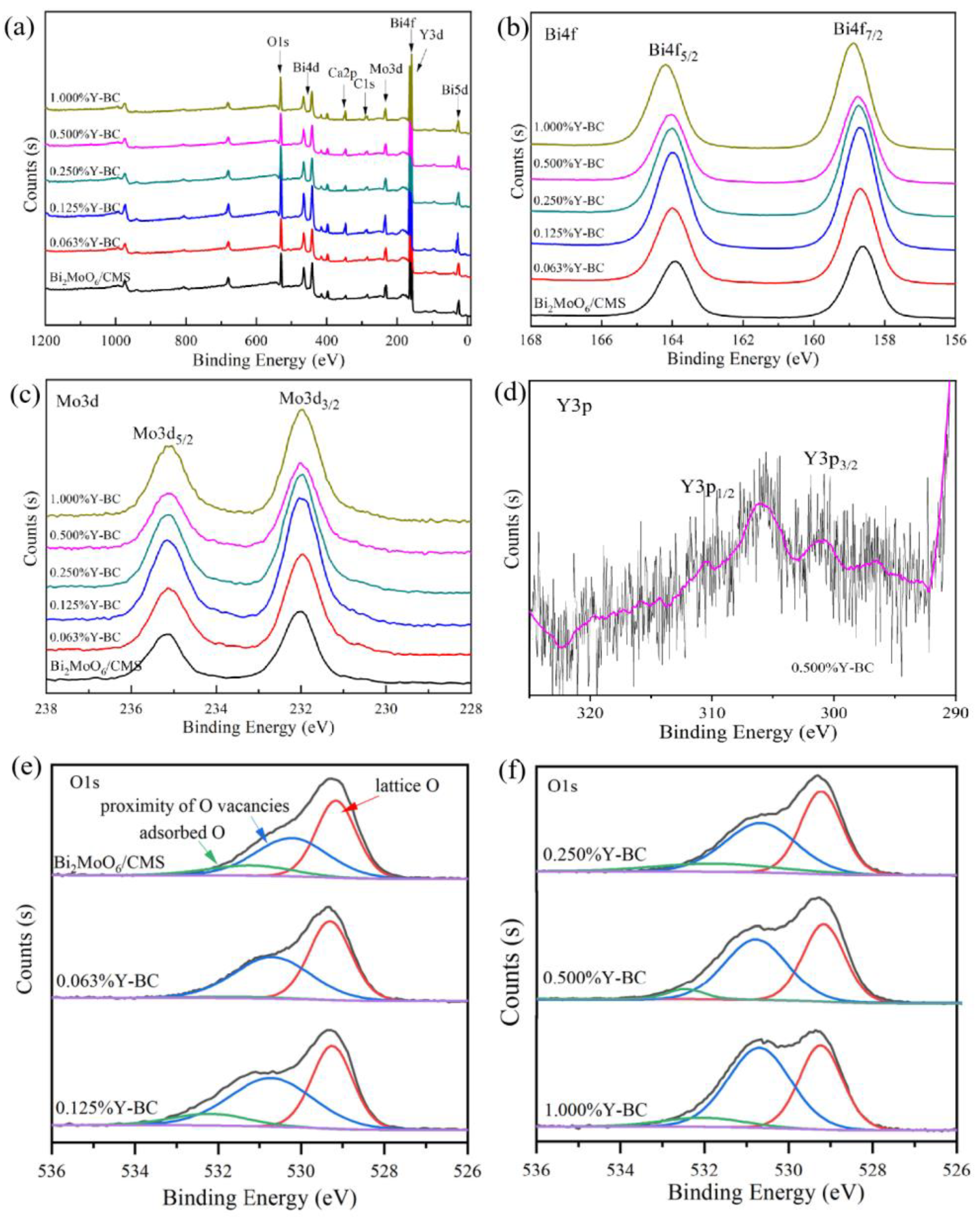
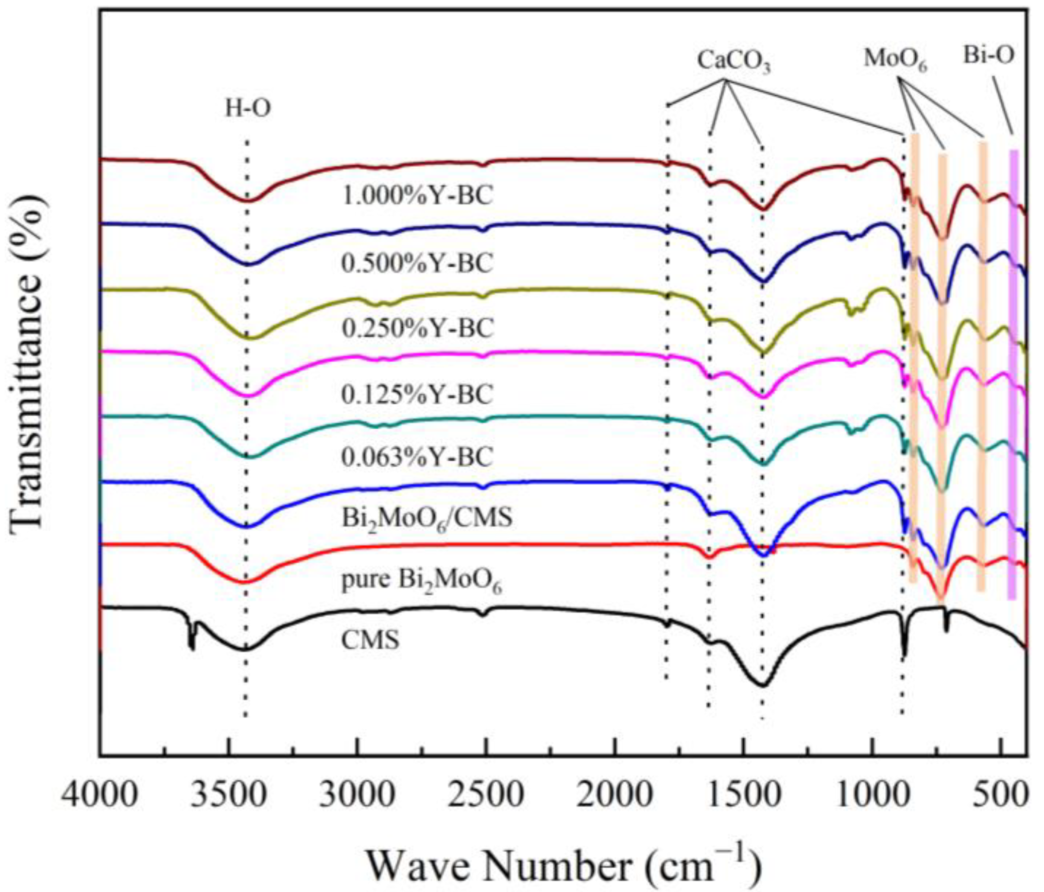
3.1. Chemical Composition Analysis

The crystal composition and structure of the as-prepared samples were analyzed using XRD measurements. Figure 1a displays the XRD patterns of the pure Bi2MoO6, CMS Bi2MoO6/CMS, and Y-doped Bi2MoO6/CMS samples. In comparison to the XRD patterns of pure Bi2MoO6 and CMS, those of Bi2MoO6/CMS and Y-doped Bi2MoO6/CMS showed the diffraction peaks of the orthorhombic phase of Bi2MoO6 and the calcite phase of CaCO3. The diffraction peaks at 28.1°, 32.3°, 46.7°, 55.3° and 58.2° were corresponded to the (131), (200), (062), (331), and (191) crystal planes of the orthorhombic phase Bi2MoO6 (JCPDS No. 76-2388). Similarly, the diffraction peaks at 29.4°, 35.9°, 43.1°, and 47.4° were attributed to the (104), (110), (202) and (024) planes of the calcite phase CaCO3 (JCPDS 83-1762). The Y-doped Bi2MoO6/CMS did not show any evident XRD peak of yttrium compounds or other impurities. However, in further amplification of the spectrum (as shown in Figure 1b), a slight shift to lower 2θ values was observed in the peak corresponding to the (131) crystal plane after Y doping. This shift indicated the substitution of some Bi3+ with Y3+ ions, which was an indication of the insertion and substitution of the Bi with rare earth ions in the lattice of Bi2MoO6 [45,46,47]. Furthermore, the crystallite size of different samples was calculated using the Scherrer equation [48], and the results are presented in Table 1. The crystalline size of Bi2MoO6 was reduced in comparison to undoped Bi2MoO6/CMS. This suggested that the introduction of Y3+ ions hinder the growth of Bi2MoO6 in the composite. This was because the insertion of Y in Bi2MoO6 created lattice defects, which further prevented the growth of Bi2MoO6 [49,50,51]. A suitable amount of lattice defects can serve as efficient photo–electron–hole capture centers, thereby promoting the separation of photogenerated electron–hole pairs and enhancing the photocatalytic activity of the catalyst [52,53].
The XPS testing was used to investigate the surface chemical composition of Bi2MoO6/CMS and Y-doped Bi2MoO6/CMS samples, as shown in Figure 2. The results revealed that the Bi2MoO6/CMS and Y-doped Bi2MoO6/CMS samples were composed of Bi, Mo, O, Ca, and C. However, the Y XPS peaks were not detected in the survey spectra due to the low amount of doping and overlap with the Bi 4f peak [45]. The high-resolution XPS spectra of Bi 4f show two strong peaks at 164.0 and 158.6 eV (Figure 2b), assigned to Bi4f5/2 and Bi4f7/2, respectively, indicating the trivalent chemical state of Bi [54,55]. The Bi peak shifted towards higher binding energy with the increase in the Y doping ratio, suggesting changes in the Bi chemical environment due to Y doping into the Bi2MoO6 lattice and charge transfer between Bi and Y [56,57]. Y doping had no significant effect on the Mo element as seen from the Mo3d3/2 and Mo3d5/2 peaks. The fine spectrum of O1s in the XPS spectrum clearly revealed the different forms of O element. As shown in Figure 2, two strong peaks at 530.7 eV and 532.3 eV were attributed to the proximity of O vacancies and lattice O, respectively, and a weak peak at 529.2 eV represented the binding energy of adsorbed O [57,58]. The relative content of three forms of oxygen could be determined based on the proportion of peak area, while the oxygen vacancies in the samples showed a significant increasing trend with the increase in Y doping amount. Furthermore, Figure 2d revealed that the binding energies for Y3p3/2 and Y3p1/2 of Y in 0.5%Y-BC were around 301.1 eV and 310.6 eV, respectively, indicating Y3+ were successfully doped into Bi2MoO6 [45].
The FTIR spectrum of samples were presented in Figure 3. The spectrum of Y-doped samples was similar to the undoped samples. The absorption bands at 841, 733, and 565 cm−1 correspond to the stretching vibration of Mo = O, tetrahedral stretching vibration of Mo-O, and bending vibration of MoO6, respectively [59]. The band at 450 cm−1 was assigned to the Bi-O stretching vibration [60]. Additionally, four peaks at 1794, 1420, 872, and 729 cm−1 were identified as the characteristic peaks of CaCO3 in CMS [61,62]. A broad peak in the 3200–3600 cm−1 could be assigned to the O-H stretching vibration, which is due to the water molecule’s adsorption on the surface area [27,63,64]. No obvious Y-O vibration peak was observed in the spectrum, likely due to the low doping amount of Y3+ in the samples.

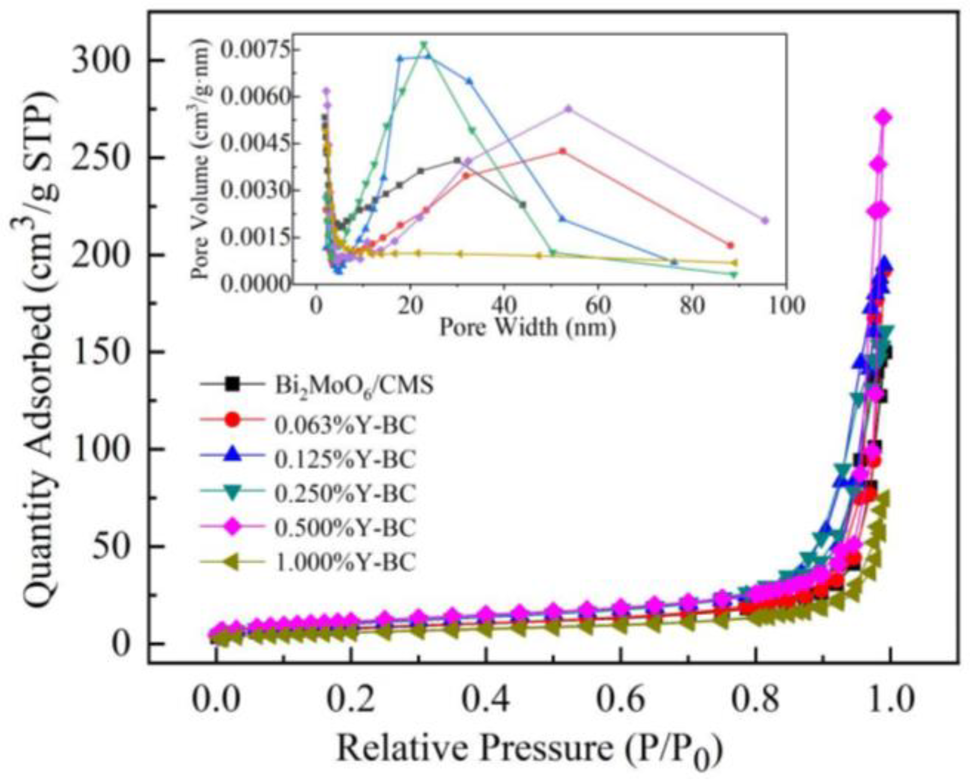
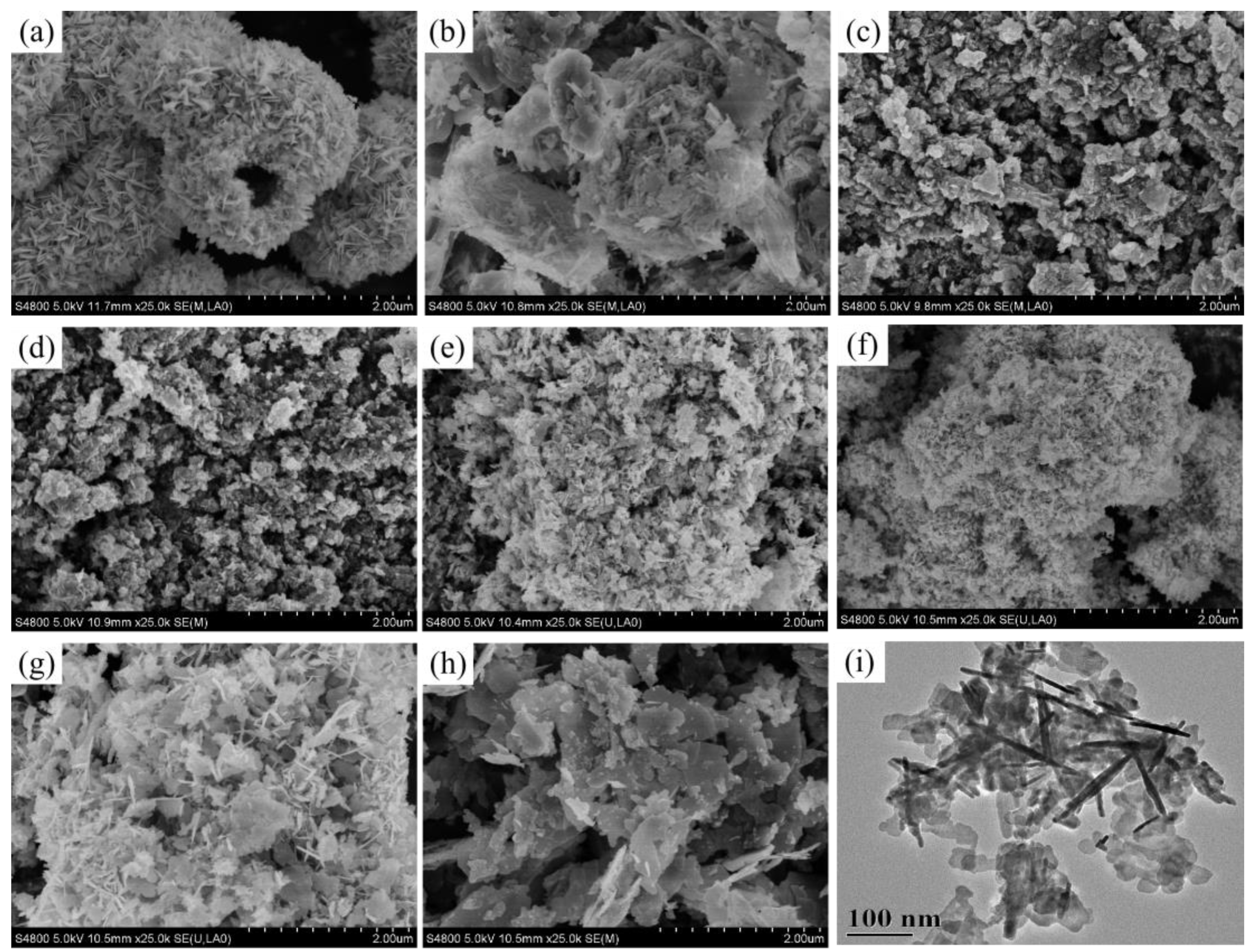
3.2. Morphology and Texture Analysis

The morphologies of samples, including pure Bi2MoO6, CMS, Bi2MoO6/CMS, and Y-doped Bi2MoO6/CMS, were characterized using SEM, and the results are presented in Figure 4. Pure Bi2MoO6 exhibited a hollow spherical structure assembled from nanosheets (Figure 4a). The CMS had an irregular block-like structure, with many nanorods on its surface (Figure 4b). Bi2MoO6/CMS exhibited a morphology composed of stacked lamellar nanosheets, primarily supported by CMS (Figure 4c). The morphologies of 0.063%Y-BC and Bi2MoO6/CMS were similar (Figure 4d), indicating that the trace amount of yttrium did not significantly alter the host materials. However, as the doping amount increased, the size of Bi2MoO6 nanosheets decreased. When the doping amount reached 0.5%, the sample displayed a homogeneous sheet-like structure that tended to aggregate into clusters (Figure 4g). Furthermore, at a Y doping concentration of 1%, the particles exhibited an increase in size and thickness, accompanied by a smooth surface (Figure 4h). As shown in Figure 5i, the TEM image of 0.5%Y-BC demonstrated that the Bi2MoO6 nanosheets were attached to the surface of the nanorods of CMS and formed a clustered structure, confirming with the SEM images.
Figure 5 displays the N2 adsorption and desorption isotherms, as well as the corresponding pore size distribution curves for Bi2MoO6/CMS and Y-doped Bi2MoO6/CMS samples. The N2 adsorption and desorption isotherms showed that all the samples exhibited type IV isotherms with an obvious H3-type hysteresis loop in the high relative pressure range of 0.9–1.0 (P/P0), indicating the presence of mesopores [65,66]. The pore size distribution curve measured via the BJH method revealed that the pore width was mainly distributed in the 2–30 nm range. The specific surface areas of Bi2MoO6/CMS, 0.063%Y-BC, 0.125%Y-BC, 0.25%Y-BC, 0.5%Y-BC, and 1%Y-BC were measured to be 28.64, 29.91, 37.94, 38.23, 42.50, and 20.60 m2/g (Table 1), respectively, and showed a gradual increase with increasing the doping amount of Y element. The sample with 0.5% doping exhibited the highest specific surface area among all the samples. However, with a further increase in the doping amount, the specific surface area decreases due to the excess Y potentially causing the aggregation of Bi2MoO6 nanosheets or blocking of pre-existing pores. The increase in specific surfaces could enhance the adsorption and degradation of organic pollutants. It also provided more active sites for the photogenerated electrons and holes, leading to an improved photocatalytic performance [24,67].

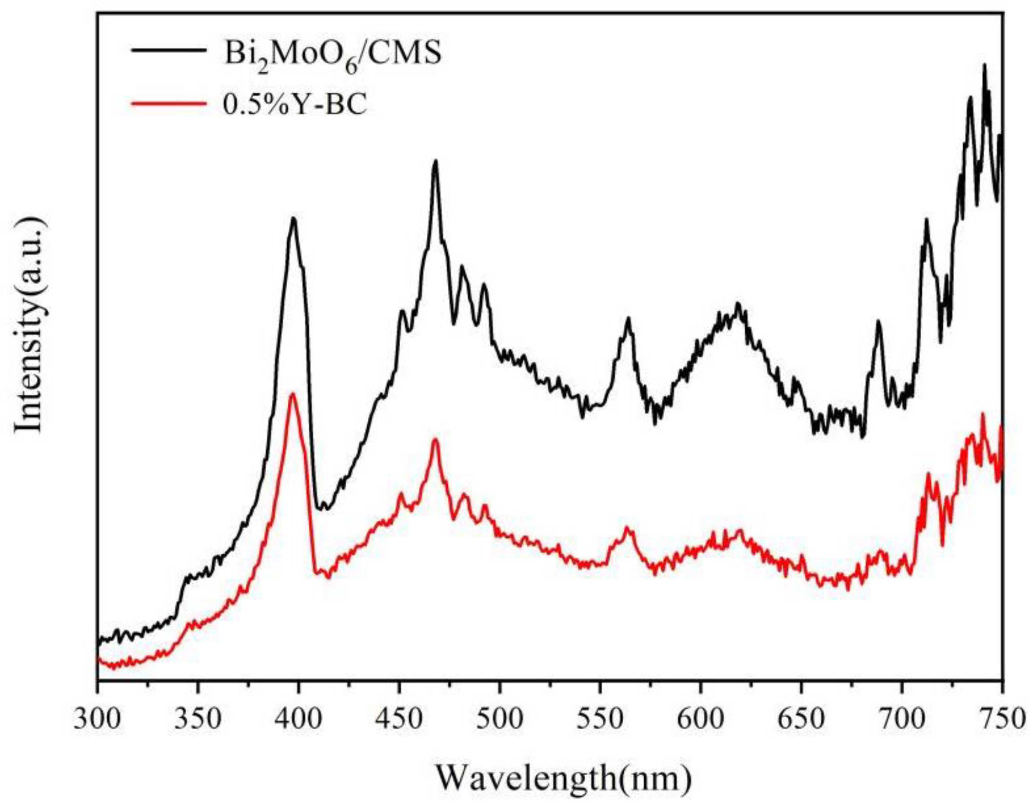
3.3. Optical Properties

The optical properties of the as-prepared samples were measured using UV–vis diffuse reflectance spectroscopy, as shown in Figure 6. All samples exhibited significant absorbance in the visible-light region. Compared to Bi2MoO6/CMS, the Y-doped Bi2MoO6/CMS samples showed a red shift of the absorption edges. The band gap value was calculated using the Kubelka–Munk equation, as follows.
(αhv)2 = A(hv − Eg),
where α, hv, A, and Eg are the absorption coefficient, photonic energy, Planck constant, and band gap, respectively [68].
Using Equation (3), the curves of these samples were fitted, and the results are presented in the inset of Figure 6. The estimated band gaps of pure Bi2MoO6, Bi2MoO6/CMS, 0.063%Y-BC, 0.125%Y-BC, 0.25%Y-BC, 0.5%Y-BC, and 1%Y-BC were approximately 2.65, 3.00, 2.98, 2.94, 2.92, 2.91 and 2.83 eV, respectively. Notably, the band gap of Y-doped Bi2MoO6/CMS had slightly decreased compared to Bi2MoO6/CMS. This decrease could be due to the charge transfer transition occurring between 4f or 5d electrons of Y elements and the conduction or valence band of Bi2MoO6 [19,69]. These findings indicated that Y-doping effectively broadened the light absorption range of the catalyst [45]. Consequently, this was considered beneficial for improving the utilization of light photons.
The efficiency of the photocatalytic material in dye degradation relies on the effective separation of the photogenerated charge carriers, which could be determined through analysis of the photocatalysts’ photoluminescence spectra (PL) [47,70]. A lower PL intensity indicated a lower recombination rate of electron–hole pairs, resulting in improved photocatalytic performance [71,72]. In Figure 7, the PL spectra of Bi2MoO6/CMS and 0.5%Y-BC were depicted, showing a strong peak at 467 nm due to photoinduced electron transfer transitions between O 2p (VB) and empty Mo 5d orbital (CB) [18]. Notably, the peak intensity of 0.5%Y-BC was lower than that of Bi2MoO6/CMS, indicating that the recombination rate of photogenerated electrons and holes in 0.5%Y-BC was lower than that of undoped Bi2MoO6/CMS.

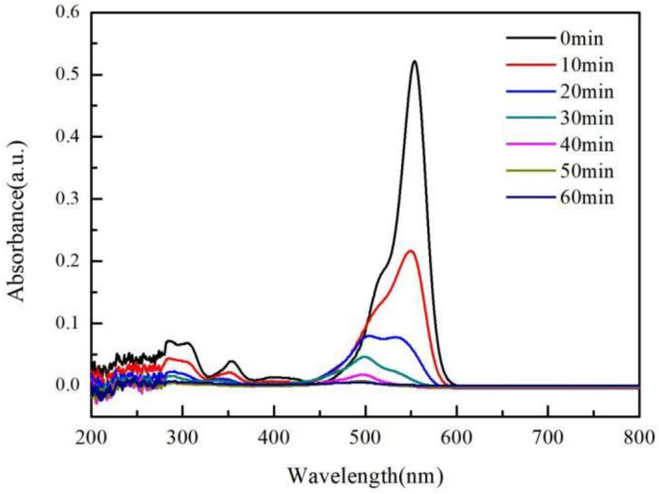
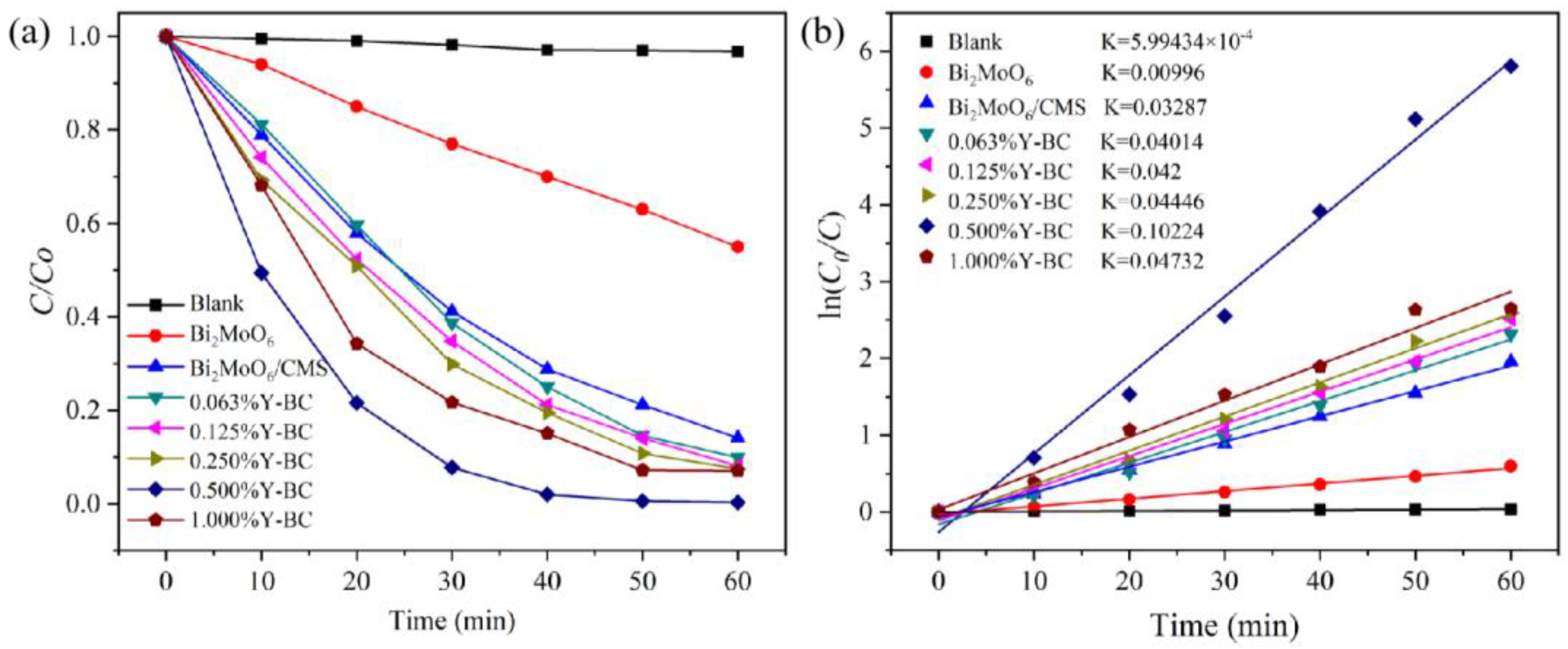
3.4. Photocatalytic Degradation of RhB

The photocatalytic activity of pure Bi2MoO6, Bi2MoO6/CMS, and Y-doped Bi2MoO6/CMS photocatalysts were investigated by testing the degradation of RhB solution under visible-light irradiation. In Figure 8a, we observed that RhB had not degraded in the absence of a photocatalyst, indicating its high structural stability. All Y-doped Bi2MoO6/CMS samples exhibited a more efficient photodegradation rate than the undoped Bi2MoO6/CMS and pure Bi2MoO6. The 0.5%Y-BC exhibited the highest photocatalytic performance, achieving a remarkable degradation rate of 99.7% after 60 min of irradiation.
Kinetic studies were conducted using the pseudo-first-order model. Figure 8b shows that 0.5% Y-BC exhibited the highest rate constant, which was three times higher than Bi2MoO6/CMS and ten times higher than pure Bi2MoO6. This is because 0.5%Y-BC has a lower recombination efficiency of photogenerated electrons and holes, higher specific surface area, and lower band gaps. However, the increase in the yttrium content to 1% would result in a lower K value. The former BET surface area analysis shows that Y doping at a high concentration could decrease the surface area. Moreover, the excess Y3+ ions in 1%Y-BC may act as a recombination center for the photogenerated charges, decreasing the photocatalytic activity [73]. Thus, it is important to carefully control the Y3+ concentration to avoid decreasing the photocatalytic efficiency caused by the recombination of electron–hole pairs. Furthermore, a comparison of the photodegradation performance of 0.5% Y-BC with other photocatalysts reported in the literature is presented in Table 2. Although the initial concentration of pollutants in this study was slightly lower than the concentrations reported in the literature, while the K value of the 0.5% Y-BC was significantly higher than that of other catalysts, indicating its superior photocatalytic performance.
Figure 9 displays the absorption spectra of RhB over time in the presence of 0.5%Y-BC under visible light. As the irradiation time increased, the absorbance of RhB at 550 nm decreased rapidly. This decrease was accompanied by a blue shift in the absorption band, which is due to the formation of a series of N-de-ethylated intermediates as RhB degrades [76]. After 60 min, RhB was entirely degraded, indicating that the dye-conjugated structure of the RhB molecule was destroyed.

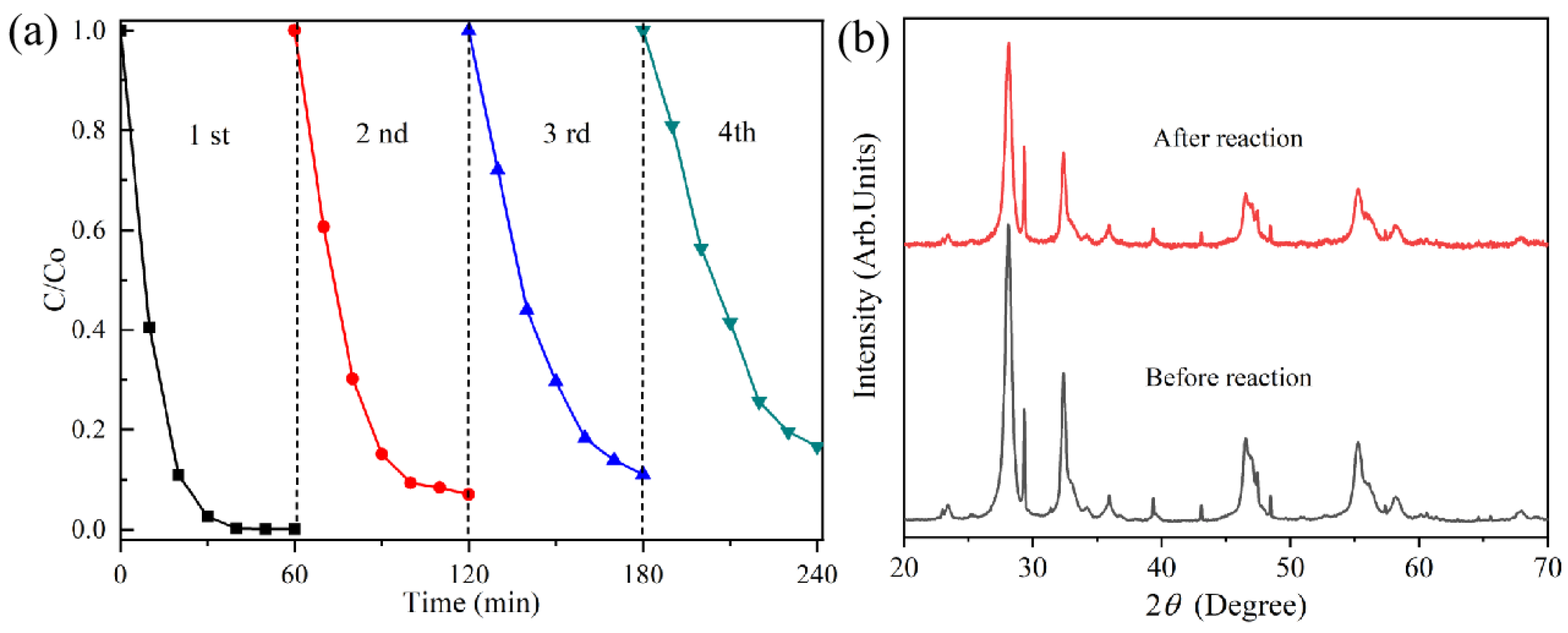
3.5. Cycle Experiment

The stability and reusability of 0.5%Y-BC was investigated via a recycling experiment, and the results are presented in Figure 10. As shown in Figure 10a, the degradation efficiency of 0.5%Y-BC for RhB decreased slightly, from 99.7% to 83.3%, after four cycles of experiments. Two factors could lead to the decrease in degradation efficiency. Firstly, there could have been a loss of catalyst mass during the centrifugation collection process. Secondly, the adsorption of the intermediate onto the photocatalyst’s surface may have resulted in a reduction in the number of exposed reactive sites, leading to a decrease in the removal efficiency for RhB. Additionally, the stability of 0.5%Y-BC was analyzed via XRD, and the results are presented in Figure 10b. Notably, the XRD data revealed that the phase of the 0.5%Y-BC remained unchanged even after four cycles of experiments, indicating its stability and reusability in conducting photocatalytic reactions.

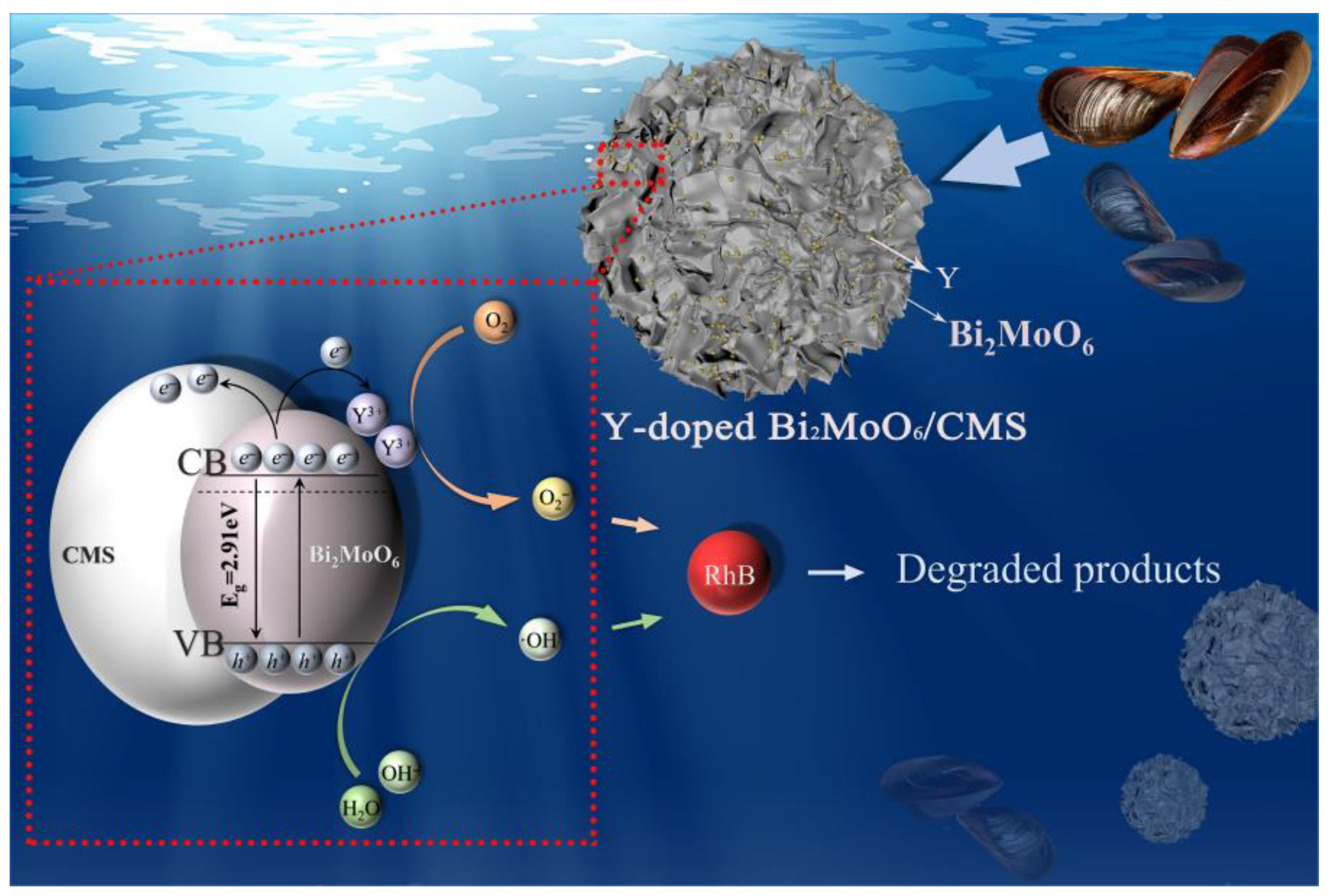
3.6. Possible Photocatalytic Mechanism of Photocatalysts

To determine the primary active radicals and explain the photocatalytic mechanism in the photocatalytic degradation of RhB by 0.5%Y-BC, active species trapping experiments were conducted. IPA, Na2C2O4, and BQ were used as scavengers of ·OH, h+, and ·O2, respectively [77]. The results, depicted in Figure 11, reveal that the photocatalytic activity of 0.5%Y-BC decreased with the addition of different active species scavengers. Specifically, the introduction of BQ and Na2C2O4 significantly lowered the photocatalytic activity of 0.5%Y-BC, suggesting that ·O2 and h+ are the primary active species involved in the RhB degradation by 0.5%Y-BC. We also observed a slight decrease in photocatalytic efficiency with the addition of IPA, indicating that ·OH also plays a crucial role in the photocatalytic reaction.
The Y3+ ion significantly enhanced the photocatalytic efficiency of Y-doped Bi2MoO6/CMS through several mechanisms. Firstly, Y doping narrowed the band gap of the photocatalysts, making it easier for photo-excited electrons to move from the CB to VB. Second, Y doping substantially increased the specific surface area of the catalyst (from 28.6385 to 42.4971 m2/g), providing more reaction sites and promoting photocatalytic degradation. Thirdly, Y doping triggered the formation of new defect sites and oxygen vacancies, which acted as electron traps to prevent the recombination of electron–hole pairs. Furthermore, mussel shells, as photocatalyst supports, have unique pores and surface structures which could offer more active sites for pollutant attachment in photocatalytic reactions than stacked catalysts [38,43]. Moreover, shells contained various trace metal elements (e.g., Se, Mn, Zn, Ti, and Sr) that could serve as doping elements, facilitating electron transfer in photocatalytic reactions, thus enhancing the photocatalytic capability of the composites [37,42]. Therefore, CMS and Y3+ ions synergistically enhanced the performance of composite photocatalysts.
A proposed mechanism, based on experimental results and the literature, explains the possible reactions in photocatalysis, as shown in Figure 12. Under visible light irradiation, Bi2MoO6 was excited, generating electron–hole pairs, with the electrons excited to the conduction band and the holes remaining in the valence band. The presence of Y3+ on the Bi2MoO6, and its specific electronic configuration with vacant 4d and 5s orbitals, could capture electrons and form Y2+, hindering the recombination of electron–hole pairs. And then, these Y2+ ions reacted with oxygen molecules to produce superoxide radicals (·O2−) [78,79,80,81]. Simultaneously, the holes in the valence band directly participate in the photo-oxidation reaction with water molecules to produce hydroxyl radicals (·OH). These active species, i.e., ·O2−, h+, and ·OH, could directly attack RhB molecules, breaking down their conjugated structure. The overall reaction could be expressed as follows:
Bi2MoO6 + hv → Bi2MoO6 (e + h+)
Y3+ + e → Y2+
Y2+ + O2 → Y3+ + ·O2
h+ + H2O → ·OH + H+
RhB + ·O2/h+/·OH → CO2 + H2O + Degradation products

4. Conclusions

In this study, a series of Y-doped Bi2MoO6/CMS was successfully developed via a simple solvothermal method based on the mussel shell. XPS results showed that Y was successfully doped into Bi2MoO6/CMS, and XRD demonstrated that Y-doped Bi2MoO6/CMS consisted of orthorhombic phase Bi2MoO6 and calcite phase CaCO3. Y-doped Bi2MoO6/CMS exhibited superior visible-light-driven catalytic performance in the degradation of RhB solution, as compared to the undoped sample. Specifically, 0.5%Y-BC with excellent stability and reusability showed the maximum degradation rate of 99.7% in 60 min, and ·O2 and h+ are the primary active species in the RhB degradation. Moreover, the improved photocatalytic activity of 0.5%Y-BC was mainly attributed to its higher surface area, narrower bandgap, and enhanced separation of electron–hole pairs. Therefore, this study provided a new approach for the high-value utilization of mussel shells, and developing high-performance photocatalysts for removing organic pollutants from wastewater.

Author Contributions

Conceptualization, L.C. and L.J.; methodology, L.C., Y.Z. and L.J.; software, L.C., Y.Z. and J.S.; validation, L.J., Y.Z. and J.G.; formal analysis, L.C. and Y.Z.; investigation, J.G. and L.C.; resources, J.G. and L.J.; data curation, L.C.; writing—original draft preparation, L.C.; writing—review and editing, L.J.; visualization, L.C. and Y.Z. All authors have read and agreed to the published version of the manuscript.

Funding

This research was funded by the Demonstration Project of Marine Economic Innovation and Development of Zhoushan City of China (NO. 2016-496), the Scientific Research Fund of Zhejiang Provincial Education Department (Y201942627) and China Scholarship Council (NO. 202008330459).

Data Availability Statement

All data that support the findings of this study are available from the corresponding author upon reasonable request.

Conflicts of Interest

The authors declare no conflict of interest.

References

  1. He, Y.; Fu, X.; Li, B.; Zhao, H.; Yuan, D.; Na, B. Highly efficient organic dyes capture using thiol-functionalized porous organic polymer. ACS Omega 2022, 7, 17941–17947. [Google Scholar] [CrossRef] [PubMed]
  2. Li, S.; Liu, W.; Zhao, S.; Li, Y.; Chen, K. Enhanced photocatalytic performance of NiS2/g-C3N4/SnS2 by improving the charge diffusion on both valence band and conduction band of carbon nitride. Chem. Sel. 2021, 6, 4440–4447. [Google Scholar] [CrossRef]
  3. Chen, W.; Wang, W.; Xing, H.; Li, W.; He, H.; Li, W.; Chu, P.K. Interfacial self-assembled dual-functional nanocomposite films for SERS monitoring of visible-light photocatalytic degradation of organic dye pollutants. Surf. Interfaces 2023, 38, 102808. [Google Scholar] [CrossRef]
  4. Ajmal, A.; Majeed, I.; Malik, R.N.; Idriss, H.; Nadeem, M.A. Principles and mechanisms of photocatalytic dye degradation on TiO2 based photocatalysts: A comparative overview. RSC Adv. 2014, 4, 37003–37026. [Google Scholar] [CrossRef]
  5. Alasri, T.M.; Ali, S.L.; Salama, R.S.; Alshorifi, F.T. Band-structure engineering of TiO2 photocatalyst by AuSe quantum dots for efficient degradation of malachite green and phenol. J. Inorg. Organomet. Polym. Mater. 2023, 33, 1729–1740. [Google Scholar] [CrossRef]
  6. Liu, C.; Zhang, Q.; Zou, Z. Recent advances in designing ZnIn2S4-based heterostructured photocatalysts for hydrogen evolution. J. Mater. Sci. Technol. 2023, 139, 167–188. [Google Scholar] [CrossRef]
  7. Hu, J.; Xia, K.; Yang, A.; Zhang, Z.; Xiao, W.; Liu, C.; Zhang, Q. Interfacial engineering of ultrathin 2D/2D NiPS3/C3N5 heterojunctions for boosting photocatalytic H2 evolution. Acta Phys.-Chim. Sin. 2023, 40, 2305043. [Google Scholar] [CrossRef]
  8. Alshorifi, F.T.; Alswat, A.A.; Mannaa, M.A.; Alotaibi, M.T.; El-Bahy, S.M.; Salama, R.S. Facile and green synthesis of silver quantum dots immobilized onto a polymeric CTS-PEO blend for the photocatalytic degradation of p-Nitrophenol. ACS Omega 2021, 6, 30432–30441. [Google Scholar] [CrossRef]
  9. Alshorifi, F.T.; Ali, S.L.; Salama, R.S. Promotional synergistic effect of Cs–Au NPs on the performance of Cs–Au/MgFe2O4 catalysts in catalysis 3,4-dihydropyrimidin-2(1H)-ones and degradation of RhB dye. J. Inorg. Organomet. Polym. Mater. 2022, 32, 3765–3776. [Google Scholar] [CrossRef]
  10. Guo, C.; Xu, J.; Wang, S.; Li, L.; Zhang, Y.; Li, X. Facile synthesis and photocatalytic application of hierarchical mesoporous Bi2MoO6 nanosheet-based microspheres. CrystEngComm 2012, 14, 3602–3608. [Google Scholar] [CrossRef]
  11. Jing, T.; Dai, Y.; Wei, W.; Ma, X.; Huang, B. Near-infrared photocatalytic activity induced by intrinsic defects in Bi2MO6(M = W, Mo). Phys. Chem. Chem. Phys. 2014, 16, 18596–18604. [Google Scholar] [CrossRef] [PubMed]
  12. Wang, J.; Zhao, C.; Yuan, S.; Li, X.; Zhang, J.; Hu, X.; Lin, H.; Wu, Y.; He, Y. One-step fabrication of Cu-doped Bi2MoO6 microflower for enhancing performance in photocatalytic nitrogen fixation. J. Colloid Interface Sci. 2023, 638, 427–438. [Google Scholar] [CrossRef]
  13. Liu, X.; Gu, S.; Zhao, Y.; Zhou, G.; Li, W. BiVO4, Bi2WO6 and Bi2MoO6 photocatalysis: A brief review. J. Mater. Sci. Technol. 2020, 56, 45–68. [Google Scholar] [CrossRef]
  14. Yang, X.; Xu, X.; Wang, J.; Chen, T.; Wang, S.; Ding, X.; Chen, H. Insights into the surface/interface modifications of Bi2MoO6: Feasible strategies and photocatalytic applications. Sol. RRL 2020, 5, 2000442. [Google Scholar] [CrossRef]
  15. Hao, Y.; Dong, X.; Wang, X.; Ma, H.; Zhang, X. Ultrathin-nanosheet-assembled Bi2MoO6 mesoporous hollow framework for realizing optimized sunlight-driven photocatalytic water oxidation. RSC Adv. 2016, 6, 102155–102158. [Google Scholar] [CrossRef]
  16. Yu, H.; Jiang, L.; Wang, H.; Huang, B.; Yuan, X.; Huang, J.; Zhang, J.; Zeng, G. Modulation of Bi2MoO6-based materials for photocatalytic water splitting and environmental application: A critical review. Small 2019, 15, 1901008. [Google Scholar] [CrossRef]
  17. Sun, S.; Wang, W. Advanced chemical compositions and nanoarchitectures of bismuth based complex oxides for solar photocatalytic application. RSC Adv. 2014, 4, 47136–47152. [Google Scholar] [CrossRef]
  18. Dutta, D.P.; Ballal, A.; Chopade, S.; Kumar, A. A study on the effect of transition metal (Ti4+, Mn2+, Cu2+ and Zn2+)-doping on visible light photocatalytic activity of Bi2MoO6 nanorods. J. Photochem. Photobiol. A Chem. 2017, 346, 105–112. [Google Scholar] [CrossRef]
  19. Yang, Z.; Wang, R.; Xu, L.; Liu, C.; Cheng, Y.; Jiang, Z.; Liu, Y.; Zhang, T.; Li, J.; Liu, X. Highly efficient flower-like Dy3+-doped Bi2MoO6 photocatalyst under simulated sunlight: Design, fabrication and characterization. Opt. Mater. 2021, 116, 111094. [Google Scholar] [CrossRef]
  20. Li, H.; Li, W.; Liu, X.; Ren, C.; Miao, X.; Li, X. Engineering of Gd/Er/Lu-triple-doped Bi2MoO6 to synergistically boost the photocatalytic performance in three different aspects: Oxidizability, light absorption and charge separation. Appl. Surf. Sci. 2018, 463, 556–565. [Google Scholar] [CrossRef]
  21. Tang, J.; Chen, Z.; Yu, X.; Tang, W.Z. Rare earth elements (lanthanum, cerium and erbium) doped black oxygen deficient Bi2O3-Bi2O3–x as novel photocatalysts enhanced photocatalytic performance. J. Rare Earth 2022, 40, 1053–1062. [Google Scholar] [CrossRef]
  22. Wang, F.; He, T.; Gao, Y.; Li, Y.; Cui, S.; Huang, H.; Yang, J. Z-scheme heterojunction Bi2MoO6/NH2-UiO-66 (Zr/Ce) for efficient photocatalytic degradation of oxytetracycline: Pathways and mechanism. Sep. Purif. Technol. 2023, 21, 124596. [Google Scholar] [CrossRef]
  23. Ge, W.; Liu, K.; Yang, P.; Deng, S.; Shen, L. Synthesis and upconversion luminescent properties of Bi2MoO6: 20%Yb3+, 2%Er3+ hollow microsphere with different W6+ ions doping. J. Solid State Chem. 2021, 297, 122064. [Google Scholar] [CrossRef]
  24. Alemi, A.A.; Kashfi, R.; Shabani, B. Preparation and characterization of novel Ln (Gd3+, Ho3+ and Yb3+)-doped Bi2MoO6 with Aurivillius layered structures and photocatalytic activities under visible light irradiation. J. Mol. Catal. A Chem. 2014, 392, 290–298. [Google Scholar] [CrossRef]
  25. Liu, X.; Zhou, W.; Li, F.; Yu, C. Eu3+ doped Bi2MoO6 nanosheets fabricated via hydrothermal-calcination route and their superior performance for aqueous volatile phenols removal. J. Taiwan Inst. Chem. Eng. 2021, 125, 276–284. [Google Scholar] [CrossRef]
  26. Moscow, S.; Kavinkumar, V.; Sriramkumar, M.; Jothivenkatachalam, K.; Saravanan, P.; Rajamohan, N.; Vasseghian, Y.; Rajasimman, M. Impact of Erbium (Er) and Yttrium (Y) doping on BiVO4 crystal structure towards the enhancement of photoelectrochemical water splitting and photocatalytic performance. Chemosphere 2022, 299, 134343. [Google Scholar] [CrossRef]
  27. Usai, S.; Obregón, S.; Becerro, A.I.; Colón, G. Monoclinic-tetragonal heterostructured BiVO4 by yttrium-doping with improved photocatalytic activity. J. Phys. Chem. C 2013, 117, 24479–24484. [Google Scholar] [CrossRef]
  28. Vaddi, D.R.; Vinukonda, K.; Patnala, R.K.; Kanithi, Y.; Gurugubelli, T.R.; Bae, J.; Koutavarapu, R.; Lee, D.Y.; Shim, J. Effect of yttrium doping on the crystal structure, optical, and photocatalytic properties of hydrothermally synthesized ZnO nanorods. Mater. Sci. Eng. B 2023, 296, 116664. [Google Scholar] [CrossRef]
  29. Vadivel, S.; Paul, B.; Maruthamani, D.; Kumaravel, M.; Vijayaraghavan, T.; Hariganesh, S.; Pothu, R. Synthesis of yttrium doped BiOF/RGO composite for visible light Photocatalytic applications. Mater. Sci. Energy Technol. 2019, 2, 112–116. [Google Scholar] [CrossRef]
  30. Wu, C.; Zhang, J.; Fang, B.; Cui, Y.; Xing, Z.; Li, Z.; Zhou, W. Self-floating biomass charcoal supported flower-like plasmon silver/carbon, nitrogen co-doped defective TiO2 as robust visible light photocatalysts. J. Clean. Prod. 2021, 329, 129723. [Google Scholar] [CrossRef]
  31. Deng, C.; Ling, X.; Peng, L.; Wang, T.; Xu, R.; Zhu, Y.; Zhang, W.; Sun, P.; Wu, Y.; Hu, H.; et al. Constructing nano CdS-decorated porous biomass-derived carbon for multi-channel synergetic photocatalytic hydrogen evolution under solar lighting. Appl. Surf. Sci. 2023, 632, 157065. [Google Scholar] [CrossRef]
  32. Alsaiari, M. Biomass-derived active carbon (AC) modified TiO2 photocatalyst for efficient photocatalytic reduction of chromium (VI) under visible light. Arab. J. Chem. 2021, 14, 103258. [Google Scholar] [CrossRef]
  33. Saleh, T.S.; Badawi, A.K.; Salama, R.S.; Mostafa, M.M.M. Design and Development of Novel Composites Containing Nickel Ferrites Supported on Activated Carbon Derived from Agricultural Wastes and Its Application in Water Remediation. Materials 2023, 16, 2170. [Google Scholar] [CrossRef]
  34. Rufford, T.E.; Hulicova-Jurcakova, D.; Zhu, Z.; Lu, G.Q. Nanoporous carbon electrode from waste coffee beans for high performance supercapacitors. Electrochem. Commun. 2008, 10, 1594–1597. [Google Scholar] [CrossRef]
  35. Ramya, S.; Vidhya, E.; Bukhari, N.A.; Hatamleh, A.A.; Nilavukkarasi, M.; Vijayakumar, S. TiO2 nanoparticles derived from egg shell waste: Eco synthesis, characterization, biological and photocatalytic applications. Environ. Res. 2022, 214, 113829. [Google Scholar] [CrossRef]
  36. Wu, Y.; Fang, X.; Shen, X.; Yu, X. Synergetic effect of photocatalytic oxidation plus catalytic oxidation on the performance of coconut shell fiber biochar decorated α-MnO2 under visible light towards BPA degradation. J. Environ. Manag. 2023, 345, 118911. [Google Scholar] [CrossRef] [PubMed]
  37. Wang, W.; Lin, F.; Yan, B.; Cheng, Z.; Chen, G.; Kuang, M.; Yang, C.; Hou, L. The role of seashell wastes in TiO2/Seashell composites: Photocatalytic degradation of methylene blue dye under sunlight. Environ. Res. 2020, 188, 109831. [Google Scholar] [CrossRef]
  38. Li, S.; Wang, C.; Liu, Y.; Xue, B.; Chen, J.; Wang, H.; Liu, Y. Facile Preparation of a Novel Bi2WO6/Calcined Mussel Shell Composite Photocatalyst with Enhanced Photocatalytic Performance. Catalysts 2020, 10, 1166. [Google Scholar] [CrossRef]
  39. Echabbi, F.; Hamlich, M.; Harkati, S.; Jouali, A.; Tahiri, S.; Lazar, S.; Lakhmiri, R.; Safi, M. Photocatalytic degradation of methylene blue by the use of titanium-doped Calcined Mussel Shells CMS/TiO2. J. Environ. Chem. Eng. 2019, 7, 103293. [Google Scholar] [CrossRef]
  40. Wanahari, W.A.; Waiho, K.; Azwar, E.; Fazhan, H.; Peng, W.; Ishak, S.D.; Tabatabaei, M.; Yek, P.N.Y.; Almomani, F.; Aghbashlo, M.; et al. A state-of-the-art review on producing engineered biochar from shellfish waste and its application in aquaculture wastewater treatment. Chemosphere 2022, 288, 132559. [Google Scholar] [CrossRef] [PubMed]
  41. Barros, M.C.; Magán, A.; Valiño, S.; Bello, P.M.; Casares, J.J.; Blanco, J.M. Identification of best available techniques in the seafood industry: A case study. J. Clean. Prod. 2009, 17, 391–399. [Google Scholar] [CrossRef]
  42. Cao, J.; Ju, P.; Chen, Z.; Dou, K.; Li, J.; Zhang, P.; Zhu, Z.; Sun, C. Trash to treasure: Green synthesis of novel Ag2O/Ag2CO3 Z-scheme heterojunctions with highly efficient photocatalytic activities derived from waste mussel shells. Chem. Eng. J. 2023, 454, 140259. [Google Scholar] [CrossRef]
  43. Cai, L.; Zhou, Y.; Wang, Z.; Chen, J.; Ji, L.; Guo, J.; Wang, Y.; Liu, J. Preparation and evaluation of a hierarchical Bi2MoO6/MSB composite for visible-light-driven photocatalytic performance. RSC Adv. 2019, 9, 38280–38288. [Google Scholar] [CrossRef]
  44. Mannaa, M.; Qasim, K.; Shorifi, F.A.; Salama, R. Role of NiO nanoparticles in enhancing structure properties of TiO2 and its applications in photodegradation and hydrogen evolution. ASC Omega 2021, 6, 30386–30400. [Google Scholar] [CrossRef]
  45. Zhang, W.; Yu, N.; Zhang, L.; Jiang, K.; Chen, Y.; Chen, Z. Synthesis of Yb3+/Er3+, co-doped Bi2WO6, nanosheets with enhanced photocatalytic activity. Mater. Lett. 2016, 163, 16–19. [Google Scholar] [CrossRef]
  46. He, M.; Li, W.; Xia, J.; Xu, L.; Di, J.; Xu, H.; Yin, S.; Li, H.; Li, M. The enhanced visible light photocatalytic activity of yttrium-doped BiOBr synthesized via a reactable ionic liquid. Appl. Surf. Sci. 2015, 331, 170–178. [Google Scholar] [CrossRef]
  47. Cao, R.; Huang, H.; Tian, N.; Zhang, Y.; Guo, Y.; Zhang, T. Novel Y doped Bi2WO6 photocatalyst: Hydrothermal fabrication, characterization and enhanced visible-light-driven photocatalytic activity for Rhodamine B degradation and photocurrent generation. Mater. Charact. 2015, 101, 166–172. [Google Scholar] [CrossRef]
  48. El-Hakam, S.A.; ALShorifi, F.T.; Salama, R.S.; Gamal, S.; El-Yazeed, W.S.A.; Ibrahim, A.A.; Ahmed, A.I. Application of nanostructured mesoporous silica/ bismuth vanadate composite catalysts for the degradation of methylene blue and brilliant green. J. Mater. Res. Technol. 2022, 18, 1963–1976. [Google Scholar] [CrossRef]
  49. Shkir, M.; Alshahrani, T. Enhancement in photo response of spray deposited Yttrium doped Bi2S3 thin films. Sens. Actuators A Phys. 2023, 351, 114169. [Google Scholar] [CrossRef]
  50. Liu, X.; Deng, H.; Yao, W.; Jiang, Q.; Shen, J. Preparation and photocatalytic activity of Y-doped Bi2O3. J. Alloys Compd. 2015, 651, 135–142. [Google Scholar] [CrossRef]
  51. Zhang, Q.; Rao, G.; Xiao, Y.; Dong, H.; Liu, G.; Zhang, Y.; Liang, J. Crystal structure, magnetic and electrical-transport properties of rare-earth-doped Sr2FeMoO6. Phys. B Condens. Matter 2006, 381, 233–238. [Google Scholar] [CrossRef]
  52. Maarisetty, D.; Mary, R.; Hang, D.R.; Mohapatra, P.; Baral, S.S. The role of material defects in the photocatalytic CO2 reduction: Interfacial properties, thermodynamics, kinetics and mechanism. J. CO2 Util. 2022, 64, 102175. [Google Scholar] [CrossRef]
  53. Zhao, K.; Liu, X.; He, Q.; Zhou, W.; Yang, K.; Tao, L.; Li, F.; Yu, C. Preparation and characterization of Sm3+/Tm3+ co-doped BiVO4 micro-squares and their photocatalytic performance for CO2 reduction. J. Taiwan Inst. Chem. Eng. 2023, 144, 104737. [Google Scholar] [CrossRef]
  54. Qin, Z.; Tian, H.; Su, T.; Ji, H.; Guo, Z. Soft template inducted hydrothermal BiYO3 catalysts for enhanced formic acid formation from the photocatalytic reduction of carbon dioxide. RSC Adv. 2016, 6, 52665–52673. [Google Scholar] [CrossRef]
  55. Zhang, J.; Xia, J.; Yin, S.; Li, H.; Xu, H.; He, M.; Huang, L.; Zhang, Q. Improvement of visible light photocatalytic activity over flower-like BiOCl/BiOBr microspheres synthesized by reactable ionic liquids. Colloids Surf. A Physicochem. Eng. Asp. 2013, 420, 89–95. [Google Scholar] [CrossRef]
  56. Krishna, R.J.; Srinivas, B.; Kumari, V.D.; Subrahmanyam, M. Sm3+-doped Bi2O3 photocatalyst prepared by hydrothermal synthesis. ChemCatChem 2009, 1, 492–496. [Google Scholar]
  57. Hu, Z.; Chen, D.; Wang, S.; Zhang, N.; Qin, L.; Huang, Y. Facile synthesis of Sm-doped BiFeO3 nanoparticles for enhanced visible light photocatalytic performance. Mater. Sci. Eng. B 2017, 220, 1–12. [Google Scholar] [CrossRef]
  58. Lu, Y.; Jia, X.; Ma, Z.; Li, Y.; Yue, S.; Liu, X.; Zhang, J. W5+-W5+ pair induced LSPR of W18O49 to sensitize ZnIn2S4 for full-spectrum solar-light-driven photocatalytic hydrogen evolution. Adv. Funct. Mater. 2022, 32, 2203638. [Google Scholar] [CrossRef]
  59. Zhang, L.; Xu, T.; Zhao, X.; Zhu, Y. Controllable synthesis of Bi2MoO6 and effect of morphology and variation in local structure on photocatalytic activities. Appl. Catal. B Environ. 2010, 98, 138–146. [Google Scholar] [CrossRef]
  60. Li, H.; Liu, J.; Hou, W.; Du, N.; Zhang, R.; Tao, X. Synthesis and characterization of g-C3N4/Bi2MoO6 heterojunctions with enhanced visible light photocatalytic activity. Appl. Catal. B Environ. 2014, 160–161, 89–97. [Google Scholar] [CrossRef]
  61. Simpson, L.J. Electrochemically generated CaCO3 deposits on iron studied with FTIR and Raman spectroscopy. Electrochim. Acta 1998, 43, 2543–2547. [Google Scholar] [CrossRef]
  62. Huang, N.; Wang, J. A TGA-FTIR study on the effect of CaCO3 on the thermal degradation of EBA copolymer. J. Anal. Appl. Pyrolysis 2009, 84, 124–130. [Google Scholar] [CrossRef]
  63. Geng, Y.; Zhang, P.; Kuang, S. Fabrication and enhanced visible-light photocatalytic activities of BiVO4/Bi2WO6 composites. RSC Adv. 2014, 4, 46054–46059. [Google Scholar] [CrossRef]
  64. Yassin, A.Y.; Abdelghany, A.M.; Salama, R.S.; Tarabiah, A.E. Structural, Optical and Antibacterial Activity Studies on CMC/PVA Blend Filled with Three Different Types of Green Synthesized ZnO Nanoparticles. J. Inorg. Organomet. Polym. Mater. 2023, 33, 1855–1867. [Google Scholar] [CrossRef]
  65. Kruk, M.; Jaroniec, M. Gas adsorption characterization of ordered organic-inorganic nanocomposite materials. Chem. Mater. 2001, 13, 3169–3183. [Google Scholar] [CrossRef]
  66. Ibrahim, A.A.; Salama, R.S.; El-Hakam, S.A.; Khder, A.S.; Ahmed, A.I. Synthesis of sulfated zirconium supported MCM-41 composite with high-rate adsorption of methylene blue and excellent heterogeneous catalyst. Colloids Surf. A Physicochem. Eng. Asp. 2021, 616, 126361. [Google Scholar] [CrossRef]
  67. Yu, J.; Yu, H.; Cheng, B.; Zhou, M.; Zhao, X. Enhanced photocatalytic activity of TiO2 powder (P25) by hydrothermal treatment. J. Mol. Catal. A Chem. 2006, 253, 112–118. [Google Scholar] [CrossRef]
  68. Wang, M.; You, M.; Guo, P.; Tang, H.; Lv, C.; Zhang, Y.; Zhu, T.; Han, J. Hydrothermal synthesis of Sm-doped Bi2MoO6 and its high photocatalytic performance for the degradation of Rhodamine B. J. Alloys Compd. 2017, 728, 739–746. [Google Scholar] [CrossRef]
  69. Wang, C.; Gu, C.; Zeng, T.; Zhang, Q.; Luo, X. Bi2WO6 doped with rare earth ions: Preparation, characterization and photocatalytic activity under simulated solar irradiation. J. Rare Earth 2021, 39, 58–66. [Google Scholar] [CrossRef]
  70. Huang, H.; Wang, S.; Tian, N.; Zhang, Y. A one-step hydrothermal preparation strategy for layered BiIO4/Bi2WO6 heterojunctions with enhanced visible light photocatalytic activities. RSC Adv. 2014, 11, 5561–5567. [Google Scholar] [CrossRef]
  71. Tian, G.; Chen, Y.; Zhai, R.; Zhou, J.; Zhou, W.; Wang, R.; Pan, K.; Tian, C.; Fu, H. Hierarchical flake-like Bi2MoO6/TiO2 bilayer films for visible-light-induced self-cleaning applications. J. Mater. Chem. A 2013, 1, 6961–6968. [Google Scholar] [CrossRef]
  72. Tian, J.; Sang, Y.; Yu, G.; Jiang, H.; Mu, X.; Liu, H. A Bi2WO6-based hybrid photocatalyst with broad spectrum photocatalytic properties under UV, visible, and near-infrared irradiation. Adv. Mater. 2013, 25, 5075–5080. [Google Scholar] [CrossRef]
  73. Yu, C.; Wu, Z.; Liu, R.; He, H.; Fan, W.; Xue, S. The effects of Gd3+ doping on the physical structure and photocatalytic performance of Bi2MoO6 nanoplate crystals. J. Phys. Chem. Solids 2016, 93, 7–13. [Google Scholar] [CrossRef]
  74. Xue, M.; Meng, F.; Ma, Y.; Zhou, S. Growing of ultra-thin Bi2MoO6 nanoflowers on Co/N-doped graphitic carbon nanoshells as attractive custom supports: Excellent photocatalytic degradation activity for pollutants. Appl. Surf. Sci. 2023, 613, 156100. [Google Scholar] [CrossRef]
  75. Varma, R.; Chaurasia, S.; Patel, N.; Bhanage, B.M. Interplay of adsorption, photo-absorption, electronic structure and charge carrier dynamics on visible light driven photocatalytic activity of Bi2MoO6/rGO (0D/2D) heterojunction. J. Environ. Chem. Eng. 2020, 8, 104551. [Google Scholar] [CrossRef]
  76. Xu, C.; Wu, H.; Gu, F.L. Efficient adsorption and photocatalytic degradation of Rhodamine B under visible light irradiation over BiOBr/montmorillonite composites. J. Hazard. Mater. 2014, 275, 185–192. [Google Scholar] [CrossRef]
  77. Liu, C.; Xiao, W.; Liu, X.; Wang, Q.; Hu, J.; Zhang, S.; Xu, J.; Zhang, Q.; Zou, Z. Rationally designed Ti3C2 MXene/CaIn2S4 Schottky heterojunction for enhanced photocatalytic Cr (VI) reduction: Performance, influence factors and mechanism. J. Mater. Sci. Technol. 2023, 161, 123–135. [Google Scholar] [CrossRef]
  78. Bouattour, S.; Rego, A.M.B.D.; Ferreira, L.F.V. Photocatalytic activity of Li+–Rb+–Y3+ doped or codoped TiO2 under sunlight irradiation. Mater. Res. Bull. 2010, 45, 818–825. [Google Scholar] [CrossRef]
  79. Zhang, Q.; Fu, Y.; Wu, Y.; Zhang, Y.N.; Zuo, T. Low-cost Y-doped TiO2 nanosheets film with highly reactive {001} facets from CRT waste and enhanced photocatalytic removal of Cr (VI) and methyl orange. Acs Sustain. Chem. Eng. 2016, 4, 1794–1803. [Google Scholar] [CrossRef]
  80. Wu, Y.; Gong, Y.; Liu, J.; Zhang, Z.; Xu, Y.; Ren, H. B and Y co-doped TiO2 photocatalyst with enhanced photodegradation efficiency. J. Alloys Compd. 2016, 695, 1462–1469. [Google Scholar] [CrossRef]
  81. Liu, J.; Zhao, W.; Wang, B.; Yan, H. Synthesis, characterization and photocatalytic properties of the Y-doped polar borate photocatalysts: Bi2ZnOB2O6: xY3+. Chem. Phys. Lett. 2019, 734, 136707. [Google Scholar] [CrossRef]
Figure 1. (a) X-ray diffraction patterns of Bi2MoO6/CMS and Y-doped Bi2MoO6/CMS; (b) magnified patterns with the 2θ between 27°and 30° X-ray diffraction patterns.
Water 15 03478 g001
Figure 2. XPS survey spectra (a) and high-resolution XPS spectra of Bi4f (b), Mo3d (c), Y3p (d) and O1s (e,f) of Bi2MoO6/CMS and Y-doped Bi2MoO6/CMS.
Water 15 03478 g002
Figure 3. FT-IR spectra of CMS, pure Bi2MoO6, Bi2MoO6/CMS, and Y-doped Bi2MoO6/CMS.
Water 15 03478 g003
Figure 4. SEM image of Bi2MoO6 (a), CMS (b), Bi2MoO6/CMS (c), 0.063%Y-BC (d), 0.125%Y-BC (e), 0.25%Y-BC (f), 0.5%Y-BC (g), 1%Y-BC (h); TEM image of 0.5%Y-BC (i).
Water 15 03478 g004
Figure 5. Nitrogen adsorption–desorption isotherms and pore size distributions of Bi2MoO6/CMS and Y-doped Bi2MoO6/CMS.
Water 15 03478 g005
Figure 6. UV-vis DRS spectra of Bi2MoO6, Bi2MoO6/CMS, and Y-doped Bi2MoO6/CMS. The inset is the plot of (αhv)2 versus hv for Bi2MoO6, Bi2MoO6/CMS, and Y-doped Bi2MoO6/CMS.
Water 15 03478 g006
Figure 7. PL spectra of Bi2MoO6/CMS and 0.5%Y-BC.
Water 15 03478 g007
Figure 8. The photocatalytic degradation curves (a) and kinetics curves (b) of RhB over Bi2MoO6, Bi2MoO6/CMS, and Y-doped Bi2MoO6/CMS.
Water 15 03478 g008
Figure 9. Time-dependent UV-vis adsorption spectral of RhB degraded by 0.5%Y-BC.
Water 15 03478 g009
Figure 10. (a) Recyclability of 0.5%Y-BC for the degradation of RhB under visible light for four cycles; (b) the XRD patterns of the original and recycled 0.5%Y-BC.
Water 15 03478 g010
Figure 11. The effects of photogenerated carriers trapping on photocatalytic degradation activity of 0.5%Y-BC.
Water 15 03478 g011
Figure 12. The mechanism of photodegradation RhB by Y-doped Bi2MoO6 composites.
Water 15 03478 g012
Table 1. Physico-chemical properties of the prepared photocatalysts.
PhotocatalystsAverage
Crystallite Size
(nm)
BET Surface Area
(m2/g)
Average Pore Diameter (nm)Pore Volume (cm3/g)Band Gap Energy
(eV)
Bi2MoO6/CMS10.8128.649.630.073.00
0.063%Y-BC10.6529.919.760.072.98
0.125%Y-BC8.4937.9414.710.142.94
0.25%Y-BC8.3838.2314.070.132.92
0.5%Y-BC8.9142.507.920.082.91
1%Y-BC9.1520.608.150.042.83
Table 2. Comparison of the photodegradation performance of Y-doped Bi2MoO6/CMS with other photocatalysts reported in the literature.
PhotocatalystCatalyst DosageLight SourceIrradiation TimePollutantsEfficiencyK (min−1)Ref.
0.5%Y-BC20 mg300 W Xe lamp60 minRhB, 6 mg/L, 40 mL99.7%0.1022This work
Bi2WO6/Calcined mussel Shell20 mg300 W Xe lamp150 minRhB, 10.0 mg/L, 100 mL98.4%0.0248[36]
TiO2/Seashell100 mgXe lamp140 minMB, 10 mg/L, 100 mL96%/[35]
TiO2/Calcined Mussel Shell40 mgUV light300 minMB, 10 mg/L, 20 mL97%/[39]
Co/N-graphitic carbon@Bi2MoO610 mg300 W Xe lamp75 minRhB/MO, 10 mg/L, 200 mL99.54%0.0335[74]
Bi2MoO6/rGO20 mg150 W Xe lamp180 minRhB, 10 mg/L,/50 mL100%/[75]
Gd3+-doped Bi2MoO6100 mg300 W Xe lamp180 minRhB, 10 mg/L,/50 mL90.2%0.0122[24]
Ho3+-doped Bi2MoO6100 mg300 W Xe lamp180 minRhB, 10 mg/L,/50 mL81.9%0.0078[24]
Yb3+-doped Bi2MoO6100 mg300 W Xe lamp180 minRhB, 10 mg/L,/50 mL79.8%0.0091[24]
Dy3+-doped Bi2MoO6100 mg300 W Xe lamp40 minRhB, 10 mg/L, 100 mL100%/[19]
Disclaimer/Publisher’s Note: The statements, opinions and data contained in all publications are solely those of the individual author(s) and contributor(s) and not of MDPI and/or the editor(s). MDPI and/or the editor(s) disclaim responsibility for any injury to people or property resulting from any ideas, methods, instructions or products referred to in the content.

