Article

Microplastic Particles’ Effects on Aquatic Organisms and Their Role as Transporters of Organic Pollutants

by
Gabriela Aguirre-Martínez
1,2,*,
Maria Virginia Carrizo
1 and
Lisette Zenteno-Devaud
3,4
1
Química y Farmacia, Facultad de Ciencias de la Salud, Universidad Arturo Prat, Avenida Arturo Prat Chacón 2120, Iquique 1100000, Chile
2
Instituto de Estudios de la Salud (IES), Universidad Arturo Prat, Casilla 121, Iquique 1100000, Chile
3
Centro de Estudios del Cuaternario Fuego-Patagonia y Antártica, Avenida España 184, Punta Arenas 6200000, Chile
4
Departamento de Ecología, Facultad de Ciencias, Universidad Católica de Santísima Concepción, Alonso De Ribera 2850, Concepción 4090541, Chile
*
Author to whom correspondence should be addressed.
Water 2023, 15(16), 2915; https://doi.org/10.3390/w15162915
Submission received: 30 June 2023 / Revised: 25 July 2023 / Accepted: 27 July 2023 / Published: 12 August 2023

Abstract

:
Microplastic (MP) contamination is considered a growing problem in terms of its production and observed impacts on aquatic organisms. In this study, we investigated the adverse effects that could occur from pure polyethylene (PE) MPs and PE contaminated with phenanthrene (Phe) and chlorpyrifos (CPF) in D. magna and podocopid ostracods. The organisms were exposed to different sizes (1–5, 27–32, 45–53, and 212–250 μm) and concentrations of MPs (0, 16, 160, 1600, 16,000 particles/mL) using a static and dynamic model of exposition. The results indicate that both daphnia and ostracods can ingest MPs, and the effect observed in most cases is directly proportional to the concentration of MPs. Exposure to pure MP did not affect the organisms. However, at 21 days, they induced a significant decrease (p < 0.05) in neonatal daphnia compared to the control. MP + CPF negatively affected the crustaceans when concentration, and exposure time were increased and when the size of the MPs was decreased. Neonatal daphnia were the most sensitive compared to juveniles and adults. MP + Phe caused mortality when increasing the concentration of MPs and in D. magna juveniles with increasing size, while in ostracods, mortality increased with decreasing particle size. The effect of the MPs in crustaceans would depend on the concentration, exposure time, size of the organisms, and size of the MPs. It is also shown that the toxicity of PE increases when these particles are associated with a contaminant, which would indicate its role as a transporter of organic contaminants.

1. Introduction

Plastic is a versatile product which is widely used in all types of consumer and industrial applications, as it works well as a thermal and electrical insulator along with being resistant to acids, alkalis, and dissolvents. Because of these properties and its low cost, its consumption has skyrocketed worldwide, leading to massive production growth over the last 50 years, particularly in recent decades [1], creating a persistent global pollution problem with particular repercussions in aquatic environments. It is estimated that over 8 million tons of plastic enter the oceans annually [2], and plastic is now found in all main oceanic gyres [3], the polar oceans [4], and deep-water sediments [5]. Plastic is even considered a main component of existing marine garbage since between 60% and 80% of this trash is plastic. In some ocean areas, this can reach as high as 95% of garbage [6]. Among the plastic components present in the various environmental compartments, polyethylene (PE) is the dominant polymer, followed by polypropylene (PP) and polystyrene (PS) [7,8,9].
Plastic is known to deteriorate and fragment in the environment [10,11], breaking down into microplastic (<5 mm) and nanoplastic (<1 µm) [12], with the smallest particles being the most prevalent in the ocean surface [13,14]. In the aquatic environment, microplastic pollution (MP) is considered a growing problem since the proportion of MP rises with its worldwide production and use [15]. The presence of MP has currently been shown on terrestrial ecosystems [16] but also in aquatic environments including water, sediment, and aquatic organisms [12,17,18] Findings revealed the appearance of MP in lakes [19], rivers [20], estuaries [21], and oceans [22], along coastlines, and even in deepwater sediments [3]. MP has been reported at concentrations of up to 100,000 particles/m3 in water [23]. Microplastics have also been described in lakes and rivers of Europe, North America, South America, Africa, and Asia [24,25].
MP can be divided into primary and secondary microplastics. Primary microplastics are produced and discharged into the environment at the micro-scale size (as with pearls used in cosmetics). Secondary microplastics arise from decomposition of larger plastic fragments due to UV radiation and physical abrasion. In the water, this degradation is slowed due to lower temperatures and limited UV radiation penetration, making plastic wastes persist and accumulate [13].
MP impacts on aquatic organisms’ health have been researched across various species. The effects can be (I) physical (due to the particles’ shape and size) and (II) chemical (due to the presence of additives and/or absorbed chemical pollutants). The physical effects of MP are mainly internal abrasions, intestinal blockage, and reduced animal feeding and energy assimilation, leading to reduced energy and fertility. The particles’ size is another important factor for aquatic animals’ health. Particles over 150 µm are generally not absorbed and only produce local inflammation. However, smaller particles can induce systemic exposure and even penetrate organs [26]. MP can also be directly ingested through trophic transference when predators consume prey contaminated with MP [27]. Acute and chronic studies indicated that microplastics and nanoplastics can cause adverse effects, such as decreased survival, immobility, abnormal behavior, feeding activity alterations, reduced survival, reproductive alterations, and abnormal development in the new generation. MP exposure can also induce histopathological alterations and oxidative stress and can even alter lipid metabolism [28].
The ecological impact of MP has been studied in aquatic ecosystems, indicating adverse effects from these particles among various species, including the zebra fish Danio rerio [29], Scrobicularia plana molluscs [30], Carcinus maenas crabs [31], and Mytilus edulis mussels [31] as well as in the crustacean Daphnia magna [12]. Immobility occurred in the latter species as particle concentrations rose [25]. There is also evidence that MP can be an important vector for transporting organic pollutants (OP) to organisms. The hydrophobic nature of plastic makes microplastic surfaces absorb hydrophobic organic pollutants (HOP) present in aqueous solutions. Due to their small size, aquatic organisms at the bottom of the food chain can ingest MP, so their pollution transfer effects could be more significant than larger plastic wastes [32]. In this sense, it has been shown that polyethylene (PE) presents a greater capacity than other plastic types for absorbing pollutants present in the environment [33].
Persistent organic pollutants (POP), including polycyclic aromatic hydrocarbons (PAH) and organochlorine pesticides (OCP), among others, are stable lipophilic chemicals which adhere to and concentrate on the hydrophobic surface of plastics in concentrations similar to those recorded in the environment [34]. PAHs are compounds generated during wood distillation, gas works, oil refineries, runoff from asphalt pavements, car emissions, oil spills, and incomplete combustion of fossil fuels and organic matter [35], which occur in the environment as complex mixtures of different chemical substances, generally comprised of 2 to 10 aromatic rings [36]. These substances are considered toxic for organisms, with mutagenic, teratogenic, and carcinogenic effects [37]. One model compound for PAH is phenanthrene (Phe), classified by the US Environmental Protection Agency (EPA) as a priority pollutant, contributing to over 49% of all PAHs in the environment. It is considered one of the main atmospheric hydrocarbon pollutants [38]. The toxicity and lethality of HAPs have been determined among some species of aquatic organisms, such as the microalgae Chlorella vulgaris [39], the amphipod Gammarus roeseli [40], D. rerio [41], the African sharptooth catfish Clarias gariepinus [42], and others [43].
One of the most commonly used OCPs is chlorpyrifos (CPF) [44], an organophosphate non-systemic insecticide leading to an inhibition of the enzyme cholinesterase [44]. This insecticide is currently in wide use for a variety of crops and is often detected in surface waters worldwide. In fact, various studies indicate CPF pollution in various countries’ waters, with levels of up to 17,000 ng/L [45]. In Chile, it is one of the most common pesticides [46]. Acute and chronic CPF toxicity among freshwater invertebrates is well documented, with evidence that environmental CPF concentrations have embryo-toxic effects in Daphnia magna [44].
Studies suggest that filter organisms, such as crustaceans, are the most likely to ingest suspended MP. Among these, D. magna (order Cladocera), has been widely used as a model organism in various toxicity bioassays to evaluate water pollutants. D. magna is an ideal indicator species. It has a wide geographical distribution, plays a key role within the zooplankton community, can be easily lab-raised, reproduces via parthenogenesis, has a short life cycle with high production of offspring, is sensitive to water quality changes, and has a transparent exoskeleton which allows for direct observation. They are also prey for many larger invertebrates and thus play a unique and crucial role in the aquatic food chain. They represent a possible trophic transference path for MP [47,48].
Benthic crustaceans of the ostracod class, and particularly from the order Podocopida, have been identified as an ideal organism for environmental studies, as they are highly sensitive to physical–chemical changes, and they are abundant, small (0.2 to 5 mm), and easy to gather and maintain, allowing for low-cost analysis. Their sensitivity to environmental variables is useful for estimating ecological alterations in bodies of water and their environmental impacts. They are also used for paleo-environmental reconstructions [49]. For example, they have been used to observe changes in diversity and abundance due to heavy metal contamination in the water [50]. However, evidence about the impact of microplastics on this organism is still scarce.
The objective of this study was to evaluate the effects which could arise from exposure to PE particles of various sizes (1–5, 27–32, 45–53, and 212–250 μm), both pure particles and those contaminated with phenanthrene (Phe) and chlorpyrifos (CPF), using D. magna and ostracods of the Podocopida subclass as model organisms.

2. Materials and Methods

2.1. Selection of Particle Sizes and Preparation of Microplastics (MP)

The microspheres of PE were acquired from Cospheric (Innovations in Microtechnology, Santa Barbara, CA, USA). MP size was selected based on the size of the particles which the chosen crustaceans could ingest (range of 1–70 µm) [51]. Particles above this range were used to evaluate the effects induced by the contact of MP with the organisms. The MP sizes used correspond to: ME with green fluorescence, 1–5 µm and a density of 1.3 g/cm3; ME with orange fluorescence, 27–32 µm and a density of 1.00 g/cm3; ME with red fluorescence, 45–53 µm and a density of 1.09 g/cm3; and ME with orange fluorescence, 212–250 µm and a density of 1.00 g/cm3.

2.2. Preparation of Pure Microplastics

A stock microplastic solution was prepared for each particle size, with the addition of Tween 20 [52] (0.1% v/v Winkler, CAS: 9005-64-5) to ensure free MP dispersion. We used an MP stock of 1–5 μm = 5 mg/L (15.520 p/mL); an MP stock of 27–32 μm = 50 mg/L (3720 p/mL); an MP stock of 45–53 μm = 200 mg/L (3240 p/mL); and an MP stock of 212–250 μm = 35.000 mg/L (5424 p/mL).
These MP stocks underwent centrifuging for 20 s to homogenize the suspension. The number of particles for each MP stock was verified using a Neubauer chamber. After this, the desired MP concentrations of 16, 160, 1600, and 16,000 particles/mL were prepared in the test medium for each size selected in a final volume of 50 mL. These concentrations were selected based on concentrations of MP found in the aquatic environment which were previously reported, as indicated in Table 1. The high particle number was selected based on the highest environmental concentration found (100,000 particles/m3, Amsterdam Canals) [23] and multiplied by a factor of 10 since MP presence in aquatic environments is rising due to plastic production increasing exponentially every year. Furthermore, Law et al. (2014) indicated that both the median and mean amount of MP in the surface layer of the North Pacific Gyre increased by around a factor of 10 from 1972–1985 and 2002–2012 [53].

2.3. MP Behavior Tests in an Aqueous Solution

A preliminary experiment was conducted to determine the most appropriate agitation method for evaluating microplastic toxicity for D. magna. To evaluate the behavior of MP in an aqueous solution, three tests were conducted with Tween 20 surfactant 0.1% v/v, 0.05% v/v, and without surfactant as well, using the Twist Shaker TW3 (FINEPCR, Gunpo-si, Korea), VWR Shaker model 3500 (Avantor, Radnor, PA, USA), and Scilogex MS-M-S10 (Scilogex, Rocky Hill, CT, USA) (dynamic methodology) units for 48 h.

2.4. Preparation of Contaminated Microplastics

The stock solution of phenanthrene (Phe) (0.04 mg/mL) (Merck CAS: 85-01-8, purity > 95%, Boston, MA, USA) was prepared using 95% ethanol (Winkler CAS: 64-17-5, Cologno Monzese, MI, USA) as a dissolvent [69] in a 1:1 proportion. For chlorpyrifos (CPF), given its moderate hydrophilic nature (log Kow = 3.31–5.27) [70], the stock solution (0.04 mg/mL) was prepared in 95% acetone (Merck, CAS: 67-64-1) in a 1:1 proportion.
To avoid any loss of these organic compounds, MPs were exposed to contaminants in amber glass bottles covered with tinfoil following the protocol established by Cornier et al., 2019 [71]. The 50 mL bottles with MP + Phe and MP + CPF were filled up to their maximum capacity to avoid any volatilization of the chemical compound and shaken for 48 h (Scilogex MS-M-S10) at room temperature (20 ± 1 °C). The contaminated particles were then washed with MilliQ water and filtered using a polycarbonate filter membrane (PCTE). Particles were recovered carefully directly from the filter membrane and stored as stock in a 50 mL amber glass container. The number of particles for each stock of MP + Phe and MP + CPF was verified using a Neubauer chamber. After this, desired MP concentrations of 16, 160, 1600, and 16,000 particles/mL were prepared in the test medium for each MP size in a final volume of 50 mL.

2.5. Organism Obtaining

Daphnia magna in newborn (0.7–1.4 mm), juvenile (1.4–2.0 mm), and adult sizes (>2.0 mm) [72] and adult Podocopida ostracods (1–2 mm) were obtained from an isolated stock provided by the Toxicology Laboratory from the Health Sciences Faculty at Universidad Arturo Prat. These organisms were raised under controlled laboratory conditions: temperature 20 ± 2 °C, hardness CaCO3 5° dKH, and total hardness 28° dGH, NH4+ < 0.2 mg/L, NH3 < 0.1 mg/L, NO2 0.1 mg/L, NO3 2 mg/L, PO4 1 mg/L, pH 7.2, O2 dissolved >82%, photoperiod od 12 h–12 h.

2.6. Bioassays

The organisms’ exposures were carried out under controlled physico-chemical parameters similar to those indicated in point 2.5 for their cultivation and maintenance. Feeding for both crustaceans was performed with a concentrated mixture of Rhodomonas minuta, Cryptomonas sp. and Spirulina sp. The bioassays were performed based on a standardized protocol (ISO 6341:2012 [73], OECD directive 202 [74], and OECD directive 211 [75]) following two models: (1) static bioassay: a system wherein the studied solution and the organisms were placed into a recipient or chamber and left there for the duration of the study without renewing the test solution [73]; (2) dynamic or rotating bioassays: a system in which the studied solution and the organisms were placed into a recipient or chamber, followed by them being constantly shaken for the duration of the experiment using a TWIST SHAKER TW3 (FINEPCR), velocity 3.5, without renewing the test solution.
The bioassays performed were of a static type for all MP sizes, although for MP particles of 1–5 and 27–32 µm, we also included a dynamic or rotatory bioassay.

2.7. Acute Toxicity Bioassays

To expose the organisms to pure MP, we used newborn, juvenile, and adult D. magna specimens, and exposure for newborns was only to MPs at the 1–5 and 27–32 µm size because newborns cannot ingest bigger particles. To evaluate the toxicity of MP + Phe, we used juvenile and adult D. magna for all particle sizes. For exposure to MP + CPF, we used microspheres of the 1–5, 27–32, and 45–53 µm sizes among juvenile and adult D. magna. To carry out the bioassays with podocopid ostracods, exposure to pure MP and MP + Phe was of a static type for all sizes since this is a benthic animal. Experiments using MP + CPF were conducted using microspheres of the 1–5, 27–32, and 45–53 µm sizes. The daphnia (n = 10) were placed into precipitated cups containing 50 mL of filtered conditioned water and MP (pure and contaminated MP + Phe and MP + CPF) at various concentrations of 0, 16, 160, 1600, and 16,000 particles/mL. Each treatment was performed in triplicate. Each precipitated cup was covered with aluminum foil to avoid contamination from outside and to reduce evaporation. The bioassays lasted 48 h. The responses (immobility, mortality) were recorded after 24 and 48 h. The presence of MP within the organism was observed using a stereomicroscope (MOTIC, ST-36, EQUILAB, Louisville, KY, USA). The daphnia were considered immobilized when they could no longer swim after 15 s of soft agitation, according to US EPA guidelines (United States Environmental Protection Agency, 1987). To evaluate microplastics’ distribution in the water column and within the organisms, we used UV light from an SFA-BLFS-RB System. This was also used for acute exposure for podocopid ostracods.

2.8. Chronic Toxicity Bioassay

This bioassay was conducted using D. magna. One newborn was placed into a precipitated cup containing each of the treatments prepared with conditioned filtered water at a final volume of 50 mL. The treatments include pure MP, MP + Phe, and MP+ CPF at a low concentration of MP (160 particles/mL), a high concentration (1600 particles/mL), and a control (0 particles). Tests were performed in triplicate, and each precipitated cup was covered with aluminum foil. The bioassays lasted 21 days, with the subjects being fed with microalgae between 1 × 10⁵ and 5 × 10⁵ cel/mL. Chronic exposure effects were examined in the basic life history parameters of D. magna (growth, total number of descendants, and mortality). MP presence was also observed within the organism via a stereomicroscope (MOTIC, ST-36, EQUILAB). To evaluate microplastics’ distribution within the water column, we used UV light from an SFA-BLFS-RB System. Exposure for MP + CPF was performed with particles at 1–5, 27–32, and 45–53 µm.

2.9. Statistical Analysis

Data were analyzed using the SPSS/PC + (15.0) statistical packet. The significant differences between the control individuals and the organisms exposed to various treatments were determined using a one-way ANOVA, followed by a Pearson multiple correlation analysis comparison. The significance level was set at 0.05. EC50 values (with 95% confidence intervals) were estimated using an interpolation method with the ICPin software (https://www.epa.gov/chemical-research/toxicity-estimation-software-tool-test, accessed on 30 June 2023) provided by USEPA [76] via a Probit analysis [77] and verified with the AAT Bioquest LC50 Calculator program.

3. Results and Discussion

3.1. MP Particles’ Behavior in an Aqueous Solution

Our observations about the behavior of PE particles in aqueous solutions showed that, for all sizes, these tend to remain on the surface and form aggregates. It is thus essential to use Tween 20 (0.1% v/v) for particles between 45–53 and 212–250 µm, although its use is optional for the smaller sizes tested in this study. However, following the action of the surfactant, the MP was deposited into the bottom of the recipient, which is why we studied a dynamic model to observe whether these particles managed to be in suspension. For the evaluation of the dynamic model, we ruled out the use of the magnetic Scilogex MS-M-S10 agitator, as it negatively impacted the survival of the test daphnia, but it was the most efficient at maintaining the particles in suspension in the body of water. The VWR Shaker model 3500 agitator also negatively impacted the organisms, since in order for the microspheres to remain in suspension in the aqueous solution, velocity had to be high enough that it harmed the water fleas. We ultimately used the Twist Shaker TW3 agitator (FINEPCR), which did not affect the behavior of D. magna. We observed that it was able to maintain the MPs in suspension for the 1–5 and 27–32 µm sizes. However, these sizes were able to remain in suspension for the static model. For the 212–250 and 45–53 µm sizes, it was not for the particles to remain in the center. The static modality was thus the most appropriate for all PE particles. No acute or chronic bioassays were performed with MP + CPF particle sizes of 212–250 µm, since during the filtration process after 48 h of contamination with pesticide, we observed that the particles’ size and integrity were altered. Static bioassays presented sedimentations of 45–53 and 212–250 µm particles, while MPs of 1–5 and 27–32 µm sizes were relatively stable in the medium. This sedimentation decreased MP concentration in the medium. However, observation during our study indicated that both organisms ingested the particles in the bottom of the beakers.

3.2. Plastic Particle Ingestion among Test Organisms

We observed that after acute exposure, D. magna and ostracod test organisms presented plastic particles both inside their bodies and stuck to their outer surface (Figure 1). Thoracic appendices and antennae, including microstructures for movement or to filter food particles, also had plastic particles adhering from the 1–5, 27–32, and 45–53 µm size ranges. Particles from the 1–5 µm (Figure 1a,d), 27–32 µm (Figure 1b,e), and 45–53 µm (Figure 1c,f) size ranges were ingested by both daphnia and ostracods, and this intake was observed during all treatments regardless of concentration, while the larger particles (212–250 µm) were not ingested by these organisms. The greatest MP density was mainly observed to accumulate in the digestive tract, with 1–5 µm microspheres being the most abundant both within the intestinal tract and adhering to daphnia’s antennae (see Figure 1a). A mass of accumulated plastic particles was observed throughout the intestinal tracts of daphnia and ostracods since plastic cannot be digested or decomposed (Figure 1). For D. magna, MP accumulation was observed directly under the microscope both with and without UV lights, while the Ostracods were observed under the microscope using UV light. The latter case was because these organisms’ characteristic carapace impedes digestive tract observations without this light.
Selected organisms are filter-feeding, and the ingestion of MP particles could affect their wellbeing since it is assumed that these creatures cannot separate or distinguish microspheres from food particles, leading to MP particles being ingested along with the food present. This action could block their digestive tracts, and thus negatively affect their nutrition, since a digestive tract saturated with particles could lead to a false sensation of fullness and lead to starvation, as they could not obtain their nutritional requirements for survival. Results similar to our own indicate that after 48, 72, and 96 h, particles of 1 μm were present within the intestines of D. magna exposed to various MP concentrations (12.5–400 mg/L) [25]. On the other hand, particles’ adherence to daphnia thoracic appendices and antennae could influence normal antenna movements, negatively impacting their swimming and feeding behavior (Figure 1a). Similar to our findings, another study showed that MP, apart from being ingested by water fleas, also became stuck in their antennae [78].

3.3. Acute Toxicity from Pure MP in D. magna and Podocopid Ostracods

Microplastics can also cause physiological damage to organisms, including abnormal metabolism and energy budget changes [79], resulting in altered behavior [80] and survival [72]. This was reflected in our results, since we observed that during experiments with juvenile water fleas, there was a mortality rate of 53.3% after 48 h following exposure to MPs of 1–5 μm at a concentration of 16,000 particles/mL, which was a significant mortality compared to the control (p < 0.05). It should be noted that mortality induced by the smaller particles of 1–5 μm (Figure 2a) was significantly dependent on the concentration (p < 0.05; r = 0.7). A study similar to our own reported an immobilization of daphnia exposed for 72 to 200 mg/L of 1 μm PE MPs [25]. However, in the present study, at the end of the acute tests performed with newborn D. magna and adult specimens, despite the visual confirmation of ingesting MPs of 1–5, 27–32, and 45–53 μm present in the digestive tract, these did not cause the organisms to die (Figure 3a–c). The same thing occurred with juvenile daphnia exposed to MPs of 27–32 and 45–53 μm (Figure 2b,c). According to our results, newborns could be less sensitive to MP particles, possibly due to a lower MP intake rate given that their size impedes any attempt to do so. Meanwhile, adults could be more resistant to particle exposure than juvenile daphnia, considering their size. All the tested organisms exposed to 212–250 μm MPs were not affected since these particles could not be ingested by the organisms. However, there was also no mortality due to contact with these particles in this species (Figure 2d and Figure 3d). Similar to our results, a study with D. magna showed that adults (9 days old) were less sensitive to polystyrene (PS) MP particles of 6 μm compared to juveniles [78]. Our results align with those reported in another study, indicating no observations of mortality in D. magna after 48 h of exposure to 100 μm PE pearls (400 mg/L) [25] and textile fibers (up to 1400 μm long, 100 mg/L) [81]. By contrast, other studies indicate adverse effects such as reduced body size and severe reproductive alterations for water fleas exposed to 0.07 μm PS MPs (100 mg/L) [82], while a different study indicated that 0.1 μm PS particles (1 mg/L) decreased feeding rates for D. magna [83].
Bioassays with podocopid ostracods indicated mortality among these organisms after 48 h of exposure to all MP sizes, although this mortality was no higher than 40% in any of the treatments compared to the control (Figure 4a–d). This mortality was significantly different from the control (p < 0.05) for treatment with particles of 212–250 μm at 48 h. This treatment presented a weak positive correlation regarding the concentration (r = 0.3). These results align with studies performed on various freshwater invertebrate species, showing that high MP concentrations caused no observable harmful effects following short-term exposure [79,84]. However, other studies have shown that MP can induce adverse effects in other lower invertebrates, such as decreased growth in the amphipod Hyalella azteca after 10 days’ exposure to polypropylene fibers (PP) (20–75 μm), due to lower food intake or interference in food processing [85]; MP from a PS of 20 μm impeded copepods’ feeding and produced sustained reductions in carbon biomass intake, giving rise to energy deficiencies and subsequent lower growth [86].
Regarding the dynamic bioassay studied in D. magna, although it was performed to make the MP particles remain in suspension within the aqueous solution and thus simulate an environment similar to that from an aquatic environment, no significant differences were observed in comparison with the static model. The results indicated that there was no mortality during the experiment (Figure 5a,b) despite the presence of MP in these organisms’ digestive tracts.
In any case, it must be noted that although MPs do not directly produce acute toxicity, MP intake can cause a series of sub-lethal effects [87]. For example, it has been suggested that MP intake can decrease feeding activity in various aquatic species, resulting in long-term physiological, behavioral, and aptitude alterations in these organisms due to energy budget and overall metabolic alterations [80,86,88]. Another study analyzed 0.1 μm PS spheres exposed to larval states of the crustaceans Amphibalanus and Artemia franciscana, with MP exposure taking place in concentrations from 0.001 to 10 mg/L during 24 and 48 h. Similar to our own results, their outcomes indicated that MP particles accumulated in these crustaceans without affecting their survival. However, their swimming activity was affected since it fell following exposure to high MP concentrations (>1 mg/L) after 48 h [89]. It should be noted that the exposure concentrations used in the present study are similar to those reported in the environment at large, and that the risk from microplastics in and of themselves is small. It should also be considered that it is likely that environmental concentrations of plastics and microplastics will rise in the future due to annual increases in plastic production and the inability of plastics to decay. For this study, we thus also considered using concentrations higher than those reported in the aquatic environment to simulate increased future MP.

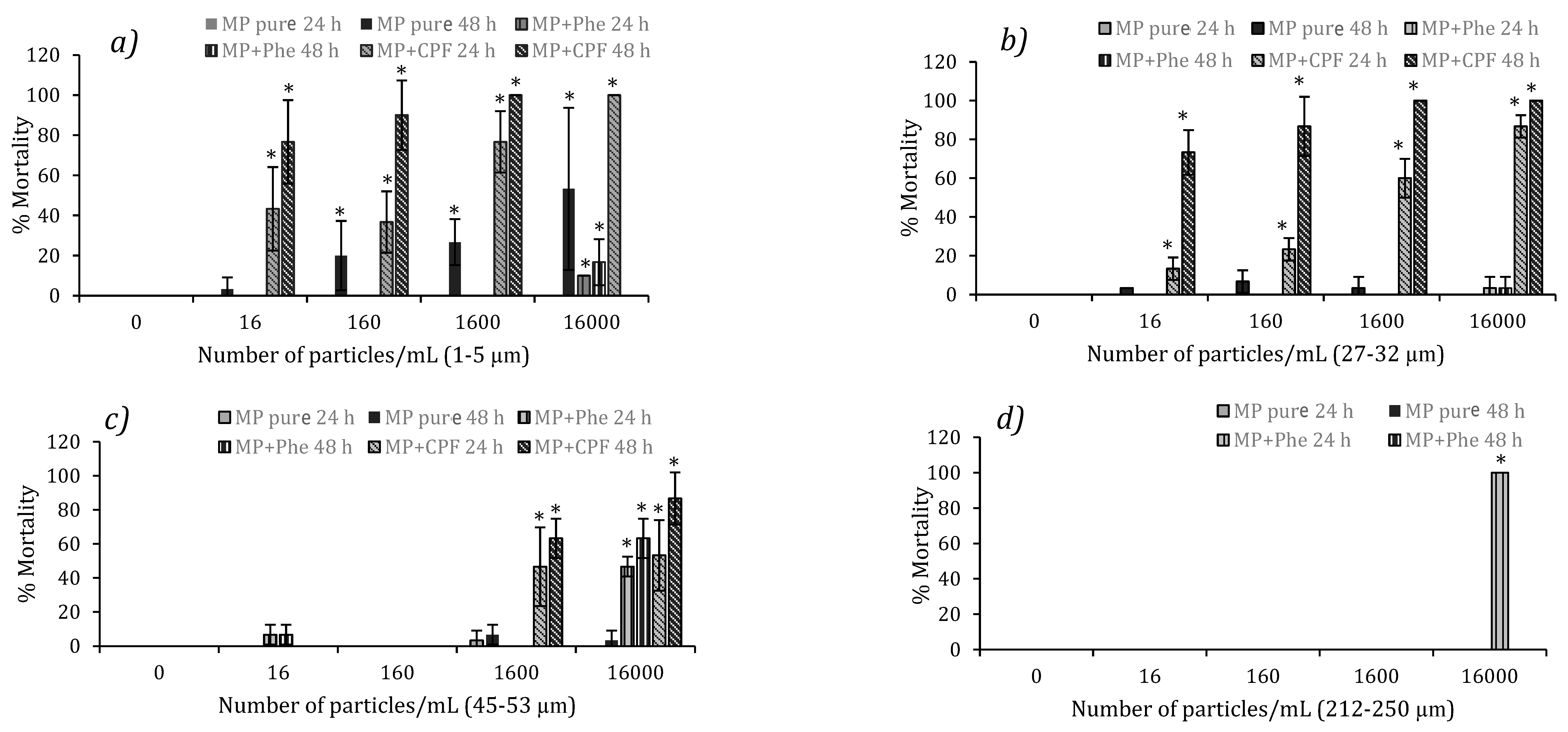
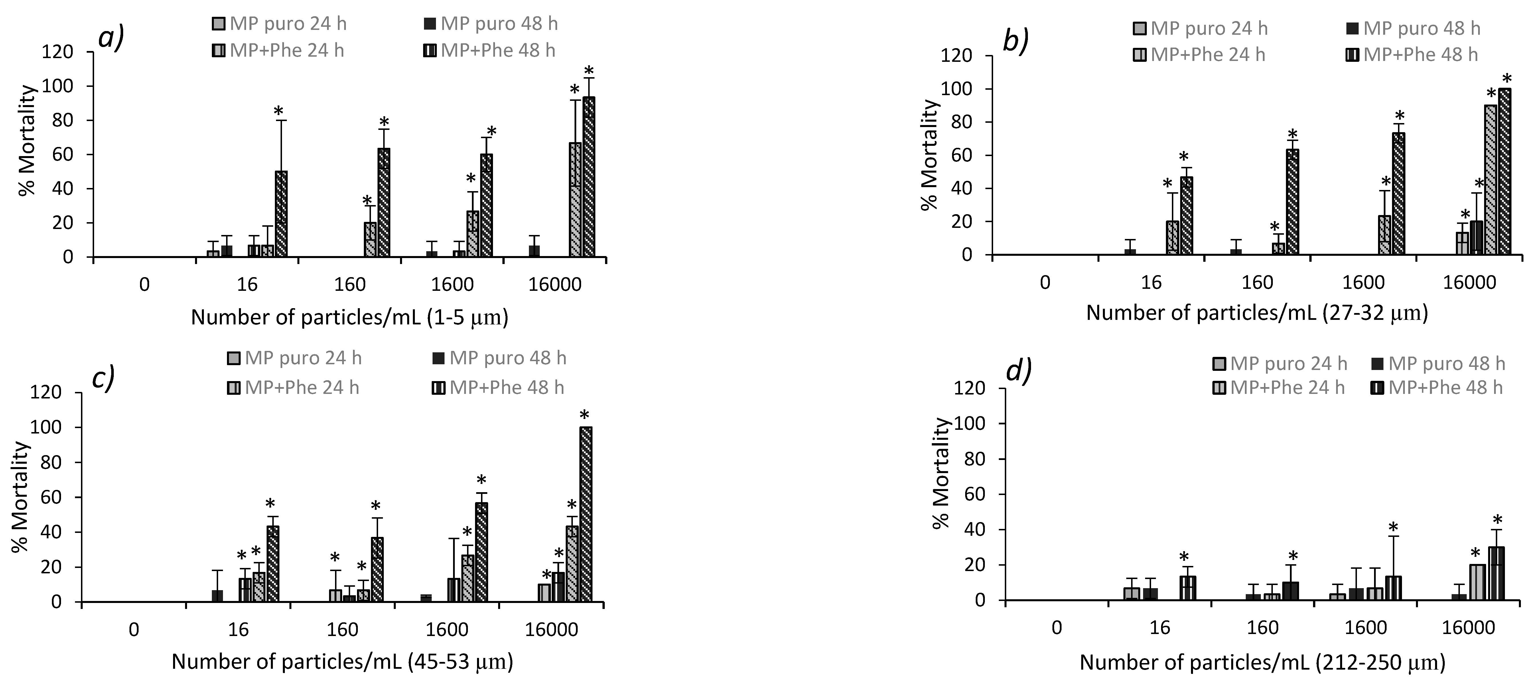
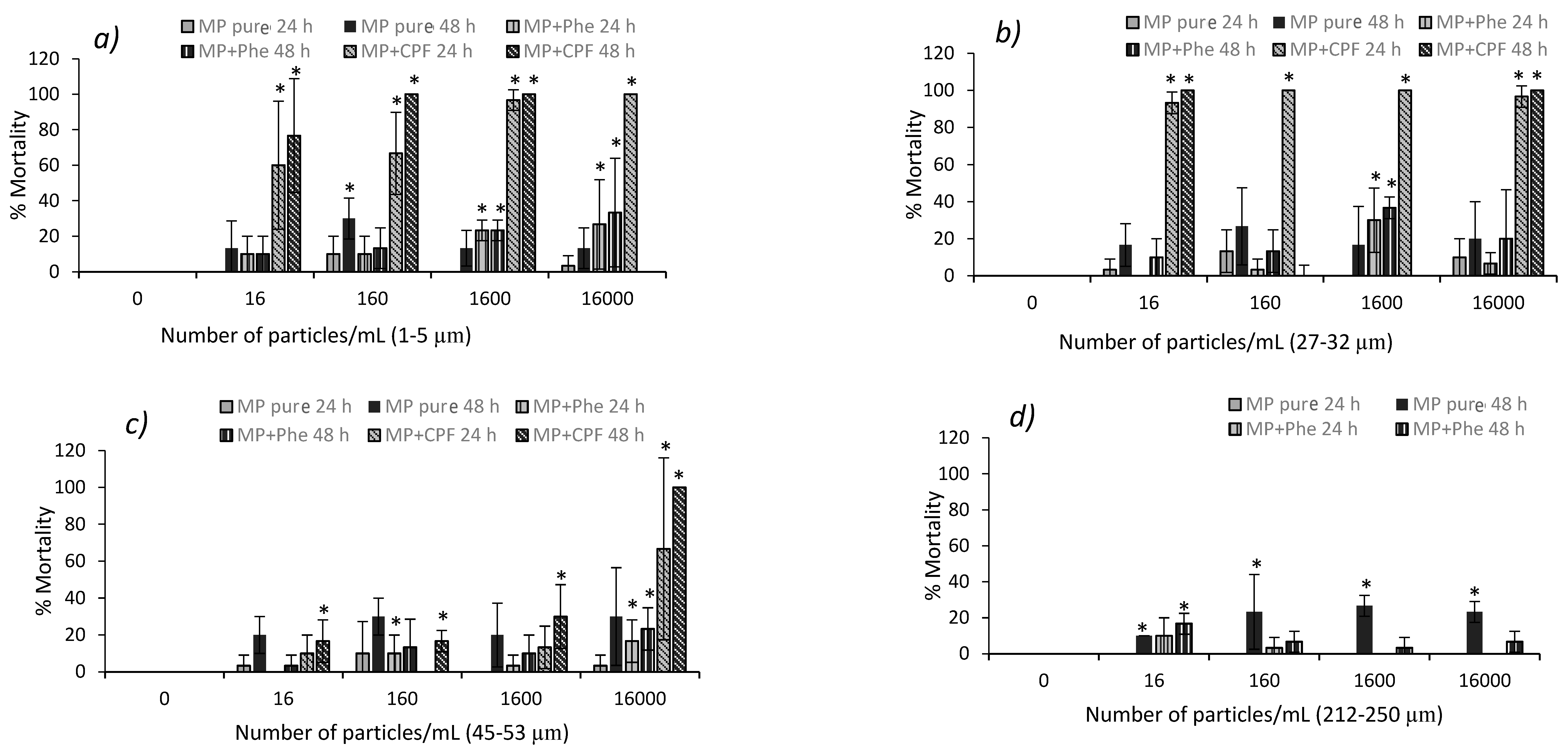
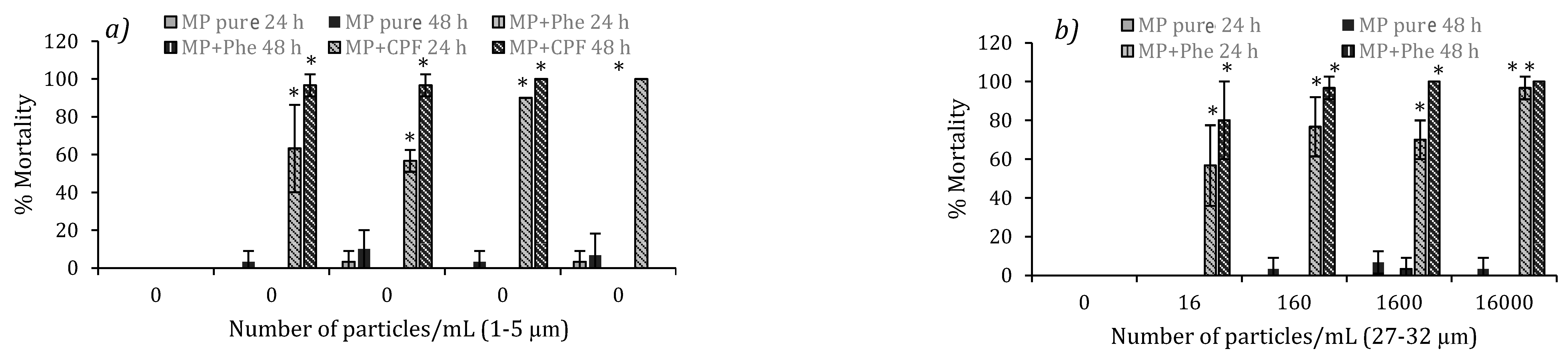
3.4. Acute Toxicity from Polluted MP in D. magna and Podocopid Ostracods

In this study, we approached the effects which could arise from the union of microplastics, and co-pollutants (Phe and CPF) absorbed into aquatic organisms via evaluation of the biological effects. The results evaluated in adult D. magna exposed to MP + Phe showed acute toxicity at 48 h—with greater mortality in organisms exposed to 212–250 μm MP particles, although these results were not above 30% (Figure 3d)—independently of the chemical union capacity (log Kow 4.57) [90]. However, juveniles presented higher mortality among water fleas exposed to 45–53 and 212–250 μm sizes at the highest concentration (16,000 particles/mL), with 100% mortality occurring after 24 h among organisms exposed to 212–250 μm MP (Figure 2d) and 46.7% at 24 h and 63.3% at 48 h in organisms exposed to 45–53 μm MP (Figure 2c). No mortality was observed during the dynamic bioassay (Figure 5a,b).
During the exposure to MP + Phe in podocopid ostracods, we observed that at the end of the bioassay, the organisms exposed to 1–5 μm MP (LC50 (48 h) = 6048 particles) presented a mortality rate of 33.3% at 48 h with the high concentration (16,000 particles/mL), which was significantly different from the control (p < 0.05) (Figure 5a). We also observed that for the same particle size, there was a positive correlation between particle concentration and mortality at 24 h (p < 0.05; r = 0.6) and 48 h (p < 0.05 r = 0.6), while organisms exposed to 27–32 μm MP presented 36.7% mortality at a concentration of 1600 particles/mL, which was significantly different from the control (p < 0.05) (Figure 4b).
With the 45–53 and 212–250 μm particles contaminated with Phe, we observed significant differences (p < 0.05) compared to the control in the highest concentration tested (16,000 particles/mL), which was the most toxic concentration for juvenile D. magna (Figure 2c,d). We observed that for 45–53 μm particles, there was a positive correlation between particle concentrations and mortality at 24 h (p < 0.055; r = 0.7) and at 48 h (p < 0.05; r = 0.6). This toxicity could be because with 45–53 and 212–250 μm MP, the surface area is greater. In other words, there is a greater absorption capacity for the hydrophobic organic pollutant. More pollutants could be transported to the organisms, revealing the pollutants’ bioavailability. It is possible that high mortality did not occur in podocopid ostracods upon exposure to MPs + Phe particles in comparison with pure MPs and that only the high concentration presented mortality, as with exposure among D. magna, since toxic Phe effects (mutagenic, teratogenic, and carcinogenic) could be expressed and observed when the organisms are chronically exposed to MPs + Phe. However, more studies are needed to understand the effects of pure and contaminated MP particles upon exposure to various organisms.
A D. magna study which evaluated the effects of nanoplastics (NPs) and MP on Phe toxicity indicated that—unlike MP particles, for which no physical damage was observed as they could be easily excreted via the intestine—the 50 nm NP and Phe had joint additive toxicity since they easily accumulated in the digestive tract and increased Phe bioaccumulation. Phe on the surface of NP can be spread into cells along with NP, which can result in direct Phe contact with the D. magna membrane. This therefore strongly increases chronic Phe toxicity, although 10 μm MP did not show an effect on Phe bioaccumulation and its residues within water fleas’ bodies. The differences can be attributed to greater Phe adsorption in 50 nm NP than in 10 μm MP [48]. Another D. magna study showed that irregular PE MP (10 and 75 µm) preincubated with Phe was more toxic for the organism than MP alone. However, the inhibitory effect of MP + Phe was not more toxic than the same concentration of Phe without MP. Water fleas’ exposure to Phe indicates an EC50 of 0.47 mg/L. By contrast, D. magna exposure to irregular MPs + Phe (0.05 g/L) resulted in an EC50 of 0.14 mg/L [91].
The present study showed a notable mortality level among D. magna and ostracods exposed to MP + CPF in all the analyzed treatments. This was significantly dependent on particle concentration (p < 0.05). We observed that in experiments with daphnia juveniles, 100% mortality occurred when they were exposed to 1–5 μm MP at a concentration of 16,000 particles/mL, which was significantly different from the control (p < 0.05) (Figure 2a) (LC50 (24h) = 1468 particles). This response positively correlated with particle concentration (p < 0.05; r = 0.7) and the 27–32 μm size (LC50 (48 h) = 1788 particles). 100% mortality appears in 1600 and 16,000 particle/mL concentrations (Figure 2b), with these responses being significantly different from the control (p < 0.05). This response is directly related to the particle concentration (p < 0.05; r = 0.7). The results in the treatments evaluated with adult D. magna indicated 95% mortality after 48 h following exposure to 1–5 μm MP (LC50 (48 h) = 3681 particles). This result correlated positively with particle concentration (r = 0.4). We also noted 100% mortality after 48 h following exposure to 27–32 μm and 45–53 μm MP in the highest concentration analyzed, which was significantly different from the control and directly correlated with particle concentrations (p < 0.05; r = 0.7 and r = 0.8 respectively) (Figure 3a–c). For all bioassays performed with MP + CPF, we observed mortality after 24 h, rising above 50% after 48 h, even at very low concentrations. This was directly proportional to particle concentrations in the study (Figure 2a–c and Figure 3a–c). However, during the bioassay with juvenile water fleas, we observed no mortality in the 16 and 160 particle/mL concentrations (Figure 2c), which could be due to juveniles having less exposure to MP and presenting lower contact with them since they had lower concentrations and larger particle size.
With juvenile D. magna exposed to MP + CPF of 1–5 μm and 27–32 μm using the dynamic methodology, the results indicated that as with the static bioassays, mortality above 50% occurred after 24 h, even at low concentrations. However, mortality was over 90% after 48 h and 100% in the 1600 and 16,000 particles/mL concentrations (Figure 5a,b), which was significantly different from the control (p < 0.05). Both sizes presented a direct dependency between mortality and particle concentration (p < 0.05; r = 0.6).
Results from the podocopid ostracods exposed to MP + CPF indicated greater sensitivity compared to D. magna, as mortality was over 95% following 24 h and 100% after 48 h of exposure at all concentrations for 27–32 μm particles. This was significantly different from the control (p < 0.05) even at the lowest concentration tested (Figure 4b). This high mortality was also observed during exposure to 1–5 μm MP. However, despite the increased mortality observed after increasing the concentration, this relation was rather weaker (p < 0.05; r = 0.3) (Figure 4a). With 45–53 μm MP, we observed 100% mortality after 48 h with the highest tested concentration, which was significantly different from the control (p < 0.05) (Figure 4c). It was also observed that the mortality produced by MPs + CPF of 45–53 μm was perfectly correlated with the particle concentration (r = 1). We should also mention that apart from the high mortality induced by CPF pesticide, we observed overstimulation of the nervous system, producing continuous repetitive movements similar to tremors, due to the constant release of nerve impulses. The effects observed after MP + CPF promotion at all sizes were thus more toxic than pure MP. This reveals that microplastic particles do act as HOP vectors, negatively affecting survival and reproduction as well as impacting the development of crustaceans’ descendants.
Our results indicate that PE MP particles can transport CPF in aquatic organisms since CPF (log Kow = 4.96, log Kd = 5.94) [92] was rapidly adsorbed onto the surface of MP used in this experiment along with presenting a relatively swift desorption rate [93], explaining the high toxicity of MP + CPF. All these results demonstrated crustaceans’ high sensitivity to organophosphate insecticide, presenting very low LC50 values. This may be due to the action mechanism of this class of chemical products, which inhibits cholesterase enzymes [94,95], giving rise to toxic effects related with biological aptitude, which can influence the density of some crustacean and bivalve populations and potentially impact aquatic food chains along with an ecological impact on the larger aquatic ecosystem. Along these lines, a study wherein Acartia tonsa copepods were exposed to PE MP of 2 to 10 μm with CPF indicated that this compound was 4–25 times more toxic when combined with MP (MP + CPF) than alone [92]. Another study evaluated the acute toxicity of PE MP (2 to 6 µm) on the microalgae Isochrysis galbana, finding that MP reduced CPF toxicity because CPF adsorption in MP surfaces modulated CPF toxicity on growth. Once CPF was adsorbed onto MP surfaces, it became less bioavailable for algae cells [96]. These findings indicate that it is not simple to obtain a clear answer and that bioaccumulation and MP and co-pollutants’ toxicity may depend on the species being studied, the plastic type, and the pollutant type, particularly its hydrophobic properties.

3.5. Chronic Toxicity of Pure MP in D. magna

Chronic toxicity lab tests using newborn D. magna exposed to two realistic concentrations of pure PE MP (160 and 1600 particles/mL) of 1–5, 27–32, 45–53, and 212–250 μm showed differences between 14 days of exposure compared to the control group regarding their first offspring and the number of young. The number of newborns produced within the 14 days of the experiment for all sizes showed a growth trend in the number of neonates compared with the control organisms, indicating a stimulation of reproduction (Figure 6a–d). Neonates rose significantly with the treatment of pure MP of 212–250 μm (p < 0.05) compared to the control group. For this response, we saw a correlation which was inversely proportional to the particle concentration (r = −0.6) (Figure 6d). However, at the end of the bioassay (21 days), there was a decrease in the number of offspring from all treatments by comparison with the control group, although this drop was not significant. This is because after 14 days, various events occurred including neonate deaths (Figure 6a–d), parental death in some treatments, and a consequent drop in the number of young. One study in which adult D. magna were exposed to high PS MP concentrations of 6 μm showed a stimulation response with higher reproductive rates than the control group. However, there was no mortality in any concentration during the reproduction test time (21 days) [78]. MP accumulation within organisms’ intestines may cause blockage, which could reduce the ingestible amount of food particles and even affect food absorption or assimilation [97]. This dimension of long-term feeding activity leads to deteriorated physiological and physical status among organisms since it affects the energy budget and, therefore, the entire metabolism [71,73]. When food availability is low, it can also reduce body length and newborns’ size [78], as energy storage in lipid form is limited [98]. A study with D. magna using PS MP of 1–5 μm, after 21 days’ exposure, saw total biomass per cup reduced in all treatments, with a 21% drop in the highest exposure concentration (105 particles/mL) compared to the control [72]. In another experiment, water fleas’ feeding rates fell by 21% in the presence of 0.1 μm particles, which could affect the energy budget, but no reproductive effect was found despite the high body loads of particles at the end of 21 days’ exposure [83].

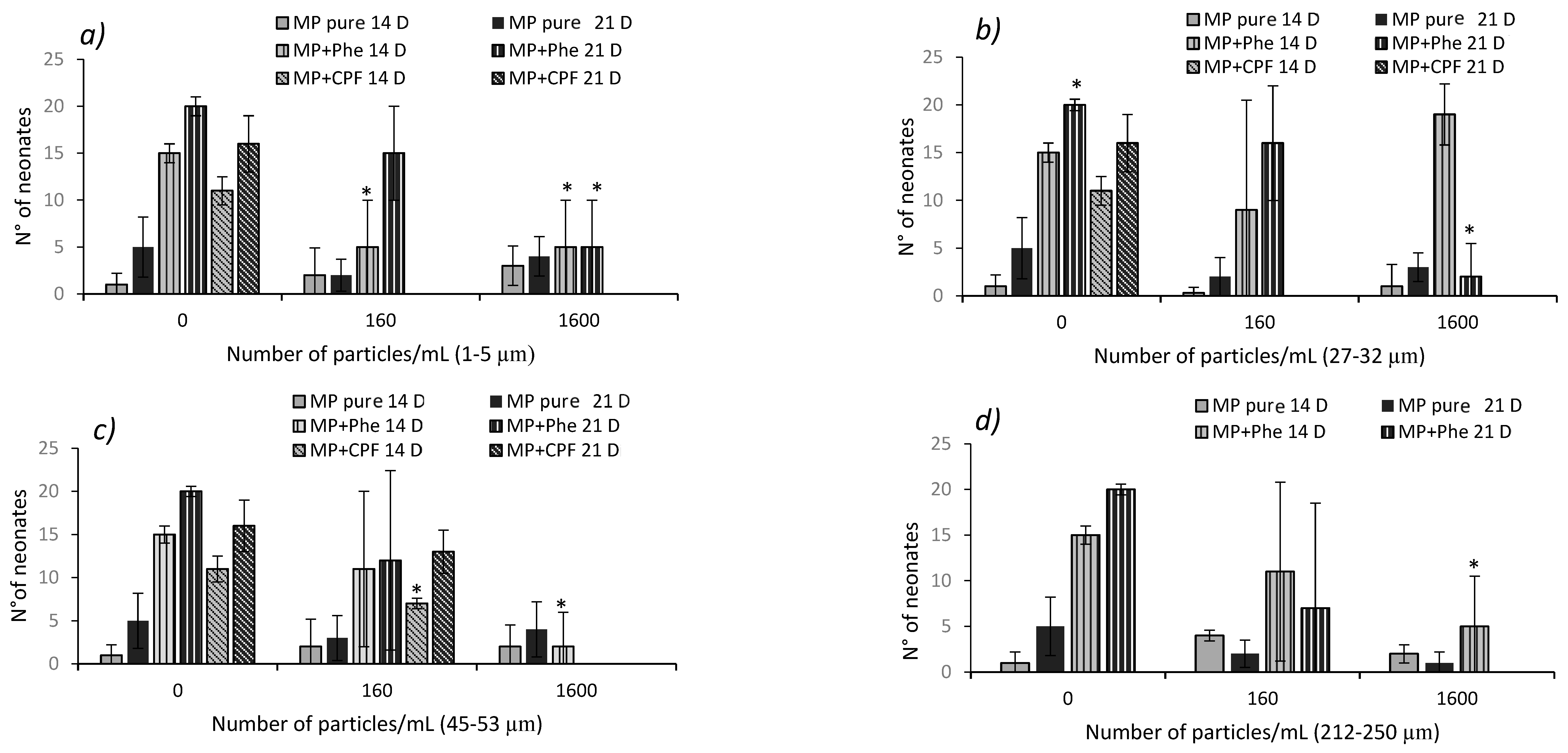
3.6. Chronic Toxicity of Contaminated MP in D. magna

During chronic bioassays with D. magna exposed to MP + Phe (160 and 1600 particles/mL), all MP sizes presented a notable decrease in reproduction compared with control groups (Figure 6a–d). For the results of these organisms exposed to 1–5 μm particles, we observed that the first generation of descendants decreased significantly at 14 days (p < 0.05) compared to the control for both concentrations in D. magna exposed to MP + Phe. We also saw a significant drop (p < 0.05) in the number of young at 21 days among daphnia exposed to 1600 particles/mL. This response is inversely proportional to the concentration at days 14 and 21 (p < 0.05; r = −0.8). Neonates exposed to 27–32 μm presented a decreased overall newborn population number compared to the control, with this response being inversely proportional to particle concentration (p < 0.05; r = −0.9). After exposing neonates to 45–53 μm MP + Phe, no survivors were observed at the end of the bioassay in any of the concentrations tested (160 and 1600 particles/mL), with this response being inversely proportional to particle concentration (p < 0.05; r = −0.8) (Figure 6c). Similarly, in newborns exposed to 212–250 μm MP + Phe, we saw a significant decrease (p < 0.05) after 14 days in the highest concentration studied, while at 21 days, this concentration killed 100% of the organisms (Figure 6d). This is inversely proportional to the particle concentration at 14 and 21 days (p < 0.05; r = −0.6 and r = −0.7, respectively).
For daphnia exposed to 1–5 μm MP + CPF, we observed 100% mortality among the organisms at 14 days (Figure 6a), which is significantly different from the control group (p < 0.05) and inversely proportional to particle concentration (p < 0.05; r = −0.6). Amounts of 27–32 μm MP significantly reduce (p < 0.05) the number of descendants among daphnia exposed to MP + Phe at 1600 particles/mL. Similarly, to observations with smaller particles, daphnia exposed to 45–53 μm MP + CPF presented 100% mortality after 14 days of the experiment (Figure 6b,c), which was a significantly different response from the control (p < 0.05). We also observed an inverse correlation regarding MP concentration (p < 0.05; r = −0.9).
The present study showed that MP exposure levels could influence the combined effects of MP and Phe in the D. magna life cycle since high MP levels and larger particle sizes increase bioavailability and possible Phe bioaccumulation among water fleas, thus affecting reproduction. Supporting our results, a study on long-term exposure to low Phe concentrations showed that this negatively affected adult fish reproduction and impacted their descendants’ development [67]. However, it is important for long-term studies to go beyond the standard 21-day time frame since the effects observed may be time-dependent. Short-term experiments must therefore go over possible effects, which are expected to be highly environmentally relevant since MPs continually enter the environment and can continually absorb additives.
During chronic exposure of D. magna to MP + CPF particles (160 and 1600 particles/mL), for the 1–5 and 27–32 μm sizes, no reproduction was observed since neonates did not survive beyond 24 hours’ exposure to the tested concentrations (Figure 6a,b). For the 45–53 μm MP, descendants were only observed for the 160 particles/mL concentration, which was lower than the control (Figure 6c). This may be due to low concentrations and larger particle sizes, leading to lower MP exposure among neonates. These results indicate that newborns are more sensitive to MP contaminated with CPF compared to other D. magna life cycle phases. This pesticide could also affect the Cladocera population. Reproduction is an essential response, since it affects entire populations. Our results are similar to laboratory studies demonstrating negative effects from CPF on Daphnia pulex reproduction during prolonged exposure at a concentration of 0.005 μg/L [99]. Another D. magna study indicated fewer descendants with exposure concentrations at or above 0.09 μg/L of CPF, and at concentrations of 0.01 and 0.03 μg/L, descendants showed more abnormalities [44].

4. Conclusions

The results obtained highlight the impact of MP on the aquatic environment, which can be related to physical effects from ingestion. This study shows that the selected MP (1–5, 27–32, 45–53 μm), apart from being ingested by water fleas and podocopid ostracods, end up trapped in their appendages. We also proved that the pure MPs tested (27–32, 45–53, and 212–250 μm) are non-toxic for these organisms despite being ingested. However, small particles of 1–5 μm induced toxic effects in the studied organisms. This would affirm that the particle size matters and that these particles’ environmental concentration could negatively affect these crustaceans without ruling out negative impacts on other aquatic species.
We highlighted the threat which MPs pose as vectors for COH, including Phe and CPF, since this study showed that MP toxicity increased after exposure to Phe and CPF. Dependent effects were observed to arise from MP concentrations, particle size, and exposure time, thereby revealing the biodisponibility of Phe and CPF during the bioassays and highlighting the high toxicity arising from the union of MP + CPF, which negatively affected the survival, reproduction, and the development of descendants among the studied crustaceans. We should mention the likelihood that MP in the aquatic environment coexists with a wide variety of organic and inorganic pollutants. Therefore, the present study adds new evidence about the effects of MP particles from PE combined with hydrophobic organic pollutants, although there is still a need for studies evaluating the effects of various MP types in combination with different organic pollutants. Finally, this study is an important contribution for future studies evaluating the adverse effects produced by MPs, demonstrating that these emerging contaminants are effectively ingested by the crustaceans chosen for this study and can transfer organic pollutants associated with them, resulting in adverse effects for the organisms. It should also be considered that these particles might be transferred to organisms at higher levels and create negative impacts on the food chain.

Author Contributions

Conceptualization, G.A.-M., M.V.C. and L.Z.-D.; methodology, G.A.-M. and M.V.C.; software, G.A.-M. and M.V.C.; validation, G.A.-M., M.V.C. and L.Z.-D.; formal analysis G.A.-M. and M.V.C.; investigation, G.A.-M. and M.V.C.; resources, G.A.-M. and L.Z.-D.; data curation, G.A.-M., M.V.C. and L.Z.-D., writing—original draft preparation G.A.-M. and M.V.C.; writing—review and editing, G.A.-M., M.V.C. and L.Z.-D.; visualization, G.A.-M. and L.Z.-D.; supervision, G.A.-M.; project administration, G.A.-M.; funding acquisition, G.A.-M. and L.Z.-D. All authors have read and agreed to the published version of the manuscript.

Funding

This research was funded by the Agencia Nacional de Investigación y Desarrollo (ANID) FONDECYT N° 11180466 and FONDECYT N° 3190455.

Data Availability Statement

Not applicable.

Conflicts of Interest

The authors declare no conflict of interest.

References

  1. Plastics Europe. ¿Qué es el Plástico? 2015. Available online: https://legacy.plasticseurope.org/es/about-plastics/what-are-plastics (accessed on 11 May 2023).
  2. Jambeck, J.; Geyer, R.; Wilcox, C.; Siegler, T.; Perryman, M.; Andrady, A.; Narayan, R.; Lavender Law, K. Plastic waste inputs from land into the ocean. Mar. Pollut. 2015, 347, 768–771. [Google Scholar] [CrossRef] [PubMed]
  3. Cózara, A.; Echevarría, F.; González-Gordillo, J.; Irigoien, X.; Úbeda, B.; Hernández-León, S.; Palma, A.; Navarro, S.; García-de-Lomas, J.; Ruiz, A.; et al. Plastic debris in the open ocean. Environ. Sci. 2014, 111, 10239–10244. [Google Scholar] [CrossRef] [PubMed]
  4. Lusher, A.; Tirelli, V.; O’Connor, I.; Officer, R. Microplastics in Arctic polar waters: The first reported values of particles in surface and sub-surface samples. Sci. Rep. 2015, 5, 14947. [Google Scholar] [CrossRef] [PubMed] [Green Version]
  5. Woodall, L.; Sanchez-Vidal, A.; Canals, M.; Paterson, G.; Coppock, R.; Sleight, V.; Calafat, A.; Rogers, A.; Narayanaswamy, B.; Thompson, R. The deep sea is a major sink for microplastic debris. R. Soc. Open Sci. 2014, 1, 140317. [Google Scholar] [CrossRef] [Green Version]
  6. Moore, C. Synthetic polymers in the marine environment: A rapidly increasing, long-term threat. Environ. Res. 2008, 108, 131–139. [Google Scholar] [CrossRef] [PubMed]
  7. Lee, H.; Shim, W.; Kwon, J. Sorption capacity of plastic debris for hydrophobic organic chemicals. Sci. Total Environ. 2014, 470–471, 1545–1552. [Google Scholar] [CrossRef]
  8. Hidalgo-Ruz, V.; Gutow, L.; Thompson, R.C.; Thiel, M. Microplastics in the marine environment: A review of the methods used for identification and quantification. Environ. Sci. Technol. 2012, 46, 3060–3075. [Google Scholar] [CrossRef]
  9. Ganesh Kumar, A.; Anjana, K.; Hinduja, M.; Sujitha, K.; Dharani, G. Review on plastic wastes in marine environment—Biodegradation and biotechnological solutions. Mar. Pollut. Bull. 2020, 150, 110733. [Google Scholar] [CrossRef]
  10. Andrady, A. Microplastics in the marine environment. Mar. Pollut. Bull. 2011, 62, 1596–1605. [Google Scholar] [CrossRef]
  11. Andrady, A. The plastic in microplastics: A review. Mar. Pollut. Bull. 2017, 119, 12–22. [Google Scholar] [CrossRef]
  12. Zhang, S.; Wu, H.; Hou, J. Progress on the Effects of Microplastics on Aquatic Crustaceans: A Review. Int. J. Mol. Sci. 2023, 24, 5523. [Google Scholar] [CrossRef]
  13. Eriksen, M.; Lebreton, L.; Carson, H.; Thiel, M.; Moore, C.; Borerro, J.; Galgani, F.; Ryan, P.; Reisser, J. Plastic Pollution in the World’s Oceans: More than 5 Trillion Plastic Pieces Weighing over 250,000 Tons Afloat at Sea. PLoS ONE 2014, 9, e111913. [Google Scholar]
  14. Tourinho, P.; Kočí, V.; Loureiro, S.; Gestel, C. Partitioning of chemical contaminants to microplastics: Sorption mechanisms, environmental distribution and effects on toxicity and bioaccumulation. Environ. Pollut. 2019, 252, 1246–1256. [Google Scholar] [CrossRef] [PubMed]
  15. Anbumani, S.; Kakkar, P. Ecotoxicological effects of microplastics on biota: A review. Environ. Sci. Pollut. Res. 2018, 25, 14373–14396. [Google Scholar] [CrossRef]
  16. Kumar, A.; Mishra, S.; Pandey, R.; Yu, Z.; Kumar, M.; Khoo, K.S.; Kumar Thakur, T.; Loke Show, P. Microplastics in terrestrial ecosystems: Un-ignorable impacts on soil characterizes, nutrient storage and its cycling. Trends Anal. Chem. 2023, 158, 116869. [Google Scholar] [CrossRef]
  17. Murray, F.; Cowie, P.R. Plastic contamination in the decapod crustacean Nephrops norvegicus (Linnaeus, 1758). Mar. Pollut. Bull. 2011, 62, 1207–1217. [Google Scholar] [CrossRef] [PubMed]
  18. Davison, P.; Asch, R.G. Plastic ingestion by mesopelagic fishes in the North Pacific Subtropical Gyre. Mar. Ecol. Prog. Ser. 2011, 432, 173–180. [Google Scholar] [CrossRef] [Green Version]
  19. Fischer, E. Microplastic pollution in lakes and lake shoreline sediments—A case study on Lake Bolsena and Lake Chiusi (central Italy). Environ. Pollut. 2016, 213, 648–657. [Google Scholar] [CrossRef]
  20. Forrest, S.; Holman, L.; Murphy, M.; Vermaire, M. Citizen science sampling programs as a technique for monitoring microplastic pollution: Results, lessons learned and recommendations for working with volunteers for monitoring plastic pollution in freshwater ecosystems. Environ. Monit. Assess. 2019, 191, 172. [Google Scholar] [CrossRef]
  21. Zhao, S.; Zhu, L.; Wang, T.; Li, D. Suspended microplastics in the surface water of the Yangtze Estuary System, China: First observations on occurrence, distribution. Mar. Pollut. Bull. 2014, 86, 562–568. [Google Scholar] [CrossRef]
  22. Desforges, J.P.; Galbraith, M.; Dangerfield, N.; Ross, P.S. Widespread distribution of microplastics in subsurface seawater in the NE Pacific Ocean. Mar. Pollut. Bull. 2014, 79, 94–99. [Google Scholar] [CrossRef]
  23. Leslie, H.A.; Brandsma, S.H.; van Velzen, M.J.; Vethaak, A.D. Microplastics en route: Field measurements in the Dutch river delta and Amsterdam canals, wastewater treatment plants, North Sea sediments and biota. Environ. Int. 2017, 101, 133–142. [Google Scholar] [CrossRef] [PubMed]
  24. Duis, K.; Coors, A. Microplastics in the aquatic and terrestrial environment: Sources (with a specific focus on personal care products), fate and effects. Environ. Sci. Eur. 2016, 28, 2. [Google Scholar] [CrossRef] [Green Version]
  25. Rehse, S.; Kloas, W.; Zarfl, C. Short-term exposure with high concentrations of pristine microplastic particles leads to immobilisation of Daphnia magna. Chemosphere 2016, 153, 91–99. [Google Scholar] [CrossRef] [PubMed]
  26. Rainieri, S.; Conlledo, N.; Larsen, B.; Granby, K.; Barranco, A. Combined effects of microplastics and chemical contaminants on the organ toxicity of zebrafish (Danio rerio). Environ. Res. 2018, 162, 135–143. [Google Scholar] [CrossRef] [PubMed]
  27. Guzzetti, E.; Sureda, A.; Tejada, S.; Faggio, C. Microplastic in marine organism: Environmental and toxicological effects. Environ. Toxicol. Pharmacol. 2018, 64, 164–171. [Google Scholar] [CrossRef] [PubMed]
  28. Cui, R.; Kim, S.; An, Y.J. Polystyrene nanoplastics inhibit reproduction and induce abnormal embryonic development in the freshwater crustacean Daphnia galeata. Sci. Rep. 2017, 7, 12095. [Google Scholar] [CrossRef] [PubMed] [Green Version]
  29. Mak, C.; Yeung, K.; Chan, K. Acute toxic effects of polyethylene microplastic on adult zebrafish. Ecotoxicol. Environ. Saf. 2019, 182, 109442. [Google Scholar] [CrossRef]
  30. Ribeiro, F.; Garcia, A.; Pereira, B.; Fonseca, M.; Mestre, N.; Fonseca, T.; Ilharco, L.; Bebianno, M. Microplastics effects in Scrobicularia plana. Mar. Pollut. Bull. 2017, 122, 379–391. [Google Scholar] [CrossRef]
  31. Farrell, P.; Nelson, K. Trophic level transfer of microplastic: Mytilus edulis (L.) to Carcinus maenas (L.). Environ. Pollut. 2013, 177, 1–3. [Google Scholar] [CrossRef]
  32. Wang, Z.; Chen, M.; Zhang, L.; Wang, K.; Yu, X.; Zheng, Z.; Zheng, R. Sorption behaviors of phenanthrene on the microplastics identified in a mariculture farm in Xiangshan Bay, southeastern China. Sci. Total Environ. 2018, 628–629, 1617–1626. [Google Scholar] [CrossRef] [PubMed]
  33. Alimi, O.; Budarz, J.; Hernandez, L.; Tufenkji, N. Microplastics and Nanoplastics in Aquatic Environments: Aggregation, Deposition, and Enhanced Contaminant Transport. Environ. Sci. Technol. 2018, 52, 1704–1724. [Google Scholar] [CrossRef] [PubMed]
  34. Caruso, G. Microplastics as vectors of contaminants. Mar. Pollut. Bull. 2019, 146, 921–924. [Google Scholar] [CrossRef] [PubMed]
  35. Tang, L.; Tang, X.Y.; Zhu, Y.G.; Zheng, M.H.; Miao, Q.L. Polycyclic aromatic hydrocarbon (PAH) contamination in urban soils of Beijing. China Reign. Ent. 2005, 31, 822–828. [Google Scholar] [CrossRef]
  36. Hylland, K. Polycyclic aromatic hydrocarbon (PAH). Ecotoxicology in marine ecosystems. Toxicol. Environ. Health Part A 2006, 69, 109–123. [Google Scholar] [CrossRef]
  37. Henkler, F.; Stolpmann, K.; Luch, A. Exposure to Polycyclic Aromatic Hydrocarbons: Bulky DNA Adducts and Cellular Responses. In Molecular, Clinical and Environmental Toxicology; Springer: Basel, Switzerland, 2012; pp. 107–131. [Google Scholar]
  38. Shimada, H.; Oginuma, M.; Hara, A.; Imamura, Y. 9,10-Phenanthrenequinone, a component of diesel exhaust particles, inhibits the reduction of 4-benzoylpyridine and all-trans-retinal and mediates superoxide formation through its redox cycling in pig heart. Chem. Res. Toxicol. 2004, 17, 1145–1150. [Google Scholar] [CrossRef]
  39. Otero-Paternina, A.; Cruz-Casallas, P.; Velasco-Santamaría, Y. Evaluación del efecto del hidrocarburo fenantreno sobre el crecimiento de Chlorella vulgaris (Chlorellaceae). Acta Biol. Colomb. 2013, 18, 87–98. [Google Scholar]
  40. Bartonitz, A.; Anyanwu, I.; Geist, J.; Imhof, H.; Reichel, J.; Graßmann, J.; Drewes, J.; Beggel, S. Modulation of PAH toxicity on the freshwater organism G. roeseli by microparticles. Environ. Pollut. 2020, 260, 113999. [Google Scholar] [CrossRef]
  41. Mai, Y.; Peng, S.; Li, H.; Lai, Z. Histological, biochemical and transcriptomic analyses reveal liver damage in zebrafish (Danio rerio) exposed to phenanthrene. Comp. Biochem. Physiol. Part C Toxicol. Pharmacol. 2019, 225, 108582. [Google Scholar] [CrossRef]
  42. Karami, A.; Romano, N.; Galloway, T.; Hamzah, H. Virgin microplastics cause toxicity and modulate the impacts of phenanthrene on biomarker responses in African catfish (Clarias gariepinus). Environ. Res. 2016, 151, 58–70. [Google Scholar] [CrossRef]
  43. Reynaud, S.; Deschaux, P. The effects of polycyclic aromatic hydrocarbons on the immune system of fish: A review. Aquat. Toxicol. 2006, 77, 229–238. [Google Scholar] [CrossRef]
  44. Palma, P.; Palma, V.L.; Fernandes, R.M.; Bohn, A.; Soares, A.; Barbosa, I. Embryo-toxic effects of environmental concentrations of chlorpyrifos on the crustacean Daphnia magna. Ecotoxicol. Environ. Saf. 2009, 72, 1714–1718. [Google Scholar] [CrossRef] [PubMed]
  45. Bertrand, L.; Marino, D.; Monferrán, M.; Amé, M. Can a low concentration of an organophosphate insecticide cause negative effects on an aquatic macrophyte? Exposure of Potamogeton pusillus at environmentally relevant chlorpyrifos concentrations. Environ. Exp. Bot. 2017, 138, 139–147. [Google Scholar] [CrossRef]
  46. Servicio Agrícola y Ganadero; División Protección Agrícola y Forestal; Subdepartamento de Plaguicidas y Fertilizantes. Informe de Venta de Plaguicidas de Uso Agrícola en Chile; SAG: Santiago, Chile, 2008. [Google Scholar]
  47. Nasser, F.; Lynch, I. Secreted protein eco-corona mediates uptake and impacts of polystyrene nanoparticles on Daphnia magna. J. Proteom. 2016, 137, 45–51. [Google Scholar] [CrossRef] [PubMed] [Green Version]
  48. Secretaría de Medio Ambiente y Recursos Naturales; Báez, M.C.; Pica, Y.; Ronco, A. Ensayo de Toxicidad Aguda con el Cladócero Daphnia magna. Ensayos Toxicológicos para la Evaluación de Sustancias Químicas en Agua y Suelo. La Experiencia en México; Secretaría de Medio Ambiente y Recursos Naturales: Mexico City, Mexico, 2008; pp. 17–32. [Google Scholar]
  49. Baltanás, A.; Mesquita-Joanes, F. Clase Ostrácoda. Orden Podocópida. Idea-Sea 2015, 74, 1–10. [Google Scholar]
  50. Palacios-Fest, M.R.; Park, L.E.; González-Porta, J.; Dix, G.R. Química de conchas de ostrácodos: Una alternativa para medir la contaminación por metales en sistemas acuáticos. Rev. Mex. Cienc. Geológicas 2003, 20, 139–153. [Google Scholar]
  51. Zocchi, M.; Sommaruga, R. Microplastics modify the toxicity of glyphosate on Daphnia magna. Sci. Total Environ. 2019, 697, 134194. [Google Scholar] [CrossRef] [PubMed]
  52. Ziajahromi, S.; Kumar, A.; Neale, P.; Leusch, F. Impact of Microplastic Beads and Fibers on Waterflea (Ceriodaphnia dubia) Survival, Growth, and Reproduction: Implications of Single and Mixture Exposures. Environ. Sci. Technol. 2017, 51, 13397–13406. [Google Scholar] [CrossRef] [PubMed] [Green Version]
  53. Law, K.L.; Morét-Ferguson, S.E.; Goodwin, D.S.; Zettler, E.R.; Deforce, E.; Kukulka, T. Distribution of surface plastic waste in the eastern Pacific Ocean from an 11-year data set. Environ. Sci. Technol. 2011, 48, 4732–4738. [Google Scholar] [CrossRef]
  54. Cole, M.; Lindeque, P.; Halsband, C.; Galloway, T. Microplastics as contaminants in the marine environment: A review. Mar. Pollut. Bull. 2011, 62, 2588–2597. [Google Scholar] [CrossRef]
  55. Woodall, L.C.; Gwinnett, C.; Packer, M.; Thompson, R.C.; Robinson, L.F.; Paterson, G.L. Using a forensic science approach to minimize environmental contamination and to identify microfibres in marine sediments. Mar. Pollut. Bull. 2015, 95, 40–46. [Google Scholar] [CrossRef]
  56. Song, Y.K.; Hong, S.H.; Jang, M.; Kang, J.H.; Kwon, O.Y.; Han, G.M. Large accumulation of micro-sized synthetic polymer particles in the sea surface microlayer. Environ. Sci. Technol. 2014, 48, 9014–9021. [Google Scholar] [CrossRef]
  57. Lechner, A.; Keckeis, H.; Lumesberger-Loisl, F.; Zens, B.; Krusch, R.; Tritthart, M. The Danube so colourful: A potpourri of plastic litter outnumbers fish larvae in Europe’s second largest river. Environ. Pollut. 2014, 188, 177–181. [Google Scholar] [CrossRef] [Green Version]
  58. de Alencastro, L.F. Évaluation de la Pollution par les Plastiques dans les Eaux de Surface en Suisse; Final Report EPFL—ENAC/IIE/GR-CEL; École Polytechnique Fédérale de Lausanne: Lausanne, Switzerland, 2014. [Google Scholar]
  59. McCormick, A.; Hoellein, T.J.; Mason, S.A.; Schluep, J.; Kelly, J.J. Microplastic is an abundant and distinct microbial habitat in an urban river. Environ. Sci. Technol. 2014, 48, 11863–11871. [Google Scholar] [CrossRef] [PubMed]
  60. Cincinelli, A.; Scopetani, C.; Chelazzi, D.; Lombardini, E.; Martellini, T.; Katsoyiannis, A.; Fossi, M.C.; Corsolini, S. Microplastic in the surface waters of the Ross Sea (Antarctica): Occurrence, distribution and characterization by FTIR. Chemosphere 2017, 175, 391–400. [Google Scholar] [CrossRef] [PubMed]
  61. Nel, H.A.; Froneman, P.W. A quantitative analysis of microplastic pollution along the south-eastern coastline of South Africa. Mar. Pollut. Bull. 2015, 101, 274–279. [Google Scholar] [CrossRef] [PubMed]
  62. Lorenz, C.; Roscher, L.; Meyer, M.S.; Hildebrandt, L.; Prume, J.; Loder, M.G.J.; Primpke, S.; Gerdts, G. Spatial distribution of microplastics in sediments and surface waters of the southern North Sea. Environ. Pollut. 2019, 252, 1719–1729. [Google Scholar] [CrossRef] [Green Version]
  63. Courtene-Jones, W.; Quinn, B.; Gary, S.F.; Mogg, A.O.M.; Narayanaswam, Y.B.E. Microplastic pollution identified in deep-sea water and ingested by benthic invertebrates in the Rockall Trough, North Atlantic Ocean. Environ. Pollut. 2017, 231, 271–280. [Google Scholar] [CrossRef] [Green Version]
  64. Faure, F.; Corbaz, M.; Baecher, H.; de Alencastro, L.F. Pollution due to plastics and microplastics in Lake Geneva and in the Mediterranean Sea. Arch Sci. 2012, 65, 157–164. [Google Scholar]
  65. Moore, C.J.; Moore, S.L.; Leecaster, M.K.; Weisberg, S.B. A comparison of plastic and plankton in the North Pacific Central Gyre Mar. Pollut. Bull. 2001, 42, 1297–1300. [Google Scholar] [CrossRef]
  66. Eriksen, M.; Mason, S.; Wilson, S.; Box, C.; Zellers, A.; Edwards, W. Microplastic pollution in the surface waters of the Laurentian Great Lakes. Mar. Pollut. Bull. 2013, 77, 177–182. [Google Scholar] [CrossRef]
  67. Free, C.M.; Jensen, O.P.; Mason, S.A.; Eriksen, M.; Williamson, N.J.; Boldgiv, B. High-levels of microplastic pollution in a large, remote, mountain lake. Mar. Pollut. Bull. 2014, 85, 156–163. [Google Scholar] [CrossRef] [PubMed]
  68. Hidalgo-Ruz, V.; Thiel, M. Distribution and abundance of small plastic debris on beaches in the SE Pacific (Chile): A study supported by a citizen science project. Mar. Environ. Res. 2013, 87–88, 12–18. [Google Scholar] [CrossRef]
  69. Ma, Y.; Huang, A.; Cao, S.; Sun, F.; Wang, L.; Guo, H.; Ji, R. Effects of nanoplastics and microplastics on toxicity, bioaccumulation, and environmental fate of phenanthrene in fresh water. Environ. Pollut. 2016, 219, 166–173. [Google Scholar] [CrossRef]
  70. Bonifacio, A.; Ballesteros, M.L.; Bonansea, R.; Filippi, I.; Amé, M.V.; Hued, A. Environmental relevant concentrations of a chlorpyrifos commercial formulation affect two neotropical fish species, Cheirodon interruptus and Cnesterodon decemmaculatus. Chemosphere 2017, 188, 486–493. [Google Scholar] [CrossRef] [PubMed]
  71. Cormier, B.; Batel, A.; Cachot, J.; Bégout, M.L.; Braunbeck, T.; Cousin, X.; Keiter, S. Multi-laboratory hazard assessment of contaminated microplastic particles by means of enhanced fish embryo test with the zebrafish (Danio rerio). Front. Environ. Sci. 2019, 7, 135. [Google Scholar] [CrossRef] [Green Version]
  72. Bosker, T.; Olthof, G.; Vijver, M.; Baas, J.; Barmentlo, S. Significant decline of Daphnia magna population biomass due to microplastic exposure. Environ. Pollut. 2019, 250, 669–675. [Google Scholar] [CrossRef] [Green Version]
  73. EN ISO 6341; Water Quality—Determination of the Inhibition of the Mobility of Daphnia magna Straus (Cladocera, Crustacea)—Acute Toxicity Test. International Organization for Standardization: Geneve, Switzerland, 2012.
  74. OECD. Test No. 202: Daphnia sp. Acute Immobilisation Test; OECD Publishing: Paris, France, 2004. [Google Scholar]
  75. OECD. OECD Guideline for Testing of Chemicals. No. 211, Daphnia magna Reproduction Test; OECD: Paris, France, 2008. [Google Scholar]
  76. Norberg-King, T.J. An Interpolation Estimate for Chronic Toxicity: The ICP Approach; NETAC Technical Report; United States Environmental Protection Agency: Duluth, MN, USA, 1988; pp. 5–88. [Google Scholar]
  77. Gulley, D.D.; Boelter, A.N.; Bergman, H.L. Toxstat/Datasys Statistical Software; Release 3.0; Department of Zoology and Physiology, University of Wyoming: Laramie, WY, USA, 1989. [Google Scholar]
  78. Eltemsah, Y.; Bøhn, T. Acute and chronic effects of polystyrene microplastics on juvenile and adult Daphnia magna. Environ. Pollut. 2019, 254 Pt A, 112919. [Google Scholar] [CrossRef]
  79. Ma, H.; Pu, S.; Liu, S.; Bai, Y.; Mandal, S.; Xing, B. Microplastics in aquatic environments: Toxicity to trigger ecological consequences. Environ. Pollut. 2020, 261, 114089. [Google Scholar] [CrossRef]
  80. De Felice, B.; Sabatini, V.; Antenucci, S.; Gattoni, G.; Santo, N.; Bacchetta, R.; Ortenzi, M.; Parolini, M. Polystyrene microplastics ingestion induced behavioral effects to the cladoceran Daphnia magna. Chemosphere 2019, 231, 423–431. [Google Scholar] [CrossRef]
  81. Jemec, A.; Horvat, P.; Kunej, U.; Bele, M.; Kržan, A. Uptake and effects of microplastic textile fibers on freshwater crustacean Daphnia magna. Environ. Pollut. 2016, 219, 201–209. [Google Scholar] [CrossRef]
  82. Besseling, E.; Wang, B.; Lürling, M.; Koelmans, A.A. Nanoplastic affects growth of S. obliquus and reproduction of D. Magna. Environ. Sci. Technol. 2014, 48, 12336–12343. [Google Scholar] [CrossRef] [PubMed]
  83. Rist, S.; Baun, A.; Hartmann, N.B. Ingestion of micro- and nanoplastics in Daphnia magna—Quantification of body burdens and assessment of feeding rates and reproduction. Environ. Pollut. 2017, 228, 398–407. [Google Scholar] [CrossRef] [Green Version]
  84. Beiras, R.; Bellas, J.; Cachot, J.; Cormier, B.; Cousin, X.; Engwall, M.; Gambardella, C.; Garaventa, F.; Keiter, S.; Le Bihanic, F.; et al. Ingestion and contact with polyethylene microplastics does not cause acute toxicity on marine zooplankton. J. Hazard. Mater. 2018, 360, 452–460. [Google Scholar] [CrossRef] [Green Version]
  85. Au, S.; Bruce, T.; Bridges, W.; Klaine, S. Responses of Hyalella azteca to acute and chronic microplastic exposures. Environ. Toxicol. Chem. 2015, 34, 2564–2572. [Google Scholar] [CrossRef] [PubMed]
  86. Cole, M.; Lindeque, P.; Fileman, E.; Halsband, C.; Galloway, T.S. The impact of polystyrene microplastics on feeding, function and fecundity in the marine copepod Calanus helgolandicus. Environ. Sci. Technol. 2015, 49, 1130–1137. [Google Scholar] [CrossRef] [PubMed]
  87. Xu, X.Y.; Lee, W.T.; Chan, A.K.Y.; Lo, H.S.; Shin, P.K.S.; Cheung, S.G. Microplastic ingestion reduces energy intake in the clam Atactodea striata. Mar. Pollut. Bull. 2017, 124, 798–802. [Google Scholar] [CrossRef]
  88. Wright, S.L.; Thompson, R.C.; Galloway, T.S. The physical impacts of microplastics on marine organisms: A review. Environ. Pollut. 2013, 178, 483–492. [Google Scholar] [CrossRef]
  89. Gambardella, C.; Morgana, S.; Ferrando, S.; Bramini, M.; Piazza, V.; Costa, E.; Garaventa, F.; Faimali, M. Effects of polystyrene microbeads in marine planktonic crustaceans. Ecotoxicol. Environ. Saf. 2017, 145, 250–257. [Google Scholar] [CrossRef]
  90. Sleight, V.A.; Bakir, A.; Thompson, R.C.; Henry, T.B. Assessment of microplastic-sorbed contaminant bioavailability through analysis of biomarker gene expression in larval zebrafish. Mar. Pollut. Bull. 2017, 116, 291–297. [Google Scholar] [CrossRef] [Green Version]
  91. Frydkjær, C.K.; Iversen, N.; Roslev, P. Ingestion and Egestion of Microplastics by the Cladoceran Daphnia magna: Effects of Regular and Irregular Shaped Plastic and Sorbed Phenanthrene. Bull. Environ. Contam. Toxicol. 2017, 99, 655–661. [Google Scholar] [CrossRef] [PubMed]
  92. Bellas, J.; Gil, I. Polyethylene microplastics increase the toxicity of chlorpyrifos to the marine copepod Acartia tonsa. Environ. Pollut. 2020, 260, 114059. [Google Scholar] [CrossRef] [PubMed]
  93. León, V.M.; García, I.; González, E.; Samper, R.; Fernández-González, V.; Muniategui-Lorenzo, S. Potential transfer of organic pollutants from littoral plastics debris to the marine environment. Environ. Pollut. 2018, 236, 442–453. [Google Scholar] [CrossRef]
  94. Solomon, K.R.; Williams, W.M.; Mackay, D.; Purdy, J.; Giddings, J.M.; Giesy, J.P. Properties and uses of chlorpyrifos in the United States. In Ecological Risk Assessment for Chlorpyrifos in Terrestrial and Aquatic Systems in the United States; Springer: New York, NY, USA, 2014; pp. 13–34. [Google Scholar]
  95. ATSDR. Toxicological Profile for Chlorpyrifos; Agency for Toxic Substances and Disease Registry: Atlanta, GA, USA, 1997; 217p.
  96. Garrido, S.; Linares, M.; Campillo, J.A.; Albentosa, M. Effect of microplastics on the toxicity of chlorpyrifos to the microalgae Isochrysis galbana, clone t-ISO. Ecotoxicol. Environ. Saf. 2019, 173, 103–109. [Google Scholar] [CrossRef]
  97. Ogonowski, M.; Schür, C.; Jarsén, Å.; Gorokhova, E. The effects of natural and anthropogenic microparticles on individual fitness in Daphnia magna. PLoS ONE 2016, 11, e0155063. [Google Scholar] [CrossRef] [PubMed] [Green Version]
  98. Goulden, C.E.; Place, A.R. Lipid accumulation and allocation in daphnid cladocera. Bull. Mar. Sci. 1993, 53, 106–114. [Google Scholar]
  99. Van der Hoeven, N.; Gerritsen, A. Effects of chlorpyrifos on individuals and populations of Daphnia pulex in the laboratory and field. Environ. Toxicol. Chem. 2009, 16, 2438–2447. [Google Scholar] [CrossRef]
Figure 1. MP particle ingestion in D. magna; (a) 1–5 μm, (b) 27–32 μm, (c) 45–53 μm. MP particle ingestion in podocopid ostracods; (d) 1–5 μm, (e) 27–32 μm, (f) 45–53 μm.
Water 15 02915 g001
Figure 2. Mortality percentages among juvenile D. magna exposed for 24 and 48 h to 0 (control), 16, 160, 1600, and 16,000 particles/mL of pure and contaminated MPs with phenanthrene (MP + Phe) and chlorpyrifos (MP + CPF) for sizes (a) 1–5 μm, (b) 27–32 μm, (c) 45–53 μm, (d) 212–250 μm. Asterisks (*) indicate significant differences compared to control (p < 0.05).
Water 15 02915 g002
Figure 3. Mortality percentages in adult D. magna exposed for 24 and 48 h to 0 (control), 16, 160, 1600, and 16,000 particles/mL of pure and contaminated MP with phenanthrene (MP + Phe) and chlorpyrifos (MP + CPF) for sizes (a) 1–5 μm, (b) 27–32 μm, (c) 45–53 μm, (d) 212–250 μm. Asterisks (*) indicate significant differences compared to control (p < 0.05).
Water 15 02915 g003
Figure 4. Mortality percentage among adult podocopid ostracods exposed for 24 and 48 h-to 0 (control), 16, 160, 1600, and 16,000 particles/mL of pure and contaminated MPs, with phenanthrene (MP + Phe) and chlorpyrifos (MP + CPF) for sizes (a) 1–5 μm, (b) 27–32 μm, (c) 45–53 μm, (d) 212–250 μm. Asterisks (*) indicate significant differences compared to control (p < 0.05).
Water 15 02915 g004
Figure 5. Mortality percentages among juvenile D. magna exposed for 24 and 48 h to 0 (control), 16, 160, 1600, and 16,000 particles/mL of pure and contaminated MPs with phenanthrene (MP + Phe) and chlorpyrifos (MP + CPF) in a dynamic modality for sizes (a) 1–5 μm, (b) 27–32 μm. Asterisks (*) indicate significant differences compared to control (p < 0.05).
Water 15 02915 g005
Figure 6. Number of neonates produced by D. magna exposed as newborns to 0 (control), 160, and 1600 particles/mL of pure and contaminated MPs with phenanthrene (MP + Phe) and chlorpyrifos (MP + CPF) for 14 and 21 days (D) for sizes (a) 1–5 μm, (b) 27–32 μm, (c) 45–53 μm, (d) 212–250 μm. Asterisks (*) indicate significant differences compared to control (p < 0.05).
Water 15 02915 g006
Table 1. MP type and concentration in aquatic environments.
MP TypeSizePlaceLocation Average MP ConcentrationReferences
(N/I)80 μmSwedish watersSweden150–2400 MP/m3 [54]
Fibers2–3 mm and diameter < 0.1 mmWater at 300 and 3500 m depthNE North Atlantic and SW Indian Ocean in the Mediterranean Sea28–800 MP/L [55]
Alkyd polymers and poli (acrilate/styrene)0.75 µm–5 mm ≈Goeje IslandSouth Korea195 MP/L [56]
NINIIOcean surface layerVarious sites0 to 8700 MP/m3 [8]
NI63 µm–5 mmNE Pacific, British Columbia coastCanada8 and 9180 MP/m3 [22]
8975% fibersNIOcean Northeastern Pacific279 MP/m3 [22]
NI0.5–5 mm ≈Yangtze EstuaryChina4137 MP/m3 [21]
NI0.5–20 mmDanube RiverBetween Vienna (Austria) and Bratislava (Slovakia)0.055 MP/m3 [57]
NI0.3–5 mmRhône RiverSwitzerland0.29 MP/m3 [58]
59NI0.33–2 mmNorthern shore cana
L
Chicago, Illinois, USA1.94 MP/m3 upstream treatment plant.
17.93 MP/m3 downstream.
[59]
Mostly fibersMostly <300 μmAmsterdam CanalsNetherlands100,000 MP/m3 [23]
Fiber fragments and others, mostly PE and PPNIRoss SeaAntarctica0.0032–1.18 MP/m3 [60]
Fibers NISoutheastern coast of South AfricaSouth Africa257.9–1215 MP/m3 [61]
PP, acrylates/polyurethane/varnish and polyamide86% <100 μmWater surfaceSouthern area of the North Sea0.1–245.4 MP/m3 [62]
PET and acrylic Fibers0.4–8.3 mm Deep sea waterNorth Atlantic Ocean, Scotland70.8 MP/m3 [63]
NI0.3–5 mmLake GenevaSwitzerland0.048 MP/m2 [64]
Plastic film and fibers<5 mmNorth PacificNorth Pacific0.33 MP/m2 [65]
NI0.3–5 mmLakes Geneva, Constance, Neuchâtel, Maggiore, Zurich, and BrienzSwitzerland0.091 MP/m2 [58]
NI0.355 to 5 mm ≈Lakes Superior, Huron and ErieGreat Lakes of North America0.043 MP/m2 [66]
Fragments, pellets/foams, facial cleanser micropearls≤1 mmLake ErieDownstream from Detroit, Cleveland, and Erie, USA0.463 MP/m2 [66]
NI0.355 to 5 mm ≈Lake HovsgolMongolia 0.001–0.044 MP/m2 [67]
NINIRapa Nui shoreline Chile805 MP/m2 [68]
Notes: PET (polyethylene terephthalate), PE (polyethylene), PP (polypropylene), ≈ (Rroughly), NI (not indicated).
Disclaimer/Publisher’s Note: The statements, opinions and data contained in all publications are solely those of the individual author(s) and contributor(s) and not of MDPI and/or the editor(s). MDPI and/or the editor(s) disclaim responsibility for any injury to people or property resulting from any ideas, methods, instructions or products referred to in the content.

