Article

Prokaryotic and Eukaryotic Communities Characteristic in the Water Column and Sediment along the Xiangjiang River, China

1
School of Hydraulic and Environmental Engineering, Changsha University of Science & Technology, Changsha 410114, China
2
Key Laboratory of Dongting Lake Aquatic Eco-Environmental Control and Restoration of Hunan Province, School of Hydraulic and Environmental Engineering, Changsha University of Science and Technology, Changsha 410004, China
3
Key Laboratory of Water-Sediment Sciences and Water Disaster Prevention of Hunan Province, Changsha 410114, China
4
School of Chemistry and Chemical Engineering, Changsha University of Science & Technology, Changsha 410114, China
*
Author to whom correspondence should be addressed.
The authors contributed equally to this work.
Water 2023, 15(12), 2189; https://doi.org/10.3390/w15122189
Submission received: 8 May 2023 / Revised: 30 May 2023 / Accepted: 8 June 2023 / Published: 10 June 2023
(This article belongs to the Section Biodiversity and Functionality of Aquatic Ecosystems)

Abstract

:
Microbial communities are central components of river ecosystems. They are involved in the transportation and transformation of certain pollutants, including nutrients discharged into surface water. Knowledge of microbial community structures is vital for understanding biochemical circulation in aquatic ecosystems. However, most of the research that is currently being conducted focuses more on bacterial diversity and less on eukaryotes, which also play key roles in the nutrient cycle. In this study, 10 sampling sites along the Xiangjiang River were selected, covering the entire reaches of Changsha City, China. Both prokaryotic and eukaryotic diversity and composition in the water and sediment samples were investigated. The results showed that conductivity, TN, and NH4+-N were the main environmental parameters influencing the distribution of microbial communities in the river water column. Proteobacteria, Acidobacteria, and Actinobacteria were the dominant bacteria in sediments. The most abundant taxa in the water samples were Proteobacteria, Actinobacteria, and Firmicutes, with Chloroplastida being the dominant eukaryote. Eukaryotes in sediments are much spatially stochastic. Function analysis showed that bacteria in the water column had more phototrophic genes than those in the sediment samples, while the latter had more nitrogen-transformation-involved genes. This suggested that river sediment is more active in the global nitrogen cycle, while the overlying water plays an important role in oxygenic photosynthesis.

1. Introduction

Microbial communities are central components of riverine ecosystems. To some degree, microbial diversity and composition can reflect the quality of the surface ecosystem; microbes are involved in the transportation and transformation of certain pollutants discharged into the surface water [1]. The composition of microbes is greatly affected by anthropogenic disturbances—such as wastewater discharge—which may lead to serious changes in the microbial community, affecting the functioning of the microbial loop, and thus, the entire aquatic food web [2,3]. Therefore, the microbial community serves as an indicator of surface water pollution. For surface water serving as a drinking water source, the dynamics of microbial communities are applied as sensitive bioindicators [4,5].
In recent years, studies on microbial communities in rivers, lakes, and reservoirs have mainly focused on the prokaryotic composition of sediments or riverines. In particular, bacteria affiliated with the phyla Proteobacteria, Actinobacteria, Acidobacteria, Bacteroidetes, Cyanobacteria, and Verrucomicrobia dominate bacterial communities in rivers [3,6]. They participate in biogeochemical circulation through photosynthesis, nitrogen fixation, nitrification, denitrification, sulfate reduction, and other biochemical pathways [7,8]. For instance, Proteobacteria play an important role in carbon metabolism and the decomposition of soluble carbohydrates, and many bacteria in this phylum have been functionally identified as organisms involved in the global carbon and nitrogen cycles [9]. Therefore, knowledge of microbial community structures is vital for understanding biochemical circulation.
Additionally, microbial eukaryotes, consisting of both algae (e.g., diatoms and chlorophytes) and protozoa (e.g., heterotrophic nanoflagellates and small ciliates), play key roles in the microbial food web and nutrient cycle [10,11]. For instance, aquatic fungi contribute to the flow of carbon through aquatic food webs and to overall ecosystem functioning. Hence, eukaryotic biodiversity and ecological roles must be evaluated in detail to gain a comprehensive understanding of surface water systems [12]. This may be due to the bias that the eukaryotic abundance is lesser than its prokaryotic counterpart and thus has little ecological importance [13].
To address these knowledge gaps, we conducted a study of the Xiangjiang (XJ) River, a primary tributary of the Yangtze River in south-central China (Figure 1). Typical pollutants, including organophosphate esters, have been reported in the surface water and sediments of this river associated with drinking water source areas [14]. Our goals were to (1) identify the overall prokaryotic and eukaryotic diversity and community composition of water and sediment samples at different sampling sites along the Xiangjiang River, (2) investigate the variations in the ecological functions of water and sediment in the Xiangjiang River, and (3) explore the relationship between planktonic and sedimentary microbial communities and water quality in surface water systems. To our knowledge, this is the first study to simultaneously investigate the prokaryotic and eukaryotic diversity in the Xiangjiang River and its sediments.

2. Material and Methods

2.1. Study Area and Sampling

The XJ River is a primary tributary of the Yangtze River, which is mainly located in Hunan Province, south-central China (Figure 1). It experiences a subtropical monsoon climate, with an average annual precipitation of 1400–1700 mm and a mean temperature of 16–19 °C. The XJ River flows from south to north through several principal cities and serves as a drinking water source for these cities, such as Changsha, the capital of Hunan Province. The current population of Changsha City is approximately 10 million. Six drinking water plants in the city obtain their influent from the XJ River, and over 80 wastewater treatment plants discharge their effluent into the river. Moreover, there were about 180 rainfall drainage outlets along this river. Consequently, water pollution from anthropogenic activities, including municipal wastewater treatment plant discharge, initial rainfall, and other non-point sources may be widespread in this region.
Ten sampling sites along the XJ River were selected, covering the total reaches of Xiangjiang Second Class Protection Zone in Changsha City, China (Figure 1). The sampling sites comprised 6 mainstream sites and 4 tributary sites. Even sampling intervals along the river were planned. However, limited by the sampling conditions, i.e., high depth and speed of the river, several sampling sites were discarded or moved forward/backward. Water and sediment samples were collected from all sites in quadruplicate during the summer of 2020. The temperature, pH values, oxidation-reduction potential (ORP), and dissolved oxygen (DO) of the water samples were examined on-site. The water samples were filtered using a 0.22 mm membrane and delivered with ice to the laboratory. All samples were stored at 4 °C and analyzed within 36 h of sampling. Sediment samples (~500 g) were collected from each site using a stainless-steel grab. Each replicate was homogenized and freeze-dried. Subsequently, DNA was individually extracted.

2.2. Chemical Analysis

The analyses of NH4+-N, total nitrogen (TN), chemical oxygen demand (COD), and total phosphorus (TP) were performed following standard methods [15].

2.3. Biological Analysis

2.3.1. DNA Extraction

A total volume of 1 L of water from each site was obtained and then filtered through 0.22 μm filter paper. The residue was resuspended in 2 mL CTAB buffer. Well-mixed sludge (0.5 g) was applied to the sediment samples. Environmental genomic DNA was extracted using the FastDNA® SPIN kit (MPbio, Inc., Irvine, CA, USA) according to the manufacturer’s instructions. The extracted DNA samples were dissolved in 30 μL of Tris-EDTA buffer and quantified using a NanoDropTM 2000 spectrophotometer (Thermo, Waltham, MA, USA).

2.3.2. Illumina Sequencing of 16S rRNA Gene Amplicons

The V3–V4 region of prokaryotic 16S rRNA was amplified by polymerase chain reaction (PCR) using the primer set 515F (5′-GTG CCA GCM GCC GCG GTA A-3′) [16] and 926R (5′-CCG YCA ATT YMT TTR AGT TT-3′) [17]. The primer sets used for eukaryotes were euk-F (5′-GGC AAG TCT GGT GCC AG-3′) and euk-R (5′-GAC TAC GAC GGT ATC TRA TCR TCT TCG-3′) [18]. The PCR products were analyzed using 1% agarose gel electrophoresis and a NanoDrop spectrophotometer. Then, all purified PCR products from three replications were mixed and sent to MajorBio, Inc. (Shanghai, China) for sequencing. Raw reads were treated as described by Chen [19].

2.3.3. Data Analysis

Trend analysis was performed using Excel 2019 (Microsoft Corp., USA). Statistical analysis was performed using SPSS software (version 20.0, IBM, Armonk, NY, USA). Mothur version v.a.30.1 was applied to calculate the diversity indices, including ACE and Chao1, which reflect on the richness, as well as Simpson and Shannon indices which indicate diversity. Redundancy analysis (RDA) was performed and plotted using R with the vegan package (version 2.5-6). Homogeneity and normality tests were performed before ANOVA. The FARPROTAX database was used for functional annotation prediction to understand biogeochemical circulation processes in the XJ River.

3. Results

3.1. Water Quality of Sampling Sites

Typical water quality parameters, including pH, conductivity, ORP, DO, NH4+-N, TN, TP, and COD are shown in Table 1. pH in all sampling sites were neural (7.02–7.95) with an average of 7.59, conductivity ranged from 172.3 to 251 S/m, and OPR from 205 to 265 mV. The average NH4+-N concentration for all sites was 0.72 mg/L, with only two sites (#6 and #8) slightly above 1 mg/L. The lowest DO concentration was observed in #8. TN and TP concentrations were approximately 0.91 ± 0.19 mg/L and 0.76 ± 0.29 mg/L, respectively. Sampling site #3 exhibited a minimum COD of 1.97 mg/L while #10 had a maximum of 49.66 mg/L. SPSS analysis revealed that only COD was significantly different among the sampling sites (Kruskal–Wallis test, p < 0.01), while the other physicochemical parameters exhibited no difference (p > 0.05).

3.2. 16S rRNA Sequencing

The 16S rRNA gene sequencing for sediment and overlying water samples generated 626,733 and 602,743 quality-filtered and chimera-free sequences, which were assigned to 7734 distinct OTUs and 965 OTUs at a 97% identity, respectively. The 7383 OTUs from sediments were classified into 73 phyla, 200 classes, 449 orders, 720 families, 1273 genera, and 2638 species whereas the 965 OTUs from water were classified into 28 phyla, 64 classes, 156 orders, 255 families, 463 genera, and 676 species. The Venn analysis illustrated in Figure S1a showed that 1026 OTUs were shared by the two habitats, 351 OTUs were unique to water samples, and 6357 OTUs were found only in sediment samples, indicating that sediments had a higher prokaryotic richness than water.
The α -diversity indices for sequencing are listed in Table S1, which shows a significant difference between the sediment and water samples (t-test, p < 0.05). The ACE, Chao1, and Shannon indices for the sediment samples were higher than that of the water samples, which confirmed a higher diversity of prokaryotes in the sediments than in the water.
Principal coordinates analysis (PCoA) was performed at the OTU level to evaluate the β -diversity of different habitats (Figure 2a). The PCoA results showed that these two axes explained 69.77% of the variation in the bacterial community. The difference between water and sediment prokaryotes was statistically significant (ANOSIM, r = 0.8167, p = 0.001).
Prokaryotic diversity for the sediment and water samples was computed and illustrated in Figure 3a. Prokaryotes in the sediment mainly consisted of 17 phyla with relative abundances higher than 1%. The most abundant phyla were Proteobacteria (22.99–39.99%) and Acidobacteria (16.04–33.17%), followed by Actinobacteria (0.37–13.36%), Planctomycetota (2.3–8.4%), Myxococcota (0.8–4.86%), Chloroflexi (1.07–7.89%), Gemmatimonadota (0.84–5.66%), and Verrucomicrobiota (1.17–7.23%). In particular, Crenarchaeota (Archaea) was found in sediments with abundance ranging from 0.09% to 2.44%. At the family level, the top 31 most abundant families in the sediment were selected and are illustrated in Table S3. Nitrosomonadaceae was the most abundant family at four sites (#3, #4, #5, and #10), no-rank-f-Vicinamibacterales (Acidobacteria) at three sites (#1, #2, and #7), Vicinamibacteraceae at two sites (#8 and #9), and Comamonnadaceae at site #6.
Only nine phyla were detected in the water samples, with relative abundances higher than 1%. The dominant taxa were Proteobacteria, Actinobacteria, and Firmicutes, which accounted for 46.94% to 85.02% of the composition. Most of the taxonomic phyla in water samples can be found in the sediments, except for Cyanobacteria (1.54–25.44%) and Deinococcota (0.62–6.3%), which were previously known as blue-green algae. It has previously been speculated that the latter tends to live in rich organic habitats. Six phyla (Bacteroidota, Firmicutes, Chloroflexi, Proteobacteria, Actinobacteria, and Planctomycetota) were associated with water and sediment. These taxa appear to play a key role in the establishment of the microbiota community in the XJ River.
At the family level, the most abundant taxa were Exiguobacteraceae, Moraxellaceae, Ilumatobacteraceae, Cyanobiaceae, Sphingomonadaceae, Sporichthyaceae, Gemmataceae, and Exiguobacteraceae. Their average abundances in the water samples were calculated and are presented in Table S3. Furthermore, ANOVA was used to analyze differences in prokaryotic composition at the family level (Table S2). Except for Sphingomonadaceae (Proteobacteria), Nocardioidaceae (Actinobacteria), and Gemmataceae (Planctomycetota), the other taxa showed remarkable differences between water and sediment.

3.3. 18S rRNA Sequencing

For 18S rRNA gene sequencing, 629,602 and 573,299 sequences were generated for the sediment and water samples, respectively. These sequences were assigned to 1268 OTUs with a 97% identity. A total of 833 OTUs from sediments were classified into 46 phyla, 103 classes, 147 orders, 173 families, 239 genera, and 405 species, while 579 OTU from water were classified into 28 phyla, 67 classes, 94 orders, 115 families, 155 genera, and 264 species. The Venn analysis in Figure S1b showed that 144 OTUs were shared by the two habitats, 435 OTUs were unique to water samples, and 689 OTUs were shared only in sediment samples, suggesting that sediment had a greater diversity of eukaryotes than the water samples in this study.
The α -diversity indices from 18S sequencing are listed in Table S1. The difference between sediment and overlying water was significant (t-test, p < 0.05). Compared with the ACE and Chao1 estimator for sediment, the water samples showed lower indices, indicating extremely low diversity and richness of eukaryotes in river water.
PCoA analysis of 18S sequencing showed that the two axes explained 62.18% of the bacterial community variation (Figure 2b). The difference between water and sediment prokaryotes was statistically significant (ANOSIM, r = 0.8102, p = 0.001).
Eukaryotic diversity in the sediment samples was calculated and is illustrated in Figure 4. This shows the remarkably diverse communities at different sampling sites. Vertebrata in the Animalia kingdom was the dominant phylum at sites #1 (60.76%), #6 (86.1%), and #7 (55.51%). Interestingly, sites #1 and #6 had Ascomycota (fungi) as the second most dominant phylum, with contents of 15.03% and 8.64%, respectively. As for site #7, Phragmoplastophyta (Chloroplastida) accounted for 22.46%, with Ascomycota accounting for only 4.74%. Annelida was dominant in #4 (27.48%) and #10 (30.53%), but was not detected in #1, #4, #6, and #7. As for the other sites, the dominant phyla were different: Phragmoplastophyta was the most abundant phyla at site #2 (31.39%), Platyhelminthes at site #3 (42.26%), and Arthropoda at site #5 (49.61%). Seven taxonomic phyla of fungi were detected in the sediments: Ascomycota, Basidiomycota, Chytridiomycota, Cryptomycota, LKM15, Mucoromycota, and Zoopagomycota. Except for Ascomycota, Chytridiomycota, and Cryptomycota, the other phyla accounted for less than 5% at each sampling site.
In contrast to the sediment microbiota, eukaryotic diversity in water samples was uniform for all the sampling sites. Chloroplastida was the most abundant kingdom in the water samples (43.53% to 94.03%) followed by two fungi (Cryptomycota and Ascomycota).

3.4. Redundancy Analysis

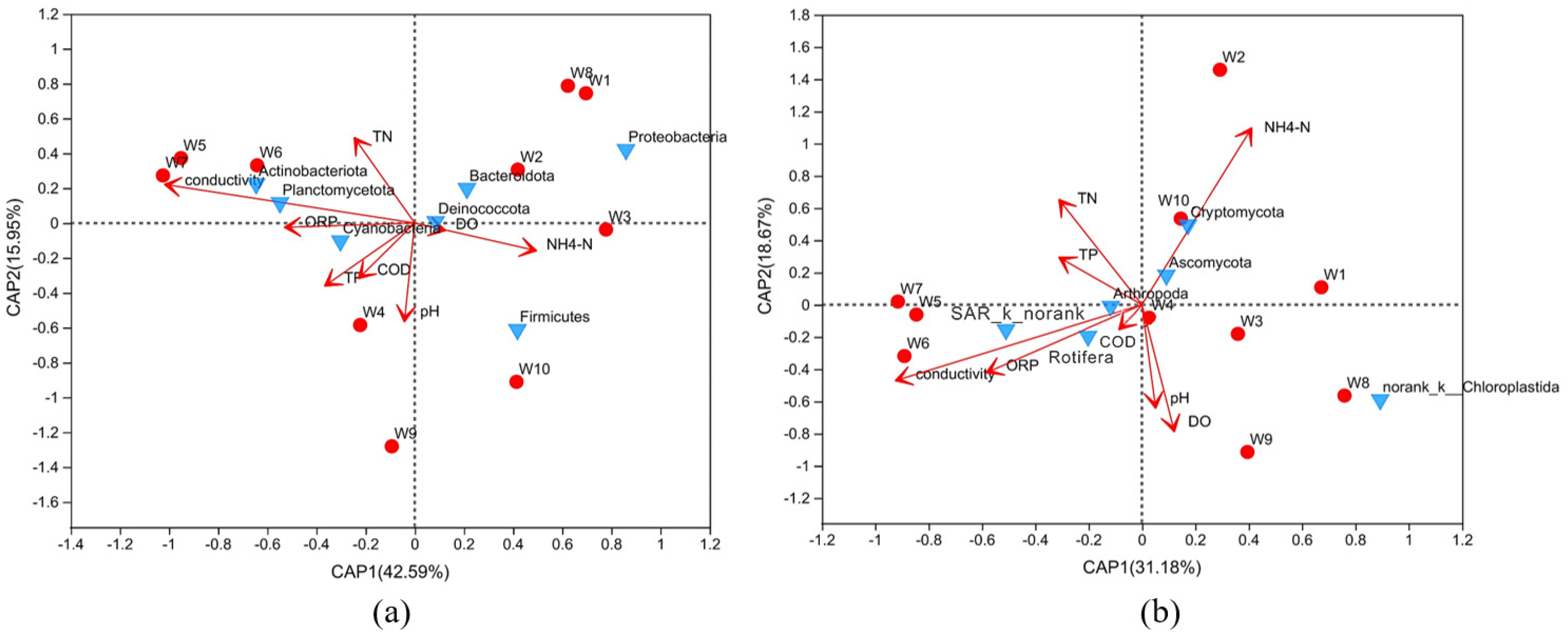
To explore the main factors affecting the distribution of microbes in the river water column, redundancy analysis was used to analyze the structures of the water samples at the phylum level and the environmental physical and chemical indicators (Figure 4). Generally, conductivity, TN, and NH4+-N are the main environmental parameters that influence the distribution of microbial communities. Conductivity, as an important index of surface water, can reflect the total dissolved solids and salts content of the water samples. Previous studies demonstrated that secondary salinization caused by anthropogenic activities generally increased the conductivity and salt content of surface water, resulting in a significant impact on water biodiversity and ecological functions [5,7]. The results showed that each sampling site exhibited various distributions. Moreover, Actinobacteriota and Planctomycetota were positively correlated with TN and conductivity, whereas Firmicutes were negatively correlated with TN. Eukaryotic Chloroplastida was negatively correlated with TN and TP in the water.

3.5. Functional Annotation

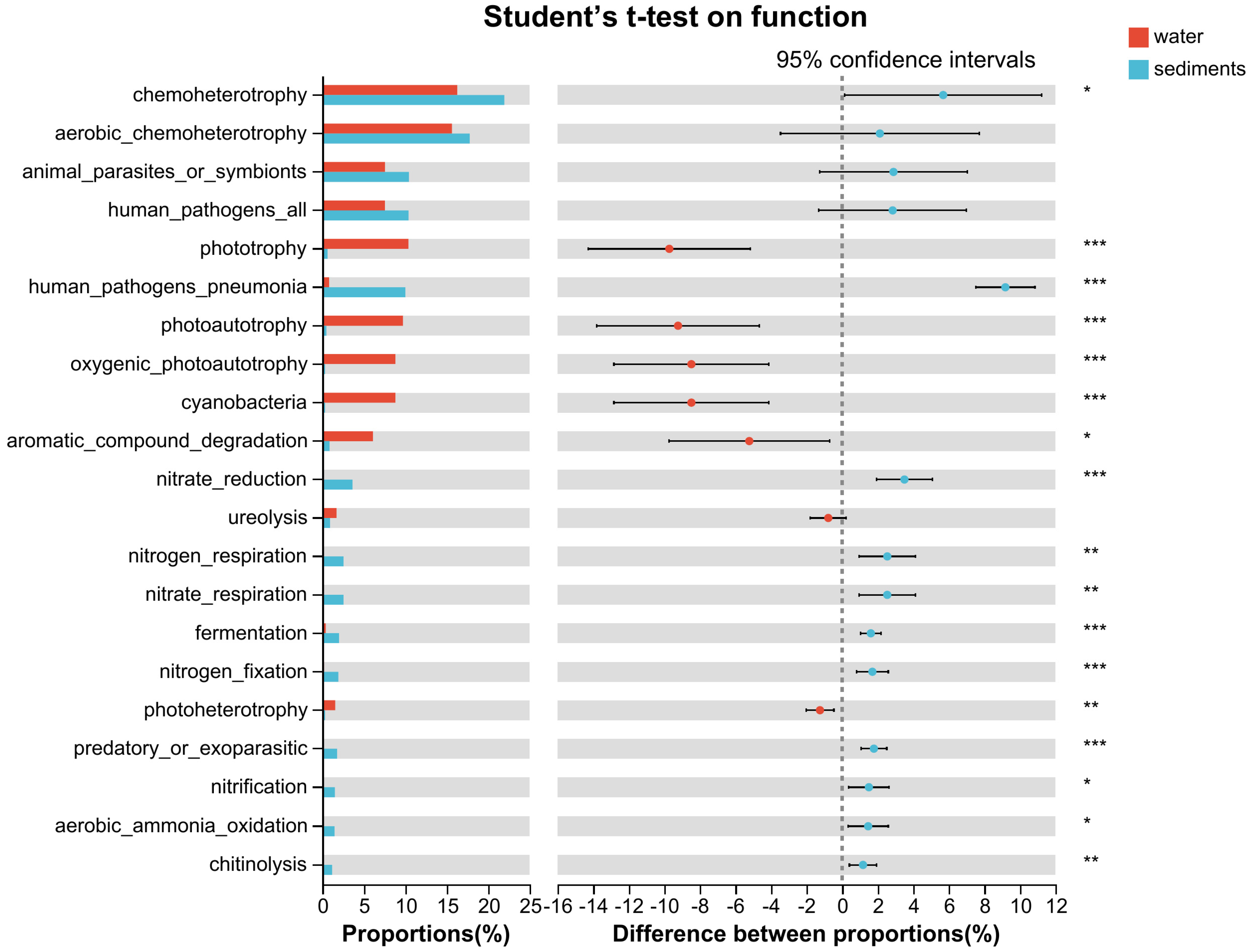
FAPROTAX is a manually constructed database that maps prokaryotic taxa to metabolic or other ecologically relevant functions (e.g., nitrification, denitrification, or fermentation) based on the literature on cultured representatives. The results have been plotted in Figure 5. A total of 49 ecological function genes were detected, with 21 genes having a relative abundance of greater than 1%. The 21 dominant functional genes participate extensively in the biogeochemical circulation of carbon and nitrogen. As shown in the figure, water has more phototrophs than sediment, which indicates that surface water has more light that promotes the growth of these planktonic bacteria. The ecological genes related to carbon were consistent, but their relative abundances differed. In addition, the relative abundance of carbon genes was high in both water and sediment, indicating that the carbon cycle was stronger than other nutrient cycles in the river.
ANOVA tests indicated that several organic degradation genes, such as aromatic compound degradation and chitinolysis, were significantly different between the two habitats (p < 0.05).
Six genes related to nitrogen transformation (nitrogen fixation, nitrate reduction, nitrification, nitrogen respiration, nitrate respiration, and aerobic ammonia oxidation) were detected in sediments. However, these genes were hardly detected in the water samples, indicating that river sediments are more involved in the nitrogen cycle than the overlying water. As for the nitrogen fixation (nif) gene, the 10 sediment samples exhibited relative abundances ranging from 0.27% to 1.93%, indicating that most of the nitrogen fixation occurred in the sediment layer rather than the water layer.

4. Discussion

4.1. Prokaryotic Variations

In this study, Proteobacteria were the most common phylum in both sediment and water, consistent with previous reports that indicate that Proteobacteria are widely distributed in various water bodies, sediments, and wetlands and play a crucial role in the degradation of organic matter [12,13,20]. Proteobacteria contain five classes (alpha, beta, gamma, delta, and Epsilon). In this study, the eight most abundant families under this phylum were classified into alpha- (Sphingomonadaceae, Rhodobacteraceae), beta- (Nitrosomonadaceae), and gamma- (Moraxellaceae, Comamonadaceae, Rhodocyclaceae, Xanthobacteraceae, Steroidobacteraceae) taxonomic classes, consistent with previous studies that found that alpha-, beta-, and gamma- were the most common Proteobacteria in surface waters [6,21,22]. Furthermore, recent research has found that nitrogen addition can increase the relative abundance of Proteobacteria [23]. Therefore, the difference in most bacteria under the phylum between water and sediment could be explained by the content of nitrogen available for the bacteria. For instance, Nitrosomonadaceae, well known as ammonium-oxidizing bacteria, mostly exist in sediment rather than water in this research. A possible reason for these differences is that the sediment has a higher substrate (ammonium) concentration than the water column.
Acidobacteria in the sediment samples were mainly represented by no-rank-f-Vicinamibacterales, no-rank_o_subgroup 7, Vicinamibacteraceae, and no-rank_o_subgroup 17, which accounted for 25.56 ± 5.82% of the composition, compared with 0.03 ± 0.027% in the water samples. The richness of Acidobacteria in sediments was consistent with recent research on the Yangtze River, which reported 20.81% to 23.76% [24]. To date, Acidobacteria has been found to be common in a wide range of soil biotopes, consistent with their known ability to compete in oligotrophic environments [25]. Few studies have been conducted on Acidobacteria in aquatic ecosystems [26]. found that several families under Acidobacteria, including Vicinamibacteraceae, were able to accumulate polyphosphate under aerobic or anoxic conditions. Therefore, the higher abundance of Acidobacteria in sediment than in water might have contributed to the higher TP availability in sediment for the bacteria to thrive.
In contrast to Acidobacteria, Cyanobacteria (mainly Cyanobium PCC-6307) were more abundant in water than in sediments. It is widely known that Cyanobacteria are phototrophic bacteria and tend to live in the surface water column to carry out oxygenic photosynthesis [27]. The average abundance of Actinobacteria, represented by Ilumatobacteraceae and Sporichthyaceae, in the water was significantly higher than that in the sediment. Actinobacteria are planktonic freshwater bacteria that are recognized early as a source of severe earthy-musty taste and odors in drinking water and have been reported to decompose all types of organic substances [21].
The relative abundance of Chloroflexi in the sediment samples (3.96 ± 2.01%) was generally higher than that in the water samples (0.95 ± 0.8%). This finding was consistent with previous reports that indicated that Chloroflexi are one of the predominant microorganisms in soil and river sediments [28]. In addition, Chloroflexi have been found to be associated with hydrocarbon-contaminated soils [5,29]. Anaerolineaceae in the phylum were the main taxonomic group in sediments, whereas water was dominant in the no-rank_c_SL56 marine group. Anaerolineaceae are mostly chemoheterotrophs and grow under anaerobic conditions [13], which is consistent with the biotope of sediments.
Another difference between the two habitats was Firmicutes, which accounted for 17.67 ± 13.20% in water and only 2.26 ± 1.66% in sediment. Only one family (Exiguobacteraceae) classified as Firmicutes showed differences between water and sediment; the other two families, Bacillaceae and Planococcaceae, exhibited no difference. Exiguobacteraceae may be aerobic or anaerobic depending on growth conditions and oxygen availability [30,31]. This bacterial taxon can reduce nitrate and is involved in the biodegradation of several complex organic compounds [32].
Overall, the variations in prokaryotes between water and sediment were probably due to environmental factors, such as DO and nutrient content. Further studies are needed to focus on the impact of environmental factors on bacterial diversity.

4.2. Eukaryotic Variations

Apart from environmental factors, biotic variables such as predation or species-species interaction could shape the distribution and structure of bacteria communities [33]. Hence, eukaryotic variations have profound roles in understanding microbial properties and functions in aquatic ecosystems.
In this study, seven out of twenty-three eukaryotic phyla exhibited remarkable differences between water and sediment. Within the Animalia kingdom, Annelida, Gastrotricha, and Vertebrata in sediment were significantly higher than in water, hinting that predation mostly happened in sediment. Chloroplastida, also known as picoplankton green algae, are the dominant eukaryotes in water. Chloroplastida, represented by two phyla, no-rank Chloroplastida, and Phragmoplastophyta showed significant differences between the water and sediment. Typically, water exhibited more no-rank Chloroplastida and fewer Phragmoplastophyta, suggesting that river water may serve as a better habitat for the former taxon.
Basidiomycota (fungi) in the sediment were also significantly different from those in water. Basidiomycota are saprotrophs and important contributors to the functioning of the ecosystem at multiple levels. However, the growth morphology of most eukaryotes remains unknown owing to the inadequacy of the current database.

4.3. Variations of Ecological Function

Bacteria play an important role in the transformation and cycling of nutrients in aquatic ecosystems. Based on the prokaryotic diversity and ecological function analysis of river water and sediment, it is found that water and sediment play equally important roles in the carbon cycle (Figure 6). Organic pollutants dissolved in water can either be biodegraded by bacteria or directly digested by zooplankton. The organics in the sediment could be eliminated not only in the same way as water but also by being involved in the transformation of nitrogen and phosphate. Nitrogen cycling occurs mostly in sediments rather than in water. The relative abundance of the nitrate reduction gene was slightly higher than that of the nitrification gene, which may be attributed to the higher nitrate concentration compared with ammonium in the river. Interestingly, nitrification, which is mediated by aerobic autotrophs and requires DO as an electron acceptor, was also detected at high levels in the sediment. The availability of oxygen may be from the overlying water, where photosynthesis and atmospheric reoxygenation occur.
Vertebrata and Annelida, which are in the phylum Amoebozoa, as well as Fungi (including Basidiomycota and Mucoromycota), were positively correlated with nitrogen and carbon metabolisms while were negatively correlated with phototrophic functions. This may be because these eukaryotes have a competitive or predatory relationship with algae and cooperate with carbon and nitrogen bacteria. In particular, Rotifera (Animalia) and no-rank Chloroplastida (Chloroplastida) were strongly correlated with the phototrophic processes. Most Rotifera are either raptorial predators, microphagous suspension feeders, or grazers; they typically require moderate oxygen conditions and abundant algae [34]. Thus, they can be used as bioindicators of water quality.

5. Conclusions

This study thoroughly investigated variations in the microbial community and ecological functions of the XJ River by collecting samples from water and sediment. 16S and 18S rRNA sequencing were performed to reveal prokaryotic and eukaryotic communities.
  • Conductivity, TN, and NH4+-N were the main environmental parameters influencing the distribution of microbial communities in the XJ River water column.
  • River sediments had higher diversity and richness than water samples.
  • Proteobacteria, Acidobacteria, and Actinobacteria were the dominant bacteria in sediments. The most abundant taxa in the water samples were Proteobacteria, Actinobacteria, and Firmicutes.
  • Eukaryotes in sediments are much spatially stochastic, while in the water column are dominated by Chloroplastida.
  • Sediments are more active in the global nitrogen cycle, while the overlying water plays an important role in oxygenic photosynthesis.
For further study in the future, we will focus on the physicochemical indices of sediments and the temporal and spatial distribution patterns of eukaryotic organisms in water bodies and sediments.

Supplementary Materials

The following supporting information can be downloaded at: https://www.mdpi.com/article/10.3390/w15122189/s1, Figure S1: Venn diagram showing the shared and unique OTUs of water and sediments for (a) prokaryotes and (b) eukaryotes. Table S1: Diversity indices for 16S and 18S rRNA sequencing. Table S2: Analysis of prokaryotic difference between sediment and water samples at family level based on ANOVA test. Table S3: Analysis of eukaryotic difference between sediment and water samples at phylum level based on ANOVA test.

Author Contributions

All authors contributed to the study’s conception and design. Material preparation, data collection, and analysis were performed by W.Z., M.L. and F.G. The first draft of the manuscript was written by S.W. and H.C. revised the manuscript, and all authors commented on previous versions of the manuscript. All authors have read and agreed to the published version of the manuscript.

Funding

This work was supported by the Education Department of Hunan Province (20A032), the Open Research Fund of Science and Technology Innovation Platform of Key Laboratory of Dongting Lake Aquatic Eco-Environmental Control and Restoration of Hunan Province (2018DT02), and the Changsha University of Science & Technology (000301606).

Data Availability Statement

All data generated or analyzed during this study are included in the paper.

Conflicts of Interest

The authors have no relevant financial or non-financial interests to disclose.

References

  1. Fu, J.; Zhao, C.; Luo, Y.; Liu, C.; Kyzas, G.Z.; Luo, Y.; Zhao, D.; An, S.; Zhu, H. Heavy metals in surface sediments of the Jialu River, China: Their relations to environmental factors. J. Hazard. Mater. 2014, 270, 102–109. [Google Scholar] [CrossRef] [PubMed]
  2. Kiersztyn, B.; Chróst, R.; Kaliński, T.; Siuda, W.; Bukowska, A.; Kowalczyk, G.; Grabowska, K. Structural and functional microbial diversity along a eutrophication gradient of interconnected lakes undergoing anthropopressure. Sci. Rep. 2019, 9, 11144. [Google Scholar] [CrossRef] [PubMed] [Green Version]
  3. Savio, D.; Sinclair, L.; Ijaz, U.Z.; Parajka, J.; Reischer, G.H.; Stadler, P.; Blaschke, A.P.; Blöschl, G.; Mach, R.L.; Kirschner, A.K.T.; et al. Bacterial diversity along a 2600 km river continuum. Environ. Microbiol. 2015, 17, 4994–5007. [Google Scholar] [CrossRef] [PubMed] [Green Version]
  4. Paerl, H.W.; Dyble, J.; Moisander, P.H.; Noble, R.T.; Piehler, M.F.; Pinckney, J.L.; Steppe, T.F.; Twomey, L.; Valdes, L.M. Microbial indicators of aquatic ecosystem change: Current applications to eutrophication studies. FEMS Microbiol. Ecol. 2003, 46, 233–246. [Google Scholar] [CrossRef]
  5. Xie, G.; Tang, X.; Shao, K.; Zhu, G.; Gao, G. Bacterial diversity, community composition and metabolic function in Lake Tianmuhu and its dammed river: Effects of domestic wastewater and damming. Ecotoxicol. Environ. Saf. 2021, 213, 112069. [Google Scholar] [CrossRef]
  6. Newton, R.J.; Jones, S.E.; Eiler, A.; McMahon, K.D.; Bertilsson, S. A guide to the natural history of freshwater lake bacteria. Microbiol. Mol. Biol. Rev. MMBR 2011, 75, 14–49. [Google Scholar] [CrossRef] [Green Version]
  7. Chen, J.; Wu, J.; Liu, M.; Li, L.; Zhang, W.; Wang, D.; Ma, T. Bacterial community structure in the surface sediments of different habitats of Baiyangdian Lake, Northern China: Effects of nutrient conditions. J. Soils Sediments 2021, 21, 1866–1874. [Google Scholar] [CrossRef]
  8. Liu, Y.; Mao, D. Integrated assessment of water quality characteristics and ecological compensation in the Xiangjiang River, south-central China. Ecol. Indic. 2020, 110, 105922. [Google Scholar] [CrossRef]
  9. Han, B.; Addo, F.G.; Mu, X.; Zhang, L.; Zhang, S.; Lv, X.; Li, X.; Wang, P.; Wang, C. Epiphytic bacterial community shift drives the nutrient cycle during Potamogeton malaianus decomposition. Chemosphere 2019, 236, 124253. [Google Scholar] [CrossRef]
  10. Chen, M.; Chen, F.; Zhao, B.; Wu, Q.L.; Kong, F. Seasonal variation of microbial eukaryotic community composition in the large, shallow, subtropical Taihu Lake, China. Aquat. Ecol. 2010, 44, 1–12. [Google Scholar] [CrossRef]
  11. Lim, E.L.; Dennett, M.R.; Caron, D.A. Identification of Heterotrophic Nanoflagellates by Restriction Fragment Length Polymorphism Analysis of Small Subunit Ribosomal DNA. J. Eukaryot. Microbiol. 2001, 48, 247–257. [Google Scholar] [CrossRef]
  12. Li, K.; Hu, J.; Li, T.; Liu, F.; Tao, J.; Liu, J.; Zhang, Z.; Luo, X.; Li, L.; Deng, Y.; et al. Microbial abundance and diversity investigations along rivers: Current knowledge and future directions. WIREs Water 2021, 8, e1547. [Google Scholar] [CrossRef]
  13. Lepère, C.; Domaizon, I.; Humbert, J.-F.; Jardillier, L.; Hugoni, M.; Debroas, D. Diversity, spatial distribution and activity of fungi in freshwater ecosystems. PeerJ 2019, 7, e6247. [Google Scholar] [CrossRef]
  14. Zhang, S.; Yang, C.; Liu, M.; Zhao, W.; Li, Y.; Meng, X.-Z.; Cai, M. Occurrence of organophosphate esters in surface water and sediment in drinking water source of Xiangjiang River, China. Sci. Total. Environ. 2021, 781, 146734. [Google Scholar] [CrossRef]
  15. APHA (American Public Health Association). Standard Methods for the Examination of Water and Wastewater, 21st ed.; American Public Health Association: Washington, DC, USA, 2005. [Google Scholar]
  16. Caporaso, J.G.; Lauber, C.L.; Walters, W.A.; Berg-Lyons, D.; Lozupone, C.A.; Turnbaugh, P.J.; Fierer, N.; Knight, R. Global patterns of 16S rRNA diversity at a depth of millions of sequences per sample. Proc. Natl. Acad. Sci. USA 2011, 108 (Suppl. S1), 4516–4522. [Google Scholar] [CrossRef] [PubMed] [Green Version]
  17. Parada, A.E.; Needham, D.M.; Fuhrman, J.A. Every base matters: Assessing small subunit rRNA primers for marine microbiomes with mock communities, time series and global field samples. Environ. Microbiol. 2016, 18, 1403–1414. [Google Scholar] [CrossRef] [PubMed]
  18. Bråte, J.; Logares, R.; Berney, C.; Ree, D.K.; Klaveness, D.; Jakobsen, K.S.; Shalchian-Tabrizi, K. Freshwater Perkinsea and marine-freshwater colonizations revealed by pyrosequencing and phylogeny of environmental rDNA. ISME J. 2010, 4, 1144–1153. [Google Scholar] [CrossRef] [Green Version]
  19. Chen, H.; Wu, J.; Huang, R.; Zhang, W.; He, W.; Deng, Z.; Han, Y.; Xiao, B.; Luo, H.; Qu, W. Effects of temperature and total solid content on biohydrogen production from dark fermentation of rice straw: Performance and microbial community characteristics. Chemosphere 2022, 286, 131655. [Google Scholar] [CrossRef]
  20. Davis, K.E.; Sangwan, P.; Janssen, P.H. Acidobacteria, Rubrobacteridae and Chloroflexi are abundant among very slow-growing and mini-colony-forming soil bacteria. Environ. Microbiol. 2011, 13, 798–805. [Google Scholar] [CrossRef] [PubMed]
  21. Betiku, O.C.; Sarjeant, K.C.; Ngatia, L.W.; Aghimien, M.O.; Odewumi, C.O.; Latinwo, L.M. Evaluation of microbial diversity of three recreational water bodies using 16S rRNA metagenomic approach. Sci. Total Environ. 2021, 771, 144773. [Google Scholar] [CrossRef] [PubMed]
  22. Korajkic, A.; Parfrey, L.W.; McMinn, B.R.; Baeza, Y.V.; VanTeuren, W.; Knight, R.; Shanks, O.C. Changes in bacterial and eukaryotic communities during sewage decomposition in Mississippi river water. Water Res. 2015, 69, 30–39. [Google Scholar] [CrossRef]
  23. Nie, Y.; Wang, M.; Zhang, W.; Ni, Z.; Hashidoko, Y.; Shen, W. Ammonium nitrogen content is a dominant predictor of bacterial community composition in an acidic forest soil with exogenous nitrogen enrichment. Sci. Total Environ. 2018, 624, 407–415. [Google Scholar] [CrossRef]
  24. Li, X.; Zhang, Y.; Song, S.; Zhou, Y.; Zhang, J. Bacterial diversity patterns differ in different patch types of mixed forests in the upstream area of the Yangtze River Basin. Appl. Soil Ecol. 2021, 161, 103868. [Google Scholar] [CrossRef]
  25. Yergeau, E.; Bokhorst, S.; Kang, S.; Zhou, J.; Greer, C.W.; Aerts, R.; Kowalchuk, G.A. Shifts in soil microorganisms in response to warming are consistent across a range of Antarctic environments. ISME J. 2012, 6, 692–702. [Google Scholar] [CrossRef] [PubMed] [Green Version]
  26. Kristensen, J.M.; Singleton, C.; Clegg, L.A.; Petriglieri, F.; Nielsen, P.H. High Diversity and Functional Potential of Undescribed “Acidobacteriota” in Danish Wastewater Treatment Plants. Front. Microbiol. 2021, 12, 643950. [Google Scholar] [CrossRef]
  27. Koblížek, M. Ecology of aerobic anoxygenic phototrophs in aquatic environments. FEMS Microbiol. Rev. 2015, 39, 854–870. [Google Scholar] [CrossRef] [Green Version]
  28. Zhang, S.; Merino, N.; Wang, N.; Ruan, T.; Lu, X. Impact of 6:2 fluorotelomer alcohol aerobic biotransformation on a sediment microbial community. Sci. Total. Environ. 2017, 575, 1361–1368. [Google Scholar] [CrossRef] [PubMed]
  29. Sutton, N.B.; Maphosa, F.; Morillo, J.A.; Abu Al-Soud, W.; Langenhoff, A.A.M.; Grotenhuis, T.; Rijnaarts, H.H.M.; Smidt, H. Impact of Long-Term Diesel Contamination on Soil Microbial Community Structure. Appl. Environ. Microbiol. 2013, 79, 619–630. [Google Scholar] [CrossRef] [PubMed] [Green Version]
  30. Kasana, R.C.; Pandey, C.B. Exiguobacterium: An overview of a versatile genus with potential in industry and agriculture. Crit. Rev. Biotechnol. 2018, 38, 141–156. [Google Scholar] [CrossRef]
  31. Zhang, L.; Zhong, M.; Li, X.; Lu, W.; Li, J. River bacterial community structure and co-occurrence patterns under the influence of different domestic sewage types. J. Environ. Manag. 2020, 266, 110590. [Google Scholar] [CrossRef]
  32. Zhang, F.; Zhang, H.; Yuan, Y.; Liu, D.; Zhu, C.; Zheng, D.; Li, G.; Wei, Y.; Sun, D. Different response of bacterial community to the changes of nutrients and pollutants in sediments from an urban river network. Front. Environ. Sci. Eng. 2020, 14, 28. [Google Scholar] [CrossRef]
  33. Yang, Y.; Gao, Y.; Huang, X.; Ni, P.; Wu, Y.; Deng, Y.; Zhan, A. Adaptive shifts of bacterioplankton communities in response to nitrogen enrichment in a highly polluted river. Environ. Pollut. 2019, 245, 290–299. [Google Scholar] [CrossRef] [PubMed]
  34. Hillis, D.G.; Lissemore, L.; Sibley, P.K.; Solomon, K.R. Effects of Monensin on Zooplankton Communities in Aquatic Microcosms. Environ. Sci. Technol. 2007, 41, 6620–6626. [Google Scholar] [CrossRef] [PubMed]
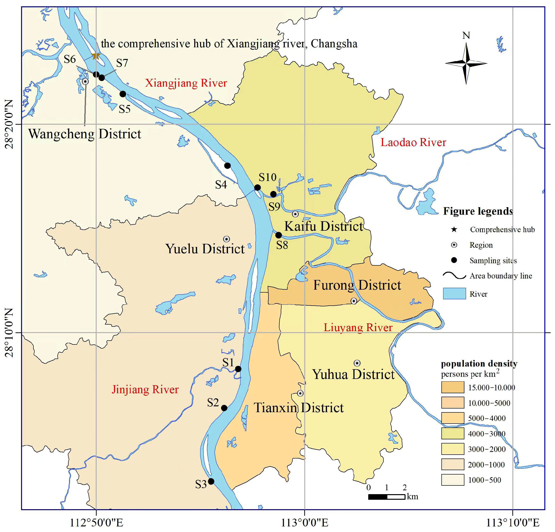
Figure 1. Geographical location of the 10 monitoring sites in the Xiangjiang River, south-central China (S1–S10).
Water 15 02189 g001
Figure 2. PCoA plot of Bray–Curtis distance for (a) prokaryotic and (b) eukaryotic communities in XJ River.
Water 15 02189 g002
Figure 3. Relative abundance of the dominant phyla of (a) prokaryotes in sediment (marked as S followed by number of sampling sites and overlying water (marked as W) with a mean abundance >1% and (b) eukaryotes in sediment (S) and overlying water (W) with the mean abundance >1%. * Signifies p value in the range of 0.01 < p ≤ 0.05, ** 0.001 < p ≤ 0.01, *** p ≤ 0.001.
Water 15 02189 g003
Figure 4. Redundancy analysis (RDA) based on Bray–Curtis distance of (a) prokaryotic and (b) eukaryotic communities on the phylum level at different sites.
Water 15 02189 g004
Figure 5. Difference of bacterial function (relative abundance > 1%) based on FAPROTAX database for sediment and water samples. * Signifies p value in the range of 0.01 < p ≤ 0.05, ** 0.001 < p ≤ 0.01, *** p ≤ 0.001.
Water 15 02189 g005
Figure 6. Proposed functions of prokaryotes and eukaryotes involved in carbon and nitrogen cycle from this study. Prokaryotes marked as P and eukaryotes marked as E.
Water 15 02189 g006
Table 1. Physicochemical indices of the surface water at sampling sites.
Sampling SiteConductivityORP
(mV)
DO
(mg/L)
pHTP
(mg/L)
NH4+-N
(mg/L)
TN
(mg/L)
CODcr
(mg/L)
#12442056.457.431.320.860.6716.16
#22482657.427.550.480.391.1317.15
#32512616.457.770.520.301.091.97
#42322316.87.380.440.760.9011.43
#52152346.77.910.660.440.7410.64
#62242356.717.950.821.070.855.32
#72242357.477.950.680.431.007.09
#8190.52155.817.020.761.361.1312.22
#9172.32166.957.480.720.710.6310.64
#102372367.67.421.220.911.0049.66
Disclaimer/Publisher’s Note: The statements, opinions and data contained in all publications are solely those of the individual author(s) and contributor(s) and not of MDPI and/or the editor(s). MDPI and/or the editor(s) disclaim responsibility for any injury to people or property resulting from any ideas, methods, instructions or products referred to in the content.