Article

Batch Studies on the Biodegradation Potential of Paracetamol, Fluoxetine and 17α-Ethinylestradiol by the Micrococcus yunnanensis Strain TJPT4 Recovered from Marine Organisms

1
Centro de Ciências da Mar, University of Algarve, Campus de Gambelas, Building 7, 8005-139 Faro, Portugal
2
Faculdade de Ciências e Tecnologias, University of Algarve, Campus de Gambelas, Building 8, 8005-139 Faro, Portugal
*
Author to whom correspondence should be addressed.
Water 2022, 14(21), 3365; https://doi.org/10.3390/w14213365
Submission received: 14 September 2022 / Revised: 9 October 2022 / Accepted: 19 October 2022 / Published: 23 October 2022
(This article belongs to the Special Issue Persistent and Emerging Organic Contaminants in Natural Environments)

Abstract

:
The emerging pollutants paracetamol, fluoxetine and ethinylestradiol are inefficiently removed by conventional wastewater treatments, entering in aquatic environments in which they are hazardous. Aiming for the obtention of bacteria with the capacity for environmental bioremediation, eight bacteria were isolated from two consortia recovered from Hymedesmia versicolor and Filograna implexa marine organisms which exhibited a high-paracetamol-removal capacity. The isolates that displayed the ability to grow in the presence of 100 mg/L paracetamol as the sole carbon source were assigned to Paenibacillus, Micrococcus and Microbacterium genera. The isolate assigned to the Micrococcus yunnanensis strain TJPT4 presented the best performance, degrading 93 ± 4% of 15 mg/L paracetamol as the sole carbon source after 360 h, and was also apparently able to degrade the produced metabolites. This strain was able to remove 82.1 ± 0.9% of 16 mg/L fluoxetine after 504 h, mainly by adsorption, but apparently a biodegradation contribution also occurred. This strain was able to remove 66.6 ± 0.2% of 13 mg/L 17α-ethinylestradiol after 360 h. As far as is known, Micrococcus yunnanensis is for the first time recovered/identified in Filograna implexa, presenting a high drug removal efficiency, thereby becoming a great candidate for treatment processes (e.g., bioaugmentation), especially in the presence of saline intrusions.

Graphical Abstract

1. Introduction

There is an increasing concern within the scientific community about pharmaceutical compounds and their problematic consequences. Wastewater treatment plants (WWTP) have limited capacity for removing pharmaceuticals and hormones due to the bioreactor characteristics, which are designed for low solid and hydraulic retention times and have no ability to withstand hydraulic overloads [1]. Moreover, the composition, diversity and degrading abilities of the bioreactor’s specialized bacterial communities are of great importance in wastewater treatments [2]. Consequently, one of the main routes by which these pollutants enter aquatic resources is through the release of inappropriately treated water into the environment [3]. Although concentrations of pharmaceuticals and their metabolites occurring in the environment are typically low from ng/L to μg/L, these compounds still have the potential to pose many threats, not only to the ecosystem they enter, but also to humans who may encounter them.
Biodegradation efficiency in a WWTP can be affected by several factors, such as saline influents/intrusions that may modify the microbial properties of the biological reactors, causing huge impacts on the biological treatments (e.g., activated sludge processes) [4]. For example, WWTPs are often subjected to saltwater infiltrations from the sea when located on coastal zones, or to industrial discharges with saline effluents or leachate discharges, in addition to cases in which marine waters are used in sanitary systems, as happens in some countries with insufficient freshwater [4]. Therefore, it is important to find halotolerant microorganisms which, in addition, have the capacity to biodegrade/remove pollutants, since these kinds of bacteria may have a central role in keeping the equilibrium of the WWTP’s biological processes near coastal areas.
This study was focused on three commonly used drugs worldwide: paracetamol, also known as acetaminophen, fluoxetine (FLX) and 17α-ethinylestradiol (EE2).
Paracetamol is one of the most commonly used analgesic and antipyretic drugs and one of the most prescribed drugs during the COVID-19 pandemic [5,6]. An important sign that paracetamol is of global concern is the fact that it was detected in seawaters, e.g., in the western Mediterranean Sea, reaching concentrations ranging from 0.468 to 1.70 ng/L [7]. Zhang et al. [8] reported that bacterial isolates from the genera Stenotrophomonas and Pseudomonas were able to use paracetamol as the sole carbon source and proposed a degradation pathway in which paracetamol may suffer an initial catalyzation by an amidohydrolase to release 4-aminophenol, with a substitution of the amino group by a hydroxyl group to form hydroquinone, and subsequent ring fission [8]. An alternative path could be the initial hydroxylation of paracetamol catalyzed by a hydrolytic enzyme to produce hydroquinone and, in either case, after the ring fission, the compounds may be converted into succinic, malonic, oxalic acid and subsequently into formic acid [8]. The mineralization of these acids into carbon dioxide and water occurs as a final step. The physicochemical characteristics of paracetamol enable the drug to access the aquatic environment easily, since it is highly soluble and has a hydrophilic nature [9]. The pseudo-first-order degradation constant (Kbiol) of paracetamol ranges from 50 to 80 L/g suspended solids (SS).day and 106 to 204 L/gSS.day [10]. This drug can be considered a highly biodegradable compound, with a Kbiol > 10 L/gSS.day, having an octanol–water partition coefficient (logKow) of 0.46 [11]. Paracetamol and its metabolites occurring in the environment may cause oxidative stress, DNA and cellular damages in the aquatic organisms [12].
Fluoxetine is a selective serotonin reuptake inhibitor (SSRI) and one of the most prescribed antidepressant drugs, being a recalcitrant pollutant containing very strong C-F bonds [13]. Fluoxetine is among the more prevalent categories of pharmaceuticals detected in the marine environment and therefore is frequently detected in coastal areas at concentrations ranging from 0.03 to 300 ng/L [14,15,16]. Khan and Murphy [17] reported that FLX was hydrolyzed by bacterial degradation to yield 4-(trifluoromethyl)phenol (TFMP) and 3-(methylamino)-1-phenylpropan-1-ol, and the final products were trifluoroacetate and fluoride ion, which may arise due to the spontaneous defluorination of the meta-cleavage products that were shown to be photosensitive [17]. The physicochemical properties of FLX and norfluoxetine (NFLX), a metabolic product of FLX excreted by humans, allow the prediction of the biodegradation and adsorption of these compounds. FLX is slightly biodegraded since it presents values of Kbiol ranging from 0.03 to 9.0, thus showing a poor to significant biodegradability, which may likely depend on the operational conditions. FLX displayed values of logKd ranging from 2.76 to 3.78 (logKd > 2.7) and a logKow higher than 4, thus behaving like a lipophilic compound and predicting a high adsorption potential [18,19,20]. FLX and its metabolite occurring in the environment may cause behavioral changes, aggressiveness and induces genotoxic, mutagenic and cytotoxic effects in aquatic organisms [21].
The endocrine-disrupting agent 17-α-ethinylestradiol (EE2) was recently included in an EU Watch List of the Water Framework Directive (WFD, EU Directive 2013/39/EU, EU Implementing Decision 2020/1161) [22] regarding emerging aquatic pollutants. The incomplete removal of EE2 by conventional wastewater treatment processes leads to the contamination of surface waters [23,24,25]. Concentrations consistently range from 0.2 to 1.5 ng/L (0.88–6.58 pM) among similar types of surface water sources [26]. EE2 is categorized as potentially biodegradable according to its kinetic reaction rate (kbiol) of 7–9 L/(gSS.day) [27]. EE2′s adsorption capacity to sewage sludge is regarded as moderate, with an octanol–water partition coefficient (logKow) of 2.8–4.2, and therefore the most likely mechanism responsible for drug removal is biodegradation [27,28]. Several metabolites were identified from the degradation of EE2; for example, Sphingobacterium sp. JCR5, an EE2-utilizing bacterium, transformed the drug into 3,4-dihydroxy-9,10-secoandrosta-1,3,5(10)- triene-9.17-dione, 2-hydroxy-2,4-dienevaleric acid and 2-hydroxy-2,4-diene-1,6-dioic acid. Several subsequent degradation steps, including the meta-cleavage of the A ring via dioxygenase, led to the production of the end products of CO2, and water [29].
EE2′s environmental contamination has a strong hazardous effect in the organisms, such as animals, causing behavioral changes and reproductive dysregulation leading to the feminization phenomena, causing regulation disorders of both proapoptotic and antiapoptotic processes and may also induce neoplastic processes [30].
Few studies exist concerning the degradation of these pollutants by marine bacteria and/or recovered from marine organisms. Bacteria collected in environments that can be considered extreme or less explored, such as marine caves, may have the capacity to grow, as for example in the presence of high concentrations of salts (halophiles) [31], and may have adapted useful metabolisms not yet explored, namely for bioremediation purposes. Therefore, bacteria from marine samples could be good candidates for the biological treatment of wastewater, especially for those cases in which the WWTPs suffer saline intrusions and may also be used in bioaugmentation processes.
The aim of this study was to search for bacterial isolates from autochthonous marine organisms with ability to degrade paracetamol, FLX and EE2 pharmaceuticals. For this purpose, bacterial consortia were recovered from these organisms collected from the marine caves of the Algarve Coast, Portugal, and a screening was performed to find bacteria with ability to degrade paracetamol, herein considered a pharmaceutical model, since it is one of the most widely used and detected drugs in wastewaters, rivers and seawaters. Often, the produced metabolites are more toxic than the parent compounds; thus, the bacterial biodegradation products were also analyzed.
Two consortia H.P4 and FI.A21 with a high capacity to degrade paracetamol were recovered from Hymedesmia versicolor and Filograna implexa marine organisms, respectively and from these consortia, eight isolates were obtained. Micrococcus yunnanensis, as far as is known, is for the first time recovered and identified in Filograna implexa (Annelida) organisms and seems to play an important role in the bioremoval of paracetamol, fluoxetine and 17α-ethinylestradiol.

2. Materials and Methods

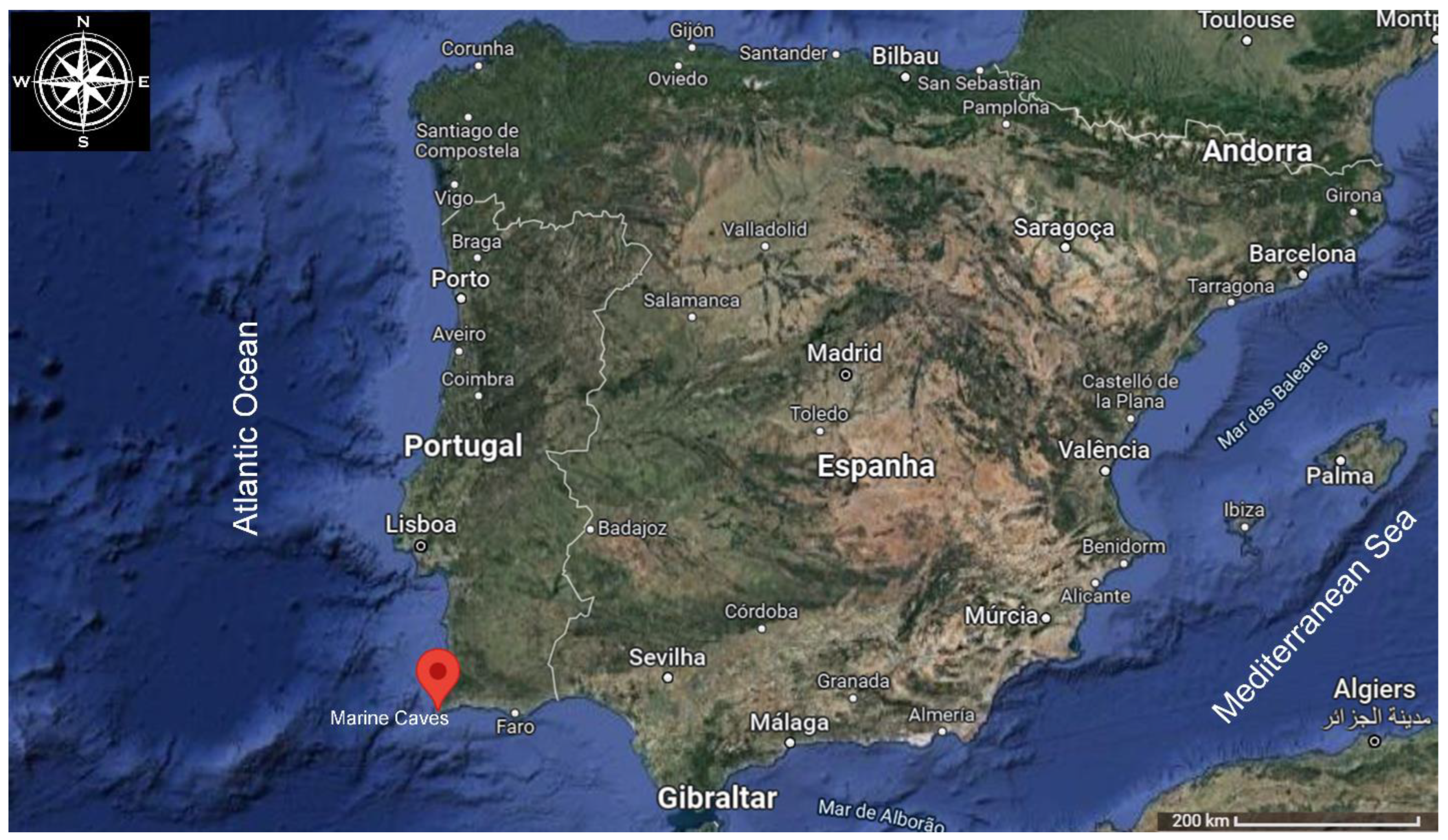
2.1. Inoculum Source

Autochthonous marine organisms were collected from Catedral and Queijo Suiço marine caves, located in Sagres, Algarve, Portugal (37°00′63.86″ N, 8°92′74.24″ W) in July 2020 (Figure 1).
A team of scientific divers collected samples of noninvasive marine species fixed on two rocky reefs. These autochthonous organisms were from the phyla Porifera, Tunicata and Annelida. Sampling was carried out by scraping and removing approximately from 3 to 6 g of each organism surface from its biota. The collected parts of the organisms were stored in seawater collected from the sampling site in 50 mL falcon tubes. After collection, the tubes were transported from Sagres to the Gambelas campus (University of Algarve) in a cooler box to maintain a stable temperature. The bacterial consortia enrichment process was carried out immediately after the arrival of the samples. Samples were stored at −80 °C.

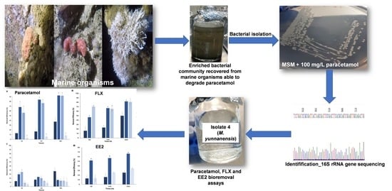
2.2. Recovery of Bacteria from Marine Organisms

From the samples collected, representative organisms of the species from different phyla were selected in order to recover the bacterial communities, namely Hymedesmia versicolor (Porifera), Didemnum sp. (Tunicates) and Filograna implexa (Annelida) (Figure S1).
The recovery of bacterial communities from the marine organisms collected in the marine caves was started by washing them with sterile seawater. This washing procedure was performed in 50 mL flasks with 1 g of the sample in 9 mL of sterile seawater under 150 rpm of orbital agitation for 4 h. Afterwards, 500 µL of the washing solution was transferred to a 50 mL flask with 4.5 mL of (1:10 dilution) of an artificial sterilized Marine Broth (MB) and incubated at 25 °C under 150 rpm during 24–48 h. After incubation, the cultures were preserved in glycerol (20 % v/v) and stored at −80 °C.

2.3. Inoculum Enrichment

The bacterial consortia recovered from the marine organisms Hymedesmia versicolor (Porifera), Didemnum sp. (Tunicates) and Filograna implexa (Annelida) were herein named as consortia H.P4, D.T18 and FI.A21, respectively.
Initial cultures of the recovered bacterial consortia were activated from the stored inoculum using 100 μL of glycerol cultures in 1 mL of MB for a period of 24 h at room temperature (23–28 °C) with 150 rpm of orbital agitation in dark conditions. To increase the biomass growth, from the previous culture, 1 mL was extracted and added to 9 mL of MB to complete the activation process, and samples were incubated for 24 h in a dark room at 28 °C.
For the enrichment, subcultures were prepared in sterilized glass flasks using 90 mL of MB and 10 mL (10% v/v) of the initial culture in the absence and presence of 50 and 100 mg/L paracetamol, incubated at room temperature (23–28 °C), under 150 rpm in the dark for 48 h. In all experiments, the bacterial growth was determined by measuring the optical density at 600 nm (OD600) at the beginning of the assay and during the incubation time using a Hach-Lange spectrophotometer DR-2800 (Hach-Lange, Sköndal, Sweden).

2.4. Pharmaceutical Bioremoval Studies Using Bacteria Recovered from Marine Organisms

After the enrichment in MB, the biodegradation assays were performed using as inoculum 10% (v/v) of each bacterial consortia H.P4, D.T18 and FI.A21 in a mineral salt medium (MSM) in the presence of 86 mg/L of paracetamol (model compound) as the sole carbon source. The MSM composition and preparation is described by Palma et al. [32] The medium was autoclaved for 25 min at 120 °C.

2.4.1. Bacterial Isolates Preparation

Bacteria were isolated from the consortia which displayed the best biodegradation abilities of paracetamol, herein used as model compound.
MB was also prepared for the positive control since it was a medium supplemented with the required carbon and energy sources for the optimal growth of bacteria recovered from marine environments. Solid media were prepared by adding 15 g/L of agar to both the MSM and MB media. Solid MSM media with/without 1 g of (NH4)2HPO4 as a nitrogen source were prepared in order to determine how this may affect the bacterial growth. This was done to determine if the bacterial isolates performed better with or without the presence of this nitrogen source. The media were sterilized by autoclaving at 120 °C for 25 min. An amount of 100 mg/L paracetamol was added to the MSM using a sterile PES syringe filter (VWR, Part of Avantor, Leuven, Belgium) for the removal of microorganisms from these heat-sensitive solutions that contained the drugs, and then distributed into petri dishes to allow them to harden.
The bacterial isolation from the consortia H.P4 and FI.A21 was performed as described by Palma et al. [32], with the exception of the step before the serial decimal dilution procedure, in which the pellet was resuspended into 9 mL of 0.1% (v/v) tryptone water supplied with 8.5 g/L NaCl, which was used as a primary enrichment medium for the growth of bacteria, with 4 h of incubation at 150 rpm in the dark at 25 °C.
The bacterial isolates were incubated at 28 °C for 48 h in the dark.
Bacterial isolates from consortia H.P4 and FI.A21 were determined based on their colony morphotypes; the protocol of bacterial selection was performed as described by Palma et al. [28,33].
Gram staining protocol and biochemical oxidase and catalase tests were performed as described by Palma et al. [28].
The DNA of each bacterial isolate was extracted using NZY Microbial gDNA Isolation kit (NZYTech, Lisbon, Portugal). The 16S rRNA gene was amplified and the direct sequencing of PCR products was performed by the Sanger method as described by Palma et al. [28,32,33]. Taxonomic identity was established as described by Palma et al. [28,33].
The phylogenetic analysis of the obtained bacterial isolates was carried out by comparison their 16S rRNA gene sequences with the type strain. The information about the type strains was listed at BacDive—the Bacterial Diversity Metadatabase, as stated in the DSMZ (https://www.dsmz.de/, accessed on 3 August 2022): species: Paenibacillus pabuli JCM 9074T with BacDive ID: 11478; Paenibacillus odorifer TOD45T with BacDive ID: 11560; Paenibacillus typhae xj7T with BacDive ID: 23180; Microbacterium ginsengisoli Gsoil 259T with BacDive ID: 7429; Micrococcus yunnanensis strain YIM 65004 T with BacDive ID: 7760; Micrococcus luteus DSM 20030T with BacDive ID: 7673; Micrococcus endophyticus strain YIM 56238T with BacDive ID: 7759.
From the eight bacterial isolates displaying the ability to grow in the presence of paracetamol, only two presented very low query covers below 90%, thus the nucleotide sequence accession numbers of the 16S rRNA sequences of the six strains were deposited in the GenBank database under accession numbers: Isolate 1_Paenibacillus pabuli (OM461362) and isolate 2_Paenibacillus typhae (OM461363) were all isolated from consortium H.P4 and isolate 4_Micrococcus yunnanensis (OM461364), isolate 5_Paenibacillus tianjinensis (OM461365), isolate 7_Microbacterium ginsengisoli (OM461366) and isolate 8_Paenibacillus pabuli (OM461367) were isolated from consortium FI.A21.

2.4.2. Removal Assays with Bacterial Isolates

After bacterial isolate identification, biodegradation experiments were carried out in MSM liquid cultures with the eight different isolates using 100, 50 and 15 mg/L of paracetamol (model compound) as the sole carbon source. The bacterial isolate which displayed the highest removal ability of paracetamol was selected for further experiments with other pharmaceutical drugs, namely FLX and EE2. Cultures were carried out following the McFarland scale as described in Palma et al. [33]; thus, 100 µL (v/v) (≈106 colony-forming unit (CFU/mL)) and 5% (1.5 mL ≈ 1.5 × 10⁷ CFU/mL) of each isolate was subcultured into 30 mL of MSM containing paracetamol or FLX or EE2.
The stock solution of 1000 mg/L of fluoxetine hydrochloride was elaborated with 200 µL of methanol to help its dissolution and the final volume was completed with milliQ water, and 17α-ethinylestradiol was prepared in methanol. To achieve each required experimental drug concentration, each stock solution was added using a sterile PES syringe filter of 0.2 μm pore size from VWR (VWR, Part of Avantor, Leuven, Belgium).
Negative controls containing the drug in the absence of the bacterial isolates were performed.
To assess the occurrence of the drug-adsorption mechanism by the interaction of the drug onto the inactivated inoculum, a bacterial biomass was produced for such a purpose: bacteria were harvested in the exponential phase (corresponding to a bacterial growth ranging OD values from 0.5 to 0.8) and were then inactivated by autoclaving during 45 min at 120 °C. The adsorption assays were performed using 10% (v/v) of the autoclaved inoculum in the presence of a given concentration of the drug. Sorption studies of FLX onto inactivated inoculum were performed applying Langmuir and Freundlich equilibrium adsorption isotherm models as mentioned in Palma et al. [34]. Biosorption of FLX onto inactivated inoculum was studied using a pseudo-second-order kinetic rate law as proposed by Ho and Mckay [35]; equations and analysis were carried out as described in Palma and Costa [34].
To investigate the possible interactions of the drug with bacterial extracellular products, bacteria were grown in the absence of the drug at 28 °C during 72 h. Then, the biomass was removed from the culture broth by centrifugation at 4000 rpm for 10 min, followed by filtration of the medium with 0.2 µm syringe filter (VWR, Part of Avantor, Leuven, Belgium). An amount of 150 mg/L of paracetamol was added to the resulting culture media (without bacteria).
The bacterial cultures were incubated at 28 °C at 150 rpm in the dark to avoid the drug’s photodegradation. Sampling was performed after 48, 144, 360 and 504 h. Positive control was made in the absence of the drug. The assays were performed in triplicate. The bacterial growth was monitored by measuring the OD600 using a Hach-Lange DR 2800 UV/visible spectrophotometer (Hach-Lange, Sköndal, Sweden). The biodegradation removal efficiencies and kinetics were determined [34].
In the present work, high paracetamol, FLX and EE2 concentrations above those found in the environment were used for several reasons, as previously explained in other works [28,32,33,36].

2.4.3. Pharmaceutical and Metabolites Analysis

Paracetamol concentrations were measured by high-performance liquid chromatography (HPLC). Paracetamol, FLX and the respective metabolic products and EE2 were analyzed using a modular Advanced Scientific Instrument KNAUER HPLC system with a Smartline UV detector 2600 (KNAUER, Berlin, Germany). The output signal was monitored and integrated using the ClarityChrom® software v.2.6.04.402. Chromatographic separation of paracetamol was achieved using a Reversed Phase C18 column Surf Extreme (250 mm × 4.6 mm, 5 µm) (functional group: a 100A pore-sized silica, which is the ideal size for small molecules separation) connected to a Guard Column Xbridge-C18 column (4.6 × 20 mm, 5 µm) purchased from ImChem (ImChem, Voisins le Bretonneux, France) and Waters (Waters Corporation, Milford, MA, USA), respectively.
The optimized mobile phase for paracetamol and metabolites detection and separation was composed by phosphate buffer/methanol (80/20, v/v), with a pH of approximately 4.6. The phosphate buffer was prepared by dissolving 4.5 g KH2PO4 and 0.0314 g K2HPO4·3H2O in 500 mL of milliQ water using ortophosphoric acid (85%) to adjust the pH if necessary. The separation was performed with the isocratic mode with a flow rate of 1.0 mL/min, and the column was maintained at room temperature. The total run time was 15 min and the injection volume was 20 µL. The detection was at 234 nm.
The mobile phase for FLX and NFLX was 10 mM of TEA:ACN (40:60, v/v) adjusted to pH 3.5 with orthophosphoric acid (85%) using a flow rate of 1.0 mL/min and a total run time of 10 min, with the column maintained at room temperature. The injection volume was 20 µL and the wavelength for detection was of 204 nm.
In the tests with EE2, samples were filtered by syringe filter of 0.2 µm and submitted to a solid phase extraction preconcentration/recuperation step using Cartridges OASIS MCX Plus, 225 mg (mixed-mode, reversed-phase/strong cation-exchange, water-wettable polymer). The recovery was done with 50% methanol:50% ACN. The mobile phase was ACN/water (55:45 v/v) and its pH was adjusted with orthophosphoric acid (85%) at 2.7. A flow rate of 1 mL/min and a total run time of 10 min at room temperature were used. The detection was at 280 nm. The metabolites produced during the experiments with EE2 were analyzed by the gas chromatography–mass spectrum (GC-MS) protocol, which is described by Palma et al. [28].
The eluents used were all HPLC gradient grade purchased from Enzymatic (Fisher Scientific, Loughborough LE, UK).
Calibration curves were constructed for each experiment to determine the concentrations of the drugs in the corresponding samples. The limits of detection (LOD) were determined as described in Palma et al. [6,28,32,33,34].

2.5. Statistical Analysis

The statistical analysis was performed by one-way of variance ANOVA (single factor) tests using the Excel Data Analysis Tools to evaluate the existence of statistical differences between the removal, in percentages, of the cultures inoculated with bacterial consortia recovered from marine organisms and the respective non-inoculated negative controls (abiotic tests only with drug).
The same statistical analysis was carried out to compare the removal efficiencies, in percentages, of the abiotic control and the cultures performed with the isolate 4, selected from the previous bacterial consortia as the best drug-degrading strain, in the presence of each drug (paracetamol, FLX and EE2) used as the sole carbon source. The removal percentages obtained in the adsorption experiments (testing the adsorption capacity of the drugs onto inactivated/autoclaved inoculum) and in the metabolic/extracellular compounds interaction experiments (testing extracellular extracts compounds interaction with the drugs) were also statistically compared with the abiotic control.
For each tested condition, 3 independent experiments were performed. Sampling was performed after 48, 144, 360 or 504 h. The sample effect is statistically significant at a significance level of 5% error (α = 0.05).

3. Results/Discussion

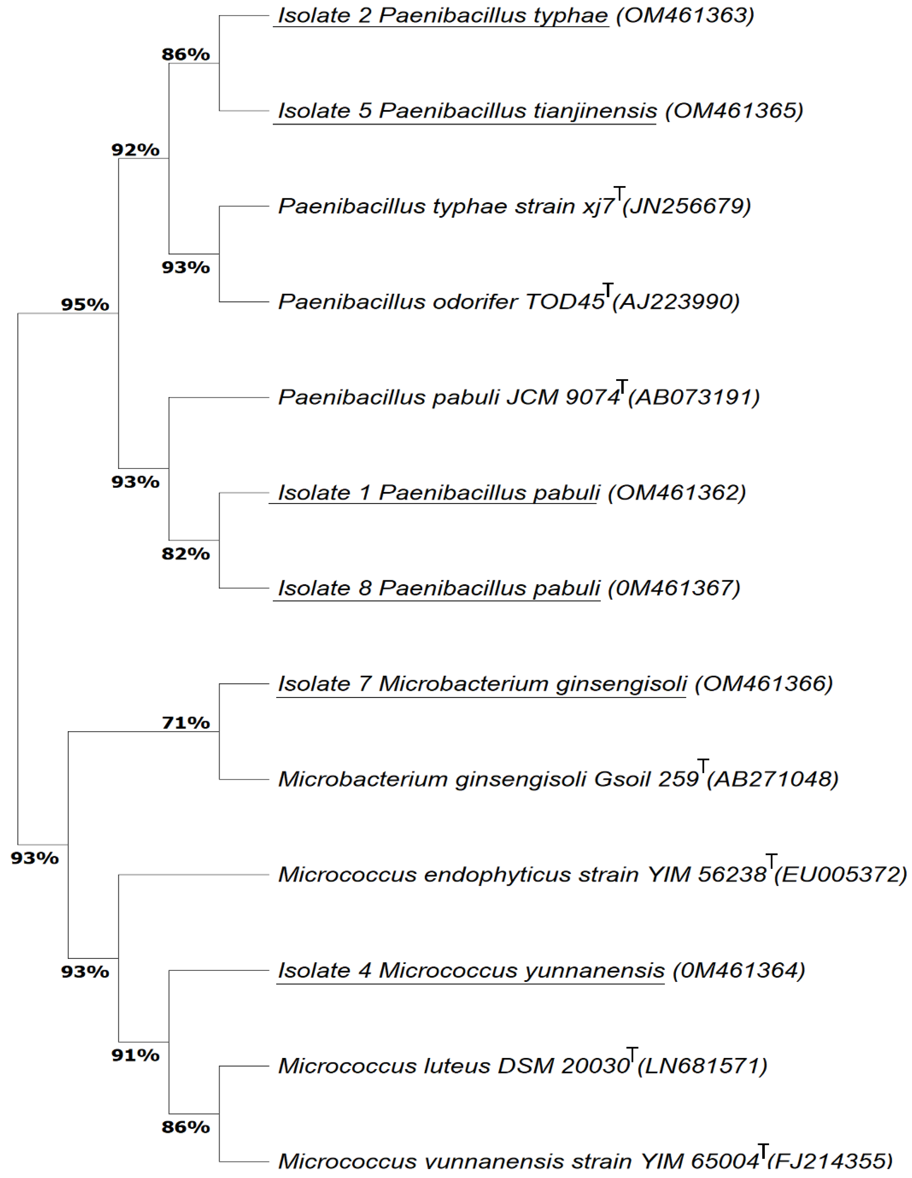
3.1. Bacterial isolation from Consortia Recovered from Marine Organisms with the Ability to Degrade Paracetamol

The assays on paracetamol biodegradation showed that the bacterial consortia H.P4, D.T18 and FI.A21 recovered from marine organisms had the ability to degrade 78 ± 14%, 59 ± 8% and 73 ± 6% of 86 mg/L, respectively, of paracetamol as the sole carbon source after 162 h. These three consortia presented a high bacterial growth, generally higher than OD600nm = 0.6, after 48 h of the experiment (Figure S2). The isolates were obtained from cultures of consortia H.P4 and FI.A21 (the most efficient in paracetamol removal) in MSM spiked with 100 mg/L of paracetamol as the sole carbon source. The colony growth was high at the initial and first dilution after 48 h at 28 °C in the dark. Bacteria were isolated from the dilutions 10−2 for consortium FI.A21 and from 10−4 for consortium H.P4. Eight isolates displaying the ability to grow in the presence of 100 mg/L of paracetamol as the sole carbon source were successfully obtained: three from consortium H.P4, named as 1, 2 and 3, and five from FI.A21, named as 4, 5, 6, 7 and 8. Each isolate was able to grow to a third generation of isolation and further in the presence of paracetamol without a nitrogen source, although presenting a different growth pattern. In the absence of the nitrogen source, the bacterial growth was diffuse, whereas in its presence, the colonies were well defined. This indicates that the bacteria may have the ability to use the nitrogen present in the drug or may withstand conditions without nitrogen present in their environment. Thus, according to this finding, all the biodegradation experiments were performed with MSM plus each drug as carbon source and supplemented with a nitrogen source, since it was evidenced that it may enhance bacterial growth and subsequently may positively affects the biodegradation efficiency, especially in the case of FLX, a fluorinated compound, and EE2, an aromatic homopolycyclic compound, whereas in the case of paracetamol, due to its chemical structure, it may serve as carbon and nitrogen source.
The isolates were identified based on their 16S rRNA sequences by inquiring into the NCBI BLAST database, according to the first entry, which displayed the highest similarity (>95%) with the following species: isolate 1_Paenibacillus pabuli (OM461362), isolate 2_Paenibacillus typhae (M461363) and isolate 3_Paenibacillus odorifer were all isolated from consortium H.P4 and isolate 4_Micrococcus yunnanensis (OM461364), isolate 5_Paenibacillus tianjinensis (OM461365), isolate 6_Paenibacillus odorifer, isolate 7_Microbacterium ginsengisoli (OM461366) and isolate 8_Paenibacillus pabuli (OM461367) were isolated from the consortium FI.A21.
For the species that have more than one item with a very high similarity, the identification was performed using the Basic Local Alignment Search Tool (BLAST) together with the biochemical characteristics (Table S1) obtained for each isolate and a phylogenetic tree in order to classify/address the bacteria into the right species. For example, isolate 4 presents a very high similarity with Micrococcus luteus and Micrococcus yunnanensis, but the first species is oxidase-positive, as with almost all of the known species of the Micrococcus genus [37], while Micrococcus yunnanensis is oxidase-negative [38], as the isolate 4. The species of this genus are halotolerant and grow in 5% of salt [37], and thus their capacity to survive in the marine environments.
With the objective to affiliate the isolates to reliably named species, a maximum likelihood phylogenetic tree based on the 16S rRNA gene was constructed by comparing the almost-complete 16S rRNA gene sequences of the obtained bacterial isolates to the corresponding type strain (Figure 2).
The resulting groups were sustained by high bootstrap values. Phylogenetic similarities in the percentage between species within groups ranged from 71 to 95% depending on the species and diversity of the group. Isolates 1 and 8 isolated from different organisms were assigned to Paenibacillus pabuli, displaying 82% of similarity among them, and both presented 93% of similarity with its type strain P. pabuli, thus defending the close relatedness with this species (Figure 2). P. typhae (Isolate 2) shared 82% similarity with P. tianjinensis (Isolate 5) and 92% with both P. typhae and P. odorifer, revealing that these isolates are closely related with these species. The Micrococcus luteus type strain and Micrococcus yunnanensis type strain species are known to be closely related and share 86% of similarity. Moreover, isolate 4, M. yunnanensis, was revealed to be very close to both type strains, displaying a similarity of 91%, although it is still closer to M. endophyticus, displaying a similarity of 93% with this type strain (Figure 2).
The percentages between groups were from 93 to 95%, revealing that all these species are closely related (Figure 2).
All the isolated bacteria are Gram-positive. Nevertheless, as far as is known, it is the first time that these bacterial isolates were recovered from Hymedesmia sp. (Porifera) and Filograna implexa (Annelida) marine organisms.
All the isolates were tested at 50 and 100 mg/L concentrations of paracetamol. The best degradation performance was shown by M. yunnanensis strain TJPT4; therefore, this isolate was then used to test its ability to remove the three different pharmaceutical compounds: paracetamol, FLX and EE2.

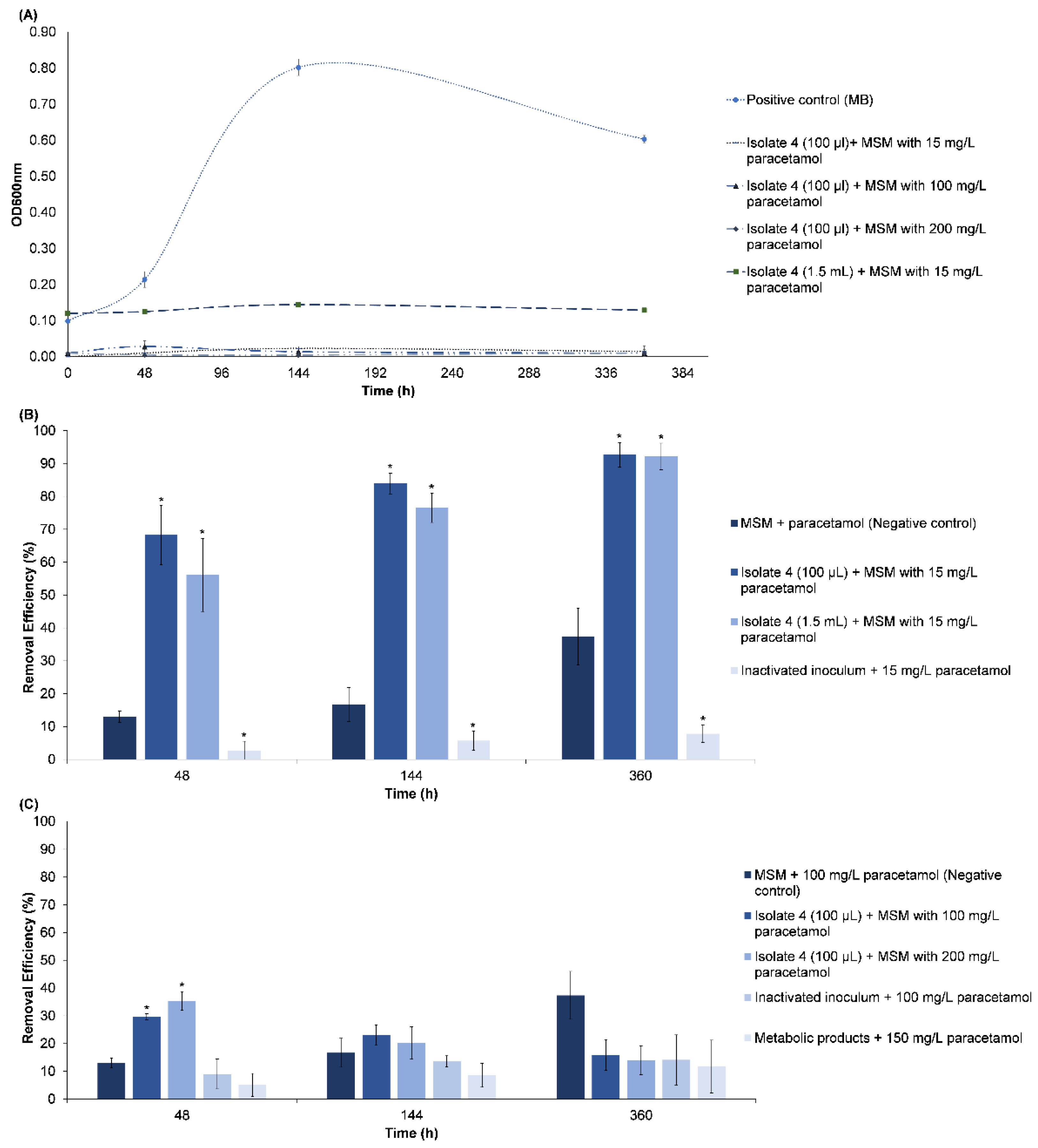
3.2. Bioremoval of Pharmaceuticals by Micrococcus yunnanensis Strain TJPT4

3.2.1. Paracetamol Removal

The bioremoval capacity of M. yunnanensis strain TJPT4 was studied in the presence of paracetamol (model drug). Different conditions were tested to investigate the mechanism behind paracetamol removal, namely whether the interactions between paracetamol and the culture medium (negative control) or the metabolic products (extracellular extract) occurs, or if adsorption to the inactivated inoculum may be responsible for the drug’s removal during the experiments. The results obtained are shown in Figure 3.
In the cultures in which 100 µL (≈106 CFU/mL) of M. yunnanensis strain TJPT4 was used as inoculum, the removal of 15 mg/L of paracetamol was of 68 ± 9%, 84 ± 3% and 93 ± 4% after 48, 144 and 360 h, respectively (Figure 3B). The removal efficiency was assumed to be 92% at a rate of 0.1481 d−1 with an R2 of 1 and a half-life (t1/2) of 4.7 days.
In the cultures with 1500 µL (1.5 mL), corresponding to 5% (v/v) of the inoculum, the isolate was able to remove 56 ± 11, 77 ± 4 and 92 ± 4% of 15 mg/L paracetamol after 48, 144 and 360 h, respectively (Figure 3B). The removal efficiency was 92% at a rate of 0.1563 d−1 with an R2 of 0.9964 and a half-life (t1/2) of 4.4 days.
In these experimental conditions, the bacterial growth was very difficult to measure, since there were formations of bacterial clumps. Although an increase in the amount of these aggregates was evidenced during the assay, it did not alter the turbidity of the medium (Figure 3A). Palma et al. [33] reported other bacterium which belong to the same actinomycetes order as M. yunnanensis, assigned to Corynebacterium nuruki, that also produced bacterial aggregates in the presence of paracetamol, which is likely a mechanism of protection from the toxicity of the drug.
In cultures with 100 and 1500 µL of inoculum in the presence of 15 mg/L of paracetamol, the removal was significantly higher (p < 0.05) than in the abiotic controls (13 ± 2, 17 ± 5 and 37 ± 9%) during the assay (Figure 3B). The removal rate of the negative control was very low (0.0291 d−1), with an R2 of 0.9683 and a half-life (t1/2) of 23.8 days.
From tests conducted with MSM in the presence of 100 mg/L of paracetamol without the isolate (abiotic control), 37 ± 9% of the drug was removed, which may be due to chemical interactions. This considerable removal of paracetamol in the abiotic control can be explained by the fact that, after a long period (360 h), the drug became unstable due to the humid conditions, leading to its hydrolysis [33].
In the present study, biodegradation under aerobic conditions appears to have a major role in the removal of paracetamol.
The metabolites detected during the degradation of 15 mg/L of paracetamol were consistent with those of 4-aminophenol (RT = 3.67 min) and hydroquinone (RT = 6.45 min) standards.
In the cultures after 48 h, the presence of a peak consistent with hydroquinone (of approximately 608 µL/L) was detected at 6.45 min, while the other peaks, which were identified at retention times of 2.65, 3.05 and 5.40 min, also appeared in the MSM medium without the presence of the drug. After 144 h, a peak which may correspond to 4-aminophenol was detected at 3.667 min with a concentration of approximately 4 mg/L, whereas hydroquinone, known to be a transient metabolite, was not detected due to its possible consumption by the bacteria. After 360 h, despite the presence of approximately 1 mg/L of paracetamol, which was not entirely degraded, no other peaks corresponding to metabolic products were detected, being most likely below the limits of detection, suggesting that M. yunnanensis can consume the metabolites produced during paracetamol biodegradation (Figure S3). The obtained results show evidence that the mechanism of paracetamol degradation by M. yunnanensis occurs in the same way as already reported in the mechanistic studies by Zhang et al. [8].
Micrococcus yunnanensis was able to remove 30 ± 1, 23 ± 4 and 16 ± 6% of 100 mg/L of paracetamol after 48, 144 and 360 h, respectively (Figure 3C). The removal rate did not follow a pseudo-first-order kinetics, since the rate constant was negative (−0.0126 d−1), with an R2 of 0.9588. The isolate was able to remove 35 ± 3, 20 ± 6 and 14 ± 5% of approximately 200 mg/L of paracetamol after 48, 144 and 360 h, respectively (Figure 3C). In this case, the removal rate also did not follow a first-order kinetics, with a negative rate constant of -0.0194 d−1 and a weak R2 of 0.7967. To follow a pseudo-first-order kinetics, the R2 from the linear fit must be greater than 0.95 [39]. At such high concentrations (100 and 200 mg/L of paracetamol), the removal of the drug seems to follow a pseudo-zero-order reaction, since the reaction rate is independent of concentration of the drug. The negative rate constants at 100 and 200 mg/L are explained by the increase of the drug from approximately 83 mg/L to 90 mg/L and from 154 mg/L to 190 mg/L, respectively, during the experiment, suggesting a reverse reaction, thereby slowing down the reaction rate, causing its decrease. This can be explained by the fact that, at high concentrations, bacteria may uptake the drug, which could be rapidly accumulated by the bacterial cells (bioaccumulation). However, these concentrations also seem to be toxic to the isolate during the assay. Therefore, after cell death and subsequent lysis, the drug may be likely released into the medium, since the bacteria appeared to be unable to efficiently metabolize such drug concentrations.
The assay in the presence of 150 mg/L paracetamol with the extracellular products (metabolites) produced by M. yunnanensis revealed that 5 ± 4, 9 ± 4 and 12 ± 9% of the drug was removed by the extracellular metabolic compounds produced by this bacterium (Figure 3C). The removal rate in the presence of the extracellular products was of 0.0092 d−1, with an R2 of 0.9622 and a half-life of 75.3 days.
The adsorption to the inoculum was measured by the contact of the inactivated biomass with 100 mg/L of paracetamol, and the removal values were of 9 ± 5, 14 ± 2 and 14 ± 9% (Figure 3C). The removal rate of this reaction was very low (0.0044 d−1), with a weak R2 of 0.602 and a half-life of 13659 days. Due to the weak R2, the paracetamol’s adsorption kinetics seems to follow a pseudo-zero-order kinetic.
The experiments with the inactivated inoculum and in the presence of the extracellular extracts were not significantly different (p > 0.05) from the negative control. Although the adsorption to the bacteria is very low, the experimental data follows a Langmuir model.

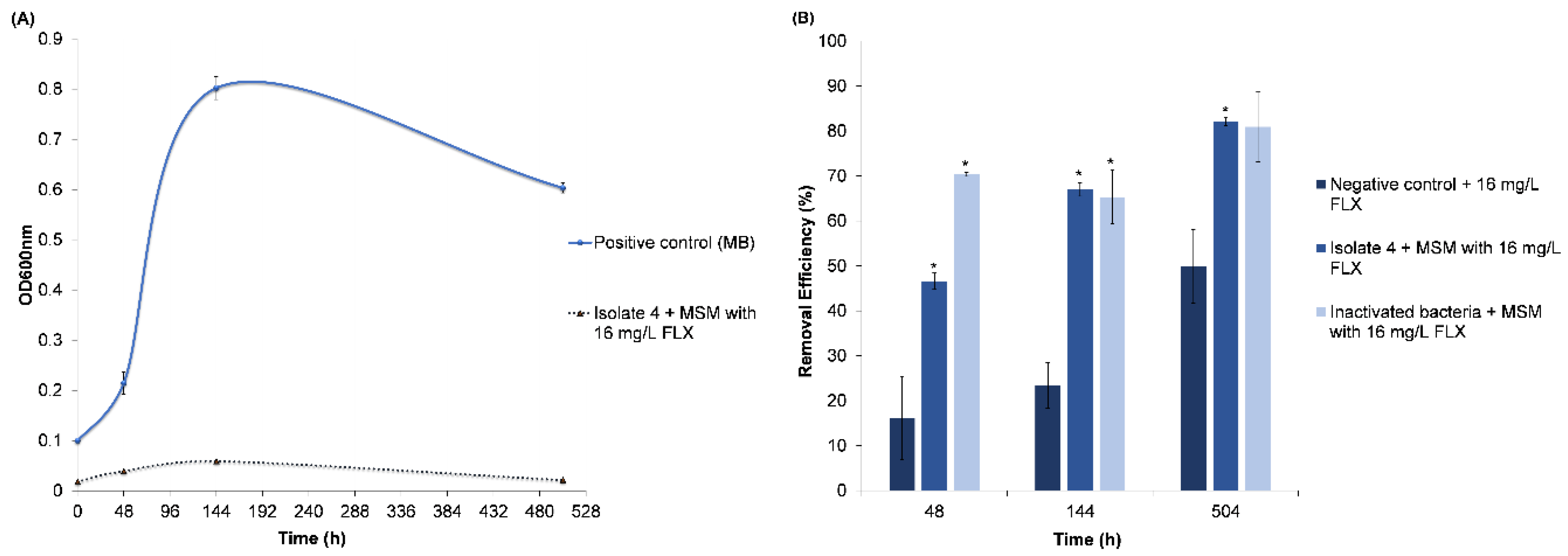
3.2.2. Fluoxetine Removal

Assays to investigate the possible biodegradation profile of FLX by Micrococcus yunnanensis and its eventual adsorption potential to the inactivated inoculum were performed (Figure 4).
In cultures using 100 µL of M. yunnanensis strain TJPT4 in the presence of 16 mg/L, the removal efficiencies were 47 ± 2%, 67 ± 2% and 82.1 ± 0.9% after 48, 144 and 504 h, respectively (Figure 4). The removal efficiency was 82% at a rate of 0.0538 d−1, with an R2 of 0.9562 and a t1/2 of 13 days (Figure 4). In this assay, 100 µL of inoculum was used to avoid/prevent adsorption effects that can be enhanced with high amounts of biomass, and thus more available adsorption sites. The cultures with the inoculum presented a FLX removal significantly higher (p < 0.05) than the in the abiotic control.
The adsorption assay was performed in the presence of 10% of inactivated inoculum and 16 mg/L of FLX, and the removal percentages obtained were of 70.4 ± 0.5, 65 ± 6 and 81 ± 8% after 48, 144 and 504 h, respectively, corresponding to a rate of 0.0625 d−1 with an R2 of 0.9689 and a t1/2 of 11 days (Figure 4). As was expected from FLX’s physicochemical characteristics, the compound had been adsorbed into the inactivated inoculum.
To investigate chemical interactions, a negative control was performed only in the presence of 16 mg/L of the drug, and the results obtained were 16 ± 9, 23 ± 5 and 50 ± 8% after 48, 144 and 504 h, respectively (Figure 4). The removal rate was 0.0305 d−1 with an R2 of 0.9967 and a t1/2 of 23 days.
The removal of FLX by the inactivated inoculum was significantly higher (p < 0.05) than in the abiotic control after 48 and 144 h. Although, the chemical interactions of the drug with the medium seem to have a minimum role during the initial steps of removal, and these apparently gained importance in the last days of the experiment. These results suggest that the adsorption mechanism has an important role in the removal of FLX, especially in the first 48 h of the assay.
In the presence of the inoculum, FLX (RT = 2.98 to 3.00 min) and its active metabolite NFLX (RT = 2.700 min) were detected. In cultures spiked with 16 mg/L FLX, it was possible to observe a decrease on both compounds after 504 h of the assay, suggesting that M. yunnanensis was able to use them as carbon sources or that the compounds may be adsorbed by the bacterial cells, or a combination of both mechanisms could have occurred. In the inactivated inoculum, it was possible to detect the transformation of FLX into NFLX in one sample. However, both appear to be adsorbed to the inactivated bacteria, since FLX decreased over time, while NFLX was not detected during and at the end of the assay. The other peaks detected are unknown, with some of them consistent with MSM constituents (Figure S4).

Adsorption of FLX onto the Inactivated Cells
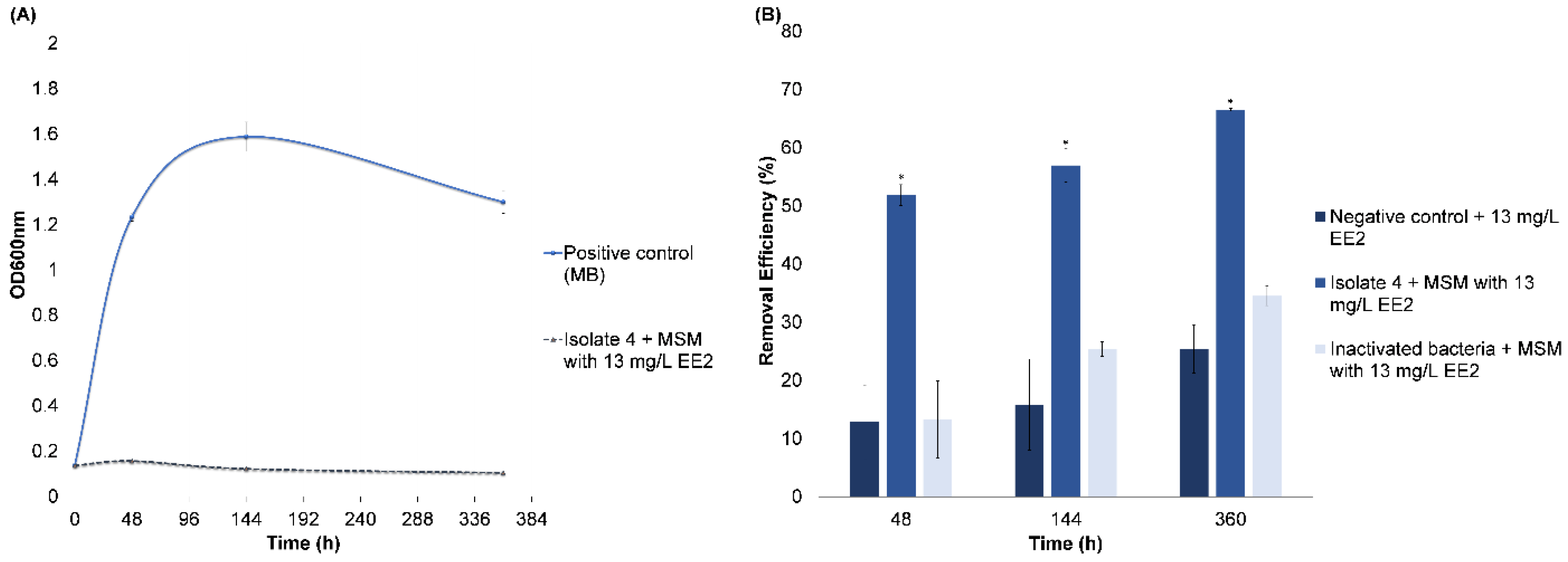
Adsorption is also an important process and is widely applied to treat and purify polluted waters with organic compounds. Several experts consider this treatment superior compared to other physical and chemical technologies. This separation process may provide a suitable cost-effectiveness benefit for water pollution control [40]. To study the interactions of FLX (adsorbate) onto the inactivated biomass surface (adsorbent), the Langmuir and Freundlich equilibrium isotherms were determined.
The Langmuir and Freundlich models do not fit the experimental data, since a negative intercept of −0.1 ± 0.1 and −0.4 ± 0.04 was obtained for both, respectively, despite the good linearity with a correlation coefficient R2 of 0.97 ± 0.04 and 0.99 ± 0.01, respectively. These results are in accordance with the study performed by Palma and Costa [34], in which the adsorption of FLX to a bacterial matrix did not follow the Langmuir or the Freundlich model. A negative intercept is considered physically impossible, and this phenomenon can occur due to the energetic heterogeneity of the adsorption sites in the adsorbate (inactivated inoculum), which is mostly constituted by cells and organic compounds that can contribute to the obstruction of the adsorption sites, thus reducing the binding energy between the surfaces [34,41].
The experimental data of FLX biosorption onto the inactivated inoculum fit a pseudo-second-order rate law according to the kinetic study described by Ho and McKay [35], in which qeq and qt (mg/g) were the amounts of the adsorbed FLX on the biosorbent (inactivated Inoculum) at eq (equilibrium) and t (time), respectively, and the second-order biosorption rate constant was given by K2 (g/mg.h). This model assumes that chemical sorption involves valency forces via the sharing or exchange of electrons between the sorbent and sorbate, and that this is the rate-limiting step. The experimental data also fit into a pseudo-second-order kinetic rate law, and the regression coefficient was high (R2 of 0.9954). The equilibrium sorption capacity was K = 0.048 ± 0.001 g/mg.days (0.002 ± 0.001 g/mg.h). The value of qeq (mg/g) (sorption capacity at equilibrium) estimated for the inactivated cultures was 14 ± 3 mg/g, which is in accordance with the experimental qeq,exp (13.2 ± 0.6 mg/g), since qeq (mg/g) and qeq,exp (mg/g) are close.

3.2.3. EE2 Removal

Assays to investigate the possible biodegradation profile of EE2 by M. yunnanensis and its adsorption potential to the inactivated inoculum were performed (Figure 5).
In the cultures with 5% (v/v) of inoculum in the presence of 13 mg/L, a removal of 52 ± 3%, 57 ± 5% (v/v) and 66.6 ± 0.4% after 48, 144 and 360 h, respectively, were obtained (Figure 5B). The removal rate of the drug was 0.0365 d−1 with an R2 of 0.9935 and a t1/2 of 19 days. The maximum bacterial growth (OD600nm = 0.157 ± 0.012) occurred after 48 h incubation (Figure 5A). In that period, a decrease of 52% of EE2′s initial concentration, as the sole carbon source, was observed (Figure 5). The optical density was difficult to measure due to the appearance of clumps, and which may also be associated to a cometabolic use of the drug by M. yunnanensis.
The removal of 13 mg/L of EE2 in the presence of the inactivated inoculum (adsorption experiments) were of 13 ± 7, 25 ± 1 and 35 ± 2% after 48, 144 and 360 h, respectively, with a reaction rate of 0.0272 d−1, an R2 of 0.9797 and a t1/2 of 25 days (Figure 5B).
In the abiotic control, only in the presence of the drug, the removal was of 13 ± 6, 16 ± 8 and 25 ± 4% after 48, 144 and 360 h, respectively, with a rate of 0.0154 d−1, an R2 of 0.9806 and a t1/2 = 45 days (Figure 5).
The removal in the negative control was not significantly different (p > 0.05) from the one obtained with the inactivated bacteria, while in the assay with the live inoculum, the drug removal was significantly higher (p < 0.05) than in the former ones (Figure 5). Hence, the results demonstrated that, after 48 h, most of the removal was due to bacterial activity (52%) and that the contribution of the adsorption mechanism seems to be approximately 13%. However, after 360 h, the contribution of adsorption may be of approximately 32%, which agrees with the literature regarding the physicochemical characteristics of this compound and its adsorption behavior.
Concerning the adsorption mechanism, the Langmuir and Freundlich models were studied, and despite a good linearity, with correlation coefficients and an R2 of 0.95 ± 0.02 and 0.986 ± 0.006, respectively, these models did not fit the obtained experimental data, since a negative intercept (−10 ± 5 and −2.9 ± 0.7) was obtained for both cases. These results are similar to those already obtained for FLX.
The equilibrium sorption capacity was K = 0.00168 ± 0.00002 g/mg.days (0.00007 ± 0.00002 g/mg.h). The value of qeq (mg/g) (sorption capacity at equilibrium) estimated for the inactivated cultures was of 4.2 ± 0.2 mg/g, which is in accordance with the experimental qeq,exp (5.5 ± 0.2 mg/g), since qeq (mg/g) and qeq,exp are close.
EE2 removal and the subsequent putative metabolites produced were analyzed by HPLC and GC-MS. Several intermediates were selected based on their detection only in the presence of bacteria, excluding the ones detected in the abiotic control.
The 17α-ethinylestradiol was detected at a retention time of 8.38 min by HPLC and at 19.433 min by GC-MS. For the detection/identification of putative metabolites in the inoculated cultures, the GC-MS spectra obtained from the samples during the assay were interpreted/inferred and analyzed based on the comparison with the National Institute and Technology (NIST) spectra database 2.0, and in some cases with the spectrum of standards and/or by the most dominant ion peak of the compound (m/z) injected under similar conditions.
A putative pathway for the biodegradation of EE2 by M. yunnanensis is hypothesized.
Along the assay, a decrease of the EE2 concentration was observed. After 48 h, a steroid compound, 3β,16α-diacetoxy-17-methyl-17α-pregna-5,13-dien-20-one, appeared at 15.888 min.
The peaks corresponding to the compounds of interest were detected by pseudo-molecular ion peaks at m/z 270, 300, 157.9 and 113, matching to estrone (E1), 3-4-dihydroxy-9,10-secoestrane-1,3,5(10)-diene-9, 17-dione, 2-hydroxy-2,4-diene-1,6-dioic acid and 2-hydroxy-2,4-dienevaleric acid, respectively. A peak at 5.722 min was detected in all samples, which is probably assigned to an impurity. Residual concentrations of each compound coexisted and were identified at 360 h of the experiment, which makes sense, since about 30% of the EE2 remains to be removed, and likely the bacteria are more active after this time of contact with the drug, being able to degrade it metabolically and/or cometabolically. A peak consistent with E1 was detected at 10.608 min (m/z 270). A peak compatible with the intermediate 3-4-dihydroxy-9,10-secoestrane-1,3,5(10)-diene-9, 17-dione was detected at 9.212 min (m/z 300), and peaks consistent with 2-hydroxy-2,4-diene-1,6-dioic acid and 2-hydroxy-2,4-dienevaleric acid were detected at 10.889 min (m/z 157.9) and 6.309 min (m/z 113) after 360 h (Figures S5 and S6). Thus, the biodegradation pathway of EE2 by M. yunnanensis seems to be similar to that proposed by Hayan et al. [29] for Spingobacterium sp. JCR5, in which its C-17 group was firstly oxygenized to the ketone group (E1) with the further hydroxylation of C-9α and its ketonization, and the subsequent cleavage of the B ring, which produces 3-4-dihydroxy-9,10-secoestrane-1,3,5(10)-diene-9, 17-dione. Afterwards, the A ring suffers hydroxylation to form 3,4-catechol, 3-hydroxy-4,5-9,10-disecoestrane-1(10),2-diene-5,9,17-trione-4-dioic, which is, in turn, transformed into two intermediates, 2-hydroxy-2,4-dienevaleric acid and 2-hydroxy-2,4-diene-1,6-dioic acid, following its mineralization.
Other recent studies have also stated the degradation and suggested the metabolic pathways for paracetamol, FLX and EE2, which are summarized in the Table 1.
In the present work, the degradation periods/kinetics and drug exposition times obtained with M. yunnanensis, although long, are compatible with the ones typically recommended for oxidation ditch systems with a solid retention time (SRT) ranging from 4 to 48 days and a hydraulic retention time (HRT) or aeration period from 6 to 30 h, such that longer times may also be beneficial for the degradation of pharmaceuticals [32,45,46].
These findings can be important to improve the biodegradation processes of these recalcitrant and harmful pollutants. Bioaugmentation is commonly used in wastewater treatment plants to restart activated sludge bioreactors in order to improve the biodegradation capacity of the indigenous microbial populations [47]. Thus, the addition of isolates such as M. yunnanensis, which are halotolerant and display the ability to degrade pharmaceuticals, may enhance the removal capacity in bioreactors that, in addition to receiving polluted influents, also suffer commonly marine intrusion, which is the case of those placed near coastal areas.

3.3. Hypothetical Genetic Potential of M. yunnanensis for Drug Biodegradation Based on Its Encoded Proteins

The obtained results suggest that M. yunnanensis may have enzymatic tools to degrade the studied drugs. This particular study was performed based on the comparison of the phenotypical characteristics of a member of the same species, the M. yunnanensis strain BCRC 80243 (Genome ID 566027.4), using the PATRIC 3.6.12 database [48]. The search of some genes known to encode for the enzymes with a particular role in paracetamol, FLX and EE2 help to infer the biodegradation ability and metabolic pathways used by this bacterium.
Studies point out that the transformation of pharmaceuticals is likely the result of the enzymatic activities of the bacterial strain.
The enzymes found in M. yunnanensis corroborate with those proposed by Zhang et al. [8] and with the results herein obtained for the degradation pathway of paracetamol, in which its metabolites are also used by the bacterium, leading to drug mineralization. This bacterium contains the gene encoding for four amidohydrolases, namely amidohydrolase Protein ID MBE1538225.1 (RefSeq locus tag Tag H4W28_000186; location 110263..111519; accession JADBEH010000002), which catalyzes paracetamol into 4-aminophenol by releasing acetate, which thereafter is transformed into hydroquinone with the replacement of the amino group by a hydroxyl group in an oxidative deamination. From several deaminase enzymes known in this bacterium, the most likely candidate is the enamine deaminase RidA (YjgF/YER057c/UK114 family), which displays enamine/imine deaminase activity. The following step is the subsequent ring fission of hydroquinone catalyzed by a dioxygenase. The investigated M. yunnanensis strain BCRC 80243 seems to not use a hydroquinone 1,2-dioxygenase, which is a type III ring-cleaving extradiol dioxygenase, but contains other important dioxygenase enzymes that may be crucial for the aromatic ring fission, as they are the beta-ketoadipate enzymes [49]. Several dioxygenase enzymes are found in M. yunnanensis (such as catechol 2,3-dioxygenase, 3-phenylpropionate dioxygenase ferredoxin subunit, etc.). Among them, 3,4-dihydroxyphenylacetate 2,3-dioxygenase, an extradiol dioxygenase (Protein ID MBE1539656.1; RefSeq locus tag H4W28_001617; location 1700379..1701545; accession JADBEH010000002), protocatechuate dioxygenase, a type II extradiol dioxygenase (PATRIC local family PLF_1269_00001118; RefSeq locus tag E2F93_09970; location 32085..33119; accession SMVL01000022) and protocatechuate 3,4-dioxygenase beta subunit (Protein ID MBE1538177.1; RefSeq locus tag H4W28_000138; location 54472..55179; accession JADBEH010000002) [48] may be the ones which play a major role in the aromatic ring fission (meta-cleavage) of these compounds.
Zhang et al. [8] suggested that aromatic compounds can be the precursors of the carboxylic acids, leading to the further mineralization of paracetamol. M. yunnanensis expresses genes encoding for acetyl-CoA synthetase (acs and yahU genes), citrate synthase, isocitrate dehydrogenase and gamma-aminobutyrate:alpha-ketoglutarate (gabT gene) aminotransferase and gamma dehydrogenase enzymes that are involved in the tricarboxylic acid (TCA) and glyoxylate cycles (isocitrate lyase) (accession JADBEH010000002) [48], which are the main energy source of the cells and in which the putative dicarboxylic acids produced during the degradation of paracetamol enter for further mineralization.
As described by Khan and Murphy [17] for other bacteria, M. yunnanensis, as the obtained results suggest, may be able to catabolize FLX; in a first step, the ether bond of FLX could suffer a hydrolyzation to yield 4-(trifluoromethyl)phenol (TFMP) and 3-(methylamino)-1-phenylpropan-1-ol by a hydrolase (M. yunnanensis has 36 known expressed hydrolases) followed by monooxygenation (M. yunnanensis has 7 monooxygenase enzymes) to 4-(trifluoromethyl)catechol. This last compound may undergo meta-cleavage by an enzyme such as catechol 2,3-dioxygenase encoded by the hpaD gene (PATRIC local family PLF_1269_00000530; RefSeq locus tag H4W28_001617; location 1700379..1701545; accession JADBEH010000002) or by protocatechuate dioxygenase. Because of the trifluoromethyl group, two potential products (6,6,6-trifluoro-5-formyl-2-hydroxy-hexa-2,4-dienoic acid or 2-hydroxy-6-oxo-4-(trifluoromethyl)hexa-2,4-dienoic acid) may be formed upon extradiol cleavage of the catechol, since the dioxygenase could attack either C-2/C-3 or C1/C6. To mineralize FLX, a fluorinated drug, and other related aromatic and aliphatic organofluorides compounds, which present a highly stable C-F bond, it is necessary that they suffer a cleavage by a dehalogenation reaction. Few microorganisms have evolved to biotransform and use fluoroacetate and aromatic compounds as carbon and energy sources by possessing a fluoroacetate dehalogenase enzyme, which belongs to an exclusive class of enzymes that specifically hydrolyze the C-F bond, yielding a fluoride ion and glycolate. M. yunnanensis possesses a gene which express one of this specific enzymes, the protein ID TFE82036.1, a haloacid dehalogenase domain protein hydrolase enzyme (PATRIC local family PLF_1269_00000697; RefSeq locus tag E2F93_01955; location 105996..106892; accession SMVL01000002). Chan et al. [50] found that members of the haloacid dehalogenase-like hydrolases superfamily were able to carry out defluorination, thus breaking the carbon–fluorine bonds. M. yunnanensis also contains a gene which expresses protein a fluoride ion transporter CrcB (PATRIC local family PLF_1269_00001369; RefSeq locus tag E2F93_00950; location 198602..199048; accession SMVL01000001) [48]. M. yunnanensis also contains two genes that encode for fluoride export (FEX) proteins, which provide tolerance and confer resistance to fluoride (F). For example, fluoride ion transporter CrcB is described as a putative integral membrane protein, which is important for reducing the fluoride toxicity decreasing its concentration in the cell, avoiding its accumulation in it [51]. Moreover, the dioxygenase enzymes may have a role in the mineralization of this drug.
In the case of EE2, an aromatic steroid, several specific enzymes are described for its degradation. Chen et al. [52] described 17β-estradiol dehydrogenase and 4-hydroxyestrone 4,5-dioxygenase as responsible for the 17-dehydrogenation and metacleavage of the estrogen A ring, respectively. These particular enzymes are not described in the PATRIC database for the M. yunnanensis strain BCRC 80243. However, Chen et al. [52] suggest that other extradiol dioxygenases are not completely excluded and may also play a role in the metacleavage of 4-hydroxyestrone. The authors mention that some metacleavage pathways include a [2Fe-2S] ferredoxin that allows the reactivation of the metal ion of extradiol dioxygenases oxidized during catalytic reactions [51]. The studied genome contains such enzymes, which are a Rieske (2Fe-2S) domain protein (PATRIC local family PLF_1269_00000566; RefSeq locus tag E2F93_04470; location 118528..119040; accession SMVL01000004) and a 3-phenylpropionate/trans-cinnamate dioxygenase ferredoxin subunit (Protein ID MBE1539663.1RefSeq locus tag H4W28_001624; location 1707978..1708307; accession SMVL01000020) [48] that, in addition to the already referred-to dioxygenases enzymes, may also have a role in this type of compound.
The comparison of the phenotypic characteristics was carried out with a strain that was likely not in contact with any of the drugs under study, so although this strain may contain the genes that code for the enzymes that are described in the literature for drug metabolism, the genes were not triggered and these specific proteins were not expressed.

4. Conclusions

Three bacterial consortia, designated by H.P4, D.T18 and FI.A21, were recovered from Hymedesmia versicolor (Porifera), Didemnum sp. (Tunicate) and Filograna implexa (Annelida) marine organisms, respectively. The results showed that the isolate assigned to the Micrococcus yunnanensis strain TJPT4 was identified for the first time in Filograna implexa (Annelida) marine organisms. This strain was able to remove 93 ± 4% of 15 mg/L paracetamol as the sole carbon source after 360 h. Apparently, it was also able to degrade its toxic metabolites. In addition, M. yunnanensis TJPT4 was able to remove 82.1 ± 0.9% of FLX from a solution containing 16 mg/L of drug as sole the carbon source after 504 h. FLX and NFLX removal occurred mainly by adsorption (~70% after 48 h), although a biodegradation contribution seems also to occur. Moreover, this strain was also able to remove 66.6 ± 0.2% of 13 mg/L of 17α-ethinylestradiol, a semisynthetic hormone, after 360 h. The capacity of M. yunnanensis for the removal of these three environmentally hazardous pollutants, in addition to its halophilic characteristics, turn it into a very promising candidate for bioaugmentation processes, especially in WWTPs with problematic saline intrusions. Thus, based on the obtained results, the following recommendations are suggested for future studies in order to onward increase the current knowledge on this subject: (i) Biodegradation studies of these and other drugs through the addition of bacterial isolates (e.g., M. yunnanensis TJPT4) that have demonstrated degradation capacity, to already existing bacterial communities (bioaugmentation) (see how such inclusion enhances the removal of the drug) and (ii) Biostimulation/bioaugmentation experiments that can be performed by adding bacterial biomass, produced in the laboratory, in a pilot system mimicking an already-implemented system (e.g., oxidation ditch), in order to improve wastewater treatment processes.

Supplementary Materials

The following supporting information can be downloaded at: https://www.mdpi.com/article/10.3390/w14213365/s1, Figure S1: Bacterial consortia were recovered from the marine organisms: Hymedesmia versicolor (Porifera) (A), Didemnum sp. (Tunicata) (B), and Filograna implexa (Annelida) (C). Samples pictured in their natural environment; Figure S2: Paracetamol removal (%) and bacterial growth (OD600) of bacterial consortia H.P4, D.T18 and FI.A21 recovered from marine organisms in MSM liquid cultures in the presence of 86 mg/L of paracetamol, after 48 h and 168 h at room temperature, 150 rpm and in dark conditions (n = 3; mean ± standard deviation); Figure S3: Representative chromatograms of the identification of putative paracetamol biodegradation products in the medium with an initial concentration of paracetamol of 15 mg/L. Detectable peaks after 48 h and after 360 h; Figure S4: Representative chromatograms of the identification FLX and NFLX its metabolic product in cultures with M. yunnanensis in the presence of 16 mg/L FLX as sole carbon source. Detectable peaks in (A) standard solutions; at (B) 48 h (C) 144 h and (D) 504 h; Figure S5: Representative chromatograms of the identification of putative EE2 biodegradation products in the medium with an initial concentration of EE2 of 13 mg/L. Negative control (C-) plus EE2; Inoculum (Inoc.) plus EE2. Detectable peaks after 48 h, 144 h and after 360 h; Figure S6: Representative chromatograms of the identification of putative EE2 biodegradation products in the medium with an initial concentration of EE2 of 13 mg/L. Detection was based on the peak known as dominant ion peak of the compound (m/z). Peaks detectable after 360 h; Table S1: Morphological and biochemical characteristics of the bacterial isolates which have maintained the ability to use paracetamol as unique carbon source.

Author Contributions

All authors contributed to the study conception and design. The first draft of the manuscript was written, reviewed and edited by T.P., who was also responsible for material preparation, methodology development, data collection, validation data and formal data evaluation. J.V. performed some experimental work in the frame of her master thesis. All authors commented on previous versions of the manuscript. M.F., V.G. and M.C. made critical, specific comments, reviewed the presentation of the data and edited the manuscript. M.C. supervised T.P. and administrated the project in which the work has been performed. All authors have read and agreed to the published version of the manuscript.

Funding

This research was funded by project 0483_PROBIOMA_5_E, cofinanced by the European Regional Development Fund within the framework of the Interreg V-A Spain-Portugal program (POCTEP) 2014–2020. This study also received Portuguese national funds from FCT—Foundation for Science and Technology through projects UIDB/04326/2020, PTDC/CTA-AMB/7782/2020, UIDP/04326/2020 and LA/P/0101/2020.

Data Availability Statement

Not applicable.

Acknowledgments

The authors acknowledge the scholarship of the International Master of Science in Marine Biological Resources (IMBRSea) program of Julia Valentine. The authors want to thank the Scientific Dive Center of CCMAR for the marine samples collection.

Conflicts of Interest

The authors declare no conflict of interest. The funders had no role in the design of the study; in the collection, analyses, or interpretation of data; in the writing of the manuscript; or in the decision to publish the results.

References

  1. Gurnham, C. Limitations of sewage treatment plants in handling industrial wastes. J. Water Pollut. Control Fed. 1960, 32, 211–215. [Google Scholar]
  2. Gu, Y.; Li, B.; Zhong, X.; Liu, C.; Ma, B. Bacterial community composition and function in a tropical municipal wastewater treatment plant. Water 2022, 14, 1537. [Google Scholar] [CrossRef]
  3. Martins, M.; Sanches, S.; Pereira, I.A. Anaerobic biodegradation of pharmaceutical compounds: New insights into the pharmaceutical-degrading bacteria. J. Hazard Mater. 2018, 357, 289–297. [Google Scholar] [CrossRef]
  4. Pólvora, S.; Aníbal, J.; Martins, A. Saline intrusions at Almargem waste water treatment plant in different tidal cycles. In Proceedings of the 2020 2nd International Congress on Engineering and Sustainability in the XXI Century INCREaSE, Faro, Portugal, 9–11 October 2019; pp. 786–798. [Google Scholar] [CrossRef]
  5. Fusi-Schmidhauser, T.; Preston, N.J.; Keller, N.; Gamondi, C. Conservative management of COVID-19 patients-emergency palliative care in action. J. Pain Symptom. Manag. 2020, 60, e27–e30. [Google Scholar] [CrossRef]
  6. Palma, T.L.; Donaldben, M.N.; Costa, M.C.; Carlier, J.D. Putative role of Flavobacterium, Dokdonella and Methylophilus strains in paracetamol biodegradation. Water Air Soil. Pollut. 2018, 229, 200. [Google Scholar] [CrossRef]
  7. Brumovský, M.; Bečanová, J.; Kohoutek, J.; Borghini, M.; Nizzetto, L. Contaminants of emerging concern in the open sea waters of the Western Mediterranean. Environ. Pollut. 2017, 229, 976–983. [Google Scholar] [CrossRef]
  8. Zhang, L.; Hu, J.; Zhu, R.; Zhou, Q.; Chen, J. Degradation of paracetamol by pure bacterial cultures and their microbial consortium. Appl. Microbiol. Biotechnol. 2013, 97, 3687–4369. [Google Scholar] [CrossRef] [PubMed]
  9. Wu, S.; Zhang, L.; Chen, J. Paracetamol in the environment and its degradation by microorganisms. Appl. Microbiol. Biotechnol. 2012, 96, 875–884. [Google Scholar] [CrossRef] [PubMed]
  10. Joss, A.; Zabczynski, S.; Göbel, A.; Hofmann, B.; Löfer, D.; McArdell, C.S.; Ternes, T.A.; Thomsen, A.; Siegrist, H. Biological degradation of pharmaceuticals in municipal wastewater treatment: Proposing a classification scheme. Water Res. 2006, 40, 1686–1696. [Google Scholar] [CrossRef] [PubMed]
  11. Ferchichi, M.; Dhaouadi, H. Sorption of paracetamol onto biomaterials. Water Sci. Technol. 2016, 74, 287–294. [Google Scholar] [CrossRef]
  12. Daniel, D.; Nunes, B.; Pinto, E.; Ferreira, I.M.P.L.V.O.; Correia, A.T. Assessment of paracetamol toxic effects under varying seawater pH conditions on the marine polychaete Hediste diversicolor using biochemical endpoints. Biology 2022, 11, 581. Available online: https://www.mdpi.com/2079-7737/11/4/581 (accessed on 3 August 2022). [CrossRef] [PubMed]
  13. Kwon, J.W.; Armbrust, K.L. Laboratory persistence and fate of fluoxetine in aquatic environments. Environ. Toxicol. Chem. 2006, 25, 2561–2568. [Google Scholar] [CrossRef]
  14. Kreke, N.; Dietrich, D.R. Physiological endpoints for potential SSRI interactions in fish. Crit. Rev. Toxicol. 2008, 38, 215–247. [Google Scholar] [CrossRef] [Green Version]
  15. Peters, J.R.; Granek, E.F.; de Rivera, C.E.; Rollins, M. Prozac in the water: Chronic fluoxetine exposure and predation risk interact to shape behaviors in an estuarine crab. Ecol. Evol. 2017, 7, 9151–9161. [Google Scholar] [CrossRef]
  16. Vasskog, T.; Anderssen, T.; Pedersen-Bjergaard, T.; Kallenborn, R.; Jensen, E. Occurrence of selective serotonin reuptake inhibitors in sewage and receiving waters at Spitsbergen and in Norway. J. Chromatogr. A 2008, 1185, 194–205. [Google Scholar] [CrossRef]
  17. Khan, M.F.; Murphy, C.D. Bacterial degradation of the anti-depressant drug fluoxetine produces trifluoroacetic acid and fluoride ion. Appl. Microbiol. Biotechnol. 2021, 105, 9359–9369. [Google Scholar] [CrossRef]
  18. Das, S.; Ray, N.M.; Wan, J.; Khan, A.; Chakraborty, T.; Ray, M.B. Micropollutants in wastewater: Fate and removal processes in: Physico-chemical wastewater treatment and resource recovery. IntechOpen 2017, 5, 76–107. [Google Scholar] [CrossRef] [Green Version]
  19. Velazquez, Y.F.; Nacheva, P.M. Biodegradability of fluoxetine, mefenamic acid, and metoprolol using different microbial consortiums. Environ. Sci. Pollut. Res. Int. 2017, 24, 6779–6793. [Google Scholar] [CrossRef] [PubMed]
  20. Wishart, D.S.; Feunang, Y.D.; Guo, A.C.; Lo, E.J.; Marcu, A.; Grant, J.R.; Sajed, T.; Johnson, D.; Li, C.; Sayeeda, Z.; et al. DrugBank 5.0: A major update to the DrugBank database for 2018. Nucleic Acids Res. 2018, 46, D1074–D1082. [Google Scholar] [CrossRef] [PubMed]
  21. Vijitkul, P.; Kongsema, M.; Toommakorn, T.; Bullangpoti, V. Investigation of genotoxicity, mutagenicity, and cytotoxicity in erythrocytes of Nile tilapia (Oreochromis niloticus) after fluoxetine exposure. Toxicol Rep. 2022, 29, 588–596. [Google Scholar] [CrossRef]
  22. Commission Implementing Decision (EU) 2020/1161 of 4 August 2020 Establishing a Watch List of Substances for Union-Wide Monitoring in the Field of Water Policy Pursuant to Directive 2008/105/EC of the European Parliament and of the Council (Notified under Document Number C(2020) 5205). Off. J. Eur. Union 2020, L257, 32–35. Available online: https://op.europa.eu/en/publication-detail/-/publication/5bd350f7-d7b1-11ea-adf7-01aa75ed71a1 (accessed on 7 June 2022).
  23. Huang, B.; Wang, B.; Ren, D.; Jin, W.; Liu, J.; Peng, J.; Pan, X. Occurrence, removal and bioaccumulation of steroid estrogens in Dianchi lake catchment. China. Environ. Int. 2013, 59, 262–273. [Google Scholar] [CrossRef]
  24. Klaic, M.; Jirsa, F. 17α-Ethinylestradiol (EE2): Concentrations in the environment and methods for wastewater treatment—An update. RSC Adv. 2022, 12, 12794–12805. [Google Scholar] [CrossRef]
  25. Ternes, T.; Stumpf, M.; Mueller, J.; Haberer, K.; Wilken, R.-D.; Servos, M. Behavior and occurrence of estrogens in municipal sewage treatment plants-I. investigations in Germany, Canada and Brazil. Sci. Total Environ. 1999, 225, 81–90. [Google Scholar] [CrossRef]
  26. Bhandari, R.K.; Deem, S.L.; Holliday, D.K.; Jandegian, C.M.; Kassotis, C.D.; Nagel, S.C.; Tillitt, D.E.; Vom Saal, F.S.; Rosenfeld, C.S. Effects of the environmental estrogenic contaminants bisphenol A and 17α-ethinyl estradiol on sexual development and adult behaviors in aquatic wildlife species. Gen. Comp. Endocrinol. 2015, 214, 195–219. [Google Scholar] [CrossRef] [PubMed]
  27. Schröder, P.; Helmreich, B.; Skrbic, B.; Carballa, M.; Papa, M.; Pastore, C.; Emre, Z.; Oehmen, A.; Langenhoff, A.; Molinos, M.; et al. Status of hormones and painkillers in wastewater effluents across several European states-considerations for the EU watch list concerning estradiols and diclofenac. Environ. Sci. Pollut. Res. Int. 2016, 23, 12835–12866. [Google Scholar] [CrossRef] [PubMed]
  28. Palma, T.L.; Shylova, A.; Costa, M.C. Isolation and characterization of bacteria from activated sludge capable of degrading 17α-ethinylestradiol, a contaminant of high environmental concern. Microbiology 2021, 167, 4. [Google Scholar] [CrossRef] [PubMed]
  29. Haiyan, R.; Shulan, J.; ud din Ahmad, N.; Dao, W.; Chengwu, C. Degradation characteristics and metabolic pathway of 17-alphaethynylestradiol by Sphingobacterium sp. JCR5. Chemosphere 2007, 66, 340–346. [Google Scholar] [CrossRef]
  30. Wojnarowski, K.; Podobiński, P.; Cholewińska, P.; Smoliński, J.; Dorobisz, K. Impact of estrogens present in environment on health and welfare of animals. Animals 2021, 11, 2152. [Google Scholar] [CrossRef]
  31. Allen, E.E.; Bartlett, D.H. Piezophiles: Microbial adaptation to the deep-sea environment. In Extremophiles; Eolss Publishers Co., Ltd.: Oxford, UK, 2002; pp. 1–26. [Google Scholar]
  32. Palma, T.L.; Shylova, A.; Carlier, J.D.; Costa, M.C. An autochthonous aerobic bacterial community and its cultivable isolates capable of degrading fluoxetine. J. Chem. Technol. Biotechnol. 2021, 96, 10. [Google Scholar] [CrossRef]
  33. Palma, T.L.; Magno, G.; Costa, M.C. Biodegradation of paracetamol by some gram positive bacterial isolates. Curr. Microbiol. 2021, 78, 2774–2786. [Google Scholar] [CrossRef]
  34. Palma, T.L.; Costa, M.C. Anaerobic biodegradation of fluoxetine using a high performance bacterial community. Anaerobe 2021, 68, 102356. [Google Scholar] [CrossRef]
  35. Ho, Y.; McKay, G. Pseudo-second order model for sorption processes. Process Biochem. 1999, 34, 451–465. [Google Scholar] [CrossRef]
  36. Çeçen, F.; Gül, G. Biodegradation of five pharmaceuticals: Estimation by predictive Models and comparison with activated sludge data. Int. J. Environ. Sci. Technol. 2021, 18, 327–340. [Google Scholar] [CrossRef]
  37. Betts, G. Other spoilage bacteria 23. In Food Spoilage Microorganisms; Woodhead Publishing Series in Food Science, Technology and Nutrition; Blackburn, C.d.W., Ed.; Woodhead Publishing; Elsevier; Unilever Research Colworth: Bedford, UK, 2006; pp. 668–693. [Google Scholar] [CrossRef]
  38. Zhao, G.Z.; Li, J.; Qin, S.; Zhang, Y.Q.; Zhu, W.Y.; Jiang, C.L.; Xu, L.H.; Li, W.J. Micrococcus yunnanensis sp. nov., a novel actinobacterium isolated from surface-sterilized Polyspora axillaris roots. Int. J. Syst. Evol. Microbiol. 2009, 59, 2383–2387. [Google Scholar] [CrossRef] [PubMed]
  39. Palma, T.L.; Vieira, B.; Nunes, J.; Lourenço, J.P.; Monteiro, O.C.; Costa, M.C. Photodegradation of chloramphenicol and paracetamol using PbS/TiO2 nanocomposites produced by green synthesis. J. Iran. Chem. Soc. 2020, 17, 2013–2031. [Google Scholar] [CrossRef]
  40. El-Naas, M.H.; Alhaija, M.A. Modeling of Adsorption Processes. In Mathematical Modelling; Brennan, C.R., Ed.; Nova Publishers Inc.: Hauppauge, NY, USA, 2011; pp. 1–22. [Google Scholar]
  41. Kumar, K.V.; Gadipelli, S.; Wood, B.; Ramisetty, K.A.; Stewart, A.A.; Howard, C.A.; Brett, D.J.L.; Rodriguez-Reinoso, F. Characterization of the adsorption site energies and heterogeneous surfaces of porous materials. J. Mater. Chem. 2019, 7, 10104–10137. [Google Scholar] [CrossRef] [Green Version]
  42. Chopra, S.; Kumar, D. Characterization, optimization and kinetics study of acetaminophen degradation by Bacillus drentensis strain S1 and waste water degradation analysis. Bioresour. Bioprocess. 2020, 7, 9. [Google Scholar] [CrossRef]
  43. Żur, J.; Wojcieszyńska, D.; Hupert-Kocurek, K.; Marchlewicz, A.; Guzik, U. Paracetamol-toxicity and microbial utilization. Pseudomonas moorei KB4 as a case study for exploring degradation pathway. Chemosphere 2018, 206, 192–202. [Google Scholar] [CrossRef]
  44. Sharma, K.; Kaushik, G.; Thotakura, N.; Raza, K.; Sharma, N.; Nimesh, S. Enhancement effects of process optimization technique while elucidating the degradation pathways of drugs present in pharmaceutical industry wastewater using Micrococcus yunnanensis. Chemosphere 2020, 238, 124689. [Google Scholar] [CrossRef]
  45. Shammas, N.K.; Wang, L.K.; Pereira, N.C.; Hung, Y. Biological Treatment Processes. In The Handbook of Environmental Engineering; Shammas, N.K., Wang, L.K., Pereira, N.C., Hung, Y., Eds.; The Humana Press: Towona, NJ, USA, 2009; pp. 513–538. [Google Scholar]
  46. Ejhed, H.; Fång, J.; Hansen, K.; Graae, L.; Rahmberg, M.; Magnér, J.; Dorgeloh, E.; Plaza, G. The effect of hydraulic retention time in onsite wastewater treatment and removal of pharmaceuticals, hormones and phenolic utility substances. Sci. Total Environ. 2018, 618, 250–261. [Google Scholar] [CrossRef]
  47. Abatenh, E.; Gizaw, B.; Tsegaye, Z.; Wassie, M. Application of microorganisms in bioremediation-review. J. Environ. Microbiol. 2017, 1, 02–09. [Google Scholar]
  48. PATRIC 3.6.12. Available online: https://www.patricbrc.org/view/Genome/566027.4 (accessed on 1 August 2022).
  49. Rios-Miguel, A.B.; Smith, G.J.; Cremers, G.; van Alen, T.; Jetten, M.S.M.; Op den Camp, H.J.M.; Welte, C.U. Microbial paracetamol degradation involves a high diversity of novel amidase enzyme candidates. Water Res. 2022, 16, X. [Google Scholar] [CrossRef] [PubMed]
  50. Chan, P.W.Y.; Chakrabarti, N.; Ing, C.; Halgas, O.; To, T.K.W.; Wälti, M.; Petit, A.-P.C.; Tran, C.; Savchenko, A.; Yakunin, A.F.; et al. Defluorination capability of l-2-haloacid dehalogenases in the HAD-like hydrolase superfamily correlates with active site compactness. ChemBioChem 2022, 23, e202100414. [Google Scholar] [CrossRef]
  51. Baker, J.L.; Sudarsan, N.; Weinberg, Z.; Roth, A.; Stockbridge, R.B.; Breaker, R.R. Widespread genetic switches and toxicity resistance proteins for fluoride. Science 2012, 335, 233–235. Available online: https://www.science.org/doi/10.1126/science.1215063 (accessed on 8 August 2022). [CrossRef] [Green Version]
  52. Chen, Y.L.; Yu, C.P.; Lee, T.H.; Goh, K.S.; Chu, K.H.; Wang, P.H.; Ismail, W.; Shih, C.J.; Chiang, Y.R. Biochemical mechanisms and catabolic enzymes involved in bacterial estrogen degradation pathways. Cell Chem. Biol. 2017, 24, 712–724.e7. [Google Scholar] [CrossRef] [PubMed]
Figure 1. Geographical location of Catedral and Queijo Suiço marine cave in Sagres (southern Portugal), from which the marine organisms were collected. Maps retrieved from Google Maps.
Water 14 03365 g001
Figure 2. Phylogenetic tree based on the 16S rRNA gene of the bacterial isolates.
Water 14 03365 g002
Figure 3. Representation of bacterial growth curve (A), removal efficiency in percentage in isolate 4 cultures in the presence of 15 mg/L (B), 100 mg/L and 200 mg/L of paracetamol (C) and the drug with inactivated sludge and extracellular metabolic products (C) over 360 h (15 days) of assay. Asterisks indicate a significant difference compared to the control at p < 0.05 *. Values are expressed as the mean ± standard error (n = 3).
Water 14 03365 g003
Figure 4. Representation of the bacterial growth curve (A) and removal efficiency in percentage (B) in the presence of fluoxetine over 504 h (21 days) of assay. Asterisks indicate a significant difference compared to the control at p < 0.05 *. Values are expressed as the mean ± standard error (n = 3).
Water 14 03365 g004
Figure 5. Representation of the bacterial growth curve (A) and removal efficiency in percentage (B) in the presence of EE2 over 360 h of assay. Asterisks indicate a significant difference compared to the control at p < 0.05 *. Values are expressed as the mean ± standard error (n = 3).
Water 14 03365 g005
Table 1. Overview of the obtained results and recent literature concerning the biodegradation of paracetamol, FLX and EE2.
Pharmaceutical Compound/Sole Carbon SourceStudyProcessStrain/
Microbial Consortium
Respiratory ConditionBiodegradationAdsorptionExtra Carbon SourcePutative Metabolic Pathway/
Degradation Products
ParacetamolThis workBatchBacterial communities/
M. yunnanensis strain TJPT4
Aerobic>60% of 15 mg/L after 48 hN.S.NoWater 14 03365 i001
Palma et al. [33]BatchBacillus cereus group;
Corynebacterium nuruki;
Enterococcus
faecium
Aerobic>90% of 200 mg/L after 48 hN.S.No4-Aminophenol; hydroquinone; 2-hexenoic acid
Chopra and Kumar [42] 20 L batch reactorBacillus drentensis strain S1Anaerobic>90% of 50–1200 mg/L after 48 hN.D.NoHydroquinone; 2-isopropyl-5-methylcyclohexanone; oxalic acid and lactic acid; formic acid; carbon dioxide
Zur et al. [43]BatchPseudomonas moorei KB4Aerobic50 mg/L—CompleteN.D.Glucose4-Aminophenol; hydroquinone; p-aminophenol and p-nitrophenol; carboxylic acids
Sharma et al. [44]BatchM. yunnanensis KGP04Aerobic>80% after 6 hN.D.Dextrose/peptoneN.D.
FluoxetineThis workBatchM. yunnanensis strain TJPT4Aerobic≈50% after 48 h>50% after 48 hNoBiodegradation/adsorption mechanisms
Palma et al. [32]BatchCommunity/Pseudomonas putida; Enterobacter ludwigii; P. nitritireducens; Alcaligenes faecalis; P. aeruginosa; P. nitroreducensAerobic>50% after 48 hN.D.Residual carbon sources existing in the raw wastewater/only FLXN.D.
Khan and Murphy [17]BatchP. knackmussii B-13Aerobic5 mM—Complete after 72 hN.D.No4-(trifluoromethyl)phenol (TFMP) and 3-(methylamino)-1-phenylpropan-1-ol; 2-hydroxy-6-oxo-7,7,7-trifuorohepta-2,4-dienoic acid (7-TFHOD)
EE2This workBatchM. yunnanensis strain TJPT4Aerobic>50% of 13 mg/L after 48 h<50% after 48 hNoWater 14 03365 i002
Palma et al. [28]BatchAcinetobacter bouvetii; Acinetobacter kokii; Pantoe agglomerans; Shinella
zoogloeoides
Aerobic>50% of 13 mg/L after 48 hN.D.NoEstrone (E1); γ-lactone compounds, 2-pentanedioic acid and 2-butenedioic acid, an intermediate metabolite of the TCA cycle
N.D. = not determined; N.S. = nonsignificant.
Publisher’s Note: MDPI stays neutral with regard to jurisdictional claims in published maps and institutional affiliations.

