Article

Embryotoxicity of Polystyrene Microspheres of Different Sizes to the Marine Medaka Oryzias melastigma (McClelland, 1839)

1
Laboratory of Marine Biology and Ecology, Third Institute of Oceanography, Ministry of Natural Resources, Xiamen 361102, China
2
State Key Laboratory of Marine Environmental Science, College of Ocean & Earth Sciences, Xiamen University, Xiamen 361005, China
*
Author to whom correspondence should be addressed.
Water 2022, 14(12), 1831; https://doi.org/10.3390/w14121831
Submission received: 13 May 2022 / Revised: 4 June 2022 / Accepted: 5 June 2022 / Published: 7 June 2022

Abstract

:
Polystyrene microplastics (PS-MPs) are potentially harmful to marine organisms, especially during the early developmental stages, although the underlying mechanism remains unclear. The present study evaluated the growth and morphological characteristics of marine medaka Oryzias melastigma (McClelland, 1839) embryos exposed to PS-MP. PS-MPs of three different sizes (0.05, 0.5, and 6.0 μm with a concentration of 106 particles/L) were subjected to waterborne exposure for 19 d. The hatching time and rate of embryos exposed to 0.5 and 6.0 μm PS-MPs were significantly lower than those of the control, while no significant difference was observed in the 0.05 μm treatment. No significant differences were observed in the mortality rate of the embryos, embryo diameter, and relevant gene expression levels, including il6, il8, il-1β, jak, stat-3, nf-κb, hif-1α, epo, cyp1a1, ahr, sod, cat, and gpx, but with the exception of vtg. Fluorescent PS-MPs were found on the embryo surfaces when the embryos were exposed to 0.5 and 6.0 μm PS-MPs, but no signals were detected inside embryos using confocal microscopy. Therefore, the results indicate that PS-MPs having a diameter of 6.0 μm can only attach to the surface or villus of embryos and not enter the embryos through the membrane pores, whereas PS-MPs with diameters of 0.05 and 0.5 μm cannot enter the embryos.

1. Introduction

Microplastics (MPs), defined as synthetic solid particles or polymeric matrices with lengths ranging from 1 to 5 mm [1], have attracted worldwide attention since a pioneering study in 2004 [2,3]. MPs are ubiquitous in terrestrial, freshwater, and marine environments [4,5,6,7,8], and can be easily ingested by an extensive range of species, including bivalves, fish, corals, and seabirds [9,10,11,12]. Mounting evidence indicates that MPs can be observed in a wide range of marine organisms, including zooplankton [13,14], fish and shellfish [15,16,17,18,19,20], birds [21], and marine mammals [22,23]. The intake of MPs may have harmful effects on aquatic organisms, such as physical or toxicological effects [24,25,26,27,28].
MP pollutants in aquatic environments can be classified into various types based on their composition, but some are predominant on the sea surface, such as polyethylene (PE), polypropylene (PP), polyamides (PA), and polystyrene (PS) [29,30]. As a commonly used polymer and a frequently found form of MP at sea [31,32,33,34], PS has a broad spectrum of applications, including food packaging, shopping bags, and household items [35]. The light weight of PS, which is one of its main properties, contribute to its spread across marine boundaries [36,37,38,39]. PS contains a styrene monomer that is a natural carcinogen [40]. Therefore, polystyrene microplastics (PS-MPs) pose a threat to marine organisms. PS is widely used in various fields, including agriculture and industry, due to its corrosion resistance, low cost, and ease of processing. Therefore, PS is widely distributed in marine environments, and its concentration is relatively high [41,42]. In addition, compared to other types of MPs, such as PE, PP, and polyvinyl chloride (PVC), PS-MPs have a stronger adsorption capacity for polycyclic aromatic hydrocarbons (PAHs), which may pose a greater ecological risk to marine species and their ecosystems [43,44].
Marine medaka O. melastigma (McClelland, 1839) is a native species to coastal waters in Pakistan, Burma, and Thailand [45]. It has been recognized as a model fish species for ecotoxicological research [46,47], due to its advantages such as small size (4.2 to 23 mm), rapid development (2 to 3 months), and transparent and distinct sexual dimorphism [46,48]. Fish embryos play an important role in marine ecosystems, and fish health and survival are critical for long-term sustainability [49,50]. Moreover, the embryonic stages of fish are more sensitive to environmental pollutants than the adult and larval stages [51,52]. Consequently, fish embryos are widely recognized and recommended as important toxicity endpoints for assessing MP contamination [53]. To date, several studies have shown the impact of PS-MPs on O. melastigma embryos [43,54], but their toxicity mechanism remains elusive, and few of these studies have focused on the size of membrane pores and PS microsphere diameter.
The present study was undertaken to: (i) determine the acute toxic effects of PS-MPs on embryo development of O. melastigma by studying mortality, hatching time, embryo diameter, hatching rate, and heartbeat rate; (ii) investigate the interaction between PS-MPs and O. melastigma embryo chorion; and (iii) analyze the mRNA expression patterns of 14 target genes, including immune- and inflammation-related, metabolic, endocrine-related, and oxidative stress-related genes.

2. Materials and Methods

2.1. Polystyrene Microplastics

Green fluorescent-labeled and fluorescent-free PS-MPs were purchased from Polysciences, Inc. (Warrington, PA, USA). To estimate whether size can affect the interaction pattern between the membrane pores and PS microspheres, PS-MPs of three different sizes (0.05, 0.5, and 6.0 μm) were used to perform waterborne exposure experiments. PS microspheres having with a size of 6.0 μm were selected because their size is similar to that of food particles consumed by O. melastigma and because this size of PS-MP is slightly larger than the average size of O. melastigma membrane pores. In addition, larger pieces of MPs will be further degraded into smaller MPs due to physical forces (such as degradation catalysis of solar radiation, wave scouring, and beating), in addition to chemical and biological forces. Therefore, PS-MPs of 0.05 and 0.5 μm were also used in the present study to better understand the interaction between PS-MPs and the embryonic membrane of O. melastigma.

2.2. Fish Maintenance

Dr. Doris Au W.T. from the City University of Hong Kong provided O. melastigma that had been bred for more than ten generations in our laboratory. Standard operating procedures for O. melastigma culture were performed as previously described [43,55]. Adult O. melastigma were maintained at 28.5 °C with a 14:10 h light:dark cycle. The stages were determined according to the method described by Kinoshita et al. [56]. O. melastigma was handled as prescribed by XMULAC20190066, a guideline approved by the ethics committee of Xiamen University.

2.3. Experimental Design

Waterborne exposure assays of PS-MPs of different sizes (0.05, 0.5, and 6.0 μm) were performed on O. melastigma embryos at a concentration of 106 particles/L for 19 d. The concentrations used in this study were based on those used in previous reports [57,58,59,60,61]. All treatments were performed in triplicate. Three developmental time points of O. melastigma embryos (3, 6, and 9 dpf) were investigated in this study, as described in our previous report [43].

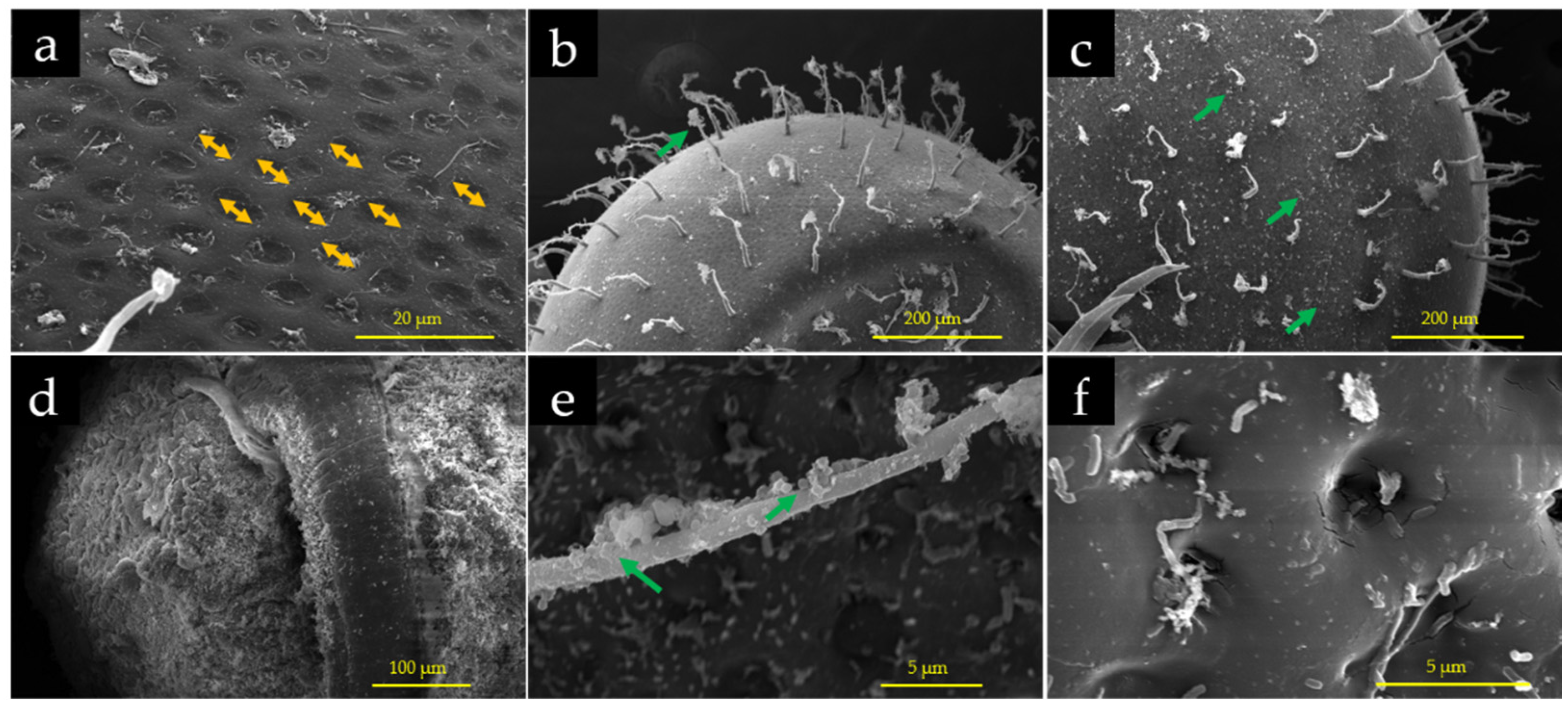
2.4. Calculation of Membrane Pore Size

On day 6 after exposure, nine embryos (n = 9) from each group were randomly selected to observe the morphological structure of the chorion using a field emission scanning electron microscope (FESEM; Quanta 450FEG, FEI, Hillsboro, OR, USA). Before observation using the FESEM, O. melastigma embryos were prepared using the CO2 critical point drying method. The average pore size of the membranes was measured after photography and calculations.

2.5. Determination of Embryonic Membrane Changes

During embryonic hatching, impurities in seawater readily attach to the embryonic surface villi, thereby altering the embryonic surface and villi. To investigate membrane changes after PS-MP exposure, nine embryos (n = 9) were randomly selected from each group to observe the diameter and chorion structure using a stereo microscope (SMZ 1270; Nikon Corp., Tokyo, Japan).

2.6. Accumulation of Polystyrene Microplastics on and Inside Chorion

Embryos that had been exposed to fluorescently labeled PS-MPs of different sizes for 3, 6, and 9 d were collected and observed using a digital inverted fluorescence microscope (Axio observer Z1; Carl Zeiss, Oberkochen, Germany), and the PS-MP distribution and accumulation patterns on the membrane surface of O. melastigma embryos were analyzed.
To investigate the distribution and accumulation of fluorescently labeled PS-MPs inside O. melastigma embryos, nine embryos (n = 9) that had been exposed to PS-MPs for 6 d were randomly collected from different treatments and photographed using a confocal fluorescence microscope (LSM 780 NLO; Carl Zeiss, Oberkochen, Germany). Fluorescence images were captured continuously in the z-axis direction every 20 nm from the top to the bottom of a single embryo.
Furthermore, to understand the PS-MP distribution on and inside the chorion of the O. melastigma embryos, the chorion was removed with tweezers after washing and prior to imaging. Thereafter, they were observed using a FESEM and images were captured.

2.7. Measurement of Mortality, Heartbeat and Hatching Rate of Fish Embryos

Dead embryos were removed and recorded during the PS-MP exposure period. Embryo mortality (%) was calculated as follows: = (dead embryos/total embryos) × 100. Ten embryos from each group were used to determine heart rate, as described in a previous study [62]. The heartbeat of O. melastigma embryos was observed at 3 d post-fertilization (dpf). Heartbeat measurement for O. melastigma larvae is difficult compared to that for embryos, as larvae can easily swim away. Therefore, embryos for heartbeat measurements in this study were selected only from 3 to 9 dpf.
The fish embryos started hatching at 8 dpf under the above experimental conditions, and the embryos were regarded as hatching failures if they did not hatch after 19 dpf. The number of hatched embryos was recorded every 24 h from 8 to 19 dpf, and the hatching rate was calculated as follows: (%) = (hatched larvae/fertile eggs) × 100.

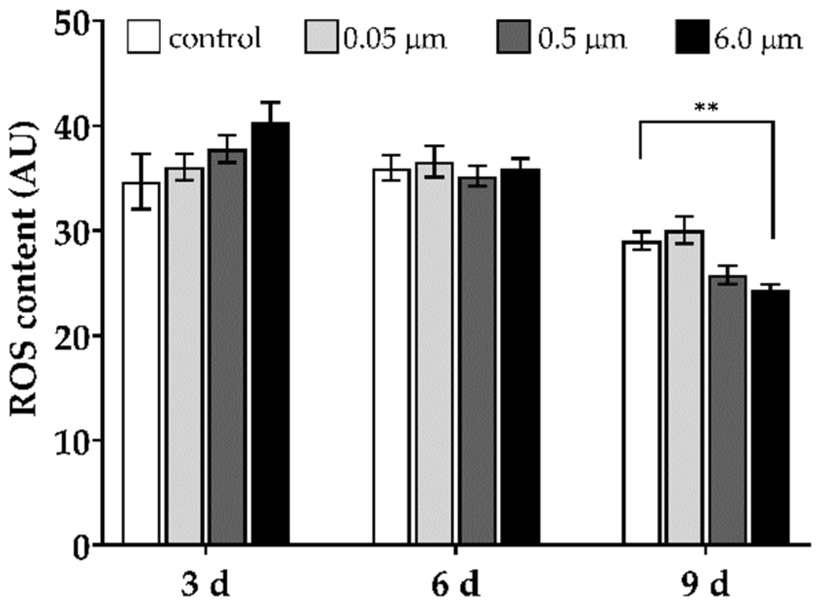
2.8. Determination of Reactive Oxygen Species (ROS) Levels

The ROS assay kit (DCFH-DA fluorescent probe staining) used in this study was purchased from Nanjing Jiancheng Bioengineering Institute (Nanjing, China). To eliminate the interference of the fluorescent signal from PS-MPs in ROS assessment, non-fluorescent PS-MPs were used to perform the ROS assay. Briefly, nine embryos (n = 9) at 3, 6, and 9 dpf after PS-MP exposure were randomly selected from each group and placed in a 6-well plate (one embryo per well). The embryos were gently washed twice with prewarmed 1 × PBS (28.5 °C, pH = 7.4), and 5 mL fluorescent probe (1:1000 DC-FHDA: ddH2O) was added to each well. The embryos were incubated at 28.5 °C for 1 h in the dark by covering them with aluminum foil, washed again with 1 × PBS, and images were captured using a digital inverted fluorescence microscope (Axio observer Z1; Carl Zeiss, Oberkochen, Germany). The images were analyzed using ImageJ v1.53 software (National Institutes of Health, Bethesda, MD, USA).

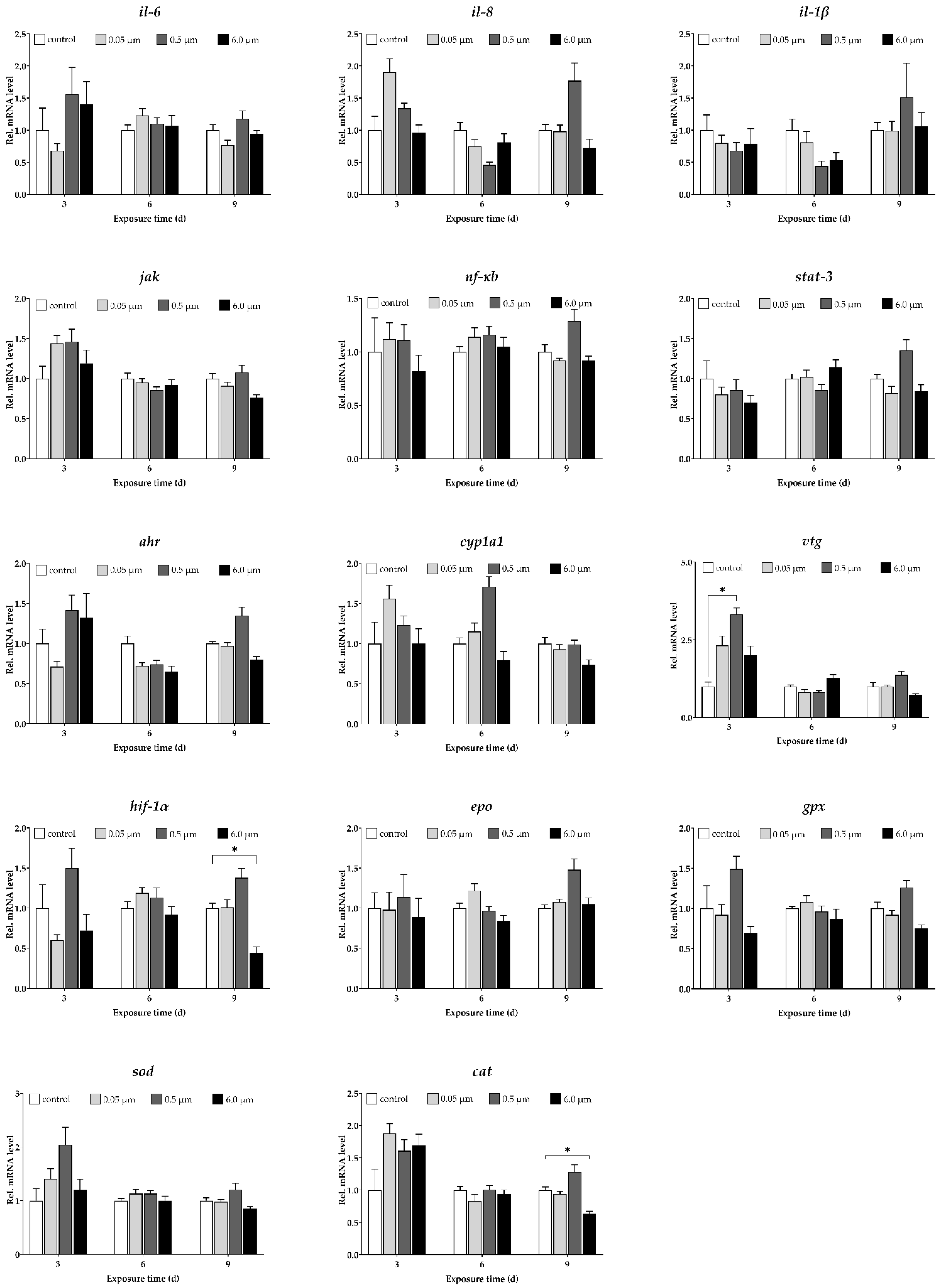
2.9. Determination of Expression Profiles of Target Genes

The following selected genes were measured using real-time reverse transcription quantitative PCR (RT-qPCR): (i) immune- and inflammation-related genes: il6 (interleukin 6), il8 (interleukin 8), il-1β (interleukin 1β), jak (Janus kinase), stat-3 (signal transducer and activator of transcription 3), nf-κb (nuclear transcription factor-κb); (ii) endocrine-related gene: vtg (vitellogenin); (iii) anaerobic-related gene: hif-1α (hypoxia-inducible factor 1-alpha) and epo (erythropoietin); (iv) metabolism-related genes: cyp1a1 (cytochrome P450 1a1) and ahr (aryl hydrocarbon receptor); and (v) oxidative stress-related genes: sod (superoxide dismutase), cat (catalase), and gpx (glutathione peroxidase). A pool of 15 embryos obtained at 3, 6, and 9 dpf from each replicate was collected from each petri dish (n = 6) and stored at −80 °C for later analysis. Total RNA analysis was conducted using TRIzol reagent (Takara Ltd., Beijing, China), according to the manufacturer’s instructions. Primer sequences used in this study are listed in Appendix A (Table A1). 18S ribosomal RNA was used as the housekeeping gene [49] and RT-qPCR was performed using SYBR Green (Roche Ltd., Basel, Switzerland) with a Rotor-Gene Q instrument (5 plex, Qiagen GmbH, Hilden, Germany). The RT-qPCR assay and data analysis were performed as previously described [63].

2.10. Statistics Analysis

All results are expressed as mean ± standard error (S.E.). All data were first analyzed for normality and homogeneity using Bartlett’s test with IBM SPSS Statistics for Windows, v22.0 (IBM Corp., Armonk, NY, USA). One-way analysis of variance (ANOVA) followed by Tukey’s HSD test (homogeneity of variance) or T3 test (uneven variance) was performed to identify significant differences between the treatment and control groups. For non-normal distribution, nonparametric tests were used to identify significant differences between the treatment and control groups. Significant differences were set at * p < 0.05, ** p < 0.01.

3. Results

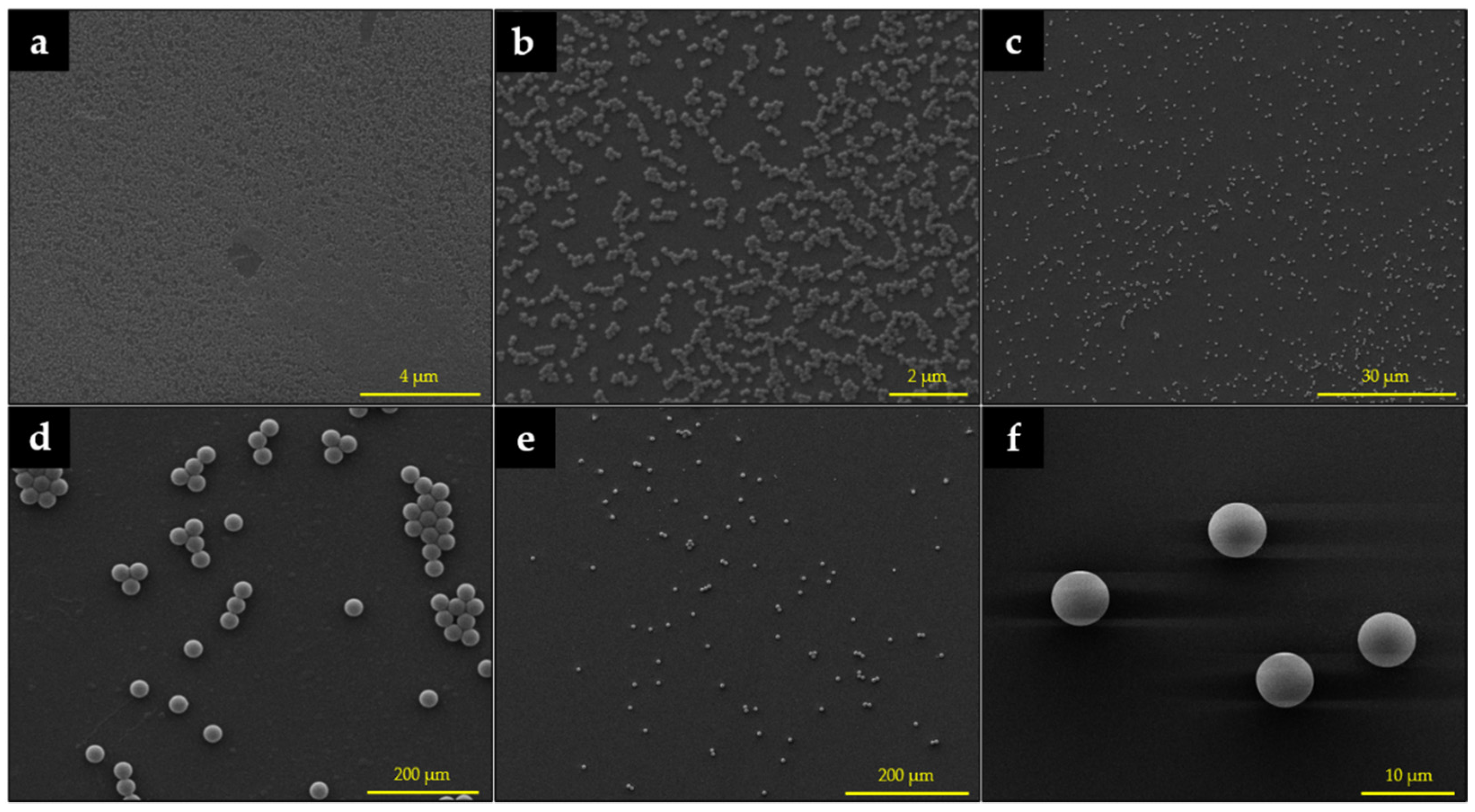
3.1. Characterization of Polystyrene Microplastics

The size and morphology of the PS-MPs were observed and verified using scanning electron microscopy (SEM, EVO-18; Carl Zeiss, Oberkochen, Germany) before the waterborne exposure experiment. The PS-MPs were spherical, their dispersion was good, and the particle sizes were consistent with the nominal sizes (Figure A1).

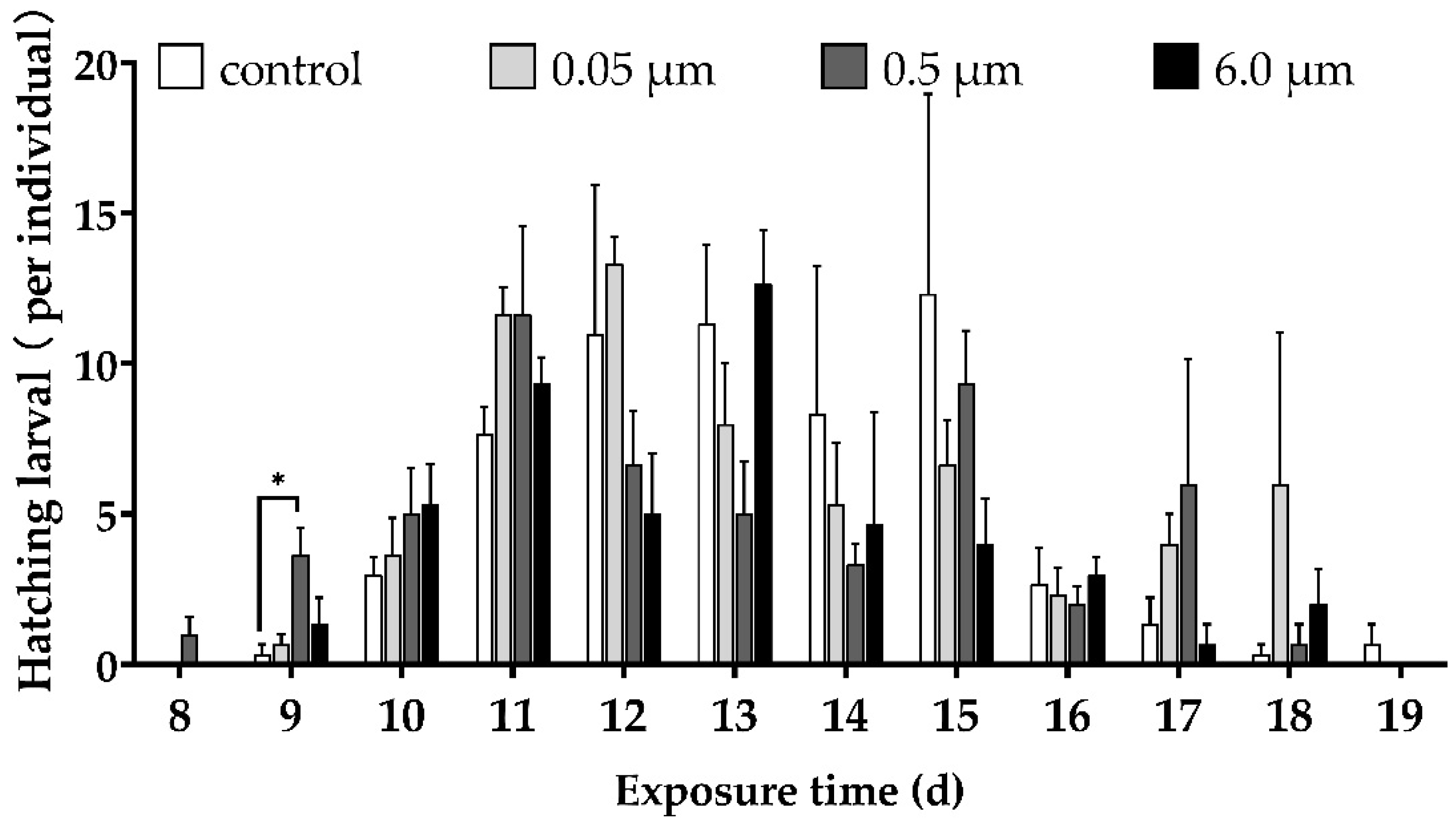
3.2. Hatching Time and Hatching Rate of Embryos

The number of hatched embryos in the 0.5 μm PS-MP treatment was significantly greater than that of the control after exposure for 9 d. The O. melastigma embryos hatched before day 19, whereas most of them hatched from days 11–15 (Figure A2). The average hatching time of O. melastigma embryos in the control group was 13.5 d, with an average hatching rate of 66.50%. The average hatching time for embryos with 0.5 and 6.0 um exposures was similar (approximately 12.8 d) and much shorter than that of the control. For the 0.05 μm PS-MP exposure, the average hatching time was similar to that of the control (Table 1).
The hatching rate of embryos decreased with increasing size of PS-MP, and the hatching rates were 61.67%, 54.33%, and 48.00% for treatment groups with diameter of 0.05, 0.5, and 6.0 μm, respectively. Compared to the control, no significant difference was observed in the 0.05 μm PS-MP exposure group, whereas the average hatching rates of the 0.5 and 6.0 μm PS-MP groups were low (Table 1).

3.3. Embryo Mortality Rate

The mortality rates of O. melastigma embryos exposed to PS-MPs of different sizes for 19 d were 29.50%, 37.00%, 40.33%, and 45.67%, respectively, which increased with increasing PS-MP size (Table 2). However, no significant difference in mortality rate was detected between the treatments.

3.4. Heart Rates

The heart rate of each treatment increased with the developmental progress of the embryo, and no significant changes in the heart rate were observed at each observation time point under exposure to 0.05 and 6.0 μm PS-MPs (Figure A3). The heart rate in the group exposed to 0.5 μm PS-MPs on day 3 was significantly higher than that of the control group on day 3 (p < 0.01), and it was significantly lower than that of the control group on day 5, while no significant difference was found at other time points (p > 0.05) (Figure A3).

3.5. Embryo Diameter

No significant difference in embryo diameter was observed between the treatment and control groups during the experiment (p > 0.05, Table A2).

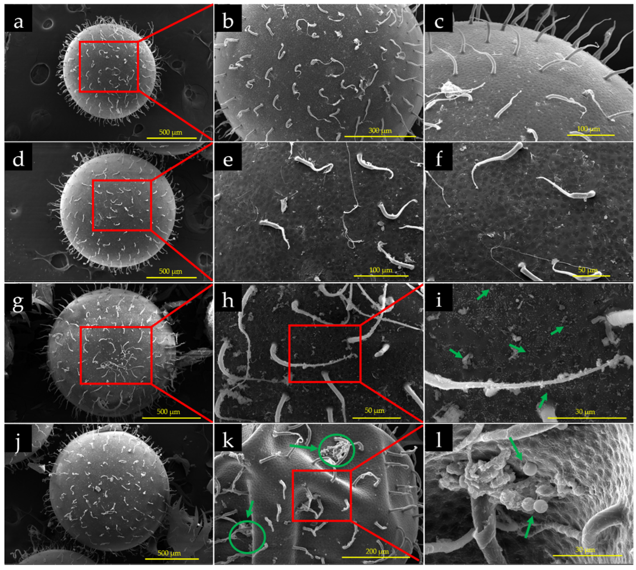
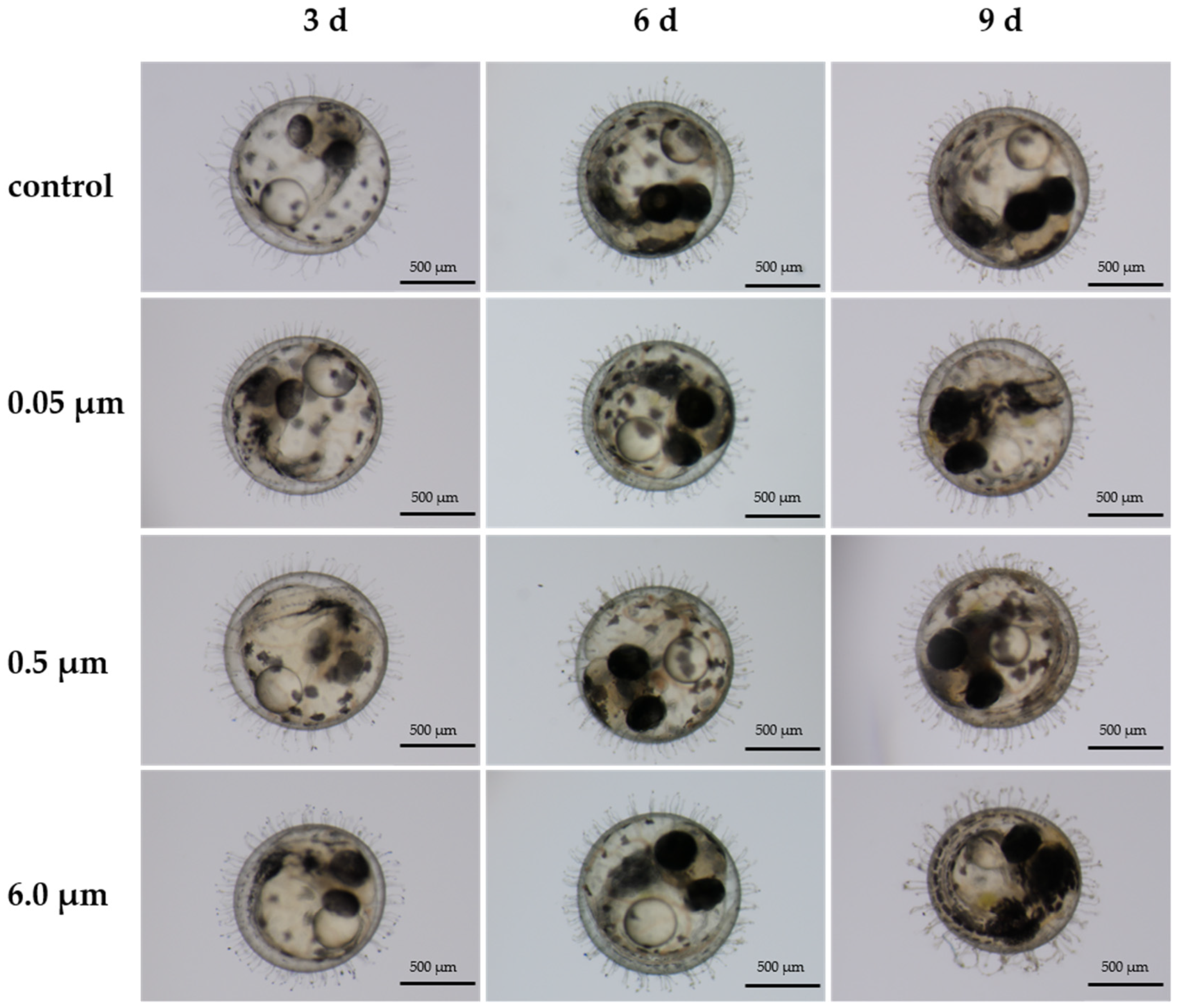
3.6. Effects of PS-MPs on Chorion of the Embryos

Villi on the chorion surface of O. melastigma embryos in the control group were clearly observed (Figure A4). However, the additives on the villi of the chorion surface accumulated over time, and were clearer when observed at 9 dpf than at 3 or 6 dpf.

3.7. Accumulation of PS-MPs on Chorion of the Embryos

No clear fluorescence signal was observed on the chorion surfaces of O. melastigma embryos (Figure A5) at 3, 6, and 9 dpf in the control group and in the group exposed to 0.05 μm PS-MPs. Embryos treated with 0.5 μm PS-MPs were observed with fluorescence signals at 3, 6, and 9 dpf, although the signal was weak. However, the fluorescence signals were clear on the embryo chorion surfaces when exposed to PS-MPs with a diameter of 6.0 μm. In addition, more PS-MPs accumulated on the chorion over time.

3.8. Accumulation of Fluorescent PS-MPs Inside the Embryos

Clearer fluorescence signals were observed on the chorion surfaces of the embryos (Figure 1). However, no evidence indicated that the PS-MPs of different sizes used in this study could enter O. melastigma embryos.

3.9. Interaction of PS-MPs with Embryos

SEM images indicated that no PS-MP signals were observed on the chorion surfaces and villi of O. melastigma embryos in the control group and the 0.05 μm PS-MP exposure group, with a smooth and clean chorion surface (Figure 2a–f). In contrast, many PS-MPs attached to the chorion surface of 0.5 and 6.0 μm PS-MP-exposed embryos (Figure 2g–l).
Can PS-MPs enter O. melastigma embryos through chorion pores? To further explore whether PS-MPs can enter O. melastigma embryos through chorion pores (5.138 ± 0.100 μm, Figure 3a), the fluorescence signal data (see Section 3.8) were analyzed using FESEM. The SEM images exhibited a clean chorion and larval surface inside the embryo, and no PS-MPs were observed (Figure 3d,f). In contrast, many PS-MPs attached to the chorion villi (Figure 3b,c,e), which indicates that PS-MPs do not enter the O. melastigma embryos but accumulate onto the villi or the membrane surface of the chorion.

3.10. ROS Production Level

Compared to the control, the embryos exposed to 6.0 μm PS-MPs resulted in a significant decrease in ROS levels at 9 dpf (Figure 4, p < 0.01), and no significant difference was found between the control and other sizes of PS-MPs at 3 and 6 dpf (p > 0.05).

3.11. Expression Profiles of Target Genes

Compared to the control, expression levels of the 14 genes, except vtg, were not significantly affected by the exposure to PS-MPs of different sizes at 3, 6, and 9 dpf (Figure 5). The expression level of vtg was significantly upregulated in the 0.5 μm PS-MP exposure at 3 dpf (p < 0.01).

4. Discussion

Marine medaka O. melastigma has been widely used in ecotoxicology studies [46,64,65]. To better understand the toxicity effects and underlying mechanisms of PS-MPs on O. melastigma, this study aimed to investigate the embryotoxicity of PS-MPs on the growth and development of O. melastigma, the accumulation and distribution of PS-MPs on/inside the surface of chorion, and the expression patterns of 14 selected genes in the present study.
The chorion, an acellular envelope, acts as a barrier between the internal and external environments of the embryo [66], plays an important role in protecting embryos from various external physical threats, such as mechanical abrasion [56], and participates in selective material transport, exchange, and respiration [67]. Zebrafish Abcb4, a cellular toxicant transporter, protects embryos and acts as an active barrier against toxic chemicals in the water [68]. In addition, electrostatic interactions and weak physical forces are generated between MPs and the chorion membrane, whereas 10 μm MPs can induce the protective effect of the chorion membrane [69]. Our results show that no significant difference was observed in the mortality of embryos exposed to PS-MPs of different sizes for 19 d. This indicates that PS-MP exposure had no obvious lethal effect on O. melastigma embryos, as the protective role of the embryo chorion may be an important cause.
No significant difference was observed in the hatching rates of O. melastigma embryos exposed to PS-MPs having a diameter of 0.05 μm. However, the hatching rate of embryos exposed to PS-MPs of 0.5 and 6.0 μm was significantly lower than that of the control; the possible reason for this is that large PS-MPs (6.0 μm) may embed or attach to the villi and the surface of membrane pores and disturb the gas and substance exchange of the embryos [53]. Therefore, the hatching rate decreased as the concentration and size of PS-MP increased. Interestingly, the hatching process of embryos was accelerated, whereas the hatching time was significantly decreased. This result is consistent with that of a previous study [53], in which PVC-MP exposure resulted in shorter hatching times and reduced hatching rates of O. melastigma embryos. Moreover, the embryo membrane may become softened, damaged, or thinned in fish embryos exposed to heavy metals such as cadmium, which makes the embryos more susceptible to mechanical damage [67]. In this study, PS-MPs caused shortened hatching times and decreased hatching rates. A possible reason for this is that a large number of MPs adhered to the chorion surface and thinned or softened the embryo membrane, thereby accelerating the larval shell-breaking process.
The heart, one of the first organs to undergo organogenesis, is vulnerable to environmental stress, which may result in an abnormal embryonic heart rate [70]. A previous study reported that the heartbeat rate was significantly changed (F0 decreased, whereas F1 increased) in O. melastigma embryos exposed to 20 μg/L PS-MPs at 11 dpf [71]. In this study, the heart rate increased over time with the different treatments. The heart rate was significantly increased in embryos exposed to 0.5 μm PS-MPs at 3 dpf, whereas it significantly decreased at 5 dpf. However, no significant change was found from 5 to 11 dpf in embryos exposed to PS-MPs of different sizes, suggesting that the embryos are more sensitive to PS-MP exposure in the early developmental stages.
The ingestion and accumulation of MPs can induce antioxidant defense systems and cause oxidative damage in aquatic organisms through ROS generation [72,73,74]. ROS can react with cellular components and cause oxidative damage if not effectively removed [75]. Normally, by increasing their activity, antioxidant defense systems in fish can eliminate excessive free radicals generated during embryonic development [73,76,77,78]. Enzymes such as superoxide dismutase (SOD) and catalase (CAT) are involved in the antioxidant defense [75]. A previous study demonstrated that ROS levels significantly increased in zebrafish exposed to PS-MPs [79]. Yang et al. (2020) also reported that MP exposure caused overproduction of ROS in C. auratus larvae, with the size of MPs influencing ROS levels [80]. Interestingly, the ROS levels in embryos exposed to 6.0 μm PS-MPs were significantly decreased at 9 dpf in our study. This phenomenon may be caused by the ingestion and accumulation of MPs that induce ROS generation and antioxidant defense systems; however, once the oxidative stress in embryos in response to PS-MPs reaches a certain threshold, it may cause the suppression of antioxidant enzyme activity and ROS metabolism in fish, which could lead to a reduction in cat expression and ROS production [81]. In the present study, the expression level of the cat gene was significantly downregulated at 9 dpf following exposure to 6.0 μm PS-MPs. Karami et al. (2017) reported a significant decrease in cat expression levels in zebrafish larvae sampled on day 20 and day 10 following exposure to graded concentrations (5, 50, or 500 μg/L) of low-density polyethylene (LDPE) fragments [82]. This is consistent with our results, indicating that the antioxidant reaction and detoxification systems are inhibited in fish embryos exposed to MPs.
Considering the size relationship between the PS-MPs and membrane pores, smaller PS-MPs (0.05 and 0.5 μm) may enter the embryo through membrane pores through non-specific diffusion [83], resulting in embryo immunotoxicity and inflammation response. It was confirmed that smaller PS-MPs (50 nm) can enter the chorion of zebrafish embryos and accumulate in the bodies of zebrafish, although the toxicity of PS-MPs was negligible [84]. In contrast, no evidence suggested that PS-MPs can enter the embryo through membrane pores, and fluorescent PS-MPs were only observed on the villi and membrane surface of the marine medaka in the present study. In addition, no clear fluorescent signal was observed inside the embryos, including those exposed to PS-MPs of smaller sizes (0.05 and 0.5 μm). It is speculated that the resolution of the microscope used in this study was limited by the size of the PS-MPs. Yamagami et al. (1992) reported that the chorion of medaka embryos becomes a rigid structure after fertilization [85]. Therefore, another possibility is that, like other small molecules, small MPs may encounter difficulties to penetrate the embryo because of the membrane system underneath the chorion. The chorion may act as a barrier to protect the embryo from contaminants in the surrounding environment.
Progressive deep hypoxia can disturb fish metabolism and swimming behavior, change the reproductive capacity and function of the endocrine system [86], promote the activation of the HIF-1α signaling pathway, and activate the expression of genes encoding proteins with known roles in oxygen homeostasis (such as hif-1α and epo) [87,88,89,90,91,92]. Overexpression of hif-1α was induced when O. melastigma embryos were exposed to hypoxic conditions [53,93]. However, no significant upregulation in the expression level of hypoxia-related gene hif-1α was found in embryos exposed to 6.0 μm PS-MPs at 3, 6, and 9 dpf in our study, suggesting that the PS-MP concentration (106 particles/L) or different sizes (0.05, 0.5, and 6.0 μm) used in this study failed to affect the oxygen flow; no hypoxic microenvironment was found on the chorion surface.
Cyp1a1 was upregulated at 3 and 6 dpf in embryos exposed to 0.05 and 0.5 μm PS-MPs, but downregulated at 9 dpf (although the difference was not significant, p < 0.05). These results indicate that the detoxification ability of O. melastigma embryos increased, with a reduction in the toxic effects of PS-MPs [55]. VTG is a commonly used biomarker in environmental endocrine disruptor studies and is significantly produced in the livers of mature females. Exogenous estrogen exposure, however, enhances VTG production in males and immature individuals [94]. Plasticizers are commonly used in the plastic manufacturing process. Plasticizers induce endocrine-disrupting effects and simulate estrogen activity [82,95,96,97]. In this study, the vtg expression level in embryos exposed to 0.5 μm PS-MPs at 3 dpf was significantly upregulated, which may be because of the endocrine disruption induced by plasticizers released from MPs [98].

5. Conclusions

In this study, we assessed the toxic effects and possible mechanisms of PS-MPs on embryonic development of the marine medaka with diameters of 0.05, 0.5 and 6.0 μm. Overall, our results revealed that PS-MP exposure exhibited size-dependent adverse effects on O. melastigma embryos, which demonstrates that larger PS-MPs (0.5 and 6.0 μm) have more toxic effects on the embryonic development of marine medaka. The results showed that larger PS-MP waterborne exposure significantly shortened the hatching time and decreased the hatching rate of the embryos. Furthermore, PS-MPs of larger sizes only attach to the chorion surface or villi of the embryos and cannot enter the embryo through the membrane pores, which suggests that the chorion plays an important role in protecting the embryo from PS-MP stress. The present study provides a new perspective to investigate the adverse effects and the underlying mechanisms caused by MPs of different sizes.

Author Contributions

Conceptualization, J.C. and J.B.; Data curation, J.C.; Investigation, J.C.; Methodology, R.Z., C.F. and J.B.; Software, J.C.; Supervision, J.B; Visualization, J.C.; Writing—original draft preparation, J.C.; Writing—review and editing, C.F. and J.B. All authors have read and agreed to the published version of the manuscript.

Funding

This research received the support of the Natural Science Foundation of Fujian Province (No. 2021J01506), First batch of the Youth Innovation Fund Project of Xiamen in 2020 (No. 3502Z20206099), the National Key Research and Development Program of China (No. 2019YFD0901101) and the National Natural Science Foundation of China (No. 41977211).

Institutional Review Board Statement

The animal study protocol was approved by Ethics Committee of Xiamen University (XMULAC20190066).

Informed Consent Statement

Not applicable.

Data Availability Statement

Not applicable.

Acknowledgments

The authors would like to thank Fukun Hong, Yulu Jiang and Min Zhang from Third Institute of Oceanography (TIO), Huiyun Chen and Jie Liu from Xiamen University (XMU) for fish culture, maintenance and technical assistance.

Conflicts of Interest

The authors declare no conflict of interest. The funders had no role in the design of the study; in the collection, analyses, or interpretation of data; in the writing of the manuscript, or in the decision to publish the results.

Appendix A

Figure A1. The morphology of polystyrene microplastics of different sizes confirmed using scanning electron microscopy (a,b) 0.05 µm, (c,d) 0.5 µm, (e,f) 6.0 µm.
Water 14 01831 g0a1
Figure A2. Effects of PS-MPs of different sizes on the numbers of hatched embryos of the O. melastigma at 3, 6 and 9 dpf (n = 100, 100 embryos for each replicate, three replicate). Asterisk indicates significant difference between the treatment group and the control (* p < 0.05).
Water 14 01831 g0a2
Figure A3. Effects of PS-MPs of different sizes on heart rate of the O. melastigma embryos (n = 9). Asterisks indicate significant difference between the treatment group and the control at each time point (* p < 0.05, ** p < 0.01).
Water 14 01831 g0a3
Figure A4. Effects of PS-MPs of different sizes on the chorion of O. melastigma embryos (n = 9).
Water 14 01831 g0a4
Figure A5. Accumulation of PS-MPs of different sizes on the chorion of O. melastigma embryos (n = 9: Scale bar = 500 μm).
Water 14 01831 g0a5
Table A1. Primer sequences used for qPCR in this study.
Gene NamePrimer Sequence (5′ End to 3′ End)
18sF: GACAAATCGCTCCACCAACT
R: CCTGCGGCTTAATTTGACCC
gpxF: GTGTGCAGAAACGACGTGGCCTGGA
R: TCGCCTTCGATGTCGCTGGTGAGGA
il-1βF: AGGCAGCGACAGCCGCAAAGTTCA
R: TGGTGTCCTTGATGCCCAGAGCCA
il-6F: GACCTTCTGGCAGGTGACGCTT
R: CACCTCTTTCTGGTGCCGTTTGG
il-8F: TGCACTGCCGCTGCATCTTGACAG
R: TCTGGGTTCAGGCAAACCTCCTGGC
nf-κbF: CATGGCTACTACGAGGCAGACC
R: AACTCCTCCTCCCACACCTTGGAC
jakF: ATCGCCACCACCTTCCAGCAC
R: TGGCAGGATGTCGCTGTCCATG
stat-3F: CGCCCTGGAAGAGAAGATTGTGGAT
R: CCAGCAACCTGACTTTGTTTGTGAA
epoF: GACCGGCTTGTGGCTGTT
R: CGTGTTGGTGACCGATGGT
vtgF: CAGGTTGCCAGCTTCACATAC
R: CTCAGTCTGTCCAGCTTTGG
hif-1αF: TGTGATATTTGCAGCATTCCA
R: CTACGACAGGAAATCCCATGA
catF: GCCAACTACCTGCAGATCCCCGTCA
R: AGTTTGGAGCGCCGCCTTGGTTGT
sodF: TGTACCAGTGCGGGGCCTCACTTCA
R: TGCGGTCACATTTCCCAGGTCCCCA
ahrF: ACTGAACATCCAGGGCAGAC
R: ATGTTCTTGGTGCGGATCTC
cyp1a1F: TGCTCTGTCTTGGTCAGTGG
R: CAGGGTAGGATTTCGGTTCA
F: forward sequence; R: reverse sequence.
Table A2. Effects of PS-MPs of different sizes on embryo diameter (n = 9).
Treatment3d Diameter (mm)6d Diameter (mm)9d Diameter (mm)
Control1.03 ± 0.011.07 ± 0.011.06 ± 0.01
0.05 μm1.02 ± 0.011.06 ± 0.011.04 ± 0.01
0.5 μm1.01 ± 0.011.04 ± 0.011.05 ± 0.01
6.0 μm1.01 ± 0.011.04 ± 0.011.05 ± 0.01
Note: Values are expressed as means ± standard error (S.E.).

References

  1. Frias, J.P.G.L.; Nash, R. Microplastics: Finding a consensus on the definition. Mar. Pollut. Bull. 2019, 138, 145–147. [Google Scholar] [CrossRef] [PubMed]
  2. Rochman, C.M. Microplastics research-from sink to source. Science 2018, 360, 28–29. [Google Scholar] [CrossRef]
  3. Thompson, R.C.; Olson, Y.; Mitchell, R.P.; Davis, A.; Rowland, S.J.; John, A.W.G.; McGonigle, D.; Russell, A.E. Lost at sea: Where is all the plastic? Science 2004, 304, 838. [Google Scholar] [CrossRef]
  4. Barros, J.; Seena, S. Plastisphere in freshwaters: An emerging concern. Environ. Pollut. 2021, 290, 118123. [Google Scholar] [CrossRef]
  5. Ma, H.; Pu, S.; Liu, S.; Bai, Y.; Mandal, S.; Xing, B. Microplastics in aquatic environments: Toxicity to trigger ecological consequences. Environ. Pollut. 2020, 261, 114089. [Google Scholar] [CrossRef]
  6. Dusaucy, J.; Gateuille, D.; Perrette, Y.; Naffrechoux, E. Microplastic pollution of worldwide lakes. Environ. Pollut. 2021, 284, 117075. [Google Scholar] [CrossRef] [PubMed]
  7. Chen, G.; Li, Y.; Wang, J. Occurrence and ecological impact of microplastics in aquaculture ecosystems. Chemosphere 2021, 274, 129989. [Google Scholar] [CrossRef] [PubMed]
  8. Campanale, C.; Galafassi, S.; Savino, I.; Massarelli, C.; Ancona, V.; Volta, P.; Uricchio, V.F. Microplastics pollution in the terrestrial environments: Poorly known diffuse sources and implications for plants. Sci. Total Environ. 2022, 805, 150431. [Google Scholar] [CrossRef]
  9. Zazouli, M.; Nejati, H.; Hashempour, Y.; Dehbandi, R.; Nam, V.T.; Fakhri, Y. Occurrence of microplastics (MPs) in the gastrointestinal tract of fishes: A global systematic review and meta-analysis and meta-regression. Sci. Total Environ. 2022, 815, 152743. [Google Scholar] [CrossRef] [PubMed]
  10. Reichert, J.; Tirpitz, V.; Anand, R.; Bach, K.; Knopp, J.; Schubert, P.; Wilke, T.; Ziegler, M. Interactive effects of microplastic pollution and heat stress on reef-building corals. Environ. Pollut. 2021, 290, 118010. [Google Scholar] [CrossRef] [PubMed]
  11. Hamm, T.; Lenz, M. Negative impacts of realistic doses of spherical and irregular microplastics emerged late during a 42 weeks-long exposure experiment with blue mussels. Sci. Total Environ. 2021, 778, 146088. [Google Scholar] [CrossRef]
  12. Amélineau, F.; Bonnet, D.; Heitz, O.; Mortreux, V.; Harding, A.M.A.; Karnovsky, N.; Walkusz, W.; Fort, J.; Grémillet, D. Microplastic pollution in the Greenland Sea: Background levels and selective contamination of planktivorous diving seabirds. Environ. Pollut. 2016, 219, 1131–1139. [Google Scholar] [CrossRef] [PubMed]
  13. Albano, M.; Panarello, G.; Di Paola, D.; Capparucci, F.; Crupi, R.; Gugliandolo, E.; Spanò, N.; Capillo, G.; Savoca, S. The influence of polystyrene microspheres abundance on development and feeding behavior of Artemia salina (Linnaeus, 1758). Appl. Sci. 2021, 11, 3352. [Google Scholar] [CrossRef]
  14. Albano, M.; Panarello, G.; Di Paola, D.; D’Angelo, G.; Granata, A.; Savoca, S.; Capillo, G. The mauve stinger Pelagia noctiluca (Cnidaria, Scyphozoa) plastics contamination, the Strait of Messina case. Int. J. Environ. Stud. 2021, 78, 977–982. [Google Scholar] [CrossRef]
  15. Bottari, T.; Savoca, S.; Mancuso, M.; Capillo, G.; GiuseppePanarello, G.; MartinaBonsignore, M.; Crupi, R.; Sanfilippo, M.; D’Urso, L.; Compagnini, G.; et al. Plastics occurrence in the gastrointestinal tract of Zeus faber and Lepidopus caudatus from the Tyrrhenian Sea. Mar. Pollut. Bull. 2019, 146, 408–416. [Google Scholar] [CrossRef]
  16. Mancuso, M.; Savoca, S.; Bottari, T. First record of microplastics ingestion by European hake MERLUCCIUS MERLUCCIUS from the Tyrrhenian Sicilian coast (Central Mediterranean Sea). J. Fish Biol. 2019, 94, 517–519. [Google Scholar] [CrossRef] [PubMed]
  17. Capillo, G.; Savoca, S.; Panarello, G.; Mancuso, M.; Branca, C.; Romano, V.; D’Angelo, G.; Bottari, T.; Spanò, N. Quali-quantitative analysis of plastics and synthetic microfibers found in demersal species from southern Tyrrhenian Sea (Central Mediterranean). Mar. Pollut. Bull. 2020, 150, 110596. [Google Scholar] [CrossRef]
  18. Savoca, S.; Matanović, K.; D’Angelo, G.; Vetri, V.; Anselmo, S.; Bottari, T.; Mancuso, M.; Kužir, S.; Spanò, N.; Capillo, G.; et al. Ingestion of plastic and non-plastic microfibers by farmed gilthead sea bream (Sparus aurata) and common carp (Cyprinus carpio) at different life stages. Sci. Total Environ. 2021, 782, 146851. [Google Scholar] [CrossRef]
  19. Savoca, S.; Bottari, T.; Fazio, E.; Bonsignore, M.; Mancuso, M.; Luna, G.M.; Romeo, T.; D’Urso, L.; Capillo, G.; Panarello, G.; et al. Plastics occurrence in juveniles of Engraulis encrasicolus and Sardina pilchardus in the Southern Tyrrhenian Sea. Sci. Total Environ. 2020, 718, 137457. [Google Scholar] [CrossRef] [PubMed]
  20. Qu, X.; Su, L.; Li, H.; Liang, M.; Shi, H. Assessing the relationship between the abundance and properties of microplastics in water and in mussels. Sci. Total Environ. 2018, 621, 679–686. [Google Scholar] [CrossRef]
  21. Zhu, C.; Li, D.; Sun, Y.; Zheng, X.; Peng, X.; Zheng, K.; Hu, B.; Luo, X.; Mai, B. Plastic debris in marine birds from an island located in the South China Sea. Mar. Pollut. Bull. 2019, 149, 110566. [Google Scholar] [CrossRef] [PubMed]
  22. Eriksson, C.; Burton, H. Origins and biological accumulation of small plastic particles in fur seals from Macquarie Island. AMBIO J. Hum. Environ. 2003, 32, 380–384. [Google Scholar] [CrossRef] [PubMed]
  23. Lusher, A.; Hernandez-Milian, G.; Berrow, S.; Rogan, E.; O’Connor, I. Microplastics and marine mammals: Studies from Ireland. In Fate and Impact of Microplastics in Marine Ecosystems; Baztan, J., Jorgensen, B., Pahl, S., Thompson, R.C., Vanderlinden, J.-P., Eds.; Elsevier: Amsterdam, The Netherlands, 2017; p. 37. ISBN 978-0-12-812271-6. [Google Scholar]
  24. Charlene, T.; Jeff, S.; Dayanthi, N. Sub-lethal responses to microplastic ingestion in invertebrates. In Particulate Plastics in Terrestrial and Aquatic Environments, 1st ed.; Bolan, N., Kirkham, M.B., Eds.; CRC Press: Boca Raton, FL, USA, 2020; p. 466. ISBN 978-1-00-305307-1. [Google Scholar] [CrossRef]
  25. Bergami, E.; Bocci, E.; Vannuccini, M.L.; Monopoli, M.; Salvati, A.; Dawson, K.A.; Corsi, I. Nano-sized polystyrene affects feeding, behavior and physiology of brine shrimp Artemia franciscana larvae. Ecotoxicol. Environ. Saf. 2016, 123, 18–25. [Google Scholar] [CrossRef]
  26. Sussarellu, R.; Suquet, M.; Thomas, Y.; Lambert, C.; Fabioux, C.; Pernet, M.E.J.; Le Goïc, N.; Quillien, V.; Mingant, C.; Epelboin, Y.; et al. Oyster reproduction is affected by exposure to polystyrene microplastics. Proc. Natl. Acad. Sci. USA 2016, 113, 2430–2435. [Google Scholar] [CrossRef] [PubMed] [Green Version]
  27. Sun, X.; Chen, B.; Li, Q.; Liu, N.; Xia, B.; Zhu, L.; Qu, K. Toxicities of polystyrene nano- and microplastics toward marine bacterium Halomonas alkaliphila. Sci. Total Environ. 2018, 642, 1378–1385. [Google Scholar] [CrossRef] [PubMed]
  28. Yin, L.; Chen, B.; Xia, B.; Shi, X.; Qu, K. Polystyrene microplastics alter the behavior, energy reserve and nutritional composition of marine jacopever (Sebastes schlegelii). J. Hazard. Mater. 2018, 360, 97–105. [Google Scholar] [CrossRef]
  29. Martellini, T.; Guerranti, C.; Scopetani, C.; Ugolini, A.; Chelazzi, D.; Cincinelli, A. A snapshot of microplastics in the coastal areas of the Mediterranean Sea. TrAC Trends Anal. Chem. 2018, 109, 173–179. [Google Scholar] [CrossRef]
  30. Kedzierski, M.; Palazot, M.; Soccalingame, L.; Falcou-Préfol, M.; Gorsky, G.; Galgani, F.; Bruzaud, S.; Pedrotti, M.L. Chemical composition of microplastics floating on the surface of the Mediterranean Sea. Mar. Pollut. Bull. 2022, 174, 113284. [Google Scholar] [CrossRef]
  31. Long, M.; Paul-Pont, I.; Hégaret, H.; Moriceau, B.; Lambert, C.; Huvet, A.; Soudant, P. Interactions between polystyrene microplastics and marine phytoplankton lead to species-specific hetero-aggregation. Environ. Pollut. 2017, 228, 454–463. [Google Scholar] [CrossRef] [Green Version]
  32. Hidalgo-Ruz, V.; Gutow, L.; Thompson, R.C.; Thiel, M. Microplastics in the marine environment: A review of the methods used for identification and quantification. Environ. Sci. Technol. 2012, 46, 3060–3075. [Google Scholar] [CrossRef]
  33. Browne, M.A.; Galloway, T.S.; Thompson, R.C. Spatial patterns of plastic debris along estuarine shorelines. Environ. Sci. Technol. 2010, 44, 3404–3409. [Google Scholar] [CrossRef]
  34. Barnes, D.K.A.; Galgani, F.; Thompson, R.C.; Barlaz, M. Accumulation and fragmentation of plastic debris in global environments. Philos. Trans. R. Soc. Lond. B Biol. Sci. 2009, 364, 1985–1998. [Google Scholar] [CrossRef] [PubMed] [Green Version]
  35. Rocha-Santos, T.; Duarte, A.C. A critical overview of the analytical approaches to the occurrence, the fate and the behavior of microplastics in the environment. TrAC Trends Anal. Chem. 2015, 65, 47–53. [Google Scholar] [CrossRef]
  36. Jeong, C.B.; Won, E.J.; Kang, H.M.; Lee, M.C.; Hwang, D.S.; Hwang, U.K.; Zhou, B.; Souissi, S.; Lee, S.J.; Lee, J.S. Microplastic size-dependent toxicity, oxidative stress induction, and p-JNK and p-P38 activation in the monogonont rotifer (Brachionus koreanus). Environ. Sci. Technol. 2016, 50, 8849–8857. [Google Scholar] [CrossRef]
  37. Jin, Y.; Xia, J.; Pan, Z.; Yang, J.; Wang, W.; Fu, Z. Polystyrene microplastics induce microbiota dysbiosis and inflammation in the gut of adult zebrafish. Environ. Pollut. 2018, 235, 322–329. [Google Scholar] [CrossRef]
  38. Brandts, I.; Teles, M.; Gonçalves, A.P.; Barreto, A.; Franco-Martinez, L.; Tvarijonaviciute, A.; Martins, M.A.; Soares, A.M.V.M.; Tort, L.; Oliveira, M. Effects of nanoplastics on Mytilus galloprovincialis after individual and combined exposure with carbamazepine. Sci. Total Environ. 2018, 643, 775–784. [Google Scholar] [CrossRef] [PubMed]
  39. Cole, M.; Lindeque, P.; Fileman, E.; Halsband, C.; Galloway, T.S. The impact of polystyrene microplastics on feeding, function and fecundity in the marine copepod Calanus helgolandicus. Environ. Sci. Technol. 2015, 49, 1130–1137. [Google Scholar] [CrossRef] [PubMed]
  40. Suman, T.Y.; Jia, P.P.; Li, W.G.; Junaid, M.; Xin, G.Y.; Wang, Y.; Pei, D.S. Acute and chronic effects of polystyrene microplastics on brine shrimp: First evidence highlighting the molecular mechanism through transcriptome analysis. J. Hazard. Mater. 2020, 400, 123220. [Google Scholar] [CrossRef] [PubMed]
  41. Alimba, C.G.; Faggio, C. Microplastics in the marine environment: Current trends in environmental pollution and mechanisms of toxicological profile. Environ. Toxicol. Pharmacol. 2019, 68, 61–74. [Google Scholar] [CrossRef]
  42. Plastics Europe. Plastics—The Facts 2021: An Analysis of European Plastics Production, Demand and Waste Data. Available online: https://plasticseurope.org/knowledge-hub/plastics-the-facts-2021/ (accessed on 2 February 2022).
  43. Chen, J.C.; Chen, M.Y.; Fang, C.; Zheng, R.H.; Jiang, Y.L.; Zhang, Y.S.; Wang, K.J.; Bailey, C.; Segner, H.; Bo, J. Microplastics negatively impact embryogenesis and modulate the immune response of the marine medaka Oryzias melastigma. Mar. Pollut. Bull. 2020, 158, 111349. [Google Scholar] [CrossRef]
  44. Rochman, C.M.; Manzano, C.; Hentschel, B.T.; Simonich, S.L.M.; Hoh, E. Polystyrene plastic: A source and sink for polycyclic aromatic hydrocarbons in the marine environment. Environ. Sci. Technol. 2013, 47, 13976–13984. [Google Scholar] [CrossRef] [Green Version]
  45. Naruse, K. Classification and phylogeny of fishes of the genus Oryzias and its relatives. Fish Biol. J. Medaka 1996, 8, 1–9. [Google Scholar]
  46. Kim, B.M.; Kim, J.; Choi, I.Y.; Raisuddin, S.; Au, D.W.T.; Leung, K.M.Y.; Wu, R.S.S.; Rhee, J.S.; Lee, J.S. Omics of the marine medaka (Oryzias melastigma) and its relevance to marine environmental research. Mar. Environ. Res. 2016, 113, 141–152. [Google Scholar] [CrossRef]
  47. Kim, H.S.; Lee, B.Y.; Han, J.; Jeong, C.B.; Hwang, D.S.; Lee, M.C.; Kang, H.M.; Kim, D.H.; Lee, D.; Kim, J.; et al. The genome of the marine medaka Oryzias melastigma. Mol. Ecol. Resour. 2018, 18, 656–665. [Google Scholar] [CrossRef]
  48. Dong, S.; Kang, M.; Wu, X.; Ye, T. Development of a promising fish model (Oryzias melastigma) for assessing multiple responses to stresses in the marine environment. BioMed Res. Int. 2014, 2014, e563131. [Google Scholar] [CrossRef] [Green Version]
  49. Steer, M.; Cole, M.; Thompson, R.C.; Lindeque, P.K. Microplastic ingestion in fish larvae in the western english channel. Environ. Pollut. 2017, 226, 250–259. [Google Scholar] [CrossRef]
  50. Wang, J.; Li, Y.; Lu, L.; Zheng, M.; Zhang, X.; Tian, H.; Wang, W.; Ru, S. Polystyrene microplastics cause tissue damages, sex-specific reproductive disruption and transgenerational effects in marine medaka (Oryzias melastigma). Environ. Pollut. 2019, 254, 113024. [Google Scholar] [CrossRef] [PubMed]
  51. Cao, L.; Huang, W.; Liu, J.; Ye, Z.; Dou, S. Toxicity of short-term copper exposure to early life stages of red sea bream, Pagrus major. Environ. Toxicol. Chem. 2010, 29, 2044–2052. [Google Scholar] [CrossRef]
  52. Mohammed, A. Why are early life stages of aquatic organisms more sensitive to toxicants than adults? In New Insights into Toxicity and Drug Testing, 1st ed.; Gowder, S., Ed.; IntechOpen: London, UK, 2013; p. 254. ISBN 978-953-51-0946-4. [Google Scholar] [CrossRef] [Green Version]
  53. Xia, B.; Sui, Q.; Du, Y.; Wang, L.; Jing, J.; Zhu, L.; Zhao, X.; Sun, X.; Booth, A.M.; Chen, B.; et al. Secondary PVC microplastics are more toxic than primary PVC microplastics to Oryzias melastigma embryos. J. Hazard. Mater. 2022, 424, 127421. [Google Scholar] [CrossRef]
  54. Li, Y.J.; Yang, G.X.; Wang, J.; Lu, X.; Li, X.; Zheng, Y.Q.; Zhang, Z.Z.; Ru, S.G. Microplastics increase the accumulation of phenanthrene in the ovaries of marine medaka (Oryzias melastigma) and its transgenerational toxicity. J. Hazard. Mater. 2022, 424 Pt D, 127754. [Google Scholar] [CrossRef]
  55. Zhang, Y.T.; Chen, M.; He, S.; Fang, C.; Chen, M.; Li, D.; Wu, D.; Chernick, M.; Hinton, D.E.; Bo, J.; et al. Microplastics decrease the toxicity of triphenyl phosphate (TPhP) in the marine medaka (Oryzias melastigma) larvae. Sci. Total Environ. 2021, 763, 143040. [Google Scholar] [CrossRef]
  56. Masato, K.; Kenji, M.; Kiyoshi, N.; Minoru, T. Looking at the medaka embryos. In Medaka: Biology, Management, and Experimental Protocols, 1st ed.; Masato, K., Kenji, M., Kiyoshi, N., Eds.; Wiley-Blackwell: Ames, IA, USA, 2009; Volume 2, pp. 165–180. ISBN 9781119575290. [Google Scholar]
  57. Li, H.; Chen, H.; Wang, J.; Li, J.; Liu, S.; Tu, J.; Chen, Y.; Zong, Y.; Zhang, P.; Wang, Z.; et al. Influence of microplastics on the growth and the intestinal microbiota composition of brine shrimp. Front. Microbiol. 2021, 12, 2671. [Google Scholar] [CrossRef] [PubMed]
  58. Phuong, N.N.; Zalouk-Vergnoux, A.; Poirier, L.; Kamari, A.; Châtel, A.; Mouneyrac, C.; Lagarde, F. Is there any consistency between the microplastics found in the field and those used in laboratory experiments? Environ. Pollut. 2016, 211, 111–123. [Google Scholar] [CrossRef] [PubMed]
  59. Da Costa Araújo, A.P.; Gomes, A.R.; Malafaia, G. Hepatotoxicity of pristine polyethylene microplastics in neotropical physalaemus Cuvieri tadpoles (Fitzinger, 1826). J. Hazard. Mater. 2020, 386, 121992. [Google Scholar] [CrossRef]
  60. Doyle, M.J.; Watson, W.; Bowlin, N.M.; Sheavly, S.B. Plastic particles in coastal pelagic ecosystems of the Northeast Pacific Ocean. Mar. Environ. Res. 2011, 71, 41–52. [Google Scholar] [CrossRef] [PubMed]
  61. Lee, K.W.; Shim, W.J.; Kwon, O.Y.; Kang, J.H. Size-dependent effects of micro polystyrene particles in the marine copepod Tigriopus japonicus. Environ. Sci. Technol. 2013, 47, 11278–11283. [Google Scholar] [CrossRef] [PubMed]
  62. Bo, J.; Cai, L.; Xu, J.H.; Wang, K.J.; Au, D.W.T. The marine medaka Oryzias melastigma—A potential marine fish model for innate immune study. Mar. Pollut. Bull. 2011, 63, 267–276. [Google Scholar] [CrossRef] [PubMed]
  63. Bo, J.; Giesy, J.P.; Ye, R.; Wang, K.J.; Lee, J.S.; Au, D.W.T. Identification of differentially expressed genes and quantitative expression of complement genes in the liver of marine medaka Oryzias melastigma challenged with Vibrio parahaemolyticus. Comp. Biochem. Physiol. Part D Genom. Proteom. 2012, 7, 191–200. [Google Scholar] [CrossRef] [PubMed]
  64. Kong, R.Y.C.; Giesy, J.P.; Wu, R.S.S.; Chen, E.X.H.; Chiang, M.W.L.; Lim, P.L.; Yuen, B.B.H.; Yip, B.W.P.; Mok, H.O.L.; Au, D.W.T. Development of a marine fish model for studying in vivo molecular responses in ecotoxicology. Aquat. Toxicol. 2008, 86, 131–141. [Google Scholar] [CrossRef]
  65. Chen, X.; Li, L.; Cheng, J.; Chan, L.L.; Wang, D.Z.; Wang, K.J.; Baker, M.E.; Hardiman, G.; Schlenk, D.; Cheng, S.H. Molecular staging of marine medaka: A model organism for marine ecotoxicity study. Mar. Pollut. Bull. 2011, 63, 309–317. [Google Scholar] [CrossRef]
  66. Jung, G.; Hug, M.; Halter, C.; Friesenhengst, A.; Walzer, J.; Czerny, T. Diffusion of small molecules into medaka embryos improved by electroporation. BMC Biotechnol. 2013, 13, 53. [Google Scholar] [CrossRef] [PubMed] [Green Version]
  67. Fausto, A.M.; Picchietti, S.; Taddei, A.R.; Zeni, C.; Scapigliati, G.; Mazzini, M.; Abelli, L. Formation of the egg envelope of a teleost, Dicentrarchus Labrax (L.): Immunochemical and cytochemical detection of multiple components. Anat. Embryol. 2004, 208, 43–53. [Google Scholar] [CrossRef]
  68. Fischer, S.; Klüver, N.; Burkhardt-Medicke, K.; Pietsch, M.; Schmidt, A.; Wellner, P.; Schirmer, K.; Luckenbach, T. Abcb4 acts as multixenobiotix transporter and active barrier against chemical uptake in zebrafish (Danio rerio) embryos. BMC Biol. 2013, 11, 69. [Google Scholar] [CrossRef] [PubMed] [Green Version]
  69. Zhang, R.; Wang, M.; Chen, X.; Yang, C.; Wu, L. Combined toxicity of microplastics and cadmium on the zebrafish embryos (Danio rerio). Sci. Total Environ. 2020, 743, 140638. [Google Scholar] [CrossRef] [PubMed]
  70. Hicken, C.E.; Linbo, T.L.; Baldwin, D.H.; Willis, M.L.; Myers, M.S.; Holland, L.; Larsen, M.; Stekoll, M.S.; Rice, S.D.; Collier, T.K.; et al. Sublethal exposure to crude oil during embryonic development alters cardiac morphology and reduces aerobic capacity in adult fish. Proc. Natl. Acad. Sci. USA 2011, 108, 7086–7090. [Google Scholar] [CrossRef] [Green Version]
  71. Wang, J.; Zheng, M.; Lu, L.; Li, X.; Zhang, Z.; Ru, S. Adaptation of life-history traits and trade-offs in marine medaka (Oryzias melastigma) after whole life-cycle exposure to polystyrene microplastics. J. Hazard. Mater. 2021, 414, 125537. [Google Scholar] [CrossRef]
  72. Capó, X.; Rubio, M.; Solomando, A.; Alomar, C.; Compa, M.; Sureda, A.; Deudero, S. Microplastic intake and enzymatic responses in Mytilus galloprovincialis reared at the vicinities of an aquaculture station. Chemosphere 2021, 280, 130575. [Google Scholar] [CrossRef]
  73. Qiao, R.; Sheng, C.; Lu, Y.; Zhang, Y.; Ren, H.; Lemos, B. Microplastics induce intestinal inflammation, oxidative stress, and disorders of metabolome and microbiome in zebrafish. Sci. Total Environ. 2019, 662, 246–253. [Google Scholar] [CrossRef] [PubMed]
  74. Solomando, A.; Capó, X.; Alomar, C.; Compa, M.; Valencia, M.J.; Sureda, A.; Deudero, S. Assessment of the effect of long-term exposure to microplastics and depuration period in Sparus aurata Linnaeus, 1758: Liver and blood biomarkers. Sci. Total Environ. 2021, 786, 147479. [Google Scholar] [CrossRef]
  75. Sureda, A.; Box, A.; Enseñat, M.; Alou, E.; Tauler, P.; Deudero, S.; Pons, A. Enzymatic antioxidant responses of a labrid fish (Coris julis) liver to environmental caulerpenyne. Comp. Biochem. Physiol. Part C Toxicol. Pharmacol. 2006, 144, 191–196. [Google Scholar] [CrossRef] [PubMed]
  76. Teles, M.; Fierro-Castro, C.; Na-Phatthalung, P.; Tvarijonaviciute, A.; Trindade, T.; Soares, A.M.V.M.; Tort, L.; Oliveira, M. Assessment of gold nanoparticle effects in a marine teleost (Sparus aurata) using molecular and biochemical biomarkers. Aquat. Toxicol. 2016, 177, 125–135. [Google Scholar] [CrossRef]
  77. Morcillo, P.; Cordero, H.; Meseguer, J.; Esteban, Á.M.; Cuesta, A. In vitro immunotoxicological effects of heavy metals on European sea bass (Dicentrarchus labrax L.) head-kidney leucocytes. Fish Shellfish Immunol. 2015, 47, 245–254. [Google Scholar] [CrossRef]
  78. Lu, K.; Qiao, R.; An, H.; Zhang, Y. Influence of microplastics on the accumulation and chronic toxic effects of cadmium in zebrafish (Danio rerio). Chemosphere 2018, 202, 514–520. [Google Scholar] [CrossRef]
  79. Umamaheswari, S.; Priyadarshinee, S.; Bhattacharjee, M.; Kadirvelu, K.; Ramesh, M. Exposure to polystyrene microplastics induced gene modulated biological responses in zebrafish (Danio rerio). Chemosphere 2020, 281, 128592. [Google Scholar] [CrossRef]
  80. Yang, H.; Xiong, H.; Mi, K.; Xue, W.; Wei, W.; Zhang, Y. Toxicity comparison of nano-sized and micron-sized microplastics to Goldfish Carassius auratus larvae. J. Hazard. Mater. 2020, 388, 122058. [Google Scholar] [CrossRef] [PubMed]
  81. Niu, Z.; Xu, W.; Na, J.; Lv, Z.; Zhang, Y. How long-term exposure of environmentally relevant antibiotics may stimulate the growth of Prorocentrum lima: A probable positive factor for red tides. Environ. Pollut. 2019, 255 Pt 1, 113149. [Google Scholar] [CrossRef]
  82. Karami, A.; Groman, D.B.; Wilson, S.P.; Ismail, P.; Neela, V.K. Biomarker responses in zebrafish (Danio rerio) larvae exposed to pristine low-density polyethylene fragments. Environ. Pollut. 2017, 223, 466–475. [Google Scholar] [CrossRef]
  83. Incardona, J.P.; Day, H.L.; Collier, T.K.; Scholz, N.L. Developmental toxicity of 4-ring polycyclic aromatic hydrocarbons in zebrafish is differentially dependent on AH receptor isoforms and hepatic cytochrome P4501A metabolism. Toxicol. Appl. Pharmacol. 2006, 217, 308–321. [Google Scholar] [CrossRef]
  84. Lee, W.S.; Cho, H.J.; Kim, E.; Huh, Y.H.; Kim, H.J.; Kim, B.; Kang, T.; Lee, J.S.; Jeong, J. Bioaccumulation of polystyrene nanoplastics and their effect on the toxicity of Au ions in zebrafish embryos. Nanoscale 2019, 11, 3173–3185. [Google Scholar] [CrossRef] [PubMed]
  85. Yamagami, K.; Hamazaki, T.S.; Yasumasu, S.; Masuda, K.; Iuchi, I. Molecular and cellular basis of formation, hardening, and breakdown of the egg envelope in fish. Int. Rev. Cytol. 1992, 136, 51–92. [Google Scholar] [CrossRef]
  86. Herbert, N.A.; Steffensen, J.F. The response of Atlantic cod, Gadus morhua, to progressive hypoxia: Fish swimming speed and physiological stress. Mar. Biol. 2005, 147, 1403–1412. [Google Scholar] [CrossRef]
  87. Ton, C.; Stamatiou, D.; Liew, C.C. Gene expression profile of zebrafish exposed to hypoxia during development. Physiol. Genom. 2003, 13, 97–106. [Google Scholar] [CrossRef] [Green Version]
  88. Catron, T.; Mendiola, M.A.; Smith, S.M.; Born, J.; Walker, M.K. Hypoxia regulates avian cardiac Arnt and HIF-1α mRNA expression. Biochem. Biophys. Res. Commun. 2001, 282, 602–607. [Google Scholar] [CrossRef] [PubMed]
  89. Royer, C.; Lachuer, J.; Crouzoulon, G.; Roux, J.C.; Peyronnet, J.; Mamet, J.; Pequignot, J.M.; Dalmaz, Y. Effects of gestational hypoxia on mRNA levels of Glut3 and Glut4 transporters, Hypoxia inducible factor-1 and thyroid hormone receptors in developing rat brain. Brain Res. 2000, 856, 119–128. [Google Scholar] [CrossRef]
  90. Terova, G.; Rimoldi, S.; Corà, S.; Bernardini, G.; Gornati, R.; Saroglia, M. Acute and chronic hypoxia affects HIF-1α mRNA levels in sea bass (Dicentrarchus labrax). Aquaculture 2008, 279, 150–159. [Google Scholar] [CrossRef]
  91. Rahman, M.S.; Thomas, P. Molecular cloning, characterization and expression of two hypoxia-inducible factor alpha subunits, HIF-1alpha and HIF-2alpha, in a hypoxia-tolerant marine teleost, Atlantic croaker (Micropogonias undulatus). Gene 2007, 396, 273–282. [Google Scholar] [CrossRef] [PubMed]
  92. Rahman, M.S.; Thomas, P. Characterization of three IGFBP mRNAs in Atlantic croaker and their regulation during hypoxic stress: Potential mechanisms of their upregulation by hypoxia. Am. J Physiol. Endocrinol. Metab. 2011, 301, E637–E648. [Google Scholar] [CrossRef] [PubMed]
  93. Yu, R.M.; Chen, E.X.; Kong, R.Y.; Ng, P.K.; Mok, H.O.; Au, D.W. Hypoxia induces telomerase reverse transcriptase (TERT) gene expression in non-tumor fish tissues in vivo: The marine medaka (Oryzias melastigma) model. BMC Mol. Biol. 2006, 7, 27. [Google Scholar] [CrossRef] [Green Version]
  94. Sugawara, T. Screening systems for endocrine disruptors. In Reproductive and Developmental Toxicology, 1st ed.; Gupta, R.C., Ed.; Elsevier Inc.: London, UK, 2011; pp. 893–902. [Google Scholar] [CrossRef]
  95. Yang, C.Z.; Yaniger, S.I.; Jordan, V.C.; Klein, D.J.; Bittner, G.D. Most plastic products release estrogenic chemicals: A potential health problem that can be solved. Environ. Health Perspect. 2011, 119, 989. [Google Scholar] [CrossRef] [PubMed]
  96. Guart, A.; Bono-Blay, F.; Borrell, A.; Lacorte, S. Migration of plasticizers phthalates, bisphenol a and alkylphenols from plastic containers and evaluation of risk. Food Addit. Contam. Part A Chem. Anal. Control Expo. Risk Assess. 2011, 28, 676–685. [Google Scholar] [CrossRef]
  97. Rochman, C.M.; Hoh, E.; Kurobe, T.; Teh, S.J. Ingested plastic transfers hazardous chemicals to fish and induces hepatic stress. Sci. Rep. 2013, 3, 3263. [Google Scholar] [CrossRef] [PubMed]
  98. Mak, C.W.; Ching-Fong Yeung, K.; Chan, K.M. Acute toxic effects of polyethylene microplastic on adult zebrafish. Ecotoxicol. Environ. Saf. 2019, 182, 109442. [Google Scholar] [CrossRef] [PubMed]
Figure 1. Accumulation of PS-MPs on/in O. melastigma embryos exposed to different PS-MPs of different sizes (n = 9): (a) control group; (b) 0.05 μm; (c) 0.5 μm; (d) 6.0 μm. Fluorescent images were captured continuously from the top to the bottom of the embryo in the z-axis every 20 nm. The numbers (1–36) show the cross-section image from the upper part to the lower part of an embryo.
Water 14 01831 g001
Figure 2. SEM images of O. melastigma embryos exposed to PS-MPs of different sizes for 6 d (n = 9). (ac) control without PS-MPs; (df) 0.05 μm; (gi) 0.5 μm; and (jl) 6.0 μm. The PS-MPs discovered on the villi or membranes of the embryos are indicated by green arrows, whereas the PS-MPs that occurred as clumps on the embryo surface are indicated by green circles.
Water 14 01831 g002
Figure 3. SEM images of different parts of O. melastigma embryos at 6 dpf after PS-MP exposure. (a) Membrane pores (from outside the membrane); (b,e) villus on membrane surface under different resolutions; (c) chorion of embryo; (d) spinal cord of larvae; (f) membrane pores (from inside the membrane). Yellow bidirectional arrows in the image indicate the position and sizes of the membrane pores, whereas green arrows indicate the PS-MPs found on the villi or membranes of the embryos.
Water 14 01831 g003
Figure 4. ROS contents of embryos exposed to PS-MPs of different sizes for 3, 6, and 9 dpf (n = 9). Asterisk indicates significant difference between the treatment group and the control (** p < 0.01).
Water 14 01831 g004
Figure 5. Changes in the relative expression levels of the related genes of embryos exposed to PS-MPs of different sizes for 3, 6, and 9 dpf (n = 6). Asterisks indicate significant difference between the treatment group and the control at each time point (* p < 0.05).
Water 14 01831 g005
Table 1. Effects of PS-MPs of different sizes on embryo hatching time and hatching rate (n = 100). Data are expressed as mean ± S.E. * p < 0.05, ** p < 0.01.
TreatmentHatching Time (d)Hatching Rate (%)
Control13.48 ± 0.1666.50 ± 7.93
0.05 μm13.34 ± 0.1861.67 ± 4.06
0.5 μm12.87 ± 0.18 **54.33 ± 1.33 *
6.0 μm12.77 ± 0.18 **48.00 ± 3.51 *
Table 2. Effects of PS-MPs of different sizes on embryo mortality (n = 100). Data are expressed as mean ± S.E.
TreatmentMortality (%)
Control29.50 ± 6.98
0.05 μm37.00 ± 3.21
0.5 μm40.33 ± 2.33
6.0 μm45.67 ± 6.17
Publisher’s Note: MDPI stays neutral with regard to jurisdictional claims in published maps and institutional affiliations.

