Article

Release of Antibiotic-Resistance Genes from Hospitals and a Wastewater Treatment Plant in the Kathmandu Valley, Nepal

1
Department of Engineering, University of Yamanashi, 4-3-11 Takeda, Kofu 400-8511, Japan
2
Interdisciplinary Center for River Basin Environment, University of Yamanashi, 4-3-11 Takeda, Kofu 400-8511, Japan
3
Department of Microbiology, Institute of Medicine, Tribhuvan University, Kathmandu 44618, Nepal
4
Department of Health Science, Kitasato University, 1-15-1 Kitasato, Minami-ku, Sagamihara 252-0373, Japan
*
Author to whom correspondence should be addressed.
Water 2021, 13(19), 2733; https://doi.org/10.3390/w13192733
Submission received: 13 August 2021 / Revised: 25 September 2021 / Accepted: 27 September 2021 / Published: 2 October 2021
(This article belongs to the Special Issue Health-Related Water Microbiology and Wastewater-Based Epidemiology)

Abstract

:
Hospitals and wastewater treatment plants (WWTPs) are high-risk point sources of antibiotic-resistance genes (ARGs) and antibiotic-resistant bacteria. This study investigates the occurrence of clinically relevant ARGs (sul1, tet(B), blaCTX-M, blaNDM-1, qnrS) and a class one integron (intI1) gene in urban rivers, hospitals, and municipal wastewater in the Kathmandu Valley, Nepal. Twenty-five water samples were collected from three rivers, six hospitals, and a wastewater treatment plant to determine the concentrations of ARGs and intI1 using quantitative polymerase chain reactions. From the results, all tested ARGs were detected in the river water; also, concentrations of ARGs in WWTP and hospital effluents varied from 6.2 to 12.5 log10 copies/L, highlighting the role of a WWTP and hospitals in the dissemination of ARGs. Except for blaNDM-1, significant positive correlations were found between intI1 and other individual ARGs (r = 0.71–0.96, p < 0.05), indicating the probable implications of intI1 in the transfer of ARGs. Furthermore, this study supports the statement that the blaNDM-1 gene is most likely to be spread in the environment through untreated hospital wastewater. Due to the interaction of surface water and groundwater, future research should focus on ARGs and factors associated with the increase/decrease in their concentration levels in drinking water sources of the Kathmandu Valley.

1. Introduction

Antibiotic resistance is one of the biggest threats to global health today and is primarily driven by the overuse of antibiotics. Between 2000 and 2015, antibiotic consumption increased by 65% and is further expected to increase by 200% in 2030 unless significant policy changes are made. The increase in antibiotic consumption was majorly driven by increased usage in low and middle-income countries [1]. Unused antibiotics are often disposed of in drains, and up to 90% of the antibiotics consumed are excreted in the original form or as metabolites [2,3]. The biodegradability test of antibiotics conducted in wastewater has also shown that antibiotics were not degraded in wastewater, resulting in the selection pressure of bacteria [4]. Extensive use of antibiotics for treatment results in the emission of large quantities of antibiotic residues and antibiotic-resistant bacteria (ARB) from hospitals [5]. Similarly, the accumulation of wastewater rich in metals, antibiotic residues, bacteria, and nutrients at wastewater treatment plants (WWTPs) provides conditions to facilitate the proliferation of antibiotic-resistance genes (ARGs) [6]. As such, inadequately treated hospital and municipal wastewater can lead to wider dissemination of ARB and ARGs, thereby posing potential threats to public health.
In developing countries, such as Nepal, the burden of antibiotic resistance is growing due to poor infection control, irrational use of antibiotics, and lack of sufficient surveillance systems for tracking antibiotic use and resistant patterns of bacteria [7,8]. Issues of water, sanitation, hygiene, and wastewater management have not been addressed appropriately. For example, only one functioning WWTP exists, and the majority of the hospitals lack an onsite wastewater treatment facility [9]. Our previous study showed that downstream and midstream sites of the Bagmati River, which flows through the Kathmandu Valley, has a higher abundance of ARGs compared to an upstream site [10]. Downstream and midstream sites are settlement areas where hospitals and WWTPs are present. There are around 115 hospitals in the Kathmandu Valley [11], and the WWTP lacks a chlorine disinfection phase, a critical step [12] in the reduction of ARGs. The WWTP in Kathmandu consists of bar screen, primary sedimentation, oxidation ditch, and secondary sedimentation (Figure S1). Another study conducted in Spain to study the impact of effluents from a hospital and WWTP has also shown that effluents, despite treatment, contributed to the spread of ARGs in receiving river sources [13]. Overall, these statements point out that existing wastewater treatment technologies do not completely remove ARGs and effluents from hospitals and WWTP are a potential source of ARB and ARGs in the Kathmandu Valley. Since the burden of antibiotic resistance and the circulating ARGs vary from one geographical region to another, regional surveillance is necessary to plan management strategies. However, information about the abundance of ARGs in effluents of such point sources and the changes in concentrations of ARGs post-treatment in WWTPs and in onsite treatment plants of hospitals remains unknown.
To address this scenario, ARGs, total bacteria, fecal indicator bacteria, and a mobile genetic element (MGE) were quantified from river water bodies, hospitals, and WWTP effluents. The ARGs/MGEs of focus in this study and the classes of antibiotics it encodes resistance for are given in Table 1. The reason for selecting these specific marker genes includes: (1) The efflux gene, tet(B), which confers resistance to both tetracycline and minocycline and exhibits the widest host range among tetracycline resistance genes [14]. (2) qnrS gene is a type of plasmid-mediated quinolone resistance (PMQR) gene. The aquatic environment has been proposed as the original source of PMQR genes, while qnrS is the most common type in the environment [15]. (3) CTX-M-β-lactamase (blaCTX-M) is the most common variant of extended-spectrum-β-lactamase (ESBL) among ESBL-producing Escherichia coli in clinical settings of Nepal [16,17]. (4) New Delhi metallo-β-lactamase-1 (blaNDM-1) originated in India [18], a neighboring country of Nepal, and has been reported as the most common gene among carbapenem-resistant E. coli at a university hospital in Kathmandu, Nepal [19]. (5) sul1 encodes for resistance to sulfonamides and has also been proposed as a marker for ARGs in the environment. (6) Integrons are the best-known MGEs responsible for the spread of antibiotic resistance [20]. Class one integrase genes (intI1) are found in greater abundance than other classes of integrons in drinking water, thereby highlighting a greater role of intI1 in ARG transfer in the environment [21].

2. Materials and Methods

2.1. Collection of Water Samples

In this study, 25 grab water samples were collected from three rivers (n = 3), six hospitals (n = 10), and a WWTP (n = 12) at different periods from August 2016 to August 2019. River water samples were collected from the surface water layer in midstream sites of the Manohara, Bagmati, and Bishnumati rivers. Similarly, both private and government hospitals with bed capacities ranging from 25 to 700 that provided consent for sampling were chosen for the study. Two hospitals were sampled twice, one of which had a treatment plant during the second round of sampling. Including that hospital, two hospitals in total had a functioning onsite treatment system during the sampling period. Therefore, wastewater samples before and after treatment were collected from these sites, making 10 hospital wastewater samples. Six samples, each of influent and treated effluents, were collected at the WWTP. All samples were collected in autoclaved 1-liter bottles, transported to the laboratory on ice packs, and processed within 4 h of collection.

2.2. Quantification of Total Coliforms (TC) and Escherichia coli in Water Samples

TC and E. coli in the water samples were quantified using the Colilert reagent (IDEXX Laboratories, Westbrook, CA, USA) according to the manufacturer’s protocols. One pack of Colilert powder was added to 100 mL of the water sample in an autoclaved glass bottle and shaken gently until it dissolved. Dilutions of up to 10−6 were prepared and used in this study. Then, the mixture was poured into a Quanti-Tray/2000 (IDEXX Laboratories) and sealed in a Quanti-Tray sealer (IDEXX Laboratories). The sealed tray was placed in an incubator (LTI-400E; EYELA, Tokyo, Japan) at 37 °C for 24 h. Yellow-colored large and small wells were counted as TC, whereas fluorescent-blue large and small wells under UV light were counted for E. coli. The most probable number (MPN) was generated using an MPN generating software 1.4.4 (IDEXX Laboratories), and the MPN per 100 mL of wastewater was calculated considering the dilution ratio.

2.3. Extraction of Bacterial DNA

Initially, 10 mL of river water and wastewater were filtered through a disposable filter unit (diameter, 47 mm; pore size, 0.22 μm; Nalgene, Tokyo, Japan). Next, DNA was extracted using a CicaGeneus DNA Extraction Reagent (Kanto Chemical, Tokyo, Japan) from the Manohara River water sample in August 2016 and the WWTP samples in September 2017, as well as April, June, and August 2018. The filter membrane was transferred into a 50-mL plastic tube, and 5 mL of Tris-EDTA buffer (pH 7.4) was added. Following repeated mixing by vortexing and shaking at 50 °C with a speed of 300 rpm, 160 µL of the resuspended sample was used to extract 350 µL DNA as described previously [24] and stored at −25 °C.
Hospital wastewater and the two other river water samples were collected and processed in February and August 2019. For DNA extraction from hospital wastewater and river water samples, the filter membrane was vortexed after adding a 10-mL elution buffer containing 0.2 g/L Na4P2O7 10H2O, 0.3 g/L C10H13N2O8Na3 3H2O, and 0.1 mL/L Tween brand polysorbate 80. Then, the supernatant was transferred into a new 50-mL tube. The same procedure was repeated with a 5-mL elution buffer that resulted in a final volume of 15 mL. Then, the obtained sample was centrifuged at 2000 × g for 10 min at 4 °C, after which the supernatant was discarded. Subsequently, two hundred microliters of phosphate-buffered saline was added to the resulting pellet and vortexed to obtain bacterial concentrate. This step was repeated until 1 mL of bacterial DNA concentrate was obtained. One hundred microliters of bacterial DNA was then extracted from 250 µL of bacterial concentrate using the DNeasy PowerSoil kit (QIAGEN, Hilden, Germany) according to the manufacturer’s instructions.

2.4. Quantitative PCR (qPCR) Assays

In all the qPCR runs, the samples were run in duplicate on a Thermal Cycler Dice Real-Time System III (Takara Bio, Kusatsu, Japan). The specific primers, probes, and amplicon sizes are listed in Table 2. Except for qnrS and tet(B), which were amplified using the SYBR Green approach, all marker genes were amplified using a probe-based assay. For SYBR Green approach amplification, a 2.5 µL DNA sample was added to a mixture containing 12.5 µL TB Green Premix Ex Taq II (Takara Bio), 0.1 µL each of forward and reverse primers (100 pmol/µL), and 9.8 µL of PCR-grade water. Thermal conditions included an initial denaturation at 95 °C for 30 s, followed by 45 cycles at 95 °C for 5 s, and at 60 °C for 30 s. Then, the melting curve analysis was performed immediately at 95 °C for 15 s, at 60 °C for 30 s, and at 95 °C for 15 s. For all TaqMan probe-based amplifications, the PCR mixture contained 2.5 µL DNA, 12.5 µL Probe qPCR mix with UNG (Takara Bio), 0.1 µL forward and reverse primers (100 pmol/µL), 0.05 µL probe (100 pmol/µL), and 9.75 µL PCR-grade water. Furthermore, the thermal conditions included incubation at 25 °C for 10 min, followed by 95 °C for 30 s, 45 cycles of 95 °C for 5 s, and 60 °C for 30 s, except for the 16S rRNA amplification that was run for 30 cycles. A standard curve was generated from the threshold cycle (Ct) values of 10-fold serial dilutions of a known concentration of artificially synthesized plasmid DNA, and the negative control comprised PCR-grade water. Cutoff values for the threshold cycle were 25 for 16S rRNA and 40 for the remaining marker genes. The PCR run efficiency was 107 ± 7.8%, while the correlation coefficient (r) of standard curves ranged from 0.989 to 0.999.

2.5. Statistical Analysis

The concentrations of all marker genes were log10 transformed. Paired t-tests and Pearson’s rank correlation test were carried out using Microsoft Excel 2016 (Microsoft Corporation, Redmond, WA, USA). The threshold for significance was p < 0.05. The log reduction value (LRV) was calculated as the log10 difference in ARG concentration in the influent and effluent samples. Heatmap was prepared with R statistical software, version 3.6.1, by using RStudio (R Core Team, Vienna, Austria).

3. Results and Discussion

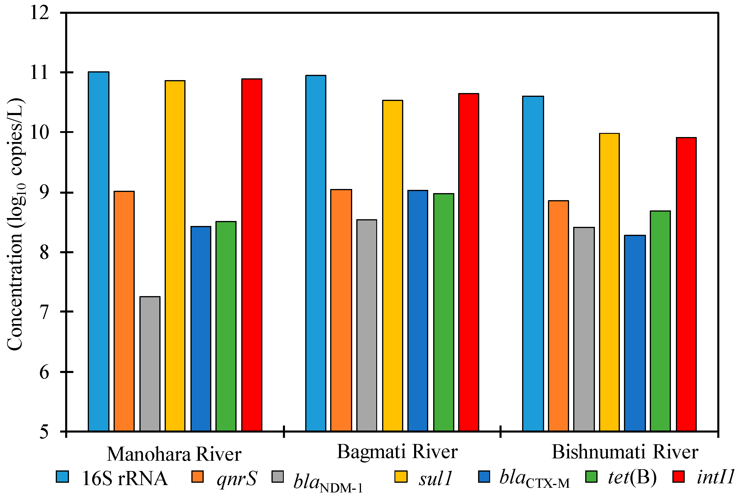
3.1. ARGs in River Water
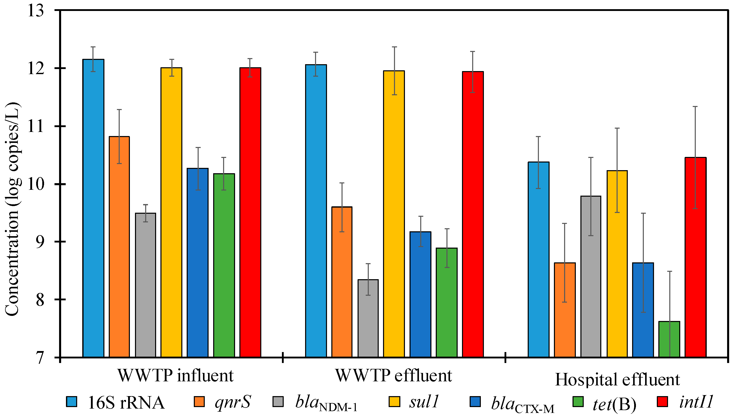
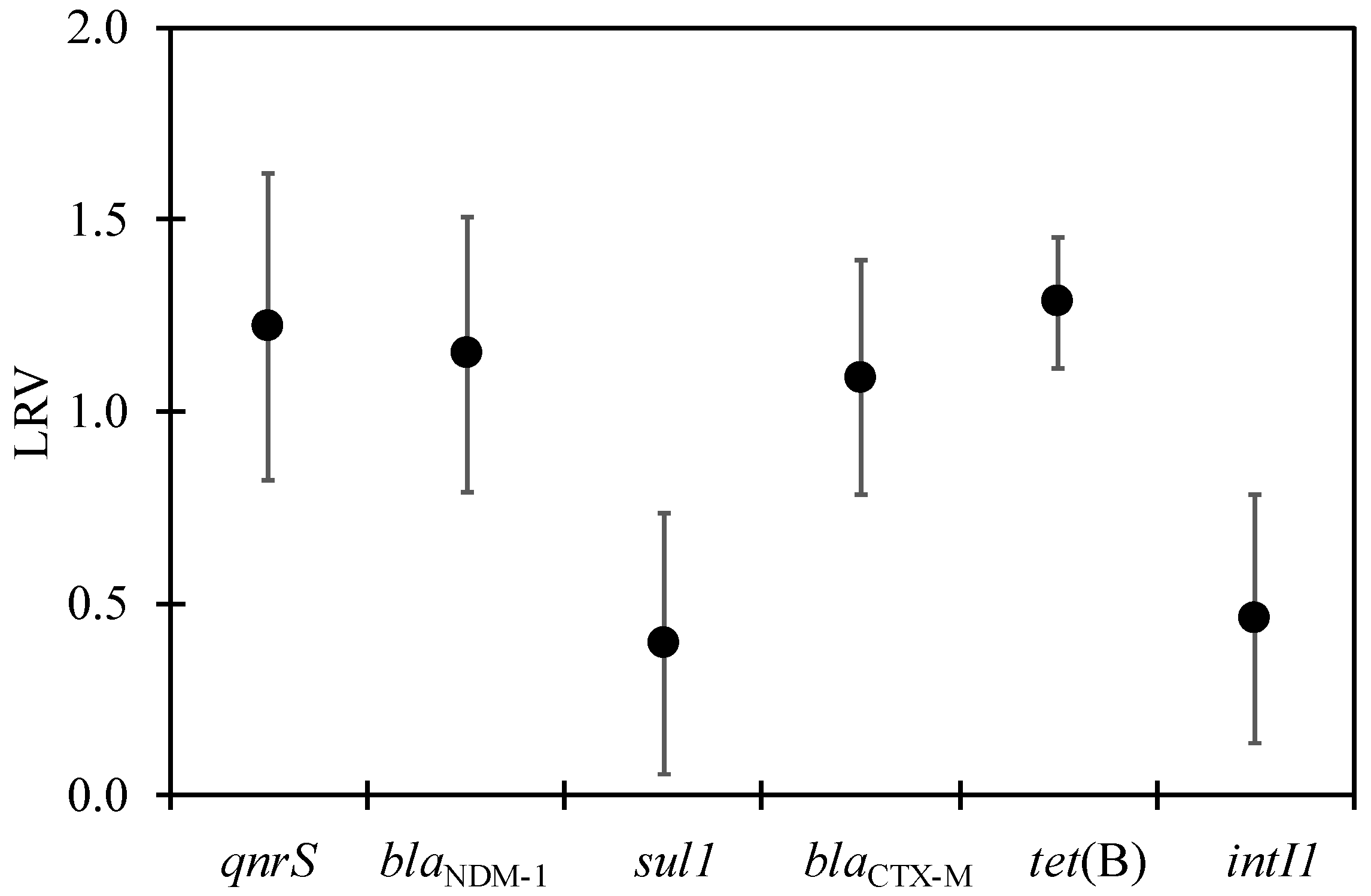
All of the marker genes tested in this study were detected in all river water samples. As illustrated in Figure 1, the concentrations of 16S rRNA, intI1, and ARGs ranged from 10.6–11, 9.9–10.9, and 7.3–10.9 log10 copies/L, respectively. High concentrations of 16S rRNA, intI1, and other ARGs have been reported in urban river sites of the Kathmandu Valley throughout the year, showing rivers in the valley are polluted with ARGs and ARB [10]. Therefore, only three river water samples were assessed to further verify those findings and our results show no improvement in ARG pollution control in the past three years. All the river water samples were collected from dense settlements in different periods and processed with different kits, making comparisons between the three rivers difficult. However, the relative abundance (a ratio of the concentration of ARGs or intI1 to that of 16S rRNA), as shown in Figure 2, was similar in all urban rivers. sul1 showed the highest relative abundance (p < 0.05), whereas the lowest was observed for blaNDM-1. sul1 encode resistance to sulfonamides, which are among the oldest antibiotics introduced and are recognized as poor or non-biodegradable compounds in the environment [32]. To date, no studies on antibiotic residues in river water and wastewater have been carried out in Nepal. However, the probable presence of sulfonamides in the environment to continuously exert selection pressure can be one of the reasons why sul1 occurred at a significantly higher concentration than other ARGs (p < 0.05).
Urban rivers in the Kathmandu Valley are polluted and not used for drinking. As a cheap source of nutrients, polluted river water is extensively used for irrigation [33]. Since bacterial DNA was extracted by filtering through a 0.22-µm pore size membrane, the detected ARGs were likely to be harbored by potentially pathogenic bacteria, and characterization of the bacterial diversity in the river water of the Kathmandu Valley has shown the presence of 111 potentially pathogenic bacteria [34]. Irrigation water is a pathway for contamination of fresh produce, and the consumption of such produce can lead to outbreaks of antibiotic-resistant bacterial pathogens in the Kathmandu Valley. In China, a study applying high-throughput qPCR to screen for ARGs/MGEs in surface water and groundwater bodies reported the sharing of 69.4% of the total detected ARGs/MGEs between river water and groundwater bodies. This finding provides evidence of surface water infiltration into groundwater [35]. However, currently, no studies that investigated the pollution profile of ARGs in groundwater in the Kathmandu Valley exist. Moreover, due to the acute shortage of drinking water, groundwater is extensively used for household activities. ARB and ARGs in the drinking water may colonize in the gut microbiome or even cause life-threatening infections, and future research should, therefore, focus on ARGs and the factors associated with the increase/decrease in concentration levels in the groundwater of the Kathmandu Valley.

3.2. ARGs in WWTP

Similar to river water samples, sul1 was the most abundant ARG, followed by qnrS, blaCTX-M, tet(B), and blaNDM-1 in both influent and effluent samples (Figure 3). In the influent of the WWTP, the concentrations of ARGs varied from 9.2 to 12.2 log10 copies/L. Sewage and drinking water pipelines are aligned very close to one another in the Kathmandu Valley. Some of the main drinking water pipes are over a century old and prone to leakage. This increases the likelihood of mixing sewage with drinking water and further contamination of drinking water with ARGs, ARB, and other pathogens. Antibiotic resistance in WWTP has been shown to mirror the pattern of antibiotic resistance in clinics [36] and our results provide further evidence of the emergence of highly resistant bacterial pathogens in the Kathmandu Valley. Figure 4 shows the LRV of ARGs and intI1 at the WWTP. ARGs are removed in biological treatment processes by the mechanism of sorption and biodegradation [37]. The highest and lowest LRVs were observed for tet(B) (1.3 ± 0.2) and sul1 (0.4 ± 0.3), respectively. Removal of ARGs is known to be affected by the treatment system used, and different ARGs displayed different reduction rates [38]. Biological treatment with oxidation ditch is among the most economical means of removing organic matter from wastewater, and the same technology is applied in Nepal. One of the concerns regarding treatment using an oxidation ditch is the long retention time, which can support interactions between bacteria and ARGs and increase exposure to antibiotic residues, thereby promoting antibiotic resistance. This interaction is proposed to be one of the reasons for the increase in the concentration of intI1 and sul1 in some effluent sample sets (Figure 3).
Except for sul1 and intI1, the relative abundance of all tested marker genes was significantly reduced in effluents (Figure 2, p < 0.05). This result is consistent with previous studies, which showed sul1 and intI1 persisted in water environments; thus, sul1 and intI1 could be used as an indicator of ARGs [7,9,39]. These marker genes are located on mobile genetic elements that can readily transfer among diverse bacteria and have disseminated throughout the bacterial community in wastewater [40]. Quick regeneration of bacteria and the possibility of horizontal transfer of genes between bacteria explain why sul1 and intI1 are persistent during wastewater treatment. Despite the reduction, about 1012 gene copies/L of individual ARGs were released into the river. Although the release of bacteria-harboring ARGs included in our study is a public health concern, high concentrations of blaNDM-1 are of special relevance. Bacteria belonging to the family of Enterobacteriaceae are associated with nosocomial and community-acquired infections, and such bacteria-harboring blaNDM-1 are resistant to all β-lactam antimicrobials except monobactam [41]. Moreover, bacteria-harboring the blaNDM-1 gene usually harbors other resistant genes [42]; thus, conferring resistance to almost all antibiotics, which seriously limits therapeutic options.
The concentration of E. coli in effluents of WWTPs ranged from 6.2 to 8.1 log10 MPN/100 mL. In this study, we did not observe the antibiotic resistance of bacterial isolates from WWTPs, but in neighboring countries of India and Bangladesh [43,44,45], E. coli, and bacteria belonging to the group TC have been shown to harbor blaNDM-1, suggesting a likelihood of certain percentage of TC and E. coli to be carbapenem-resistant and blaNDM-1 positive in our samples. TC and E. coli belong to the family Enterobacteriaceae, and carbapenem-resistant Enterobacteriaceae have been placed in the critical category of the World Health Organization’s list for an urgent need for new antibiotics due to an increase in resistance towards available antibiotics. In addition, blaNDM-1 has been found to transfer at 30 °C more frequently than at other temperatures among the Enterobacteriaceae family via conjugation [46]. Since the temperature of 30 °C is common during summer, environmental transfer of blaNDM-1 among pathogens can occur.

3.3. ARGs in the Hospital Wastewater

The occurrence of ARGs in the hospital wastewater is shown in Figure 3. In some sample sets, sul1, blaNDM-1, and intI1 were found to be in higher concentrations than 16Sr RNA marker genes. Similar results have been reported previously [45,47], reflecting high concentrations of bacteria harboring the ARGs. The concentration of blaNDM-1in the hospital wastewater of New Delhi, India, has been reported to be as high as 16 log10 copies/L [43], which is six logs higher than our findings in Nepal. Despite the widespread dissemination of blaNDM-1 in the Indian subcontinent [42], the difference in concentrations of blaNDM-1 in the hospital wastewater of these two countries could be explained by the larger size of hospitals surveyed in India, where overcrowding by up to three times the designated capacity was also noted. In comparison to WWTP samples, the relative abundance of only blaNDM-1 was significantly high (p < 0.05) in hospital effluent samples, as shown in Figure 2. The use of carbapenem drugs is more common in hospitals, and bacteria-harboring genes encoding resistance to carbapenems in rivers have been shown to be of hospital origin [48]. In Dhaka, Bangladesh, blaNDM-1 was detected in 71% of wastewater samples from hospital adjacent areas in comparison to 12.1% of wastewater samples from community areas [44]. The variation in the ARG profile in hospital wastewater suggests hospital effluents as a diffusion reservoir of blaNDM-1-producing bacteria outside clinical settings and deserves special attention [49]. In comparison to hospital wastewater, ARGs have been extensively studied in WWTP. Hospital wastewater can be often overlooked since it is extensively diluted in municipal sewage and contributes less than one percent of total wastewater in WWTP [50]. A limitation of our study is that we only screened for a limited number of ARGs, and thus, we may have missed other important ARGs. Subsequent studies based on a metagenomics approach that will provide clear information of the overall ARG profile to picture the antibiotic resistance landscape of the study area is suggested.
Among the two hospitals with an onsite wastewater treatment facility, one used biological treatment, and another used ultrafiltration with an activated carbon filter as secondary treatment after primary sedimentation. Both treatment methods reduced similar levels of ARGs and intI1 with LRVs ranging from 0.3 to 1.1. However, we were unable to draw concrete conclusions because the sampling was done only once. Regardless, our results indicate hospital wastewater as an important reservoir of ARGs. To combat antibiotic resistance, the prudent use of antibiotics should be adopted, and installing effective wastewater treatment at hospitals is needed to eliminate ARGs at the point source. Treatment strategies that include tertiary treatment steps to effectively remove ARGs are recommended.

3.4. Correlation between Individual ARGs, intI1, and Fecal Indicator Bacteria

As summarized in Table 3 and Figures S2–S4 in the Supplemental Material, Pearson’s rank correlation test was performed on all data sets. Except for blaNDM-1, significant positive correlations were found between intI1 and all individual ARGs (0.71–0.96, p < 0.05), which indicate the probable implications of intI1 in the transfer of ARGs. In contrast, TC concentrations did not correlate with concentrations of ARGs (p > 0.05). Fecal pollution is often associated with ARG contamination, exemplifying the impact of sewage, but high-throughput sequencing to determine the bacterial community at the genus level revealed pathogenic or opportunistic pathogens of genera Acinetobacter, Aeromonas, Myroides, Proteus, Pseudomonas, and Streptococcus to be highly prevalent with ARGs in the hospital wastewater, along with Escherichia [47,51]. Especially the species Acinetobacter baumannii is a frequent cause of infection and is reported to be resistant to many antibiotics used in hospitals [52,53]. The possibility of a higher percentage of bacteria belonging to different genera harboring these ARGs can explain the non-correlation between TC and ARGs.

4. Conclusions

Antibiotic resistance is a global public health problem. However, regional surveillance is the first step to prevent the spread of antibiotic resistance as variation in the burden of antibiotic resistance and circulating ARGs can be observed. Our results demonstrated river water to be contaminated with the studied ARGs. Up to 1012 copies/L of individual ARGs were discharged in WWTP and hospital effluents, highlighting the role of WWTPs and hospitals as important point sources of ARGs in the Kathmandu Valley. Positive correlations between intI1 and most ARGs also indicate the probable implications of intI1 in the transfer of ARGs. To conclude, more centralized WWTPs, tertiary treatment for WWTP and hospitals with an onsite treatment plant, and effective treatment strategies for hospitals without any treatment is recommended to combat the growing threat of antibiotic resistance via wastewater management.

Supplementary Materials

The following are available online at https://www.mdpi.com/article/10.3390/w13192733/s1, Figure S1: Schematic diagram of WWTP, Figure S2: Correlation of E. coli with ARGs, Figure S3: Correlation of TC with ARGs, Figure S4: Correlation of intI1 with ARGs.

Author Contributions

Conceptualization, O.T. and E.H.; investigation, O.T., B.M., S.T., N.S., S.R., T.F., K.S. and E.H.; methodology, O.T., B.M. and E.H.; validation, T.F., K.S., J.B.S. and E.H.; formal analysis, O.T.; resources, J.B.S. and E.H.; writing—original draft preparation, O.T.; writing—review and editing, T.F., K.S., J.B.S. and E.H.; supervision, E.H.; funding acquisition, E.H. All authors have read and agreed to the published version of the manuscript.

Funding

This study was supported by the Science and Technology Research Partnership for Sustainable Development (SATREPS) program of Japan International Cooperation Agency (JICA) and Japan Science and Technology Agency (JST), entitled “Hydro-microbiological Approach for Water Security in Kathmandu Valley, Nepal” and the Japan Society for the Promotion of Science (JSPS) through Fund for the Promotion of Joint International Research (Fostering Joint International Research (B)) (grant number JP18KK0297).

Institutional Review Board Statement

Not applicable.

Informed Consent Statement

Not applicable.

Acknowledgments

The authors thank Rajani Ghaju Shrestha, Takahiro Doyama, Taizo Mochizuki, and Masaaki Ito (University of Yamanashi, Japan) for their support during the water sampling and laboratory analysis. The authors also thank the workers of WWTP and hospitals for their kind coopereation in sample collection.

Conflicts of Interest

The authors declare no conflict of interest. The funders had no role in the design of the study; in the collection, analyses, or interpretation of data; in the writing of the manuscript, or in the decision to publish the results.

References

  1. Klein, E.Y.; Van Boeckel, T.P.; Martinez, E.M.; Pant, S.; Gandra, S.; Levin, S.A.; Goossens, H.; Laxminarayan, R. Global increase and geographic convergence in antibiotic consumption between 2000 and 2015. Proc. Natl. Acad. Sci. USA 2018, 115, E3463–E3470. [Google Scholar] [CrossRef] [PubMed] [Green Version]
  2. Toranzos, G.; Steyn, M.; Santiago-Rodriguez, T.; Sano, D. Editorial: Bacterial antibiotic resistance in the water environment. J. Water Health 2020, 18, 855–857. [Google Scholar] [CrossRef] [PubMed]
  3. Sarmah, A.K.; Meyer, M.T.; Boxall, A.B.A. A global perspective on the use, sales, exposure pathways, occurrence, fate and effects of veterinary antibiotics (VAs) in the environment. Chemosphere 2006, 65, 725–759. [Google Scholar] [CrossRef] [PubMed]
  4. Kümmerer, K.; Al-Ahmad, A.; Mersch-Sundermann, V. Biodegradability of some antibiotics, elimination of the genotoxicity and affection of wastewater bacteria in a simple test. Chemosphere 2000, 40, 701–710. [Google Scholar] [CrossRef]
  5. Kümmerer, K.; Henninger, A. Promoting resistance by the emission of antibiotics from hospitals and households into effluent. Clin. Microbiol. Infect. 2003, 9, 1203–1214. [Google Scholar] [CrossRef] [Green Version]
  6. Rizzo, L.; Manaia, C.; Merlin, C.; Schwartz, T.; Dagot, C.; Ploy, M.C.; Michael, I.; Fatta-Kassinos, D. Urban wastewater treatment plants as hotspots for antibiotic resistant bacteria and genes spread into the environment: A review. Sci. Total Environ. 2013, 447, 345–360. [Google Scholar] [CrossRef] [PubMed] [Green Version]
  7. Acharya, K.P.; Wilson, R.T. Antimicrobial Resistance in Nepal. Front. Med. 2019, 6, 105. [Google Scholar] [CrossRef] [Green Version]
  8. Dahal, R.H.; Chaudhary, D.K. Microbial Infections and Antimicrobial Resistance in Nepal: Current Trends and Recommendations. Open Microbiol. J. 2018, 12, 230–242. [Google Scholar] [CrossRef] [Green Version]
  9. Joshi, H.D.; Acharya, T.; Ayer, R.; Dhakal, P.; Karki, K.B.; Dhimal, M. Health Care Waste Management Practice in Health Care Institutions of Nepal. J. Nepal Health Res. Counc. 2017, 15, 7–11. [Google Scholar] [CrossRef] [Green Version]
  10. Thakali, O.; Tandukar, S.; Brooks, J.P.; Sherchan, S.P.; Sherchand, J.B.; Haramoto, E. The Occurrence of Antibiotic Resistance Genes in an Urban River in Nepal. Water 2020, 12, 450. [Google Scholar] [CrossRef] [Green Version]
  11. Hyoju, R.K.; Shrestha, R. Efficiency of General Hospitals in Kathmandu Valley. 2014. Available online: http://conference.ioe.edu.np/ioegc2014/papers/IOE-CONF-2014-39.pdf (accessed on 11 May 2021).
  12. Thakali, O.; Brooks, J.P.; Shahin, S.; Sherchan, S.P.; Haramoto, E. Removal of Antibiotic Resistance Genes at Two Conventional Wastewater Treatment Plants of Louisiana, USA. Water 2020, 12, 1729. [Google Scholar] [CrossRef]
  13. Rodriguez-Mozaz, S.; Chamorro, S.; Marti, E.; Huerta, B.; Gros, M.; Sànchez-Melsió, A.; Borrego, C.M.; Barceló, D.; Balcázar, J.L. Occurrence of antibiotics and antibiotic resistance genes in hospital and urban wastewaters and their impact on the receiving river. Water Res. 2015, 69, 234–242. [Google Scholar] [CrossRef] [PubMed]
  14. Chopra, I.; Roberts, M. Tetracycline Antibiotics: Mode of Action, Applications, Molecular Biology, and Epidemiology of Bacterial Resistance. Microbiol. Mol. Biol. Rev. 2001, 65, 232–260. [Google Scholar] [CrossRef] [PubMed] [Green Version]
  15. Poirel, L.; Cattoir, V.; Nordmann, P. Plasmid-Mediated Quinolone Resistance; Interactions between Human, Animal, and Environmental Ecologies. Front. Microbiol. 2012, 3, 24. [Google Scholar] [CrossRef] [PubMed] [Green Version]
  16. Sherchan, J.B.; Hayakawa, K.; Miyoshi-Akiyama, T.; Ohmagari, N.; Kirikae, T.; Nagamatsu, M.; Tojo, M.; Ohara, H.; Sherchand, J.B.; Tandukar, S. Clinical epidemiology and molecular analysis of extended-spectrum-β-lactamase-producing Escherichia coli in Nepal: Characteristics of sequence types 131 and 648. Antimicrob. Agents Chemother. 2015, 59, 3424–3432. [Google Scholar] [CrossRef] [PubMed] [Green Version]
  17. Pokhrel, R.; Thapa, B.; Kafle, R.; Shah, P.; Tribuddharat, C. Co-existence of beta-lactamases in clinical isolates of Escherichia coli from Kathmandu, Nepal. BMC Res. Notes 2014, 7, 694. [Google Scholar] [CrossRef] [PubMed] [Green Version]
  18. Yong, D.; Toleman, M.A.; Giske, C.G.; Cho, H.S.; Sundman, K.; Lee, K.; Walsh, T.R. Characterization of a new metallo-β-lactamase gene, blaNDM-1, and a novel erythromycin esterase gene carried on a unique genetic structure in Klebsiella pneumoniae sequence type 14 from India. Antimicrob. Agents Chemother. 2009, 53, 5046–5054. [Google Scholar] [CrossRef] [Green Version]
  19. Shrestha, B.; Tada, T.; Shimada, K.; Shrestha, S.; Ohara, H.; Pokhrel, B.M.; Sherchand, J.B.; Kirikae, T. Emergence of Various NDM-Type-Metallo-β-Lactamase-Producing Escherichia coli Clinical Isolates in Nepal. Antimicrob. Agents Chemother. 2017, 61, 1–6. [Google Scholar] [CrossRef] [Green Version]
  20. Rowe-Magnus, D.A.; Mazel, D. The role of integrons in antibiotic resistance gene capture. Int. J. Med. Microbiol. 2002, 292, 115–125. [Google Scholar] [CrossRef]
  21. Zhang, K.; Xin, R.; Zhao, Z.; Ma, Y.; Zhang, Y.; Niu, Z. Antibiotic Resistance Genes in drinking water of China: Occurrence, distribution and influencing factors. Ecotoxicol. Environ. Saf. 2020, 188, 109837. [Google Scholar] [CrossRef]
  22. Narciso-da-Rocha, C.; Varela, A.R.; Schwartz, T.; Nunes, O.C.; Manaia, C.M. blaTEM and vanA as indicator genes of antibiotic resistance contamination in a hospital–urban wastewater treatment plant system. J. Glob. Antimicrob. Resist. 2014, 2, 309–315. [Google Scholar] [CrossRef] [PubMed] [Green Version]
  23. World Health Organization. WHO Model List of Essential Medicines, 21st List. 2019. Available online: https://www.who.int/publications/i/item/WHOMVPEMPIAU2019.06 (accessed on 5 May 2021).
  24. Malla, B.; Ghaju Shrestha, R.; Tandukar, S.; Bhandari, D.; Thakali, O.; Sherchand, J.B.; Haramoto, E. Detection of Pathogenic Viruses, Pathogen Indicators, and Fecal-Source Markers within Tanker Water and Their Sources in the Kathmandu Valley, Nepal. Pathogens 2019, 8, 81. [Google Scholar] [CrossRef] [PubMed] [Green Version]
  25. Suzuki, M.T.; Taylor, L.T.; DeLong, E.F. Quantitative Analysis of Small-Subunit rRNA Genes in Mixed Microbial Populations via 5′-Nuclease Assays. Appl. Environ. Microbiol. 2000, 66, 4605–4614. [Google Scholar] [CrossRef] [PubMed] [Green Version]
  26. Luo, Y.; Mao, D.; Rysz, M.; Zhou, Q.; Zhang, H.; Xu, L.; Alvarez, P.J.J. Trends in Antibiotic Resistance Genes Occurrence in the Haihe River, China. Environ. Sci. Technol. 2010, 44, 7220–7225. [Google Scholar] [CrossRef]
  27. Rutgersson, C.; Fick, J.; Marathe, N.; Kristiansson, E.; Janzon, A.; Angelin, M.; Johansson, A.; Shouche, Y.; Flach, C.-F.; Larsson, D.G.J. Fluoroquinolones and qnr Genes in Sediment, Water, Soil, and Human Fecal Flora in an Environment Polluted by Manufacturing Discharges. Environ. Sci. Technol. 2014, 48, 7825–7832. [Google Scholar] [CrossRef] [PubMed]
  28. Colomer-Lluch, M.; Jofre, J.; Muniesa, M. Antibiotic resistance genes in the bacteriophage DNA fraction of environmental samples. PLoS ONE 2011, 6, e17549. [Google Scholar] [CrossRef] [Green Version]
  29. Naas, T.; Ergani, A.; Carrër, A.; Nordmann, P. Real-Time PCR for Detection of NDM-1 Carbapenemase Genes from Spiked Stool Samples. Antimicrob. Antimicrob. Agents Chemother. 2011, 55, 4038–4043. [Google Scholar] [CrossRef] [Green Version]
  30. Czekalski, N.; Berthold, T.; Caucci, S.; Egli, A.; Bürgmann, H. Increased levels of multiresistant bacteria and resistance genes after wastewater treatment and their dissemination into Lake Geneva, Switzerland. Front. Microbiol. 2012, 3, 1–18. [Google Scholar] [CrossRef] [Green Version]
  31. Barraud, O.; Baclet, M.C.; Denis, F.; Ploy, M.C. Quantitative multiplex real-time PCR for detecting class 1, 2 and 3 integrons. J. Antimicrob. Chemother. 2010, 65, 1642–1645. [Google Scholar] [CrossRef] [Green Version]
  32. Baran, W.; Adamek, E.; Ziemiańska, J.; Sobczak, A. Effects of the presence of sulfonamides in the environment and their influence on human health. J. Hazard. Mater. 2011, 196, 1–15. [Google Scholar] [CrossRef]
  33. Shrestha, S.; Shrestha, S.; Shindo, J.; Sherchand, J.B.; Haramoto, E. Virological quality of irrigation water sources and pepper mild mottle virus and tobacco mosaic virus as index of pathogenic virus contamination level. Food Environ. Virol. 2018, 10, 107–120. [Google Scholar] [CrossRef] [PubMed]
  34. Shrestha, R.G.; Tandukar, S.; Bhandari, D.; Sherchan, S.P.; Tanaka, Y.; Sherchand, J.B.; Haramoto, E. Prevalence of Arcobacter and Other Pathogenic Bacteria in River Water in Nepal. Water 2019, 11, 1416. [Google Scholar] [CrossRef] [Green Version]
  35. Wu, D.-L.; Zhang, M.; He, L.-X.; Zou, H.-Y.; Liu, Y.-S.; Li, B.-B.; Yang, Y.-Y.; Liu, C.; He, L.-Y.; Ying, G.-G. Contamination profile of antibiotic resistance genes in ground water in comparison with surface water. Sci. Total Environ. 2020, 715, 136975. [Google Scholar] [CrossRef] [PubMed]
  36. Parnanen, K.M.M.; Narciso-da-Rocha, C.; Kneis, D.; Berendonk, T.U.; Cacace, D.; Do, T.T.; Elpers, C.; Fatta-Kassinos, D.; Henriques, I.; Jaeger, T.; et al. Antibiotic resistance in European wastewater treatment plants mirrors the pattern of clinical antibiotic resistance prevalence. Sci. Adv. 2019, 5, eaau9124. [Google Scholar] [CrossRef] [Green Version]
  37. Bairán, G.; Rebollar-Pérez, G.; Chávez-Bravo, E.; Torres, E. Treatment processes for microbial resistance mitigation: The technological contribution to tackle the problem of antibiotic resistance. Int. J. Environ. Res. Public Health 2020, 17, 8866. [Google Scholar] [CrossRef]
  38. Amarasiri, M.; Sano, D.; Suzuki, S. Understanding human health risks caused by antibiotic resistant bacteria (ARB) and antibiotic resistance genes (ARG) in water environments: Current knowledge and questions to be answered. Crit. Rev. Environ. Sci. Technol. 2019, 50, 2016–2059. [Google Scholar] [CrossRef]
  39. Su, H.; Liu, S.; Hu, X.; Xu, X.; Xu, W.; Xu, Y.; Li, Z.; Wen, G.; Liu, Y.; Cao, Y. Occurrence and temporal variation of antibiotic resistance genes (ARGs) in shrimp aquaculture: ARGs dissemination from farming source to reared organisms. Sci. Total Environ. 2017, 607–608, 357–366. [Google Scholar] [CrossRef]
  40. Gillings, M.R.; Gaze, W.H.; Pruden, A.; Smalla, K.; Tiedje, J.M.; Zhu, Y.-G. Using the class 1 integron-integrase gene as a proxy for anthropogenic pollution. ISME J. 2015, 9, 1269–1279. [Google Scholar] [CrossRef]
  41. Sherchan, J.B.; Tada, T.; Shrestha, S.; Uchida, H.; Hishinuma, T.; Morioka, S.; Shahi, R.; Twi, R.T.; Kirikae, T.; Sherchand, J.B. Emergence of clinical isolates of highly carbapenem-resistant Klebsiella pneumonia co-harboring blaNDM-5 and blaOXA-181 or -232 in Nepal. Int. J. Infect. Dis. 2020, 92, 247–252. [Google Scholar] [CrossRef] [Green Version]
  42. Nordmann, P.; Poirel, L.; Walsh, T.R.; Livermore, D.M. The emerging NDM carbapenemases. Trends Microbiol. 2011, 19, 588–595. [Google Scholar] [CrossRef]
  43. Lamba, M.; Graham, D.W.; Ahammad, S.Z. Hospital Wastewater Releases of Carbapenem-Resistance Pathogens and Genes in Urban India. Environ. Sci. Technol. 2017, 51, 13906–13912. [Google Scholar] [CrossRef] [PubMed] [Green Version]
  44. Islam, M.A.; Islam, M.; Hasan, R.; Hossain, M.I.; Nabi, A.; Rahman, M.; Goessens, W.H.F.; Endtz, H.P.; Boehm, A.B.; Faruque, S.M. Environmental Spread of New Delhi Metallo-β-Lactamase-1-Producing Multidrug-Resistant Bacteria in Dhaka, Bangladesh. Appl. Environ. Microbiol. 2017, 83, 1–11. [Google Scholar] [CrossRef] [PubMed] [Green Version]
  45. Lamba, M.; Gupta, S.; Shukla, R.; Graham, D.W.; Sreekrishnan, T.R.; Ahammad, S.Z. Carbapenem resistance exposures via wastewaters across New Delhi. Environ. Int. 2018, 119, 302–308. [Google Scholar] [CrossRef]
  46. Walsh, T.R.; Weeks, J.; Livermore, D.M.; Toleman, M.A. Dissemination of NDM-1 positive bacteria in the New Delhi environment and its implications for human health: An environmental point prevalence study. Lancet Infect. Dis. 2011, 11, 355–362. [Google Scholar] [CrossRef]
  47. Wang, Q.; Wang, P.; Yang, Q. Occurrence and diversity of antibiotic resistance in untreated hospital wastewater. Sci. Total Environ. 2018, 621, 990–999. [Google Scholar] [CrossRef] [PubMed]
  48. Cerdeira, L.; Fernandes, M.R.; Ienne, S.; Souza, T.A.; Garcia, D.d.O.; Lincopan, N. Draft genome sequence of an environmental multidrug-resistant Klebsiella pneumoniae ST340/CC258 harbouring blaCTX-M-15 and blaKPC-2 genes. J. Glob. Antimicrob. Resist. 2017, 8, 108–109. [Google Scholar] [CrossRef]
  49. Zhang, C.; Qiu, S.; Wang, Y.; Qi, L.; Hao, R.; Liu, X.; Shi, Y.; Hu, X.; An, D.; Li, Z.; et al. Higher Isolation of NDM-1 Producing Acinetobacter baumannii from the Sewage of the Hospitals in Beijing. PLoS ONE 2013, 8, 6–11. [Google Scholar] [CrossRef] [PubMed]
  50. Karkman, A.; Do, T.T.; Walsh, F.; Virta, M.P.J. Antibiotic-Resistance Genes in Waste Water. Trends Microbiol. 2018, 26, 220–228. [Google Scholar] [CrossRef] [Green Version]
  51. Marathe, N.P.; Berglund, F.; Razavi, M.; Pal, C.; Dröge, J.; Samant, S.; Kristiansson, E.; Joakim Larsson, D.G. Sewage effluent from an Indian hospital harbors novel carbapenemases and integron-borne antibiotic resistance genes. Microbiome 2019, 7, 1–11. [Google Scholar] [CrossRef] [Green Version]
  52. Seruga Music, M.; Hrenovic, J.; Goic-Barisic, I.; Hunjak, B.; Skoric, D.; Ivankovic, T. Emission of extensively-drug-resistant Acinetobacter baumannii from hospital settings to the natural environment. J. Hosp. Infect. 2017, 96, 323–327. [Google Scholar] [CrossRef]
  53. Shrestha, S.; Tada, T.; Miyoshi-Akiyama, T.; Ohara, H.; Shimada, K.; Satou, K.; Teruya, K.; Nakano, K.; Shiroma, A.; Sherchand, J.B.; et al. Molecular epidemiology of multidrug-resistant Acinetobacter baumannii isolates in a university hospital in Nepal reveals the emergence of a novel epidemic clonal lineage. Int. J. Antimicrob. Agents 2015, 46, 526–531. [Google Scholar] [CrossRef] [PubMed]
Figure 1. Absolute abundance of ARGs in river water.
Water 13 02733 g001
Figure 2. Heat map showing the relative abundance of ARGs and intI1.
Water 13 02733 g002
Figure 3. The abundance of 16S rRNA, ARGs, and intI1 in WWTPs and hospitals.
Water 13 02733 g003
Figure 4. LRVs of ARGs and intI1 in WWTPs. n = 6, except for sul1 (n = 4) and intI1 (n = 3).
Water 13 02733 g004
Table 1. Marker genes and the class of drugs to which they encode resistance.
GeneClass of Antibiotics/Importance
16SrRNAProxy for total bacterial load
tet(B)Tetracycline b
qnrSFluoroquinolones a
blaCTX-MCephalosporins a,b, Penicillin, Monobactams b
blaNDM-1Carbapenems b, Cephalosporins a,b, Penicillin
sul1Sulfonamides
intI1Proxy for the spread of antibiotic-resistance genes [22]
a Includes watch group antibiotics; b includes reserve group antibiotics (“last resort options”) [23].
Table 2. Nucleotide sequences of primers and probes used in the study.
AssayFunctionSequence (5′–3′)Size (bp)Reference
16S rRNAForward primerCGGTGAATACGTTCYCGG142[25]
Reverse primerGGWTACCTTGTTACGACTT
TaqMan probeFAM-CTTGTACAC/ZEN/ACCGCCCGTC-IBFQ
tet(B)Forward primerCGAAGTAGGGGTTGAGACGC192[26]
Reverse primerAGACCAAGACCCGCTAATGAA
qnrSForward primerGATATCGAAGGCTGCCACTT115[27]
Reverse primerCACGGAACTCTATACCGTAGCA
blaCTX-MForward primerACCAACGATATCGCGGTGAT101[28]
Reverse primerACATCGCGACGGCTTTCT
TaqMan probeFAM-TCGTGCGCCGCTG-BHQ1
blaNDM-1Forward primerATTAGCCGCTGCATTGAT154[29]
Reverse primerCATGTCGAGATAGGAAGTG
TaqMan probeFAM-CTGCCAGACATTCGGTGC-TAMRA
sul1Forward primerCCGTTGGCCTTCCTGTAAAG67[30]
Reverse primerTTGCCGATCGCGTGAAGT
TaqMan probeFAM-CAGCGAGCCTTGCGGCGG-BHQ1
intI1Forward primerGCCTTGATGTTACCCGAGAG196[31]
Reverse primerGATCGGTCGAATGCGTGT
TaqMan probeFAM-ATTCCTGGCCGTGGTTCTGGGTTTT- BHQ1
Y denotes C or T; FAM, 6-carboxyfluorescein; ZEN, ZEN internal quencher; IBFQ, Iowa Black fluorescent quencher; BHQ1, black hole quencher 1; TAMRA, 5-carboxytetramethylrhodamine.
Table 3. Pearson’s correlation coefficient between intI1, ARGs, and fecal indicator bacteria.
Indicatorsul1tet(B)qnrSblaCTX-MblaNDM-1
intI10.96 *0.73 *0.71 *0.80 *−0.04
TC−0.160.160.220.180.33
E. coli0.58 *0.74 *0.61 *0.60 *0.06
* p < 0.05.
Publisher’s Note: MDPI stays neutral with regard to jurisdictional claims in published maps and institutional affiliations.

