Article

Quantification of Polycyclic Aromatic Hydrocarbons (PAHs) in Various Fruit Types: A Comparative Analysis

1
Department of Agricultural, Environmental and Food Sciences (DiAAA), University of Molise, Via De Sanctis, 86100 Campobasso, Italy
2
Department of Biosciences and Territory, University of Molise, c/da Fonte Lappone, 86090 Isernia, Italy
3
Institute of Atmospheric Pollution Research, Division of Rome, c/o Ministry of Environment and Energy Security, 00147 Rome, Italy
*
Author to whom correspondence should be addressed.
Atmosphere 2024, 15(9), 1028; https://doi.org/10.3390/atmos15091028
Submission received: 28 June 2024 / Revised: 13 August 2024 / Accepted: 22 August 2024 / Published: 25 August 2024
(This article belongs to the Section Air Quality)

Abstract

:
The exposure of humans to polycyclic aromatic hydrocarbons (PAHs) through fruits is a scarcely investigated topic. The atmospheric deposition of PAHs could contribute to such an issue. The present paper would like to propose an easy, fast, and routinary analytical method to extract and quantify PAHs in apples, pears, and grapes. Dispersive liquid–liquid microextraction allowed us to recover PAHs ranging between 68.0 ± 1.2 and 96.2 ± 0.8% from fruit. Gas chromatography equipped with flame ionization detector analysis showed satisfactory analytical parameters, with details like R2 > 0.9912 in a concentration range of 0.5–500 µg mL−1, with a variability ranging within 0.7–2.3%. Rural fruit samples were found to be more contaminated by PAHs compared to urban samples, likely due to the use of non-green fuels in rural areas considered in this study. Further in-depth research on this topic is strongly recommended due to the relevance of fruits in the Mediterranean diet.

1. Introduction

Environmental and food matrix contamination are strongly interconnected. Atmospheric polycyclic aromatic hydrocarbons (PAHs) are notoriously carcinogenic compounds. They could lead to the contamination of exposed food matrices through atmospheric deposition [1,2]. Consequently, PAHs can be introduced into the human body through the food chain. Studies have indicated that human exposure to PAHs predominantly occurs via the consumption of contaminated food [3]. Smoking processes applied to foods (such as smoked meats, smoked fish, and related products) have been identified as the primary sources of human exposure to PAHs. Consequently, the European Union (previously known as the European Community) has implemented Regulation No. 1881/2006, which sets maximum allowable levels of PAHs in these products (i.e., 5 and 2 µg kg−1, respectively). Similarly, processed foods (i.e., oils and fats) intended for human consumption, processed cereal-based foods for infants and young children, processed infant foods, infant formula, and seafood, including crustaceans, cephalopods (excluding crabs and lobsters), and mollusks, have also been regulated for maximum tolerable levels by the same regulation (i.e., 2, 1, 1, and 5 µg kg−1, respectively) [4]. Some scientific studies identify dried and/or smoked foods, as well as foods cooked at high temperatures (i.e., grilled, fried, and roasted), as the primary sources of dietary PAH contamination [5]. However, the release of PAHs from various anthropogenic activities contributes to their dissemination across environmental compartments, including air, water, and soil, leading to the contamination of adjacent plant and animal species [3]. Environmental contamination by PAHs could lead to the contamination of cultivated foods, such as fruit (and vegetables), ruling out the possibility that only processed foods are a source of PAH exposure for humans [6]. The PAH contamination of fruits has been proposed, but the mechanisms underlying this contamination remain unclear. Specifically, if PAHs are present in the air, they may primarily contaminate fruits through surface deposition.
Conversely, if contamination occurs through the uptake of PAHs from soil, PAHs would be mainly present in the internal tissues [7]. The waxy surface of fruit can concentrate low-molecular-weight PAHs through surface adsorption; high-molecular-weight PAHs bound to particles can contaminate the surface due to atmospheric deposition. Indeed, it was reported that the peels are generally more contaminated than cores; furthermore, the removal of peels reduces the PAH ingestion by humans [8]. For instance, studies seemed to prove the occurrence of PAHs on fruits. In this regard, Rojo Camargo and Teledo (2003) [9] reported average PAH contamination levels of 4.05 µg kg−1 in apples and 3.87 µg kg−1 in pears, identifying benzo(a)anthracene as the most representative compound. The significant impact of environmental contamination was suggested; emissions of PAHs from automobile traffic and industrial activities have been shown to affect the levels and profiles of PAHs in nearby grown vegetables and fruits. As recently reported, the contamination of vegetables and fruits is mainly due to the deposition of PAHs, therefore, the health status of the area where fruit and vegetables are grown becomes the principal variable. Indeed, the impact of the type of cultivation area on PAH levels in fruits and vegetables has been suggested. Specifically, lettuce grown in an industrialized area showed a PAH contamination higher than that grown in rural areas (i.e., fluoranthene, 6.19 ± 1.54 µg kg−1 vs. 2.29 ± 0.28 µg kg−1; pyrene, 2.59 ± 0.72 µg kg−1 vs. 0.73 ± 0.19 µg kg−1; benzo(a)anthracene, 0.35 ± 0.02 µg kg−1 vs. 0.23 ± 0.03 µg kg−1; and benzo(a)pyrene, 0.12 ± 0.01 µg kg−1 vs. 0.07 ± 0.01 µg kg−1) [9]. Their contamination by PAHs is relevant as fruits are frequently used in the human diet. Even though fruits seem to be lowly contaminated by PAHs, the high frequency of their ingestion by humans makes them a remarkable source of PAHs [10]. In 2023, a concentration ranging within 10.32 and 204.85 ng g−1 was found in several types of fruits (i.e., pear, grape, strawberries, orange, banana, apple, and mango), where the most abundant was phenanthrene, followed by anthracene and benzo[a]pyrene [11]. Similarly, it was recently reported that apple and banana samples collected in India exhibited PAH levels ranging from 0.34 to 2.46 ng g−1 and from 0.33 to 2.39 ng g−1, respectively, while watermelon samples showed levels ranging from 0.03 to 0.29 ng g−1 [12]. Furthermore, a study conducted in Iran has recently underlined the contamination by PAHs of fruits; the findings reported levels of PAHs in apples, banana, plums, grapes, peaches, and kiwi of 153.4, 190.1, 209.3, 252.4, 156.7, and 194.9 µg kg−1 [13].
The present paper would like to investigate the levels and the contribution of PAHs in various fruit types, specifically, details like apples, pears, and grapes by means of gas chromatography equipped with flame ionization detector (FID). To assess whether contamination occurs by atmospheric deposition or by absorption from the soil, a qualitative and quantitative analysis was carried out on both the peel and pulp. Before the chemical analyses, the entire methodology was tested on spiked apple samples. In particular, the author would like to underline the use of a really common detector such as FID for the analysis. The aim is to set up a methodology that can be routinely used worldwide. For this scope, the authors make the confirmation by means of GC-MS.

2. Materials and Methods

2.1. Materials

All standards of the eight PAHs investigated in the present study, namely, Fluoranthene (Fla), Fluorene (Fle), Benzo[a]pyrene (B[a]P), Benzo[B]fluoranthene (B[b]P), Phenanthrene (Ph), Pyrene (Py), Acenaphthene (Ace), and Acenaphthylene (Acy), were purchased from Sigma-Aldrich (Milan, Italy). The Internal Standard (IS) used for octacosane in a concentration of 80 µg mL−1 in acetone is obtained from Sigma Aldrich (Milan, Italy). The concentration of the standard solution of PAHs was 500 µg mL−1 in acetone.

2.2. Sample Collection

In the present study, apple (n = 20, 10 from rural and 10 from urban area), pear (n = 20, 10 from rural and 10 from urban area), and grape (n = 20, 10 from rural and 10 from urban area) samples were collected from areas with known varying levels of pollution. The samples were gathered from both an agricultural area and an urban area. For the development and validation of the analytical method, samples were purchased from a supermarket, carefully washed, and stored prior to analysis.

2.3. Sample Pre-Treatment

For the validation of the extraction method, fruit samples, with details like apple, pear, and grape samples, were carefully peeled to separate the skin from the pulp. The skin and pulp samples were then stored in appropriately cleaned high-density polyethylene (HDPE) containers. Therefore, the sample were spiked with an appropriate amount of PAH solution (500 μg mL−1) prior to lyophilization using the Lio 5P freeze dryer for 24 h at −56 °C. The final concentration expected on each sample was 100 μg mL−1. After the freeze-drying procedure, samples were stored in HDPE container until the extraction.

2.4. Extraction Methodology Investigation

For the extraction of PAHs from the fruit samples, two extraction techniques were investigated: ultrasound-vortex-assisted dispersive liquid–liquid microextraction (UVA-DLLME) and solid–liquid extraction (SLE).

2.4.1. UVA-DLLME Procedure

For the DLLME, 100 mg of lyophilized sample (both skin and pulp) of apple, pear, and grape was placed in a 10 mL Pyrex container. Then, 10 mL of distilled water buffered to pH 4.5 was added to the real sample. Additionally, the IS (80 µg mL−1) and 250 μL of heptane were added. The solution was vortexed for 5 min and sonicated for 6 min in an ultrasonic bath. The solution was then centrifuged at 4000 rpm for 30 min. After centrifugation, 1 μL of the solvent film that formed on the top of the Pyrex container was collected with a syringe and injected into the gas chromatograph system.

2.4.2. SLE Procedure

For the SLE, the following method was followed: 500 mg of lyophilized sample (both skin and pulp) were placed in a vial, where 5 mL of heptane and the IS at a concentration of 80 µg mL−1 were added. The solution was vortexed for 5 min, sonicated for 6 min, and then centrifuged at 4000 rpm for 30 min. Subsequently, the supernatant was collected and concentrated under a nitrogen flow to bring the solution to a known volume, achieving the desired concentration. For the SLE, both heptane and a heptane–acetone mixture were used as solvents to compare recovery rates and select the solvent or solvent mixture with the highest extraction capacity. Finally, 1 μL of the obtained solution was injected into the gas chromatograph.

2.5. GC-FID Conditions

In this study, a Master GC Dani (Monza, Italy) gas chromatographic system equipped with a flame ionization detector (FID) was used. The analysis was conducted using Clarity v.2.6.3 software (Data Apex 2007, Prague, Czech Republic). A fused-silica capillary column with a chemically bonded phase (SE-54, 5% phenyl—95% dimethylpolysiloxane, Teknokroma, Rome, Italy) was employed. The column had the following chromatographic characteristics: 30 m × 250 μm i.d.; film thickness, 0.25 μm; theoretical plate number, N, 120,000 for n-dodecane at 90 °C; capacity factor, kf, 7.3; optimum linear velocity of the carrier gas, hydrogen, uopt, 34.5 cm/s; and utilization of theoretical efficiency, 95%.
Hydrogen served as the carrier gas for the gas chromatography analysis at a linear and constant velocity of 38 cm/s. A programmed temperature vaporizer (PTV) injector was used to inject 1 μL of the sample in splitless mode. The vaporizer was heated from 110 °C to 280 °C at 800 °C/min, five seconds after injection, and maintained for 5 min. After 2 min, the splitter valve was opened. The oven temperature was programmed from 100 °C to 280 °C at a rate of 10 °C/min, and the FID temperature was set to 310 °C.

2.6. Gas Chromatography Ion Trap Mass Spectrometry Analysis (GC-IT/MS)

GC-IT/MS was employed to confirm the presence of PAHs in unspiked samples of apple, pear, and grape. Precisely, the analysis utilized a Finning TraceGC ULTRA model (Thermo Fisher Scientific, Milan, Italy) equipped with a PolarisQ mass selective detector. A fused-silica capillary column with the following specifications was used: chemically bonded phase SE-54 ((5%-phenyl)(1%-vinyl)-methylpolysiloxane) (Teknokroma, Barcelona, Spain); 30 m × 250 μm i.d.; film thickness, 0.25 μm.
Helium served as the carrier gas at a constant flow rate of 1 mL/min for the GC analysis. A 1 μL sample was injected into the separation system using a programmed temperature vaporizer (PTV) injector in splitless mode. Five seconds after injection, the vaporizer temperature was ramped from 280 °C to 325 °C and held constant for 5 min. Two minutes post-injection, the splitter valve was opened. The oven temperature was initially held at 100 °C for 1 min, then increased to 300 °C at a rate of 10 °C/min. The transfer line temperature was maintained at 275 °C, and the ion trap temperature was set at 250 °C. Data acquisition was performed using SIM mode, and data processing was conducted using XcaliburTM analysis software (Thermo Fisher Scientific, Milan, Italy).

3. Results and Discussion

In the present study, two extraction procedures were tested and compared for the isolation of PAHs from apples, pears, and grapes. Before the extraction methods were applied, the fruit samples were freeze-dried. The removal of water prior to extraction was necessary in order to ensure precision in the grams of fruit analyzed.
SLE and UVA-DLLME were performed on spiked fruit samples. The percentage recoveries (% R) obtained are reported in Table 1.
The experimental evidence showed a higher extraction efficiency of UVA-DLLME compared to SLE for all fruits tested (Table 1).
The average % Rs of PAHs from apple pulp using UVA-DLLME and SLE were 77.8 ± 10.1% and 61.6 ± 16.2%, respectively.
For pear and grape pulps, the results confirm the higher extraction efficiency of UVA-DLLME. SLE extracted 58.5 ± 16.2% compared to 77.6 ± 11.1% obtained with UVA-DLLME for pear pulp. For grape pulp, SLE extracted 57.3 ± 16.6% compared to 79.4 ± 10.2% obtained using UVA-DLLME. The higher extraction efficiency of UVA-DLLME was also confirmed for the skins of apple, pear, and grape. % Rs of 80.1 ± 10.5%, 82.3 ± 9.1%, and 79.7 ± 8.2% were extracted using UVA-DLLME compared to 60.2 ± 12.3%, 61.8 ± 11.4%, and 59.3 ± 12.1% obtained with SLE for apple, pear, and grape, respectively. The extraction efficiency of UVA-DLLME for the isolation of PAHs from food matrices is well-documented in the scientific literature. UVA-DLLME has been used for extracting several chemicals from different matrices; for instance, Ianiri et al. (2022) [14] applied UVA-DLLME for the isolation of phthalate residues from hot drink samples by vending machines. Furthermore, the same approach was used for extracting phthalic acid esters and bisphenol-A from honey with an average % R > 90% [15]. Recently, the extraction versatility of DLLME was confirmed through tests for the simultaneous extraction of pesticides, phthalates, and PAHs from infant food samples. For such target analytes, the recoveries were >78.5% [16]. Similarly, DLLME extracted PAHs from infant formula milk (% R 89–97%), confirming its feasibility for such an aim [17]. On the other hand, the use of DLLME to extract PAHs from fruits is scarcely documented. For example, Zhao et al. (2009) [15] used DLLME for PAH extraction from fruit juice, reporting a % R ranging from 64.8% to 101.1% using tetrachloroethane for solvent extraction [18].
In the present study, a slightly modified DLLME was applied. Precisely, the disperser solvent was replaced by vortexing and ultrasound, achieving the dispersion of the extracting solvent through the application of physical force, without the use of an additional solvent (i.e., disperser solvent). This approach significantly reduces the use of organic solvents. In traditional DLLME, the solvent used in the larger volume is the disperser solvent (e.g., 3 mL) [18], compared to the extracting solvent, which is used in the microliter range [19,20]. Therefore, the extraction was performed using n-heptane (polarity index 0.1) for solvent extraction. It was selected based on the scientific evidence that the extraction of PAHs (which are generally non-polar) using polar solvents did not lead to a satisfactory % R. For example, the use of chloroform (polarity index of 4.1) resulted in a % R ranging from 40.8% to 69.4%, with a higher standard deviation compared to tetrachloroethane (a non-polar solvent) [18]. Therefore, using the UVA-DLLME protocol, the authors were able to achieve a % R of 80.1 ± 10.5%, 82.3 ± 9.1%, and 79.7 ± 8.2% for the skins of apple, pear, and grape, and 77.8 ± 10%, 77.6 ± 11.1, and 79.4 ± 10.2% for the pulp of apple, pear, and grape, respectively, from spiked samples (100 μg mL−1).
Regarding the analytical parameters, the results are showed in Table 2, which were determined for the apple samples. Briefly, the PAHs considered in this study showed a R2 > 0.9912 within the investigated range of 0.5–500 μg mL−1, except for B[a]P and B[b]P, which were studied between 0.7 and 500 μg mL−1. Further, the limits of detection (LODs) and limits of quantification (LOQs), experimentally determined, were satisfactory for detecting target analytes in samples investigated also from a legislative point of view. The LODs and LOQs were determined following the International Conference on Harmonization [21]; therefore, for the LODs, the chromatographic peaks considered were equal to three times the standard deviation of the baseline noise, whereas, for the LOQs, the chromatographic peaks were seven times the standard deviation of the baseline noise. Moreover, the repeatability of the methodology proposed allowed the determination of its precision and accuracy; therefore, we carried out intra-day and inter-day calculations, with the relative standard deviation (RSD).
Finally, Figure 1 reported a typical chromatogram obtained using GC-FID after the UVA-DLLME performed on apple sample spiked with 100 µg mL−1 of PAHs. As shown in Figure 1, all peaks were well-solved. The data obtained demonstrate that the method provides a % R ranging from 68.0 ± 1.2 to 96.2 ± 0.8%. Both the intra-day and inter-day values are satisfactory. The % RSD values ranging from 0.7 to 2.3% indicate a negligible variability of the methodology.

3.1. Application to Real Samples

The proposed extraction methodology and quali–quantification analysis were applied to real samples to confirm the feasibility of such a protocol for the routine analyses of PAH occurrence in fruits. The UVA-DLLME-GC-FID protocol was applied to apples, pears, and grapes collected from urban and rural areas. Therefore, both the pulps and skins of apples, pears, and grapes were treated through UVA-DLLME and chemically quantified using GC-FID. All pulps of apples, pears, and grapes analyzed did not show PAH occurrence (all PAH concentrations were <LODs). An example of a UVA-DLLME-GC-FID chromatogram of a pulp apple is reported in Figure 2.
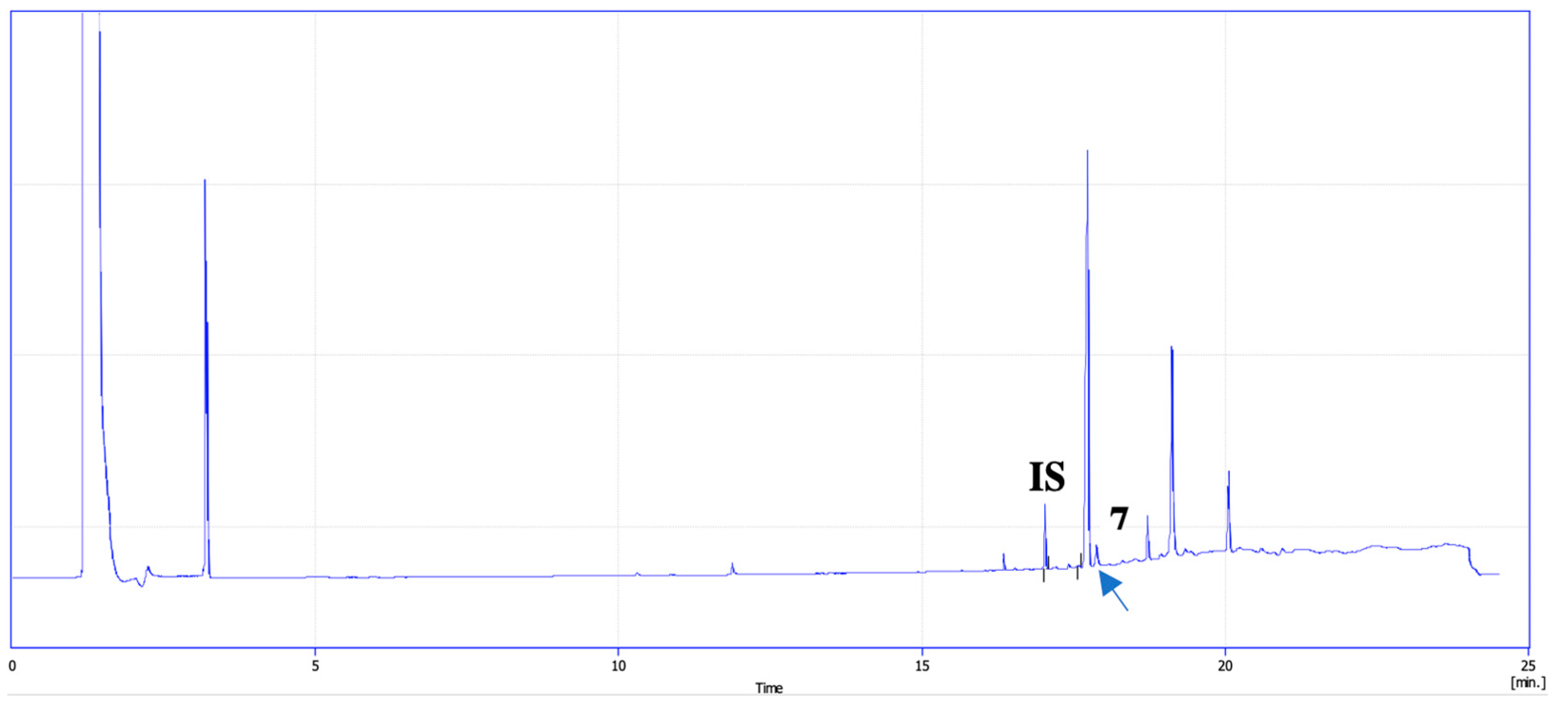
Regarding the apple, pear, and grape skins analyzed, n = 5 samples of apples, n = 2 of pears, and n = 1 of grapes showed the presence of PAHs. Precisely, four out of five samples of apples, found to be contaminated by PAHs, were collected from rural areas, whilst only one was from the urban one. Apple skin showed the presence of B[b]P in an average concentration of 22.1 ± 0.7 µg g−1 (Figure 3). It is worth specifying that all peaks in the chromatogram were analyzed to confirm whether they were PAHs as well. The analyses confirm the different nature of the analytes detected.
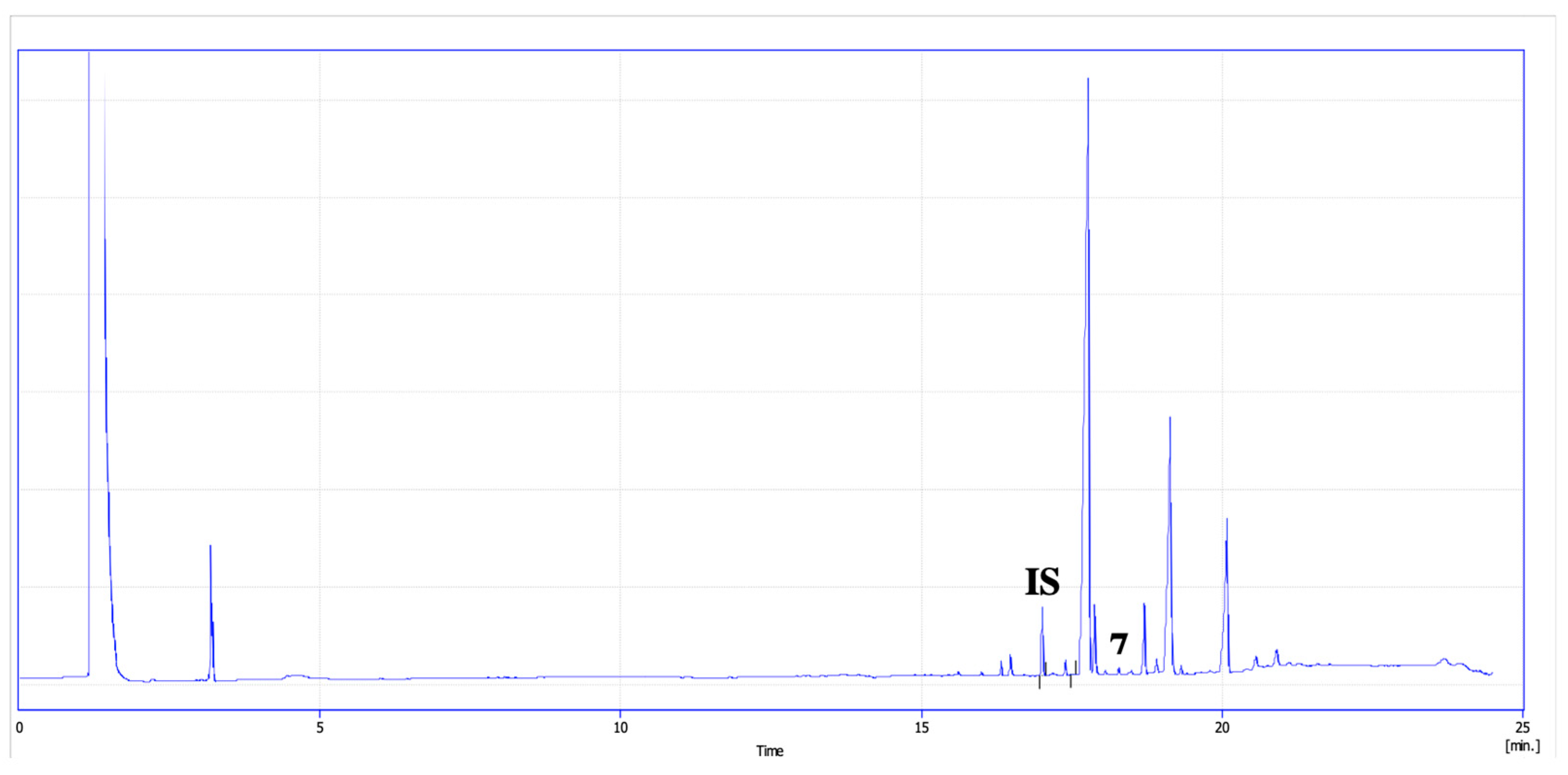
Two (n = 2) pear skin samples, collected from a rural area, were found to be contaminated by PAHs. Both samples showed the presence of B[b]P (as shown in Figure 4). The average concentration revealed was 28.2 ± 1.2 µg g−1.

3.2. Fruit Contamination in Urban and Rural Areas

The results obtained from this study show higher contamination levels in fruit samples collected from rural areas, specifically in the peel samples. Four out of five apple peels, with an average concentration of B[b]P of 22.0 ± 0.7 µg g−1, were sampled from a rural area in the Molise region (Southern Italy), and only one sample showed contamination from an urban area in the Molise region. Similarly, the contaminated pear peel samples (mean, 28.2 ± 1.2 µg g−1) and one contaminated grape (19.1 ± 0.3 µg g−1) peel sample (n = 2) were both collected from rural areas characterized by agricultural activities. A recent study by Di Fiore et al. (2023) [1] investigated PAH contamination levels in the rural areas of the Molise region, approximately 0.3 km from the fruit sampling area. PAH sampling was conducted using sentinel bees (i.e., Apis mellifera); the PAH levels were found to be higher in the rural area compared to the urban area. For instance, B[a]P levels were higher in the rural area compared to the urban area (<2 µg kg−1 vs. ~10 µg kg−1) [1]. This trend was also detected in other Italian regions; for example, rural areas in the Campania region showed higher mean concentrations of PAHs compared to urban ones (Ʃ PAHs 11,353 ng g−1 vs. 755 ng g−1) [22]. On the contrary, it was reported that cucumber cultivated in urban areas showed higher concentration of PAHs than those of rural areas (Ʃ PAHs 6.7 µg kg−1 vs. 4.6 µg kg−1) [23]. Our results have confirmed contamination exclusively on the peel of the investigated fruit, suggesting that the primary pathway of contamination is atmospheric deposition. It is necessary to specify that the authors assumed atmospheric deposition as the pathway based on the absence of PAHs in the pulp. Consequently, it can be hypothesized that the gaseous phase deposition of PAHs contributed to the presence of B[b]P in the fruit samples that tested positive in the experimental assays [2]. Analyses of air pollution by PAHs in the fruit sampling area suggest low levels of PAHs. However, the orographic characteristics of the area appear to influence the behavior of PAHs. The orographic trapping of PAHs could increase the exposure of fruits [24]. In such an area, the less volatile ones (e.g., B[b]P) distribute among vegetation, soil, and air, acting as primary reservoirs for these chemicals and facilitating their entry into the food chain [25]. The origin of B[b]P is not yet clear. The authors hypothesize its presence in rural areas due to the higher abundance of plant material and the use of non-green fuels. Specifically, higher alkanes present in fuels and plant material form PAHs through the process of pyrolysis, which involves the cracking of organic compounds. It was reported that heavy-duty diesel, generally used for agricultural tractors and other agricultural machinery, can emit up to 1000 µg kg−1. In addition to this, the burning of agricultural waste, under sub-optimum conditions, significantly contributes to the emission of atmospheric PAHs [26].

4. Conclusions

The present study aimed to develop an analytical method for the extraction and quantitative determination of PAHs in the pulp and peel of apples, pears, and grapes. The validated analytical method demonstrated a satisfactory performance, with R2 > 0.9912 over the concentration range of 0.5–500 μg mL−1. Recovery percentages (% R) ranged from 68.0 ± 1.2 to 96.2 ± 0.8%, showing variability within acceptable limits (0.7 to 2.3%). The developed methodology was applied to real fruit samples (apples, pears, and grapes), both peel and pulp, collected from rural and urban areas in Southern Italy. Our results indicated that fruits grown in rural areas are also affected by PAHs. The use of non-green diesel fuels appears to be the primary source of PAH exposure in fruits. The exposure of unprocessed foods such as fruit to PAHs is underexplored in the scientific literature, and, consequently, regulations are lacking on this issue. Given the importance of fruit in the Mediterranean diet, further comprehensive research on this topic is recommended.

Author Contributions

Conceptualization, P.A.; methodology, C.D.F. and P.A.; software, I.N. validation, C.D.F. and M.M.; formal analysis, C.D.F., M.M. and I.N.; investigation, C.D.F. and M.M.; resources, M.M.; data curation, C.D.F. and M.M.; writing—original draft preparation, C.D.F.; writing—review and editing, C.D.F. and P.A.; visualization, P.A.; supervision, P.A.; project administration, P.A.; funding acquisition, P.A. All authors have read and agreed to the published version of the manuscript.

Funding

This research received no external funding.

Institutional Review Board Statement

Not applicable.

Informed Consent Statement

Not applicable.

Data Availability Statement

The original contributions presented in the study are included in the article, further inquiries can be directed to the corresponding author.

Acknowledgments

The author would like to thank Alessia Iannone and Fabiana Carriera for their contribution to this paper.

Conflicts of Interest

The authors declare no conflicts of interest.

References

  1. Di Fiore, C.; De Cristofaro, A.; Nuzzo, A.; Notardonato, I.; Ganassi, S.; Iafigliola, L.; Sardella, G.; Ciccone, M.; Nugnes, D.; Passarella, S.; et al. Biomonitoring of Polycyclic Aromatic Hydrocarbons, Heavy Metals, and Plasticizers Residues: Role of Bees and Honey as Bioindicators of Environmental Contamination. Environ. Sci. Pollut. Res. 2023, 30, 44234–44250. [Google Scholar] [CrossRef] [PubMed]
  2. Jia, J.; Bi, C.; Zhang, J.; Chen, Z. Atmospheric Deposition and Vegetable Uptake of Polycyclic Aromatic Hydrocarbons (PAHs) Based on Experimental and Computational Simulations. Atmos. Environ. 2019, 204, 135–141. [Google Scholar] [CrossRef]
  3. Paris, A.; Ledauphin, J.; Poinot, P.; Gaillard, J.-L. Polycyclic Aromatic Hydrocarbons in Fruits and Vegetables: Origin, Analysis, and Occurrence. Environ. Pollut. 2018, 234, 96–106. [Google Scholar] [CrossRef] [PubMed]
  4. Bansal, V.; Kim, K.-H. Review of PAH Contamination in Food Products and Their Health Hazards. Environ. Int. 2015, 84, 26–38. [Google Scholar] [CrossRef]
  5. Zelinkova, Z.; Wenzl, T. The Occurrence of 16 EPA PAHs in Food—A Review. Polycycl. Aromat. Compd. 2015, 35, 248–284. [Google Scholar] [CrossRef]
  6. Naghashan, M.; Kargarghomsheh, P.; Nazari, R.R.; Mehraie, A.; Tooryan, F.; Shariatifar, N. Risk Assessment of PAHs in Fruit Juice Samples Marketed in City of Tehran, Iran. Environ. Sci. Pollut. Res. 2022, 30, 20077–20088. [Google Scholar] [CrossRef]
  7. Samsøe-Petersen, L.; Larsen, E.H.; Larsen, P.B.; Bruun, P. Uptake of Trace Elements and PAHs by Fruit and Vegetables from Contaminated Soils. Environ. Sci. Technol. 2002, 36, 3057–3063. [Google Scholar] [CrossRef]
  8. Ashraf, M.W.; Salam, A. Polycyclic Aromatic Hydrocarbons (PAHs) in Vegetables and Fruits Produced in Saudi Arabia. Bull. Environ. Contam. Toxicol. 2012, 88, 543–547. [Google Scholar] [CrossRef]
  9. Rojo Camargo, M.C.; Toledo, M.C.F. Polycyclic Aromatic Hydrocarbons in Brazilian Vegetables and Fruits. Food Control 2003, 14, 49–53. [Google Scholar] [CrossRef]
  10. Lee, Y.-N.; Lee, S.; Kim, J.-S.; Kumar Patra, J.; Shin, H.-S. Chemical Analysis Techniques and Investigation of Polycyclic Aromatic Hydrocarbons in Fruit, Vegetables and Meats and Their Products. Food Chem. 2019, 277, 156–161. [Google Scholar] [CrossRef]
  11. Al-Farhan, B.S.; Said, T.O.; El-Ghamdi, S.A.; Al-Alamie, A.Y. Distribution and Health Risk Assessment on Dietary Exposure of PAHs in Vegetables and Fruits of Asir, Saudi Arabia. Waste Manag. Bull. 2023, 1, 94–102. [Google Scholar] [CrossRef]
  12. Abdou, A.A.L.G.; Al Touby, S.S.J.; Hossain, M.A. Polycyclic Aromatic Hydrocarbons in Different Highly Consumes Local and Imported Fruits. Green Energy Environ. Technol. 2024. [Google Scholar]
  13. Khalili, F.; Shariatifar, N.; Dehghani, M.H.; Yaghmaeian, K.; Nodehi, R.N.; Yaseri, M. The analysis and probabilistic health risk assessment of PAHs in vegetables and fruits samples marketed Terhan Chemiometric. Glob. NEST J. 2021, 23, 497–508. [Google Scholar]
  14. Ianiri, G.; Di Fiore, C.; Passarella, S.; Notardonato, I.; Iannone, A.; Carriera, F.; Stillittano, V.; De Felice, V.; Russo, M.V.; Avino, P. Methodology for Determining Phthalate Residues by Ultrasound–Vortex-Assisted Dispersive Liquid–Liquid Microextraction and GC-IT/MS in Hot Drink Samples by Vending Machines. Analytica 2022, 3, 213–227. [Google Scholar] [CrossRef]
  15. Notardonato, I.; Passarella, S.; Ianiri, G.; Di Fiore, C.; Russo, M.V.; Avino, P. Analytical Scheme for Simultaneous Determination of Phthalates and Bisphenol A in Honey Samples Based on Dispersive Liquid–Liquid Microextraction Followed by GC-IT/MS. Effect of the Thermal Stress on PAE/BP-A Levels. MPs 2020, 3, 23. [Google Scholar] [CrossRef]
  16. Petrarca, M.H.; Vicente, E.; Amelia Verdiani Tfouni, S. Single-Run Gas Chromatography-Mass Spectrometry Method for the Analysis of Phthalates, Polycyclic Aromatic Hydrocarbons, and Pesticide Residues in Infant Formula Based on Dispersive Microextraction Techniques. Microchem. J. 2024, 197, 109824. [Google Scholar] [CrossRef]
  17. Faridi, A.; Nemati, M. Determination of Benzo(α)Pyrene in Infant Formula Using High Performance Liquid Chromatography and Dispersive Liquid–Liquid Microextraction. Pharm. Sci. 2023, 29, 246–251. [Google Scholar] [CrossRef]
  18. Zhao, X.; Fu, L.; Hu, J.; Li, J.; Wang, H.; Huang, C.; Wang, X. Analysis of PAHs in Water and Fruit Juice Samples by DLLME Combined with LC-Fluorescence Detection. Chroma 2009, 69, 1385–1389. [Google Scholar] [CrossRef]
  19. Pérez, R.A.; Albero, B. Ultrasound-assisted extraction methods for the determination of organic contaminants in solid and liquid samples. Trends Anal. Chem. 2023, 166, 117204. [Google Scholar] [CrossRef]
  20. Quigley, A.; Cummins, W.; Connolly, D. Dispersive Liquid-Liquid Microextraction in the Analysis of Milk and Dairy Products: A Review. J. Chem. 2016, 2016, 4040165. [Google Scholar] [CrossRef]
  21. Hubert, P.; Nguyen-Huu, J.-J.; Boulanger, B.; Chapuzet, E.; Chiap, P.; Cohen, N.; Compagnon, P.-A.; Dewé, W.; Feinberg, M.; Lallier, M.; et al. Harmonization of Strategies for the Validation of Quantitative Analytical Procedures. J. Pharm. Biomed. Anal. 2004, 36, 579–586. [Google Scholar] [CrossRef] [PubMed]
  22. Thiombane, M.; Albanese, S.; Di Bonito, M.; Lima, A.; Zuzolo, D.; Rolandi, R.; Qi, S.; De Vivo, B. Source Patterns and Contamination Level of Polycyclic Aromatic Hydrocarbons (PAHs) in Urban and Rural Areas of Southern Italian Soils. Environ. Geochem. Health 2019, 41, 507–528. [Google Scholar] [CrossRef] [PubMed]
  23. Soceanu, A.; Dobrinas, S.; Stanciu, G.; Popescu, V. Polycyclic aromatic hydrocarbons in vegetables grown in urban and rural areas. Environ. Eng. Manag. J. 2014, 9, 2311–2315. [Google Scholar]
  24. Zhou, R.; Yang, R.; Jing, C. Polycyclic Aromatic Hydrocarbons in Soils and Lichen from the Western Tibetan Plateau: Concentration Profiles, Distribution and Its Influencing Factors. Ecotoxicol. Environ. Saf. 2018, 152, 151–158. [Google Scholar] [CrossRef] [PubMed]
  25. Davidson, D.A.; Wilkinson, A.C.; Blais, J.M.; Kimpe, L.E.; McDonald, K.M.; Schindler, D.W. Orographic Cold-Trapping of Persistent Organic Pollutants by Vegetation in Mountains of Western Canada. Environ. Sci. Technol. 2003, 37, 209–215. [Google Scholar] [CrossRef]
  26. Ravindra, K.; Sokhi, R.; Vangrieken, R. Atmospheric Polycyclic Aromatic Hydrocarbons: Source Attribution, Emission Factors and Regulation. Atmos. Environ. 2008, 42, 2895–2921. [Google Scholar] [CrossRef]
Figure 1. UVA-DLLME-GC-FID chromatogram of an apple sample spiked with 10 µg mL−1 of a PAH solution. Peaks legend: (1) Acy, (2) Ace; (3) Fle; (4) Ph; (5) Fla; (6) Py; (7) B[b]P; (8) B[a]P; and (IS) Internal Standard. For the acronyms, see Section 2.1.
Atmosphere 15 01028 g001
Figure 2. UVA-DLLME-GC-FID chromatogram of an apple pulp sample. All PAHs were <LODs. Peaks legend: (IS) Internal Standard.
Atmosphere 15 01028 g002
Figure 3. UVA-DLLME-GC-FID chromatogram of an apple skin collected from a rural area. All PAHs were <LODs except for B[b]P. Peaks legend: (IS) Internal Standard; and (7) B[b]P.
Atmosphere 15 01028 g003
Figure 4. UVA-DLLME-GC-FID chromatogram of a pear skin collected from a rural area. All PAHs were <LODs except for B[b]P. Peaks legend: (IS) Internal Standard; and (7) B[b]P.
Atmosphere 15 01028 g004
Table 1. % R obtained using SLE and UVA-DLLME performed on spiked pulp of apple, pear, and grape.
PAHSLE (% R)UVA-DLLME (% R)
Sample 1ApplePearGrapeApplePearGrape
Acy37.1 ± 2.636.2 ± 4.335.1 ± 3.368.0 ± 1.269.1 ± 0.970.2 ± 0.5
Ace37.1 ± 1.932.5 ± 3.131.2 ± 3.168.4 ± 0.567.4 ± 1.269.1 ± 0.4
Fla75.5 ± 4.068.1 ± 3.866.2 ± 4.378.4 ± 0.680.0 ± 1.279.1 ± 0.5
Fle57.4 ± 3.253.1 ± 2.650.2 ± 2.387.4 ± 1.388.5 ± 1.489.1 ± 0.3
B[a]P65.8 ± 2.164.1 ± 3.261.2 ± 3.567.2 ± 1.368.3 ± 1.269.8 ± 1.2
B[b]P71.2 ± 2.170.1 ± 2.372.2 ± 4.178.4 ± 0.579.1 ± 0.280.1 ± 0.2
Ph75.3 ± 2.372.1 ± 3.672.3 ± 3.196.2 ± 0.898.0 ± 0.498.3 ± 1.3
Py73.6 ± 3.472.1 ± 2.570.2 ± 3.378.4 ± 0.380.1 ± 0.779.7 ± 1.2
1 % R is relative to three extractions for each fruit type for both extraction approaches tested. For the acronyms, see Section 2.1.
Table 2. Correlation coefficients (R2) in the range 0.5–500 μg mL−1, LOD (µg mL−1), LOQ (µg mL−1), recoveries, and intra-day and inter-day precision, along with RDS, in apple samples of each PAH investigated by means of UVA-DLLME approach. For acronyms, please see Section 2.1.
PAHR2LODLOQ% R aIntra-Day (1) bIntra-Day (2) bInter-Day cRDS (%) d
Acy0.99340.120.2768.0 ± 1.266.3 ± 2.265.2 ± 3.265.6 ± 5.60.8
Ace0.99240.090.2168.4 ± 0.567.2 ± 1.565.3 ± 3.566.7 ± 4.51.5
Fla0.99450.060.2678.4 ± 0.675.4 ± 1.376.2 ± 3.476.0 ± 4.40.6
Fle0.99140.080.1487.4 ± 1.385.3 ± 2.186.1 ± 4.785.8 ± 4.50.5
B[a]P0.99120.280.6267.2 ± 1.365.1 ± 2.068.1 ± 3.367.3 ± 4.32.3
B[b]P0.99230.250.5278.4 ± 0.576.3 ± 1.574.1 ± 2.775.3 ± 3.71.5
Ph0.99320.120.3196.2 ± 0.893.6 ± 1.396.3 ± 2.194.8 ± 4.11.4
Py0.99150.110.3878.4 ± 0.376.3 ± 1.475.2 ± 3.475.7 ± 3.50.7
a For % R, see Table 1; the mean value reported is derived from three tests conducted on the same day and it is expressed as % recovery ± standard deviation; b % R ± standard deviation; the mean value reported is derived from three tests conducted on the same day. c The reported mean value is derived from the average of results obtained from tests conducted on three different days and it is expressed as % R ± standard deviation; d Relative Standard Deviation. For the acronyms, see Section 2.1.
Disclaimer/Publisher’s Note: The statements, opinions and data contained in all publications are solely those of the individual author(s) and contributor(s) and not of MDPI and/or the editor(s). MDPI and/or the editor(s) disclaim responsibility for any injury to people or property resulting from any ideas, methods, instructions or products referred to in the content.