Article

Characterization of the Cultivable Microbiota Components of Marine Bioaerosols in the North Tropical Atlantic

by
Gabriela Cristina Chagas Moura
1,*,
Yasmin Marques Ayres
2,
Anna Luisa de Carvalho Brito
2,
Edmilson Ferreira de Souza Júnior
2,
Rafael dos Santos Rocha
2,
Paulo Miguel Vieira De Sousa
2,
Antônio Geraldo Ferreira
2,
Oscarina Viana de Sousa
2 and
Doris Veleda
1
1
Oceanography Department (DOCEAN), Universidade Federal de Pernambuco, Recife 50670-901, Brazil
2
Marine Science Institute (LABOMAR), Universidade Federal do Ceará, Fortaleza 60356-001, Brazil
*
Author to whom correspondence should be addressed.
Atmosphere 2023, 14(10), 1470; https://doi.org/10.3390/atmos14101470
Submission received: 7 August 2023 / Revised: 2 September 2023 / Accepted: 13 September 2023 / Published: 22 September 2023
(This article belongs to the Section Aerosols)

Abstract

:
Microorganisms are key to balancing marine ecosystems and have complex interactions at the ocean–atmosphere interface, affecting global climate and human health. This research investigated the diversity of cultivable bacteria and fungi in marine bioaerosols in the North Tropical Atlantic Ocean. Using the technique of spontaneous sedimentation in selective culture media, samples were collected during oceanographic expeditions. After isolation and purification, microbial strains were identified by phenotypic and genetic analyses. Fungi isolated included Acrophialophora, Aspergillus, Chrysosporium, Cladosporium, Fonsecaea, Mucor, Rhodotorula, Schizophyllum, Stemphylium, Candida, Curvularia, Cystobasidium, Exophiala, Neotestudina, Penicillium, Pestalotiopsis, and Preussia. The bacterial isolates belonged to the Bacillota, Pseudomonadota, Enterobacteriaceae family, Bacillus genus, and Serratia liquefaciens groups. About 40% of bacteria and 42% of fungi were identified as potential human pathogens, suggesting a relationship between human actions and the microbiota present in bioaerosols on the high seas. Sea surface temperature (SST) and wind speed influenced microorganisms. More studies and analyses in different scenarios should be conducted considering environmental and climate variables in order to deepen knowledge and generate information on the subject, so that standards can be established, and quality parameters determined.

1. Introduction

Microorganisms are present in all environments on planet Earth, although in some of these, in loco sampling is impossible due to difficult access and/or the current absence of compatible technologies. The oceans, until the last century, were included in this group. The presence of marine microorganisms in the air over the Atlantic Ocean had not been documented until the mid-twentieth century, with the first articles published on the subject in the 1950s [1,2]. Research efforts in an attempt to fill these gaps on oceanic microorganisms in the atmosphere, diversity, biogeography, interactions, and participation in global biogeochemistry are evident from the increasing number of papers published in the following decades. We performed a quick search on a free research platform using a combination of keywords: marine microbial, atmospheric communities, aerosol; this search resulted in a finding increasing numbers of articles produced on the subject throughout the 1960s until the early 2020s.
Bioaerosols are conceptualized as solid particles carried by the air originating from biological organisms [3], which are aerosolized from the marine environment to the atmosphere through the continuous generation of bubbles caused by the friction of the wind on the surface of the water [4,5]. The Sea Surface Microlayer (SML) is the main contributor of organic matter and inorganic salts to the atmosphere [6], including microorganisms, such as bacteria and fungi [3,6,7], viruses, pollen, and their derivatives, such as endotoxins and mycotoxins [7].
This particulate biological material suspended in the atmosphere has functions for the Earth system, the climate and public health at the local, regional, and global scales, as they act directly in the dispersion and absorption of solar radiation or, even indirectly, in the formation of cloud condensation nuclei, influencing droplet sizes, cloud cover, and albedo [4,8,9]. Its distribution, abundance, and diversity are affected by different meteo-oceanographic factors such as temperature, precipitation, relative humidity, and wind speed, as well as climate change and global warming [10], thus making it essential to collect abiotic data complementary for the thematic discussion, on a relatively long-time scale and at the time of sampling.
Among the main factors that impact the microbiological diversity in the atmosphere, there is the air temperature [11]. In the Tropical Atlantic Ocean, this factor oscillates meridionally, leading to the Intertropical Convergence Zone (ITCZ), which migrates seasonally, being more north in June, July, and August and reaching its southernmost position in December, January, and February. The trade winds are associated with the migration of sea surface temperature (SST) and ITCZ. The SE trade winds move towards the NH (SH) as the ITCZ is further north (south) [12].
In addition, high air temperatures and low air humidity values favor the transport of bioaerosols, due to the atmospheric water vapor absorbed by the particles, favoring their increase and weight and, consequently, their gravitational deposition [13,14].
As well as the dispersion of material from the ocean–atmosphere interface by wind, in combination with surface tension gradients, it represents a unique mechanism that ensures microbiological transport. Additionally, changes in physical forcings such as wind speed and water circulation affect the composition and molecular arrangement of biofilms in the ocean surface microlayer and, consequently, the composition of bioaerosols.
The increase in pressure, as it reflects the increase in the amount of cold air, which alone can facilitate the release and transport of microorganisms, which can lead to a high abundance in the atmosphere [13]. Finally, climate change implies a change in the global atmospheric circulation, which contains not only chemical compounds but also bioaerosols, mainly fungal spores and fungal hyphae clefts [10]. These climate changes can change the seasonal and characteristic systems of each location, changing the patterns of meteo-oceanographic variables and, consequently, affecting bioaerosols.
In this sense, despite efforts, information on the diversity and distribution of these organisms throughout the oceans, especially the Atlantic Ocean, is still scarce. Thus, this article aimed to analyze and identify the diversity of cultivable bacteria and fungi present in marine bioaerosols collected along the North Tropical Atlantic Ocean (NTA) and also to collect oceanographic and meteorological variables and possibly associating them with the diversity found, thus evidencing possible determining relationships between oceanographic and meteorological variables and microbial diversity.

2. Materials and Methods

2.1. Study Area

The study area comprised the North Tropical Atlantic, from 0° to 15° N (Figure 1a). Samplings were carried out during an expedition by the Prediction and Research Moored Array in the Tropical Atlantic Program (PIRATA) around meteo-oceanographic buoys (Figure 1b) and corresponded to the following points: P1 (15° N 38° W), P2 (12° N 38° W), P3 (8° N 38° W), P4 (4° N 38° W), and P5 (0° 35°W). The samples were collected in November and December of 2017, on board the Hydro-oceanographic (NPqHo) ship “Vital de Oliveira” of the Brazilian Navy, and in June 2022, on board the oceanographic ship “Antares” of the Brazilian Navy.

2.2. Samplings

Microbial particles were collected using the spontaneous sedimentation technique on the surface of selective culture media in Petri dishes [15,16]. Briefly, selective culture media were exposed to air for 30 min with the opening of the Petri dishes, then closed and conditioned. Sets, in duplicate, of selective medium for bacteria [Plate Count Agar (PCA, Difco®) diluted in sea water at 10 ppm] and fungi [Potato Dextrose Agar (PDA) added with 10 µg/mL of Ampicillin] were used. The collected material was then transported to the Laboratório de Microbiologia Ambiental e do Pescado (LAMAP–UFC).

2.3. Sample Processing: Isolation and Identification of the Microbiota

In the laboratory, the grown bacterial colonies were selected and isolated. Cultures were checked for purity, morphology, and cell wall characteristics by Gram-staining technique [17,18]. Then, the cultures were submitted to biochemical tests according to the Bergey’s Manual [17] arranged as a dichotomous key in the following phenotypes: production of catalase and oxidase enzymes, motility, production of hydrogen sulfide (H2S), production of indole, test of methyl red (VM) and Voges–Proskauer (VP), urea broth, Simmons citrate [19,20]. Due to identification by biochemical tests, genetic identification by PCR and sequencing was not performed on the isolated bacteria.
For the isolation of fungal cultures, plates with growth were rehydrated (10 ppm NaCl saline solutions) and diluted. Inoculated plates were incubated at 28 °C for 7 days [14]. After isolation, the samples were purified and, when isolated colonies were obtained, DNA extraction was performed following an existing protocol with adaptations for the samples [21], the polymerase chain reaction (PCR) using the ITS-1 primers (5′-TCCGTAGGTGAACCTGCGG-3′) and ITS4 (5′-TCCTCCGCTTATTGATATGC-3′) and, finally, the sequencing of the amplicons achieved.
The amplicons obtained were sent to ACTGene Analises Moleculares, for sequencing, using an automatic sequencer ABI 3500 Genetic Analyzer (Applied Biosystems, Waltham, MA, USA). The sequences obtained were analyzed using the 3500 Data Collection software and compared to sequences from the GenBank database of the National Center for Biotechnology Information (NCBI).

2.4. Oceanographic and Meteorological Variables

Meteo-oceanographic data were collected together at the time of collection and were obtained through equipment present in the facilities of the ships of each shipment. The variables obtained were wind speed, relative humidity, atmospheric pressure, and air temperature. During the expeditions, no CTD stations were carried out so, seeking to complement the abiotic data, SST data from satellite databases available online was included. In this work, 20 years of monthly SST and surface winds (10 m) from January 2003 to December 2022 were analyzed. The SST data had a 9 Km spatial resolution and were available in the MODIS Aqua Level 3 product, derived from the MODIS sensor from NASA aboard the Aqua satellite http://apdrc.soest.hawaii.edu/ (accessed on 20 June 2023). The surface wind database was from the ERA5 reanalysis model, with 0.25 degrees of resolution. ERA5 is the fifth generation European Center for Medium-Range Weather Forecasts (ECMWF) reanalysis for global climate. The SST data were used to analyze the climatology of the study area and to estimate the SST anomalies during the collection period. The SST anomaly was obtained by removing the monthly climatological average. The anomaly values made it possible to identify whether the SST was above or below the historical average during the collection analysis period. In addition, the satellite data allowed for extracting the values at the same points as the collections analyzed here.

2.5. Statistical Analysis

Statistical analyses were performed using the Rstudio software, which uses the R statistical programming language, (version 4.2.3, The R Foundation, Vienna, Austria) readxl, RVAideMemoire, car and the tidyverse packages were used. Initially, the Shapiro–Wilk test was performed to verify the possible normality of the meteo-oceanographic data. Since these data did not present a normal distribution and due to the sample size, non-parametric tests were chosen. The Levene test was also performed for homogeneity of median variances for the 2017 and 2022 groups. The Mann–Whitney test was performed for two independent groups of meteo-oceanographic variables between the years 2017 and 2022. The Kruskal–Wallis test (ANOVA one-way for non-parametric data) with the chi-square value for groups 2017 and 2022.
Quality assurance and control (QA/QC) was performed throughout the field and laboratory sampling and analysis process with the use of certified inputs (culture media and reagents), manually calibrated equipment (following the manufacturers’ instructions) and internationally validated methodologies.

3. Results

3.1. Microbiota Cultivable in Marine Bioaerosols

According to the morphological characteristics of the isolated fungal colonies, 19 colonies belonging to 9 different genera were identified: Acrophialophora sp. (5%), Aspergillus sp. (27%), Chrysosporium sp. (5%), Cladosporium sp. (16%), Fonsecaea sp. (27%), Mucor sp. (5%), Rhodotorula sp. (5%), Schizophyllum sp. (5%), and Stemphylium sp. (5%). Of the bacterial isolates, 56 isolates representing the phyla Bacillota (35%) and Pseudomonadota (20%) were characterized; family Enterobacteriaceae (25%); genera Bacillus (5%); and Serratia liquefaciens (15%) (Table 1).
At the first collection point (P1), bacterial strains belonging to the Phyla Pseudomonadota and Bacillota, and the Enterobacteriacea family were found, as for fungal strains, only the genus Aspergillus was identified.
At the second (P2) and third (P3) collection points, it was not possible to isolate bacterial strains in the collected samples. In the selective media, there was growth of fungal colonies with characteristics of the genera: Aspergillus sp., Fonsecaea sp., Rhodotorula sp., and Acrophialophora sp. (P2), and Fonsecaea sp., Schizophyllum sp., Stemphylium sp., Cladosporium sp., Chrysosporium sp., and Aspergillus sp. (P3).
At the fourth collection point (P4), different qualities of bacteria were found, including: Serratia liquefaciens, phylum Bacillota, phylum Pseudomonadota, Enterobacteriacea family, and Bacillus genus. As for the fungal samples, only the genus Mucor was possibly identified.
At the last collection point (P5), only bacterial microorganisms corresponding to the Enterobacteriaceae family, phylum Bacillota, and Serratia liquefaciens were found.
As for the results of fungal sequencing, 19 colonies were genetically identified, these data were compiled in the table below (Table 2):
Of the microorganisms identified, 10 divergent identifications were found, among which 4 were identified at the species level, the genus Aspergillus sp. was the most represented (26%), followed by Candida sp. (11%) and Curvularia sp. (11%) and the remaining Cladosporium sp. (5%), Cystobasidium sp. (5%), Exophiala dermatitidis (5%), Neotestudina sp. (5%), Penicillium sp. (5%), Pestalotiopsis sp. (5%), Preussia sp. (5%), and Rhodotorula sphaerocarpa (5%).

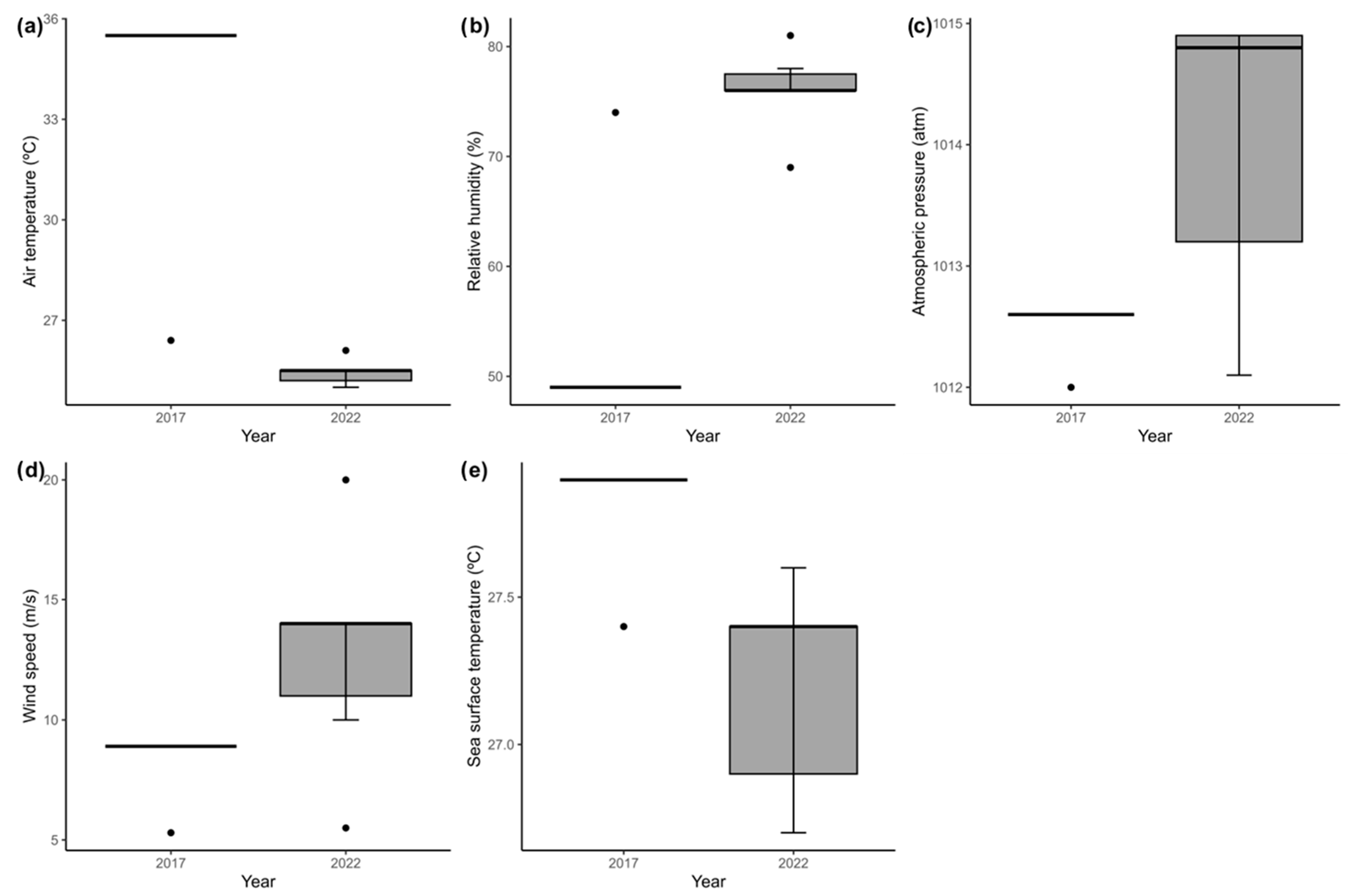
3.2. Oceanographic and Meteorological Variables

The parameters that were obtained were organized in Table 3:
Statistical tests were performed to verify the presence of significant differences (Figure 2). Thus, the results indicate significant differences in the abiotic variables (p < 0.05) between the collection years (2017 and 2022), which results in possible significant differences in the organisms found between the collection years (2017 and 2022).
SST and surface wind data allow to analyze the climatology and anomalies of the study area (Figure 3 and Figure 4). Figure 3 shows the seasonal climatology of SST and surface winds. As expected, in the NTA, the SST is lower during the DJF and higher during the JJA, oscillating around 2 °C throughout the year. This migration drives the trade winds and the ITCZ, which is north in JJA and south in DJF.
SST and wind anomalies were calculated for the period of the oceanographic campaigns, removing the monthly climatological average for the specific months and years of the oceanographic campaigns analyzed in this study. It would show us if the oceanic and atmospheric conditions were close to the climatology or presented atypical conditions. Figure 4 shows the SST anomalies and surface winds values for November 2017 and June 2022.
During the 2017 shipment, SST varied little concerning the historical average. However, it showed negative anomalies mainly at points P3 to P5. During the 2022 shipment, the same region presented positive SST anomalies. This characteristic agrees with the statistical results that bring significant annual differences between the variables. Therefore, there were significant yearly variations of SST, which were below the climatological averages in 2017 and above the climatological average in 2022, indicating once again that differences in this parameter can influence and cause changes in the microbiota found.

4. Discussion

Multiple microbiological genera were found in the study area, evidencing a plurality that, until now, is at least little known and studied. In addition, the relationship between specimens and anthropogenic presence or action is remarkable.
As for the fungal genera, Fonsecaea, Rhodotorula, Mucor, and Acrophialophora are widely associated in the literature with human pathogens and are present in the air of different environments, especially in urban areas and coastal cities [22,23,24,25]. The presence of these representatives that are potentially pathogenic for humans in marine bioaerosols along the ocean can be caused due to different reasons, including the intense ship traffic and its relationship with tourist activities, industrialization, and global warming [26,27]. These mentioned anthropogenic activities impact and interfere with the sustainability of the marine environment due to changes in symbiotic relationships and microorganisms in the marine environment [28].
Chrysosporium is a genus present in different marine ecosystems including algae, sponges, and fish, making its presence in the study area consistent and expected. It is also important to emphasize the biotechnological capacity that the genus presents through the production of basic structures for the production of new drugs [29].
From pioneering research into atmospheric microbiota diversity to current research into the marine environment, the genus Aspergillus is found [30]. In addition to the amplitude in its presence, Aspergillus sporulation capacity and its association with atmospheric CO2 concentrations are known, increasing the sporulation of organisms and, consequently, their presence in the environment [10]. Historically, species of the genus Cladosporium are often found in oceanic air masses and marine environments, especially in mangrove sediments [30]. The presence of Cladosporium can be explained due to the tendency of these spores to thrive in environments with a hot climate and air currents, parameters that are remarkable in the collected sites; in addition, their spores are mainly affected by variations in average temperature and pressure [10,31,32]. Fungal organisms of the Cladosporium and Aspergillus genera have previously been found in bioaerosol samples from Antarctica [33], evidencing, however, the lack of recent studies on the subject throughout the study area, or even possible relationships between the study area and the places where they are already present.
Species of the genus Stemphylium have a long record of detection in bioaerosols in different regions and altitudes, including along the Atlantic Ocean [34]. Despite the higher frequency in polar air masses, exposure to different temperatures and pressure can modify sporulation, possibly increasing it, which prolongs and favors its permanence and transport of fungal organisms to other locations [31,32].
Species of the genus Schizophyllum are cosmopolitan in occurrence and different concentrations of carbon dioxide can affect the reproduction of species of this genus, and low concentrations of this gas allow the formation of more complex fungal structures [35].
Regarding the bacterial material found, it was composed of bacteria that have significant pathogenic potential, such as those of the Enterobacteriaceae family, which, as they are not considered inhabitants of marine environments, are associated with pollution from the mainland, or even through ship traffic in the region associated with tourist and industrial activities [27]. In some of the older studies, the genus Bacillus appears in abundance [5], however, according to our results, in the NTA, only one isolate corresponded to this genus.
This could possibly be explained due to the fact that Gram-negative bacteria are more commonly found in the marine environment, in agreement with works by other authors in relation to the marine environment [36]. The Bacillota phylum was the most abundant in strains isolated from marine bioaerosols (35%), according to [3] in their research on the spatial distribution of bacterial populations in marine bioaerosol samples during a cruise from the North Sea to the Baltic Sea, identified representatives of the phylum Bacillota in 8.3% of the isolates. According to [37], in their study on bacterioplankton in estuaries, the phylum Bacillota did not appear to be an abundant component, with the exception of the estuary which had significant values of suspended particulate matter, indicating that they are more represented in waters of great turbidity, as well as the surface waters of the study area, which have high values of wind speed that carry the microbiota from the waters and transport it to the air. The phylum Pseudomonadota represented 20% of the isolates, the second most abundant in the results, corresponding with the results of pioneering studies such as [38], in which most of the isolates belonged to the Phylum Bacillota and Pseudomonadota, converging with the results obtained and also more recent studies [36,37]. The Serratia liquefaciens species is characterized by inhabiting different environments, such as sediment, water, and has already been found in marine organisms [39], in addition to being associated with the production of histamine that causes contamination in humans [40]. Therefore, given its presence in the microbiota of marine organisms, it is possible that it was carried and, therefore, was found in the bioaerosols collected in this study.
The biogeographical patterns of microorganisms are little known and are controlled by multiple factors (selection, drift, dispersion, and mutation) [41]. When focusing on bioaerosols, the most important of them becomes dispersal, which may depend on the properties of each taxon such as initial abundance in the community of origin. The dispersion of bioaerosols is directly linked to the atmospheric dispersion capacity that works as a limiting factor, considering environmental variables such as the rate of UV radiation [42]. That is, the richness found in this work is directly associated with the ability of this rate to survive and adapt to dispersion in the atmospheric environment.
When we compare the characterization data with the sequencing data, it is possible to notice that there was an overlap of some genera, among them Aspergillus sp., Cladosporium sp., Rhodotorula sp. This overlap corresponds to approximately 48% of the samples that were characterized. Indicating that, despite the divergences between the samples that were identified and gives greater security of genetic identification, the characterization is able to provide some significant idea of the local microbiota.
In addition, we also obtained a coincidence in the genera that were found according to each buoy when we compared the data obtained in relation to characterization and sequencing. In P1, P2, and P3, in both types of identification, it was possible to identify the genus Aspergillus sp., and Rhodotorula was identified in P2 in both identification methodologies.
Candida sp. is a pathogenic fungal genus in which some species are considered multi-resistant pathogens [43] and have already been found in the marine environment forming association with sponges [44] after the discharge of effluents at the site [45] opening up the possibility of human interference in the aerial microbiome of the study area.
Curvularia sp. Is considered a genus of worldwide distribution and some species have already been found in aquatic environments and in the air [46,47]; however, it is widely known to be a pathogenic fungus among different species of animals and plants, including humans. It is worth noting that this genus, in addition to being cosmopolitan, has been described through the recent discovery of new species [48] and may in the future become a risk in view of its pathogenic capacity and its identification in the study area.
Yeasts of the genus Cystobasidium sp. Were recently found in association with marine animals such as sponges [44] and also attached to thermal insulation boards present in marine environments [49], that is, these yeasts are present in the marine air close, at least, to the region of the North Atlantic Ocean and North Pacific.
Exophiala dermatitidis is a fungal species that is associated with skin and lung infections. Organisms of this genus are present in different types of environments and have the ability to withstand extreme conditions such as high temperatures or salinity and variable pH [50,51]. In addition, this organism is often found in tropical regions, natural conditions that are consistent with the study area, justifying its occurrence and bringing the problem of pathogenic organisms overlapping in relation to others due to changes in climatic conditions such as increased temperature [52].
We believe that it is the first time that Neotestudina sp. was isolated and cultured from marine samples, especially in the air of the study region (12° N and 15° N). Bearing in mind that this genus can be isolated from soils in tropical regions [53] and has historically been associated with skin diseases [54,55,56], it is suggested that its transport from land to marine aerosols is due to human influence in the study area, probably through the maritime flow of vessels, modifying the local microbiota. Another important point is that this species was only found in the 2022 collection, suggesting that its presence is seasonal, or that climate anomalies may be associated with changes in the microbiota of the area, since, in 2020, another species of the same genus was also found for the first time in soil samples in Nigeria [53].
Penicillium sp. Is a genus widely studied in the literature and, consequently, already isolated several times from the marine environment; as a recent example, it was found in seawater, macroalgae, and tree trunks resulting in 16 different species of Penicillium sp. Isolated beyond the description of a new species [57]. This genus was also found in several urban and coastal regions and is considered a pollutant in the region’s aerosols, which may indicate, with its presence in the study area, that the air quality needs to be better evaluated [25].
As for the genus Pestalotiopsis sp., it is known how this is present in mangrove environments and is currently shown to be an advancing tool for biotechnology research since endophytic fungi are prolific resources for secondary bioactive metabolites and, as a result of its structural complexity, this genus has proven to be efficient [58,59]. With that, several studies bring the research and identification of new species of this genus, which encourages the possibility of using this isolate for future biotechnological tests [60].
Preussia sp., like other genera found, is used in biotechnology both due to its anti-inflammatory effects and in enhancing the growth of agricultural products [61,62]. Therefore, like Pestalotiopsis sp., it results in a new possibility of obtaining biotechnological products. Rhodotorula sphaerocarpa is a species of a fungal genus marked by being associated with human pathogens and is easily present in the environment. Studies indicate that this species increases its respiration in environments with higher temperature values, even though its reproduction is dependent on this environmental variable [63]. In addition, authors indicate that species of this genus, derived from marine water samples, may present biotechnological activity as antioxidants and bioconverter of marine residues, allowing, once again, the use of isolates obtained as possible biotechnological products [64,65].
It is necessary to emphasize how, within this richness found throughout the NTA, the most significant part of the results suggest microorganisms that are associated with the presence and actions of human beings. That is, in addition to the indirect anthropic impact associated with the increase in emissions of global warming gases, such as carbon dioxide, and how these interfere both in environmental variables that interfere in the local microbiota and in the reproduction and viability of microorganisms, there is also the impact direct that occurs through the ship traffic that historically occurs in the area and that directly deposits exotic species in the region, altering the microbiome of the area.
Microorganisms are directly impacted by different environmental variables (air temperature, wind speed, UV radiation, etc.) that control both the survival and permanence as well as the ability to reproduce and viability of microbiological spores, which varies according to each species [66].
With that, we have the association between the fungal diversity found and the climatic parameters, where according to the means and distributions found (Figure 2), it is possible to notice differences in the values of the two years collected (p < 0.05), these significant differences of abiotic data can influence and result in changes in the microbiota present in the study area. The average temperature in 2017 was 28.5 °C, while in 2022, it was 25.3 °C. Initially, it was expected that this difference could represent a possible greater fungal diversity, since high temperatures are generally associated with the reproduction of microorganisms; however, the sample that presented the greatest diversity was that of 2022 (nine different identifications) in which, despite the average being lower, presented much more homogeneous values (sd = 0.44), which was different from the first sample (sd = 4.03). The Relative Humidity of the Air values were also higher and more homogeneous in the 2022 collections, agreeing with the diversity results, since due to the atmospheric water vapor being absorbed by the particles, weight gain is favored and, consequently, the gravitational deposition of the particles, making it possible, through the collection of spontaneous sedimentation, to obtain a greater number of isolates and, consequently, fungal diversity. The wind speed in the year 2022 was more intense, which also corroborates the greater diversity found in this year. Taking into account the importance that winds have in the formation of bioaerosols through the bursting of bubbles in the oceanic surface microlayer, therefore, higher values of wind speed may be associated with the generation of more particles of microorganisms present in the ocean and, consequently, a greater diversity.
The availability of long-term data from satellites and models allowed an assessment of SST and wind conditions for November 2017 and June 2022 (Figure 3). The maps of SST anomalies indicate that in 2017, the temperatures were slightly below average (weak negative anomalies), and in 2022, the same region had above-average temperature values (positive anomaly).
When the SST was above the climatological average in 2022, the diversity was greater, highlighting a possible relationship between the two variables. Therefore, when we group these data, the SST may significantly influence the microbiological diversity more than the air temperature, considering the diversity data and its relationship with the meteo-oceanographic data.
The results represent the existing microbiota in the NTA ocean but admit the limitations of culture and microbiological survival.

5. Conclusions

Different microbiological taxonomic groups were present in the air of the North Tropical Atlantic Ocean, among them a great variety of fungi and bacteria, corresponding to nine and five types, respectively. These findings shed light on the airborne microbiome present in the study area, as well as the possibility of studying the origin and fate of these microorganisms. A considerable part of the microorganisms found are associated with human pathogens or related to anthropic actions, evidencing possible impacts of human actions in the region since changes in microbiomes can lead to serious changes in the entire food chain of ecosystems.
It is also noteworthy that the results show the need for regional studies to obtain complete data that can compile quantifications and non-cultivable diversities, given the importance of microorganisms for different biogeochemical cycles and, therefore, their global relevance. Finally, these studies have been carried out and continued in search of a complete volume of data by the production team of this work.

Author Contributions

G.C.C.M., O.V.d.S., A.G.F. and D.V. designed the study. G.C.C.M. collected the samples. G.C.C.M., Y.M.A., A.L.d.C.B. and P.M.V.D.S. processed, isolated and sequenced the samples. G.C.C.M., E.F.d.S.J. and R.d.S.R. analyzed the data. All authors have read and agreed to the published version of the manuscript.

Funding

This research is funding by the SIMOPEC Project—Ocean-atmosphere coupled modeling operating system for the monitoring and forecasting of climate extremes in the Northeast of Brazil, with funding from the National Council for Scientific and Technological Development—CNPq: 406707/2022. The first author received a scholarship from Improvement of Higher Education (CAPES).

Institutional Review Board Statement

Not applicable.

Data Availability Statement

Data sharing is not applicable to this article.

Acknowledgments

The first author G.M. thanks the National Council for the Improvement of Higher Education (CAPES). O.V.S. is grateful to the Brazilian Navy and the Prediction and Research Moored Array in the Tropical Atlantic Program (PIRATA) and CNPq by the Fellowship of Research Productivity (PQ2: 316342/2021-1). D.V. is grateful to the Brazilian National Research Council (CNPq) Research Fellowship PQ2, under the project “Teleconnections between Pacific and Atlantic: impacts on climate variability in South America”, grant number: 314879/2020-0. D.V. also thanks the SIMOPEC Project—Ocean-atmosphere coupled modeling operating system for the monitoring and forecasting of climate extremes in the Northeast of Brazil, with funding from the National Council for Scientific and Technological Development—CNPq Grant 406707/2022, and the TRIATLAS project, which has received funding from the European Union’s Horizon 2020 research and innovation program under grant Agreement Number 817578.

Conflicts of Interest

The authors declare no conflict of interest.

References

  1. Pady, S.M.; Kapica, L. Fungi in Air Over the Atlantic Ocean. Mycologia 1955, 47, 34–50. [Google Scholar] [CrossRef]
  2. Pady, S.M.; Kelly, C.D. Aerobiological studies of fungi and bacteria over the Atlantic Ocean. Can. J. Bot. 1954, 32, 202–212. [Google Scholar] [CrossRef]
  3. Seifried, J.S.; Wichels, A.; Gerdts, G. Spatial distribution of marine airborne bacterial communities. Microbiol. Open 2015, 4, 475–490. [Google Scholar] [CrossRef] [PubMed]
  4. Alves, C.A. Química entre a microcamada superficial oceânica e os aerossóis marinhos. Química Nova 2014, 37, 1382–1400. [Google Scholar] [CrossRef]
  5. Cho, B.C.; Hwang, C.Y. Prokaryotic abundance and 16S rRNA gene sequences detected in marine aerosols on the East Sea (Korea). FEMS Microbiol. Ecol. 2011, 76, 327–341. [Google Scholar] [CrossRef]
  6. Fröhlich-Nowoisky, J.; Burrows, S.M.; Xie, Z.; Engling, G.; Solomon, P.A.; Fraser, M.P.; Mayol-Bracero, O.L.; Artaxo, P.; Begerow, D.; Conrad, R.; et al. Biogeography in the air: Fungal diversity over land and oceans. Biogeosciences 2012, 9, 1125–1136. [Google Scholar] [CrossRef]
  7. Yao, M. Bioaerosol: A bridge and opportunity for many scientific research fields. J. Aerosol Sci. 2018, 115, 108–112. [Google Scholar] [CrossRef]
  8. Aller, J.Y.; Kuznetsova, M.R.; Jahns, C.J.; Kemp, P.F. The sea surface microlayer as a source of viral and bacterial enrichment in marine aerosols. J. Aerosol Sci. 2005, 36, 801–812. [Google Scholar] [CrossRef]
  9. Liss, P.S.; Duce, R.A. The Sea Surface and Global Change; Cambridge University Press: Cambridge, UK, 1997. [Google Scholar]
  10. Ceter, T. Effects of global-warming and climate-changes on atmospheric fungi spores distribution. Commun. Fac. Sci. Univ. Ank. Ser. C Biol. Geol. Eng. Geophys. Eng. 2018, 27, 263–272. [Google Scholar] [CrossRef]
  11. Kowalski, M.; Pastuszka, J.S. Effect of ambient air temperature and solar radiation on changes in bacterial and fungal aerosols concentration in the urban environment. Ann. Agric. Environ. Med. 2018, 25, 259–261. [Google Scholar] [CrossRef]
  12. Melo, A.D.; Cavalcanti, I.D.A.; Souza, P.P.D. Zona de convergência intertropical do Atlântico. In Tempo e Clima no Brasil; Cavalcanti, I.F.A., Ferreira, N.J., Silva, M.G.A.J., Dias, M.A.F.S., Eds.; Oficina de Textos: São Paulo, Brazil, 2009; pp. 25–41. [Google Scholar]
  13. Zhen, Q.; Deng, Y.; Wang, Y.; Wang, X.; Zhang, H.; Sun, X.; Ouyang, Z. Meteorological factors had more impact on airborne bacterial communities than air pollutants. Sci. Total Environ. 2017, 601–602, 703–712. [Google Scholar] [CrossRef] [PubMed]
  14. Qi, J.; Shao, Q.; Xu, W.; Gao, D.; Jin, C. Seasonal distribution of bioaerosols in the coastal region of Qingdao. J. Ocean Univ. China 2014, 13, 57–65. [Google Scholar] [CrossRef]
  15. Pasquarella, C.; Pitzurra, O.; Savino, A. The index of microbial air contamination. J. Hosp. Infect. 2000, 46, 241–256. [Google Scholar] [CrossRef]
  16. Pasquarella, C.; Sansebastiano, G.E.; Ferretti, S.; Saccani, E.; Fanti, M.; Moscato, U.; Giannetti, G.; Fornia, S.; Cortellini, P.; Vitali, P.; et al. A mobile laminar airflow unit to reduce air bacterial contamination at surgical area in a conventionally ventilated operating theatre. J. Hosp. Infect. 2007, 66, 313–319. [Google Scholar] [CrossRef]
  17. Madigan, M.T.; Martinko, J.M.; Bender, K.S.; Buckley, D.H.; Stahl, D.A.; da Fonseca, F.G. Microbiologia de Brock, 12th ed.; Artmed: Porto Alegre, Brazil, 2010. [Google Scholar]
  18. Tortora, G.J.; Funke, B.R.; Case, C.L. Microbiologia, 10th ed.; Artmed: Porto Alegre, Brazil, 2012. [Google Scholar]
  19. Brenner, D.J.; Krieg, N.R.; Staley, J.T.; Garrity, G.M.; Boone, D.R.; Vos, P.; Goodfellow, M.; Rainey, F.A.; Schleifer, K.-H. (Eds.) Bergey’s Manual of Systematic Bacteriology: The Pseudomonadota, 2nd ed.; Springer: New York, NY, USA, 2005. [Google Scholar] [CrossRef]
  20. Winn, W.; Allen, S.; Janda, W.; Koneman, E.; Procop, G.; Schreckenberger, P.; Woods, G. Koneman’s Color Atlas and Textbook of Diagnostic Microbiology, 6th ed.; Lippincott Williams & Wilkins: Washington, DC, USA, 2006. [Google Scholar]
  21. Aamir, S.; Sutar, S.; Singh, S.K.; Baghela, A. A rapid and efficient method of fungal genomic DNA extraction, suitable for PCR based molecular methods. Plant Pathol. Quar. 2015, 5, 74–81. [Google Scholar] [CrossRef]
  22. Sendrasoa, F.A.; Ratovonjanahary, V.T.; Rasamoelina, T.; Ramarozatovo, L.S.; Rapelanoro Rabenja, F. Treatment responses in patients with chromoblastomycosis to itraconazole in Madagascar. Med. Mycol. 2022, 60, myac086. [Google Scholar] [CrossRef] [PubMed]
  23. Samaddar, A.; Sharma, A.; Shrimali, T. Pulmonary infection due to Acrophialophora fusispora in a patient with underlying mixed connective tissue disease and chronic pulmonary aspergillosis: A case report and review of literature. J. Mycol. Med. 2020, 30, 100932. [Google Scholar] [CrossRef] [PubMed]
  24. Ziaee, A.; Zia, M.; Goli, M. Identification of saprophytic and allergenic fungi in indoor and outdoor environments. Environ. Monit. Assess. 2018, 190, 574. [Google Scholar] [CrossRef]
  25. Stanley, H.O.; Amesi, M.E. Air Quality Assessment of Port Harcourt Urban Slums and Health Implications. Int. J. Pathog. Res. 2021, 6, 50–57. [Google Scholar] [CrossRef]
  26. de Leeuw, G.; Guieu, C.; Arneth, A.; Bellouin, N.; Bopp, L.; Boyd, P.W.; van der Gon, H.A.D.; Desboeufs, K.V.; Dulac, F.; Facchini, M.C.; et al. Ocean–Atmosphere Interactions of Particles. In Ocean-Atmosphere Interactions of Gases and Particles, 1st ed.; Liss, P., Johnson, M.T., Eds.; Springer: Berlin/Heidelberg, Germany, 2014; pp. 172–247. [Google Scholar] [CrossRef]
  27. Zrnčić, S. Microbial Diseases of Marine Organisms. J. Mar. Sci. Eng. 2022, 10, 1682. [Google Scholar] [CrossRef]
  28. Aprill, A. Marine Animal Microbiomes: Toward Understanding Host–Microbiome Interactions in a Changing Ocean. Front. Mar. Sci. 2017, 4, 222. [Google Scholar] [CrossRef]
  29. Wang, Y.; Yang, X.; Li, Y.; Wang, B.; Shi, T. The Genus Chrysosporium: A Potential Producer of Natural Products. Fermentation 2023, 9, 76. [Google Scholar] [CrossRef]
  30. Yang, Z.; Shi, Y.; Wang, J.; Wang, L.; Li, X.; Zhang, D. Unique functional responses of fungal communities to various environments in the mangroves of the Maowei Sea in Guangxi, China. Mar. Pollut. Bull. 2021, 173, 196–199. [Google Scholar] [CrossRef] [PubMed]
  31. Olsen, Y. Airborne Cladosporium and Alternaria spore concentrations through 26 years in Copenhagen, Denmark. Aerobiologia 2019, 36, 141–157. [Google Scholar] [CrossRef]
  32. Grinn-Gofroń, A.; Nowosad, J.; Bosiacka, B.; Camacho, I.; Pashley, C.; Belmonte, J.; De Linares, C.; Ianovici, N.; Manzano, J.M.M.; Sadyś, M.; et al. Airborne Alternaria and Cladosporium fungal spores in Europe: Forecasting possibilities and relationships with meteorological parameters. Sci. Total Environ. 2019, 653, 938–946. [Google Scholar] [CrossRef]
  33. Pearce, D.; Bridge, P.; Hughes, K.; Sattler, B.; Psenner, R.; Russell, N. Microorganisms in the atmosphere over Antarctica. FEMS Microbiol. Ecol. 2009, 69, 143–157. [Google Scholar] [CrossRef]
  34. Damialis, A.; Kaimakamis, E.; Konoglou, M.; Akritidis, I.; Traidl-Hoffmann, C.; Gioulekas, D. Estimating the abundance of airborne pollen and fungal spores at variable elevations using an aircraft: How high can they fly? Sci. Rep. 2017, 7, 44535. [Google Scholar] [CrossRef]
  35. Klein, J. Role of Pde2 in the CO2 Sensing during Fruiting in Schizophyllum commune. Master’s Thesis, Utrecht University, Utrecht, The Netherlands, 2022. [Google Scholar]
  36. Sanz-Sáez, I.; Salazar, G.; Sánchez, P.; Lara, E.; Royo-Llonch, M.; Sà, E.L.; Lucena, T.; Pujalte, M.J.; Vaqué, D.; Duarte, C.M.; et al. Diversity and distribution of marine heterotrophic bacteria from a large culture collection. BMC Microbiol. 2020, 20, 207. [Google Scholar] [CrossRef]
  37. Ghosh, A.; Bhadury, P. Exploring biogeographic patterns of bacterioplankton communities across global estuaries. Microbiol. Open 2019, 8, e00741. [Google Scholar] [CrossRef]
  38. Urbano, R.; Palenik, B.; Gaston, C.J.; Prather, K.A. Detection and phylogenetic analysis of coastal bioaerosols using culture dependent and independent techniques. Biogeosciences 2011, 8, 301–309. [Google Scholar] [CrossRef]
  39. Begrem, S.; Jérôme, M.; Leroi, F.; Delbarre-Ladrat, C.; Grovel, O.; Passerini, D. Genomic diversity of Serratia proteamaculans and Serratia liquefaciens predominant in seafood products and spoilage potential analyses. Int. J. Food Microbiol. 2021, 354, 109326. [Google Scholar] [CrossRef] [PubMed]
  40. Oktariani, A.F.; Ramona, Y.; Sudaryatma, P.E.; Dewi, I.A.M.M.; Shetty, K. Role of Marine Bacterial Contaminants in Histamine Formation in Seafood Products: A Review. Microorganisms 2022, 10, 1197. [Google Scholar] [CrossRef]
  41. Hanson, C.; Fuhrman, J.; Horner-Devine, C.; Martiny, J. Beyond biogeographic patterns: Processes shaping the microbial landscape. Nat. Rev. Microbiol. 2012, 10, 497–506. [Google Scholar] [CrossRef] [PubMed]
  42. Šantl-Temkiv, T.; Sikoparija, B.; Maki, T.; Carotenuto, F.; Amato, P.; Yao, M.; Morris, C.E.; Schnell, R.; Jaenicke, R.; Pöhlker, C.; et al. Bioaerosol field measurements: Challenges and perspectives in outdoor studies. Aerosol Sci. Technol. 2020, 54, 520–546. [Google Scholar] [CrossRef]
  43. Dadar, M.; Tiwari, R.; Karthik, K.; Chakraborty, S.; Shahali, Y.; Dhama, K. Candida albicans—Biology, molecular characterization, pathogenicity, and advances in diagnosis and control—An update. Microb. Pathog. 2018, 117, 128–138. [Google Scholar] [CrossRef]
  44. Kaewkrajay, C.; Putchakarn, S.; Limtong, S. Cultivable yeasts associated with marine sponges in the Gulf of Thailand, South China Sea. Antonie van Leeuwenhoek 2021, 114, 253–274. [Google Scholar] [CrossRef] [PubMed]
  45. Michalska, M.; Kurpas, M.; Zorena, K.; Wąż, P.; Marks, R. Mold and Yeast-Like Fungi in the Seaside Air of the Gulf of Gdańsk (Southern Baltic) after an Emergency Disposal of Raw Sewage. J. Fungi 2021, 7, 219. [Google Scholar] [CrossRef] [PubMed]
  46. Manamgoda, D.S.; Rossman, A.Y.; Castlebury, L.; Chukeatirote, E.; Hyde, K. A taxonomic and phylogenetic re-appraisal of the genus Curvularia (Pleosporaceae): Human and plant pathogens. Phytotaxa 2015, 212, 175–198. [Google Scholar] [CrossRef]
  47. Verma, P.; Singh, S.; Singh, R. Seven species of Curvularia isolated from three lakes of Bhopal. Adv. Life Sci. Technol. 2013, 8, 13–15. [Google Scholar]
  48. Marin-Felix, Y.; Hernández-Restrepo, M.; Crous, P.W. Multi-locus phylogeny of the genus Curvularia and description of ten new species. Mycol. Progress 2020, 19, 559–588. [Google Scholar] [CrossRef]
  49. Parracha, J.L.; Borsoi, G.; Veiga, R.; Flores-Colen, I.; Nunes, L.; Viegas, C.A.; Moreira, L.M.; Dionísio, A.; Glória Gomes, M.; Faria, P. Durability assessment of external thermal insulation composite systems in urban and maritime environments. Sci. Total Environ. 2022, 849, 157828. [Google Scholar] [CrossRef] [PubMed]
  50. Babič, M.; Zupancic, J.; Gunde-cimerman, N.; Hoog, S. Ecology of the Human Opportunistic Black Yeast Exophiala dermatitidis Indicates Preference for Human-Made Habitats. Mycopathologia 2018, 183, 201–212. [Google Scholar] [CrossRef] [PubMed]
  51. Yazdanparast, S.; Mohseni, S.; Hoog, G.; Aslani, N.; Sadeh, A.; Badali, H. Consistent high prevalence of Exophiala dermatitidis, a neurotropic opportunist, on railway sleepers. J. Mycol. Med. 2017, 27, 180–187. [Google Scholar] [CrossRef] [PubMed]
  52. Thitla, T.; Kumla, J.; Khuna, S.; Lumyong, S.; Suwannarach, N. First Report of Environmental Isolation of Exophiala spp. in Malaysia. Curr. Microbiol. 2020, 77, 2915–2924. [Google Scholar] [CrossRef]
  53. Ezekiel, C.; Kraak, B.; Sandoval-Denis, M.; Sulyok, M.; Oyedele, O.A.; Ayeni, K.; Makinde, O.; Akinyemi, O.; Krska, R.; Crous, P.; et al. Diversity and toxigenicity of fungi and description of Fusarium madaense sp. nov. from cereals, legumes and soils in north-central Nigeria. MycoKeys 2020, 67, 95–124. [Google Scholar] [CrossRef]
  54. Segretain, G.; Destombes, P. Description of a new agent for maduromycosis, Neotestudina rosatii, n. gen., n. sp., isolated in Africa. CR Hebd. Seances Acad. Sci. 1961, 253, 2577–2579. [Google Scholar]
  55. Baylet, R.; Camain, R.; Chabal, J.; Izarn, R. Recent contribution to the study of mycetoma in Senegal. Neotestudina rosatii, Pyrenochaeta romeroi, Aspergillus nidulans. Bull. Soc. Medicale D’Afr. Noire Lang. Francaise 1968, 13, 311–313. [Google Scholar]
  56. Destombes, P.; Mariat, F.; Rosati, L.; Segretain, G. Mycetoma in Somalia—Results of a survey done from 1959 to 1964. Acta Trop. 1977, 34, 355–373. [Google Scholar]
  57. Gonçalves, M.F.M.; Santos, L.; Silva, B.M.V.; Abreu, A.C.; Vicente, T.F.L.; Esteves, A.C.; Alves, A. Biodiversity of Penicillium species from marine environments in Portugal and description of Penicillium lusitanum sp. nov., a novel species isolated from sea water. Int. J. Syst. Evol. Microbiol. 2019, 69, 3014–3021. [Google Scholar] [CrossRef]
  58. Xu, Z.; Wu, X.; Li, G.; Feng, Z.; Xu, J. Pestalotiopisorin B, a new isocoumarin derivative from the mangrove endophytic fungus Pestalotiopsis sp. HHL101. Nat. Prod. Res. 2019, 34, 1002–1007. [Google Scholar] [CrossRef]
  59. Zhou, J.; Zheng, D.Y.; Xu, J. Two new polyketides from endophytic fungus Pestalotiopsis sp. HQD-6 isolated from the Chinese mangrove plant Rhizophora mucronate. J. Asian Nat. Prod. Res. 2021, 24, 52–58. [Google Scholar] [CrossRef]
  60. Wu, C.; Wang, Y.; Yang, Y. Pestalotiopsis Diversity: Species, Dispositions, Secondary Metabolites, and Bioactivities. Molecules 2022, 27, 8088. [Google Scholar] [CrossRef] [PubMed]
  61. Al-Hosni, K.; Shahzad, R.; Latif Khan, A.; Muhammad Imran, Q.; Al Harrasi, A.; Al Rawahi, A.; Asaf, S.; Kang, S.M.; Yun, B.W.; Lee, I.J. Preussia sp. BSL-10 producing nitric oxide, gibberellins, and indole acetic acid and improving rice plant growth. J. Plant Interact. 2018, 13, 112–118. [Google Scholar] [CrossRef]
  62. Youn, U.; Seo, S.; Yim, J.; Kim, C.; Han, S. Chemical constituents from the culture filtrate of a Himalayan soil fungus, Preussia sp. and their anti-inflammatory activity. Korean J. Microbiol. 2018, 54, 18–23. [Google Scholar] [CrossRef]
  63. Heitger, M.; Baltar, F. Respiration, Production, and Growth Efficiency of Marine Pelagic Fungal Isolates. J. Fungi 2023, 9, 417. [Google Scholar] [CrossRef]
  64. Sarkar, A.; Philip, A.M.; Thakker, D.P.; Wagh, M.S.; Rao, K.B. In vitro Antioxidant activity of extracellular L-glutaminase enzyme isolated from marine yeast Rhodotorula sp. DAMB1. Res. J. Pharm. Technol. 2020, 13, 209–215. [Google Scholar] [CrossRef]
  65. Miranda, A.F.; Nham Tran, T.L.; Abramov, T.; Jehalee, F.; Miglani, M.; Liu, Z.; Rochfort, S.; Gupta, A.; Cheirsilp, B.; Adhikari, B.; et al. Marine Protists and Rhodotorula Yeast as Bio-Convertors of Marine Waste into Nutrient-Rich Deposits for Mangrove Ecosystems. Protist 2020, 171, 125738. [Google Scholar] [CrossRef]
  66. Martinez-Bracero, M.; Markey, E.; Clancy, J.H.; McGillicuddy, E.J.; Sewell, G.; O’Connor, D.J. Airborne Fungal Spore Review, New Advances and Automatisation. Atmosphere 2022, 13, 308. [Google Scholar] [CrossRef]
Figure 1. (a) Location map of the study area; (b) standard meteo-oceanographic buoy referring to the PIRATA Project.
Atmosphere 14 01470 g001
Figure 2. Boxplot comparing abiotic data annually (2017 and 2022). (a) Air Temperature, (b) relative humidity, (c) atmospheric pressure, (d) wind speed, and (e) sea surface temperature.
Atmosphere 14 01470 g002
Figure 3. Seasonal SST climatology for (a) DJF, (b) MAM, (c) JJA, and (d) SON.
Atmosphere 14 01470 g003aAtmosphere 14 01470 g003b
Figure 4. Sea surface temperature (MODIS Aqua) and wind (ERA5) anomalies for (a) November 2017 and (b) June 2022. Positive (red) and negative (blue) anomalies indicate oscillations in monthly climatology. Positions P1 to P5 are indicated by circles.
Atmosphere 14 01470 g004
Table 1. Operational Taxonomic Units (OTUs) of bacteria and fungi detected in bioaerosols collected at points along the coast of Brazil in the NTA ocean and the Equator line phenotypically identified.
P1P2P3P4P5
BacteriaPseudomonadotaX**X
EnterobacteriaceaeXXX
BacillotaXXX
S. liquefaciens XX
Bacillus XX
FungiAspergillus sp.XXX
Fonsecaea sp. XX
Rhodotorula sp. X
Stemphylium sp. X
Acrophialophora sp. X
Chrysosporium sp. X
Schizophyllum sp. X
Cladosporium sp. XX
Preussia sp. X
Neotestudina sp.XX
Preussia sp. X
Curvularia sp. XX
Penicullium sp. XX
Exophiala dermatitidis X
Candida sp. X X
Pestalotiopsis sp. X
Cystobasidium sp. X
Mucor sp. X
* It was not possible to isolate strains.
Table 2. Microbiological diversity results according to each collection point.
Collection PointCode
Sample
IdentificationSimilarityAccess
(GenBank)
Nucleotide Coverage (%)E-Value
P11Aspergillus sp.95%MN788648.11000
12Neotestudina sp.97%OW983104.11000
P206.1Rhodotorula sphaerocarpa86%MT355634.11000
7Aspergillus sp.89%MK725871.11000
5Preussia sp.95%KC013967.11000
7Cladosporium sp.84%KX788174.11000
16Curvularia sp.86%MN215656.11000
23Penicillium sp.82% MN634532.11000
06.2Neotestudina sp.92%OW983104.11000
P38Penicillium citrinum87% OP163524.11000
9Aspergillus sp.93% KY203991.11000
11Candida sp.95%KY911171.11000
14Aspergillus sp.92%MN700120.11000
15Aspergillus japonicus83%GQ359413.11000
16Curvularia sp.73%MN540246.11000
18Pestalotiopsis sp.81% ON681710.11000
P426Cystobasidium sp.87%LC424143.11000
P529Candida sp.88% KP131656.11000
30Exophiala dermatitidis86% MN410630.11000
Table 3. Meteorological and oceanographic parameters of the moments of microbiological collections.
Environmental ParametersCollect Points 2017Collect Points 2022
P1P2P3P4P5P1P2P3P4P5
Air Temperature (°C)28.426.435.525.726.525.225.525.126.125
Relative humidity (%)637449827869.076.085.078.081.0
Atmospheric Pressure (hPa)101310121012.61008.61008.51014.71014.91012.91012.11012.7
Wind Speed (knots)2.45.38.915.618201495.510
Sea Surface Temperature (°C)26.927.427.927.626.725.425.9527.3528.1627.8
Disclaimer/Publisher’s Note: The statements, opinions and data contained in all publications are solely those of the individual author(s) and contributor(s) and not of MDPI and/or the editor(s). MDPI and/or the editor(s) disclaim responsibility for any injury to people or property resulting from any ideas, methods, instructions or products referred to in the content.

